colcemid and the mitotic cycle
TRANSCRIPT

Journal of Cell Science 102, 387-392 (1992)Printed in Great Britain © The Company of Biologists Limited 1992
387
COMMENTARY
Colcemid and the mitotic cycle
CONLY L. RIEDER*
Wadsworth Center for Labs and Research, P.O. Box 509, Albany, NY 12201-0509, USA and Department of Biomedical Sciences, StateUniversity of New York, Albany, NY 12222, USA
and ROBERT E. PALAZZO
Marine Biological Laboratory, Woods Hole, MA 02543, USA
*Author for correspondence at Wadsworth Center for Labs and Research
Introduction
The precise segregation of replicated chromosomes todaughter cells during mitosis depends on the formationof a bipolar spindle composed primarily of microtubules(MTs). Since MTs are highly dynamic structures whosespatial organization is critical for proper spindlefunction, physical and chemical agents that interferewith MT behavior invariably disrupt mitosis. Perhapsthe most notable of these agents is colchicine, derivedfrom plants of the genus Colchicum, which has longbeen known to be a potent inhibitor of cell divisionthrough its effects on spindle MT assembly (reviewedby Eigsti and Dustin, 1955; Dustin, 1978; Sluder, 1991).Over the years the action of colchicine, and the closelyrelated but less-toxic compound demecolcine (Colce-mid), has been mostly elucidated and other drugs (e.g.podophyllotoxin, steganacin, vinblastine, Nocodazole)have been discovered that interfere similarly withmitosis through their action on MTs (e.g. see Eigsti andDustin, 1955; Deysson, 1968; Mareel and DeMets,1984).
The functional basis of how colchicine and Colcemiddisrupt the spindle is now well understood (e.g. seeTaylor, 1965; Wilson et al., 1976; Dustin, 1978; Mareeland DeMets, 1984). However, much of our knowledgeof how mitosis proceeds in the presence of these drugs(C-mitosis; Levan, 1938) is based on cytologicalexaminations of fixed cells conducted prior to 1955(summarized by Eigsti and Dustin, 1955; Dustin, 1978).Although these pioneering studies provided fundamen-tal data regarding the effects of colchicine/Colcemid onspindle formation in plants and animals, and estab-lished much of the terminology still used to characterizethe process of C-mitosis, few addressed the ultimatefate of C-mitotics in animal tissues. Moreover, thosethat did failed to reach a consensus concerning the ex-tent that colchicine/Colcemid permanently blocks cellsin mitosis, or whether these drugs inhibit the disjunc-tion (i.e. anaphasic separation) of replicated chromo-somes. Both of these issues are germane to, and have
been impacted by, recent and important findings on thecontrol mechanisms by which the cell monitors progressthrough, and ultimately exits, mitosis (e.g. see Hartwelland Weinert, 1989; Murray and Kirschner, 1989).
The aim of this commentary is to oultine the processof C-mitosis in plant and animal cells with an emphasison new data that provide possible explanations for whyvarious cell types behave differently during mitosis inthe presence of drugs that disrupt MT function.Although our focus is on colchicine/Colcemid, many ofthe conclusions may be applicable to similar drugs thatdisrupt mitosis through their action on MTs.
The 'mitotic block'
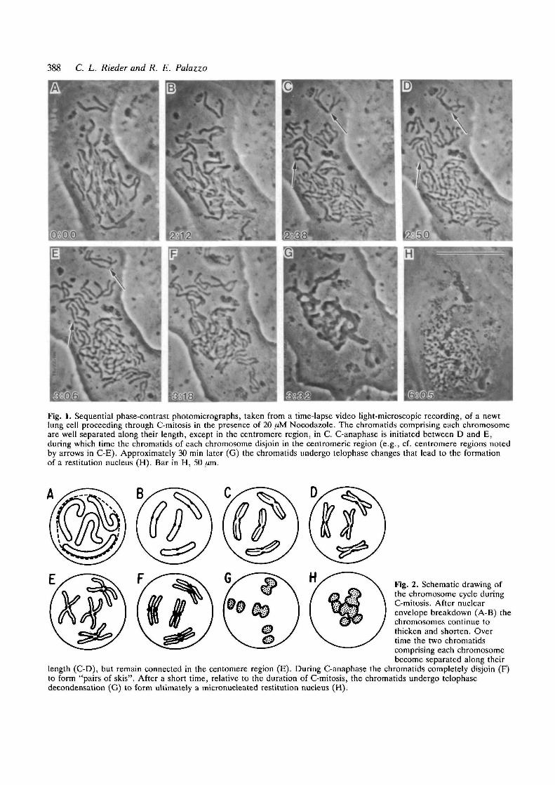
Over a wide range of concentrations colchicine andColcemid do not affect the rate at which cells entermitosis (reviewed by Eigsti and Dustin, 1955; Sluder,1979). When applied well before nuclear envelopebreakdown (NEB) and in a sufficient concentrationthese drugs completely inhibit the formation of spindleMTs. As a result, during NEB the chromosomes arereleased into the cytoplasm where they remain ran-domly dispersed throughout the prolonged period of C-mitosis (Fig. 1). It is noteworthy that the chromosomecondensation cycle (see Mazia, 1987) continues duringC-mitosis (Fig. 2), so that over time the chromosomesmay become quite condensed, reducing their regularlength by 1-1.5x (Ludford, 1936; Bajer, 1959; reviewedby Eigsti and Dustin, 1955; Mazia, 1961). During thelater stages of condensation the sister chromatidsusually separate along their length, except in thecentromeric region, to form X-shaped chromosomes or"C-pairs" (for plants, see Levan, 1938; Ostergren, 1943;Mole-Bajer, 1958; for animals, see Ludford, 1936;Stubblefield, 1964; Cooke et al., 1987; Figs 1,2).
In his classic 1938 paper on the effects of colchicine
Key words: colcemid, mitotic cycle, microtubules, cell cycle.
388 C. L. Rieder and R. E. Palazzo
Fig. 1. Sequential phase-contrast photomicrographs, taken from a time-lapse video light-microscopic recording, of a newtlung cell proceeding through C-mitosis in the presence of 20 fjM Nocodazole. The chromatids comprising each chromosomeare well separated along their length, except in the centromere region, in C. C-anaphase is initiated between D and E,during which time the chromatids of each chromosome disjoin in the centromeric region (e.g., cf. centromere regions notedby arrows in C-E). Approximately 30 min later (G) the chromatids undergo telophase changes that lead to the formationof a restitution nucleus (H). Bar in H, 50 /jm.
Fig. 2. Schematic drawing ofthe chromosome cycle duringC-mitosis. After nuclearenvelope breakdown (A-B) thechromosomes continue tothicken and shorten. Overtime the two chromatidscomprising each chromosomebecome separated along their
length (C-D), but remain connected in the centomere region (E). During C-anaphase the chromatids completely disjoin (F)to form "pairs of skis". After a short time, relative to the duration of C-mitosis, the chromatids undergo telophasedecondensation (G) to form ultimately a micronucleated restitution nucleus (H).

C-mitosis 389
Levan states "the prophases arrive at metaphase andare kept at that state for a long period...". Thisstatement was based on Strasburger's (1884; see page120 of Wilson, 1925) terminology of the time, whichseparated the mitotic cycle into prophase, metaphase,anaphase and telophase without an intervening stage ofprometaphase. The impetus for establishing "prometa-phase" as a distinct stage of mitosis occurred betweenthe publication of Schrader's first (1944) and second(1953) books on mitosis, well after Levan's initialstudies. As first emphasized by Nebel and Ruttle in 1938(see also Ostergren, 1943), and more recently by Sluder(1979, 1988), C-mitotics are blocked in prometaphasenot metaphase. Indeed, after prolonged periods in C-mitosis, recovering sea urchin cells still require thesame 10 minute prometaphase interval to construct aspindle and congress chromosomes that is normallyrequired in untreated controls (Sluder, 1979; see alsoBrinkley et al., 1967). Regardless, the erroneous notionthat colchicine/Colcemid blocks the mitotic cycle atmetaphase is still perpetuated as evidenced by thecontinued widespread use of the terms "metaphasearrest", "C-mitotic metaphase", "maintained in meta-phase", "held in metaphase", "colchicine (or C)-metaphase", "metaphase-blocked", etc.
A clear distinction between a mitotic block atprometaphase and metaphase should not be viewed as atrivial matter. It becomes increasingly important asmolecular-genetic and cell-free systems are used todissect more closely, and to define, the sequence ofbiochemical events comprising mitosis. Indeed, theterm "metaphase arrest" is commonly used to charac-terize various somatic cell mutants blocked in mitosis,and to describe the outcome of experimental treatmentson mitotic cells, even under conditions in which spindleformation is largely or completely inhibited. These"metaphase arrested" cells contrast sharply with thoseoocytes that are naturally arrested at true metaphase Ior II of meiosis with fully fomed spindles (reviewed byLongo, 1973), and those (few) somatic cells that can beinduced by various treatments to arrest permanently inmitosis with fully formed (e.g. see Shoji-Kasai et al.,1987; Jordan et al., 1991) or nearly fully formed (Hiranoet al., 1988) spindles.
Escaping the mitotic block
Most, if not all plant cells undergo repeated cell cyclesin the presence of colchicine (e.g. see Levan, 1938;Nebel and Ruttle, 1938; Eigsti and Dustin, 1955), a factthat has been widely utilized for generating polyploidstrains of commercially valuable crops. Similarly, manytypes of animal cells, including some from Chinesehamsters (Stubblefield, 1964), newts (Fig. 1), ratkangaroos (Jensen et al., 1987), mice (Kung et al.,1990), humans (Chamla et al., 1980) and sea urchins(Sluder, 1979), are capable of completing one or morerounds of C-mitosis in the presence of the drug. Thus,contrary to the implications of such common terms as"mitotic arrest", "stathmokinesis", "metaphase ar-
rest", "blocked or arrested in mitosis", "C-mitoticarrest", "halted at metaphase", etc., colchicine, Colce-mid and drugs with similar actions do not permanentlyblock plant and many animal cells in mitosis. Rather,when compared with controls, most drug-treated cellsinvariably spend a significantly greater period of time(up to 10-fold; Eigsti and Dustin, 1955) in (prometa-phase of) mitosis prior to entering interphase of thenext cell cycle.
The prolongation of the mitotic period during C-mitosis is not a unique response to the destruction of thespindle by colchicine and similar drugs. On thecontrary, concentrations of Colcemid or vinblastinethat have little or no discernable effect on spindleformation in sea urchins (Sluder, 1988) or HeLa-S3 cells(Jordon et al., 1991) significantly prolong mitosis (seaurchins) or may even permanently arrest the cells attrue metaphase (HeLa). Similarly, prolongation of themitotic period is not a unique response to Colcemid orother drugs that disrupt MTs; the duration of prometa-phase in untreated cells is greatly extended by thepresence of mal-oriented chromosomes (Mazia, 1961;Zirkle, 1970; Rieder and Alexander, 1989), and/or bythe absence of normal spindle bipolarity (Sluder andBegg, 1983; Hunt et al., 1992).
It has been proposed by Hartwell and Weinert (1989)that cells possess control mechanisms, termed "check-points", which function to ensure that the events of thecell cycle are properly coordinated. The fact that theonset of anaphase is considerably delayed by partial ortotal disruption of the spindle (as in C-mitosis),treatments that minimally compromise MT function, bythe lack of spindle bipolarity, and/or by mal-orientedchromosomes on a bipolar spindle, reveals that theprocess of spindle formation is "monitored" by such asurveillance checkpoint. As emphasized by Mazia(1961,1987), and more recently by others (Hartwell andWeinert, 1989; Murray and Kirschner, 1989), thischeckpoint appears to control cell entry into anaphase,and passage through this point triggers a cascadingseries of events that allow a rapid escape from mitosis,advancing the cell to interphase of the next cell cycle.
It has recently become clear that the nuclear andcytoplasmic events that lead to mitosis are regulated, inpart, by the sequential synthesis and accumulation of"cyclin" proteins A and B. These proteins are cofactorsrequired for the catalytic activity of the protein kinase,p34cdc2 (Solomon et al., 1990). Cyclin synthesis drivescells into mitosis (Murray and Kirschner, 1989), whilethe initiation of anaphase and the cells' subsequent exitfrom mitosis is coincident with the rapid proteolyticdestruction of these proteins (Evans et al., 1983;reviewed by Murray and Kirschner, 1989; Whitfield etal., 1990). More specifically, in somatic cells cyclin Aappears to reach peak levels just before NEB, and isthen degraded during prometaphase as the spindleforms. By contrast, the cyclin B level remains high untilthe metaphase-anaphase transition, at which time itdrops precipitously. Importantly, cyclin A is degradedbut cyclin B levels remain high throughout theprolonged prometaphase exhibited by C-mitotics (Kung

390 C. L. Rieder and R. E. Palazzo
et al., 1990; Whitfield et al., 1990) and cells containingmonopolar spindles (Hunt et al., 1992). Together thesedata strongly support the argument that passagethrough the spindle-formation surveillance checkpointis triggered by declining levels of cyclin B. If true, it willbecome important to elucidate how the life expectancyof cyclin B is determined by the "state of microtubulesand form of the spindle" (Hunt et al., 1992). The recentisolation of mitotic arrest-deficient (mad) mutants inyeast (Hoyt et al., 1991; Li and Murray, 1991), in whichthe cells fail to arrest at mitosis in response to loss ofMT function, offers a promising approach for under-standing how the cell monitors spindle formation.
Not all animal cells ultimately pass through C-mitosisand enter the next cell cycle in the presence of drugsthat disrupt MT function. For example, cells of certainmammalian lines (including HeLa S3, Vero, Tera2) dieafter 72 h in C-mitosis (see references quoted by Eigstiand Dustin, 1955; Kung et al., 1990), possibly from theirinability to synthesize mRNA (Dustin, 1959). In somecases, a significant proportion of the cells within amitotically arrested population escape the block whileothers die in mitosis (i.e. the block is leaky; e.g. seeShoji-Kasai et al., 1987; Jordan et al., 1991). Kung et al.(1990) have recently shown that the ability of a cell typeto survive C-mitosis is somewhat species-specific, and ispositively correlated with its ability to degrade cyclin Bduring the prolonged mitotic period. Although theseexperiments do not distinguish whether cyclin Bdegradation causes, or simply results from, the bio-chemical changes leading to escape from mitosis, theformer does provide a possible molecular basis for whysome cells are truly "arrested" in mitosis by colchicineor Colcemid while others can ultimately advance tointerphase. Clearly, "the stringency of the [spindleformation surveillance checkpoint]...varies among dif-ferent cell lines" (Kung et al., 1990).
Chromatid disjunction in the absence of aspindle
In actively cycling cells the initiation of anaphase, andthus exit from mitosis, is signaled by the disjunction ofreplicated chromatids. In some types of cells thechromatids of each replicated chromosome separate atthe centromeric region near the end of the C-mitoticperiod. This "C-anaphase" (Levan, 1938) phenomenonappears to occur in all plant cells (reviewed by Levan,1954; Eigsti and Dustin, 1955), where it has beenespecially well characterized owing to the absence ofrounding during the division process (Mole-Bajer, 1958;Lambert, 1980). In Haemanthus each pair of replicatedchromosomes requires a 1-2 min period to separate (seeFig. 6 of Lambert, 1980), and all chromatids of thegenome separate in near but not perfect synchrony (seeEigsti and Dustin, 1955; Mole-Bajer, 1958; Lambert,1980) in the complete absence of MTs (Lambert, 1980).Shortly after separation the chromatids begin to swelland undergo telophase events to form a 4N or polyploidrestitution nucleus. The total duration of C-anaphase is
similar to the time of anaphase in untreated cells (Mole-Bajer, 1958).
There is currently no consensus concerning the extentto which C-anaphase occurs in animal cells (e.g. seeLevan, 1954; Mazia, 1961; Rao and Engelberg, 1966;Mclntosh, 1979), and there are several obvious reasonsfor this confusion. Unlike plants, the ultimate fate ofindividual chromosomes during C-mitosis in animals isdifficult to follow clearly because most cells progress-ively round throughout this process. Moreover, fewinvestigators have studied the course of C-mitosis inanimal cells with the explicit goal of determiningwhether the chromatids disjoin.
C-anaphase figures are seen in many types of animalcells when assayed by using squashed or droppedchromosome preparations. These include, but are notlimited to, grasshopper spermatogonium (Sokolow,1939), human lymphocytes (Gabarron et al., 1986),mouse carcinoma (Ludford, 1936), ascites tumor(Levan, 1954), Chinese hamster ovary (Stubblefield,1964), rat kangaroo kidney epithelia (Vig, 1981) andDrosophila neuroblasts (Gonzalez et al., 1991). (SeeEigsti and Dustin (1955), for additional references onC-anaphase in chromosome spreads of animal cells.)Studies on premature centromere separation (e.g. seeFitzgerald et al., 1975), and the sequence of centromereseparation (e.g. see Vig, 1981), reveal that the harshpreparative treatments used for these analyses (hypo-tonic swelling, fixation in acetic acid/ethanol, squashingor dropping onto slides) are not likely to inducechromatid separation artificially.
C-anaphase has also been clearly demonstrated in seaurchin embryos fixed and lightly flattened between twocoverslips (Sluder, 1979). Moreover, C-anaphase fig-ures represent approx. 1-2% of all mitotics in PtKcultures fixed after 18 h in a concentration (20 fxM) ofnocodazole sufficient to deplete the cells of MTs (C.L.Rieder and R.W. Cole, unpublished). We have alsoobserved the process of C-anaphase directly by time-lapse video light microscopy of similarly treated newtlung cells (Fig. 1). With respect to these findings it isnoteworthy that individual chromosomes within thecytoplasm of PtK (Brenner et al., 1980) and newt(Rieder and Alexander, 1989) cells, which fail to attachto the normally forming spindle, still separate theirchromatids at the onset of anaphase. Chromatiddisjunction also occurs during monopolar mitosis innewts (Rieder et al., 1986) and sea urchins (Mazia etal., 1981).
The mechanism responsible for chromatid separationremains mysterious. It is clear from studies on C-mitotics that it is not dependent on antagonistic pullingforces, generated by the spindle, that act on sisterkinetochores within the centromeric region. Thisconclusion contrasts sharply with those models forchromatid separation in yeast, generated to explain theapparent need for spindle MT-dependent forces duringDNA decatenation by topoisomerase II (Holm et al.,1985, 1989; Uemura and Yanagida, 1986). In someanimal cells chromatid disjunction exhibits a close tem-poral coupling to the Ca2+-mediated inactivation of the

C-mitosis 391
p34cdc2/cyclin B complex and the destruction of cyclin B(Hunt et al., 1992; Shamu and Murray, 1992). It alsoprobably requires DNA topoisomerase II activity(Downes et al., 1991; Shamu and Murray, 1992) andperhaps the modification of INCENP (Cooke et al.,1987) and CLiP (Rattner et al., 1988), proteins uniqueto that region of the centromere spanning the sisterkinetochores. In this context it is noteworthy thatchromatids maintain firm centromeric connectionsprior to C-anaphase, after becoming separated alongthe remainder of their length (see above). Thus theprocessing of chromatin that leads to chromatidseparation is multi-phasic (i.e. the decatenation andsubseqeunt separation of chromosome arms and telo-meres occurs well before that of the centromeres).
It remains to be determined whether C-anaphase is acharacteristic feature of C-mitosis in all animal cells.Statements that it does not occur must be re-evaluatedin the context of those considerations that tend to maskits appearance. However, as discussed above some celltypes ultimately die in C-mitosis, apparently becausethey cannot degrade cyclin B to initiate those anaphaseevents that allow them to exit the mitotic cycle (Kung etal., 1990; Whitfield et al., 1990; Hunt etal., 1992). Sincethe initiation of anaphase is normally heralded bychromatid separation, cells that are unable to exit C-mitosis may never disjoin their chromatids. In such cellsspindle formation would be necessary for chromatidseparation (i.e. anaphase) only because it is a prerequi-site for initiating cyclin B degradation to allow passagethrough the spindle-formation surveillance checkpoint,not because chromatid separation is based on forcesgenerated by the spindle (e.g. see Gonzalez et al.,1991).
Although chromatid disjunction is normally tempor-aly coincident with cyclin B destruction, it may not bemediated, even indirectly, by declining cyclin B levelsbut by some other independent signal. Under thesecircumstances cells would be able to separate theirchromatids without necessarily initiating those otherevents of anaphase that allow them to exit mitosis.Reports that certain mutant human cells appear toremain arrested for considerable periods of time inmitosis, with some or all of their chromatids disjoined(Fitzgerald et al., 1975; Rudd et al., 1983; Gabarron etal., 1986), argue in favor of this hypothesis. By contrast,it is also possible that the events of anaphase that allowthe cell to exit mitosis can occur in the absence ofchromatid separation. Such a "relief of dependence"(Hartwell and Weinert, 1989) is suggested by theobservation that treatments that inhibit chromatidseparation in mammalian cells do not necessarilyprevent exit from mitosis (Downes et al., 1991).
Conclusions
We have reviewed the evidence that, for many cells,disruption of the mitotic spindle with Colcemid,colchicine and similar drugs delays but does not inhibitprogression through the mitotic cycle. Whether a
particular cell type can exit C-mitosis depends on itsability to overcome the spindle-formation surveillancecheckpoint in the absence of a spindle, an ability thatmay depend on whether the cell can ultimately degradecyclin B while in C-mitosis. C-mitotics capable ofpassing through this checkpoint normally advance tointerphase by way of a C-anaphase. C-anaphase isindicated by the separation of sister chromatids and thisevent does not depend on forces generated by thespindle.
We thank Drs. G. Sluder, S.P. Alexander, S.S. Bowser andJ.G. Ault for their scientific comments, and Ms. S. Nowo-grodzki for editorial assistance. This work was supported, inpart, by grants from the National Institutes of Health,General Medical Sciences R01-40198 (to C.L.R.) and R01-43264 (to R.E.P.), by grant no. 2725 from the Council forTobacco Research (to R.E.P.), and by American CancerSociety grant JFRA 62121 (to R.E.P.).
References
Bajer, A. S. (1959). Change of length and volume of mitoticchromosomes in living cells. Hereditas 45, 579-596.
Brenner, S. L., Liaw, L.-H. and Berns, M. W. (1980). Lasermicroirradiation of kinetochores in mitotic PtK2 cells. Cell.Biophys. 2, 139-151.
Brinkley, B. R., Stubblefield, E. and Hus, T. C. (1967). The effects ofcolcemid inhibition and reversal on the fine structure of the mitoticapparatus of Chinese hamster cells in vivo. J. Ultrastruct. Res. 19,1-18.
Chamla, Y., Roumy, M., Lassegues, M. and Battin, J. (1980). Alteredsensitivity to colchicine and PHA in human cultured cells. Hum.Genet. 53, 249-253.
Cooke, C. A., Heck, M. S. and Earnshaw, W. C. (1987). The innercentromere protein (INCENP) antigens: Movement from innercentromere to midbody during mitosis. J. Cell Biol. 105, 2053-2067.
Deysson, G. (1968). Antimitotic substances. Int. Rev. Cytol. 24, 99-148.
Downes, C. S., Mullinger, A. M. and Johnson, R. T. (1991).Inhibitors of DNA topoisomerase II prevent chromatid separationin mammaliancells but do not prevent exit from mitosis. Proc. Nat.Acad. Sci. USA 88, 8895-8899.
Dustin. P. (1959). The quantitative estimation of mitotic growth in thebone marrow of the rat by the stathmokinetic (colchicinic) method.In The Kinetics of Cellular Proliferation (ed. F. Stohlman, Jr), pp.50-56. New York, London: Grune and Stratton.
Dustin, P. (1978). Microtubules, pp. 452. New York: Springer Verlag.Eigsti, O. J. and Dustin, P. (1955). Colchicine in Agriculture,
Medicine, Biology and Chemistry, pp. 470. Aimes, Iowa: IowaState Coll. Press.
Evans, T., Rosenthal, E. T., Youngblom, J., Distel, D. and Hunt, T.(1983). Cyclin: A protein specified by maternal mRNA in seaurchin eggs that is destroyed at each cleavage division. Cell 33, 389-396.
Fitzgerald, P. H., Pickering, A. F., Mercer, J. M. and Miethke, P. M.(1975). Premature centromere division: A mechanism of non-disjunction causing X chromosome aneuploidy in somatic cells ofman. Ann. Hum. Genet. 38, 417-428.
Gabarron, J., Jimenez, J. and Glover, G. (1986). Prematurecentromere division dominantly inherited in a subfertile family.Cytogenet. Cell Genet. 43, 69-71.
Gonzalez, C , Jimenez, J. C , Ripoll, P. and Sunkel, C. E. (1991). Thespindle is required for the process of sister chromatid separation inDrosophila neuroblasts. Exp. Cell Res. 192, 10-15.
Hartwell, L. H. and Weinert, T. A. (1989). Checkpoints: Controlsthat ensure the order of cell cycle events. Science 246, 629-634.
Hirano, T., Hiraoka, Y. and Yanagida, M. (1988). A temperature-sensitive mutation of the Schizosaccharomyces pombe gene nuc2+that encodes a nuclear-scaffold-like protein blocks spindleelongation in anaphase. J. Cell Biol. 106, 1171-1183.

392 C. L. Rieder and R. E. Palazzo
Holm, C , Goto, T., Wang, J. C. and Botstein, D. (1985). DNAtopoisomerase II is required at the time of mitosis in yeast. Cell 41,553-563.
Holm, C , Stearns, T. and Botstein, D. (1989). DNA topoisomerase IImust act at mitosis to prevent nondisjunction and chromosomebreakage. Mol. Cell Biol. 9, 159-168.
Hoyt, M. A., Totis, L. and Roberts, B. T. (1991). S. cerevisiae genesrequired for cell cycle arrest in response to loss of microtubulefunction. Cell 66, 507-517.
Hunt, T., Luca, F. C. and Ruderman, J. V. (1992). The requirementsfor protein synthesis and degredtion, and the control of destructionof cyclins A and B in the meiotic and mitotic cell cycles of teh clamembryo. / . Cell Biol. 116, 707-724.
Jensen, C. G., Davison, E. A., Bowser, S. S. and Rieder, C. L. (1987).Primary cilia cycle in PtKl cells: Effects of colcemid and taxol oncilia formation and resorption. Cell Modi. Cyloskel. 7, 187-197.
Jordan, M. A., Thrower, P. and Wilson, L. (1991). Mechanism ofinhibition of cell proliferation by vinca alkaloids. Cancer Res. 51,2212-2222.
Rung, A. L., Sherwood, S. W. and Schimke, R. T. (1990). Cell line-specific differences in the control of cell cycle progression in theabsence of mitosis. Proc. Nat. Acad. Sci. USA 87, 9553-9557.
Lambert, A-M. (1980). The role of chromosomes in anaphase triggerand nuclear envelope activity in spindle formation. Chromosoma76, 295-308.
Levan, A. (1938). The effect of colchicine on root mitoses in Allium.Hereditas 24, 471-486.
Levan, A. (1954). Colchicine-induced C-mitosis in two mouse ascitestumors. Hereditas 40, 1-64.
Li, R. and Murray, A. W. (1991). Feedback control of mitosis inbudding yeast. Cell 66, 519-531.
Longo, F. J. (1973). Fertilization: A comparative ultrastructuralreview. Biol. Reprod. 9, 149-215.
Ludford, R. J. (1936). The action of toxic substances upon thedivision of normal and malignant cells in vitro and in vivo. Arch.Exp. Zellforsch. 18, 411-441.
Mareel, M. M. and DeMets, M. (1984). Effect of microtubuleinhibitors on invasion and on related activities of tumor cells. Int.Rev. Cytol. 90, 125-168.
Mazia, D. (1961). Mitosis: the physiology of cell division. In The Cell.vol. Ill (ed. J. Brachet and A.E. Mirksy), pp. 78-412. New York:Academic Press.
Mazia, D. (1987). The chromosome cycle and the centrosome cycle inthe mitotic cycle. Int. Rev. Cytol. 100, 49-92.
Mazia, D., Paweletz, N., Sluder, G. and Finze, E.-M. (1981).Cooperation of kinetochores and pole in the establishment ofmonopolar mitotic apparatus. Proc. Nat. Acad. Sci. USA 78, 377-381.
Mclntosh, J. R. (1979). Cell division. In Microtubules (ed. K. Robertsand J.S. Hyams), pp. 382-441. New York: Academic Press.
Mole-Bajer, J. (1958). Cine-micrographic analysis of C-mtosis inendosperm. Chromosoma 9, 332-358.
Murray, A. W. and Kirschner, M. W. (1989). Dominoes and clocks:The union of two views of the cell cycle. Science 246, 614-621.
Nebel, B. R. and Ruttle, M. L. (1938). The cytological and geneticalsignificance of colchicine. J. Hered. 29, 3-9.
Ostergren, G. (1943). Elastic chromosome repulsions. Hereditas 29,444-450.
Rao, P. N. and Engelberg, J. (1966). Mitotic non-disjunction of sisterchromatids and anomalous mitosis induced by low temperatures inHeLa cells. Exp. Cell Res. 43, 332-342.
Rattner, J. B., Kingwell, B. G. and Fritzler, M. J. (1988). Detectionof distinct structural domains within the primary constriction usingautoantibodies. Chromosoma 96, 360-367.
Rieder, C. L. and Alexander, S. P. (1989). The attachment ofchromosomes to the mitotic spindle and the production ofaneuploidy in newt lung cells. In Mechanisms of ChromosomeDistribution and Aneuploidy (ed. M.A. Resnick and B.K. Vig), pp.185-194. New York: Alan R. Liss.
Rieder, C. L., Davison, E. A., Jensen, L. C. W., Cassimeris, L. andSalmon, E. D. (1986). Oscillatory movements of monoorientedchromosomes and their position relative to the spindle pole resultfrom the ejection properties of the aster and half-spindle. J. CellBiol. 103, 581-591.
Rudd, N. L., Teshima, I. E., Martin, R. H., Sisken, J. E. andWeksberg, R. (1983). A dominantly inherited cytogenetic anomaly:A possible cell division mutant. Hum. Genet. 65, 117-121.
Schrader, F. (1944). Mitosis, 1st cdn, pp. 110. Columbia Univ. Press,New York.
Schrader, F. (1953). Mitosis, 2nd edn, pp. 170. Columbia Univ. Press,New York.
Shamu, C. E. and Murray, A. W. (1992). Sister chromatid separationin frog egg extracts requires DNA topoisomerase II activity duringanaphase. J. Cell Biol. (in press).
Shoji-Kasai, Y., Senshu, M., Iwashita, S. and Imahori, K. (1987).Thiol proteasc-spccific inhibitor E-64 arrests human cpidermoidcarcinoma A431 cells at mitotic metaphasc. Proc. Nat. Acad. Sci.USA 85, 146-150.
Sluder, G. (1979). Role of spindle microtubules in the control of cellcycle timing. J. Cell Biol. 80, 674-691.
Sluder, G. (1988). Control mechanisms of Mitosis: The role of spindlemicrotubules in the timing of mitotic events. Zool. Sci. 5, 653-665.
Sluder, G. (1991). The practical use of colchicine and colcemid toreversibly block microtubule assembly in living cells. In AdvancedTechniques in Chromosome Research (ed. K.W. Adolph), pp. 427-447. New York: Marcel Dckker.
Sluder, G. and Begg, D. A. (1983). Control mechanisms of the cellcycle: Role of the spatial arrangement of spindle components in thetiming of mitotic events. J. Cell Biol. 97, 877-886.
Sokolow, I. (1939). Einfluss des Colchicins auf dieSpermatogenialmitosen bei den Orthopetcren. CR (Doklady)Acad. Sci. URSS 24, 298-300.
Solomon, M. J., Glotzer, M., Lee, T., Philippe, M. and Kirschner, M.(1990). Cyclin activation of p34cdc2. Cell 63, 1013-1024.
Stubblefield, E. (1964). DNA synthesis and chromosomalmorphology of Chinese hamster cells cultured in media containingN-deacetyl-N-methylcolchicine (Colcemid). In Cytogenetics ofCells in Culture (ed. R.J. Harris), pp. 223-248. New York:Academic Press.
Taylor, E. W. (1965). The mechanism of colchicine inhibition ofmitosis. J. Cell Biol. 25, 145-160.
Uemura, T. and Yanagida, M. (1986). Mitotic spindle pulls but fails toseparate chromosomes in type II DNA topoisomerase mutants:uncoordinated mitosis. EMBO J. 5. 1003-1010.
Vig, B. K. (1981). Sequence of centromere separation: An analysis ofmitotic chromosomes from long-term cultures of Potorus cells.Cytogenet. Cell Genet. 31, 129-136.
Whitfield, W. G. F., Gonzalez, C , Maldonado-Codina, G. andGlover, D. M. (1990). The A- and B-type cyclins of Drosophila areaccumulated and destroyed in temporally distinct events that defineseparable phases of the G2-M transition. EMBO J. 9, 2563-2572.
Wilson, E. B. (1925). The Cell in Development and Heredity, pp. 1232.New York: MacMillan Co.
Wilson, L., Anderson, K. and Chin, D. (1976). Nonstoichiometricpoisoning of microtubule polymerization: A model for themechanism of action of the vinca alkaloids, popodhylcotoxin andcolchicine. In Cell Motility (ed. Goldman, R.. Pollard, T. andRosenbaum, J.), vol. 3, pp. 1051-1064. Cold Spring HarborLaboratory Press, NY.
Zirkle, R. E. (1970). Ultraviolet-microbeam irradiation of newt-cellcytoplasm: Spindle destruction, false anaphase, and delay of trueanaphase. Radial. Res. 41, 516-537.
Note added in proofWhile this manuscript was at the printers Gosh andPaweletz (Exp. Cell Res. 200, 215-217, 1992) reportedthat okadaic acid inhibits sister chromatid separation inHeLa cells without inhibiting exit from mitosis. As aresult, at the next mitosis diplochromosomes areformed that contain 4 unseparated chromatids. Thesedata support the hypotheses that phosphatase 1 activityis required for sister chromatid separation, and thatchromatid separation is not an obligatory event forescape from mitosis.