collagen diseases in adolescents dr.chandrika rao professor and hod. m.s.ramaiah medical college and...
TRANSCRIPT

COLLAGEN DISEASES IN ADOLESCENTS
DR.CHANDRIKA RAOPROFESSOR AND HOD.M.S.RAMAIAH MEDICAL COLLEGE AND HOSPITALBANGALORE

Definition
•Collagen-Greek-`Glue producer`
•Collagen diseases-A small group of
disorders due to structural or metabolic
defects in collagen.

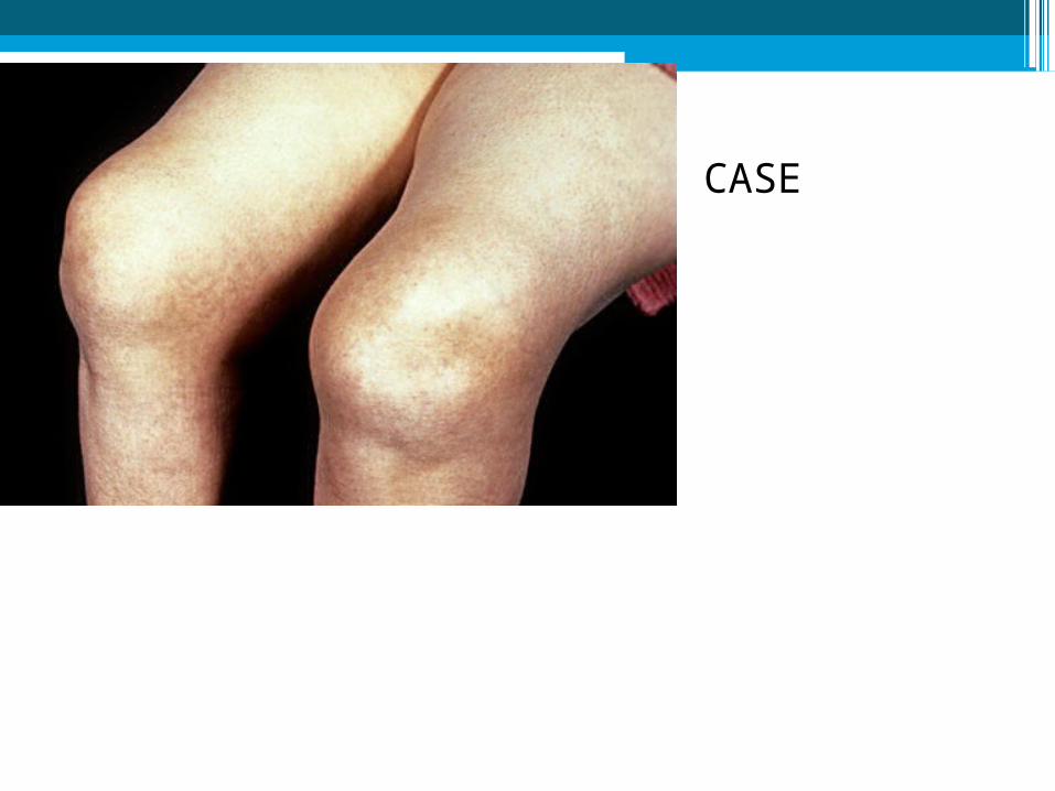
CASE
6'4",
Armspan of 6'7“. He has hypermobile joints in his knees, shoulders and ankles.

CASE
CASE

Collagen-Structure
•Generic term for proteins forming a triple helix of three polypeptide chains .
•All members of the collagen family form these in the extracellular matrix .
•Size, function and tissue distribution vary considerably.
•N=28

Type Collagen Distribution Disorders
I
Fibril-I,II,III,V,XI
Most abundant .Scar tissue,tendons, skin, artery walls, cornea, bones and teeth.
Osteogenesis imperfecta,
Ehlers–Danlos syndrome especially type IV,
Infantile cortical hyperostosis aka Caffey's disease
II Hyaline cartilage,. Vitreous humour . Collagenopathy, types II and XI
IIIGranulation tissue, Reticular fiber. artery walls, skin, intestines and the uterus
Ehlers–Danlos syndrome, Dupuytren's contracture
IV Basal lamina; eye lens, glomeruli Alport syndrome, Goodpasture's syndrome

VIMost interstitial tissue, assoc. with type I
Ulrich myopathy, Bethlem myopathy, Atopic dermatitis
VIIForms anchoring fibrils in dermoepidermal junctions
Epidermolysis bullosa dystrophica
VIII Some endothelial cells Posterior polymorphous corneal dystrophy
IXFACIT collagen-(IX,XII,XIV,XV), cartilage, assoc. with type II and XI fibrils
EDM2 and EDM3
XHypertrophic and mineralizing
cartilageSchmid metaphyseal dysplasia

Collagen vascular disorders
•Discoid lupus erythematosus•Systemic lupus erythematosus•Neonatal lupus erythematosus•Juvenile dermatomyositis•Childhood scleroderma

Approach
•Detailed history•Progressive•Multiple areas involved•Skin,Musculo-skeletal involvement•Family history•Remmision and relapse

Gene location mutation Syndrome
COL1A1 17q22 Null alleles OI type I
Partial deletions; C-terminal substitutions
OI type II
N-terminal substitutions OI types I, III or IV
Deletion of exon 6 EDS type VII
COL1A2 7q22.1 Splice mutations; exon deletions OI type I
C-terminal mutations OI type II, IV
N-terminal substitutions OI type III
Deletion of exon 6 EDS type VII
GENETICS
Different Types of Mutations in Collagen I Chain Genes Cause Different Disease Severities

Family Tree
?
13 aortic aneurysm
44
69
28 aortic aneurysm, aneurysm of kidney
28 AA
28 AA
31 AA, cerebral hemorrhage
45 AAA
45 ?valve replacement


Ehlers-Danlos syndrome
The signs and symptoms of Ehlers-Danlos syndrome vary from mildly loose joints to life-threatening complications•

Diseases of Elastic Fiber• Cutis laxa• Williams syndrome• Buschke-Ollendorff syndrome • Menkes disease • Pseudoxanthoma elasticum, • Marfan's syndrome

SLERevised Diagnostic Criteria
1. Malar rash2. Discoid rash3. Photosensitivity4. Oral ulcers5. Arthritis6. Serositis7. Renal disorder8. Haematologic disorder9. Neurologic disoder10.Immunologic disoder11.ANA 4/11 are present serially or simultaneously.


Inclusion Criteria Exclusion Criteriaa Laboratory Criteriaa
Signs and symptoms suggestive of a CTD but not fulfilling the diagnostic or classification criteria for any of the defined CTDs b for at least 3 years c
Malar rashDiscoid LupusCutaneous sclerosisHeliotrope rashGottron papulesErosive arthritis
Anti-dsDNAAnti-SmithAnti-U1-RNPAnti-Scl70AnticentromereAnti-La/SSBAnti-Jo1Anti-Mi2
Presence of antinuclear antibodies determined on two different occasions
Preliminary Classification Criteria for Undifferentiated Connective-Tissue Disease

Investigation •CBC,ESR,CRP X-ray•Urinalysis CT Scan•Serum creatinine Early renal
biopsy•Rheumatoid factor (RF)•ANA-IFA
•Other:•CK,C3, ,TSH,Anti-Ro/SSA,Anti-La/SSB•Anti-Smith,Anti-cardiolipins,•Lupus anticoagulant•Vitamin D - 25(OH)D3

Treatment
•Early diagnosis.HEADDSS
•May still utilize tried & tested agents initially
•Phenytoin, Cyclosporine,Ca Channel blocker-
Nifedipine-stimulatory drugs.
•Newer immunosuppressive &
immunomodulatory-NASAIDS,Cox-2inhibitor,
Cyclosporine, Azathioprine, Ca channel blocker.

CONCLUSION
• A multidisciplinary approach.
• The aim should be to reduce disease activity to a
minimum level and to allow treatment free
intervals, so that the growth, development, and
fertility of these children are ensured.


•Thank you
•Q:Acquired collagen disorder?