colloquium paper an ancestral bacterial division system is ... · 9.02.2015 · an ancestral...
TRANSCRIPT
An ancestral bacterial division system is widespreadin eukaryotic mitochondriaMichelle M. Legera, Markéta Petr�ub, Vojt�ech Žárskýb, Laura Emea, �Cestmír Vlcekc, Tommy Hardinga, B. Franz Langd,Marek Eliáše, Pavel Dole�zalb, and Andrew J. Rogera,1
aCentre for Comparative Genomics and Evolutionary Bioinformatics (CGEB), Department of Biochemistry and Molecular Biology, Dalhousie University,Halifax, NS, Canada, B3H 4R2; bBiotechnology and Biomedicine Centre of the Academy of Sciences and Charles University in Vestec (BIOCEV) Group,Department of Parasitology, Faculty of Science, Charles University in Prague, 128 44 Prague, Czech Republic; cLaboratory of Genomics and Bioinformatics,Institute of Molecular Genetics, Academy of Sciences of the Czech Republic, 142 20 Prague 4, Czech Republic; dRobert Cedergren Centre for Bioinformaticsand Genomics, Département de Biochimie, Université de Montréal, Montreal, QC, Canada, H3T 1J4; and eDepartment of Biology and Ecology, Faculty ofScience, University of Ostrava, 710 00 Ostrava, Czech Republic
Edited by Patrick J. Keeling, University of British Columbia, Vancouver, BC, Canada, and accepted by the Editorial Board February 24, 2015 (received for reviewJanuary 14, 2015)
Bacterial division initiates at the site of a contractile Z-ring com-posed of polymerized FtsZ. The location of the Z-ring in the cellis controlled by a system of three mutually antagonistic proteins,MinC, MinD, and MinE. Plastid division is also known to bedependent on homologs of these proteins, derived from theancestral cyanobacterial endosymbiont that gave rise to plastids.In contrast, the mitochondria of model systems such as Saccharo-myces cerevisiae, mammals, and Arabidopsis thaliana seem tohave replaced the ancestral α-proteobacterial Min-based divisionmachinery with host-derived dynamin-related proteins that formouter contractile rings. Here, we show that the mitochondrial di-vision system of these model organisms is the exception, ratherthan the rule, for eukaryotes. We describe endosymbiont-derived,bacterial-like division systems comprising FtsZ and Min proteins indiverse less-studied eukaryote protistan lineages, including jako-bid and heterolobosean excavates, a malawimonad, strameno-piles, amoebozoans, a breviate, and an apusomonad. For two ofthese taxa, the amoebozoan Dictyostelium purpureum and thejakobid Andalucia incarcerata, we confirm a mitochondrial locali-zation of these proteins by their heterologous expression in Sac-charomyces cerevisiae. The discovery of a proteobacterial-likedivision system in mitochondria of diverse eukaryotic lineagessuggests that it was the ancestral feature of all eukaryotic mito-chondria and has been supplanted by a host-derived system mul-tiple times in distinct eukaryote lineages.
mitochondria | mitochondrial division | Min proteins | MinCDE |mitochondrial fission
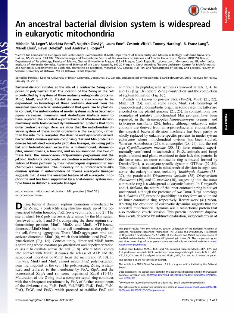
During bacterial division, septum formation is mediated bythe Z-ring, a contractile ring structure made up of the po-
lymerized tubulin homolog FtsZ (reviewed in refs. 1 and 2). Thesite at which FtsZ polymerizes is determined by the Min system(reviewed in refs. 1 and 3–5), comprising the three septum site-determining proteins MinC, MinD, and MinE. ATP-bound,dimerized MinD binds the inner cell membrane at the poles ofthe cell, forming aggregates. These MinD aggregates bind andactivate dimerized MinC (6), which then inhibits local FtsZ po-lymerization (Fig. 1A). Concomitantly, dimerized MinE formsa spiral ring whose constant polymerization and depolymerizationcauses it to oscillate across the cell (7, 8). Where MinE comesinto contact with MinD, it causes the release of ATP and thesubsequent liberation of MinD from the membrane (9, 10). Inthis way, MinD and MinC cannot inhibit FtsZ polymerizationnear the midpoint of the cell. The polymerizing Z-ring is stabi-lized and tethered to the membrane by FtsA, ZipA, and thenonessential ZapA and (in some organisms) ZapB (11–15).Maturation of the Z-ring into a complete septal ring continueswith the subsequent recruitment by FtsA of further componentsof the divisome (i.e., FtsB, FtsE, FtsI/PBP3, FtsK, FtsL, FtsN,FtsQ, FtsW, and FtsX), which proceed to stabilize FtsZ and
contribute to peptidoglycan synthesis (reviewed in refs. 3, 4, 16and 17) (Fig. 1B) before Z-ring constriction and the completionof septum formation (Fig. 1C).Plastids are known to possess FtsZ (18–20), MinD (21, 22),
MinE (21, 23), and, in some cases, MinC (24) homologs ofcyanobacterial endosymbiotic origin; in some cases, the latter areencoded on the plastid genome (21, 25). In contrast, only twoexamples of putative mitochondrial Min proteins have beenreported, in the stramenopiles Nannochloropsis oceanica andEctocarpus siliculosus (26). Indeed, although eukaryotic mito-chondria are derived from an α-proteobacterial endosymbiont,the ancestral bacterial division machinery has been partly orwholly replaced by eukaryote-specific proteins in model systemeukaryotes where mitochondrial division has been studied.Whereas Amoebozoa (27), stramenopiles (28, 29), and the redalga Cyanidioschyzon merolae (30, 31) have retained experi-mentally confirmed mitochondrial FtsZ, animals and fungi(opisthokonts) and plants examined to date lack this protein. Inthe latter taxa, an outer contractile ring is instead formed byDnm1p/Drp1, a eukaryote-specific dynamin GTPase (32–34).This protein is implicated in mitochondrial division in organismsacross the eukaryotic tree, including Arabidopsis thaliana (35–37), the parabasalid Trichomonas vaginalis (38), Dictyosteliumdiscoideum (39), and C. merolae (40), suggesting that the outercontractile ring is a widespread eukaryotic feature. In T. vaginalisand A. thaliana, the nature of the inner contractile ring is not yetunderstood, although the presence of two Dnm1/Drp1 homologsin A. thaliana (37) raises the possibility that they form an outer andan inner contractile ring, respectively. Recent work (41) recon-structing the evolution of eukaryotic dynamins suggests that theancestral mitochondrial dynamin was a bifunctional protein thatalso mediated vesicle scission. This protein underwent duplica-tion events, followed by subfunctionalization, independently in at
This paper results from the Arthur M. Sackler Colloquium of the National Academy ofSciences, “Symbioses Becoming Permanent: The Origins and Evolutionary Trajectoriesof Organelles,” held October 15–17, 2014, at the Arnold and Mabel Beckman Center ofthe National Academies of Sciences and Engineering in Irvine, CA. The complete programand video recordings of most presentations are available on the NAS website at www.nasonline.org/Symbioses.
Author contributions: M.M.L., M.P., and P.D. designed research; M.M.L., M.P., �C.V., andT.H. performed research; B.F.L. contributed new reagents/analytic tools; M.M.L., M.P.,V.Ž., L.E., �C.V., and M.E. analyzed data; and M.M.L., M.P., T.H., and A.J.R. wrote the paper.
The authors declare no conflict of interest.
This article is a PNAS Direct Submission. P.J.K. is a guest editor invited by the EditorialBoard.
Data deposition: The sequences reported in this paper have been deposited in the GenBankdatabase (accession nos. KP271960–KP271964, KP324909–KP324912, KP258196–KP258204,and KP738110).1To whom correspondence should be addressed. Email: [email protected].
This article contains supporting information online at www.pnas.org/lookup/suppl/doi:10.1073/pnas.1421392112/-/DCSupplemental.
www.pnas.org/cgi/doi/10.1073/pnas.1421392112 PNAS Early Edition | 1 of 8
CELL
BIOLO
GY
COLLOQUIUM
PAPE
R
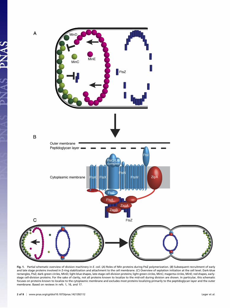
Fig. 1. Partial schematic overview of division machinery in E. coli. (A) Roles of Min proteins during FtsZ polymerization. (B) Subsequent recruitment of earlyand late stage proteins involved in Z-ring stabilization and attachment to the cell membrane. (C) Overview of septation initiation at the cell level. Dark-bluerectangles, FtsZ; dark-green circles, MinD; light-blue shapes, late-stage cell-division proteins; light-green circles, MinC; magenta circles, MinE; red shapes, early-stage cell-division proteins. For the sake of clarity, not all proteins known to localize to the mid-cell during division are shown. In particular, this schematicfocuses on proteins known to localize to the cytoplasmic membrane and excludes most proteins localizing primarily to the peptidoglycan layer and the outermembrane. Based on reviews in refs. 1, 16, and 17.
2 of 8 | www.pnas.org/cgi/doi/10.1073/pnas.1421392112 Leger et al.
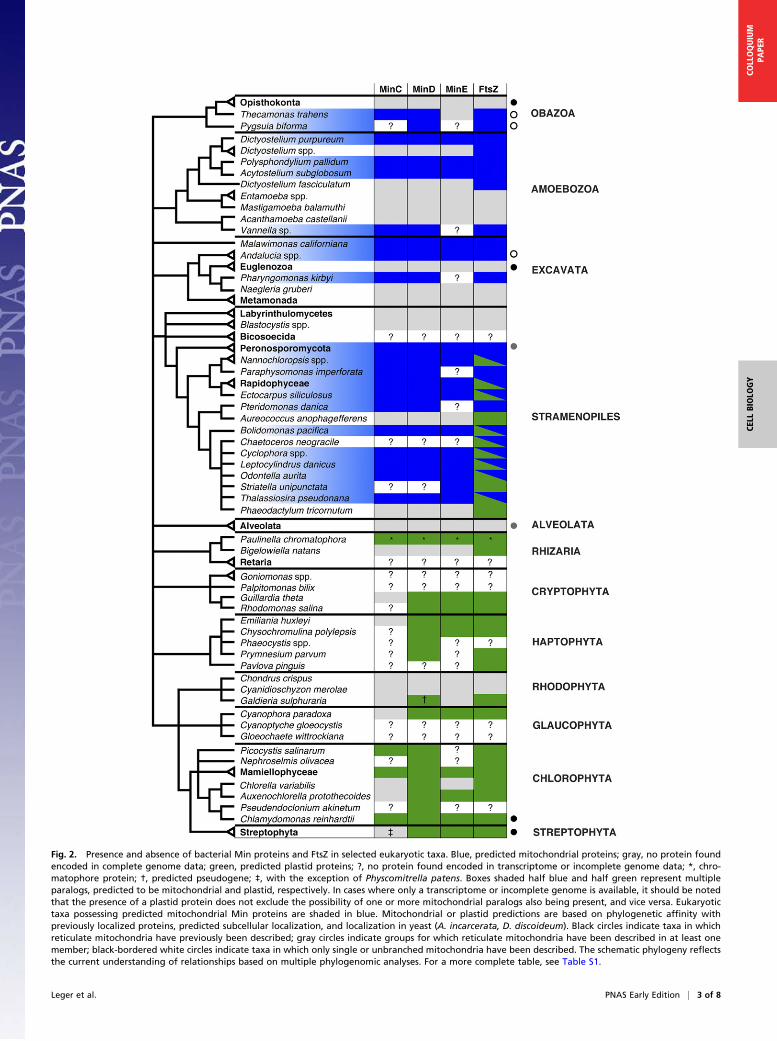
Fig. 2. Presence and absence of bacterial Min proteins and FtsZ in selected eukaryotic taxa. Blue, predicted mitochondrial proteins; gray, no protein foundencoded in complete genome data; green, predicted plastid proteins; ?, no protein found encoded in transcriptome or incomplete genome data; *, chro-matophore protein; †, predicted pseudogene; ‡, with the exception of Physcomitrella patens. Boxes shaded half blue and half green represent multipleparalogs, predicted to be mitochondrial and plastid, respectively. In cases where only a transcriptome or incomplete genome is available, it should be notedthat the presence of a plastid protein does not exclude the possibility of one or more mitochondrial paralogs also being present, and vice versa. Eukaryotictaxa possessing predicted mitochondrial Min proteins are shaded in blue. Mitochondrial or plastid predictions are based on phylogenetic affinity withpreviously localized proteins, predicted subcellular localization, and localization in yeast (A. incarcerata, D. discoideum). Black circles indicate taxa in whichreticulate mitochondria have previously been described; gray circles indicate groups for which reticulate mitochondria have been described in at least onemember; black-bordered white circles indicate taxa in which only single or unbranched mitochondria have been described. The schematic phylogeny reflectsthe current understanding of relationships based on multiple phylogenomic analyses. For a more complete table, see Table S1.
Leger et al. PNAS Early Edition | 3 of 8
CELL
BIOLO
GY
COLLOQUIUM
PAPE
R
least three lineages (opisthokonts, land plants, and alveolates), butthe ancestral bifunctional form seems to have been retained inamoebozoa such as D. discoideum, the red alga C. merolae, andstramenopiles (and possibly additional eukaryotes that have cur-rently less well-characterized dynamins). The distribution of an-cestral-like bifunctional mitochondrial/vesicle fission dynaminsthus seems to mirror that of mitochondrial FtsZ (41).Here, we hypothesize that the complete loss of the α-proteo-
bacterial division system is the exception, rather than the rule,for eukaryotes. We show that mitochondria-targeted homologsof bacterial Min proteins are patchily but widely distributedamong diverse eukaryote lineages; and we further demonstratethat Min proteins from two of these lineages, the amoebozoanDictyostelium purpureum and the jakobid excavate Andaluciaincarcerata, localize to mitochondria when expressed in yeast.
Materials and MethodsDatabase Searches. Publicly available databases and sequencing projectswere searched using the Basic Local Alignment Search Tools (BLAST) blastp andtblastn (42). A large number of databases containing eukaryotic sequenceswere screened with these tools using query sequences from D. purpureum(XP_003286111, XP_003292258, XP_003293637, XP_642499), E. siliculosus(CBJ32744, CBJ31561, CBJ28079, CBJ48312) A. incarcerata, Pseudomonas flu-orescens (AEV64338, AEV64339, AEV64340, AEV64767), and Anabaena sp. 90(YP_006998153, AFW94434, YP_006996248, YP_006996249). The databasessearched included the Nucleotide collection (nr/nt), National Center for Bio-technology Information (NCBI) Genomes, Whole-Genome Shotgun contigs,Expressed Sequence Tags, High-throughput Genomic Sequences and Tran-scriptome Shotgun Assembly divisions of GenBank (43) (last accessed February9, 2015); the Broad Institute project databases (44) (accessed April 23, 2014);the Joint Genome Institute (JGI) genome databases (45, 46) (last accessedFebruary 9, 2015); dictyBase, 2013 release (47–49); the EnsemblProtists data-base (50) (last accessed February 9, 2015); the Eukaryotic Pathogen DatabaseResources (EuPathDB) (51) (last accessed February 9, 2015); and the MarineMicrobial Eukaryote Transcriptome Sequencing Project (52) (accessed June 3,2014), via the Community cyberinfrastructure for Advanced Microbial EcologyResearch and Analysis (CAMERA) portal (53) (for a full list of sequencesidentified, see Table S1 and Dataset S1). In addition, we searched our ownunpublished genome or transcriptome assemblies from several protist taxa ofkey evolutionary interest: two jakobids (A. incarcerata and Andalucia godoyi),the heterolobosean Pharyngomonas kirbyi, and Malawimonas californiana.Potential homologs identified were screened manually to exclude con-taminants from bacterial or other eukaryotic sources, by searching forintrons and excluding sequences with a notably high degree of similarity tobacterial or distantly related eukaryotic homologs. Subcellular localizationand targeting peptides were predicted using TargetP, using “plant”
parameters for plastid-bearing taxa and “nonplant” parameters for taxalacking plastids (54, 55).
Sequence Generation. P. kirbyi strain AS12B (56, 57) was cultivated at 37 °C in10% (wt/vol) salt medium (NaCl 1.6 M, KCl 34.0 mM, MgCl2 44.2 mM, CaCl24.0 mM, MgSO4 4.5 mM) supplemented with Citrobacter sp. as a food sourcebefore RNA isolation. RNA was extracted using TRIzol (Life Technologies) fol-lowing the manufacturer’s instructions and stored at −80 °C. The RNA samplewas treated with Turbo DNase (Life Technologies) before conversion to cDNAusing the GeneRacer kit with SuperScript III reverse transcriptase (Life Technol-ogies) and stored at −20 °C. Primers were designed to amplify genes of interestusing available sequences. Primer sequences were as follows: MinCF, 5′-ATGT-CACGTCGATGGTTAGT-3′; MinCR, 5′-TAATACAAAAAAAAAACA-3′; MinDF,5′-ATGTATCGATCAACGAGTTC-3′; andMinDR, 5′-TTAGTTCCTGCTAAATAATC-3′.PCR reactions were done using the Phusion high-fidelity DNA polymerase(New England BioLabs) where the initial denaturation at 98 °C for 30 s wasfollowed by 30 cycles of DNA denaturation at 98 °C for 10 s, primer annealingat 40 °C for 30 s, and strand elongation at 72 °C for 60 s, with a final extensionat 72 °C for 10 s. PCR products were purified by gel extraction using theNucleospin Extract II kit (Macherey-Nagel) and were directly sequenced usingthe PCR primers.
Phylogenetic Analyses. For each protein, alignments were generated fromdatasets including all known eukaryotic homologs and bacterial homologsharvested from NCBI using MUSCLE v.3.8.31 (58) or MAFFT-L-INSI v7.149b(59–61), and trimmed using BMGE 1.1 (62) (-m BLOSUM30; all other pa-rameters default). Preliminary phylogenies were generated using FastTree,and datasets were manually refined. Twenty independent Maximum Like-lihood (ML) tree estimates and 200 bootstrap replicates were generatedusing RAxML v.8.0.23 (63) under the PROTGAMMALG4X (64) model ofamino acid substitution. Bayesian inference posterior probabilities werecalculated using PhyloBayes v.3.3f (65) under the catfix C20 model of evo-lution. We tested whether specific phylogenetic hypotheses were rejectedby the data using the approximately unbiased (AU) test implemented inCONSEL v.1.20 (66) (Table S2). Maximum-likelihood trees given specificconstraints (i.e., corresponding to specific hypotheses) were generated usingRAxML. In addition, the 200 trees from bootstrap replicates were included inthe hypothesis-testing analyses performed with CONSEL.
Yeast Culture, Transformation, and Microscopy. Saccharomyces cerevisiae strainYPH499 was grown at 30 °C on YPD medium or selective medium withouturacil after lithium-acetate transformation. For ectopic expression of AiMinC,-D, and -E, the complete AiMinC, -D, and -E ORFs were amplified by PCR fromA. incarcerata cDNA. For ectopic expression of DpMinC, -D, and -E, the com-plete DpMinC, -D, and -E ORFs were amplified by PCR from synthesized DNAfragments, containing Escherichia coli codon-optimized sequences. Theresulting PCR products were cloned separately into pUG35 using XbaI/ClaIrestriction sites (AiMinD and DpMinC and -E) or BamHI/HindIII restriction sites
Fig. 3. Min proteins from D. purpureum (A) and A. incarcerata (B) expressed in S. cerevisiae to confirm predicted mitochondrial targeting. Differentialinterference contrast (DIC) images of S. cerevisiae cells expressing Min fusion proteins (Left); in green, MinC, MinD, or MinE expressed with the C-terminal GFPtag in S. cerevisiae; in red, mitochondria labeled with MitoTracker Red CMXRos (Mito); merged images (Merge) show mitochondrial localization of all Minproteins. (Scale bars: 5 μm.)
4 of 8 | www.pnas.org/cgi/doi/10.1073/pnas.1421392112 Leger et al.
(AiMinC and -E and DpMinD), allowing the expression of GFP on the C-ter-minus of each protein. For fluorescence microscopy, cells were incubated withMitoTracker Red CMXRos (1:10,000) for 10 min, washed once in PBS, andmounted in 2% low-melting agarose. Cells were viewed using an OlympusIX81 microscope and a Hamamatsu Orca-AG digital camera using the cell Rimaging program at 100× magnification.
Results and DiscussionWe identified sequences encoding at least one Min protein froma number of eukaryotic taxa (Fig. 2 and Table S1), including an-cestrally plastid-lacking lineages such as the apusomonad Theca-monas trahens, the breviate Pygsuia biforma, the jakobid excavatesA. godoyi and A. incarcerata, the malawimonad M. californiana,and several amoebozoan lineages, such as D. purpureum. Threepreviously reported FtsZ sequences identified in haptophytes(Gephyrocapsa oceanica and Pleurochrysis carterae) and a glau-cophyte (Cyanophora paradoxa) (29) were excluded as probableα-proteobacterial contaminants, based on their position in pre-liminary phylogenies, their high degree of similarity to α-pro-teobacterial sequences, and, in the case of C. paradoxa, ourinability to recover the reported mitochondrial FtsZ sequencefrom the genome sequence (67). All complete genomes encodingat least one Min protein also encoded at least one FtsZ homolog;however, the reverse was not true. Min proteins were retainednot only in lineages with typical aerobic mitochondria, but also inlineages possessing mitochondrion-related organelles (MROs)such as A. incarcerata (68) and P. biforma (69).Most of these Min and FtsZ homologs possess predicted mito-
chondrial targeting peptides (Table S1). To confirm these pre-dictions, we expressed GFP-tagged homologs of Min proteinsin S. cerevisiae, in conjunction with the mitochondrial stainMitoTracker Red CMXRos (Fig. 3). We chose Min proteinsfrom two representative taxa lacking plastids: the amoebozoanD. purpureum (Fig. 3A) and the jakobid excavate A. incarcerata(Fig. 3B). In both cases, the GFP signal colocalized with the Mito-tracker signal, supporting the predicted targeting of A. incarcerataand D. discoideumMin proteins to the inside of the mitochondria.Single-protein phylogenies of MinC, -D, -E, and FtsZ recover
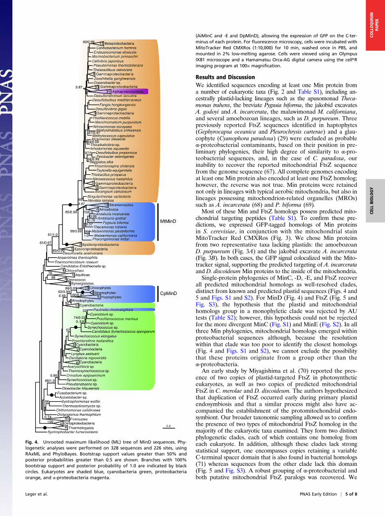
all predicted mitochondrial homologs as well-resolved clades,distinct from known and predicted plastid sequences (Figs. 4 and5 and Figs. S1 and S2). For MinD (Fig. 4) and FtsZ (Fig. 5 andFig. S3), the hypothesis that the plastid and mitochondrialhomologs group in a monophyletic clade was rejected by AUtests (Table S2); however, this hypothesis could not be rejectedfor the more divergent MinC (Fig. S1) and MinE (Fig. S2). In allthree Min phylogenies, mitochondrial homologs emerged withinproteobacterial sequences although, because the resolutionwithin that clade was too poor to identify the closest homologs(Fig. 4 and Figs. S1 and S2), we cannot exclude the possibilitythat these proteins originate from a group other than theα-proteobacteria.An early study by Miyagishima et al. (70) reported the pres-
ence of two copies of plastid-targeted FtsZ in photosyntheticeukaryotes, as well as two copies of predicted mitochondrialFtsZ in C. merolae and D. discoideum. The authors hypothesizedthat duplication of FtsZ occurred early during primary plastidendosymbiosis and that a similar process might also have ac-companied the establishment of the protomitochondrial endo-symbiont. Our broader taxonomic sampling allowed us to confirmthe presence of two types of mitochondrial FtsZ homolog in themajority of the eukaryotic taxa examined. They form two distinctphylogenetic clades, each of which contains one homolog fromeach eukaryote. In addition, although these clades lack strongstatistical support, one encompasses copies retaining a variableC-terminal spacer domain that is also found in bacterial homologs(71) whereas sequences from the other clade lack this domain(Fig. 5 and Fig. S3). A robust grouping of α-proteobacterial andboth putative mitochondrial FtsZ paralogs was recovered. We
Fig. 4. Unrooted maximum likelihood (ML) tree of MinD sequences. Phy-logenetic analyses were performed on 328 sequences and 226 sites, usingRAxML and PhyloBayes. Bootstrap support values greater than 50% andposterior probabilities greater than 0.5 are shown. Branches with 100%bootstrap support and posterior probability of 1.0 are indicated by blackcircles. Eukaryotes are shaded blue, cyanobacteria green, proteobacteriaorange, and α-proteobacteria magenta.
Leger et al. PNAS Early Edition | 5 of 8
CELL
BIOLO
GY
COLLOQUIUM
PAPE
R
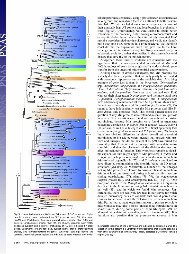
subsampled these sequences, using γ-proteobacterial sequences asan outgroup, and reanalyzed them in an attempt to better resolvethis clade. We also excluded amoebozoan sequences because oftheir unusually high AT content and long branches in preliminarytrees (Fig. S3). Unfortunately, we were unable to obtain betterresolution of the branching order among α-proteobacterial andeukaryotic clades. Nevertheless, the C-terminally truncated FtsZproteins were identified only in eukaryotes, and we did not identifymore than one FtsZ homolog in α-proteobacteria. We thereforeconclude that the duplication event that gave rise to the FtsZparalogs found in extant eukaryotes likely occurred early ineukaryotic evolution, rather than earlier, in the α-proteobacteriallineage that gave rise to the mitochondrion.Altogether, these lines of evidence are consistent with the
hypothesis that the nuclear-encoded mitochondrial Min andFtsZ homologs of eukaryotes originated by endosymbiotic genetransfer from the ancestral mitochondrial endosymbiont.Although found in diverse eukaryotes, the Min proteins are
sparsely distributed, a pattern that can only partly be reconciledwith taxonomic representation in the available data. A strikingexample of gene loss is seen in the Mycetozoa (Dictyosteliumspp., Acytostelium subglobosum, and Polysphondylium pallidum).Here, D. discoideum, Dictyostelium citrinum, Dictyostelium inter-medium, and Dictyostelium firmibasis have retained only FtsZwhereas their sister taxon D. purpureum and the more basal taxaP. pallidum, Polysphondylium violaceum, and A. subglobosumhave additionally maintained all three Min proteins. Meanwhile,the yet more distantly related Dictyostelium fasciculatum (72, 73)seems to have independently lost the Min proteins and, like D.discoideum, only possesses FtsZ. This overall pattern raises thequestion of why Min proteins were retained in some taxa, yet lostin others. No correlation was found with mitochondrial cristaemorphology, because Min proteins were found in organismspossessing discoid (e.g., P. kirbyi) (56) or tubular (e.g., A. godoyi)(74) cristae, as well as in lineages with MROs that apparently lackcristae entirely (e.g., A. incarcerata and P. biforma) (68, 69). Nor isthere any obvious difference in either overall mitochondrialmorphology or lifestyle between lineages that possess Min pro-teins and lineages that do not. Kiefel et al. (29) have raised thepossibility that FtsZ is lost in lineages with reticulate mito-chondria, and thus the placement of the division site may notaffect mitochondrial function. This hypothesis remains a plausi-ble explanation that might apply to Min proteins; A. godoyi andP. biforma each possess a single mitochondrion or mitochon-drion-related organelle (74, 75), and T. trahens is predicted tohave discrete, nonbranching mitochondria based on 3D recon-structions (76) (Fig. 2). Meanwhile, a number of the lineageslacking Min proteins are known to possess reticulate mitochon-dria in at least one tissue and during at least one life stage, in-cluding opisthokonts (77), plants (78, 79), the euglenozoanEuglena gracilis (80), and apicomplexa (81, 82) (Fig. 2). Oneexception seems to be Phytophthora cinnamomi, an organismdescribed in the literature as having 3–4 reticulate mitochondriaper cell (83), and in which we found Min homologs. Un-fortunately, there are relatively few taxa in our survey for whichdetailed microscopy data are available that would permit con-clusions to be drawn about the 3D structure of their mitochon-dria. Furthermore, many organisms known to possess reticulatemitochondria may also possess unbranched mitochondria insome tissues, during some parts of their life cycle (79), oralongside reticulate mitochondria, as in P. cinnamomi (83). It istherefore also possible that the presence or absence of MinFig. 5. Unrooted maximum likelihood (ML) tree of FtsZ sequences. Phylo-
genetic analyses were performed on 327 sequences and 257 sites, usingRAxML and PhyloBayes. Bootstrap support values greater than 50% andposterior probabilities greater than 0.5 are shown. Branches with 100%bootstrap support and posterior probability of 1.0 are indicated by blackcircles. Eukaryotes are shaded blue, cyanobacteria green, proteobacteriaorange, and α-proteobacteria magenta. Eukaryotic paralogs lacking thevariable C-terminal spacer region are indicated by stars whereas those with
incomplete sequence at the C-terminus are indicated by question marks. Theexception to this pattern is a Corethron hystrix sequence that, despite branchingwith other stramenopiles in the MtFtsZ1 clade, possesses a C-terminal variableregion (Fig. S3).
6 of 8 | www.pnas.org/cgi/doi/10.1073/pnas.1421392112 Leger et al.
proteins reflects some unknown transient mitochondrial mor-phological feature specific to replication. Clearly, genetic andfunctional studies of mitochondrial Min systems are greatlyneeded to understand their precise roles.Further questions are raised by the apparent absence of
homologs of all other components of the bacterial divisome fromthe surveyed eukaryotes, including ZipA, ZapA, FtsA, FtsB,FtsE, FtsI, FtsK, FtsL, FtsN, FtsQ, FtsW, and FtsX. Searches ofdatabases using α-proteobacterial and E. coli homologs of theseproteins as queries yielded no candidate homologs. The bacterialdivisome components recruited late in the division process (FtsB,FtsE, FtsI, FtsL, FtsN, FtsQ, FtsW, and FtsX) are primarily in-volved in facilitating peptidoglycan synthesis, and so their ap-parent absence is perhaps not surprising, given the lack ofa peptidoglycan wall in any mitochondria. However, it is notclear how the Z-ring remains stabilized and anchored to themembrane in the absence of FtsA, ZipA, or ZapA. ZED,a coiled-coil domain protein with 25.8% sequence identity toZapA, is reported to be involved in mitochondrial Z-ring for-mation in the red alga C. merolae (84). However, we were unableto identify any homologs of this protein in other eukaryotes. Thetwo distinct FtsZ paralogs may form an alternating copolymerthat forms the Z-ring; or the Z-ring might be composed ofa single paralog whereas the second paralog might instead beinvolved in attachment of the Z-ring to the membrane. In eithercase, the anchoring mechanism of FtsZ remains a mystery.Recent work (85) implicates the endoplasmic reticulum (ER)
in the control of the mitochondrial division site location andsubsequent Dnm1p recruitment in yeast. This type of externaldivision site control contrasts with that of the Min protein sys-tem, which regulates division site location from the mitochon-drial matrix. The contrast between these control mechanismsraise the questions of when the role of the ER in mitochondrial
division may have emerged; whether any taxa possess both Minproteins and Dnm1p/Drp1; and how these organisms (if theyexist) recruit Dnm1p/Drp1 in the absence of ER-mediated di-vision site control. Therefore, an important avenue of furtherstudy is the taxonomic distribution of mitochondrial Dnm1p/Drp1 and its functional interplay with FtsZ. Study of this dis-tribution is hampered by the fact that multiple paralogs ofdynamins have different functions within eukaryotic cells (41),including vesicular trafficking in yeast (86), and unknown func-tions in less-studied organisms such as T. vaginalis (38). Theseproteins lack N-terminal targeting peptides, and so, in the ab-sence of localization data, a mitochondrial function cannotclearly be ascribed to any one of them based on sequence dataalone. In any case, investigations into the molecular mechanismsgoverning the coordination of the various kinds of inner andouter contractile rings are critically needed in diverse eukaryotelineages to fully understand what are features of the divisionsystem of the last eukaryotic common ancestor and what aremore recent lineage-specific innovations.
ACKNOWLEDGMENTS. We thank Dr. Michael W. Gray for planting the seedsof the collaboration leading to this paper. We thank the Acytostelium ge-nome consortium for kindly providing A. subglobosum gene sequences.M.M.L. thanks Yana Eglit for help in taming Adobe Illustrator. M.M.L. wassupported by the National Research Fund, Luxembourg (FNR), and by theNova Scotia Health Research Foundation (NSHRF). L.E. was supported bya Centre for Comparative Genomics and Evolutionary Bioinformatrics post-doctoral fellowship from the Tula Foundation. T.H. was supported by theNatural Sciences and Engineering Research Council of Canada. This work wassupported by Regional Partnerships Program Grant FRN 62809 from theCanadian Institutes of Health Research and the NSHRF (to A.J.R.), CzechScience Foundation Grant 13-24983S (to M.E.), Czech Science FoundationGrant 13-29423S (to P.D.), and a European Regional Development Fundaward to the Biomedicine Center of the Academy of Sciences and CharlesUniversity (CZ.1.05/1.1.00/02.0109).
1. de Boer PA (2010) Advances in understanding E. coli cell fission. Curr Opin Microbiol13(6):730–737.
2. Meier EL, Goley ED (2014) Form and function of the bacterial cytokinetic ring. CurrOpin Cell Biol 26:19–27.
3. Natale P, Pazos M, Vicente M (2013) The Escherichia coli divisome: Born to divide.Environ Microbiol 15(12):3169–3182.
4. Lutkenhaus J, Pichoff S, Du S (2012) Bacterial cytokinesis: From Z ring to divisome.Cytoskeleton (Hoboken) 69(10):778–790.
5. Lutkenhaus J (2007) Assembly dynamics of the bacterial MinCDE system and spatialregulation of the Z ring. Annu Rev Biochem 76:539–562.
6. Ghosal D, Trambaiolo D, Amos LA, Löwe J (2014) MinCD cell division proteins formalternating copolymeric cytomotive filaments. Nat Commun 5:5341.
7. Hale CA, Meinhardt H, de Boer PA (2001) Dynamic localization cycle of the cell di-vision regulator MinE in Escherichia coli. EMBO J 20(7):1563–1572.
8. Loose M, Fischer-Friedrich E, Ries J, Kruse K, Schwille P (2008) Spatial regulators forbacterial cell division self-organize into surface waves in vitro. Science 320(5877):789–792.
9. Park KT, WuW, Lovell S, Lutkenhaus J (2012) Mechanism of the asymmetric activationof the MinD ATPase by MinE. Mol Microbiol 85(2):271–281.
10. Hu Z, Lutkenhaus J (2001) Topological regulation of cell division in E. coli. spatio-temporal oscillation of MinD requires stimulation of its ATPase by MinE and phos-pholipid. Mol Cell 7(6):1337–1343.
11. Hale CA, de Boer PA (1997) Direct binding of FtsZ to ZipA, an essential component ofthe septal ring structure that mediates cell division in E. coli. Cell 88(2):175–185.
12. Wang X, Huang J, Mukherjee A, Cao C, Lutkenhaus J (1997) Analysis of the interactionof FtsZ with itself, GTP, and FtsA. J Bacteriol 179(17):5551–5559.
13. Pichoff S, Lutkenhaus J (2002) Unique and overlapping roles for ZipA and FtsA inseptal ring assembly in Escherichia coli. EMBO J 21(4):685–693.
14. Hale CA, de Boer PA (1999) Recruitment of ZipA to the septal ring of Escherichia coliis dependent on FtsZ and independent of FtsA. J Bacteriol 181(1):167–176.
15. Galli E, Gerdes K (2010) Spatial resolution of two bacterial cell division proteins: ZapArecruits ZapB to the inner face of the Z-ring. Mol Microbiol 76(6):1514–1526.
16. Egan AJ, Vollmer W (2013) The physiology of bacterial cell division. Ann N Y Acad Sci1277:8–28.
17. den Blaauwen T, Andreu JM, Monasterio O (2014) Bacterial cell division proteins asantibiotic targets. Bioorg Chem 55:27–38.
18. Fraunholz MJ, Moerschel E, Maier UG (1998) The chloroplast division protein FtsZ isencoded by a nucleomorph gene in cryptomonads. Mol Gen Genet 260(2-3):207–211.
19. Sato M, Nishikawa T, Yamazaki T, Kawano S (2005) Isolation of the plastid ftsZ genefrom Cyanophora paradoxa (Glaucocystophyceae, Glaucocystophyta). Phycol Res 53:93–96.
20. Osteryoung KW, Vierling E (1995) Conserved cell and organelle division. Nature
376(6540):473–474.21. Wakasugi T, et al. (1997) Complete nucleotide sequence of the chloroplast genome
from the green alga Chlorella vulgaris: The existence of genes possibly involved in
chloroplast division. Proc Natl Acad Sci USA 94(11):5967–5972.22. Colletti KS, et al. (2000) A homologue of the bacterial cell division site-determining
factor MinD mediates placement of the chloroplast division apparatus. Curr Biol 10(9):
507–516.23. Itoh R, Fujiwara M, Nagata N, Yoshida S (2001) A chloroplast protein homologous to
the eubacterial topological specificity factor minE plays a role in chloroplast division.
Plant Physiol 127(4):1644–1655.24. Miyagishima SY, Kabeya Y, Sugita C, Sugita M, Fujiwara T (2014) DipM is required for
peptidoglycan hydrolysis during chloroplast division. BMC Plant Biol 14:57.25. Douglas SE, Penny SL (1999) The plastid genome of the cryptophyte alga, Guillardia
theta: Complete sequence and conserved synteny groups confirm its common an-
cestry with red algae. J Mol Evol 48(2):236–244.26. Vieler A, et al. (2012) Genome, functional gene annotation, and nuclear trans-
formation of the heterokont oleaginous alga Nannochloropsis oceanica CCMP1779.
PLoS Genet 8(11):e1003064.27. Gilson PR, et al. (2003) Two Dictyostelium orthologs of the prokaryotic cell division
protein FtsZ localize to mitochondria and are required for the maintenance of normal
mitochondrial morphology. Eukaryot Cell 2(6):1315–1326.28. Beech PL, et al. (2000) Mitochondrial FtsZ in a chromophyte alga. Science 287(5456):
1276–1279.29. Kiefel BR, Gilson PR, Beech PL (2004) Diverse eukaryotes have retained mitochondrial
homologues of the bacterial division protein FtsZ. Protist 155(1):105–115.30. Takahara M, et al. (2000) A putative mitochondrial ftsZ gene is present in the uni-
cellular primitive red alga Cyanidioschyzon merolae. Mol Gen Genet 264(4):452–460.31. Takahara M, Kuroiwa H, Miyagishima S, Mori T, Kuroiwa T (2001) Localization
of the mitochondrial FtsZ protein in a dividing mitochondrion. Cytologia (Tokyo)
66:421–425.32. Bleazard W, et al. (1999) The dynamin-related GTPase Dnm1 regulates mitochondrial
fission in yeast. Nat Cell Biol 1(5):298–304.33. Labrousse AM, Zappaterra MD, Rube DA, van der Bliek AM (1999) C. elegans
dynamin-related protein DRP-1 controls severing of the mitochondrial outer
membrane. Mol Cell 4(5):815–826.34. Smirnova E, Griparic L, Shurland DL, van der Bliek AM (2001) Dynamin-related protein
Drp1 is required for mitochondrial division in mammalian cells. Mol Biol Cell 12(8):
2245–2256.
Leger et al. PNAS Early Edition | 7 of 8
CELL
BIOLO
GY
COLLOQUIUM
PAPE
R
35. Arimura S, Tsutsumi N (2002) A dynamin-like protein (ADL2b), rather than FtsZ, isinvolved in Arabidopsis mitochondrial division. Proc Natl Acad Sci USA 99(8):5727–5731.
36. Mano S, Nakamori C, Kondo M, Hayashi M, Nishimura M (2004) An Arabidopsisdynamin-related protein, DRP3A, controls both peroxisomal and mitochondrialdivision. Plant J 38(3):487–498.
37. Arimura S, Aida GP, Fujimoto M, Nakazono M, Tsutsumi N (2004) Arabidopsisdynamin-like protein 2a (ADL2a), like ADL2b, is involved in plant mitochondrialdivision. Plant Cell Physiol 45(2):236–242.
38. Wexler-Cohen Y, Stevens GC, Barnoy E, van der Bliek AM, Johnson PJ (2014) A dy-namin-related protein contributes to Trichomonas vaginalis hydrogenosomal fission.FASEB J 28(3):1113–1121.
39. Wienke DC, Knetsch ML, Neuhaus EM, Reedy MC, Manstein DJ (1999) Disruption ofa dynamin homologue affects endocytosis, organelle morphology, and cytokinesis inDictyostelium discoideum. Mol Biol Cell 10(1):225–243.
40. Nishida K, et al. (2003) Dynamic recruitment of dynamin for final mitochondrialseverance in a primitive red alga. Proc Natl Acad Sci USA 100(4):2146–2151.
41. Purkanti R, Thattai M (2015) Ancient dynamin segments capture early stages of host-mitochondrial integration. Proc Natl Acad Sci USA 112(9):2800–2805.
42. Altschul SF, et al. (1997) Gapped BLAST and PSI-BLAST: A new generation of proteindatabase search programs. Nucleic Acids Res 25(17):3389–3402.
43. Benson DA, et al. (2014) GenBank. Nucleic Acids Res 42(Database issue):D32–D37.44. Origins of Multicellularity Sequencing Project, Broad Institute of Harvard and MIT
www.broadinstitute.org/. Accessed April 23, 2014.45. Grigoriev IV, et al. (2012) The genome portal of the Department of Energy Joint
Genome Institute. Nucleic Acids Res 40(Database issue):D26–D32.46. Nordberg H, et al. (2014) The genome portal of the Department of Energy Joint
Genome Institute: 2014 updates. Nucleic Acids Res 42(Database issue):D26–D31.47. Kreppel L, et al. (2004) dictyBase: A new Dictyostelium discoideum genome database.
Nucleic Acids Res 32(Database issue):D332–D333.48. Basu S, et al. (2013) DictyBase 2013: Integrating multiple Dictyostelid species. Nucleic
Acids Res 41(Database issue):D676–D683.49. Fey P, Dodson RJ, Basu S, Chisholm RL (2013) One stop shop for everything Dictyos-
telium: dictyBase and the Dicty Stock Center in 2012. Methods Mol Biol 983:59–92.50. Flicek P, et al. (2014) Ensembl 2014. Nucleic Acids Res 42(Database issue):D749–D755.51. Aurrecoechea C, et al. (2007) ApiDB: Integrated resources for the apicomplexan bi-
oinformatics resource center. Nucleic Acids Res 35(Database issue):D427–D430.52. Keeling PJ, et al. (2014) The Marine Microbial Eukaryote Transcriptome Sequencing
Project (MMETSP): Illuminating the functional diversity of eukaryotic life in theoceans through transcriptome sequencing. PLoS Biol 12(6):e1001889.
53. Sun S, et al. (2011) Community cyberinfrastructure for Advanced Microbial EcologyResearch and Analysis: The CAMERA resource. Nucleic Acids Res 39(Database issue):D546–D551.
54. Emanuelsson O, Nielsen H, Brunak S, von Heijne G (2000) Predicting subcellular lo-calization of proteins based on their N-terminal amino acid sequence. J Mol Biol300(4):1005–1016.
55. Emanuelsson O, Brunak S, von Heijne G, Nielsen H (2007) Locating proteins in the cellusing TargetP, SignalP and related tools. Nat Protoc 2(4):953–971.
56. Park JS, Simpson AG (2011) Characterization of Pharyngomonas kirbyi (= “Macro-pharyngomonas halophila” nomen nudum), a very deep-branching, obligately hal-ophilic heterolobosean flagellate. Protist 162(5):691–709.
57. Harding T, et al. (2013) Amoeba stages in the deepest branching heteroloboseans,including Pharyngomonas: Evolutionary and systematic implications. Protist 164(2):272–286.
58. Edgar RC (2004) MUSCLE: A multiple sequence alignment method with reduced timeand space complexity. BMC Bioinformatics 5:113.
59. Katoh K, Misawa K, Kuma K, Miyata T (2002) MAFFT: A novel method for rapidmultiple sequence alignment based on fast Fourier transform. Nucleic Acids Res30(14):3059–3066.
60. Katoh K, Kuma K, Toh H, Miyata T (2005) MAFFT version 5: Improvement in accuracyof multiple sequence alignment. Nucleic Acids Res 33(2):511–518.
61. Katoh K, Standley DM (2013) MAFFT multiple sequence alignment software version 7:improvements in performance and usability. Mol Biol Evol 30(4):772–780.
62. Criscuolo A, Gribaldo S (2010) BMGE (Block Mapping and Gathering with Entropy): Anew software for selection of phylogenetic informative regions from multiple se-quence alignments. BMC Evol Biol 10:210.
63. Stamatakis A (2014) RAxML version 8: A tool for phylogenetic analysis and post-analysis of large phylogenies. Bioinformatics 30(9):1312–1313.
64. Le SQ, Dang CC, Gascuel O (2012) Modeling protein evolution with several amino acidreplacement matrices depending on site rates. Mol Biol Evol 29(10):2921–2936.
65. Lartillot N, Lepage T, Blanquart S (2009) PhyloBayes 3: A Bayesian softwarepackage for phylogenetic reconstruction and molecular dating. Bioinformatics25(17):2286–2288.
66. Shimodaira H, Hasegawa M (2001) CONSEL: For assessing the confidence of phylo-genetic tree selection. Bioinformatics 17(12):1246–1247.
67. Price DC, et al. (2012) Cyanophora paradoxa genome elucidates origin of photosyn-thesis in algae and plants. Science 335(6070):843–847.
68. Simpson AG, Patterson DJ (2001) On core jakobids and excavate taxa: The ultra-structure of Jakoba incarcerata. J Eukaryot Microbiol 48(4):480–492.
69. Stairs CW, et al. (2014) A SUF Fe-S cluster biogenesis system in the mitochondrion-related organelles of the anaerobic protist Pygsuia. Curr Biol 24(11):1176–1186.
70. Miyagishima SY, et al. (2004) Two types of FtsZ proteins in mitochondria and red-lineage chloroplasts: The duplication of FtsZ is implicated in endosymbiosis. J Mol Evol58(3):291–303.
71. TerBush AD, Yoshida Y, Osteryoung KW (2013) FtsZ in chloroplast division: Structure,function and evolution. Curr Opin Cell Biol 25(4):461–470.
72. Heidel AJ, et al. (2011) Phylogeny-wide analysis of social amoeba genomes high-lights ancient origins for complex intercellular communication. Genome Res 21(11):1882–1891.
73. Romeralo M, Cavender JC, Landolt JC, Stephenson SL, Baldauf SL (2011) An expandedphylogeny of social amoebas (Dictyostelia) shows increasing diversity and new mor-phological patterns. BMC Evol Biol 11:84.
74. Lara E, Chatzinotas A, Simpson AGB (2006) Andalucia (n. gen.): The deepest branchwithin jakobids (Jakobida; Excavata), based on morphological and molecular study ofa new flagellate from soil. J Eukaryot Microbiol 53(2):112–120.
75. Brown MW, Sharpe SC, Silberman JD, Heiss AA, Lang BF, et al. (2013) Phylogenomicsdemonstrates that breviate flagellates are related to opisthokonts and apusomonads.P Roy Soc B-Biol Sci 280(1769).
76. Heiss AA, Walker G, Simpson AG (2013) The microtubular cytoskeleton of the apu-somonad Thecamonas, a sister lineage to the opisthokonts. Protist 164(5):598–621.
77. Barberà MJ, et al. (2010) Sawyeria marylandensis (Heterolobosea) has a hydro-genosome with novel metabolic properties. Eukaryot Cell 9(12):1913–1924.
78. Bendich AJ, Gauriloff LP (1984) Morphometric analysis of cucurbit mitochondria: Therelationship between chondriome volume and DNA content. Protoplasma 119:1–7.
79. Seguí-Simarro JM, Coronado MJ, Staehelin LA (2008) The mitochondrial cycle ofArabidopsis shoot apical meristem and leaf primordium meristematic cells is definedby a perinuclear tentaculate/cage-like mitochondrion. Plant Physiol 148(3):1380–1393.
80. Buetow DE (1989) The mitochondrion. The Biology of Euglena: Subcellular Bio-chemistry and Molecular Biology, ed Buetow DE (Academic, San Diego), Vol 4, pp247–314.
81. HoganMJ, Yoneda C, Feeney L, Zweigart P, Lewis A (1960) Morphology and culture ofToxoplasma. Arch Ophthalmol 64:655–667.
82. van Dooren GG, et al. (2005) Development of the endoplasmic reticulum, mito-chondrion and apicoplast during the asexual life cycle of Plasmodium falciparum.MolMicrobiol 57(2):405–419.
83. Hardham AR (1987) Ultrastructure and serial section reconstruction of zoospores ofthe fungus Phytophthora cinnamomi. Exp Mycol 11(4):297–306.
84. Yoshida Y, et al. (2009) The bacterial ZapA-like protein ZED is required for mito-chondrial division. Curr Biol 19(17):1491–1497.
85. Friedman JR, et al. (2011) ER tubules mark sites of mitochondrial division. Science334(6054):358–362.
86. Miyagishima SY, Nishida K, Kuroiwa T (2003) An evolutionary puzzle: chloroplast andmitochondrial division rings. Trends Plant Sci 8(9):432–438.
8 of 8 | www.pnas.org/cgi/doi/10.1073/pnas.1421392112 Leger et al.