colon and rectum - new brunswick colorecta… · web view... evaluation of histologic factors...
TRANSCRIPT
Protocol for the Examination of Specimens from Patients with Primary Carcinoma of the Colon and RectumWell-differentiated neuroendocrine neoplasms (carcinoid tumors) are not included.This modified NB CAP version has not been reviewed, verified or approved by CAP. NB specific modifications are noted in blue.
Based on AJCC/UICC TNM, 7th editionProtocol web posting date: February 1, 2011
Procedures• Excisional Biopsy (Polypectomy)• Local Excision (Transanal Disk Excision)• Colectomy (Total, Partial, or Segmental Resection)• Rectal Resection (Low Anterior Resection or Abdominoperineal Resection)
AuthorsKay Washington, MD, PhD, FCAP*
Department of Pathology, Vanderbilt University Medical Center, Nashville, TNJordan Berlin, MD
Department of Medicine, Vanderbilt University Medical Center, Nashville, TNPhilip Branton, MD, FCAP
Department of Pathology, Inova Fairfax Hospital, Falls Church, VALawrence J. Burgart, MD, FCAP
Allina Laboratories, Abbott Northwestern Hospital, Minneapolis, MNDavid K. Carter, MD, FCAP
Department of Pathology, St. Mary’s/Duluth Clinic Health System, Duluth, MNPatrick Fitzgibbons, MD, FCAP
Department of Pathology, St. Jude Medical Center, Fullerton, CAWendy L. Frankel, MD, FCAP
Department of Pathology, Ohio State University Medical Center, Columbus, OHKevin C. Halling, MD, PhD, FCAP
Department of Laboratory Medicine and Pathology, Mayo Clinic, Rochester, MNJohn Jessup, MD
Division of Cancer Treatment and Diagnosis, National Cancer Institute, Bethesda, MDSanjay Kakar, MD, FCAP
Department of Pathology, University of California San Francisco and the Veterans Affairs Medical Center, San Francisco, CA
Bruce Minsky, MDDepartment of Radiation Oncology, University of Chicago, Chicago, IL
Raouf Nakhleh, MD, FCAPDepartment of Pathology, St. Luke’s Hospital, Jacksonville, FL
Carolyn C. Compton, MD, PhD, FCAP†Office of Biorepositories and Biospecimen Research, National Cancer Institute, Bethesda, MD
For the Members of the Cancer Committee, College of American Pathologists
*denotes primary author. † denotes senior author. All other contributing authors are listed alphabetically.
Gastrointestinal • Colon and RectumColonRectum 3.1.0.0
Previous lead contributors: Donald E. Henson, MD; Robert V.P. Hutter, MD; Leslie H. Sobin, MD; Harold E. Bowman, MD
2
Gastrointestinal • Colon and RectumColonRectum 3.1.0.0
© 2011 College of American Pathologists (CAP). All rights reserved.
The College does not permit reproduction of any substantial portion of these protocols without its written authorization. The College hereby authorizes use of these protocols by physicians and other health care providers in reporting on surgical specimens, in teaching, and in carrying out medical research for nonprofit purposes. This authorization does not extend to reproduction or other use of any substantial portion of these protocols for commercial purposes without the written consent of the College.
The CAP also authorizes physicians and other health care practitioners to make modified versions of the Protocols solely for their individual use in reporting on surgical specimens for individual patients, teaching, and carrying out medical research for non-profit purposes.
The CAP further authorizes the following uses by physicians and other health care practitioners, in reporting on surgical specimens for individual patients, in teaching, and in carrying out medical research for non-profit purposes: (1) Dictation from the original or modified protocols for the purposes of creating a text-based patient record on paper, or in a word processing document; (2) Copying from the original or modified protocols into a text-based patient record on paper, or in a word processing document; (3) The use of a computerized system for items (1) and (2), provided that the Protocol data is stored intact as a single text-based document, and is not stored as multiple discrete data fields.
Other than uses (1), (2), and (3) above, the CAP does not authorize any use of the Protocols in electronic medical records systems, pathology informatics systems, cancer registry computer systems, computerized databases, mappings between coding works, or any computerized system without a written license from CAP. Applications for such a license should be addressed to the SNOMED Terminology Solutions division of the CAP.
Any public dissemination of the original or modified Protocols is prohibited without a written license from the CAP.
The College of American Pathologists offers these protocols to assist pathologists in providing clinically useful and relevant information when reporting results of surgical specimen examinations of surgical specimens. The College regards the reporting elements in the “Surgical Pathology Cancer Case Summary (Checklist)” portion of the protocols as essential elements of the pathology report. However, the manner in which these elements are reported is at the discretion of each specific pathologist, taking into account clinician preferences, institutional policies, and individual practice.
The College developed these protocols as an educational tool to assist pathologists in the useful reporting of relevant information. It did not issue the protocols for use in litigation, reimbursement, or other contexts. Nevertheless, the College recognizes that the protocols might be used by hospitals, attorneys, payers, and others. Indeed, effective January 1, 2004, the Commission on Cancer of the American College of Surgeons mandated the use of the checklist elements of the protocols as part of its Cancer Program Standards for Approved Cancer Programs. Therefore, it becomes even more important for pathologists to familiarize themselves with these documents. At the same time, the College cautions that use of the protocols other than for their intended educational purpose may involve additional considerations that are beyond the scope of this document.
The inclusion of a product name or service in a CAP publication should not be construed as an endorsement of such product or service, nor is failure to include the name of a product or service to be construed as disapproval.
3
Gastrointestinal • Colon and RectumColonRectum 3.1.0.0
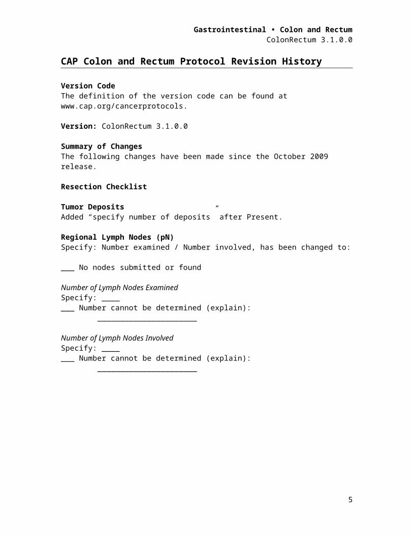
CAP Colon and Rectum Protocol Revision History
Version CodeThe definition of the version code can be found at www.cap.org/cancerprotocols.
Version: ColonRectum 3.1.0.0
Summary of ChangesThe following changes have been made since the October 2009 release.
Resection Checklist
Tumor DepositsAdded “specify number of deposits” after Present.
Regional Lymph Nodes (pN)Specify: Number examined / Number involved, has been changed to:
___ No nodes submitted or found
Number of Lymph Nodes ExaminedSpecify: _______ Number cannot be determined (explain): ______________________
Number of Lymph Nodes InvolvedSpecify: _______ Number cannot be determined (explain): ______________________
4
CAP Approved Gastrointestinal • Colon and RectumColonRectum 3.1.0.0
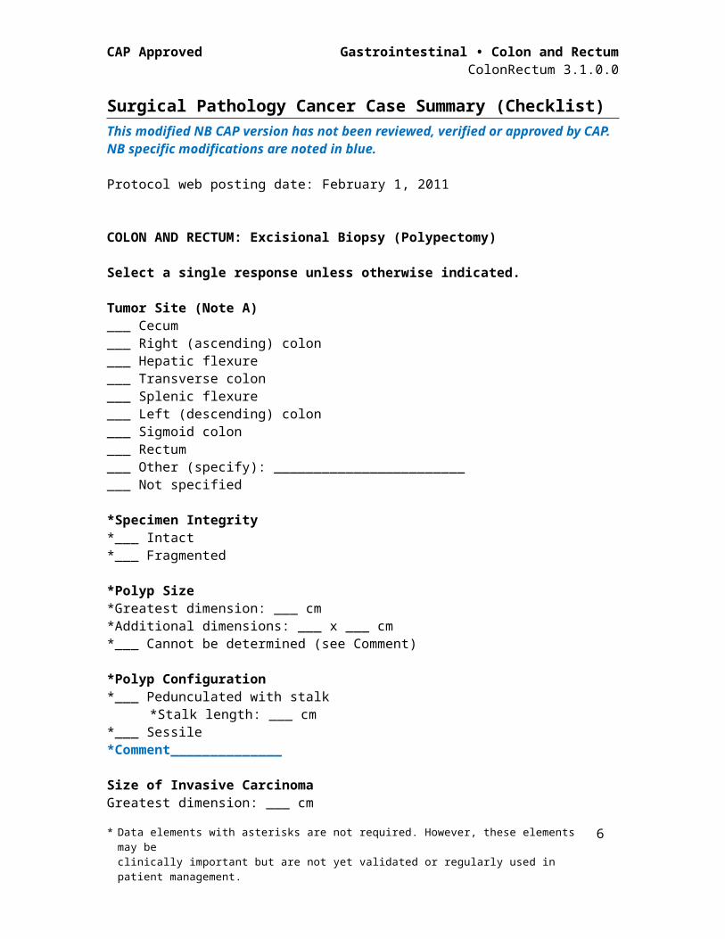
Surgical Pathology Cancer Case Summary (Checklist)This modified NB CAP version has not been reviewed, verified or approved by CAP. NB specific modifications are noted in blue.
Protocol web posting date: February 1, 2011
COLON AND RECTUM: Excisional Biopsy (Polypectomy)
Select a single response unless otherwise indicated.
Tumor Site (Note A)___ Cecum___ Right (ascending) colon___ Hepatic flexure___ Transverse colon___ Splenic flexure___ Left (descending) colon___ Sigmoid colon___ Rectum___ Other (specify): ___________________________ Not specified
*Specimen Integrity*___ Intact*___ Fragmented
*Polyp Size*Greatest dimension: ___ cm*Additional dimensions: ___ x ___ cm*___ Cannot be determined (see Comment)
*Polyp Configuration *___ Pedunculated with stalk
*Stalk length: ___ cm*___ Sessile*Comment______________
Size of Invasive Carcinoma Greatest dimension: ___ cm*Additional dimensions: ___x ___ cm___ Cannot be determined (see Comment)
* Data elements with asterisks are not required. However, these elements may be clinically important but are not yet validated or regularly used in patient management.
5
CAP Approved Gastrointestinal • Colon and RectumColonRectum 3.1.0.0
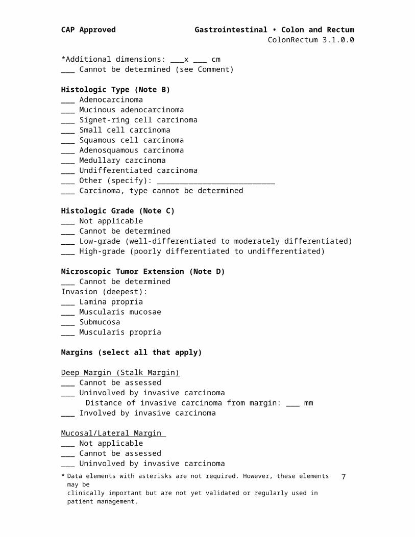
Histologic Type (Note B)___ Adenocarcinoma___ Mucinous adenocarcinoma ___ Signet-ring cell carcinoma ___ Small cell carcinoma ___ Squamous cell carcinoma ___ Adenosquamous carcinoma___ Medullary carcinoma___ Undifferentiated carcinoma___ Other (specify): _____________________________ Carcinoma, type cannot be determined
Histologic Grade (Note C)___ Not applicable___ Cannot be determined___ Low-grade (well-differentiated to moderately differentiated)___ High-grade (poorly differentiated to undifferentiated)
Microscopic Tumor Extension (Note D) ___ Cannot be determinedInvasion (deepest):___ Lamina propria___ Muscularis mucosae___ Submucosa___ Muscularis propria
Margins (select all that apply)
Deep Margin (Stalk Margin)___ Cannot be assessed___ Uninvolved by invasive carcinoma
Distance of invasive carcinoma from margin: ___ mm___ Involved by invasive carcinoma
Mucosal/Lateral Margin ___ Not applicable___ Cannot be assessed___ Uninvolved by invasive carcinoma___ Involved by invasive carcinoma___ Involved by adenoma
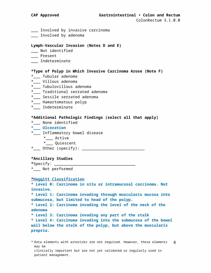
Lymph-Vascular Invasion (Notes D and E)___ Not identified___ Present___ Indeterminate
* Data elements with asterisks are not required. However, these elements may be clinically important but are not yet validated or regularly used in patient management.
6
CAP Approved Gastrointestinal • Colon and RectumColonRectum 3.1.0.0
*Type of Polyp in Which Invasive Carcinoma Arose (Note F)*___ Tubular adenoma*___ Villous adenoma*___ Tubulovillous adenoma*___ Traditional serrated adenoma*___ Sessile serrated adenoma*___ Hamartomatous polyp*___ Indeterminate
*Additional Pathologic Findings (select all that apply)*___ None identified*___ Ulceration*___ Inflammatory bowel disease
*___ Active*___ Quiescent
*___ Other (specify): ___________________________
*Ancillary Studies *Specify: ___________________________________*___ Not performed
* Haggitt Classification * Level 0: Carcinoma in situ or intramucosal carcinoma. Not invasive.* Level 1: Carcinoma invading through muscularis mucosa into submucosa, but limited to head of the polyp.* Level 2: Carcinoma invading the level of the neck of the adenoma* Level 3: Carcinoma invading any part of the stalk* Level 4: Carcinoma invading into the submucosa of the bowel wall below the stalk of the polyp, but above the muscularis propria.
*Comment(s)
* Data elements with asterisks are not required. However, these elements may be clinically important but are not yet validated or regularly used in patient management.
7
CAP Approved Gastrointestinal • Colon and RectumColonRectum 3.1.0.0
Surgical Pathology Cancer Case Summary (Checklist)This modified NB CAP version has not been reviewed, verified or approved by CAP. NB specific modifications are noted in blue.
Protocol web posting date: February 1, 2011
COLON AND RECTUM: Resection, Including Transanal Disk Excision of Rectal Neoplasms
Select a single response unless otherwise indicated.
Specimen (select all that apply) (Note A)___ Terminal ileum___ Cecum___ Appendix___ Ascending colon___ Transverse colon___ Descending colon___ Sigmoid colon___ Rectum___ Anus___ Other (specify): _____________________________________ Not specified
Procedure ___ Right hemicolectomy___ Transverse colectomy___ Left hemicolectomy___ Sigmoidectomy___ Rectal/rectosigmoid colon (low anterior resection)___ Total abdominal colectomy___ Abdominoperineal resection___ Transanal disk excision (local excision)___ Other (specify): _______________________________ Not specified
*Specimen Length (if applicable)*Specify: ___ cm
* Data elements with asterisks are not required. However, these elements may be clinically important but are not yet validated or regularly used in patient management.
8
CAP Approved Gastrointestinal • Colon and RectumColonRectum 3.1.0.0
Tumor Site (select all that apply) (Note A)___ Cecum___ Right (ascending) colon___ Hepatic flexure___ Transverse colon___ Splenic flexure___ Left (descending) colon___ Sigmoid colon___ Rectosigmoid ___ Rectum___ Colon, not otherwise specified___ Cannot be determined (see Comment)
Tumor SizeGreatest dimension: ___ cm*Additional dimensions: ___ x ___ cm___ Cannot be determined (see Comment)
Macroscopic Tumor Perforation (Note G)___ Present___ Not identified___ Cannot be determined
Macroscopic Intactness of Mesorectum (Note H) This is mandatory in NB.___ Not applicable___ Complete___ Near complete___ Incomplete___ Cannot be determined
Histologic Type (Note B)___ Adenocarcinoma___ Mucinous adenocarcinoma ___ Signet-ring cell carcinoma ___ Small cell carcinoma ___ Squamous cell carcinoma ___ Adenosquamous carcinoma___ Medullary carcinoma___ Undifferentiated carcinoma___ Other (specify): _____________________________ Carcinoma, type cannot be determined
Histologic Grade (Note C)___ Not applicable___ Cannot be assessed___ Low-grade (well-differentiated to moderately differentiated)___ High-grade (poorly differentiated to undifferentiated)___ Other (specify): ____________________________
* Data elements with asterisks are not required. However, these elements may be clinically important but are not yet validated or regularly used in patient management.
9
CAP Approved Gastrointestinal • Colon and RectumColonRectum 3.1.0.0
*Histologic Features Suggestive of Microsatellite Instability (Note I)
* Intratumoral Lymphocytic Response (tumor-infiltrating lymphocytes) *___ None*___ Mild to moderate (0-2 per high-power [X400] field)*___ Marked (3 or more per high-power field)
*Peritumor Lymphocytic Response (Crohn-like response)*___ None*___ Mild to moderate*___ Marked *Tumor Subtype and Differentiation (select all that apply)*___ Mucinous tumor component (specify percentage: ____)*___ Medullary tumor component*___ High histologic grade (poorly differentiated)
Microscopic Tumor Extension___ Cannot be assessed___ No evidence of primary tumor___ Intramucosal carcinoma, invasion of lamina propria___ Tumor invades submucosa___ Tumor invades muscularis propria___ Tumor invades through the muscularis propria into the subserosal adipose tissue or
the nonperitonealized pericolic or perirectal soft tissues but does not extend to the serosal surface
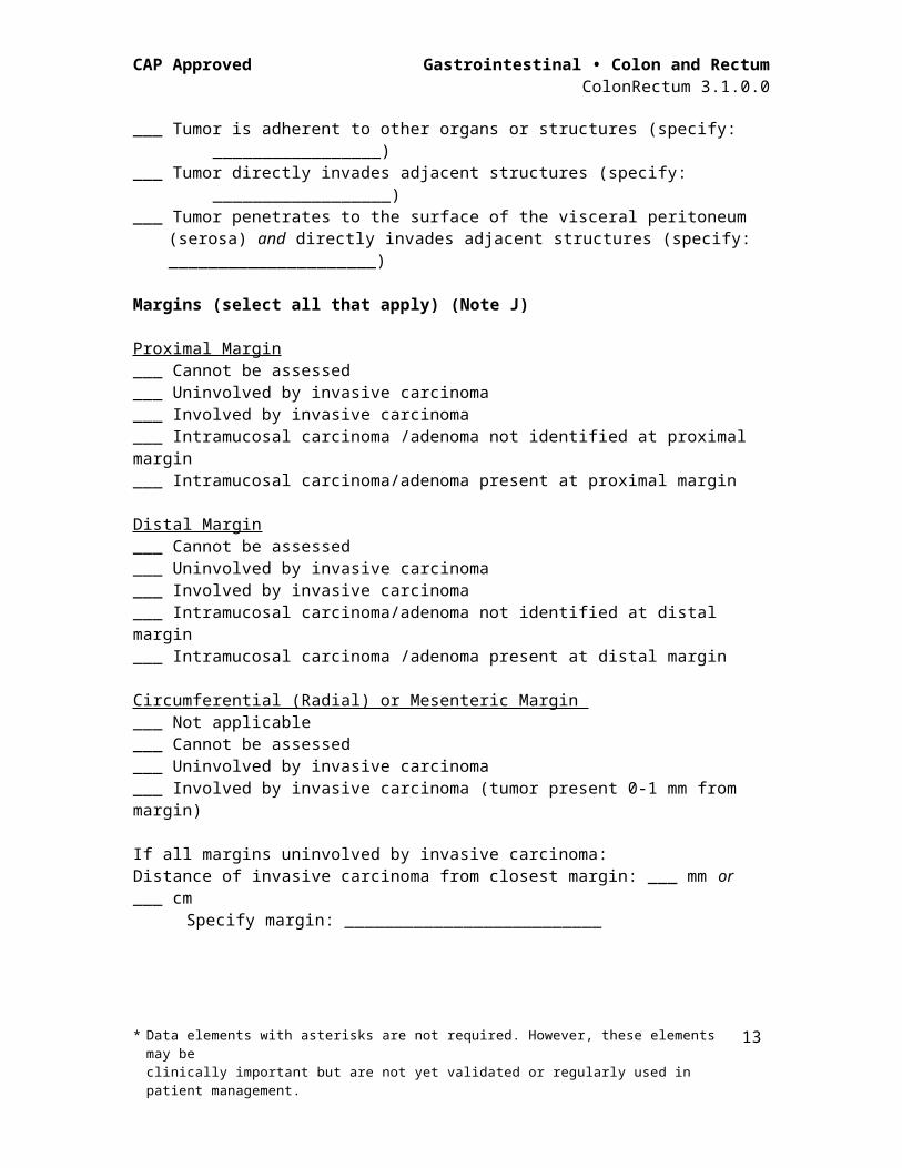
___ Tumor penetrates to the surface of the visceral peritoneum (serosa) ___ Tumor is adherent to other organs or structures (specify: _________________) ___ Tumor directly invades adjacent structures (specify: __________________)___ Tumor penetrates to the surface of the visceral peritoneum (serosa) and directly
invades adjacent structures (specify: _____________________)
Margins (select all that apply) (Note J)
Proximal Margin___ Cannot be assessed___ Uninvolved by invasive carcinoma___ Involved by invasive carcinoma___ Intramucosal carcinoma /adenoma not identified at proximal margin___ Intramucosal carcinoma/adenoma present at proximal margin
Distal Margin___ Cannot be assessed___ Uninvolved by invasive carcinoma___ Involved by invasive carcinoma___ Intramucosal carcinoma/adenoma not identified at distal margin___ Intramucosal carcinoma /adenoma present at distal margin
* Data elements with asterisks are not required. However, these elements may be clinically important but are not yet validated or regularly used in patient management.
10
CAP Approved Gastrointestinal • Colon and RectumColonRectum 3.1.0.0
Circumferential (Radial) or Mesenteric Margin ___ Not applicable___ Cannot be assessed___ Uninvolved by invasive carcinoma___ Involved by invasive carcinoma (tumor present 0-1 mm from margin)
If all margins uninvolved by invasive carcinoma:Distance of invasive carcinoma from closest margin: ___ mm or ___ cm
Specify margin: __________________________
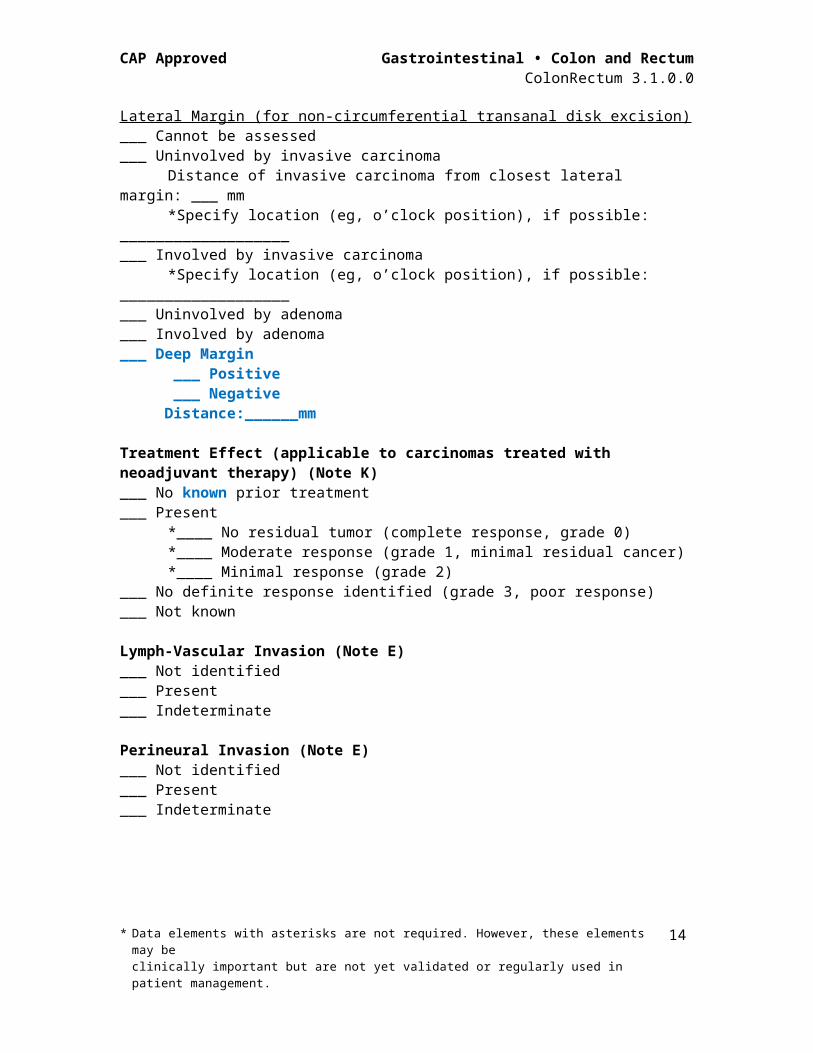
Lateral Margin (for non-circumferential transanal disk excision)___ Cannot be assessed___ Uninvolved by invasive carcinoma
Distance of invasive carcinoma from closest lateral margin: ___ mm*Specify location (eg, o’clock position), if possible: ___________________
___ Involved by invasive carcinoma*Specify location (eg, o’clock position), if possible: ___________________
___ Uninvolved by adenoma ___ Involved by adenoma___ Deep Margin ___ Positive ___ Negative Distance:______mm
Treatment Effect (applicable to carcinomas treated with neoadjuvant therapy) (Note K)___ No known prior treatment___ Present
*____ No residual tumor (complete response, grade 0)*____ Moderate response (grade 1, minimal residual cancer)*____ Minimal response (grade 2)
___ No definite response identified (grade 3, poor response)___ Not known
Lymph-Vascular Invasion (Note E)___ Not identified___ Present___ Indeterminate
Perineural Invasion (Note E)___ Not identified___ Present___ Indeterminate
* Data elements with asterisks are not required. However, these elements may be clinically important but are not yet validated or regularly used in patient management.
11
CAP Approved Gastrointestinal • Colon and RectumColonRectum 3.1.0.0
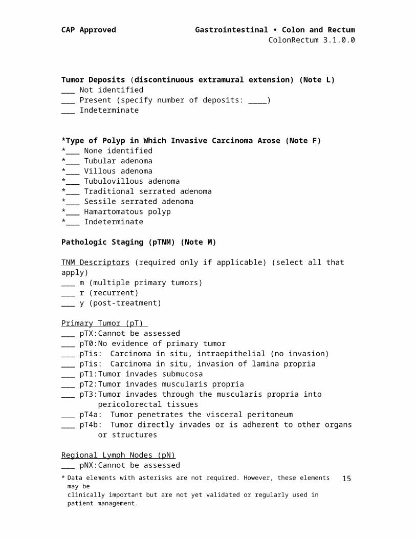
Tumor Deposits (discontinuous extramural extension) (Note L)___ Not identified___ Present (specify number of deposits: ____)___ Indeterminate
*Type of Polyp in Which Invasive Carcinoma Arose (Note F)*___ None identified*___ Tubular adenoma*___ Villous adenoma*___ Tubulovillous adenoma*___ Traditional serrated adenoma*___ Sessile serrated adenoma*___ Hamartomatous polyp*___ Indeterminate
Pathologic Staging (pTNM) (Note M)
TNM Descriptors (required only if applicable) (select all that apply)___ m (multiple primary tumors)___ r (recurrent)___ y (post-treatment)
Primary Tumor (pT) ___ pTX: Cannot be assessed___ pT0: No evidence of primary tumor___ pTis: Carcinoma in situ, intraepithelial (no invasion)___ pTis: Carcinoma in situ, invasion of lamina propria___ pT1: Tumor invades submucosa___ pT2: Tumor invades muscularis propria___ pT3: Tumor invades through the muscularis propria into pericolorectal tissues___ pT4a: Tumor penetrates the visceral peritoneum___ pT4b: Tumor directly invades or is adherent to other organs or structures
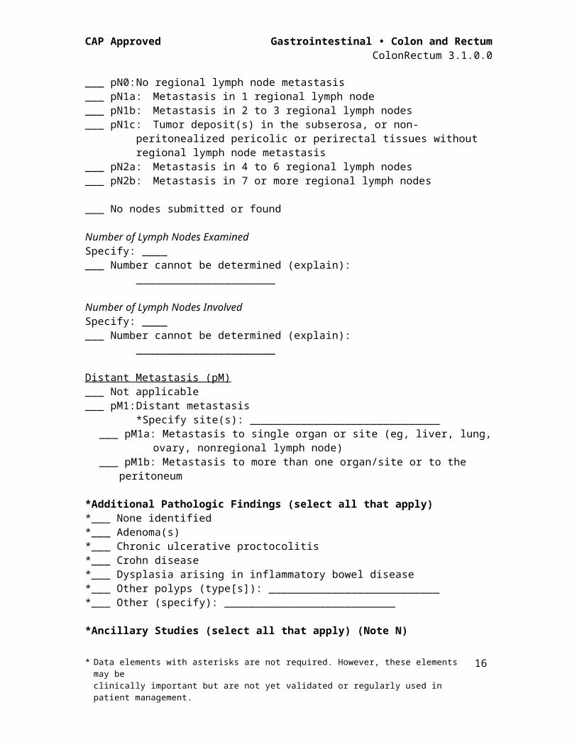
Regional Lymph Nodes (pN)___ pNX: Cannot be assessed___ pN0: No regional lymph node metastasis___ pN1a: Metastasis in 1 regional lymph node___ pN1b: Metastasis in 2 to 3 regional lymph nodes___ pN1c: Tumor deposit(s) in the subserosa, or non-peritonealized pericolic or
perirectal tissues without regional lymph node metastasis___ pN2a: Metastasis in 4 to 6 regional lymph nodes___ pN2b: Metastasis in 7 or more regional lymph nodes
___ No nodes submitted or found
* Data elements with asterisks are not required. However, these elements may be clinically important but are not yet validated or regularly used in patient management.
12
CAP Approved Gastrointestinal • Colon and RectumColonRectum 3.1.0.0
Number of Lymph Nodes ExaminedSpecify: _______ Number cannot be determined (explain): ______________________
Number of Lymph Nodes InvolvedSpecify: _______ Number cannot be determined (explain): ______________________
Distant Metastasis (pM)___ Not applicable ___ pM1: Distant metastasis
*Specify site(s): _________________________________ pM1a: Metastasis to single organ or site (eg, liver, lung, ovary, nonregional
lymph node)___ pM1b: Metastasis to more than one organ/site or to the peritoneum
*Additional Pathologic Findings (select all that apply)*___ None identified*___ Adenoma(s)*___ Chronic ulcerative proctocolitis*___ Crohn disease*___ Dysplasia arising in inflammatory bowel disease*___ Other polyps (type[s]): ___________________________*___ Other (specify): ___________________________
*Ancillary Studies (select all that apply) (Note N)
*___ Microsatellite instability (specify testing method: ___________________)*___ Stable*___ Low*___ High
*Immunohistochemistry Studies for Mismatch Repair Proteins*___ MLH1
*___ Intact nuclear positivity, tumor cells*___ Loss of nuclear positivity, tumor cells*___ Pending*___ Other (specify): _____________________
*___ MSH2*___ Intact nuclear positivity, tumor cells*___ Loss of nuclear positivity, tumor cells*___ Pending*___ Other (specify): _____________________
*___ MSH6*___ Intact nuclear positivity, tumor cells*___ Loss of nuclear positivity, tumor cells*___ Pending*___ Other (specify): _____________________
* Data elements with asterisks are not required. However, these elements may be clinically important but are not yet validated or regularly used in patient management.
13
CAP Approved Gastrointestinal • Colon and RectumColonRectum 3.1.0.0
*___ PMS2*___ Intact nuclear positivity, tumor cells*___ Loss of nuclear positivity, tumor cells*___ Pending*___ Other (specify): _____________________
*Mutational Analysis*___ BRAF V600E mutational analysis (specify testing method: _________________)
*___ Mutant BRAF detected *___ No mutant BRAF detected (wild type BRAF allele)*___ Other (specify):______________________
*___ KRAS mutational analysis (specify testing method: _________________)*___ Mutant KRAS detected (specify mutation_____________)*___ No mutant KRAS detected (wild type KRAS allele)*___ Other (specify):___________________________
*Other, specify: ___________________________________
*____ Not performed
*Comment(s)
* Data elements with asterisks are not required. However, these elements may be clinically important but are not yet validated or regularly used in patient management.
14
Background Documentation Gastrointestinal • Colon and RectumColonRectum 3.1.0.0
Explanatory Notes
A. Anatomic SitesThe protocol applies to all carcinomas arising in the colon and rectum.1 It excludes carcinomas of the vermiform appendix and low-grade neuroendocrine neoplasms (carcinoid tumors).
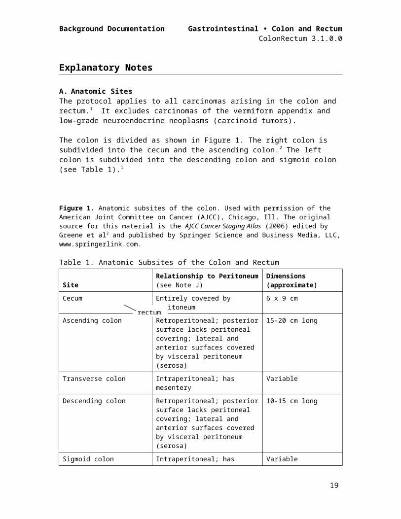
The colon is divided as shown in Figure 1. The right colon is subdivided into the cecum and the ascending colon.2 The left colon is subdivided into the descending colon and sigmoid colon (see Table 1).1
Figure 1. Anatomic subsites of the colon. Used with permission of the American Joint Committee on Cancer (AJCC), Chicago, Ill. The original source for this material is the AJCC Cancer Staging Atlas (2006) edited by Greene et al2 and published by Springer Science and Business Media, LLC, www.springerlink.com.
Table 1. Anatomic Subsites of the Colon and Rectum
SiteRelationship to Peritoneum (see Note J)
Dimensions (approximate)
Cecum Entirely covered by peritoneum 6 x 9 cm
Ascending colon Retroperitoneal; posterior surface lacks peritoneal covering; lateral and anterior surfaces covered by visceral peritoneum (serosa)
15-20 cm long
Transverse colon Intraperitoneal; has mesentery Variable
Descending colon Retroperitoneal; posterior surface lacks peritoneal covering; lateral and anterior surfaces covered by visceral peritoneum (serosa)
10-15 cm long
Sigmoid colon Intraperitoneal; has mesentery Variable
Rectum Upper third covered by peritoneum on anterior and lateral surfaces; middle third covered by peritoneum only on anterior surface; lower third has no peritoneal covering
12 cm long
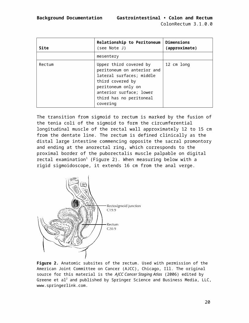
The transition from sigmoid to rectum is marked by the fusion of the tenia coli of the sigmoid to form the circumferential longitudinal muscle of the rectal wall approximately 12 to 15 cm from the dentate line. The rectum is defined clinically as the distal large intestine commencing opposite the sacral promontory and ending at the anorectal ring, which corresponds to the proximal border of the puborectalis muscle palpable on digital rectal examination1 (Figure 2). When measuring below with a rigid sigmoidoscope, it extends 16 cm from the anal verge.
15
rectum
Background Documentation Gastrointestinal • Colon and RectumColonRectum 3.1.0.0
Figure 2. Anatomic subsites of the rectum. Used with permission of the American Joint Committee on Cancer (AJCC), Chicago, Ill. The original source for this material is the AJCC Cancer Staging Atlas (2006) edited by Greene et al2 and published by Springer Science and Business Media, LLC, www.springerlink.com.
Tumors located at the border between 2 subsites of the colon (eg, cecum and ascending colon) are registered as tumors of the subsite that is more involved. If 2 subsites are involved to the same extent, the tumor is classified as an "overlapping" lesion.
A tumor is classified as rectal if its inferior margin lies less than 16 cm from the anal verge or if any part of the tumor is located at least partly within the supply of the superior rectal artery.3 A tumor is classified as rectosigmoid when differentiation between rectum and sigmoid according to the previously mentioned guidelines is not possible.4
B. Histologic TypesFor consistency in reporting, the histologic classification proposed by the World Health Organization (WHO) is recommended and is shown below.5
WHO Classification of Colorectal CarcinomaAdenocarcinomaMucinous (colloid) adenocarcinoma (greater than 50% mucinous)Signet-ring cell carcinoma (greater than 50% signet-ring cells)#
Squamous cell carcinomaAdenosquamous carcinomaMedullary carcinoma##
Small cell carcinoma# (high-grade neuroendocrine carcinoma)
Undifferentiated carcinoma#
Other (specify)###
# By convention, signet-ring cell carcinomas, small cell carcinomas, and undifferentiated carcinomas are high grade (see Note C). The only histologic types of colorectal
16
Background Documentation Gastrointestinal • Colon and RectumColonRectum 3.1.0.0
carcinoma that have been shown to have adverse prognostic significance independent of stage are signet-ring cell carcinoma6 and small cell carcinoma (high-grade neuroendocrine carcinoma).7
## Medullary carcinoma is a distinctive histologic type strongly associated with high levels of microsatellite instability (MSI-H), indicative of defects in normal DNA repair gene function. Medullary carcinoma may occur either sporadically8 or in association with hereditary nonpolyposis colon cancer (HNPCC).9 This tumor type is characterized by solid growth in nested, organoid, or trabecular patterns, with no immunohistochemical evidence of neuroendocrine differentiation. Medullary carcinomas are also characterized by numerous tumor infiltrating lymphocytes (see Note I).
### The term "carcinoma, NOS" (not otherwise specified) is not part of the WHO classification.
C. Histologic GradeA number of grading systems for colorectal cancer have been suggested, but a single widely accepted and uniformly used standard for grading is lacking. Most systems stratify tumors into 3 or 4 grades as follows:
Grade 1 Well-differentiatedGrade 2 Moderately differentiatedGrade 3 Poorly differentiatedGrade 4 Undifferentiated
Despite a significant degree of interobserver variability,10 histologic grade has repeatedly been shown by multivariate analysis to be a stage-independent prognostic factor.11 Specifically, it has been demonstrated that high tumor grade is an adverse prognostic factor. It is noteworthy that in the majority of studies documenting the prognostic power of tumor grade, the number of grades has been collapsed to produce a 2-tiered stratification for data analysis as follows:
Low-grade: Well-differentiated and moderately differentiatedHigh-grade: Poorly differentiated and undifferentiated
The widest variations in grading concern the stratification of low-grade tumors into well- or moderately differentiated categories, while interobserver variability in diagnosing high-grade carcinoma is relatively small. Therefore, in light of its proven prognostic value, relative simplicity, and reproducibility, a 2-tiered grading system for colorectal carcinoma (ie, low-grade and high-grade) is recommended. The following criteria for grading based on gland formation alone are suggested.12
Low-grade = Greater than or equal to 50% gland formationHigh-grade = Less than 50% gland formation
17
Background Documentation Gastrointestinal • Colon and RectumColonRectum 3.1.0.0
D. Carcinoma in an Adenomatous Polyp: Microscopic Tumor Extension and High-risk Features Colorectal adenomas containing invasive adenocarcinoma that extends through the muscularis mucosae into the submucosa have been defined as "malignant polyps.”13 This term encompasses cases in which the entire polyp head is replaced by carcinoma and adenomas with focal malignancy, but the definition excludes adenomas with high-grade dysplasia (intraepithelial carcinoma) or intramucosal carcinoma (invasive carcinoma limited to the lamina propria or invading no deeper than the muscularis mucosae), because these polyps possess negligible biological potential for metastasis14
(see Tis in Note M).
Malignant polyps removed by endoscopic polypectomy require evaluation of histologic factors related to the risk of adverse outcome (ie, lymph node metastasis or local recurrence from residual malignancy) following polypectomy.13,15 Factors shown to have independent prognostic significance and are important in determining the need for further surgical treatment include:• Histologic grade• Status of the resection margin• Lymphatic/venous vessel involvement
An increased risk of adverse outcome has been shown to be associated with:• High-grade carcinoma• Tumor at or less than 1 mm from the resection margin• Lymphatic/venous vessel involvement14
E. Lymph-Vascular and Perineural InvasionVenous invasion has been demonstrated by multivariate analysis to be an independent adverse prognostic factor.11 Invasion of extramural veins, in particular, has been shown to be an independent indicator of unfavorable outcome and increased risk of occurrence of hepatic metastasis.16 The significance of intramural venous invasion is less clear, because data specific to this issue are lacking.
In several studies, both lymphatic invasion17 and perineural invasion18 have been shown by multivariate analysis to be independent indicators of poor prognosis. The prognostic significance, if any, of the anatomic location of these structures is not defined. Furthermore, it is not always possible to distinguish lymphatic vessels from postcapillary venules, because both are small, thin-walled structures. Thus, the presence or absence of tumor invasion of small, thin-walled vessels should be reported in all cases.
F. PolypsDistinction should be made between traditional serrated adenomas, which exhibit cytologic features of adenomas, and the newly described sessile serrated adenomas.19 The sessile serrated adenoma may be the precursor lesion for colorectal carcinomas with high levels of microsatellite instability (MSI-H); they are more commonly found in the right colon and are characterized by serrated architecture with bulbous dilatation of deep crypts and lack of overt nuclear atypia, in most cases.
18
Background Documentation Gastrointestinal • Colon and RectumColonRectum 3.1.0.0
G. PerforationTumor perforation is an uncommon complication of colorectal cancer, but one that is associated with a poor outcome, including high in-hospital mortality and morbidity.20 Perforation of the uninvolved colon proximal to an obstructing tumor is also associated with high mortality because of generalized peritonitis and sepsis. Reported perforation rates range from 2.6% to 9%. Perforation is more likely to occur in older patients.
H. Mesorectal EnvelopeThe quality of the surgical technique is a key factor in the success of surgical treatment for rectal cancer, both in the prevention of local recurrence and in long-term survival. Numerous studies have demonstrated that total mesorectal excision (TME) improves local recurrence rates and the corresponding survival by as much as 20%. This surgical technique entails precise sharp dissection within the areolar plane outside (lateral to) the visceral mesorectal fascia to remove the rectum. This plane encases the rectum, its mesentery, and all regional nodes and constitutes Waldeyer’s fascia. High-quality TME surgery reduces local recurrence from 20% to 30%, to 8% to 10% or less, and increases 5-year survival from 48% to 68%.21,22 Adjuvant therapy in the presence of a high-quality TME may further reduce local recurrence (from 8% to 2.6%).22
Pathologic evaluation of the resection specimen has been shown to be a sensitive means of assessing the quality of rectal surgery. It is superior to indirect measures of surgical quality assessment, such as perioperative mortality, rates of complication, number of local recurrences, and 5-year survival. It has been shown that macroscopic pathologic assessment of the completeness of the mesorectum of the specimen, scored as complete, partially complete, or incomplete, accurately predicts both local recurrence and distant metastasis.22 Microscopic parameters, such as the status of the circumferential resection margin, the distance between the tumor and nearest circumferential margin (ie, “surgical clearance”), and the distance between the tumor and the closest distal margin, are all important predictors of local recurrence and may be affected by surgical technique. There is strong evidence that the status of the circumferential resection margin is a powerful predictor of local recurrence but is inconsistently evaluated and under-reported.
The nonperitonealized surface of the fresh specimen is examined circumferentially, and the completeness of the mesorectum is scored as described below.22 The entire specimen is scored according to the worst area.
Incomplete Little bulk to the mesorectumDefects in the mesorectum down to the muscularis propriaAfter transverse sectioning, the circumferential margin appears very irregular
Nearly Complete Moderate bulk to the mesorectumIrregularity of the mesorectal surface with defects greater than 5 mm, but none
extending to the muscularis propriaNo areas of visibility of the muscularis propria except at the insertion site of the levator
ani muscles
19
Background Documentation Gastrointestinal • Colon and RectumColonRectum 3.1.0.0
Complete Intact bulky mesorectum with a smooth surfaceOnly minor irregularities of the mesorectal surfaceNo surface defects greater than 5 mm in depthNo coning towards the distal margin of the specimenAfter transverse sectioning, the circumferential margin appears smooth
I. Histopathologic Features Suggestive of Microsatellite InstabilityIdentification of MSI-H colorectal tumors is important, as mismatch repair deficiency may serve as a prognostic marker of patient outcome, a predictive marker of response to chemotherapy, and as a screening tool for hereditary nonpolyposis colon cancer (HNPCC) (Lynch syndrome). Revised Bethesda guidelines for HNPCC detection recommend testing colorectal tumors for microsatellite instability under the following circumstances23:
1. Colorectal cancer diagnosed in a patient who is younger than 50 years.2. Presence of synchronous, metachronous, or other HNPCC-associated tumors
(endometrial, stomach, ovarian, pancreas, ureter and renal pelvis, biliary tract, small bowel, and brain tumors and sebaceous adenomas and keratoacanthomas), regardless of age.
3. Colorectal cancer with MSI-H histology# in a patient who is younger than 60 years.4. Colorectal cancer in 1 or more first-degree relatives with an HNPCC-related tumor,
with 1 of the cancers being diagnosed in a person younger than 50 years.5. Colorectal cancer diagnosed in 2 or more first- or second-degree relatives with
HNPCC-related tumors, regardless of age.
# MSI-H histologic features are defined as presence of tumor-infiltrating lymphocytes, Crohn-like lymphocytic reaction, mucinous/signet-ring cell differentiation, or medullary growth pattern.23
Tumor-infiltrating lymphocytes are closely associated with microsatellite instability and medullary architecture (see above) and should be distinguished from Crohn-like peritumoral infiltrates (lymphoid aggregated or follicles are the tumor edge, not associated with pre-existing lymph node).24 Although absolute cut-off values have not been established, only moderate- and high-density intratumoral lymphocytes (approximately 3 or more per high-power field using hematoxylin-and-eosin [H&E]-stained sections) should be considered significant.25
Other pathologic features associated with MSI-H status in colorectal carcinomas include right-sided location, high-grade histology, and lack of dirty necrosis.25
J. Margins It may be helpful to mark the margin(s) closest to the tumor with ink following close examination of the serosal surface for puckering and other signs of tumor involvement. Margins marked by ink should be designated in the macroscopic description of the surgical pathology report. The serosal surface (visceral peritoneum) does not constitute a surgical margin.
20
Background Documentation Gastrointestinal • Colon and RectumColonRectum 3.1.0.0
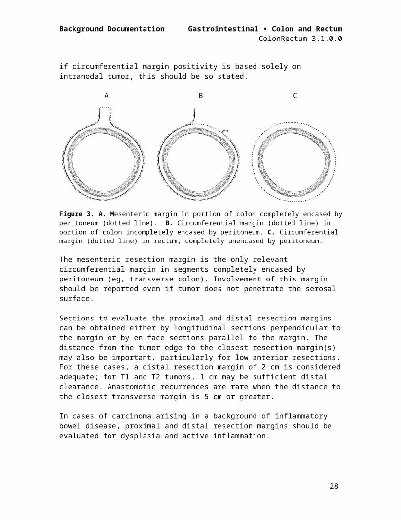
In addition to addressing the proximal and distal margins, the circumferential (radial) margin (Figure 3A-3C) must be assessed for any segment either unencased (Figure 3C) or incompletely encased by peritoneum (Figure 3B) (see Note A). The circumferential (radial) margin represents the adventitial soft tissue margin closest to the deepest penetration of tumor and is created surgically by blunt or sharp dissection of the retroperitoneal or subperitoneal aspect respectively. Multivariate analysis has suggested that tumor involvement of the circumferential (radial) margin is the most critical factor in predicting local recurrence in rectal cancer.26 A positive circumferential (radial) margin in rectal cancer increases the risk of recurrence by 3.5-fold and doubles the risk of death from disease. For this reason, the circumferential (radial) margin should be assessed in all rectal carcinomas as well as colonic segments with nonperitonealized surfaces. The distance between the tumor and circumferential (radial) margin should be reported (see Note H). The circumferential (radial) margin is considered negative if the tumor is more than 1 mm from the inked nonperitonealized surface but should be recorded as positive if tumor is located 1 mm or less from the nonperitonealized surface because local recurrence rates are similar with clearances of 0 to 1 mm. This assessment includes tumor within a lymph node as well as direct tumor extension, but if circumferential margin positivity is based solely on intranodal tumor, this should be so stated.
A B C
Figure 3. A. Mesenteric margin in portion of colon completely encased by peritoneum (dotted line). B. Circumferential margin (dotted line) in portion of colon incompletely encased by peritoneum. C. Circumferential margin (dotted line) in rectum, completely unencased by peritoneum.
The mesenteric resection margin is the only relevant circumferential margin in segments completely encased by peritoneum (eg, transverse colon). Involvement of this margin should be reported even if tumor does not penetrate the serosal surface.
Sections to evaluate the proximal and distal resection margins can be obtained either by longitudinal sections perpendicular to the margin or by en face sections parallel to the margin. The distance from the tumor edge to the closest resection margin(s) may also be important, particularly for low anterior resections. For these cases, a distal resection margin of 2 cm is considered adequate; for T1 and T2 tumors, 1 cm may be
21
Background Documentation Gastrointestinal • Colon and RectumColonRectum 3.1.0.0
sufficient distal clearance. Anastomotic recurrences are rare when the distance to the closest transverse margin is 5 cm or greater.
In cases of carcinoma arising in a background of inflammatory bowel disease, proximal and distal resection margins should be evaluated for dysplasia and active inflammation.
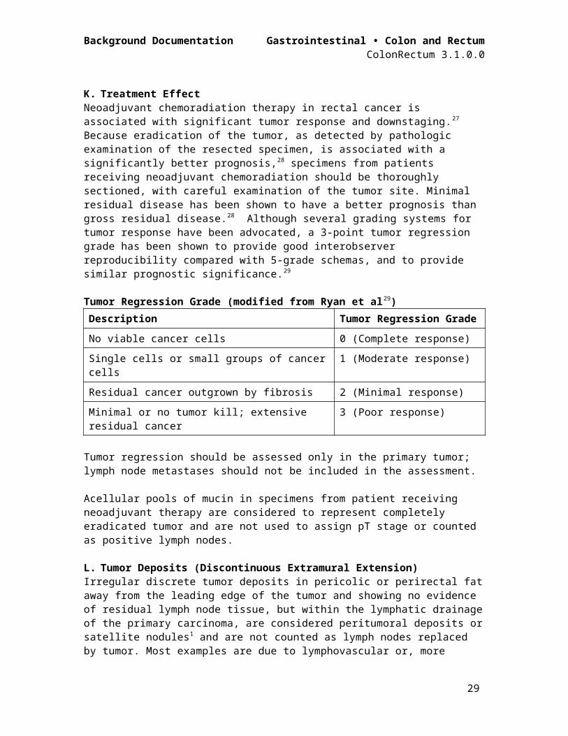
K. Treatment Effect Neoadjuvant chemoradiation therapy in rectal cancer is associated with significant tumor response and downstaging.27 Because eradication of the tumor, as detected by pathologic examination of the resected specimen, is associated with a significantly better prognosis,28 specimens from patients receiving neoadjuvant chemoradiation should be thoroughly sectioned, with careful examination of the tumor site. Minimal residual disease has been shown to have a better prognosis than gross residual disease.28 Although several grading systems for tumor response have been advocated, a 3-point tumor regression grade has been shown to provide good interobserver reproducibility compared with 5-grade schemas, and to provide similar prognostic significance.29
Tumor Regression Grade (modified from Ryan et al29)Description Tumor Regression Grade
No viable cancer cells 0 (Complete response)
Single cells or small groups of cancer cells 1 (Moderate response)
Residual cancer outgrown by fibrosis 2 (Minimal response)
Minimal or no tumor kill; extensive residual cancer 3 (Poor response)
Tumor regression should be assessed only in the primary tumor; lymph node metastases should not be included in the assessment.
Acellular pools of mucin in specimens from patient receiving neoadjuvant therapy are considered to represent completely eradicated tumor and are not used to assign pT stage or counted as positive lymph nodes.
L. Tumor Deposits (Discontinuous Extramural Extension)Irregular discrete tumor deposits in pericolic or perirectal fat away from the leading edge of the tumor and showing no evidence of residual lymph node tissue, but within the lymphatic drainage of the primary carcinoma, are considered peritumoral deposits or satellite nodules1 and are not counted as lymph nodes replaced by tumor. Most examples are due to lymphovascular or, more rarely, perineural invasion. Because these tumor deposits are associated with reduced disease-free and overall survival,30,31 their number should be recorded in the surgical pathology report. If tumor deposits are observed in lesions that would otherwise be classified as pT1 (tumor confined to submucosa) or pT2 (tumor confined to muscularis propria), then the primary tumor classification is not changed, but the nodule is recorded in a separate N category as N1c1 (see Note M).
22
Background Documentation Gastrointestinal • Colon and RectumColonRectum 3.1.0.0
M. TNM and Anatomic Stage/Prognostic GroupingsSurgical resection remains the most effective therapy for colorectal carcinoma, and the best estimation of prognosis is derived from the pathologic findings on the resection specimen. The anatomic extent of disease is by far the most important prognostic factor in colorectal cancer.
The protocol recommends the TNM staging system of the American Joint Committee on Cancer (AJCC) and the International Union Against Cancer (UICC)1 but does not preclude the use of other staging systems.
By AJCC/UICC convention, the designation “T” refers to a primary tumor that has not been previously treated. The symbol “p” refers to the pathologic classification of the TNM, as opposed to the clinical classification, and is based on gross and microscopic examination. pT entails a resection of the primary tumor or biopsy adequate to evaluate the highest pT category, pN entails removal or biopsy of nodes adequate to validate lymph node metastasis, and pM implies microscopic examination of distant lesions. Clinical classification (cTNM) is usually carried out by the referring physician before treatment during initial evaluation of the patient or when pathologic classification is not possible.
TNM DescriptorsFor identification of special cases of TNM or pTNM classifications, the “m” suffix and “y” and “r” prefixes are used. Although they do not affect the stage grouping, they indicate cases needing separate analysis.
The “m” suffix indicates the presence of multiple primary tumors in a single site and is recorded in parentheses: pT(m)NM.
The “y” prefix indicates those cases in which classification is performed during or following initial multimodality therapy (ie, neoadjuvant chemotherapy, radiation therapy, or both chemotherapy and radiation therapy). The cTNM or pTNM category is identified by a “y” prefix. The ycTNM or ypTNM categorizes the extent of tumor actually present at the time of that examination. The “y” categorization is not an estimate of tumor prior to multimodality therapy (ie, before initiation of neoadjuvant therapy).
The “r” prefix indicates a recurrent tumor when staged after a documented disease-free interval, and is identified by the “r” prefix: rTNM.
T Category Considerations (Figures 4-6)pTis. For colorectal carcinomas, "carcinoma in situ" (pTis) as a staging term includes cancer cells confined within the glandular basement membrane (intraepithelial carcinoma, synonymous with high-grade dysplasia) or invasive into the mucosal lamina propria, up to but not through the muscularis mucosae (intramucosal carcinoma). Tumor extension through the muscularis mucosae into the submucosa is classified as T1.
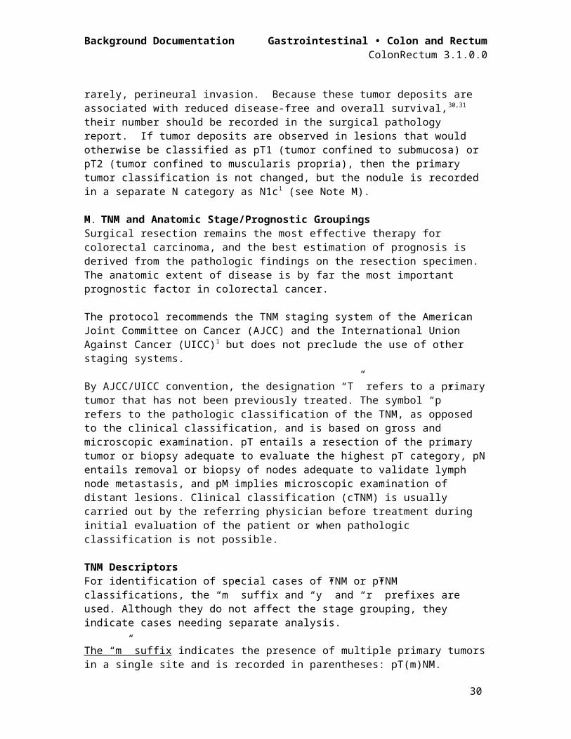
pT4. Direct invasion of other organs or structures includes invasion of other segments of colorectum by way of the serosa or mesocolon (eg, invasion of the sigmoid colon by carcinoma of the cecum) is classified as pT4 (Figure 6). In such a case, both an
23
Background Documentation Gastrointestinal • Colon and RectumColonRectum 3.1.0.0
adjacent organ and the visceral peritoneum are penetrated by tumor. Intramural extension of tumor from 1 subsite (segment) of the large intestine into an adjacent subsite or into the ileum (eg, for a cecal carcinoma) or anal canal (eg, for a rectal carcinoma) does not affect the pT classification.
Figure 4. T4 (left side) with involvement of serosa (visceral peritoneum) by tumor cells in a segment of colorectum with a serosal covering. In contrast, the right side of the diagram shows T3 with macroscopically positive circumferential margin (designated R2 in AJCC staging system), corresponding to gross disease remaining after surgical excision. Used with permission of the American Joint Committee on Cancer (AJCC), Chicago, Ill. The original source for this material is the AJCC Cancer Staging Atlas (2006) edited by Greene et al2 and published by Springer Science and Business Media, LLC, www.springerlink.com.
24
Background Documentation Gastrointestinal • Colon and RectumColonRectum 3.1.0.0
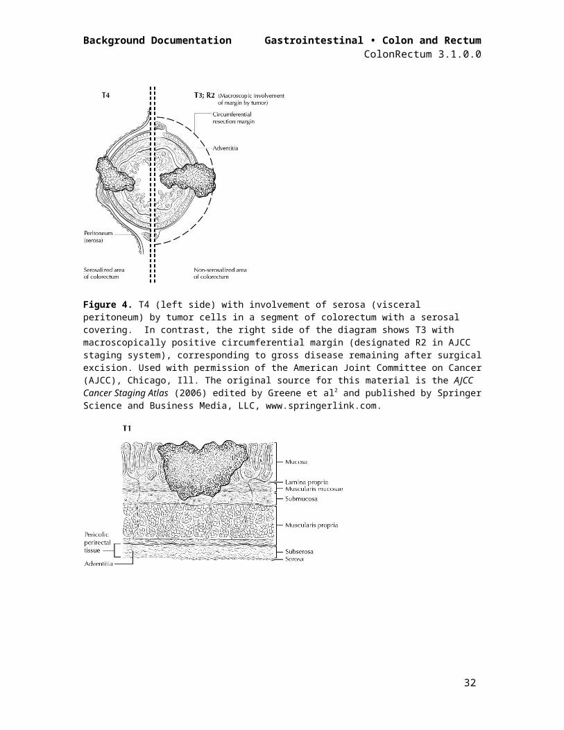
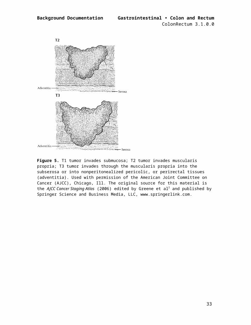
Figure 5. T1 tumor invades submucosa; T2 tumor invades muscularis propria; T3 tumor invades through the muscularis propria into the subserosa or into nonperitonealized pericolic, or perirectal tissues (adventitia). Used with permission of the American Joint Committee on Cancer (AJCC), Chicago, Ill. The original source for this material is the AJCC Cancer Staging Atlas (2006) edited by Greene et al2 and published by Springer Science and Business Media, LLC, www.springerlink.com.
25

Background Documentation Gastrointestinal • Colon and RectumColonRectum 3.1.0.0
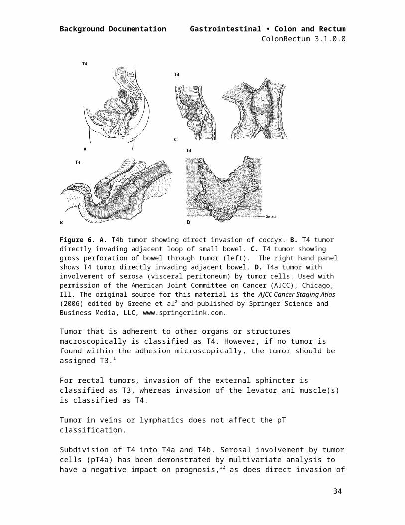
Figure 6. A. T4b tumor showing direct invasion of coccyx. B. T4 tumor directly invading adjacent loop of small bowel. C. T4 tumor showing gross perforation of bowel through tumor (left). The right hand panel shows T4 tumor directly invading adjacent bowel. D. T4a tumor with involvement of serosa (visceral peritoneum) by tumor cells. Used with permission of the American Joint Committee on Cancer (AJCC), Chicago, Ill. The original source for this material is the AJCC Cancer Staging Atlas (2006) edited by Greene et al2 and published by Springer Science and Business Media, LLC, www.springerlink.com.
Tumor that is adherent to other organs or structures macroscopically is classified as T4. However, if no tumor is found within the adhesion microscopically, the tumor should be assigned T3.1
For rectal tumors, invasion of the external sphincter is classified as T3, whereas invasion of the levator ani muscle(s) is classified as T4.
Tumor in veins or lymphatics does not affect the pT classification.
Subdivision of T4 into T4a and T4b. Serosal involvement by tumor cells (pT4a) has been demonstrated by multivariate analysis to have a negative impact on prognosis,32 as does direct invasion of adjacent organs (pT4b). Visceral peritoneal involvement can be missed without thorough sampling and/or sectioning, and malignant cells have been identified in serosal scrapings in as many as 26% of specimens categorized as pT3 by histologic examination alone.33 Although the absence of standard guidelines for assessing peritoneal involvement may contribute to underdiagnosis, the following findings are considered to represent serosal involvement by tumor:1. Tumor present at the serosal surface with inflammatory reaction, mesothelial
hyperplasia, and/or erosion/ulceration 2. Free tumor cells on the serosal surface (in the peritoneum) with underlying
ulceration of the visceral peritoneum32
Both types of peritoneal involvement are associated with decreased survival.
Although small studies suggested that serosal involvement was associated with worse outcome than invasion of adjacent organs, data from a large cohort of more than 100,000 colon cancer cases33 indicate that penetration of the visceral peritoneum carries a 10% to 20% better 5-year survival than locally invasive carcinomas for each category of N. Therefore, designation of the T4 subsets was changed in the seventh edition of the AJCC Cancer Staging Manual to reflect these new findings.
N Category ConsiderationsThe regional lymph nodes for the anatomical subsites of the large intestine (Figure 7) are as follows:
Cecum: anterior cecal, posterior cecal, ileocolic, right colicAscending colon: ileocolic, right colic, middle colicHepatic flexure: middle colic, right colicTransverse colon: middle colicSplenic flexure: middle colic, left colic, inferior mesentericDescending colon: left colic, inferior mesenteric, sigmoid
26
Background Documentation Gastrointestinal • Colon and RectumColonRectum 3.1.0.0
Sigmoid colon: inferior mesenteric, superior rectal sigmoidal, sigmoid mesentericRectosigmoid: perirectal, left colic, sigmoid mesenteric, sigmoidal, inferior mesenteric,
superior rectal, middle rectalRectum: perirectal, sigmoid mesenteric, inferior mesenteric, lateral sacral, presacral,
internal iliac, sacral promontory, superior rectal, middle rectal, inferior rectal
Figure 7. The regional lymph nodes of the colon and rectum. Used with permission of the American Joint Committee on Cancer (AJCC), Chicago, Ill. The original source for this material is the AJCC Cancer Staging Atlas (2006) edited by Greene et al2 and published by Springer Science and Business Media, LLC, www.springerlink.com.
Nodes along the sigmoid arteries are considered pericolic nodes, and their involvement is classified as N1 or N2 according to the number involved.
Perirectal lymph nodes include the mesorectal (paraproctal), lateral sacral, presacral, sacral promontory (Gerota), middle rectal (hemorrhoidal), and inferior rectal (hemorrhoidal) nodes. Metastasis in the external iliac or common iliac nodes is classified as distant metastasis.1
Submission of Lymph Nodes for Microscopic Examination. All grossly negative or equivocal lymph nodes are to be submitted entirely.12 Grossly positive lymph nodes may be partially submitted for microscopic confirmation of metastasis.
The accuracy and predictive value of stage II assignment are directly proportional to the thoroughness of the surgical technique in removing all regional nodes and the pathologic examination of the resection specimen in identifying and harvesting all regional lymph nodes for microscopic assessment. It has been suggested that 12 lymph nodes be considered the minimal acceptable harvest from a careful specimen dissection.12 In 2007, the National Quality Forum listed the presence of at least 12 lymph nodes in a surgical resection among the key quality measures for colon cancer
27
Background Documentation Gastrointestinal • Colon and RectumColonRectum 3.1.0.0
care in the United States (see http://www.facs.org/cancer/qualitymeasures.html, June 2, 2009).
Increasingly, however, evidence indicates that this bar should be raised, as the greater the number of nodes examined, the greater the likelihood that metastasis will be found, suggesting that no minimum number of nodes accurately or reliably stages all patients.34,35
More importantly, it has been shown that clinical outcome is linked to lymph node harvest in stage II disease. Numerous studies have shown that conventional pathologic examination of increased numbers of lymph nodes is itself associated with an increased survival advantage in stage II disease,36 indicating a positive effect of optimal mesenteric resection by the surgeon, optimal lymph node harvest from the resection specimen by the pathologist, or both.
The number of lymph nodes recovered from resection specimen is dependent on several factors. Surgical technique, surgery volume, and patient factors (eg, age and anatomic variation) alter the actual number of nodes in a resection specimen, but the diligence and skill of the pathologist in identifying and harvesting lymph nodes in the resection specimen also are major factors. Lymph nodes may be more difficult to identify in specimens from patients who are obese37 or elderly, or after neoadjuvant therapy.38 Because it has been shown that nodal metastasis in colorectal cancer is often found in small lymph nodes (<5 mm in diameter), diligent search for lymph nodes is required on gross examination of resection specimens. If fewer than 12 lymph nodes are found, re-examining the specimen for additional lymph nodes, with or without visual enhancement techniques, should be considered.12 The pathology report should clearly state the total number of lymph nodes examined and the total number involved by metastases. Data are insufficient to recommend routine use of tissue levels or special/ancillary techniques.
Nonregional Lymph Nodes. For microscopic examination of lymph nodes in large resection specimens, lymph nodes must be designated as regional versus nonregional, according to the anatomic location of the tumor. Metastasis to nonregional lymph nodes is classified as distant metastasis and designated as M1.
Lymph Nodes Replaced by Tumor. A tumor nodule in the pericolonic/perirectal fat without histologic evidence of residual lymph node tissue is classified as a tumor deposit (peritumoral deposit or satellite nodule) and is not considered a positive lymph node. Such tumor deposits may represent discontinuous spread, lymph-vascular spread with extravascular extension, or totally replaced lymph nodes. In the absence of unequivocal lymph node metastases, tumor deposits are recorded as N1c.1
Micrometastasis and Isolated Tumor Cells. A micrometastasis is defined as tumor measuring greater than 0.2 mm but less than or equal to 2.0 mm in greatest dimension. Micrometastases are classified as N1(mic) or M1(mic) in lymph nodes or at distant sites, respectively. Isolated tumor cells (ITCs) are defined as single tumor cells or small clusters of tumor cells measuring 0.2 mm or less, usually found by special techniques such as immunohistochemical staining, and are classified as N0.4 Because the biologic
28
Background Documentation Gastrointestinal • Colon and RectumColonRectum 3.1.0.0
significance of ITCs (either a single focus in a single node, multiple foci within a single node, or micrometastatic involvement of multiple nodes) remains unproven, N0 is considered justified. The number of lymph nodes involved by micrometastases or ITCs should be clearly stated.
Routine assessment of regional lymph nodes is limited to conventional pathologic techniques (gross assessment and histologic examination), and data are currently insufficient to recommend special measures to detect micrometastasis or ITCs. Thus, neither multiple levels of paraffin blocks nor the use of special/ancillary techniques such as immunohistochemistry are recommended for routine examination of regional lymph nodes.
TNM Anatomic Stage/Prognostic GroupingsPathologic staging is usually performed after surgical resection of the primary tumor. Pathologic staging depends on pathologic documentation of the anatomic extent of disease, whether or not the primary tumor has been completely removed. If a biopsied tumor is not resected for any reason (eg, when technically unfeasible), and if the highest T and N categories or the M1 category of the tumor can be confirmed microscopically, the criteria for pathologic classification and staging have been satisfied without total removal of the primary cancer.
TNM Stage GroupingsStage 0 Tis N0 M0#
Stage I T1 N0 M0T2 N0 M0
Stage IIA T3 N0 M0Stage IIB T4a N0 M0Stage IIC T4b N0 M0Stage IIIA T1-T2 N1 M0
T1 N2a M0Stage IIIB T3-T4a N1 M0
T2-T3 N2a M0T1-T2 N2b M0
Stage IIIC T4a N2a M0T3-T4a N2b M0T4b N1-N2 M0
Stage IVA Any T Any N M1aStage IVB Any T Any N M1b
# M0 is defined as no distant metastasis.1
N. Ancillary StudiesDetection of defects in mismatch repair in colorectal carcinomas is important for detection of Lynch syndrome (a subset of HNPCC accounting for approximately 2% of all colorectal carcinomas), and examination of the tissue for defective DNA mismatch repair is recommended if any of the criteria in the revised Bethesda guidelines23 (Note I) are met. In addition, emerging data suggest that MIS-H in sporadic colon cancers are associated with better outcome and may serve as a predictor of response to 5-FU
29
Background Documentation Gastrointestinal • Colon and RectumColonRectum 3.1.0.0
based chemotherapy,39 although these latter indications for testing are not clearly established and have not been accepted as standard of care. MSI Testing
Scientific Rationale: Most tumors from patients with HNPCC exhibit MSI-H due to defective DNA mismatch repair. Patients whose colorectal tumors do not exhibit an MSI-H phenotype are very unlikely to have HNPCC. MSI testing has high sensitivity but not necessarily high specificity for HNPCC, because an MSI-H phenotype can be observed in approximately 15% of sporadic colorectal cancer. The specificity of MSI testing can be increased by using it primarily on at-risk populations, such as colorectal cancer patients younger than 50 years or patients with a strong family history of HNPCC associated tumors (eg, colorectal, endometrial, gastric, or upper urinary tract urothelial carcinoma).23
Clinical Rationale: MSI testing can be used to cost-effectively screen at-risk colorectal cancer patients for possible HNPCC. Patients with an MSI-H phenotype may have a germline mutation in one of several DNA mismarch repair (MMR) genes (eg, MLH1, MSH2, MSH6, or PMS2) and after appropriate genetic counseling may want to consider having such testing. Follow-up germline testing for HNPCC may help in making a definitive diagnosis of the disorder and aid in the presymptomatic detection of carriers in at-risk individuals. Presymptomatic detection of carriers could lead to increased surveillance and potentially reduce morbidity and mortality.
Best Method: MSI testing is generally performed with at least 5 microsatellite markers, generally mononucleotide or dinucleotide repeat markers. In 1998, a National Institutes of Health consensus panel proposed that laboratories use a 5-marker panel consisting of 3 dinucleotide and 3 mononucleotide repeats for MSI testing.40 Recent data suggest that dinucleotide repeats may have lower sensitivity and specificity for identifying tumors with an MSI-H phenotype. As a consequence, there has been a move towards including more mononucleotides and fewer dinucleotides in MSI testing panels. Many laboratories now use a commercially available kit for MSI testing that utilizes 5 mononucleotide markers.
Quality Assurance: The detection of MSI in a tumor by microsatellite analysis requires that the DNA used for the analysis be extracted from a portion of the tumor that contains approximately 40% or more tumor cells. Thus, pathologists should help identify areas of the tumor for DNA isolation that have at least this minimum content of tumors cells. MSI testing is frequently done in conjunction with immunohistochemical (IHC) testing for DNA MMR protein expression (ie, MLH1, MSH2, MSH6, PMS expression). If the results of DNA MMR IHC and MSI testing are discordant (eg, MSI-H phenotype with normal IHC or abnormal IHC with MSS phenotype), then the laboratory should make sure that the same sample was used for MSI and IHC testing and that there was no sample mix-up. External proficiency testing surveys are available through the College of American Pathologists Molecular Oncology resource committee and other organizations. These surveys are invaluable tools to ensure that the laboratory assays are working as expected.
30

Background Documentation Gastrointestinal • Colon and RectumColonRectum 3.1.0.0
Reporting Guidelines: Ideally, the results of DNA MMR IHC and MSI testing should be incorporated into the surgical pathology report for the colorectal cancer case and an interpretation of the clinical significance of these findings provided. If DNA MMR IHC has not been performed, this testing should be recommended for any cases that show an MSI-H phenotype, because this information will help identify the gene that is most likely to have a germline mutation (eg, a patient whose tumor shows loss of MSH2 and MSH6 expression, but retention of MLH1 and PMS2 expression, is likely to have an MSH2 germline mutation). Examination of expression of MLH1, MSH2, MSH6, and PMS2 is the most common IHC testing method used for suspected MSI-H cases; antibodies to these MMR proteins are commercially available. Any positive reaction in the nuclei of tumor cells is considered as intact expression (normal), and it is common for intact staining to be somewhat patchy. An interpretation of expression loss should be made only if positive reaction is seen in internal control cells, such as the nuclei of stromal, inflammatory, or non-neoplastic epithelial cells. Intact expression of all 4 proteins indicates that MMR enzymes tested are intact but does not entirely exclude Lynch syndrome, as approximately 5% of families may have a missense mutation (especially in MLH1) that can lead to a nonfunctional protein with retained antigenicity. Defects in lesser-known MMR enzymes may also lead to a similar result, but this situation is rare. Loss of expression of MLH1 may be due to Lynch syndrome or methylation of the promoter region (as occurs in sporadic MSI colorectal carcinoma). Genetic testing is ultimately required for this distinction, although a specific BRAF mutation is present in many sporadic cases, but not familial cancers. Loss of MSH2 expression essentially always implies Lynch syndrome. PMS2 loss is often associated with loss of MLH1 and is only independently meaningful if MLH1 is intact. MSH6 is similarly related to MSH2.
Analysis for somatic mutations in the V600E hot spot in BRAF may be indicated for tumors that show MSI-H, as this mutation has been found in sporadic MSI-H tumors but not in HNPCC-associated cancers.41 Use of BRAF mutational analysis as a step before germline genetic testing in patients with MSI-H tumors may be a cost-effective means of identifying patients with sporadic tumors for whom further testing is not indicated.42
The presence of the K-ras gene (KRAS) mutation has been shown to be associated with lack of clinical response to therapies targeted at the epidermal growth factor receptor (EGFR), such as cetuximab43 and panitumumab.44 While clinical guidelines for KRAS mutational analysis are evolving, current provisional recommendations from the American Society for Clinical Oncology are that all patients with stage IV colorectal carcinoma who are candidates for anti-EGFR antibody therapy should have their tumor tested for KRAS mutations (http://www.asco.org/, June 2, 2009). Anti-EGFR antibody therapy is not recommended for patients whose tumors show mutation in KRAS codon 12 or 13.
References1. Edge SB, Byrd DR, Carducci MA, Compton CC, eds. AJCC Cancer Staging
Manual. 7th ed. New York, NY: Springer; 2009.2. Greene FL, Compton CC, Fritz AG, Shah J, Winchester DP, eds. AJCC Cancer
Staging Atlas. New York, NY: Springer; 2006.
31
Background Documentation Gastrointestinal • Colon and RectumColonRectum 3.1.0.0
3. Fielding LP, Arsenault PA, Chapuis PH, et al. Clinicopathological staging for colorectal cancer: an International Documentation System (IDS) and an International Comprehensive Anatomical Terminology (ICAT). J Gastroenterol Hepatol. 1991;6(4):325-344.
4. Wittekind C, Henson DE, Hutter RVP, Sobin LH, eds. TNM Supplement: A Commentary on Uniform Use. 2nd ed. New York, NY: Wiley-Liss; 2001.
5. Hamilton SR, Vogelstein B, Kudo S, et al. Carcinoma of the colon and rectum. In: Hamilton SR, Aaltonen LA, eds. World Health Organization Classification of Tumours: Pathology and Genetics of Tumours of the Digestive System. Lyon, France: IARC Press; 2000:103-143.
6. Kang H, O'Connell JB, Maggard MA, Sack J, Ko CY. A 10-year outcomes evaluation of mucinous and signet-ring cell carcinoma of the colon and rectum. Dis Colon Rectum. 2005;48(6):1161-1168.
7. Bernick PE, Klimstra DS, Shia J, et al. Neuroendocrine carcinomas of the colon and rectum. Dis Colon Rectum. 2004;47(2):163-169.
8. Wick MR, Vitsky JL, Ritter JH, Swanson PE, Mills SE. Sporadic medullary carcinoma of the colon: a clinicopathologic comparison with nonhereditary poorly differentiated enteric-type adenocarcinoma and neuroendocrine colorectal carcinoma. Am J Clin Pathol. 2005;123:56-65.
9. Jass JR. Pathology of hereditary nonpolyposis colorectal cancer. Ann N Y Acad Sci. 2000;910:62-73; discussion 73-64.
10. Jass JR, Atkin WS, Cuzick J, et al. The grading of rectal cancer: historical perspectives and a multivariate analysis of 447 cases. Histopathology. 1986;10(5):437-459.
11. Compton CC. Colorectal cancer. In: Gospodarowicz MK, O'Sullivan B, Sobin LH, eds. Prognostic Factors in Cancer. New York, NY: Wiley-Liss; 2006:133-137.
12. Compton CC, Fielding LP, Burgart LJ, et al. Prognostic factors in colorectal cancer. College of American Pathologists Consensus Statement 1999. Arch Pathol Lab Med. 2000;124(7):979-994.
13. Cooper HS. Pathology of endoscopically removed malignant colorectal polyp. Curr Diagn Pathol. 2007;13(6):423-437.
14. Cooper HS, Deppisch LM, Gourley WK, et al. Endoscopically removed malignant colorectal polyps: clinical pathological correlations. Gastroenterology. 1995;108(6):1657–1665.
15. Coverlizza S, Risio M, Ferrari A, Fenoglio-Preiser CM, Rossini FP. Colorectal adenomas containing invasive carcinoma: pathologic assessment of lymph node metastatic potential. Cancer. 1989;64(9):1937-1947.
16. Blenkinsopp WK, Stewart-Brown S, Blesovsky L, Kearney G, Fielding LP. Histopathology reporting in large bowel cancer. J Clin Pathol. 1981;34(5):509-513.
17. Di Fabio F, Nascimbeni R, Villanacci V, et al. Prognostic variables for cancer-related survival in node-negative colorectal carcinomas. Dig Surg. 2004;21(2):128-133.
18. Fujita S, Shimoda T, Yoshimura K, Akasu T, Moriya Y. Prospective evaluation of prognostic factors in patients with colorectal cancer undergoing curative resection. J Surg Oncol. 2003;84(3):127-131.
19. Snover DC, Jass JR, Fenoglio-Preiser C, Batts KP. Serrated polyps of the large intestine: a morphologic and molecular review of an evolving concept.[see comment]. Am J Clin Pathol. 2005;124(3):380-391.
32
Background Documentation Gastrointestinal • Colon and RectumColonRectum 3.1.0.0
20. Anwar MA, D'Souza F, Coulter R, Memon B, Khan IM, Memon MA. Outcome of acutely perforated colorectal cancers: experience of a single district general hospital. Surg Oncol. 2006;15(2):91-96.
21. Arbman G, Nilsson E, Hallbook O, Sjodahl R. Local recurrence following total mesorectal excision for rectal cancer. Br J Surg. 1996;83(3):375-379.
22. Kapiteijn E, Marijnen CA, Nagtegaal ID, et al. Preoperative radiotherapy combined with total mesorectal excision for resectable rectal cancer. N Engl J Med. 2001;345(9):638-646. [summary for patients in Med J Aust. 2002 Nov 18;177(10):563-564; PMID: 12429007]
23. Umar A, Boland CR, Terdiman JP, et al. Revised Bethesda guidelines for hereditary nonpolyposis colorectal cancer (Lynch syndrome) and microsatellite instability. J Natl Cancer Inst. 2004;96(4):261-268.
24. Alexander J, Watanabe T, Wu T-T, Rashid A, Li S, Hamilton SR. Histopathological identification of colon cancer with microsatellite instability. Am J Pathol. 2001;158(2):527-535.
25. Greenson JK, Bonner JD, Ben-Yzhak O, et al. Phenotype of microsatellite unstable colorectal carcinomas. Am J Surg Pathol. 2003;27(5):563-570.
26. Birbeck KF, Macklin CP, Tiffin NJ, et al. Rates of circumferential resection margin involvement vary between surgeons and predict outcomes in rectal cancer surgery. Ann Surg. 2002;235(4):449-457.
27. Ruo L, Tickoo S, Klimstra DS, et al. Long-term prognostic significance of extent of rectal cancer response to preoperative radiation and chemotherapy. Ann Surg. 2002;236(1):75-81.
28. Gavioli M, Luppi G, Losi L, et al. Incidence and clinical impact of sterilized disease and minimal residual disease after preoperative radiochemotherapy for rectal cancer. Dis Colon Rectum. 2005;48(10):1851-1857.
29. Ryan R, Gibbons D, Hyland JMP, et al. Pathological response following long-course neoadjuvant chemoradiotherapy for locally advanced rectal cancer. Histopathology. 2005;47(2):141-146.
30. Lo DS, Pollett A, Siu LL, et al. Prognostic significance of mesenteric tumor nodules in patients with stage III colorectal cancer. Cancer. 2008;112(1):50-54.
31. Puppa G, Maisonneuve P, Sonzogni A, et al. Pathological assessment of pericolonic tumor deposits in advanced colonic carcinoma: relevance to prognosis and tumor staging. Mod Pathol. 2007;20(8):843-855.
32. Shepherd NA, Baxter KJ, Love SB. The prognostic importance of peritoneal involvement in colonic cancer: a prospective evaluation. Gastroenterology. 1997;112(4):1096-1102.
33. Gunderson LL, Jessup JM, Sargent DJ, Greene FL, Stewart A. TN categorization for rectal and colon cancers based on national survival outcome data. J Clin Oncol. 2008;26(15S):4020.
34. Cserni G, Vinh-Hung V, Burzykowski T. Is there a minimum number of lymph nodes that should be histologically assessed for a reliable nodal staging of T3N0M0 colorectal carcinomas? J Surg Oncol. 2002;81(2):63-69.
35. Goldstein NS. Lymph node recoveries from 2427 pT3 colorectal resection specimens spanning 45 years. Am J Surg Pathol. 2002;26(2):179-189.
36. Chang GJ, Rodriguez-Bigas MA, Skibber JM, Moyer VA. Lymph node evaluation and survival after curative resection of colon cancer: systematic review. [Review]. J Natl Cancer Inst. 2007;99(6):433-441.
33
Background Documentation Gastrointestinal • Colon and RectumColonRectum 3.1.0.0
37. Gorog D, Nagy P, Peter A, Perner F. Influence of obesity on lymph node recovery from rectal resection specimens. Pathol Oncol Res. 2003;9(3):180-183.
38. Wijesuriya RE, Deen KI, Hewavisenthi J, Balawardana J, Perera M. Neoadjuvant therapy for rectal cancer down-stages the tumor but reduces lymph node harvest significantly. Surg Today. 2005;35(6):442-445.
39. Ribic CM, Sargent DJ, Moore MJ, et al. Tumor microsatellite-instability status as a predictor of benefit from fluoruracil-based adjuvant chemotherapy for colon cancer. N Engl J Med. 2003;249(3):247-257.
40. Boland CR, Thibodeau SN, Hamilton SR, et al. A National Cancer Institute Workshop on Microsatellite Instability for cancer detection and familial predisposition: development of international criteria for the determination of microsatellite instability in colorectal cancer. Cancer Res. 1998;58(22):5248-5257.
41. Domingo E, Niessen RC, Oliveira C, et al. BRAF-V600E is not involved in the colorectal tumorigenesis of HNPCC in patients with functional MLH1 and MSH2 genes. Oncogene. 2005;24(24):3995-3998.
42. Bessa X, Balleste B, Andreu M, et al. A prospective, multicenter, population-based study of BRAF mutational analysis for Lynch syndrome screening. Clin Gastroenterol Hepatol. 2008;6(2):206-214.
43. Lievre A, Bachet J-B, Le Corre D, et al. KRAS mutation status is predictive of response to cetuximab therapy in colorectal cancer. Cancer Res. 2006;66(8):3992-3995.
44. Amado RG, Wolf M, Peters M, et al. Wild-type KRAS is required for panitumumab efficacy in patients with metastatic colorectal cancer. J Clin Oncol. 2008;26(10):1626-1634.
34