colorado state university veterinary diagnostic...
TRANSCRIPT

Colorado State University Veterinary Diagnostic Laboratories
Volume 4, Number 2 Spring 2000 OOLetter from the Director This issue of LabLines has articles covering a wide variety of species from dogs, cats, and horses, to pigs, sheep, and cattle. The subjects cover disciplines ranging from pathology and toxicology, to parasitology, bacteriology, and virology. In every issue, we try to provide this wide range of articles so there is a little something for everyone. We hope you enjoy these items and welcome any feedback. We also recently completed our 1999 Annual Report which has a wide variety of information, including disease prevalences and antimicrobial susceptibility patterns. Call us if you would like a copy of this report. Since our last issue, we have had some changes in our sample entry area with Erica James leaving, but replaced by Amber Reeve. We are very pleased to announce a new dermatohistopathology service with the addition of Dr. Sonya Bettenay, a clinical dermatologist who has a joint appointment in clinical sciences and the Diagnostic Laboratory. See inside for more information on this already popular new service. We also continue to make ad-vances in our diagnostic techniques with new molecular tests and updated equipment, see inside for more details on this. We had a successful meeting in January with our external advisory committee that spent half a day advising us as to the future directions of the laboratory. We greatly appreciate their help and are pleased to welcome Dr. Tom Welsh and Robyn Elmslie to the committee. We also thank Dr. Steve Wheeler who left the committee after many years of service. We enjoyed seeing some of you at the Annual Conference in January at Colorado State University, and look forward to seeing you at the Colorado Veterinary Medical Association meeting in Snowmass in September.
Barb Powers, DVM/PhD
Colorado State University Arkansas Valley Diagnostic Laboratories Animal Disease Diagnostic Laboratory 300 West Drake 27847 Road 21/Rocky Ford, CO 81067 Fort Collins, CO 80523 Phone 719/254-6382 Fax 719/254-6055 Phone 970/491-1281 Western Slope Fax 970/491-0320 Animal Diagnostic Laboratory email: [email protected] 425-29 Road/Grand Junction, CO 81501 http: //dxlab.cvmbs.colostate.edu/dlab Phone 970/243-0673 Fax 970/242-0003

VIGILANCE EVER!
Darrel Schweitzer/Grand Junction
ith the mild winter nearly over and spring approaching, our pets have had and will continue to have opportunity
for contact with wildlife. We would do well to be ever vigilant for those zoonotic diseases which may result from such con-tacts, as the following case illustrates. A Western Slope clinic was presented with two, one-year-old female cats from the same household. These animals presented with anorexia and lethargy of several days’ duration. The first cat was observed to be markedly depressed with a temperature of 105.2oF. It was holding its mouth open and the lower jaw appeared swollen. Saliva had a cloudy, purulent appearance. Submandibular and prescapular lymph nodes were 3cm in di-ameter. Saliva and a lymph node aspirate were obtained and submitted for culture and fluorescent antibody stain (FA). The cat was started on gentamicin and amoxicillin. The following day its temperature was 105.5oF. The prescapular nodes were normal in size, but there was no change in size of the subman-dibular nodes. Several oral ulcerations had developed. On day two of antibiotic therapy, the cat’s temperature decreased to 102.3oF. The submandibular nodes were decreased in size and oral ulcerations were resolving. Antibiotic therapy was changed to tetracycline. The cat continued to improve and was sent home one week after admission. The second cat had a tem-perature of 105.2oF on admission. The submandibular nodes were enlarged. There were no other physical abnormalities. She was also treated with gentamicin and amoxicillin for two days, and then switched to tetracycline. By day three, her tem-perature was 102.0oF. She was likewise sent home after six days. The owner reported that these cats, as well as the family dog, were seen chewing on a wild rabbit carcass about one week prior to this incident. The dog had developed vomiting and di-arrhea for several days, which resolved with no treatment. From a laboratory standpoint, this was a difficult disease to diagnose. The lymph node aspirate and saliva were initially cultured at the Western Slope Laboratory. An organism grew from the lymph node aspirate, but only normal flora was recovered from the mouth culture. Although the organism that grew had cultural characteristics of Francisella tularensis, identification by BBL Crystal methodology was inconclusive, and not compatible with the above organism. The samples were also submitted to the Centers for Disease Control (CDC) at Fort Collins for plague and tularemia FA, both were negative. The organism was submitted at the Fort Collins Laboratory, where identification was inconclusive again. This culture was then submitted to CDC, where FA on the culture organism was negative for plague and equivocal for tularemia. Subcultures at CDC were subsequently typed to be Francisella tularensis, type A (virulent), and also were identified as such by gas chromatography by a Cornell University laboratory.
This case illustrates several points. Given the history and physi-cal findings, the veterinarian immediately instituted biosecurity measures appropriate for the suspected diseases (plague and tularemia). While generally transmitted in the wild from animal to animal by arthropods, both Yersinia pestis, the plague organ-ism, and Francisella tularensis, the organism causing tularemia, can be transmitted from infected animals to humans by contact. Contact transmission is especially likely by Francisella tularen-sis, as this organism can penetrate intact skin or mucous mem-branes, as well as being transmitted by airborne droplets. Several years ago at another clinic on the western slope, an ani-mal technician contracted plague from a cat. Fortunately, that case had a favorable outcome, but it was a most unpleasant and terrifying experience for the technician. A second point is that these animals were hospitalized for the duration of their illness, where biosecurity could be maintained, rather than being sent home where they could pose a danger to their owners. This was very prudent despite the lack of early laboratory confirmation. In fact, the final identification only came several weeks after the animals were presented, illustrating a third point, namely that because we deal with bio-logic systems, definitive answers are not always immediately forthcoming. While many of us recognize and deal with this, some animal owners may have difficulty understanding this fact, and may not understand why safety measures must be taken. It’s our job to explain this point to them.
NEW MICROBIAL IDENTIFICATION SYSTEM IN PLACE
Doreene R. Hyatt
e have recently purchased a piece of laboratory equipment that, when integrated, will help to further
identify bacterial microorganisms. This equipment is currently in use at the CDC and many industrial laboratories. The new system is capable of identifying over 1,500 different species of bacteria, both aerobes and anaerobes. The identifica-tion system incorporates a revolutionary redox chemistry to per-form carbon utilization tests for bacteria identification. This new, patented chemistry responds to the process of metabolism (i.e., respiration) rather than to the metabolic by-products (e.g., acid) that our current systems use. The identification plates are prefilled and dried with 95 different carbon sources. The re-sulting 95-test color/turbidity patterns provide high resolution identification at species and subspecies level. The system can be used in epidemiological studies to reveal subtle metabolic differences between strains. This identification system also is unique in that it allows the construction of databases to differ-entiate any organisms that are not currently in their system. We will be using this system on organisms that are generally hard to identify, namely the fastidious organisms or the organ-isms that do not ferment many of the sugars that we currently
W
W

use for identification. We look forward to improved and more efficient identification of these difficult bacteria. ______________________ Bacterial culture: Submit swab and/or Port-a-cul. Fee=$11 for aerobes, $31 for aerobes and anaerobes, and $9 additional for antimicrobial susceptibility.
NEOSPOROSIS
John M. Cheney
n the mid-1980s, a cyst-forming protozoan parasite resembling Toxoplasma gondii was reported from three suc-
cessive litters from a Boxer bitch in Norway. However, none of the pups had antibodies to T. gondii in their serum. A retro-spective study of a toxoplasmosis -like dis ease in 23 dogs ex-amined at Angel Memorial Hospital in the late 1980s indicated that 10 dogs had the same unidentified protozoan parasite. These researchers described the parasite and named it Neospora caninum. The predicted coccidian life cycle of the parasite was confirmed in 1998 when its oocysts were recovered from dog feces. Dogs can serve as both the definitive and intermediate hosts. Natural infections have been reported in cattle, horses, sheep, goats, and deer that also serve as the intermediate hosts. Neosporosis has emerged as a serious disease of cattle and dogs during the past five years. Neosporosis affects both beef and dairy cattle, and has been reported from all continents. It is a major cause of abortions in dairy cattle in the United States, New Zealand, and the Nether-lands. As many as 100% of the cattle in certain areas have been exposed to N. caninum. In California, 18% to 24% of the fe-tuses submitted to diagnostic laboratories had neosporosis. Neosporosis is transmitted vertically (from cow to calf) in utero, perhaps for several generations. Until the recent discovery of oocysts of N. caninum in dog feces, horizontal transmission within an infected herd was not known. It is now known that cows can become infected by the ingestion of tissue cysts or by ingestion of infective oocysts from dog feces. Only calves less than two months of age have clinical signs of the disease. Abortions are the only clinical sign seen in adult cows. Cows of any age may abort from three months of gesta-tion to term; however, most Neospora abortions occur between four to six months of gestation. Neospora abortions may occur at anytime of the year and can affect only a few cows or as many as 30% of the herd. Cows with anti-N. caninum antibodies in their serum (seropositive) are more likely to abort than are seronegative cows. At birth, calves infected in utero with N. caninum may have neurological signs, be underweight, unable to stand, or appear normal. The backlegs or frontlegs may be flexed or hyperextended. Calves also may have exoph-thalmia or an asymmetrical appearance to the eyes. Identification of anti-N. caninum antibodies in the sera of cows that aborted is only indicative of exposure of N. caninum, and thus, a negative test may exc lude a diagnosis of neosporosis. Examination of the fetus is necessary for a definitive diagnosis. When possible, send the entire aborted fetus to the diagnostic
laboratory. If the entire fetus cannot be submitted, then submit the brain, heart, liver (fresh and fixed in 10% neutral buffered formalin), and the dam’s serum. There are no pathognomonic gross lesions of neosporosis. Histopathology and immunohisto-chemistry are necessary to identify the organism in tissues for a definitive diagnosis. Several serologic tests, including the indirect fluorescent anti-body test, the direct agglutination test, polymerase-chain reac-tion (PCR) test and the ELIZA test can be used to detect anti-N. caninum antibodies in the serum or other body fluids. At the present time, no serologic test definitively identifies N. caninum as the etiologic agent of abortions in cattle. Finding anti-N. ca-ninum in sera from fetuses or suckling calves indicates the cow was infected, but a negative test in the fetus is less useful as antibody synthesis in the fetus is dependent upon the stage of gestation.
Life cycle of Neospora caninum (Dubey, JP. “Neosporosis in cattle: biology and economic impact.” JAVMA, 214:8,
April15, 1999, pp. 1160)) Neospora is transmitted vertically in cows, therefore, culling seropositive cows is the only way to prevent transmission from the cow to the calf. Since most heifer calves born to infected cows will be infected, it is recommended to cull these calves. Alternatively, since a non-infected heifer may be born to a sero-positive cow, heifers may be tested serologically either prior to colostral consumption, or after six months of age once passive antibodies have disappeared. All replacements (cows and heifers) should test seronegative before entering the herd. Since N. caninum oocysts are shed by dogs, it is prudent to protect feed and water from contamination with dog feces. Also, dogs should not be allowed to eat fetal membranes, aborted fetuses, or dead calves.
I

A killed Neospora caninum vaccine has recently been marketed by Bayer Corporation. At this time, the vaccine has a condi-tional license while efficacy and potency test studies are in progress. Drugs that will prevent transmission of the parasite from cow to fetus are not available. ____________________ Neospora diagnosis: ELIZA Serology—Submit 1-2ml serum on ice. Fee=$10 each for 1-10 tests; $7 each for >10 tests. Fetal testing—Submit entire fetus or tissue as described above. Fee=$60. (includes complete work-up for other abortifactive agents)
TIPS TO HELP GET MORE OUT OF YOUR SKIN BIOPSY
Sonya Bettenay
hich skin lesions should be biopsied? —Any skin lesion which appears unusual, lesions which fail to respond to
empirical therapy, and lesions that are nodular and suspected to be neoplastic. Finally, skin biopsies are indicated to exclude diagnoses even if one does not necessarily obtain a definitive diagnosis. Preparation of the site—With the exception of excisional biopsy of nodules, no surgical preparation of the site should be employed. Even topical application of alcohol with air drying may alter the epidermis. Clip and gently remove the overlying hair. If crusts are present, it may be less traumatic to use scis -sors than electric clippers, as crusts should be left on the skin and submitted with the biopsy. Include a note stating ‘please cut in crusts’ on the request form. Local anesthesia can be ad-ministered subcutaneously with the needle entry point outside the biopsy area. Infection as a result of lack of surgical prepa-ration is rare. Selection of the site—Spend five minutes looking for primary lesions and the full range of lesions. Carefully examine the en-tire animal for the most representative samples, and try to form a differential diagnosis list prior to biopsy. Sample depigment-ing lesions in an area of active depigmentation, ie, a gray color rather than the final stage of depigmentation which is white. Sample alopecia in the center of the worst area, as well as in junctional and normal areas. Sample areas of ulceration with a wedge biopsy, including adjacent intact skin. Do not expect a pathologist to be able to describe more than an ulcer if an ulcer is biopsied. Always include a normal sample, if possible from
the dorsal trunk, not ventrally. Obtain multiple samples (six is a good rule-of-thumb) so that they represent the range of lesions from normal to worst. Sampling—Handle the biopsy specimen carefully, treat it like tissue paper even when excising. Hold the biopsy punch at right angles to the surface of the skin and gently place over the se-lected lesion. Apply firm continuous pressure and rotate the punch in ONE direction until a sufficient depth has been reached to free the dermis from its underlying attachment. Re-move the punch and blot any blood carefully. Grasp the section of tissue at the base, which should be the panniculus, and sever the subcutaneous attachment. Do not grasp the dermis or epi-dermis with forceps as this leads to ‘crush artifact’, destroying the cells and architecture of the sample. In the case of a thin sample, place it panniculus down, onto a rigid piece of cardboard or broken tongue depressor. This pre-vents the tissue from curling when placed in formalin. The vol-ume of formalin required is approximately 10 times the volume of the sample. Large nodules can be partially transected at 1cm intervals to allow adequate penetration of the formalin into the center of the lesion. Wedge versus punch—Wedge excision is indicated with vesicles, suspected cases of panniculitis, and when biopsying through the edge of a lesion, including adjacent normal tissue. This allows correct orientation of the lesion by the technician for sectioning. The punch biopsy is quick, relatively atraumatic and usually employed with suspected infectious, inflammatory, and endo-crine dermatoses. Disposable biopsy punches are readily avail-able in 4, 6, and 8mm diameter sizes. Use 6 to 8mm punches except on noses and feet, where smaller punches may be needed. For canine claw disease, please contact us for details of a special new biopsy technique which spares the toe! Submission of biopsy samples—Give us a complete history and physical findings with a list of clinical differential diagno-ses. Many dermatoses are diagnosed using a combination of signalment (age/breed/sex), clinical presentation (distribu-tion/type of primary lesions, if present), history (in particular, previous response to therapy), and supportive histopathology. Truly diagnostic pathology is uncommon with skin biopsies. We frequently use the information submitted with the biopsy sample to try to prioritize or discard the different clinical possi-bilities. Furthermore, if the clinical differential diagnosis list fails to correlate with the histopathology, then a review of the sections, recuts of the tissue, or special stains may be ordered to help clarify the picture. Classical textbook histopathology is certainly seen, but as in the clinical situation, not all patients read the textbooks. Careful completion of our skin biopsy re-quest form will greatly improve the chances of establishing a diagnosis in these “gray zone” cases. _______________________ Dermatohistopathology: Submit skin biopsies as described above. Fee=$50 for histopathology and clinical dermatologist assessment.
W

EYELID TEST FOR PRE-CLINICAL SCRAPIE IN DOMESTIC SHEEP
Terry Spraker
crapie, a transmissible spongiform encephalopathy that causes fatal neurologic disease in domestic sheep, is asso-
ciated with altered prion protein. We are now offering a new test to aid in the diagnosis of pre-clinical scrapie in sheep. This test is an eyelid test developed over the last several years by Dr. Katherine O’Rourke at the Animal Research Services/USDA in Pullman, WA. Our laboratory, along with several other labora-tories, played a role in the validation of this eyelid test. For this test, take a tissue biopsy from the lymphoid follicles of the me-dial lower bulbar surface of the third eyelid of the sheep. Then preserve the biopsy in 10% neutral buffered formalin and send it to us. We soak the formalin-preserved tissues in 95% formic acid to inactivate the prion, routinely process and stain the tissue with two monoclonal antibodies directed against different epi-topes of the prion protein of scrapie. In previous studies, this test demonstrated a high degree of accuracy and can probably detect scrapie 8-to-12 months before clinical signs occur. False positive cases have not yet been documented. False negative cases can be seen if the number of lymphoid follicles in the eyelid biopsy is inadequate and if the disease is in an extremely early phase. Sheep should be 18 months of age or older before a biopsy is taken. A negative test does not verify that the animal does not have or will not develop scrapie in the future. A positive test means that scrapie-associated prion can be detected in the lymphoid tissue of the eyelid. This test does not confirm that the sheep has scrapie. However, if a sheep is found positive with this eyelid test, the test results will be reported to the State Veterinarian. If an animal is found with positive staining, THE ANIMAL SHOULD NOT BE SOLD, but it should be euthanized and ex-amined at necropsy. A complete necropsy with histological examination of the brain with the use of immunohistochemical staining with monoclonal antibody specifically for the scrapie prion is necessary to confirm a diagnosis of scrapie. The pri-mary use for this test is for a producer to screen his flock for scrapie and to kill all suspect animals. ____________________ Scrapie eyelid test: Submit eyelid in formalin. Fee=$25. A NEWLY RECOGNIZED OUTBREAK OF CORYNE- BACTERIUM PSEUDOTUBERCULOSIS IN HORSES (PIGEON FEVER)
Doreene R. Hyatt
igeon fever or Pigeon breast is an infectious disease caused by Corynebacterium pseudotuberculosis. The disease is
characterized by mild to severe abscesses on the ventral and pectoral regions of horses. The disease is usually self-limiting
and treatment consists of lancing the abscess and appropriate antibiotic use. When the disease is recognized, control pro-grams typically consist of collecting and properly disposing the abscess exudate and instituting insect control programs. We have recognized an increase in isolations of C. pseudotuber-culosis from horse abscesses and wounds since September 1999. Since that time, there has been an increase from an average of two isolations per year to 19 isolations. The samples were sub-mitted from 11 different veterinary clinics and the horses were owned by 18 different owners. The veterinary clinics were all located in Larimer County. In collaboration with the State Vet-erinarian’s office, we are investigating this outbreak in an at-tempt to learn more about the disease. Please call us with any information or questions regarding this disease outbreak.
SURGICAL PATHOLOGY: INK THOSE MARGINS!
Gary Mason
e continue to enjoy an increasing mail-in surgical pathology caseload. Requests for diagnosis of the nature
of mass lesions remain the most common reason for biopsy submission. If neoplasia is discovered, the presence or absence of tumor free margins, as well as their dimensions, are of interest. A few moments of your time devoted to marking the excisional surface of a biopsy can greatly aid in the accuracy of our interpretation of tumor margins. “Inked” margins are easy to prepare. After excision, simply blot excess blood from the cut surface; paint the cut surface with India ink, let dry for ten minutes or so, and immersion fix as usual. India ink is inexpensive and obtainable from many office supply or art hobby shops. Various colored inks are marketed for surgical pathology but these are more expensive and can be more difficult to see than India ink. Of these, the yellow col-ored ink is the most visible. Multiple ink colors can be used when more than one mass is submitted in a single container, however, if the masses are of similar size, tagging with suture to designate location or submission in separate containers remains the best policy. Why do we need this assistance? As in most diagnostic labora-tories, the size of our caseload precludes involvement of the pathologist in preparation (“trimming”) of fixed tissues for em-bedding and processing. Typically, the first time a pathologist sees the tissue is on a stained histopathology slide. The gross orientation of the lesion is sometimes difficult to reconstruct at that time. Our technical staff are very good and trim mass le-sions with subsequent margin identification as a goal. However, despite their best efforts, there are times when it is difficult to perceive where the excisional margin existed. This problem is inherent in the process. The most common problem is with cu-taneous tumors in which a margin of fat is subjacent to the mass and removed intact with the mass. This adipose tissue has little structural integrity after removal from the animal and may slide off to one side of the mass, or fail to stick to the slide. It is often
S
P
W

difficult to determine if the fatty tissue has become artifactually detached or if the lesion was “shelled out” at surgery. Inked margins are readily visible microscopically and remove uncer-tainty about the site of the excisional margin. Although inking margins aids in reading completeness of excision, tagging sus-pected narrow margins with suture draws attention to these areas and this practice should continue. Inking margins is becoming routine in veterinary practice and markedly increases confidence in the interpretation of surgical margins. This simple practice benefits your patients, your prac-tice, and your pathologists. Give it a try! ________________________ Surgical Pathology: Submit tissues in fixative (and ink those margins!). Fee=$22 (1-3 slides) plus shipping. NEW FIXATIVE--We have been using a new non-toxic alco-hol-based fixative in our mailers for safer shipping. It is OK to mix this fixative with formalin if you need to fix tissues in for-malin first and transfer to this fixture for mailing, or to add more volume for large samples.
GET TO KNOW YOUR LAB/Meet our Bacteriology Laboratory Staff
ayle Thompson’s name and voice should sound familiar to most clients calling the bacteriology section of the labora-
tory as she is working on her 15th year here as a medical tech-nologist. Her primary responsibilities include anaerobic, fungal and mycobacterial culturing. Gayle also is currently working on increasing the sensitivity of the mycobacterium culture tech-nique to decrease the four-month growing period. Results for this new method will be available late spring when both methods will be used. Cindy Hirota joined the laboratory in 1988 as a medical tech-nologist. Cindy primarily spends her time performing the sero-logical tests including Johne’s (ELISA), Brucella ovis (ELISA), Lepto-5 titers, as well as immunoglobulin quantitations. In ad-dition, Cindy is preparing to offer new tests such as the syner-gistic hemolysis inhibition assay for Corynebacterium pseudotuberculosis, Johne’s (AGID) and Clostridium difficile (ELISA) toxin test. Denise Bolte started working at the laboratory shortly after graduating from Colorado State University in 1995. After a short time in our sample entry area, she moved into the bacteriology section and is currently in charge of special projects which frequently come into the laboratory. She now splits her time among a large Equine Clostridium Study, an in-house Salmonella surveillance for the Veterinary Teaching Hos-pital, and the normal set-up of incoming samples. Denise also is
cross-trained to provide back-up for the other technologists in their absence.
Bacteriology Laboratory (f-b)—Jean Muirhead/Cindy Hirota/Barbara McClelland/Denise Bolte
(Gayle Thompson absent)
Barbara McClelland was welcomed in 1997 after spending 17 years as a microbiologist supervisor in a human hospital setting. In addition to setting up and reading our aerobic cultures, she maintains and updates the BIOMIC, our new automated Kirby Bauer antimicrobial susceptibility system. Jean Muirhead is one employee whom most clients will never talk to or meet. Jean has spent five years with us as a technolo-gist covering the weekend duty so that our other technicians can enjoy the weekend off. Although serology tests, anaerobic and fungal cultures are not set-up or read on the weekend, all aero-bic cultures and sensitivities, both on-going and arriving, are set-up, read and reported. This enables you to receive results without waiting until Monday. Samples that you wish to be set-up over the weekend can either be dropped off at the laboratory or sent via Federal Express with the Saturday Delivery box marked. Please call on Friday and let Jean know of anything needing special attention over the weekend.
CHRONIC WASTING DISEASE UPDATE
Dan Gould
hronic wasting disease (CWD) is a transmissible spongi-form encephalopathy (TSE) that may affect as many as 5%
to 6% of the free-roaming deer in areas of Larimer County. In the fall of 1998, a geographically targeted survey of adult cattle was initiated to evaluate the possibility of CWD being trans-mitted from deer to cattle. Twenty-two ranches where cattle co-mingled with free-roaming deer throughout the years cooperated
G
C

in this geographically targeted survey. For cattle to qualify, they had to be at least four years old or have spent a minimum of four years in the herd. The original goal was to examine the brains of 200 slaughtered cows for lesions of TSE. Because of the exceptional participation of area ranchers, 282 heads were submitted. Many of these were older cows that were being eliminated from the herd due to age or not being pregnant. A certain number of the brains could not be used because they were damaged during slaughter or identification was compro-mised. Ultimately, 262 brains could be evaluated for the altera-tions indicative of a TSE. We completed the analysis of all 262 brains and none have any indications of CWD. The evaluation included the microscopic interpretation of many parts of the brain, including those areas most commonly affected by the TSEs, as well as uncommonly affected areas. In addition, tissue from each animal was sub-jected to immunohistochemical staining in order to detect the presence of the suspected disease agent, the prion protein. Evi-dence of the agent was not present in any of the animals. Evidence of transmission of CWD from deer to cattle under free-roaming conditions could not be demonstrated. Funding for this project was provided by the Colorado Beef Board, the Colorado Cattlemen’s Association (Floyd Cross and Leonard Horn Foundations), and the CSU Research Foundation. This work complements mechanistic studies funded by the Colorado State Legislature. Much appreciation is expressed to these sponsors and to those ranchers cooperating in the project.
STRYCHNINE
Dwayne Hamar and Cathy Bedwell
trychnine is a plant indole alkaloid that has been used as an animal poison since the 16th century. Most commercial
strychnine is isolated from strychnos seeds found in Southeast Asia. Strychnine is readily absorbed from the intestinal tract, metabolized by the liver, and excreted in the urine. Strychnine antagonizes the inhibitory neurotransmitter glycine at post-synaptic neurons in the spinal cord and medulla. This results in stimulation-sensitive tetanic seizures with resultant opisthotonos, extensor rigidity, and death. Rigor mortis occurs very rapidly after death. Treatment of poisoned animals is se-dation, most commonly with pentobarbital. Activated charcoal may decrease absorption of strychnine. Clinical signs of strych-nine poisoning usually begin within two hours of consuming the poison, and will last for 24-48 hours. Confirming strychnine poisoning usually involves analysis of the vomitus/stomach content and/or urine, although strychnine may sometimes be detected in the liver. In suspected cases of strychnine intoxication, it is good practice to obtain stomach contents and urine. We had a case where strychnine was de-tected in the urine sample but not in the stomach contents. Strychnine will not be found in the urine during the first 1-2
hours following exposure, since not enough has been absorbed. Strychnine will not normally be found if the animal lives for 24 hours after exposure, since most of the absorbed strychnine has been metabolized and/or excreted. Sometimes a majority of the strychnine has been absorbed by the time the stomach contents are sampled, or if the animal vomited, the remaining stomach contents are not useful diagnostically.
UV Spectra of Extracted Stomach Contents, in dilute acid
Strychnine at one time could readily be purchased at phar-macies, since it was thought to have some medicinal properties. At present it is sold as a treated grain or pellets with 0.5 % or less strychnine. Formulations of greater than 0.5 % strychnine are federally restricted and permits are required for its purchase. Normally, these formulations contain an added dye, which may be any color. The colored baits we have seen are a blue/green color. In some cases, the dose is so high that the stomach con-tents take on the color of the dye. Therefore, you should closely exam stomach contents for the presence of dye and/or foreign grains. Most of our cases of suspected strychnine poisoning are mali-cious poisoning of dogs. Over the last three years, confirmed cases of strychnine poisoning include 10 canines, one coyote and one equine. There was strong evidence supporting mali-cious poisoning in the horse case. Near the same location, another horse was successfully treated for what appeared to be strychnine intoxication. Other diagnostic laboratories have also confirmed strychnine poisoning in horses. Strychnine poisoning in the horse is normally accidental. Since most strychnine poisonings are malicious, it is a good practice to handle all suspected cases of poisoning as potential legal cases. Complete records of clinical signs, treatment and necropsy results are very important, especially if called to court.
S

Samples collected should be split. Either freeze one for future analysis, or submit it to a second laboratory, preferably one that performs the analysis using a different technique than the pri-mary laboratory. There are several different methods used to detect strychnine, including UV spectra, HPLC, GC/MS or colorimeter. We use the UV spectra technique. _____________________ Strychnine testing: Submit stomach contents, vomitus and/or urine. Fee=$20. THE ZOONOTIC POTENTIAL OF SWINE INFLUENZA
Randy Basaraba
wine influenza (SI) is an acute respiratory disease of swine caused by influenza virus A, an RNA virus in the family
Orthomyxoviridae. Influenza in pigs is characterized by high morbidity and low mortality, and is often associated with movement of pigs or other environmental stress factors such as those that occur in late fall and winter. The severity of clinical signs varies with age, level of immune protection, and the presence or absence of other respiratory pathogens. Uncompli-cated infections with SI are characterized by acute onset with relatively rapid recovery. Serologic studies have demonstrated that more than 50% of market pigs in some regions of North America have been exposed to SI. Many different species can be infected by influenza viruses in-cluding humans, lower mammals, including marine mammals, and birds. Subclinical infections of migrating waterfowl repre-sent an immense reservoir for all different subtypes of influenza in nature. Because the genome of influenza virus is segmented, exchange of genetic material between viruses (reassortment) can result in the development of new viral strains. New strains also result from changes in surface-exposed glycoprotein antigens, resulting in a drift or shift in antigenic specificity. Additionally, cross-species transmission of influenza viruses can result in the generation of new viral strains. Pigs may play an important role in the epidemiology and ecology of influenza in other species including humans. Unlike humans, pigs are susceptible to in-fection with influenza of both human and avian origin, thus, making them uniquely suitable as a natural host in which virus reassortment can occur. Feral pigs are known to be exposed to SI by serologic surveys and can serve as a source of exposure to other wild species. Two surface glycoproteins—hemagglutinin (H) and neuraminidase (N)—are responsible for viral attachment and release from infected cells. There are 15 different hemaggluti-nin and nine neuraminidase glycoproteins recognized among all influenza A viruses. Differences in these glycoproteins are used to classify antigenic subtypes of viral isolates from the various species. H1N1 viruses are the most common subtype in swine and were first isolated in the 1930s, but were recognized clini-cally since 1918, which closely paralleled the human influenza pandemic that claimed more than 20 million lives worldwide. While there are no viral isolates available from the 1918 out-
break, advances in molecular biology recently have allowed scientists to partially characterize the human pandemic strains from archival samples. Based on gene sequence analysis, it was found that 1918 human influenza isolates are more closely re-lated to swine influenza than to avian strains, and were likely circulating within the human population for several years prior to 1917. Classical H1N1 SI viruses are zoonotic and can be transmitted directly to people through contact with infected pigs. Numerous SI infections of humans have been documented in the United States and worldwide since the early 1970s. Most cases of SI in humans are mild infections that rarely lead to death. A number of atypical influenza viruses have been isolated from swine in the past 10-to-20 years. Recently, H1N2 influenza A viruses were isolated from the lungs of pigs in Japan. Results of phylogenetic analysis of viral gene sequences revealed that the N2 was of human origin and other gene segments were of clas-sical H1N1. This virus variant has become well-established in populations of pigs in Japan. Recently in the United States, H3N2 strain of SI were isolated from a herd of pigs exhibiting clinical signs of influenza. The virus has since spread to the upper Midwest where manifestations of endemic and epidemic infections have been documented. Sow herds with endemic forms of the disease are associated with high fever, anorexia, and sporadic abortions with decreased conception rate. This occurrence represents a new type of influenza that is similar to the human isolates although the risk of transmission to people appears minimal since most people have immunity against hu-man strains of H3N2. It is important to recognize that SI is a potential human patho-gen and influenza viruses in general are constantly changing. Recombination of genetic material from viruses of different species, cross-species infection, and antigenic drift and shift can result in the development of new viral subtypes. There have been and continue to be a number of atypical influenza viruses isolated from swine around the world that have the potential to significantly influence the swine industry. A definitive diagno-sis through laboratory testing and further virus characteristics are an important means of monitoring ever-changing SI isolates. Producers and veterinary practitioners should realize that infec-tion with atypical viruses may have atypical clinical presenta-tions and that every attempt to diagnose and further characterize viral isolates should be made. __________________________ SI diagnostics/Serology—Submit 1-2ml of serum. Fee=$5. Virology—Submit fresh lung or nasal swab for ELISA. Fee=$15.
CAUSES OF DEATH AND CATASTROPHIC MUSCULOSKELETAL INJURY IN COLORADO RACEHORSES—1995-2000
Robert Norrdin
his program is a collaborative project among the Diagnostic Laboratory, the Equine Orthopaedic Research Group, and
the Colorado Racing Commission. The objectives are:
S
T
• To provide documentation of the cause of death and find-ings in horses that are euthanized or die while training or racing at Colorado racetracks.
• To survey the musculoskeletal lesions of the limb. • To collect samples for specific studies on musculoskeletal
problems that are of importance to Colorado horse racing. We performed necropsies on 49 horses over a five-year period. Thirty-five were thoroughbreds, 2-to-11 years old, with most between 3 and 6 years of age. The other 14 were quarter horses, 2-to-5 years of age, with eight being 2-year-old animals, a rela-tively younger population than the thoroughbreds. The cause of death in 16 horses was attributed to non-musculo-skeletal conditions. The gastrointestinal system accounted for nine deaths; six were colics, two were gastric ruptures, and one was suspected Salmonellosis. The respiratory system was con-sidered a principal site of disease responsible for the death or euthanasia of four horses. Infection or hemorrhage was promi-nent in the upper respiratory tract of two horses and the lower respiratory system was primarily involved in two horses. Bloody nasal discharge following racing is a frequent occur-rence in racehorses and indicative of hemorrhage into the upper or lower respiratory tract associated with the stress of racing. Three thoroughbreds died of extensive hemorrhages in the sub-cutaneous tissue, pharyngeal region, skeletal musculature, lungs, and abdominal and pleural cavities. No cause for these hemor-rhages could be found and the term “hemorrhage diathesis” was used to describe these cases. Reports indicate that similar cases have been seen occasionally at racetracks in other states. Catastrophic injury to the musculoskeletal system was responsi-ble for euthanasia or death in 33 (67%) of the cases. Most of these were fractures that occurred during racing or training. Proximal sesamoid bone fractures were the most common serious musculoskeletal injury with nine horses affected. Frac-tures were always unilateral and involved the right and left forelimbs equally. There were seven of 35 thoroughbreds and only two of 14 quarter horses, indicating this is more likely in the thoroughbreds. The age range was 2 to 9 years, indicating that older horses remain susceptible to this injury. Slab fractures of the carpus were seen in five horses 2-to-5 years of age, indicating that this is a problem of early and mid-career racehorses. Four were in thoroughbreds, and all involved the right carpus. One quarter horse and two thoroughbreds had bi-lateral fractures. The third carpal bone was the most commonly affected with the fourth carpal, radial carpal, and intermediate carpal bones also being affected. Three horses 2-to-4 years of age had fractures of the front can-non (metacarpal bone). Two were quarter horses; one was a 2-year-old gelding that had bilateral distal shaft fractures of the front cannon bones, and the other was a 4-year-old gelding with proximal metacarpal fracture and carpal/metacarpal luxation that was presumed to be secondary to the fracture. The third was a 2-year-old thoroughbred female that had both a transverse and condylar fracture of the right front cannon bone.
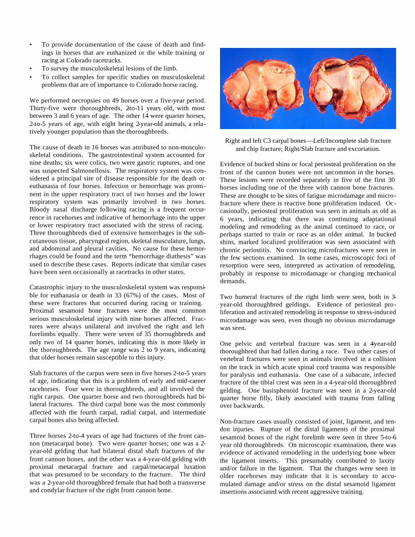
Right and left C3 carpal bones—Left/Incomplete slab fracture and chip fracture; Right/Slab fracture and excoriation.
Evidence of bucked shins or focal periosteal proliferation on the front of the cannon bones were not uncommon in the horses. These lesions were recorded separately in five of the first 30 horses including one of the three with cannon bone fractures. These are thought to be sites of fatigue microdamage and micro-fracture where there is reactive bone proliferation induced. Oc-casionally, periosteal proliferation was seen in animals as old as 6 years, indicating that there was continuing adaptational modeling and remodeling as the animal continued to race, or perhaps started to train or race as an older animal. In bucked shins, marked localized proliferation was seen associated with chronic periostitis. No convincing microfractures were seen in the few sections examined. In some cases, microscopic foci of resorption were seen, interpreted as activation of remodeling, probably in response to microdamage or changing mechanical demands. Two humeral fractures of the right limb were seen, both in 3-year-old thoroughbred geldings. Evidence of periosteal pro-liferation and activated remodeling in response to stress-induced microdamage was seen, even though no obvious microdamage was seen. One pelvic and vertebral fracture was seen in a 4-year-old thoroughbred that had fallen during a race. Two other cases of vertebral fractures were seen in animals involved in a collision on the track in which acute spinal cord trauma was responsible for paralysis and euthanasia. One case of a subacute, infected fracture of the tibial crest was seen in a 4-year-old thoroughbred gelding. One basisphenoid fracture was seen in a 2-year-old quarter horse filly, likely associated with trauma from falling over backwards. Non-fracture cases usually consisted of joint, ligament, and ten-don injuries. Rupture of the distal ligaments of the proximal sesamoid bones of the right forelimb were seen in three 5-to-6 year old thoroughbreds. On microscopic examination, there was evidence of activated remodeling in the underlying bone where the ligament inserts. This presumably contributed to laxity and/or failure in the ligament. That the changes were seen in older racehorses may indicate that it is secondary to accu-mulated damage and/or stress on the distal sesamoid ligament insertions associated with recent aggressive training.

Three joint luxations were seen. One involved the car-pal/metacarpal joint associated with slab fractures of the carpus. Two luxations were seen in the pastern joint, both in quarter horse geldings that were 2-to-4 years of age. The cause of such lesions is not clear although a misstep or right/left interference must be considered. Two cases of laminitis were seen, both of a subacute nature. Generally, the results of these studies are consistent with those of larger studies of necropsies in racehorses like those in California, but some differences are noteworthy. We experienced a somewhat greater incidence of gastrointestinal disease in our collection and hence, the proportion of musculo-skeletal system lesions primarily responsible for death or eutha-nasia were less than in other studies (67% as opposed to 80%). The thoroughbreds in our study were generally older than else-where but the higher incidence of musculoskeletal problems in younger, 2-to-3 year-old quarter horses is similar. The highest frequency of fractures involving sesamoid bones also is consis -tent. However, carpal slab fractures were the second most common fractures in our study while metacarpal bone fractures were second and considerably more common in other studies. This may indicate a relatively higher incidence of carpal frac-tures in our Colorado population. The reason for this possible difference is unknown.
NEW MOLECULAR TEST FOR VIRULENCE TESTING OF ENTEROTOXIGENIC ESCHERICHIA COLI
Doreene R. Hyatt
e are in the process of final testing a new polymerase chain reaction (PCR) test for the detection of virulence
determinants of Enterotoxigenic Escherichia coli (ETEC). Animal diseases due to ETEC are typically severe watery diar-rhea during the first few days of life. The two most important virulence attributes of ETEC are adhesins and toxins. For ani-mal ETEC, the most common adhesins are the pili on the sur-face of the bacterial cell (i.e., K88, K99, 987P, F41, F42, F165, F17, and F18). The enterotoxins are designated as heat-liable (LT) or heat stable (ST) with further subdivisions (LTh-I, LTp-I, LTIIa, LTIIb, StaH, StaP, STb). The PCR will detect the presence of up to nine genes that can be produced by pathogenic Escherichia coli. These genes are K99, K88, F41, 987P, F107, LT, Sta, STb, SLT-IIv. While this test cannot detect whether the pilin or toxin proteins are being pro-duced (genes that are being expressed), it can detect the presence of the genes, and therefore, the possibility of the proteins being present. We expect this test to be available early in the year and it can be requested by writing in “E. coli PCR” on the diagnostic labora-tory submission forms. ________________________ Fecal Culture: Submit feces or intestinal segments. Fee=$11.
BROOM SNAKEWEED AND NITRATE POISONING
Charles W. Dickie/Rocky Ford
ranch in southeastern Colorado lost 14 head of mature Hereford cows over a 10-day period. One string of dead
cows was found in a particularly succulent patch of broomweed. Many green leaves were showing at the base of the plants, while the abundant range grass was dry. Deaths occurred in two pastures, and the cattle were moved to a third pasture where the death loss stopped. Very little broomweed was present in the third pasture. Over a period of several days, we received eight syringes of ocular fluid from each of seven dead cows and one in utero near-term fetus. We received 10 bags of various forage samples, including Kochia scoparia, and one bag of commercial cattle feed pellets. Three water samples were submitted along with brain, liver, spleen, kidney, lung, intestine, and ruminal contents from two cows and a heifer with her unborn near-term fetus. Examination of rumen contents revealed well-masticulated plant parts which could have been broom snakeweed. Three of the eight ocular fluid samples were negative for nitrite and the other five were strongly positive. Interestingly, the heifer was strongly positive, while her in utero calf was negative, suggesting that the heifer died very rapidly without much transplacental movement of nitrite. Testing of broom snake-weed and the 10 bags of forage samples was negative for toxic nitrate levels except for the Kochia scoparia. The feed pellets were tested for urea levels and were not toxic. All water sam-ples were negative for toxic nitrate levels. Nitrate levels in plants could go from toxic to non-toxic within as short a time as one day, hence, toxic plants in one short time period might test negative a little while later. Since the kochia was dry and unpalatable, we suspect the nitrate came from the snakeweed, even though it tested nitrate negative, but this needs confirma-tion by testing these plants for nitrate content over a period of time. Histopathology showed acute cellular swelling in the livers. The kidneys showed minimal glomerular hemorrhage and as-cending/descending tubular hydropic degeneration with tubular hemorrhage. The histopathology of the kidneys fit well with snakeweed poisoning, while the livers did not show the hydropic degeneration typically described. Texas toxicologists, who have much experience with snakeweed poisoning, indicate that hepatic hydropic degeneration is more with chronic poisoning. The kidney, however, was more quickly affected and showed the typical changes. This case presented lesions consistent with acute broom snakeweed poisoning, and the cattle had ingested this weed. In addition, there was evidence of nitrite toxicity in five out of eight ocular fluid samples. The source of the nitrate remains obscure. Approximately four weeks later, 74 heifers had started calving. Forty calved with 15 retained membranes and two stillborn at
W
A

our last communication. Retained membranes are very characteristic of broom snakeweed poisoning, as is abortion. Ocular fluids from the two stillborn calves were negative for nitrate/nitrite. Liver enzymes, BUNs, and creatinines were not elevated in the dams of the two calves at the time of delivery, whereas levels in sick cattle four weeks earlier were markedly elevated. Broom snakeweed continues to present diagnostic and practical problems throughout the West. The widespread existence of this plant on western ranges suggests we need to practice co-existence, since it probably won’t go away soon.
PCR NEWS
Jane Carman
e continue to adapt new polymerase chain reaction (PCR) tests to improve accuracy of disease diagnosis. Below is
a listing of our new PCR tests implemented recently or soon to be introduced.
BOVINE TRICHOMONIASIS IN COLORADO—Trichomoniasis is a venereal disease of cattle which results in varying degrees of reproduction inefficiency (usually early embryonic death). In beef cattle on western ranges, the first indication of a problem is usually finding 20-40% of the cows open at the time of fall pregnancy test. In the past few years, the number of infected herds has increased. Recently, infected herds have been identified in the following Colorado counties—Alamosa (3), Conejo (5), Arapahoe (1), Mesa (2), Montrose (2), Eagle (2), and Montezuma (1). The Animal Health Welfare Committee will discuss the situation and what can and should be done to protect Colorado cattle herds from the disease. This meeting is scheduled for 8-9:30AM on Tuesday, June 20, 2000, at the CCA Annual Convention in Pueblo, CO. If you have questions prior to this meeting, contact Dr. Marv Hamann, Chairman, Animal Health and Welfare Committee at 719/542-6075, the CCA office at 303/431-6422, or John Cheney at the Diagnostic Laboratory at 970-491-1281.
Disease Agent Sample Needed Price PCR Tests Introduced in the Last Year Canine Distemper Virus Whole blood (EDTA), Conjunctival scraping $28.00 Fresh and/or formalin-fixed lung/brain/urinary bladder Porcine Reproductive and Fresh lung $28.00 Respiratory Virus NOTE: Currently working on protocol for Formalin-fixed tissue Caprine Arthritis and Encephalitis Whole blood (EDTA) $23.00 Ovine Progressive Pneumoniae Whole blood (EDTA) $23.00 New PCR Tests Introduced this Year Blue Tongue Virus Whole blood (EDTA), fresh spleen $28.00 Epizootic Hemorrhagic Disease Whole blood (EDTA), fresh spleen $28.00 Bovine Viral Diarrhea Virus in Bulk Tank Milk, fresh (local dairies only) or frozen $36.00 Bulk Tank Milk Canine Adenovirus, type 1/type 2 Fresh and/or formalin-fixed liver/brain $23.00 Canine Parvovirus, type 1/type 2 Fresh and/or formalin-fixed lung (CPV-1)/ and Feline Panleukopenia Virus intestine/feces (CPV-2) $23.00 Fresh and/or formalin-fixed intestine/feces Feline Immunodeficiency Virus Whole blood (EDTA) $23.00 NOTE: Currently working on protocol for formalin-fixed tissue Mycoplasma gallisepticum/synoviae Isolate, fresh lung/joint tap $23.00 NOTE: Currently working on protocol for various transport/growth media
W

WE HAVE A NEW DERMATOLOGY SERVICE! An article inside introduces Dr. Sonya Bettenay, a board-certified clinical dermatologist, who works closely with our pathologists to provide you with a clinical interpretation of the histopathologic findings of your skin biopsy. She will include treatment suggestions for you; if appropriate. We welcome her as a member of our laboratory and are pleased to offer you this valuable new dermatohistopathology service. Call 970-491-1281 for more information.
PLEASE NOTE--We have been receiving many samples without identification on the specimen containers, especially with biopsy submissions. Please remember to always fill-out paperwork completely, with appropriate test(s) requests and LABEL YOUR SAMPLES!
WHAT’S INSIDE THIS ISSUE OF LABLINES
• Get More Out of Your Skin Biopsies • Eyelid Test for Scrapie in Sheep • Broom Snakeweed and Nitrate Poisoning • Chronic Wasting Disease Update • PCR News • Strychnine Poisoning • New Microbial Identification System • Pigeon Fever in Horses • Colorado Racehorse Injuries • Plague and Tularemia Require Vigilance • Neosporosis • Inking Margins for Tumors • Swine Influenza • Enterotoxigenic E. coli