colorectal cancer molecular pathways and the … · tumor suppressor genes. microsatellite...
TRANSCRIPT

The First CRC Screening & Quality in Colonoscopy SymposiumApril 28th, 2018Beirut, Lebanon
COLORECTAL CANCER MOLECULAR PATHWAYS AND THE SERRATED POLYP
Charles J. Kahi, MD, MSIndiana University School of Medicine
Richard L. Roudebush VA Medical CenterIndianapolis, Indiana
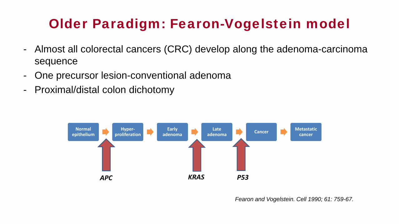
Older Paradigm: Fearon-Vogelstein model
- Almost all colorectal cancers (CRC) develop along the adenoma-carcinoma sequence
- One precursor lesion-conventional adenoma- Proximal/distal colon dichotomy
Normal epithelium
Hyper-proliferation
Early adenoma
Late adenoma Cancer Metastatic
cancer
APC KRAS P53
Fearon and Vogelstein. Cell 1990; 61: 759-67.
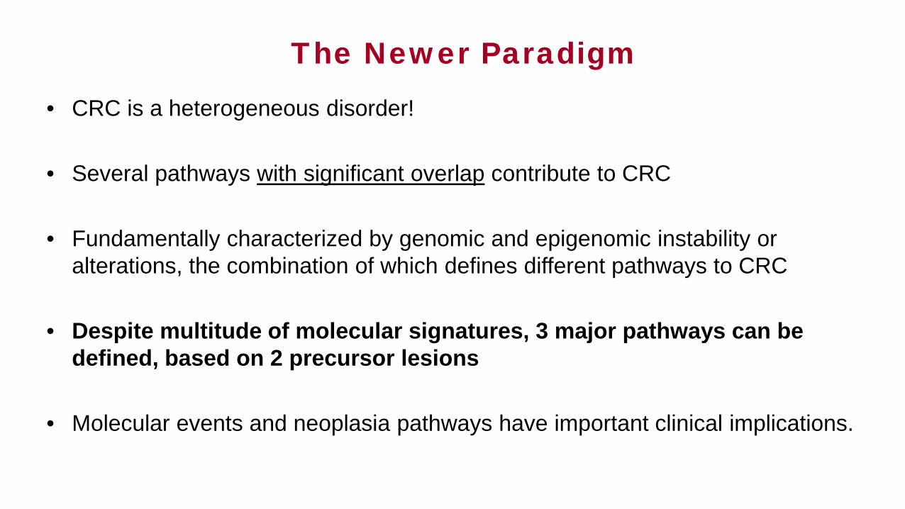
• CRC is a heterogeneous disorder!
• Several pathways with significant overlap contribute to CRC
• Fundamentally characterized by genomic and epigenomic instability or alterations, the combination of which defines different pathways to CRC
• Despite multitude of molecular signatures, 3 major pathways can be defined, based on 2 precursor lesions
• Molecular events and neoplasia pathways have important clinical implications.
The Newer Paradigm
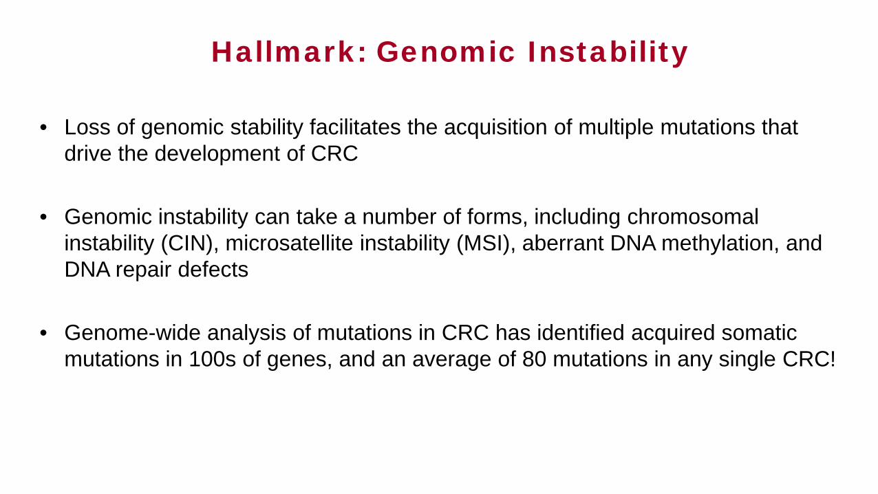
• Loss of genomic stability facilitates the acquisition of multiple mutations that drive the development of CRC
• Genomic instability can take a number of forms, including chromosomal instability (CIN), microsatellite instability (MSI), aberrant DNA methylation, and DNA repair defects
• Genome-wide analysis of mutations in CRC has identified acquired somatic mutations in 100s of genes, and an average of 80 mutations in any single CRC!
Hallmark: Genomic Instability

Gene or group of genes Description Mechanism for mutation increasing CRC risk
APC Tumor suppressor gene Inactivating mutation causes loss of regulation of spindle microtubules during mitosis
TP53 Tumor suppressor gene Inactivating mutation causes loss of regulation of cell-cycle arrest and cell death
RAS Oncogene Activating mutations drive cell growth through MAPK pathway
BRAF Oncogene Activating mutations drive cell growth through MAPK pathway
PIK3CA OncogeneActivating mutation upregulates PI3 K pathway,
enhancing prostaglandin E2 synthesis and inhibiting apoptosis
MLH1, MSH2, MSH6, PMS2 Mutation Mismatch Repair genes Inactivating mutation impairs ability to repair strand slippage within nucleotide repeats
EPCAM Codes for transmembrane glycoprotein epithelial cell adhesion molecule
Deletion of 3′ end of EPCAM leads to epigenetic silencing of MSH2
MYH Base excision repair gene Germline inactivating mutation of MYH leads to somatic mutation of APC
Ewing I, et al. Frontline Gastroenterology 2014;5:26–30
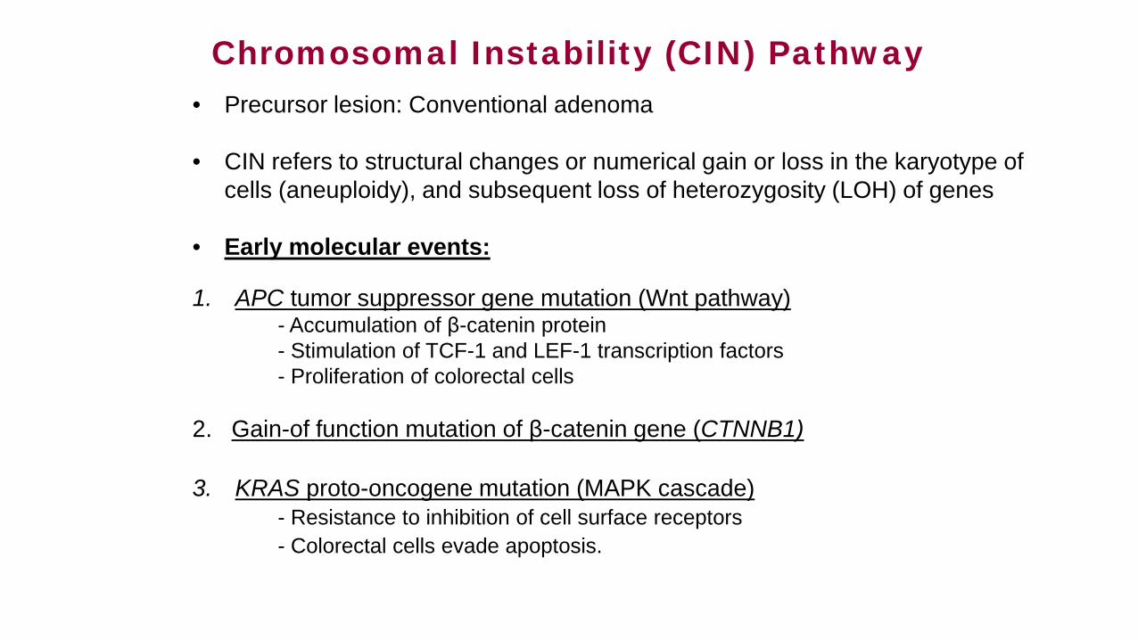
Chromosomal Instability (CIN) Pathway• Precursor lesion: Conventional adenoma
• CIN refers to structural changes or numerical gain or loss in the karyotype of cells (aneuploidy), and subsequent loss of heterozygosity (LOH) of genes
• Early molecular events:
1. APC tumor suppressor gene mutation (Wnt pathway) - Accumulation of β-catenin protein - Stimulation of TCF-1 and LEF-1 transcription factors- Proliferation of colorectal cells
2. Gain-of function mutation of β-catenin gene (CTNNB1)
3. KRAS proto-oncogene mutation (MAPK cascade)- Resistance to inhibition of cell surface receptors- Colorectal cells evade apoptosis.
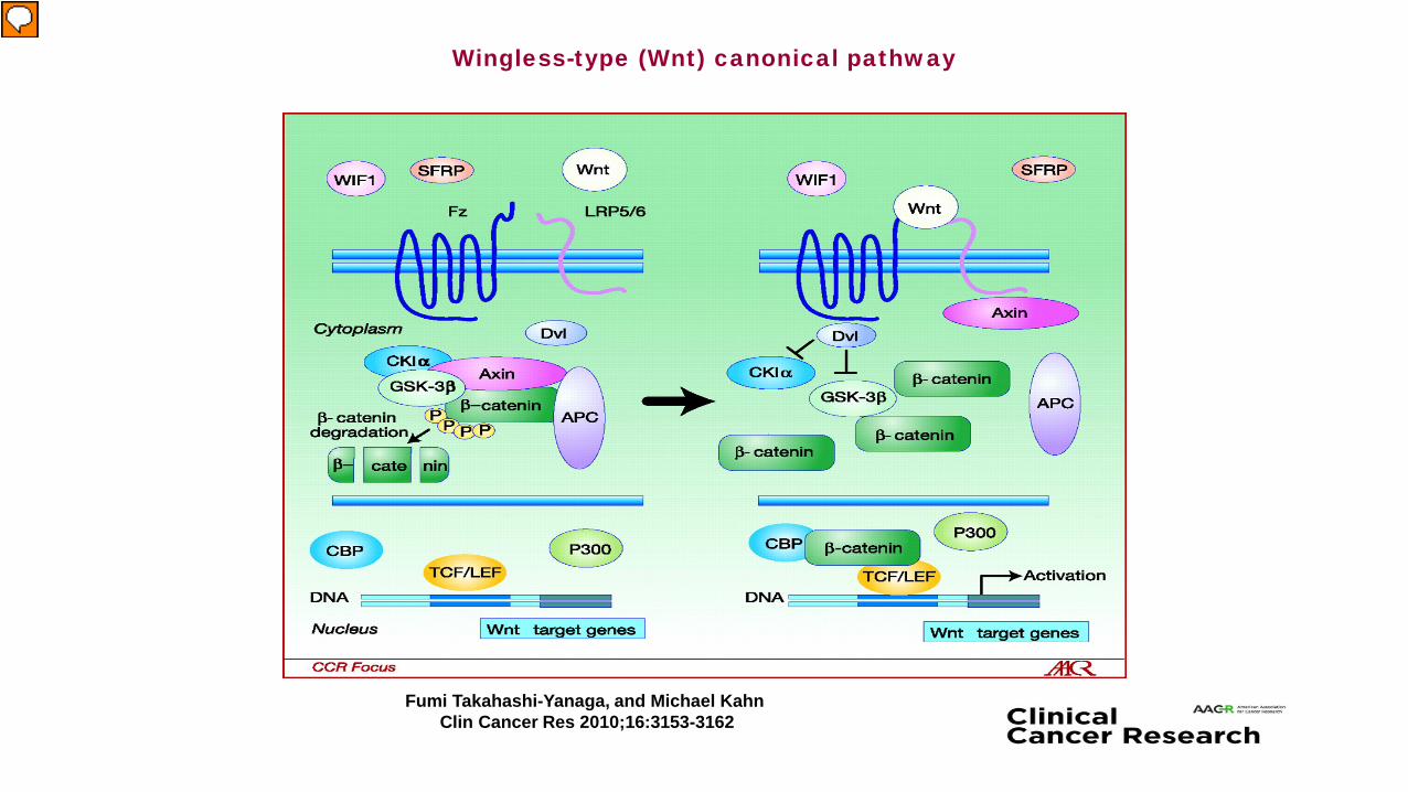
Wingless-type (Wnt) canonical pathway
Fumi Takahashi-Yanaga, and Michael KahnClin Cancer Res 2010;16:3153-3162
Chromosomal Instability (CIN) Pathway
• Later molecular events:1. Mutation of p53 tumor suppressor gene
- p53 (Chr 17p) a.k.a. “guardian of the genome”- Mutation in p53 prevalence increases from adenoma, to HGD, to CRC- Unclear if absolutely required
2. LOH (Chr 18q)- Occurs in 70% of CRC- Often synchronous with p53 loss- Affects Deleted in Colorectal Carcinoma (DCC), SMAD4, and SMAD2 which are involved in regulation of cell proliferation and apoptosis
3. Other events- PI3K pathway mutations—accelerated cell growth- microRNA upregulation or downregulation: Effectively act like oncogenes and tumor suppressor genes.
Microsatellite Instability (MSI) Pathway
• Precursor lesion: Conventional adenoma
• 15% of CRC have MSI - 20% have hereditary cause-Lynch syndrome)
- Remaining associated with hypermethylation of MLH1 (serrated pathway)
• Microsatellites are mononucleotide or dinucleotide repeats found throughout the genome, vulnerable to transcription errors during replication
• Mutation Mismatch Repair (MMR) system: Identifies and corrects errors
• Germline mutations leading to loss of function have been identified in four genes involved in MMR: MLH1, MSH2, MSH6 and PMS2
• Germline deletion mutations in the EPCAM gene leads to epigenetic silencing of the neighboring MSH2 MMR gene.
Microsatellite Instability (MSI) Pathway
• Most labs assess MSI using a panel of 5 mononucleotide markers - BAT-25, BAT-26, NR-21, NR-24, MONO-27
• At least 2 of 5 (40%): MSI or MSI-High- MSI-Low versus MSS distinction is controversial
• Additional molecular alterations in hereditary MSI pathway:
- Wnt pathway activation and β-catenin gene mutation
- K-RAS mutations are common
- TGF-β gene mutation (inhibitor of epithelial cell growth)
- BAX gene mutation leads to evasion from apoptosis (similar to p53).
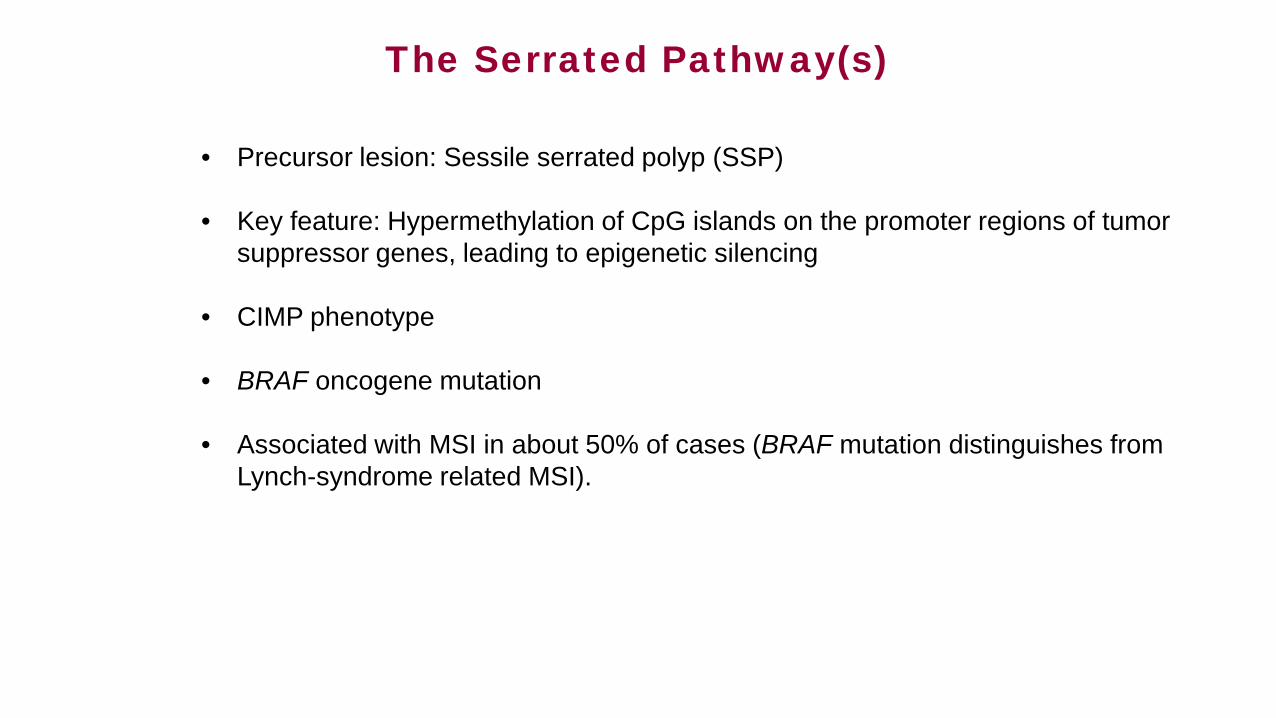
The Serrated Pathway(s)
• Precursor lesion: Sessile serrated polyp (SSP)
• Key feature: Hypermethylation of CpG islands on the promoter regions of tumor suppressor genes, leading to epigenetic silencing
• CIMP phenotype
• BRAF oncogene mutation
• Associated with MSI in about 50% of cases (BRAF mutation distinguishes from Lynch-syndrome related MSI).
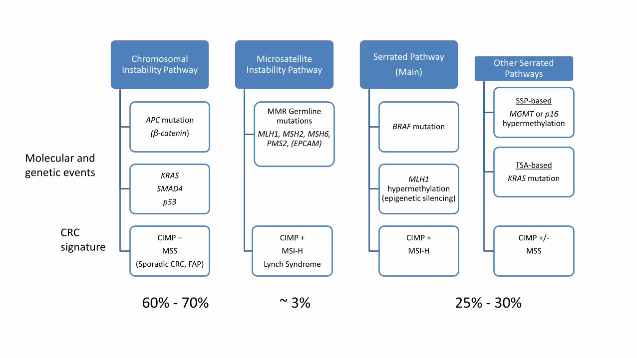
Chromosomal Instability Pathway
APC mutation(β-catenin)
KRASSMAD4
p53
CIMP –MSS
(Sporadic CRC, FAP)
Microsatellite Instability Pathway
MMR Germline mutations
MLH1, MSH2, MSH6, PMS2, (EPCAM)
CIMP +MSI-H
Lynch Syndrome
Serrated Pathway(Main)
BRAF mutation
MLH1hypermethylation
(epigenetic silencing)
CIMP +MSI-H
Other Serrated Pathways
SSP-basedMGMT or p16
hypermethylation
TSA-basedKRAS mutation
CIMP +/-MSS
60% - 70% ~ 3% 25% - 30%
Molecular and genetic events
CRC signature

Clinical Applications and Implications• Fecal FIT-DNA test for average-risk CRC screening
- Detection of abnormal DNA shed in stool by CRC
- KRAS, aberrantly methylated BMP3 and NDRG4 promoter areas, beta-actin
- Sensitivity for CRC 92% (FIT 74%)
- Highest sensitivity for advanced polyps among all non-invasive tests (40%)
- Sensitivity for large SSP about 42%Imperiale et al. New Engl J Med 2014; 370: 1287-97USMSTF-Gastrointest. Endosc. 2017 86(1): 18-33.
• Chemoprevention with aspirin/NSAIDs (degradation of β-catenin)
• Genetic testing for familial CRC and polyposis syndromes- APC for FAP and its variants
- SMAD4 for juvenile polyposis…
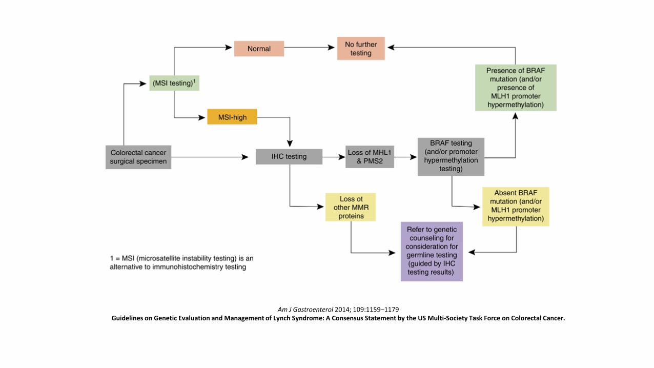
Am J Gastroenterol 2014; 109:1159–1179Guidelines on Genetic Evaluation and Management of Lynch Syndrome: A Consensus Statement by the US Multi-Society Task Force on Colorectal Cancer.
Clinical Applications and Implications
• Prognostic- MSI CRC (both Lynch and serrated pathways) has better prognosis than MSS CRC,
even with adjustment for stage
- BRAF mutation in MSS CRC marker of worse prognosis
- 18q LOH associated with worse prognosis in stage II and III CRC
• Predictive- KRAS and BRAF mutant CRC do not respond to anti-EGFR therapy (cetuximab)
- MSI CRC require oxalaplatin addition to 5-FU for response
- MLH1 hypermethylated CRC: Poor response to 5-FU, but respond to irinotecan
- Stage IV MSI CRC (but not MSS) respond to immunotherapy (pembrolizumab).
Obuch and Ahnen. Gastroenterol Clin N Am 2016; 45: 459-76.

Yamauchi et al. Gut. 2012 Jun; 61(6): 847–854.
Dichotomy concept is obsolete: The frequencies of CIMP-high, MSI-high, and BRAF mutation in CRC increase gradually from rectum to ascending colon.
Serrated Pathway and Post Colonoscopy CRC: Overlap of Molecular Signatures
• Compared to sporadic CRC, PCCRC is more likely to:
- Be located in the proximal colon- Demonstrate MSI- Be associated with CIMP
Sawhney et al. Gastroenterology 2006; 131: 1700-5Arain et al. Am J Gastroenterol 2010; 105: 1189-95Nishihara et al. NEJM 2013; 369: 1095-1105.
Serrated polyps: Old-New lesions
• Prior to 1990: Adenoma or hyperplastic (benign)
• Polyps with “mixed” features Longacre et al. Am J Surg Pathol 1990;14:524–37
• 1996: “sessile serrated adenoma” coined to distinguish the polyps of “giant hyperplastic polyposis” from true hyperplastic polyps,
Torlakovic et al. Gastroenterology 1996; 110: 748-755
• Histologic features further refined in 2003Torlakovic et al. Am J Surg Pathol 2003: 27; 65-81.
Classification of Serrated Lesions of the Colorectum
• Hyperplastic Polyp- Microvesicular HP (MVHP)- Goblet-cell rich HP (GCHP)- Mucin-poor HP (MPHP)
• Sessile Serrated Adenoma/Polyp (SSA/P)- SSA/P without cytological dysplasia- SSA/P with cytological dysplasia
• Traditional Serrated Adenoma (TSA)
Snover D, et al. WHO classification of tumours. Pathology and genetics. Tumours of the digestive system. 4th edition. Berlin: Springer-Verlag. 2010.
The Serrated Pathway: How Long?
• Pathological review of 2319 SSP:- Median age of patients (years):
SSP without dysplasia: 61 SSP with LGD: 66 SSP with HGD: 72 SSP with CA: 76
Lash et al. J Clin Pathol 2010; 63: 681-6
• Other studies have shown more aggressive behavior, related in part to patient age (inactivation of MLH1 associated with advancing age)
Sweetser et al. Clin Gastroenterol Hepatol 2013; 11:760-7Jass JR. Dis Colon Rectum 2001; 44:163-6Goldstein NS. Am J Clin Pathol 2006; 125: 132-45
• Serrated lesion-carcinoma sequence duration is variable• May take 10-15 years, but key event is MLH1 methylation/silencing,
which accelerates progression to CRC.
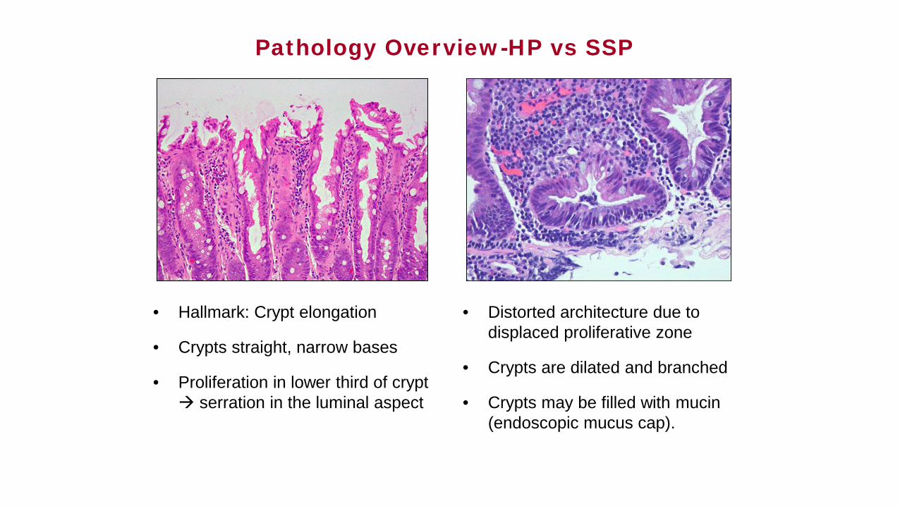
Pathology Overview-HP vs SSP
• Hallmark: Crypt elongation
• Crypts straight, narrow bases
• Proliferation in lower third of crypt serration in the luminal aspect
• Distorted architecture due to displaced proliferative zone
• Crypts are dilated and branched
• Crypts may be filled with mucin (endoscopic mucus cap).
Pathology Considerations
• Often difficult to distinguish HP from SSP, criteria not validated• Considerable inter-observer variation, even among expert pathologists
Khalid O et al. World J Gastroenterol 2009;15:3767-70
- Contributes to high variability in serrated polyp endoscopic detection ratesPayne et al. Clin Gastroenterol Hepatol 2014;12:1119–26.
- Pathologic diagnoses of SSP are increasing over timeGill et al. J Clin Pathol 2013; 66: 655-8
- Right-sided location and larger size predictive of reclassification of HP to SSPSingh et al. Gastrointest Endosc 2012; 76: 1003-8
Practical Bottom lines: - Communicate with your pathologist- Treat right-sided HP ≥ 1 cm as SSP
Rex et al. Am J Gastroenterol 2012; 107: 1315-29.
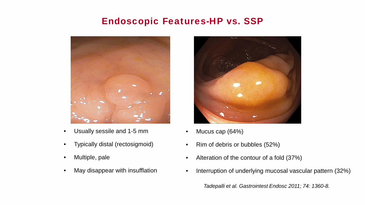
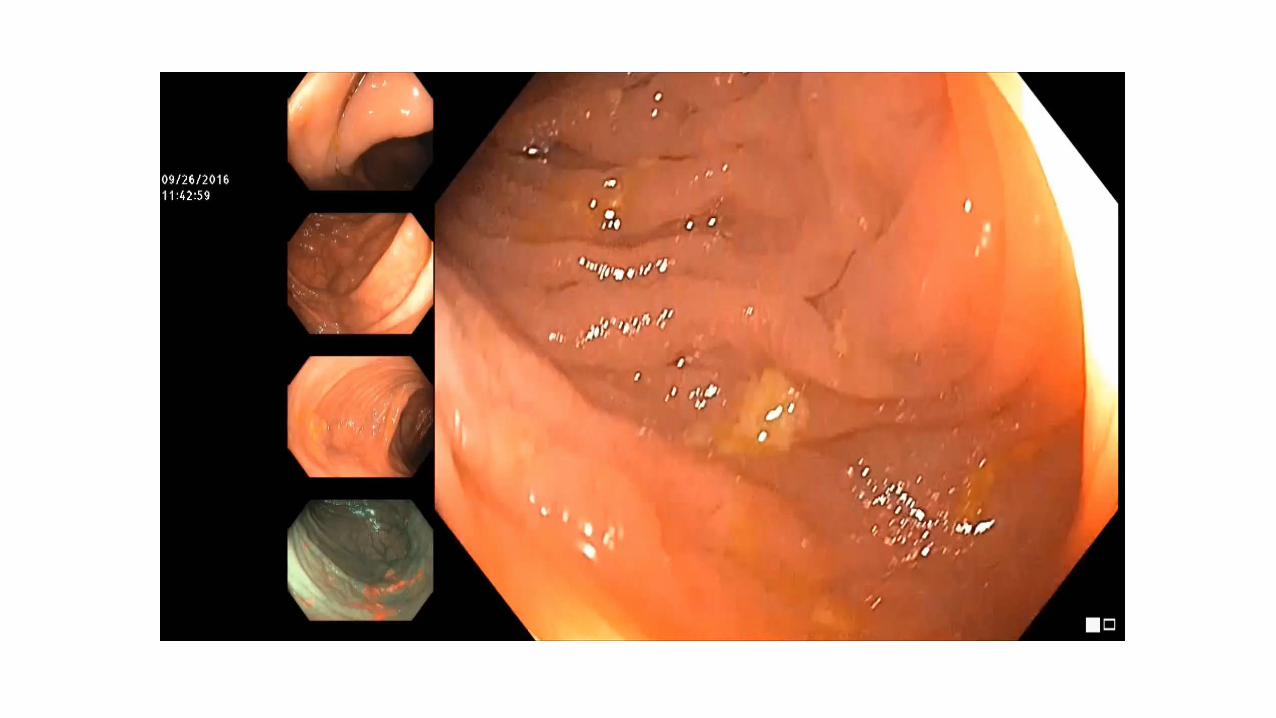
Endoscopic Features-HP vs. SSP
• Usually sessile and 1-5 mm
• Typically distal (rectosigmoid)
• Multiple, pale
• May disappear with insufflation
• Mucus cap (64%)
• Rim of debris or bubbles (52%)
• Alteration of the contour of a fold (37%)
• Interruption of underlying mucosal vascular pattern (32%)
Tadepalli et al. Gastrointest Endosc 2011; 74: 1360-8.

Hazewinkel et al. Gastrointest Endosc. 2013;77:916–924.
Indistinct borders (OR ~ 2)
Cloud-like appearance(OR ~ 5)
Dark spots inside the crypts(OR ~ 2)
Endoscopic Prediction SSP vs. HP
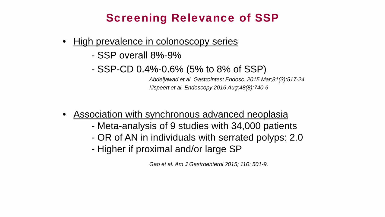
Screening Relevance of SSP
• High prevalence in colonoscopy series- SSP overall 8%-9%- SSP-CD 0.4%-0.6% (5% to 8% of SSP)
Abdeljawad et al. Gastrointest Endosc. 2015 Mar;81(3):517-24IJspeert et al. Endoscopy 2016 Aug;48(8):740-6
• Association with synchronous advanced neoplasia - Meta-analysis of 9 studies with 34,000 patients- OR of AN in individuals with serrated polyps: 2.0- Higher if proximal and/or large SP
Gao et al. Am J Gastroenterol 2015; 110: 501-9.
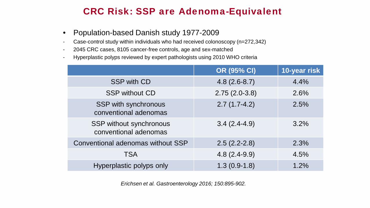
CRC Risk: SSP are Adenoma-Equivalent
• Population-based Danish study 1977-2009- Case-control study within individuals who had received colonoscopy (n=272,342)- 2045 CRC cases, 8105 cancer-free controls, age and sex-matched- Hyperplastic polyps reviewed by expert pathologists using 2010 WHO criteria
Erichsen et al. Gastroenterology 2016; 150:895-902.
OR (95% CI) 10-year riskSSP with CD 4.8 (2.6-8.7) 4.4%
SSP without CD 2.75 (2.0-3.8) 2.6%SSP with synchronous
conventional adenomas2.7 (1.7-4.2) 2.5%
SSP without synchronous conventional adenomas
3.4 (2.4-4.9) 3.2%
Conventional adenomas without SSP 2.5 (2.2-2.8) 2.3%TSA 4.8 (2.4-9.9) 4.5%
Hyperplastic polyps only 1.3 (0.9-1.8) 1.2%

Serrated polyposis syndrome (SPS)
I. At least 5 serrated polyps proximal to the sigmoid, of which ≥ 2 are ≥ 10 mm in size
II. At least one serrated polyp proximal to sigmoid, in first-degree relative of patient with SPS
III. > 20 serrated polyps throughout the colon.
Snover D, et al. WHO classification of tumours. Pathology and genetics. Tumours of the digestive system. 4th edition. Berlin: Springer-Verlag. 2010.

SPS: Most Common Polyposis Syndrome
• Prevalence estimates higher than previously thought- FIT/FOBT screening cohorts: 1:111 to 1:127 colonoscopies- Screening colonoscopy cohorts: 1:238 colonoscopies
Rivero-Sanchez et al. Endoscopy 2017; 49: 44-53Ijspeert JEG et al. Gut 2017; 66: 1225-32
• Unusual syndrome--Genetic basis unclear/uncertain- Criterion II alone uncommon- Average age of diagnosis > 50 years
Carballal et al. Gut 2016; 65: 1829-37
- About 50% have a family hx of CRCRex et al. Am J Gastroenterol 2012; 107:1315–1329.
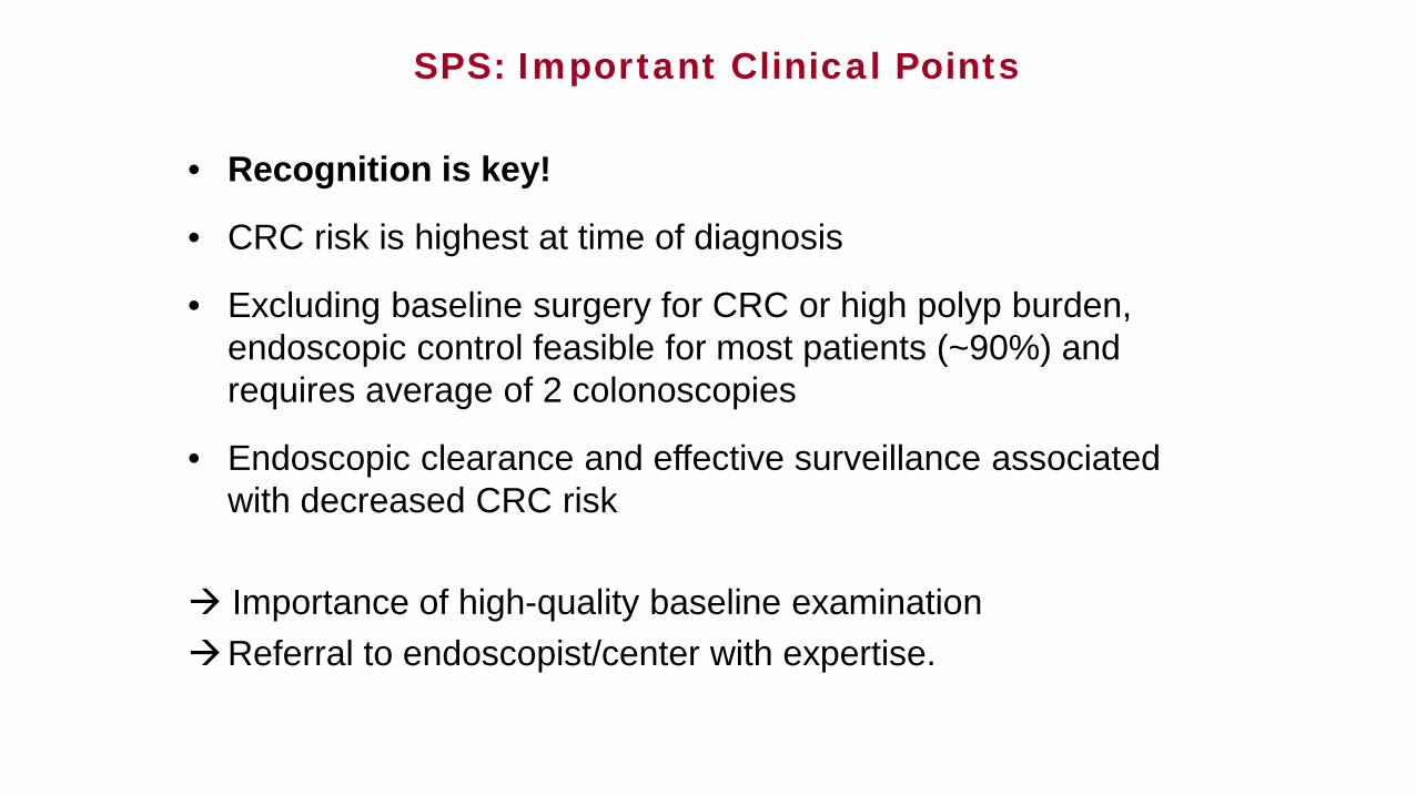
SPS: Important Clinical Points
• Recognition is key!
• CRC risk is highest at time of diagnosis
• Excluding baseline surgery for CRC or high polyp burden, endoscopic control feasible for most patients (~90%) and requires average of 2 colonoscopies
• Endoscopic clearance and effective surveillance associated with decreased CRC risk
Importance of high-quality baseline examinationReferral to endoscopist/center with expertise.
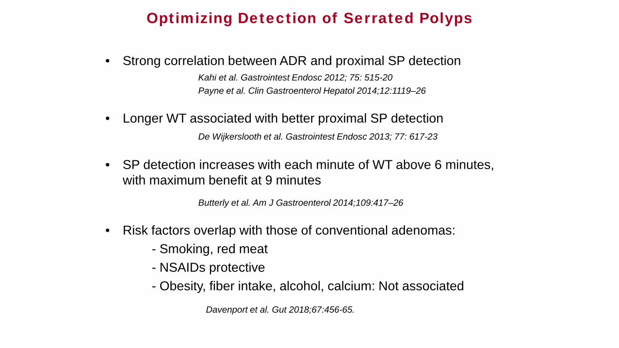
Optimizing Detection of Serrated Polyps
• Strong correlation between ADR and proximal SP detectionKahi et al. Gastrointest Endosc 2012; 75: 515-20Payne et al. Clin Gastroenterol Hepatol 2014;12:1119–26
• Longer WT associated with better proximal SP detectionDe Wijkerslooth et al. Gastrointest Endosc 2013; 77: 617-23
• SP detection increases with each minute of WT above 6 minutes, with maximum benefit at 9 minutes
Butterly et al. Am J Gastroenterol 2014;109:417–26
• Risk factors overlap with those of conventional adenomas:- Smoking, red meat- NSAIDs protective- Obesity, fiber intake, alcohol, calcium: Not associated
Davenport et al. Gut 2018;67:456-65.
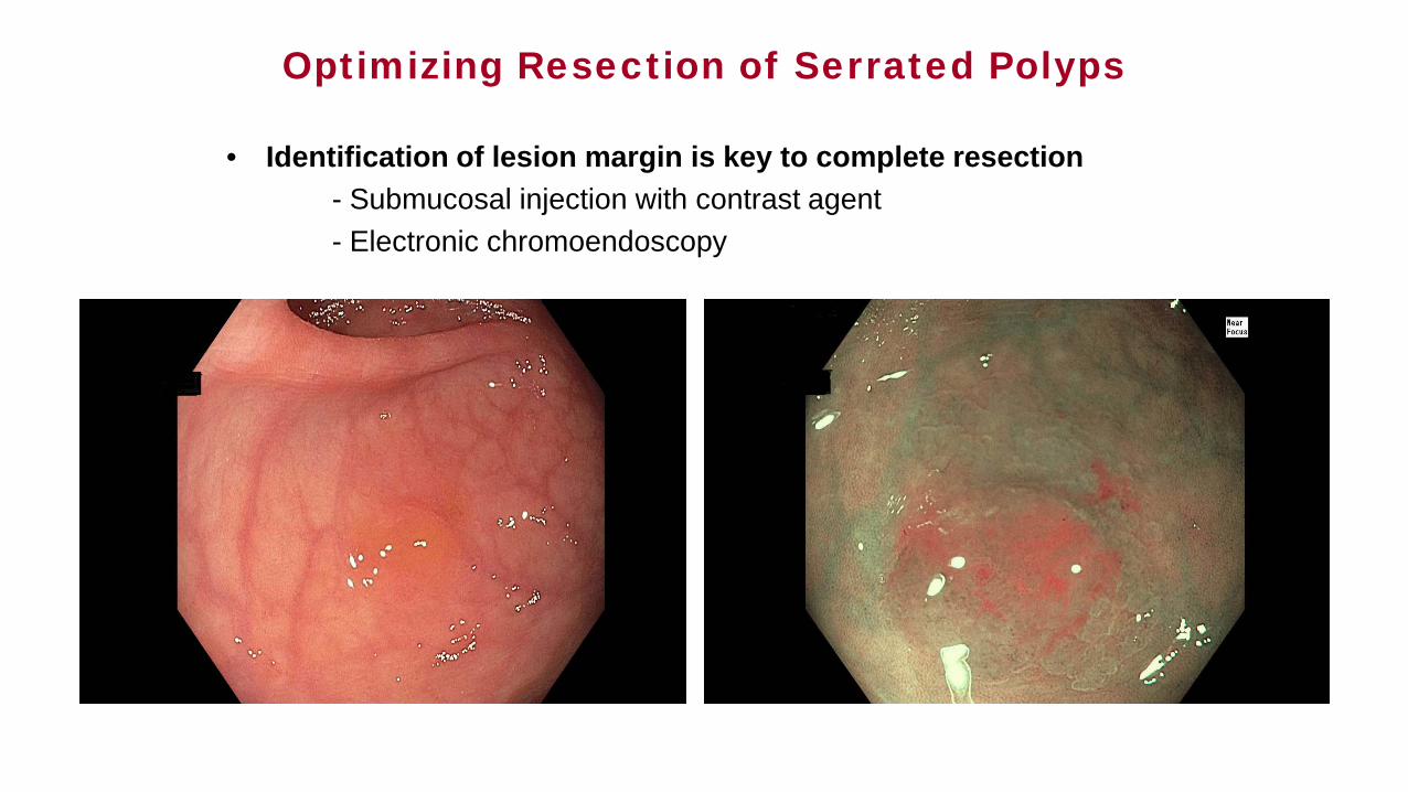
Optimizing Resection of Serrated Polyps
• Identification of lesion margin is key to complete resection- Submucosal injection with contrast agent- Electronic chromoendoscopy

Optimizing Resection of Serrated Polyps
- Australian Multicenter study (ACE):246 patients with 323 SSP ≥ 20 mm
SSP Adenomas P-valueEradication rate 99.1% 94.5% < 0.001
Intraprocedure bleeding 6.9% 16.7% < 0.001Delayed bleeding 5.7% 6.3% 0.7
Perforation 0.4% 0.4% 0.972-year cumulative recurrence rate 7% 28.4% < 0.001
Pellise et al. Gut Apr 2017; 66 (4): 644-53.
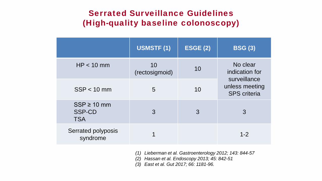
(1) Lieberman et al. Gastroenterology 2012; 143: 844-57(2) Hassan et al. Endoscopy 2013; 45: 842-51(3) East et al. Gut 2017; 66: 1181-96.
USMSTF (1) ESGE (2) BSG (3)
HP < 10 mm 10(rectosigmoid) 10
No clear indication for surveillance
unless meeting SPS criteria
SSP < 10 mm 5 10
SSP ≥ 10 mmSSP-CDTSA
3 3 3
Serrated polyposis syndrome 1 1-2
Serrated Surveillance Guidelines(High-quality baseline colonoscopy)