colour vision examination
TRANSCRIPT


• Color vision is the capacity of an organism or machine to distinguish objects based on the wavelengths (or frequencies) of the light they reflect, emit, or transmit.
• Perception of colors is a subjective process where the brain and nervous system responds to the visual stimuli that emerge when incident light reacts with the several types of cone photoreceptors in the eye
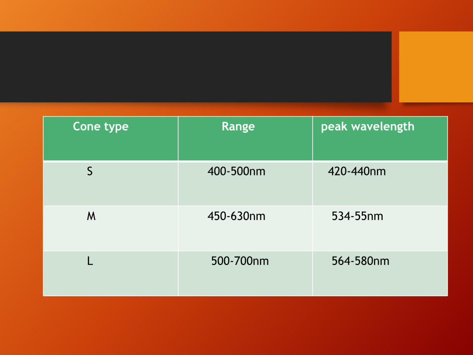
Cone type Range peak wavelength
S 400-500nm 420-440nm
M 450-630nm 534-55nm
L 500-700nm 564-580nm
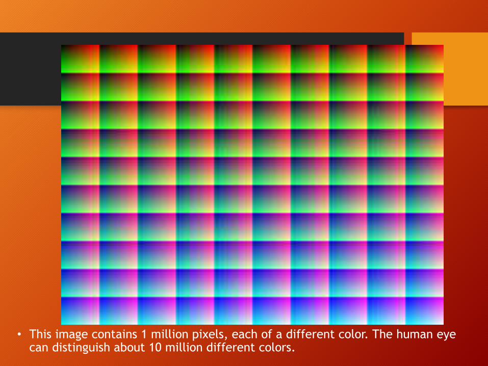
• This image contains 1 million pixels, each of a different color. The human eye can distinguish about 10 million different colors.

Color Vision Deficiency
the inability to distinguish certain colours.
one or more of the cone types is missing or
defective to any extent.
abnormal colour matching and colour confusions.
Marked reduction in the no. of separate colours
that can be distinguishedin the spectrum.
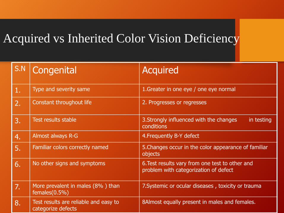
Acquired vs Inherited Color Vision Deficiency
S.N Congenital Acquired
1. Type and severity same 1.Greater in one eye / one eye normal
2. Constant throughout life 2. Progresses or regresses
3. Test results stable 3.Strongly influenced with the changes in testing conditions
4. Almost always R-G 4.Frequently B-Y defect
5. Familiar colors correctly named 5.Changes occur in the color appearance of familiar objects
6. No other signs and symptoms 6.Test results vary from one test to other and problem with categorization of defect
7. More prevalent in males (8% ) than females(0.5%)
7.Systemic or ocular diseases , toxicity or trauma
8. Test results are reliable and easy to categorize defects
8Almost equally present in males and females.
Importance of color vision examination
• An acquired color deficiency may be the earliest sign of
a sight threatening condition
• Handicapped in a number of ways
• Impact in pursuit of a career / everyday activities
• Minimize future disappointment and assist in counselling
for alternate career choices
• Sketching the nature of the color deficiency will help
illustrate potential problem.
Occupational aspects of color vision testing
-Textiles, garments, Paints and other
industries(exact color matching)
-Armed forces, Aviation,Electrical and
telecommunication, trades, Maritime,
Commercial driving, Railroad etc.
At any cone pigment may deficient, Or absent totally.
• Trichromatism (Normal sight) Which person can differentiate all colors.
-All 3 cones although not necessary functioning perfectly.
• Anomalous trichomatism can differentiate all colors but on reduced or displaced sensitivities.
- Protanomaly red displaced sensitivity.
- Deutranomaly green displaced sensitivity.
- Tritanomaly blue displaced sensitivity .
Types of colour vision defects
• Dichromatism absence of one cone
- Tritanopia blue is missing (red ,green are present).
- Deutranopia green is missing (red and blue are present)
- Protanopia red is missing while blue and green are
present.
• Monochromatism totally unable to dedifferentiate colors of equal
brightness.
Patient Selection(Indications)
• unexplained reduction in visual acquity or low visual
acquity e.g. 20/25
• recent color disturbances or any difference in color
vision between the two eyes
• Any patient who exhibits a sign of abnormality in
the fundus
Color Vision Tests
• There are a no. of clinical color vision tests which aim to
identify , classify and grade the severity of color vision
deficiency or are designed to determine the occupational
suitability
Color Vision Tests
• Pseudoisochromatic Test Plates
-Ishihara Plates ,F2 Plates ,AOHRR Plates
• Hue discrimination/ Arrangement tests-The Farnsworth D-15 test-The Farnsworth Munsell-100 Hue test
• Anamaloscopes
-The Nagel Anamaloscope
-The Neitz Anamaloscope
• Color naming and color sorting
-Lantern tests,Yarn test
• Notes for color vision testing
- Use proper illumination (day light).
- Explain test for the patient.
- In screening for congenital diseased test is done binocularly and monocularly for acquired abnormality.
- Patient should use his or her near correction.
- Avoid tinted spectacles or contact lenses.

Pseudoisochromatic Plates
• Design Principle
• -To identify a colored symbol made up of colored dots of varying sizes
embeded in a background of differently colored dots
-The figure and background colors are chosen so that they are confused
(isochromatic) by color deficient but discerned by the normal
-The distance between the colors should exceed the minimum required for them
to be discriminated by person with normal color vision
• Examples –Ishihara plates ,AO-HRR plates ,Devorine e.tc.

Types
• Transformed Plates –
2 to 9 plates in ishihara 38. Both normal and color deficient will see differently
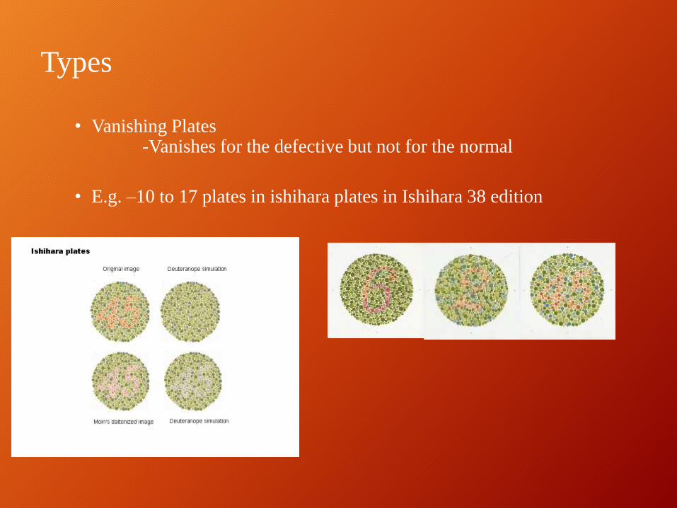
Types
• Vanishing Plates-Vanishes for the defective but not for the normal
• E.g. –10 to 17 plates in ishihara plates in Ishihara 38 edition

Types
• Hidden digit plates
-18 to 21 plates in 38 edition
Concealed from person with normal color vision but is visible to
severely color defective
Types
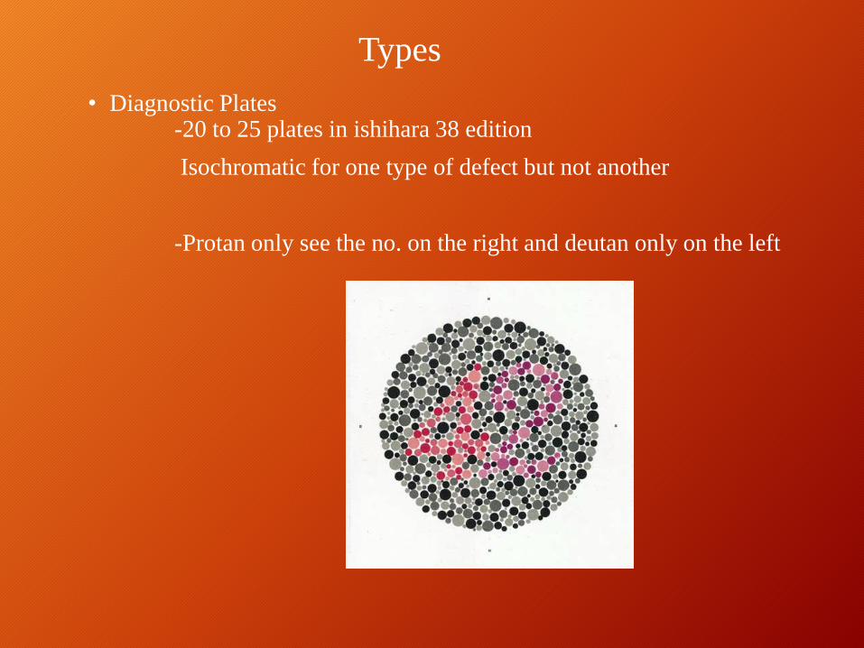
• Diagnostic Plates-20 to 25 plates in ishihara 38 edition
Isochromatic for one type of defect but not another
-Protan only see the no. on the right and deutan only on the left
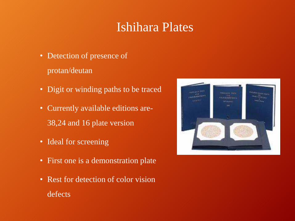
Ishihara Plates
• Detection of presence of
protan/deutan
• Digit or winding paths to be traced
• Currently available editions are-
38,24 and 16 plate version
• Ideal for screening
• First one is a demonstration plate
• Rest for detection of color vision
defects
Testing Guidelines
• VA > 6/60
• Illumination
= 500-600 lux
=20 to 60 ft cd or day light illumination
• Testing distance= 75 to100 cm or at arm length
• Observation time = 3 to5 secs per plate
( 10 secs for winding
paths)
Interpretation
• Count the no. of plates misread
• Exclude the demonstration plate from this total
• More than the indicated no. of errors made protan/deutan defect almost certain to be present
• 38 Plate edition= 4 or less – normal=8 or more –deficient
• 24 Plate edition = 2 or less –normal= 6 or more –deficient
• 16 Plate edition = 2 or less – normal
= 4 or more deficient
The no. of errors is Not A reliable estimate of the severity of any color
vision defect.
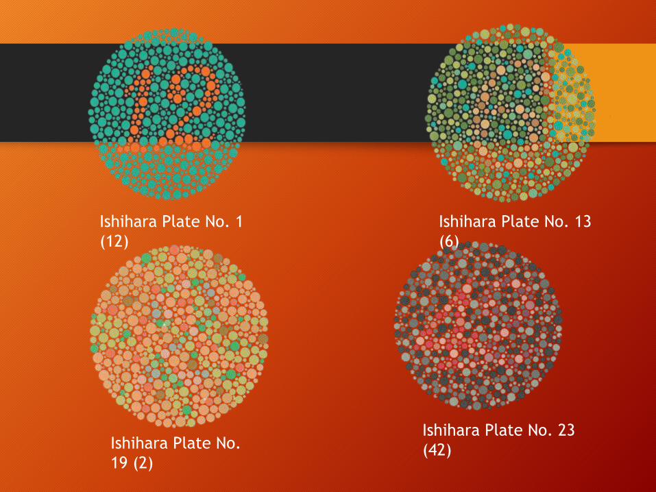
Ishihara Plate No. 1
(12)
Ishihara Plate No. 13
(6)
Ishihara Plate No.
19 (2)
Ishihara Plate No. 23
(42)
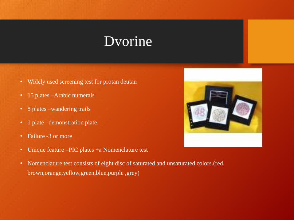
Dvorine
• Widely used screening test for protan deutan
• 15 plates –Arabic numerals
• 8 plates –wandering trails
• 1 plate –demonstration plate
• Failure -3 or more
• Unique feature –PIC plates +a Nomenclature test
• Nomenclature test consists of eight disc of saturated and unsaturated colors.(red,
brown,orange,yellow,green,blue,purple ,grey)
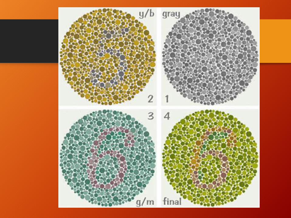
- Consists of two sections:
i.e for screening and fordiagnosisand severity
-screening plates:
4 demonstration and 6 vanishing plates( two for tritan and 4 for protan deutan defect)
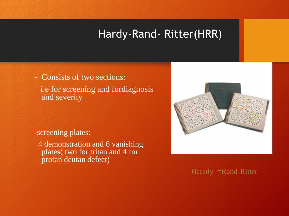
Harady –Rand-Ritter
Hardy-Rand- Ritter(HRR)
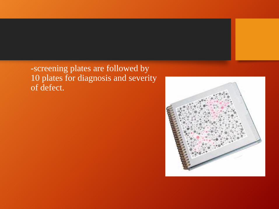
-screening plates are followed by 10 plates for diagnosis and severity of defect.

Hue discrimination tests
• Qualitative test for hue discrimination
• Diagnosis of the type and degree of color vision defect
• Cannot distinguish between dichromats and anomalous trichromats
• Consists of colored caps of different hue to be arranged in serial order of
hue
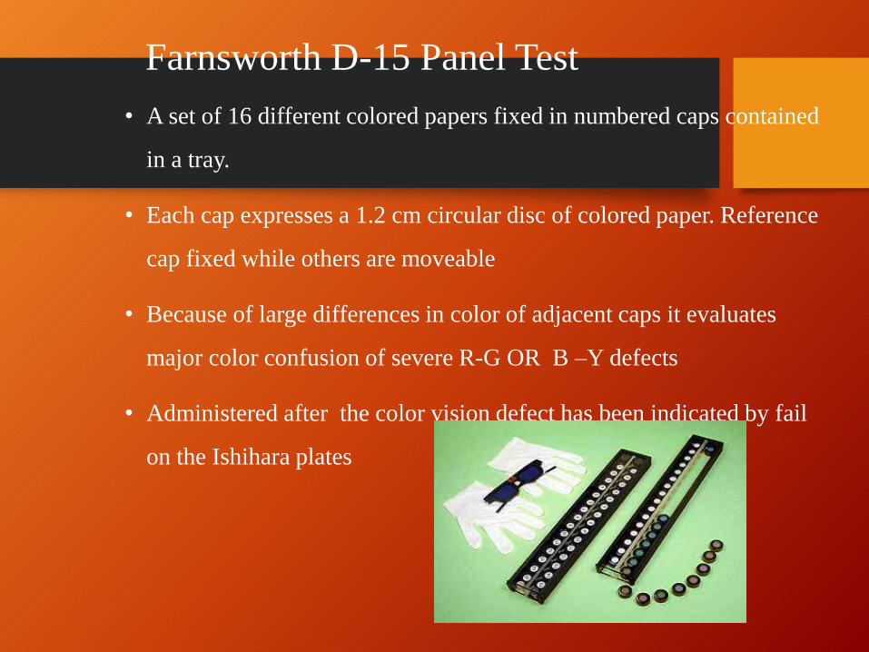
Farnsworth D-15 Panel Test
• A set of 16 different colored papers fixed in numbered caps contained
in a tray.
• Each cap expresses a 1.2 cm circular disc of colored paper. Reference
cap fixed while others are moveable
• Because of large differences in color of adjacent caps it evaluates
major color confusion of severe R-G OR B –Y defects
• Administered after the color vision defect has been indicated by fail
on the Ishihara plates
Testing Guidelines
• Illumination :at least 270 lux or almost natural
daylight
• Distance: 50 cm
• Present the test to the RE first then to the LE
Interpretation
• Record the order in which the caps were arranged
• Plot the cap sequence on the hue circle
• More than two crossings made ,determine the orientation of the
confusion axes
• If two or fewer crossings = defect not severe
• If mire than two crossings made , severe defect and orientation
tells the type of defect

Interpretation
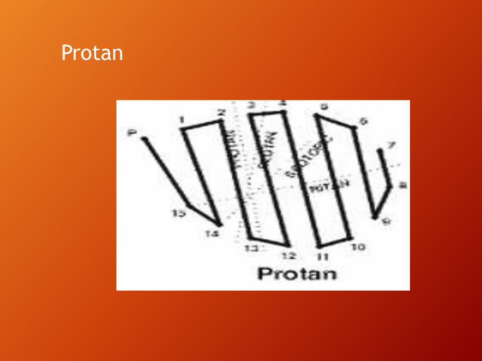
Protan
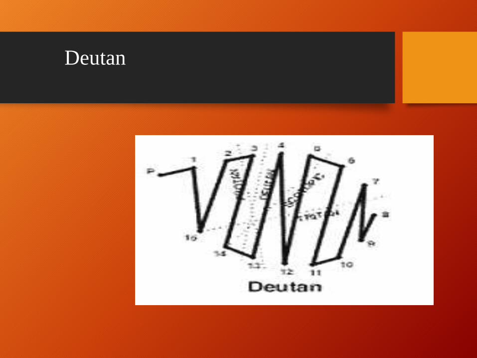
Deutan
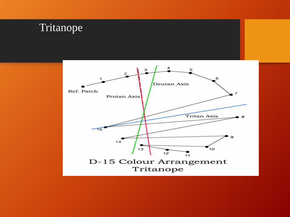
Tritanope
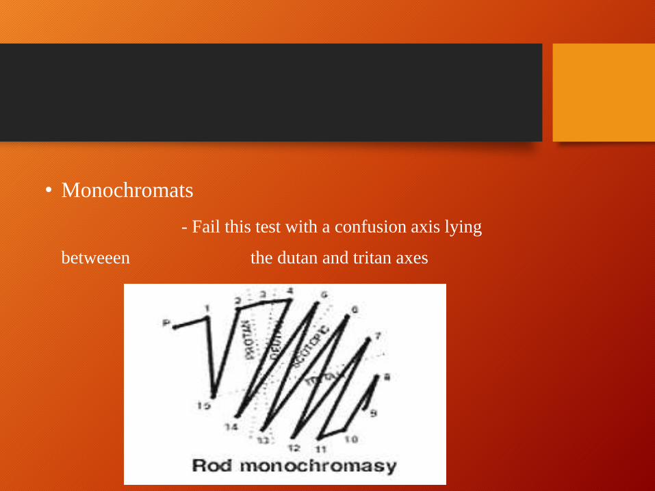
• Monochromats
- Fail this test with a confusion axis lying
betweeen the dutan and tritan axes
Farnsworth Munsell 100 hue test
• An expanded version of Panel D-15 TEST
• Consists of 85 caps divided approximately equally in four boxes
• Main purpose
-To classify type of CVD
-To measure the severity
• Can also be used to assess the progression of an acquired CVD
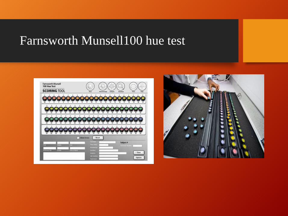
Farnsworth Munsell100 hue test

Testing Guidelines• Almost similar to that of D-15
• Time allowed per box is usually 3 minutes
• Record the sequence of numbers for each box that includes a polar co-
ordinate graph for plotting the error score for each cap.
• Error score for a cap = sum of the absolute difference
between the no. of the cap and
those adjacent to it
Recordings and Interpretation
• Record the cap sequence in the score sheet.
• A cap out of sequence is marked, the cap positioned in its place is recorded in
the line under it.
• A score for each cap is computed as –
Correct sequence is (22,23,24) = score 2
For a sequence (13,17,12) = score 9
• For the smallest possible error score, a point is marked on the innermost
circle in the sheet.
• Poorest score is 14.
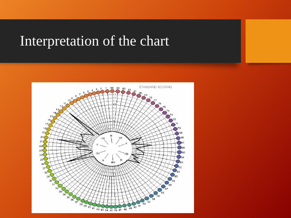
Interpretation of the chart
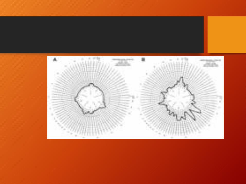

• If the plot is horizontally extended,
then protan defect is present.
• For obliquely oriented- deutran
• For vertical- tritan

H-16 test
• Designed by Farnsworth
- Has 16 colored samples
- Not commercially available unfortunately.
- For identifying dichromats
- Higher Munsell chroma than D-15
Color matching tests
• One color is to be matched mixing two colors
• Examples
-Nagel Anomaloscope
-City University Test
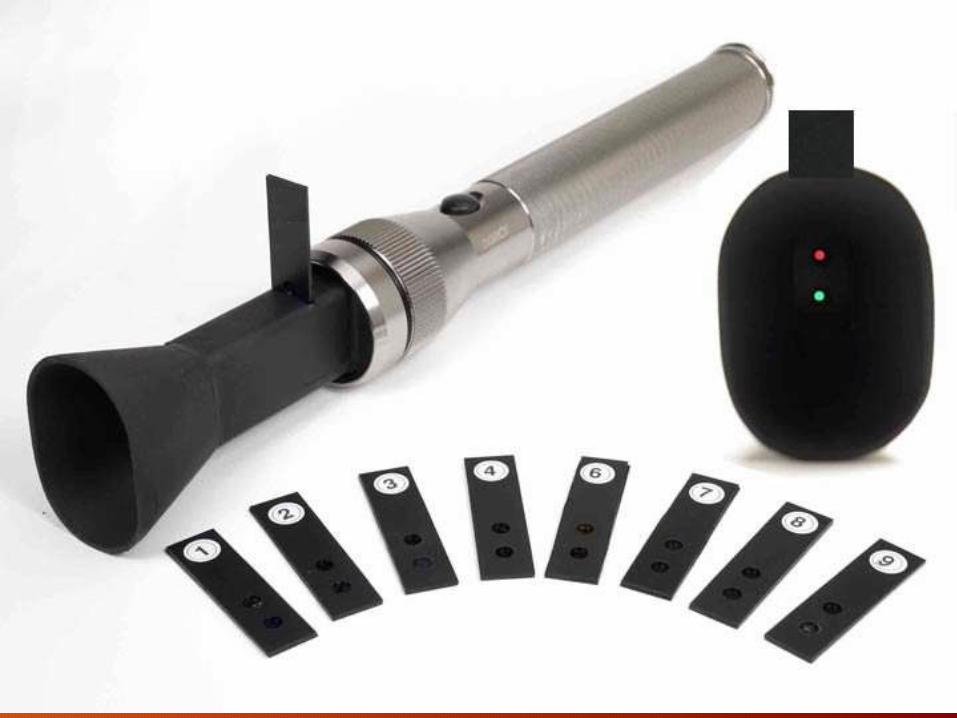
• Anomaloscope -
examines color matching behaviour - pt. mix the
monochromatic R AND G color in a proportion to match a
given yellow color discs
- judgement made for relative amount of R and G

AnomaloscopeTypes:
1 Nagel Anomaloscope
2 Pickford Nicholson Anomaloscope
3 Neitz OT Anomaloscope
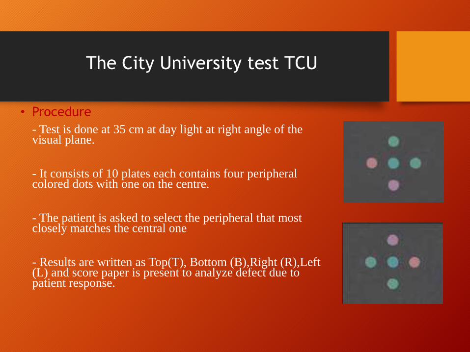
• Procedure
- Test is done at 35 cm at day light at right angle of the visual plane.
- It consists of 10 plates each contains four peripheral colored dots with one on the centre.
- The patient is asked to select the peripheral that most closely matches the central one
- Results are written as Top(T), Bottom (B),Right (R),Left (L) and score paper is present to analyze defect due to patient response.
The City University test TCU

Color naming and color sorting
•Lantern test
-To identify the color of a signal light in lantern
-Hue , brightness and size can be varied-Judgment made by the mistakes
-Not popular
• Names
- Farnsworth Lantern test
-Holmes Wright Lantern test

Yarn test
• Holmgren Wools test- oldest color sorting test
• To select from the pile of colored yarns those
which resembles a "standard skin“
• Not effective for diagnostic purpose
• Yarns fade and become dirty with handeling in a
short time.