coma and haemodynamic instability in the context of trauma · netherlands journal of critical care...
TRANSCRIPT
Netherlands Journal of Critical Care
210 NETH J CRIT CARE - VOLUME 25 - NO 6 - NOVEMBER 2017
Submitted January 2017; Accepted June 2017
C A S E R E P O R T
Coma and haemodynamic instability in the context of trauma
A. Gietler1, E. Pragt1, J.W.E.M. Sels1, P.F.W. Hannemann2, E.A. Bouman1, D.C.J.J. Bergmans1, W.N.K.A. van Mook1
Department of 1Intensive Care Medicine and 2Surgery, Maastricht University Medical Centre+, Maastricht, the Netherlands
Correspondence
A. Gietler - [email protected]
Keywords - propranolol intoxication, treatment of beta-blocker intoxication, coma and hemodynamic instability
AbstractComa and haemodynamic instability in a trauma patient due to propranolol intoxication. A 51-year-old female was involved in a car accident. Following this event she first became comatose followed by haemodynamic instability (hypotension and later bradycardia). Eventually it proved that these were attributable to a beta-blocker intoxication (propranolol) and not to the trauma. In case of propranolol intoxication (which is highly lipophilic) coma can precede the haemodynamic symptoms. Furthermore, hypotension can precede bradycardia because the negative inotropic effects occur earlier than the negative chronotropic effects.
IntroductionImmediately after trauma, it is not uncommon that patients are comatose and haemodynamically unstable. From a differential diagnostic perspective, the highly diverse causes for both conditions should be considered as will become clear from the following illustrative case.
CaseA 51-year-old woman hit a small parked bus with her car. When the ambulance staff found her in her car the Glasgow Coma Scale (GCS) was 2-4-2. She was taken to the emergency department after application of a cervical collar and immobilisation on a long spine board, ventilation with oxygen via a non-rebreathing mask (100% O2, 15 litres per minute), insertion of an oropharyngeal cannula and starting an IV drip infusion. The blood glucose level was 6.9 mmol/l. In the patient’s car a box with medication was found by the ambulance staff, containing various types of tablets, including diazepam, venlafaxine and paracetamol. Also, three empty blister packs (total of 30 capsules) of ‘propranolol retard’ (extended release formulation) 80 mg capsules (in total 2400 mg) were found. Empty blister packs of other drugs were not found.
When arriving at the emergency department, the GCS was 1-1-1, respiratory rate 10/minute, oxygen saturation was 100%, blood pressure 84/55 mmHg with a sinus rhythm of 79/minute. The further neurological examination did not show any other abnormalities such as anisocoria, lateralisation or pathological reflexes. Because of inadequate breathing, combined with a low GCS score, the patient was intubated uneventfully. Subsequently, over the period of an hour she gradually became more hypotensive (RR 64/40 mmHg) with a sinus rhythm of 70/minute, which did not respond to a total 1 mg of phenylephrine IV, 2.5 mmol calcium chloride IV, noradrenalin 0.1 µg/kg/min IV and intravenous fluids (1 litre NaCl 0.9%, 1 litre gelofusine and 1 litre Ringer's solution). The chest X-ray showed no abnormalities. On the abdominal echography a small amount of free fluid was seen in the lower abdomen. The CT of the abdomen showed fluid near the hepatic hilum and around the head of the pancreas, without any indication for a parenchymal lesion and an intact duodenum. Further trauma screening, including a CT scan of the cerebrum, did not show any abnormalities. Furthermore, there were no injuries to the face. Due to stable haemoglobin values and only a small amount of free fluid, a laparotomy was not performed. The ECG showed a sinus rhythm of 70/min, PR 0.15 sec, QRS 0.086 sec and QTc 0.42 sec, without abnormalities.
With reference to the coma and the haemodynamic instability following the trauma, various aetiologies were considered (table 1). The intensivist was consulted.
Because no explanation for the coma was found on CT cerebrum, further heteroanamnesis was explored. The woman’s medical history was obtained and it mentioned depression and a borderline personality disorder, as well as prior intoxications with beta-blockers (propranolol), benzodiazepines and paracetamol. The beta-blockers were prescribed for her in
Netherlands Journal of Critical Care
NETH J CRIT CARE - VOLUME 25 - NO 6 - NOVEMBER 2017 211
Coma and haemodynamic instability with trauma
Belgium for migraine symptoms. In September 2007, she had also been treated on our intensive care unit due to an intoxication with diazepam, propranolol, haloperidol and nortriptyline. At that time, she had also been comatose, with a GCS score of 1-1-1, blood pressure 60/40 mmHg and on the ECG, a sinus rhythm of 60/minute, an incomplete right bundle branch block, QRS 0,132 sec, QTc 0,517 sec. At that time IV flumazenil was given, after which the GCS score improved to 10; moreover, the persistent hypotension reacted well to the administration of norepinephrine.
Based on this new information, flumazenil 0.5 mg IV was now administered several times, however without any effect on the GCS score. IV acetylcysteine was started and activated coal was administered via a nasogastric tube. A transthoracic echocardiography (TTE) that was carried out in the emergency department showed a significantly decreased contractility, with a left ventricle ejection fraction of 18%. A working diagnosis of propranolol intoxication was made and dobutamine 5 µg/kg/min IV was started and two doses of glucagon 5 mg IV were given according to the National Institute for Public Health and the Environment (RIVM) guidelines.[1] As a result, the haemodynamics improved to a blood pressure of 100/60 mmHg, with a sinus rhythm of 60/min. In the ICU, both medications were continued: dobutamine 5 µg/kg/min and glucagon 5 mg/h IV. With that treatment, she remained haemodynamically stable for two days. Neurologically, our patient recovered fully without any neurological deficits.After cessation of dobutamine and glucagon infusion, TTE showed a left ventricle ejection fraction of 40%. In an evaluation by a psychiatrist, the patient was considered legally capable. On that same day, she was allowed to leave the ICU, and was given an appointment for further psychiatric follow-up and treatment. Toxicology screening showed intermediate values for benzodiazepines and low values for opiates and paracetamol. Because propranolol levels do not correlate well with the clinical effect[2,3] and analysis takes too long to be clinically relevant[4] serum concentrations were not determined.
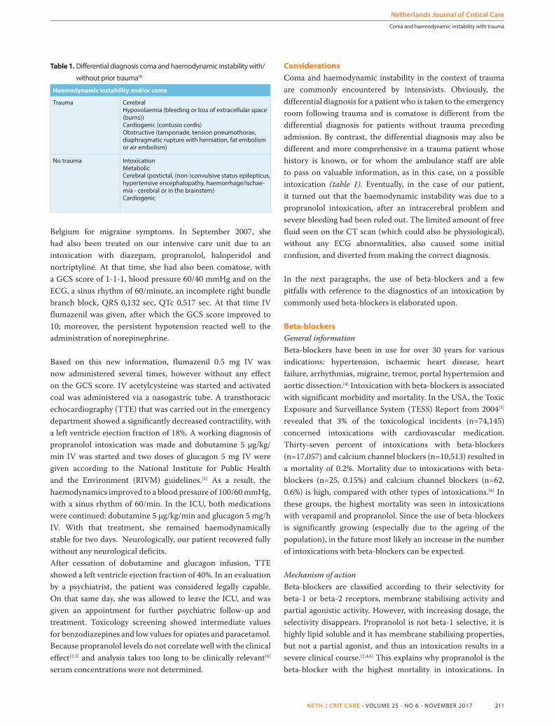
ConsiderationsComa and haemodynamic instability in the context of trauma are commonly encountered by intensivists. Obviously, the differential diagnosis for a patient who is taken to the emergency room following trauma and is comatose is different from the differential diagnosis for patients without trauma preceding admission. By contrast, the differential diagnosis may also be different and more comprehensive in a trauma patient whose history is known, or for whom the ambulance staff are able to pass on valuable information, as in this case, on a possible intoxication (table 1). Eventually, in the case of our patient, it turned out that the haemodynamic instability was due to a propranolol intoxication, after an intracerebral problem and severe bleeding had been ruled out. The limited amount of free fluid seen on the CT scan (which could also be physiological), without any ECG abnormalities, also caused some initial confusion, and diverted from making the correct diagnosis.
In the next paragraphs, the use of beta-blockers and a few pitfalls with reference to the diagnostics of an intoxication by commonly used beta-blockers is elaborated upon.
Beta-blockers General informationBeta-blockers have been in use for over 30 years for various indications: hypertension, ischaemic heart disease, heart failure, arrhythmias, migraine, tremor, portal hypertension and aortic dissection.[4] Intoxication with beta-blockers is associated with significant morbidity and mortality. In the USA, the Toxic Exposure and Surveillance System (TESS) Report from 2004[5]
revealed that 3% of the toxicological incidents (n=74,145) concerned intoxications with cardiovascular medication. Thirty-seven percent of intoxications with beta-blockers (n=17,057) and calcium channel blockers (n=10,513) resulted in a mortality of 0.2%. Mortality due to intoxications with beta-blockers (n=25, 0.15%) and calcium channel blockers (n=62, 0.6%) is high, compared with other types of intoxications.[6] In these groups, the highest mortality was seen in intoxications with verapamil and propranolol. Since the use of beta-blockers is significantly growing (especially due to the ageing of the population), in the future most likely an increase in the number of intoxications with beta-blockers can be expected.
Mechanism of actionBeta-blockers are classified according to their selectivity for beta-1 or beta-2 receptors, membrane stabilising activity and partial agonistic activity. However, with increasing dosage, the selectivity disappears. Propranolol is not beta-1 selective, it is highly lipid soluble and it has membrane stabilising properties, but not a partial agonist, and thus an intoxication results in a severe clinical course.[2,4,6] This explains why propranolol is the beta-blocker with the highest mortality in intoxications. In
Table 1. Differential diagnosis coma and haemodynamic instability with/
without prior trauma[4]
Haemodynamic instability and/or coma
Trauma CerebralHypovolaemia (bleeding or loss of extracellular space (burns)) Cardiogenic (contusio cordis)Obstructive (tamponade, tension pneumothorax, diaphragmatic rupture with herniation, fat embolism or air embolism)
No trauma IntoxicationMetabolic Cerebral (postictal, (non-)convulsive status epilepticus, hypertensive encephalopathy, haemorrhage/ischae-mia - cerebral or in the brainstem)Cardiogenic
Netherlands Journal of Critical Care
212 NETH J CRIT CARE - VOLUME 25 - NO 6 - NOVEMBER 2017
Coma and haemodynamic instability with trauma
addition, propranolol is a cardiac potassium channel blocker which can induce QRS widening and subsequent correlated ventricular arrhythmias (see below).
Pharmacology and toxicityAbsorption of beta-blockers after oral administration is fast, with the maximum effect after 1-2 hours. Propranolol also
has an active metabolite (4-hydroxypropranolol) following metabolisation in the liver of which the half-life is prolonged. Thus, elimination of propranolol and its metabolites is delayed in case of liver disease, reduced hepatic blood circulation or liver enzyme inhibition.[3] The half-life of propranolol slow release is 10-20 hours. Propranolol is the most lipid soluble beta-blocker, hence it passes the blood-brain barrier easily and, as a result,
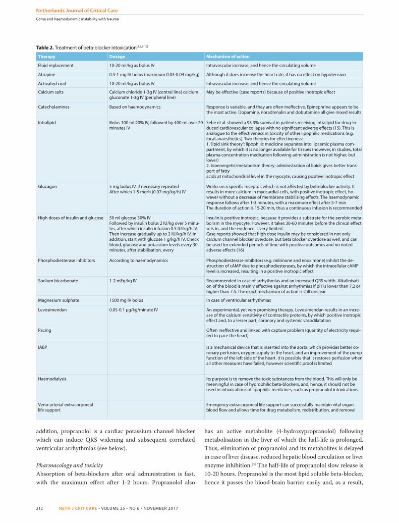
Table 2. Treatment of beta-blocker intoxication[2,5,7-16]
Therapy Dosage Mechanism of action
Fluid replacement 10-20 ml/kg as bolus IV Intravascular increase, and hence the circulating volume
Atropine 0.5-1 mg IV bolus (maximum 0.03-0.04 mg/kg) Although it does increase the heart rate, it has no effect on hypotension
Activated coal 10-20 ml/kg as bolus IV Intravascular increase, and hence the circulating volume
Calcium salts Calcium chloride 1-3g IV (central line) calcium gluconate 1-3g IV (peripheral line)
May be effective (case reports) because of positive inotropic effect
Catecholamines Based on haemodynamics Response is variable, and they are often ineffective. Epinephrine appears to be the most active. Dopamine, noradrenalin and dobutamine all give mixed results
Intralipid Bolus 100 ml 20% IV, followed by 400 ml over 20 minutes IV
Sebe et al. showed a 93.3% survival in patients receiving intralipid for drug-in-duced cardiovascular collapse with no significant adverse effects (15). This is analogue to the effectiveness in toxicity of other lipophilic medications (e.g. local anaesthetics). Two theories for effectiveness: 1. ‘lipid sink theory’: lipophilic medicine separates into lipaemic plasma com-partment, by which it is no longer available for tissues (however, in studies, total plasma concentration medication following administration is not higher, but lower) 2. bioenergetic/metabolism theory: administration of lipids gives better trans-port of fatty acids at mitochondrial level in the myocyte, causing positive inotropic effect
Glucagon 5 mg bolus IV, if necessary repeatedAfter which 1-5 mg/h (0.07 mg/kg/h) IV
Works on a specific receptor, which is not affected by beta-blocker activity. It results in more calcium in myocardial cells, with positive inotropic effect, ho-wever without a decrease of membrane stabilising effects. The haemodynamic response follows after 1-3 minutes, with a maximum effect after 5-7 min The duration of action is 15-20 min, thus a continuous infusion is recommended
High doses of insulin and glucose 50 ml glucose 50% IVFollowed by insulin bolus 2 IU/kg over 5 minu-tes, after which insulin infusion 0.5 IU/kg/h IV. Then increase gradually up to 2 IU/kg/h IV. In addition, start with glucose 1 g/kg/h IV. Check blood, glucose and potassium levels every 30 minutes, after stabilisation, every
Insulin is positive inotropic, because it provides a substrate for the aerobic meta-bolism in the myocyte. However, it takes 30-60 minutes before the clinical effect sets in, and the evidence is very limited.Case reports showed that high dose insulin may be considered in not only calcium channel blocker overdose, but beta blocker overdose as well, and can be used for extended periods of time with positive outcomes and no noted adverse effects (16)
Phosphodiesterase inhibitors According to haemodynamics Phosphodiesterase inhibitors (e.g. milrinone and enoximone) inhibit the de-struction of cAMP due to phosphodiesterases, by which the intracellular cAMP level is increased, resulting in a positive inotropic effect
Sodium bicarbonate 1-2 mEq/kg IV Recommended in case of arrhythmias and an increased QRS width. Alkalinisati-on of the blood is mainly effective against arrhythmias if pH is lower than 7.2 or higher than 7.5. The exact mechanism of action is still unclear
Magnesium sulphate 1500 mg IV bolus In case of ventricular arrhythmias
Levosimendan 0.05-0.1 µg/kg/minute IV An experimental, yet very promising therapy. Levosimendan results in an incre-ase of the calcium sensitivity of contractile proteins, by which positive inotropic effect and, to a lesser part, coronary and systemic vasodilatation
Pacing Often ineffective and linked with capture problem (quantity of electricity requi-red to pace the heart)
IABP Is a mechanical device that is inserted into the aorta, which provides better co-ronary perfusion, oxygen supply to the heart, and an improvement of the pump function of the left side of the heart. It is possible that it restores perfusion when all other measures have failed, however scientific proof is limited
Haemodialysis Its purpose is to remove the toxic substances from the blood. This will only be meaningful in case of hydrophilic beta-blockers, and, hence, it should not be used in intoxications of lipophilic medicines, such as propranolol intoxications
Veno-arterial extracorporeallife support
Emergency extracorporeal life support can successfully maintain vital organ blood flow and allows time for drug metabolism, redistribution, and removal
Netherlands Journal of Critical Care
NETH J CRIT CARE - VOLUME 25 - NO 6 - NOVEMBER 2017 213
Coma and haemodynamic instability with trauma
has the strongest central nerve system toxicity[2-6] of all beta-blockers. The propranolol levels in the brain are 10-20 times as high as in plasma.
Clinical symptoms in case of intoxicationsThe most common symptoms in beta-blocker intoxications are hypotension, bradycardia and cardiogenic shock as a result of conduction disorders and reduced contractility.[2-4] Various ECG anomalies may occur, including first-degree atrioventricular block, increased QRS width, prolonged QTc interval and a Brugada pattern (ST elevation in V1-3 ≥0.2 mV, a right bundle branch block and a prolongation of the PR interval). The negative inotropic effects can precede the negative chronotropic effects. Therefore, it is possible that severe intoxication, combined with deep shock, can be seen with an initial normal heart rate. It is less well known that central nervous system symptoms may precede cardiovascular toxicity, especially in case of highly lipophilic beta-blockers. Also in the case of our patient, a coma preceded the haemodynamic symptoms, namely in the form of hypotension without bradycardia, however combined with a significantly reduced cardiac contractility.
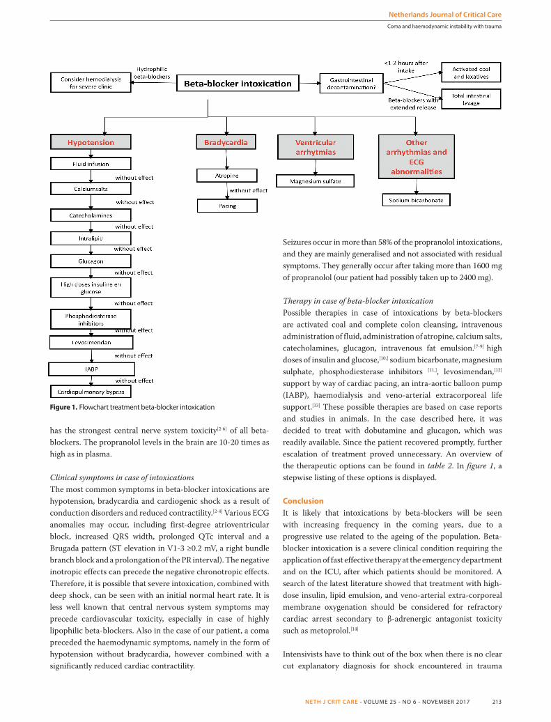
Figure 1. Flowchart treatment beta-blocker intoxication
Seizures occur in more than 58% of the propranolol intoxications, and they are mainly generalised and not associated with residual symptoms. They generally occur after taking more than 1600 mg of propranolol (our patient had possibly taken up to 2400 mg).
Therapy in case of beta-blocker intoxicationPossible therapies in case of intoxications by beta-blockers are activated coal and complete colon cleansing, intravenous administration of fluid, administration of atropine, calcium salts, catecholamines, glucagon, intravenous fat emulsion.[7-9] high doses of insulin and glucose,[10,] sodium bicarbonate, magnesium sulphate, phosphodiesterase inhibitors [11,], levosimendan,[12] support by way of cardiac pacing, an intra-aortic balloon pump (IABP), haemodialysis and veno-arterial extracorporeal life support.[13] These possible therapies are based on case reports and studies in animals. In the case described here, it was decided to treat with dobutamine and glucagon, which was readily available. Since the patient recovered promptly, further escalation of treatment proved unnecessary. An overview of the therapeutic options can be found in table 2. In figure 1, a stepwise listing of these options is displayed.
ConclusionIt is likely that intoxications by beta-blockers will be seen with increasing frequency in the coming years, due to a progressive use related to the ageing of the population. Beta-blocker intoxication is a severe clinical condition requiring the application of fast effective therapy at the emergency department and on the ICU, after which patients should be monitored. A search of the latest literature showed that treatment with high-dose insulin, lipid emulsion, and veno-arterial extra-corporeal membrane oxygenation should be considered for refractory cardiac arrest secondary to β-adrenergic antagonist toxicity such as metoprolol.[14]
Intensivists have to think out of the box when there is no clear cut explanatory diagnosis for shock encountered in trauma
Netherlands Journal of Critical Care
214 NETH J CRIT CARE - VOLUME 25 - NO 6 - NOVEMBER 2017
Coma and haemodynamic instability with trauma
patients. In case of a suspected intoxication by lipophilic beta-blockers, such as propranolol, there are two things that intensivists should be aware of. Firstly, an intoxication by such a beta-blocker may also cause central nerve system symptoms, including coma, which may also temporally precede the haemodynamic symptoms. Secondly, haemodynamic instability may already occur without any bradycardia being present at that moment; thus the negative inotropic side effect sometimes precedes the negative chronotropic effects. In our case the patient’s blood pressure was 84/55 mmHg with a sinus rhythm of 79/minute on arrival to the emergency department.
DisclosuresAll authors declare no conflict of interest. No funding or financial support was received.
References
1. www.rivm.nl/Onderwerpen/V/Vergiftigingen/Propranolol.2. Shepherd G. Treatment of poisoning by β-adrenergic and calcium-channel
blockers. Am J Health-Syst Parm. 2006;63:1828-45.3. Shannon MW. Haddad and Winchester's clinical management of poisoning and
drug overdose 4th edition: Saunders Elsevier; 2007.
4. www.uptodate.com. Beta blocker poisoning, Causes of coma, Shock trauma ddx.5. Watson WA, Litovitz TL, Rodgers GC, Jr, et al. 2004 Annual report of the American
Association of Poison Control Centers Toxic Exposure Surveillance System. Am J Emerg Med. 2005;23:589-666.
6. Beta receptor antagonists. In: Brent J, Burkhart K, Phillips D, Donovan J, (eds). Critical Care Toxicology. Diagnosis and Management of the Critically Poisoned Patient. 2nd edition, Springer, 2005:771-86.
7. Dean P, Ruddy JP, Marshall S. Intravenous lipid emulsion in propranolol [corrected] overdose. Anaesthesia. 2011;65:1148-50.
8. Weinberg GL. Lipid infusion therapy: translation to clinical practice. Anesth Analg. 2008;106:1340-2.
9. Samuels TL, Uncles DR, Willers JW, Monteiro R, Halloran C. Logging the potential for intravenous lipid emulsion in propranolol and other lipophilic drug overdoses. Anaesthesia. 2011;66:221-2.
10. Megarbane B, Karyo S, Baud FJ. The role of insulin and glucose (hyperinsulinaemia/euglycaemia) therapy in acute calcium channel antagonist and beta-blocker poisoning. Toxicolog Rev. 2004;23:215-22.
11. Sandroni C, Cavallaro F, Caricato A, Scapigliati A, Fenici P, Antonelli M. Enoximone in cardiac arrest caused by propranolol: two case reports. Acta Anaesthesiol Scand. 2006;50:759-61.
12. Leppikangas H, Ruokonen E, Rutanen J, Kiviniemi V, Lindgren L, Kurola J. Levosimendan as a rescue drug in experimental propranolol-induced myocardial depression: a randomized study. Ann Emerg Med. 2009;54:811-7 e1-3.
13. Kolcz J, Pietrzyk J, Januszewska K, Procelewska M, Mroczek T, Malec E. Extracorporeal life support in severe propranolol and verapamil intoxication. J Intensive Care Med. 2007;22:381-5.
14. Escajeda JT, Katz KD, Rittenberger JC. Successful treatment of metoprolol-induced cardiac arrest with high-dose insulin, lipid emulsion, and ECMO. Am J Emerg Med. 2015;33:1111.e1-4.
15. Sebe A, Dişel NR, Açıkalın Akpınar A, Karakoç E. Role of intravenous lipid emulsions in the management of calcium channel blocker and β-blocker overdose: 3 years experience of a university hospital. Postgrad Med. 2015;127:119-24.
16. lsion therapy for cardiogenic shock induced by intentional calcium-channel blocker and Beta-blocker overdose: a case series. J Emerg Med. 2014;46:486.
CURS
US
CURS
US
CURS
US
CURS
US
www.nvic.nl
Consolidatiecursus echogra� eVrijdag 15 december 2017