combinatorial extracellular matrices for human embryonic ... yang biomacromolecules 2010.pdf ·...
TRANSCRIPT
Combinatorial Extracellular Matrices for Human EmbryonicStem Cell Differentiation in 3D
Fan Yang,† Seung-Woo Cho,‡ Sun Mi Son,§ Sarah P. Hudson,§ Said Bogatyrev,§
Lily Keung,§ Daniel S. Kohane,| Robert Langer,§,⊥ and Daniel G. Anderson*,⊥
David H. Koch Institute for Integrative Cancer Research, Massachusetts Institute of Technology,45 Carleton Street, E25-342, Cambridge, Massachusetts 02139, Departments of Orthopaedic Surgery and
Bioengineering, Stanford University, 300 Pasteur Drive, Stanford, California 94304, Department ofBiotechnology, Yonsei University, Seoul 120-749, Korea, Department of Chemical Engineering,
Massachusetts Institute of Technology, 77 Massachusetts Avenue, Cambridge, Massachusetts 02139, andDepartment of Anesthesiology, Division of Critical Care Medicine, Children’s Hospital,
Harvard Medical School, 300 Longwood Avenue, Boston, Massachusetts 02115
Received March 31, 2010; Revised Manuscript Received June 14, 2010
Embryonic stem cells (ESCs) are promising cell sources for tissue engineering and regenerative medicine. Scaffoldsfor ESC-based tissue regeneration should provide not only structural support, but also signals capable of supportingappropriate cell differentiation and tissue development. Extracellular matrix (ECM) is a key component of thestem cell niche in vivo and can influence stem cell fate via mediating cell attachment and migration, presentingchemical and physical cues, as well as binding soluble factors. Here we investigated the effects of combinatorialextracellular matrix proteins on controlled human ESC (hESC) differentiation. Varying ECM compositions in 3Dmarkedly affects cell behavior, and optimal compositions of ECM hydrogels are identified that facilitate specific-lineage differentiation of stem cells. To our knowledge, this is the first combinatorial analysis of ECM hydrogelsfor their effects on hESC differentiation in 3D. The 3D matrices described herein may provide a useful platformfor studying the interactive ECM signaling in influencing stem cell differentiation.
1. Introduction
Embryonic stem cells (ESCs) are attractive candidates fortissue regeneration due to their ability to self-renew indefinitelyand differentiate into any type of cells in our body. Before ESCscan be widely used for therapeutic purposes, methods must bedeveloped to control their differentiation toward specificlineages. Increasing evidence has shown that the interplaybetween stem cells and their surrounding microenvironmentsis critical for regulating stem cell behavior.1 Stem cells residein a complex microenvironment in vivo, where they constantlyinteract and respond to multiple types of signals such as solubleand insoluble factors and mechanical forces.2 Soluble factors,including cytokines and biochemical molecules, can induce ESCdifferentiation toward desired specific lineages.3-6
The extracellular matrix (ECM) is an insoluble molecularnetwork consisting of a variety of elements, including interstitialmatrix proteins and the basement membrane. Collagens arefibrillar proteins that provide structural support, and are the mostabundant proteins in the ECM.7 Fibronectin acts as a generalcell adhesion molecule and provides binding sites for cell surfacereceptors and other ECM components.8 Laminin provides thescaffolding for basement membranes and can influence cellbehavior.9 Both fibronectin and laminin bind to collagen andplay roles in mediating cell-ECM interactions.
Previous studies of ECM effects on stem cell differentiationhave mainly focused on examining the role of the ECM inregulating mouse ESC differentiation toward lineages such ashepatocytes and cardiomyocytes.10,11 However, comparativestudies of human ESCs and mouse ESCs have demonstratedmajor differences in the signaling pathways required to regulatethe stem cell fate.12 Therefore, how ECM regulates human ESCfate remains to be resolved. A recent study has reported thathuman mesenchymal stem cells in 3D synthetic hydrogels canbe induced to differentiate down different pathways by modify-ing the material surface using five different small-moleculefunctional groups.13 This study shows the promise of regulatingstem cell fate via matrix cues.
Due to the complexity of cell-ECM and ECM-ECMinteractions, optimizing ECM effects on cell behavior remainschallenging. To overcome these limitations, combinatorialmethods have been developed to screen cell responses to ECMproteins. Flaim and co-workers developed ECM protein mi-croarrays.11 In this study, different combinations of natural ECMproteins such as collagen, laminin and fibronectin were depositedon a hydrogel slide, and their effects on mouse ESC differentia-tion to cardiac lineage were studied. Several other studies havealso developed high-throughput microarray approaches forscreening cell material interactions.14-18 However, these studiesexamined cell-material interactions on 2D surfaces, whereasstem cells niche in vivo is a 3D environment. In some settings,three dimensionality of the cell microenvironment is importantfor proper regulation of cell behavior.19-22 Furthermore,cell-matrix interactions in 3D matrices can differ from thoseon 2D substrates.21,22 Recently, a combinatorial library ofsynthetic 3D polymeric scaffolds was developed and screeningwith osteoblasts showed that such synthetic libraries may be
* To whom correspondence should be addressed. E-mail: [email protected].† Stanford University.‡ Yonsei University.§ Department of Chemical Engineering, Massachusetts Institute of
Technology.| Harvard Medical School.⊥ David H. Koch Institute for Integrative Cancer Research, Massachusetts
Institute of Technology.
Biomacromolecules 2010, 11, 1909–1914 1909
10.1021/bm100357t 2010 American Chemical SocietyPublished on Web 07/09/2010
used for rapidly identifying scaffold formulations that caninfluence osteoblast adhesion and proliferation.23
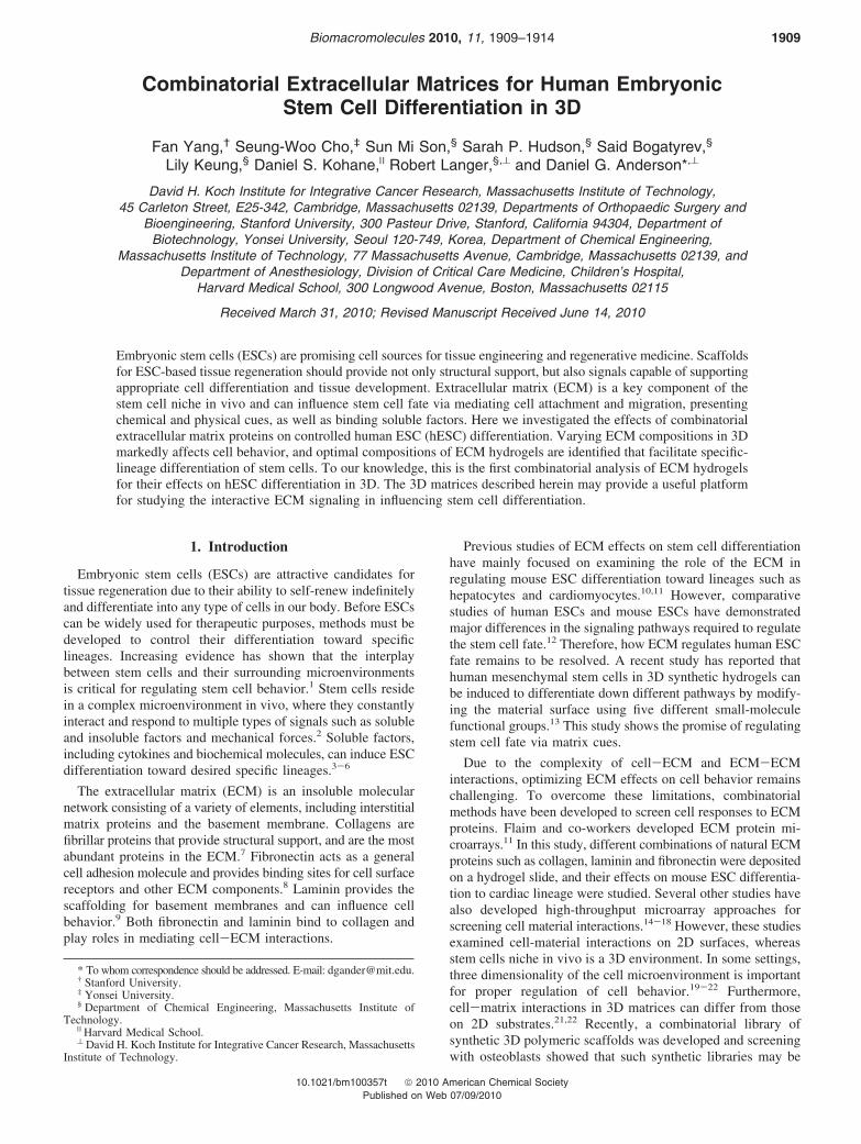
Here we develop a combinatorial array of 3D ECMs forosteogenic and endothelial differentiation of hESC-derived cells(hESdCs). We hypothesize that differentiation of hESdCs in3D can be modulated by varying the physical structure and thedensity of cellular binding domains from ECM proteins andthat optimal ECM compositions may exist to facilitate lineagespecific differentiation. Therefore, we examined 3D hydrogelsbased on several key ECM proteins including collagens,fibronectin, and laminin (Figure 1). We chose collagen type I(Col I) to be the major component of the scaffold as Col Iaccounts for 90% of bone matrix protein content.7 To elucidatethe role of ECM cues in controlling the differentiation ofhESdCs, Col I hydrogel was interspersed with fibronectin (FN),laminin (LM), and collagen type IV (Col IV) at differentconcentrations. Specifically, a total of 36 hydrogel compositionswere generated in parallel with different amounts of FN (10,25, 50 µg/mL), LM (10, 50, 100 µg/mL), and Col IV (10, 50,100 µg/mL; Figure 1). Cells were encapsulated in the hydrogelsin 3D and cultured in osteogenic differentiation medium orendothelial differentiation medium for 3 weeks before analysesof cellular organization and differentiation.
2. Experimental Section
2.1. Human Embryonic Stem Cell (hESC) Culture. hESC lineH9 (WiCell Research Institute, Madison, WI) was grown on inactivatedmouse embryonic fibroblasts in hESC growth medium consisting of80% knockout Dulbecco’s modified Eagle medium (DMEM, Gibco,Gaithersburg, MD) supplemented with 20% knockout serum, 4 ng/mLbasic fibroblast growth factor (bFGF), 1 mM L-glutamine, 0.1 mM2-mercaptoethanol, and 1% nonessential amino acids (Invitrogen,Carlsbad, CA). For embryoid body (EB) formation, hESC colonies weredissociated into small clumps by incubating at 37 °C for 15 min with2 mg/mL collagenase IV (Gibco). Human embryonic stem cell-derivedcells (hESdCs) used in this study were obtained as previous described.24
The hESC clumps were pelleted, resuspended in hESC growth mediumwithout bFGF, and cultured in Petri dishes for 10 days with mediumchange every other day. The EBs were then transferred to 0.1% (w/v)gelatin-coated plates. Upon 70% confluence, hESC-derived cells(hESdCs) were subcultured in mesenchymal stem cell growth mediumconsisting of DMEM, 10% fetal bovine serum, 100 mM sodium
pyruvate (Gibco), 100 unit penicillin, and 100 µg/mL streptomycin.Cells were cultured until passage 3 to induce further homogeneousdifferentiation before use. The hESdCs exhibit a similar morphologyto mesenchymal stem cells (MSC) and express MSC surface markersas previously reported.24
2.2. Extracellular Matrix (ECM) Hydrogels. Bovine dermalcollagen type I (Col I) hydrogels (1.5 mg/mL) were prepared by dilutingthe Col I solution (3.0 mg/mL, BD Biosciences, San Jose, CA) with10× phosphate buffered saline (Gibco; 9:1 v/v) and adjusting the pHto 7.4 using NaOH (Sigma, St. Louis, MO). The solution was thenadded into 48-well plates and incubated at 37 °C for 1 h to inducegelation. No cross-linking agent was used. To create a combinatorialcollection ECM hydrogels, Col I-based hydrogels (1.5 mg/mL) wasinterspersed with variable amounts of fibronectin (FN), laminin (LM),or collagen type IV (Col IV). Specifically, FN (1.0 mg/mL, Sigma)stock solution was added to the Col I solution (1.5 mg/mL) to obtaina final FN concentration of 10, 25, or 50 µg/mL. LM (1.0 mg/mL,Sigma) or Col IV (0.7 mg/mL, BD Biosciences) solutions were addedto Col I solution to reach a final concentration of 10, 50, or 100 µg/mL, respectively. The solutions were then neutralized to pH 7.4, andincubated in 48-well plates for 1 h at 37 °C for gelation. The resultingECM hydrogel collection has 36 different compositions in total (Figure1), and all experiments were done in triplicates.
2.3. Human Embryonic Stem Cell-Derived Cell (hESdC) Dif-ferentiation in ECM-Hydrogels. hESdCs were suspended in the ColI solutions with different compositions of ECM proteins (1.0 × 10⁷cells per 1 mL of each solution) and incubated for 1 h at 37 °C to formthe gels. All groups were cultured in osteogenic medium as previouslydefined26 or endothelial differentiation medium (EGM-2, Lonza,Walkersville, MD). Osteogenic medium consists of high-glucoseDulbecco’s modified Eagle’s medium (DMEM; Gibco), 100 nMdexamethasone (Sigma), 50 mg/mL ascorbic acid-2-phosphate (Sigma),10 mM �-glycerophosphate (Sigma), 10% fetal bovine serum (Gibco),100 unit/mL penicillin, and 100 mg/mL streptomycin (Gibco). Allsamples were incubated for three weeks before harvest, with mediumchange three times a week.
2.4. Scanning Electron Microscopy (SEM). For SEM, samples werefirst fixed overnight in 2.5% glutaraldehyde at room temperature. Sampleswere then lyophilized and sputter-coated with gold-plutonium alloy (10nm thickness) under vacuum before SEM analysis. SEM analysis wasconducted using a FEI/Philips XL30 FEG ESEM (FEI, Hillsboro, OR).
2.5. Quantitative Real-Time Polymerase Chain Reaction(PCR). Gene expression of osteogenic or endothelial markers byhESdCs cultured in hydrogels was examined using quantitative real-
Figure 1. Schematic illustration of compositions and layout of the combinatorial ECM hydrogels. Stem cell-seeded ECM mixtures were depositedinto 48-well plates, and cultured in either osteogenic differentiation medium or endothelial differentiation medium. All experiments were done intriplicate. Col I: collagen I; FN: fibronectin; LM: laminin; Col IV: collagen IV.
1910 Biomacromolecules, Vol. 11, No. 8, 2010 Communications
time PCR (n ) 3 per group). Total RNA was extracted from hydrogelsamples as previously described.25 A reverse transcription reaction wasperformed with 1 µg of total RNA using SuperScript III reversetranscriptase (Invitrogen). Real-time PCR was performed using a 7500Fast Real-Time PCR System (Applied Biosystems, Foster City, CA).Osteocalcin expression was quantified using SYBR Green detectingreagents, with primer sequence as follows: Osteocalcin, forward 5′-GAC TGT GAC GAG TTG GCT GA-3′ and reverse 5′-CTG GAGAGG AGC AGA ACT GG-3′; �-actin, forward 5′-TGG CAC CACACC TTC TAC AAT GAG C-3′ and reverse 5′-GCA CAG CTT CTCCTT AAT GTC ACG C-3′. Endothelial marker von Willebrand Factor(vWF) expression was quantified using Universe Fast PCR Master Mix(Applied Biosystems) with TaqMan Gene Expression Assays (AppliedBiosystems) for target (vWF: Hs01109438_m1) and endogenous control(glyceraldehyde 3-phosphate dehydrogenase (GAPDH): Hs02758991_g1).All samples were analyzed in triplicates. The expression level of targetgene was first normalized to endogenous control (�-actin or GAPDH),26
and results are presented as relative fold changes in all groups usingnormalized mRNA level in group 1 as controls.
2.6. Histology. Cell-hydrogel constructs were harvested at 3 weeksafter culture. Specimens were fixed in 10% (v/v) buffered formaldehyde,dehydrated with a graded ethanol series, and embedded in paraffin.Specimens were sliced into 4 µm sections and stained with hematoxylinand eosin (H&E) and Masson’s trichrome to detect tissue morphologyand collagen deposition.
2.7. Immunofluorescent Staining. Tissue sections were immunof-luorescently stained using primary antibodies against osteocalcin (Chemi-con, Temecula, CA) and collagen type II (DAKO, Carpenteria, CA).Section stained with nonspecific primary antibody is used as a negativecontrol. The staining signals were visualized with FITC-conjugatedsecondary antibodies (Invitrogen). The stained sections were examinedusing a fluorescent microscope (Carl Zeiss, Oberkochen, Germany).
2.8. Statistical Analysis. Quantitative data are expressed as mean( standard deviation. Statistical analysis was performed by the analysisof variance (ANOVA) using a Bonferroni test. A value of p < 0.05was considered statistically significant.
3. Results and Discussion
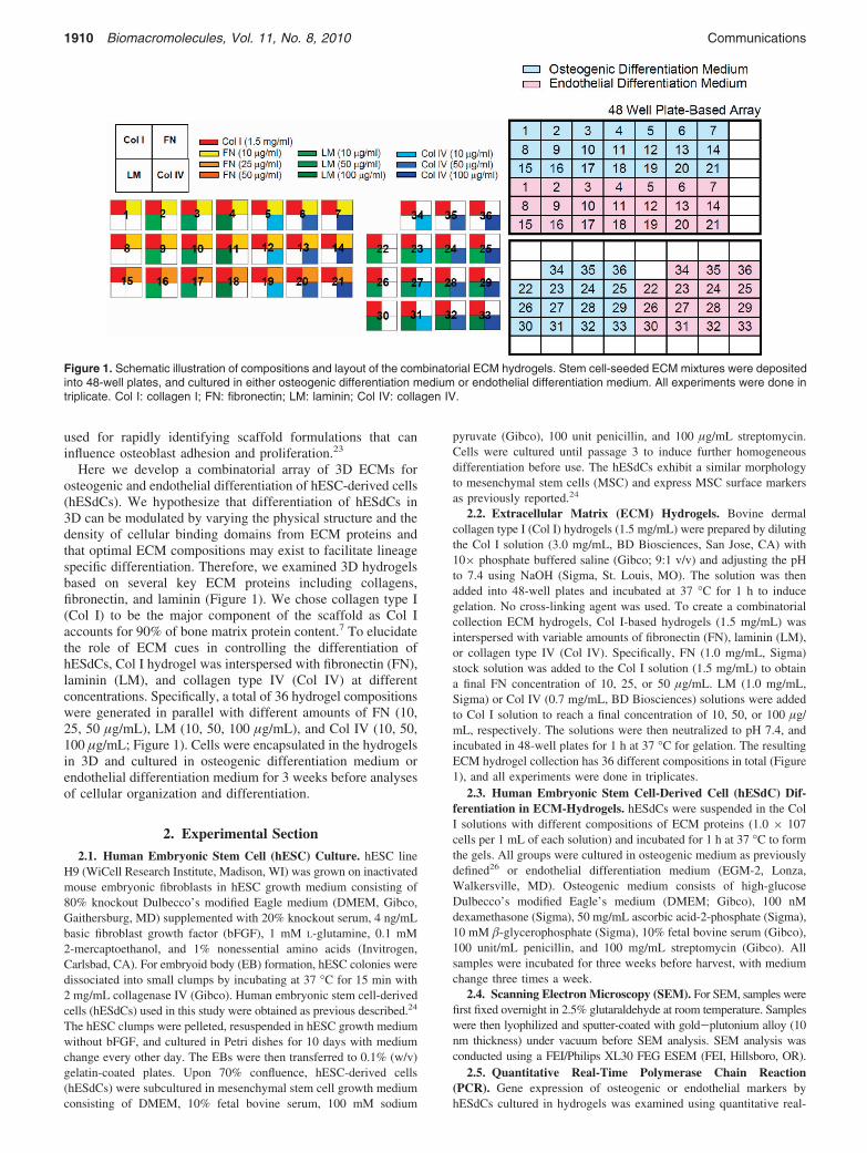
3.1. Morphological Analyses of ECM Hydrogel Network.The microscopic structure of ECM hydrogel network wasexamined by scanning electron microscopy (SEM). Col I
matrices alone showed a homogeneous fibrillar network consist-ing of fibers of approximately 200-300 nm diameter (Figure2). Interspersing other ECM components (FN, LM, and Col IV)significantly changed the structures and morphology of thenetwork (Figure 2). For example, the addition of FN (25 µg/mL; Group 8) increased matrix heterogeneity, with someaggregates and amorphous regions interspersed in and alongthe collagen fibers. Further addition of LM (50 µg/mL) to FN-Col I hydrogel (Group 10) induced a much denser fibrillarnetwork, with thinner fibers and smaller pore size (Figure 2).Interspersing Col IV (50 µg/mL) to FN-Col I network (Group13) resulted in a fibrillar network structure similar to Col Inetwork alone, with higher heterogeneity in pore size and fibersize (Figure 2). A previous study has examined the effects ofFN and LM on structural property of 3D collagenous network,where it was reported that FN and LM were organized inaggregates, interspersed in collagen gel, and distributed alongcollagen fibers in thin fibrils.10
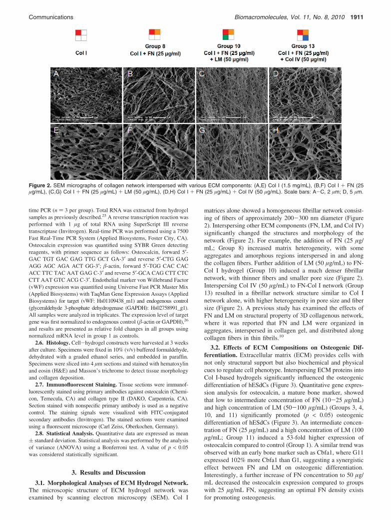
3.2. Effects of ECM Compositions on Osteogenic Dif-ferentiation. Extracellular matrix (ECM) provides cells withnot only structural support but also biochemical and physicalcues to regulate cell phenotype. Interspersing ECM proteins intoCol I-based hydrogels significantly influenced the osteogenicdifferentiation of hESdCs (Figure 3). Quantitative gene expres-sion analysis for osteocalcin, a mature bone marker, showedthat low to intermediate concentration of FN (10-25 µg/mL)and high concentration of LM (50-100 µg/mL) (Groups 3, 4,10, and 11) significantly promoted (p < 0.05) osteogenicdifferentiation of hESdCs (Figure 3). An intermediate concen-tration of FN (25 µg/mL) and a high concentration of LM (100µg/mL; Group 11) induced a 53-fold higher expression ofosteocalcin compared to control (Group 1). A similar trend wasobserved with an early bone marker such as Cbfa1, where G11expressed 102% more Cbfa1 than G1, suggesting a synergisticeffect between FN and LM on osteogenic differentiation.Interestingly, a further increase of FN concentration to 50 µg/mL decreased the osteocalcin expression compared to groupswith 25 µg/mL FN, suggesting an optimal FN density existsfor promoting osteogenesis.
Figure 2. SEM micrographs of collagen network interspersed with various ECM components: (A,E) Col I (1.5 mg/mL), (B,F) Col I + FN (25µg/mL), (C,G) Col I + FN (25 µg/mL) + LM (50 µg/mL), (D,H) Col I + FN (25 µg/mL) + Col IV (50 µg/mL). Scale bars: A-C, 2 µm; D, 5 µm.
Communications Biomacromolecules, Vol. 11, No. 8, 2010 1911
Our results also indicate that multiple ECM interactions canplay a role in regulating stem cell differentiation. FN alone(Group 1, 8, and 15) did not affect the osteogenic differentiationprocess (Figure 3). However, in the presence of LM and ColIV, FN significantly enhanced osteogenic differentiation ofhESdCs within a specific concentration range (Figure 3). LMalone (Groups 22, 26, and 30) showed no or moderate effectson osteogenesis, but increased osteocalcin expression 53-foldwith FN (25 µg/mL). Col IV alone did not increase osteocalcinexpression (Groups 34-36) (Figure 3), but increased osteocalcinexpression 16-fold in the presence of FN (Group 21) and LM(Group 25; p < 0.05). Our results confirm that ECM composi-tions interact in a complex manner and cellular response toindividual ECM components cannot necessarily be used topredict the cellular response.
Specific lineage differentiation of stem cells may also bemodulated by the mechanical property of the matrix on whichthe cells are grown.27 By tuning the elastic modulus of ColI-coated polyacrylamide hydrogel, mesenchymal stem cells werereported to differentiate toward various pathways includingneurogenic, myogenic, or osteogenic lineages.14 In our study,the matrix is a Col I-based hydrogel and a previous report hasshown that interspersing Col I hydrogel with other ECMcomponents such as FN or LM does not significantly alter themechanical property of the network10 within the concentrationsexamined here. Therefore, we believe the altered stem cellresponses to various ECM groups observed in our study werelikely not caused by the mechanical property of the matrices.There are several other factors that may be responsible for theobserved cellular response, including matrix ligand presentation,surface topography of the fibers, network pore size, and so on.For example, encapsulated stem cells may respond to the matrixligands through focal adhesion complexes, which lead tocorresponding cytoskeleton reorganization. Focal adhesion ki-nase signaling has been reported to play an important role inregulating ECM-induced osteogenic differentiation of humanmesenchymal stem cells28 and may influence the hESdCosteogenesis in a similar manner. Furthermore, varying ECMcompositions led to an obvious change in the network density,which affects the transport of soluble factors that are involvedin cell differentiation.
Previous work has examined the effects of various ECMcompositions on stem cell adhesion and differentiation and
showed that ECM proteins such as FN and LM may promotemesenchymal stem cell (MSC) adhesion and differentiationtoward bone pathway.29-31 While the effects of individual ECMproteins have been extensively explored, the influence ofcombinatorial ECM proteins on stem cell differentiation in 3Dremain unclear, and optimal concentration is yet to be deter-mined. Furthermore, most previous work examined cell-materialinteractions on 2D surfaces, while cell-matrix interactions in3D matrices can differ significantly from those on 2Dsubstrates.21,22 The findings from this study will provide valuableguidance for designing next generation of 3D synthetic stemcell niche to mimic the ECM microenvironment and promotespecific stem cell differentiation.
3.3. Effects of ECM Compositions on Tissue Formation.While the gene expression assay directly quantifies the effectsof ECM matrices on osteogenic differentiation, histologyprovides information on the effects of ECM compositions ontissue formation. Our results demonstrated that varying ECMcompositions in 3D markedly affected cellular organization andtissue formation. Histological analysis (hematoxylin and eosin(H&E) and Masson’s trichrome staining; Figure 4) revealed thatinterspersing Col I gel with singular additional ECM component(FN, LM, or Col IV) did not increase the cellularity comparedto the control (Group 1). In contrast, interspersing Col I gelwith two additional ECM components markedly promotedcellular organization and tissue formation. In general, groupswith high level of osteocalcin expression (Groups 3, 4, 10, 11,14, 18, 21, and 25) demonstrated denser structure, significantlyhigher cellularity, and stronger collagen matrix deposition(Figure 4). In one ECM condition (Group 11), hESdCs producedan evenly distributed matrix (Figure 5A,B). Homogeneous andintense staining of collagen type II and mature bone markerosteocalcin was detected throughout the construct, suggestingthese cells underwent endochondral ossification (Figure 5C,D).While the processes of osteogenesis and matrix formation arecorrelated and exhibited similar trends, we also noticed somevariance in the fold of change. For example, the matrixformation in Group 13 is much higher than Group 3 andcomparable to Group 14, yet the osteocalcin expression in Group13 is not as significantly upregulated. Such difference may bedue to the multifactorial changes in the ECM matrices inducedby varying ECM compositions and concentrations in 3D. Ourscanning electron microscopy data (Figure 2) showed thatvarying ECM compositions led to an obvious change in thenetwork density and pore size, which directly affects thetransport of soluble factors that are involved in cell differentia-tion. Meanwhile, varying ECM compositions may also changecellular remodeling dynamics and matrix metalloproteinase(MMP) activity, which will directly influence the matrixformation. The extracellular matrix also serves as a depot forgrowth factors, from which they can be released into thesurrounding microenvironment and regulate cell phenotype andtissue formation.32,33 Varying ECM compositions may alsoinfluence the ECM-growth factor interactions, which wouldin turn influence stem cell fate via autocrine or paracrine cellsignaling. All these factors, in part or together, likely contributeto the observed changes in stem cell differentiation and tissueformation.
3.4. Effects of ECM Compositions on Endothelial Dif-ferentiation. Bone is a highly vascularized tissue, and an idealscaffold for bone tissue engineering should promote both bonetissue formation and angiogenesis. To identify an optimal ECM-based scaffold for angiogenesis induction, hESdCs were en-capsulated in the 3D hydrogels and cultured in endothelial
Figure 3. Quantitative gene expression of osteocalcin, a maturebone marker, by the encapsulated hESdCs after 3 weeks of culturein ECM gels under osteogenic conditions. All experiments weredone in triplicates and results were presented as relative foldchanges in all groups using normalized mRNA level in group 1 ascontrol (*p < 0.05).
1912 Biomacromolecules, Vol. 11, No. 8, 2010 Communications
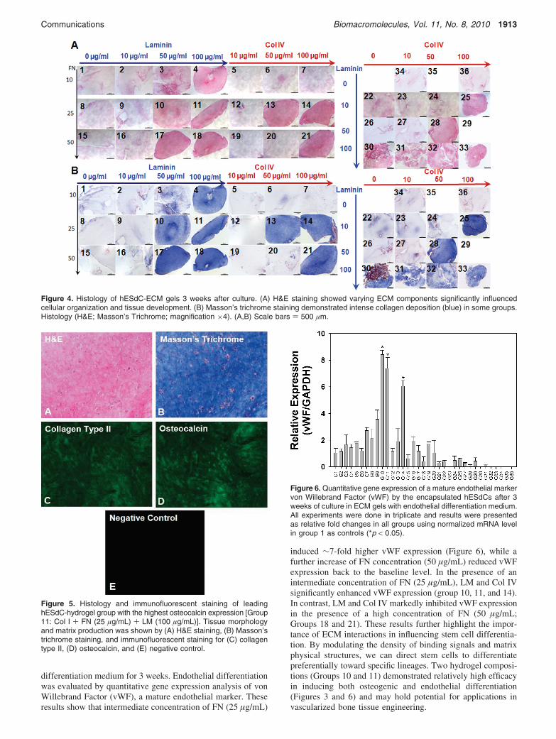
differentiation medium for 3 weeks. Endothelial differentiationwas evaluated by quantitative gene expression analysis of vonWillebrand Factor (vWF), a mature endothelial marker. Theseresults show that intermediate concentration of FN (25 µg/mL)
induced ∼7-fold higher vWF expression (Figure 6), while afurther increase of FN concentration (50 µg/mL) reduced vWFexpression back to the baseline level. In the presence of anintermediate concentration of FN (25 µg/mL), LM and Col IVsignificantly enhanced vWF expression (group 10, 11, and 14).In contrast, LM and Col IV markedly inhibited vWF expressionin the presence of a high concentration of FN (50 µg/mL;Groups 18 and 21). These results further highlight the impor-tance of ECM interactions in influencing stem cell differentia-tion. By modulating the density of binding signals and matrixphysical structures, we can direct stem cells to differentiatepreferentially toward specific lineages. Two hydrogel composi-tions (Groups 10 and 11) demonstrated relatively high efficacyin inducing both osteogenic and endothelial differentiation(Figures 3 and 6) and may hold potential for applications invascularized bone tissue engineering.
Figure 4. Histology of hESdC-ECM gels 3 weeks after culture. (A) H&E staining showed varying ECM components significantly influencedcellular organization and tissue development. (B) Masson’s trichrome staining demonstrated intense collagen deposition (blue) in some groups.Histology (H&E; Masson’s Trichrome; magnification ×4). (A,B) Scale bars ) 500 µm.
Figure 5. Histology and immunofluorescent staining of leadinghESdC-hydrogel group with the highest osteocalcin expression [Group11: Col I + FN (25 µg/mL) + LM (100 µg/mL)]. Tissue morphologyand matrix production was shown by (A) H&E staining, (B) Masson’strichrome staining, and immunofluorescent staining for (C) collagentype II, (D) osteocalcin, and (E) negative control.
Figure 6. Quantitative gene expression of a mature endothelial markervon Willebrand Factor (vWF) by the encapsulated hESdCs after 3weeks of culture in ECM gels with endothelial differentiation medium.All experiments were done in triplicate and results were presentedas relative fold changes in all groups using normalized mRNA levelin group 1 as controls (*p < 0.05).
Communications Biomacromolecules, Vol. 11, No. 8, 2010 1913
4. Conclusion
In conclusion, we have demonstrated that a combinatorialanalysis of ECM gels can facilitate the identification of optimalECM matrices for hESdC differentiation in 3D. To ourknowledge, this is the first combinatorial analysis of ECMhydrogels for their effects on hESC differentiation in 3D. Thisapproach may be useful for the study of other clinically relevanthuman stem cell types or be combined with various solublefactors to study stem cell differentiation in the context ofinteracting ECM and growth factor signaling. Successful tissueregeneration strategies rely on optimized stem cell niche designand extensive efforts are being dedicated in developing bioma-terials to direct stem cell fate.34 Advances in understandingcell-extracellular matrix interactions and signaling wouldprovide valuable insight in guiding the design of biomimeticmaterials to regulate stem cell fate for various tissue regenerationapplications.
Acknowledgment. This work was supported by NationalInstitutes of Health (NIH) grants (DE016516 and EB000244)and a Juvenile Diabetes Research Foundation (JDRF) grant (17-2007-1063). F.Y. would also like to thank NIH for postdoctoralfellowship support.
References and Notes(1) Scadden, D. T. Nature 2006, 441, 1075–1079.(2) Metallo, C. M.; Mohr, J. C.; Detzel, C. J.; Pablo, J. J.; Wie, B. J.;
Palecek, S. P. Biotechnol. Prog. 2007, 23, 18–23.(3) D’Amour, K. A.; Agulnick, A. D.; Eliazer, S.; Kelly, O. G.; Kroon,
E.; Baetge, E. E. Nat. Biotechnol. 2005, 23, 1534–1541.(4) D’Amour, K. A.; Bang, A. G.; Eliazer, S.; Kelly, O. G.; Agulnick,
A. D.; Smart, N. G.; Moorman, M. A.; Kroon, E.; Carpenter, M. K.;Baetge, E. E. Nat. Biotechnol. 2006, 24, 1392–1401.
(5) Darabi, R.; Gehlbach, K.; Bachoo, R. M.; Kamath, S.; Osawa, M.;Kamm, K. E.; Kyba, M.; Perlingeiro, R. C. Nat. Med. 2008, 14, 134–143.
(6) Yang, L.; Soonpaa, M. H.; Adler, E. D.; Roepke, T. K.; Kattman,S. J.; Kennedy, M.; Henckaerts, E.; Bonham, K.; Abbott, G. W.;Linden, R. M.; Field, L. J.; Keller, G. M. Nature 2008, 453, 524–528.
(7) Kern, B.; Shen, J.; Starbuck, M.; Karsenty, G. J. Biol. Chem. 2001,276, 7101–7107.
(8) Pankov, R.; Yamada, K. M. J. Cell Sci. 2002, 115, 3861–3863.(9) Timpl, R.; Rohde, H.; Robey, P. G.; Rennard, S. I.; Foidart, J. M.;
Martin, G. R. J. Biol. Chem. 1979, 254, 9933–9937.
(10) Battista, S.; Guarnieri, D.; Borselli, C.; Zeppetelli, S.; Borzacchiello,A.; Mayol, L.; Gerbasio, D.; Keene, D. R.; Ambrosio, L.; Netti, P. A.Biomaterials 2005, 26, 6194–6207.
(11) Flaim, C. J.; Chien, S.; Bhatia, S. N. Nat. Methods 2005, 2, 119–125.(12) Wei, C. L.; Miura, T.; Robson, P.; Lim, S. K.; Xu, X. Q.; Lee, M. Y.;
Gupta, S.; Stanton, L.; Luo, Y. Q.; Schmitt, J.; Thies, S.; Wang, W.;Khrebtukova, I.; Zhou, D. X.; Liu, E. T.; Ruan, Y. J.; Rao, M.; Lim,B. Stem Cells 2005, 23, 166–185.
(13) Benoit, D. S.; Schwartz, M. P.; Durney, A. R.; Anseth, K. S. Nat.Mater. 2008, 7, 816–823.
(14) MacBeath, G.; Schreiber, S. L. Science 2000, 289, 1760–1763.(15) Anderson, D. G.; Levenberg, S.; Langer, R. Nat. Biotechnol. 2004,
22, 863–866.(16) Anderson, D. G.; Putnam, D.; Lavik, E. B.; Mahmood, T. A.; Langer,
R. Biomaterials 2005, 26, 4892–4897.(17) Simon, C. G., Jr.; Eidelman, N.; Kennedy, S. B.; Sehgal, A.l.; Khatri,
C. A.; Washburn, N. R. Biomaterials 2005, 26, 6906–6915.(18) Nakajima, M.; Ishimuro, T.; Kato, K.; Ko, I.-K.; Hirata, I.; Arima,
Y.; Iwata, H. Biomaterials 2007, 28, 1048–1060.(19) Cukierman, E.; Pankov, R. D.; Stevens, R.; Yamada, K. M. Science
2001, 294, 1708–1712.(20) Grinnell, F. Trends Cell Biol. 2003, 13, 264–269.(21) Chun, T. H.; Hotary, K. B.; Sabeh, F.; Saltiel, A. R.; Allen, E. D.;
Weiss, S. J. Cell 2006, 125, 577–591.(22) Hotary, K. B.; Allen, E. D.; Brooks, P. C.; Datta, N. S.; Long, M. W.;
Weiss, S. J. Cell 2003, 114, 33–45.(23) Yang, Y.; Bolikal, D.; Becker, M.; Kohn, J.; Zeiger, D.; Simon, C.
AdV. Mater. 2008, 20, 2037–2043.(24) Hwang, N. S.; Varghes, S.; Zhang, Z.; Elisseeff, J. Tissue Eng. 2006,
12, 2695–2706.(25) Yang, F.; Williams, C. G.; Wang, D. A.; Lee, H.; Manson, P. N.;
Elisseeff, J. Biomaterials 2005, 26, 5991–5998.(26) Mori, R.; Wang, Q.; Danenberg, K. D.; Pinski, J. K.; Danenberg, P. V.
Prostate 2008, 68, 1555–1560.(27) Engler, A. J.; Sen, S.; Sweeney, H. L.; Discher, D. E. Cell 2006, 126,
677–689.(28) Salasznyk, R. M.; Klees, R. F.; Williams, W. A.; Boskey, A.; Plopper,
G. E. Exp. Cell Res. 2007, 313, 22–37.(29) Gronthos, S.; Simmons, P. J.; Graves, S. E.; Robey, P. G. Bone 2001,
28, 174–181.(30) Keselowsky, B. G.; Collard, D. M.; Garcia, A. J. Proc. Natl. Acad.
Sci. U.S.A. 2005, 102, 5953–5957.(31) Hidalgo-Bastida, L. A.; Cartmell, S. H. Tissue Eng., Part B 2010,
Apr 7, Epub ahead of print.(32) Discher, D. E.; Mooney, D. J.; Zandstra, P. W. Science 2009, 324,
1673–1677.(33) Uebersax, L.; Merkle, H. P.; Meinel, L. Tissue Eng., Part B 2009,
15, 263–289.(34) Designing materials to direct stem-cell fate. Lutolf, M. P.; Gilbert,
P. M.; Blau, H. M. Nature 2009, 462, 433–441.
BM100357T
1914 Biomacromolecules, Vol. 11, No. 8, 2010 Communications