combinedreversedphasehplc,massspectrometry,andnmr...
TRANSCRIPT

Hindawi Publishing CorporationJournal of Biomedicine and BiotechnologyVolume 2011, Article ID 385786, 8 pagesdoi:10.1155/2011/385786
Research Article
Combined Reversed Phase HPLC, Mass Spectrometry, and NMRSpectroscopy for a Fast Separation and Efficient Identification ofPhosphatidylcholines
Jan Willmann,1 Herbert Thiele,2 and Dieter Leibfritz1
1 Institute of Organic Chemistry, University of Bremen, NW2C, 28359 Bremen, Germany2 Bruker Daltonik GmbH, Fahrenheitstr. 4, 28359 Bremen, Germany
Correspondence should be addressed to Dieter Leibfritz, [email protected]
Received 14 May 2010; Accepted 21 July 2010
Academic Editor: Olav Kvalheim
Copyright © 2011 Jan Willmann et al. This is an open access article distributed under the Creative Commons Attribution License,which permits unrestricted use, distribution, and reproduction in any medium, provided the original work is properly cited.
In respect of the manifold involvement of lipids in biochemical processes, the analysis of intact and underivatised lipids of bodyfluids as well as cell and tissue extracts is still a challenging task, if detailed molecular information is required. Therefore, theadvantage of combined use of high-pressure liquid chromatography (HPLC), mass spectrometry (MS), and nuclear magneticresonance (NMR) spectroscopy will be shown analyzing three different types of extracts of the ubiquitous membrane componentphosphatidylcholine. At first, different reversed phase modifications were tested on phosphatidylcholines (PC) with the sameeffective carbon number (ECN) for their applicability in lipid analysis. The results were taken to improve the separation of threenatural PC extract types and a new reversed phase (RP)-HPLC method was developed. The individual species were characterizedby one- and two-dimensional NMR and positive or negative ion mode quadrupole time of flight (q-TOF)-MS as well as MS/MStechniques. Furthermore, ion suppression effects during electrospray ionisation (ESI), difficulties, limits, and advantages of theindividual analytical techniques are addressed.
1. Introduction
The analysis of native and underivatized lipids within bodyfluids as well as cell and tissue extracts is still a challengingtask, in particular, if the molecular structure of individualcomponents needs to be identified in decently short time.The lipid composition consists of different main classes suchas fatty acids, neutral lipids, and lipids with positively ornegatively charged head groups with manifold subclasses ofstructural diversity. Variations within the lipid compositionwere attributed to different pathologies such as neoplasticand neurodegenerative diseases, diabetes mellitus, and manyothers. Furthermore, some lipid classes are involved in celldeath (apoptosis, necrosis), cellular signaling and are precur-sors for lysophospholipids (i.e., lysophosphatidylcholine),diacylglycerols, and phosphatic and arachidonic acid [1–25].
1,2-Diacyl-sn-glycero-3-phosphatidylcholine (PC) rep-resents a major constituent of cell membranes. It consists of
the polar head group phosphorylcholine attached to the sn-3position of glycerol and differing saturated and unsaturatedfatty acids esterified to the sn-1 and sn-2 position, wherebyfatty acids in position sn-1 are preferentially saturated as arule. Numerous studies dealt with PCs in the past because oftheir utmost biochemical and clinical importance and manydifferent analytical techniques have been proposed to get aninsight into metabolic turnover or to characterize pathophys-iological deviations of the native lipid composition. Mostof these techniques suffer from various drawbacks as beingtime-consuming, insensitive, destructive, or not related toindividual substructures. Gas chromatography (GC) [26–28], thin layer chromatography (TLC) [29–31], and high-performance liquid chromatography (HPLC) [32–37] arecommonly used for lipid analysis. GC-based techniques arequantitative but require time-consuming sample preparationtechniques. GC is often used in combination with TLC forthe lipid class separation. The spots on a TLC plate are

2 Journal of Biomedicine and Biotechnology
scratched out and their fatty acid residues are analyzed uponderivatization into a volatile substrate and recorded by GC.However, the precise molecular structure of an individuallipid is lost because of the preceding hydrolysis of the lipids.Enzymatic cleavage of the ester bond using phospholipasesallows a successive hydrolysis of the sn-2 and sn-1 fatty acid,but it is rather time-consuming due to intense laboratorywork and already minor contamination of the enzyme leadsto false results. HPLC offers the separation of lipid classesusing the normal phase mode (NP) and additionally theseparation according to the different fatty acid residues of anindividual lipid in the reversed phase mode (RP). In this case,a successful separation depends distinctly on the appropriateselection of the stationary phase. Alternatively, MS-basedtechniques are widely used, as they are fast, sensitive andrequire only minor sample preparation [38]. The use ofhigh resolution MS systems give access to the molecularformula. In addition, characteristic fragmentations identifythe lipid class and molecular structure. When coupled witha HPLC-system their selectivity is much higher and benefitsfrom both techniques. NMR spectroscopy is capable tomeasure intact biomaterials nondestructively without anypreceding derivatization. Especially 31P-NMR is well-suitedto quantify phospholipid class analysis and needs only lesssample preparation [39–44]. Again only minor informationis obtained with respect to the fatty acid residues. 1H-NMR measurements are also widely used, as they containmore information about the fatty acids in general, butthe connection to the glycerol backbone is missing due tomassive signal overlap. 2D-NMR involving the 13C nucleusprovides a lot more resolution and more information aboutindividual species, but the low NMR sensitivity of the 13Cisotope prevents a fast and wide application of this techniquein a routine analysis [44–48].
This paper presents an efficient RP-HPLC setup to sepa-rate phosphatidylcholines, which ultimately will be extend-able to separate other polar phospholipids. Subsequentlythe HPLC tool is combined with the highly informativemolecular assignment potentials of MS and NMR [49]. Fivedifferent types of silica-based reversed phase modificationswere tested with respect to their capability to separate lipidscontaining fatty acids with an equivalent carbon number(ECN), which is the number of carbon atoms within a fattyacid chain minus twice the number of double bonds. Theextension of a lipid by a C=C double bond will not changethe hydrophobicity. The performance of all columns wastested on a mixture of five PCs with the same ECN whereastwo of them are even constitutional isomers concerning the1,2-positions of glycerol, which hampers the separation evenmore.
Then, the HPLC column with best performance wasused to achieve an efficient baseline separation of threenative PC extracts (soy bean, bovine brain, and egg yolk).Furthermore, the MS fragmentation behavior in the positiveand negative ion mode is investigated for individual PCs toidentify characteristic fragmentation patterns for this lipidclass and its fatty acid residues. 1D and 2D high-resolutionNMR spectra were also acquired to confirm the molecularstructure.
2. Material and Methods
2.1. Chemicals. Methanol-d4 and deuterated chloroform,methanol (LC-MS grade), all fatty acids, the test mixturecompounds dipalmitoyl-phosphatidylcholine (DPPC), pal-mitoyl-oleoyl-phosphatidylcholine (POPC), oleoyl-palmi-toyl-phosphatidylcholine (OPPC), dioleoyl-phosphatidyl-choline (DOPC), stearoyl-linoleoyl-phosphatidylcholine(SLPC), and also the soy bean, bovine brain, and egg yolkextracts were purchased from Sigma-Aldrich Chemie GmbH(Taufkirchen, Germany). The double distilled water wastaken from the in-house system.
2.2. High-Performance Liquid Chromatography. A HP 1100series HPLC system (Agilent Technologies, Waldbronn,Germany) was used. The injection volume was 3 μL of thestandard prepared in methanol. Five columns with differentstationary phases were tested with respect to their separationperformance for lipid analysis:
(1) type A silica-based endcapped C18 (Nucleosil 100-5C18, 250 × 3 mm),
(2) type A silica-based phenyl (Nucleosil 100-5 C6H5,250 × 4 mm),
(3) type B silica-based high density C18 (Nucleodur C18Gravity, 5 μm, 250 × 3 mm),
(4) type B silica-based polymer/cross linked C18 (Nucle-odur C18 Isis, 5 μm, 250 × 3 mm),
(5) type B silica-based mixed mode phenyl/C18 (Nucleo-dur Sphinx RP, 5 μm, 250 × 3 mm).
All HPLC columns and materials were a kind gift ofMacherey-Nagel (Duren, Germany).
The 3 mm columns were operated at flow rate of0.6 mL/min and the 4 mm column at 1 mL/min. The mobilephase was optimized by adapting the methanol content indifferent runs between 90% and 100% for the alkyl phasesand between 80% and 100% for the phenyl phase withrespect to the hydrophobic interaction of the analytes withthe RP packing.
An 8 mm Nucleodur Sphinx RP was operated underisocratic conditions at 4.1 mL/min flow with a mobile phaseconsisting of methanol and water (90 : 10) for the semipreparative approach. To collect the individual species forNMR measurements a Gilson 215 liquid handler (GilsonInternational B.V., Bad Camberg, Germany) was used. Thecolumn temperature was kept at 40◦C in all runs.
2.3. Mass Spectrometry. An esquire LC iontrap system(Bruker Daltonik GmbH, Bremen, Germany) was used formass spectrometric detection for positive and negative ionmode mass and MS/MS spectra of each PC compound wererecorded. The capillary voltage was set to −3800 V and theend plate offset to 500 V in positive ion mode. For the HPLCthe nebulizer gas was set to 40 psi, dry gas and dry heat wereset to 10 L/min and 300◦C, respectively. In case of directinfusion via a syringe pump, the dry and nebulizer gaseswere reduced to 5 L/min and 5 psi, respectively. The collision

Journal of Biomedicine and Biotechnology 3
energy for MS/MS experiments was optimized with respectto the precursor ion stability.
A micrOTOF-Q-equipped with the Apollo ESI ion source(Bruker Daltonik GmbH, Bremen, Germany) was used forprecision mass detection. The capillary voltage was set to4500 V and the end plate offset to −500 V in negative ionmode. The nebulizer gas was set to 0.4 bar, dry gas and dryheat were set to 4 L/min and 200◦C, respectively. For MS/MSexperiments the collision energy of the quadrupole was−42 eV/z. The molecular formula was generated by matchinghigh mass accuracy and isotopic pattern (SigmaFit, BrukerDaltonik GmbH, Bremen, Germany).
2.4. Nuclear Magnetic Resonance. All samples were storedat −80◦C before the measurements. In case of dissolvedsamples, the solvents were evaporated by a gentle streamof nitrogen and redissolved in CDCl3/ CD3OD (2 : 1). 1D(1H, 13C) and high-resolution 2D (HSQC, HSQC-TOCSY,HMBC) NMR spectra with a digital resolution of 1k datapoints in F1 and 4k data point in F2 dimension of eachPC species were acquired on a Bruker DRX 600 MHzNMR spectrometer equipped with 5 mm TXI probe (BrukerBioSpin GmbH, Rheinstetten/Karlsruhe, Germany).
3. Results
3.1. High-Performance Liquid Chromatography. A compar-ison of five different reversed phase columns revealed thefollowing behavior: The separation of the test mixture ontype A silica-based materials showed only poor results forall PC compounds. Although, it seems that the Nucleosilmaterial separates all peaks very well (see Table 1), theextreme peak broadening and a distinct tailing spoils thepretended peak separation. In contrast, the type B silicabased materials separated DPPC, DOPC, SLPC, and POPCor OPPC very well. However, the two lipid isomers POPCand OPPC were only well separated (Table 1) on the polymercross link RP packing (ISIS). With all RP materials it waspossible to separate lipids containing two monounsaturatedfatty acids from lipids with one or two saturated or onepolyunsaturated fatty acid. The shortest separation timeswith sharp chromatographic peaks were achieved by themixed mode stationary phase (Sphinx). Therefore, thisstationary phase was selected to separate the individualcompounds within the lipid extracts of natural sources.
3.1.1. Separation of the Phosphatidylcholine Extracts. Themixed mode stationary phase (Sphinx) and mobile phaseof 90% methanol and 10% water allows the baselineseparation of all species within the three different extracts(i.e., extraction residues). The separation of the PC species isnot influenced by the type or contaminations of the extract.The results are listed in Table 2.
Altogether, twelve PC components and one plasmalogene(bovine brain extract) were identified. All species contained asaturated fatty acid in position sn-1 (i.e., myristic (1 species),palmitic (7 species) or stearic acid (4 species). Palmitic orstearic acid were esterified to the sn-2 position within three
A
B
C
D
FE
300 400 500 600 700
(m/z)
281
255281.1
255
281.1255
281.1
462.1
480.1
506.1
480.1
480.1
744.4
744.4
744.4
744.4
744.4
(m/z)
(m/z)
(m/z)
(m/z)
Incr
easi
ngfr
agm
enta
tion
ener
gy
Figure 1: Variation of the fragmentation energy of negative ionmode ESI-MS2 of POPC (744.4).
compounds. The other nine compounds contained a mono-(four times) or polyunsaturated fatty acid (five times) inposition sn-2. The plasmalogen consists of octadecanol (sn-1) and oleic acid (sn-2). Most species were identified inbovine brain extract (11 + 1) and the fewest in the egg yolkextract (8).
3.1.2. Preparation of the Individual Compounds for NMRMeasurement. The same mobile phase (90% methanol and10% water) and column type (MN Sphinx) were usedfor the semipreparative HPLC runs as for the analyticalmeasurements. However, the HPLC column dimension wasupscaled (8 mm instead of 3 mm). The flow was splitted afterthe column and a small amount was used for peak detectionand identification into the micro-TOF-Q. The residual eluentwas collected for NMR measurements.
3.2. Mass Spectrometry. The ionization efficiencies of allequimolar concentrated saturated fatty acids were recordedrelative to the internal standard undecanoic acid in differentmeasurements using the negative ESI ion mode. The ioncounts of undecanoic acid were set to 100 percent in allcases and the ion counts of all other fatty acids wererecalculated with respect to this value (Table 3). All fattyacids with a shorter chain length as the internal standardshow lower ionization efficiencies and all fatty acids with 12or more carbon atoms show higher ionization efficiencies.The ionization efficiency of the fatty acids increases notlinearly.
3.2.1. Fragmentation. The positive ion mode MS spectrashowed better signal to noise ratios than in the negativeion mode. In positive ion mode, the base peak results fromthe adsorption of a sodium ion. In negative ion mode, thebase peak results from the demethylation of the parent ionduring the transfer into the ion trap mass spectrometer.[M + A]−, whereby A is chloride or formate, was observedto a small extent only. In positive ion mode, MS/MS thepolar headgroup of phosphorylcholine was cleaved off. Innegative ion mode, MS/MS spectra the fatty acids of each
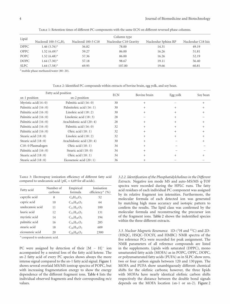
4 Journal of Biomedicine and Biotechnology
Table 1: Retention times of different PC-components with the same ECN on different reversed phase columns.
LipidColumn type
Nucleosil 100-5 C6H5 Nucleosil 100-5 C18 Nucleodur C18 Gravity Nucleodur Sphinx RP Nucleodur C18 Isis
DPPC 1.46 (5.76)∗ 56.82 78.00 14.31 49.19
OPPC 1.52 (6.49)∗ 59.27 86.00 16.26 51.81
POPC 1.52 (6.48)∗ 57.36 86.00 16.26 52.19
DOPC 1.64 (7.30)∗ 57.18 98.67 19.11 56.40
SLPC 1.64 (7.58)∗ 69.95 107.00 19.66 60.81∗mobile phase methanol/water (80 : 20).
Table 2: Identified PC compounds within extracts of bovine brain, egg yolk, and soy bean.
Fatty acid positionECN Bovine brain Egg yolk Soy bean
sn-1 position sn-2 position
Myristic acid(14 : 0) Palmitic acid (16 : 0) 30 + − +
Palmitic acid (16 : 0) Palmitoleic acid (16 : 1) 30 + + +
Palmitic acid (16 : 0) Linoleic acid (18 : 2) 30 + + +
Palmitic acid (16 : 0) Linolenic acid (18 : 3) 28 − + −Palmitic acid (16 : 0) Arachidonic acid (20 : 4) 28 + + +
Palmitic acid (16 : 0) Palmitic acid (16 : 0) 32 + + +
Palmitic acid (16 : 0) Oleic acid (18 : 1) 32 + + +
Stearic acid (18 : 0) Linoleic acid (18 : 2) 32 + + +
Stearic acid (18 : 0) Arachidonic acid (20 : 4) 30 + − +
C18 : 0 Plasmalogen Oleic acid (18 : 1) 34 + − −Palmitic acid (16 : 0) Stearic acid (18 : 0) 34 + − −Stearic acid (18 : 0) Oleic acid (18 : 1) 34 + + +
Stearic acid (18 : 0) Eicosenoic acid (20 : 1) 36 + − −
Table 3: Electrospray ionization efficiency of different fatty acidcompared to undecanoic acid (pKs = 4,69 for all acids).
Fatty acid Number ofcarbons
Empiricalformula
Ionisationefficiency∗ (%)
caprylic acid 8 C8H16O2 52
capric acid 10 C10H20O2 64
undecanoic acid 11 C11H22O2 100
lauric acid 12 C12H24O2 131
myristic acid 14 C14H28O2 194
palmitic acid 16 C16H32O2 285
stearic acid 18 C18H36O2 609
eicosanoic acid 20 C20H40O2 2300∗compared to undecanoic acid.
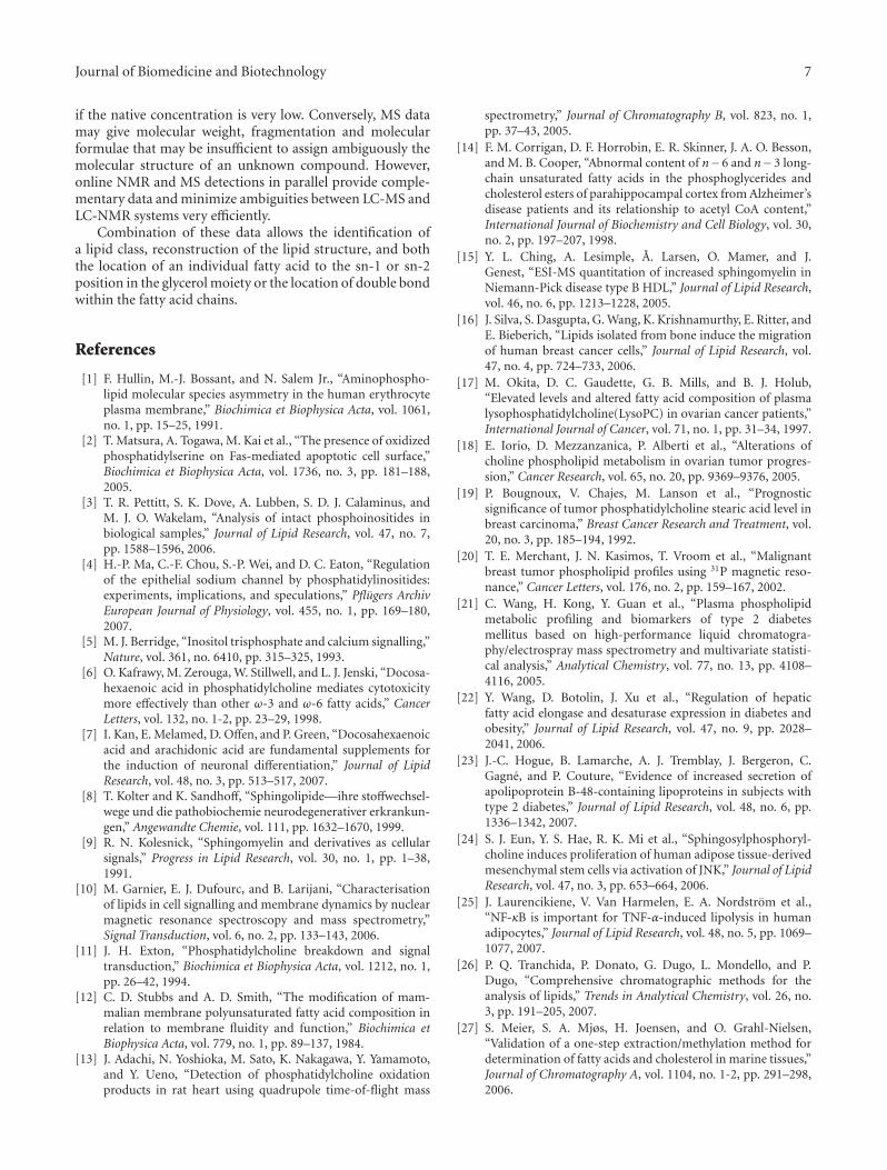
PC were assigned by detection of their [M − H]− ionaccompanied by a neutral loss of the fatty acid ketene. Thesn-2 fatty acid of every PC species shows always the moreintense signal compared to the sn-1 fatty acid signal. Figure 1shows several overlaid MS/MS iontrap spectra of POPC, butwith increasing fragmentation energy to show the energydependence of the different fragment ions. Table 4 lists theindividual observed fragments and their corresponding m/zvalues.
3.2.2. Identification of the Phosphatidylcholines in the DifferentExtracts. Negative ion mode MS and auto-MS/MS q-TOFspectra were recorded during the HPLC runs. The fattyacid residues of each individual PC component was assignedby its relative fragment ion intensities. Furthermore, themolecular formula of each detected ion was generatedby matching high mass accuracy and isotopic pattern toconfirm the results. The lipid class was confirmed by themolecular formula and reconstructing the precursor ionof the fragment ions. Table 2 shows the indentified specieswithin the three different extracts.
3.3. Nuclear Magnetic Resonance. 1D-(1H and 13C) and 2D-(HSQC, HSQC-TOCSY, and HMBC) NMR spectra of thefive reference PCs were recorded for peak assignment. TheNMR parameters of all reference compounds are listedin the supplements. Lipids with saturated (DPPC), monounsaturated fatty acids (MOFA) as in POPC, OPPC, DOPC,or polyunsaturated fatty acids (PUFA) as in SLPC show zero,two or four carbon signals between 120 and 130 ppm. TheMOFA and PUFA show unambiguously different chemicalshifts for the olefinic carbons; however, the three lipidswith MOFAs have nearly identical olefinic carbon shiftsrespectively the distance between the double bond signalsdepends on the MOFA location (sn-1 or sn-2). Figure 2

Journal of Biomedicine and Biotechnology 5
Table 4: Assignment of the individual observed fragments of POPC.
Fragment Assignment m/z
A palmitic acid [M −H]− 255.0
B oleic acid [M − H]− 281.0
C
HC
HC
H2C CH3
CH3
CH3
O−
O
O
O
OOP N
462.1
D HC
H2C
H2C
CH3
CH3
CH3O
O
O
OOP N
OH
O−
480.1
EHC
H2C
H2C
CH3
CH3
CH3O−
O
OO
OO
P N
OH
506.1
F HC
H2C
H2C
CH3
CH3
CH3
CH3
OO
O
OO
OO
P N
O−
744.4
129.6 129.5 129.4 129.3
(ppm)
Figure 2: Overlaid 13C-NMR spectral sections of olefinic carbons ofPCs with MOFAs (top OPPC, middle POPC, and bottom DOPC).
shows the overlaid 13C NMR spectra of POPC, OPPC, andDOPC. The chemical shift difference between these twosignals is about 50 Hz for POPC and about 47 Hz for OPPC.The corresponding 1H-NMR spectra show identical signalsfor the olefinic protons (not shown). Nonetheless, a lipidwith two MOFAs can be deduced from the intensity ratio of
the olefinic protons to the glycerol protons in the 1H-NMRspectra.
3.3.1. NMR of the PC Extracts. HPLC-MS structural resultswere confirmed for all molecular species using 1D-(1H) and2D-(HSQC, HSQC-TOCSY, and HMBC) NMR techniques.The results are shown in Table 2. No branched chain oroxidized fatty acids were observed. The NMR parameter ofunsaturated fatty acids have been measured separately (seeSupplement) and are identical within the mixtures.
4. Discussion
Lipids with equal ECN should have the same hydropho-bicity, which is the discriminating factor in reversed phasechromatography. This holds for PCs with the same ECNwithin a mixture and even more, if they are stereoisomerssuch as POPC and OPPC. The successful reversed phaseseparation of hydrophobic and zwitterionic molecules likephosphatidylcholines depends on very well endcapped silicamaterials, as Coulomb interactions of the choline group

6 Journal of Biomedicine and Biotechnology
with the silica gel lead to peak broadening, retention timeshifts (data not shown), and peak tailing. Packing materialswith hydrophobic van der Waals interactions only (i.e., highdensity C18 materials) show no separation of isomeric lipids.Additional interactions like steric effects by polymer cross-linked RP packings can overcome this problem. Stationaryphases with only aromatic modifications are not hydropho-bic enough to achieve a good phospholipid separation. Themixed mode stationary phase contains additionally alkylligands and offers therefore more hydrophobic interactionsand good silica gel coverage. This column showed theshortest phospholipid retention times for all alkyl stationaryphases and a good separation with narrow chromatographicpeaks. Saturated lipids have the lowest and polyunsaturatedlipids the highest retention times. Comparing lipids of thesame ECN with two MOFAs or one PUFA and one saturatedfatty acid the retention of the lipid with the PUFA is higher, astheir spatial demand is larger and the π-π interactions with aPUFA residue is not affected by the other fatty acid.
4.1. Separation of the Phosphatidylcholine Extracts. By meansof the HPLC separation it was possible to get semiquan-titative information of the individual compounds in theextracts. Referring to the results of the separation of thelipid standard mixture it was possible to separate all specieswithin the extracts. The elution order of lipids with the sameECN is in the same line as for the reference mixture (lipidswith saturated fatty acids, one MOFA, two MOFAs, onePUFA). However, in case of PUFAs with four double bonds(i.e., arachidonic acid; 20 : 4) the π-π interactions becomeso strong that this compound elutes as last compoundin the next higher ECN group (see Table 2). Nonetheless,they are unambiguously identified by means of their MSfragmentation or NMR spectra.
4.2. Preparation of the Individual Compounds for NMRMeasurement. The upscaling with the same type of RPpacking of the newest generation is no problem, althougha peak broadening can occur because of the higher sampleload.
4.3. Ion Suppression Effects. The ionization efficiency of thedifferent fatty acids during ESI increases nonlinearly. Theresults can be correlated with the molecules hydrophobicity,which shows the same progression. The hydrophobicityis obtained from the octanol-water partitioning coefficient(log pow) (data not shown). There are no differences betweenthe pKa values of the individual fatty acids (Table 3), sothat the ionization efficiency of the fatty acids depends onlyon the molecules hydrophobicity, which increases by thenumber of carbon atoms.
As POPC and OPPC or SLPC and DOPC, respectively,have the same molecular weight/precision mass, they cannotbe assigned based on their mol peak only. However, theyare distinguished by MS/MS spectra, as the sn-2 fatty acidis always cleaved off at lower fragmentation energies due tosterical effects. In addition, the intensity ratio of the sn-2fatty acid anion to the sn-1 fatty acid anion is constant
for a particular fragmentation energy. A semiquantitativeor even quantitative analysis of stereoisomeric PCs withina mixture (f.e., POPC and OPPC) still is difficult, becausethe anion signal of an individual fatty acid will be the sameregardless the position on the glycerol backbone and noother distinguishing signal is observed for one of the isomers.However, using the intensity ratio of the sn-2 fatty acid anionto the sn-1 fatty acid anion for the pure reference compoundone can calculate the approximate POPC to OPPC ratio.
The MS detection limits also benefits from the isocraticHPLC method with high-organic solvent concentration inthe mobile phase. In case of gradient HPLC, methodsthe ionization efficiency varies with the varying organicpercentage of the mobile phase. Furthermore, the risk of ionsuppression during the ionization process is minimized bythe sample introduction after HPLC separation.
The identification of the individual species in the dif-ferent extracts was achieved by MS/MS and comparing theindividual fragment intensities as described earlier. The useof high-resolution spectra acquired by the micro-TOF-Qallows the generation of the compound’s molecular formulaby matching high mass accuracy and isotopic pattern.Abnormalities like oxidation of the double bond, and soforth. were not observed.
It may be noted, that not only diacyl PLs can be identifiedwithin a mixture, but also alkyl/alkenyl, acyl PLs because ofthe different fragmentation pattern of the fatty acid residuecompared to an ether link.
The HPLC and MS results were confirmed by NMRspectroscopy, especially the configuration and location ofdouble bonds in the fatty acid residues. Only NMR havingthe highest qualitative and quantitative structure elucidationpotential allows a complete structure elucidation. 1H-, 13C-,or 31P-NMR spectra are capable to identify the various lipidclasses. Beyond this, the degree of unsaturation is obtainedfrom the proton signal intensity ratio of the double bondsignal versus the choline group signal. MOFAs and PUFAsare differentiated by the number of carbon signals withinthe double bond region. The location (sn-1 or sn-2) of theMOFA follows from the 13C-NMR spectra or from ESI-MS.The risk of peak overlapping in the NMR spectra was avoidedby recording 2D-NMR spectra by preceding separation ofindividual phosphatidylcholines by HPLC.
5. Conclusion
The separation of lipids with equivalent chain lengths incomplex mixtures can be improved using RP-HPLC packingsof the newer generation containing additional discrimina-tors. Already simple PC mixture cannot be assigned bya single analytical technique, while the combination ofHPLC separation power, MS sensitivity with accurate massmeasurement of molecular and fragment ions and NMRstructure elucidation power will meet most suitably thechallenge. They overcome the limits of any single techniqueand also proof the potential of their combination ultimatelyto analyze native (lipid) mixtures. The molecular structureof a novel compound may not be evaluated by NMR alone,
Journal of Biomedicine and Biotechnology 7
if the native concentration is very low. Conversely, MS datamay give molecular weight, fragmentation and molecularformulae that may be insufficient to assign ambiguously themolecular structure of an unknown compound. However,online NMR and MS detections in parallel provide comple-mentary data and minimize ambiguities between LC-MS andLC-NMR systems very efficiently.
Combination of these data allows the identification ofa lipid class, reconstruction of the lipid structure, and boththe location of an individual fatty acid to the sn-1 or sn-2position in the glycerol moiety or the location of double bondwithin the fatty acid chains.
References
[1] F. Hullin, M.-J. Bossant, and N. Salem Jr., “Aminophospho-lipid molecular species asymmetry in the human erythrocyteplasma membrane,” Biochimica et Biophysica Acta, vol. 1061,no. 1, pp. 15–25, 1991.
[2] T. Matsura, A. Togawa, M. Kai et al., “The presence of oxidizedphosphatidylserine on Fas-mediated apoptotic cell surface,”Biochimica et Biophysica Acta, vol. 1736, no. 3, pp. 181–188,2005.
[3] T. R. Pettitt, S. K. Dove, A. Lubben, S. D. J. Calaminus, andM. J. O. Wakelam, “Analysis of intact phosphoinositides inbiological samples,” Journal of Lipid Research, vol. 47, no. 7,pp. 1588–1596, 2006.
[4] H.-P. Ma, C.-F. Chou, S.-P. Wei, and D. C. Eaton, “Regulationof the epithelial sodium channel by phosphatidylinositides:experiments, implications, and speculations,” Pflugers ArchivEuropean Journal of Physiology, vol. 455, no. 1, pp. 169–180,2007.
[5] M. J. Berridge, “Inositol trisphosphate and calcium signalling,”Nature, vol. 361, no. 6410, pp. 315–325, 1993.
[6] O. Kafrawy, M. Zerouga, W. Stillwell, and L. J. Jenski, “Docosa-hexaenoic acid in phosphatidylcholine mediates cytotoxicitymore effectively than other ω-3 and ω-6 fatty acids,” CancerLetters, vol. 132, no. 1-2, pp. 23–29, 1998.
[7] I. Kan, E. Melamed, D. Offen, and P. Green, “Docosahexaenoicacid and arachidonic acid are fundamental supplements forthe induction of neuronal differentiation,” Journal of LipidResearch, vol. 48, no. 3, pp. 513–517, 2007.
[8] T. Kolter and K. Sandhoff, “Sphingolipide—ihre stoffwechsel-wege und die pathobiochemie neurodegenerativer erkrankun-gen,” Angewandte Chemie, vol. 111, pp. 1632–1670, 1999.
[9] R. N. Kolesnick, “Sphingomyelin and derivatives as cellularsignals,” Progress in Lipid Research, vol. 30, no. 1, pp. 1–38,1991.
[10] M. Garnier, E. J. Dufourc, and B. Larijani, “Characterisationof lipids in cell signalling and membrane dynamics by nuclearmagnetic resonance spectroscopy and mass spectrometry,”Signal Transduction, vol. 6, no. 2, pp. 133–143, 2006.
[11] J. H. Exton, “Phosphatidylcholine breakdown and signaltransduction,” Biochimica et Biophysica Acta, vol. 1212, no. 1,pp. 26–42, 1994.
[12] C. D. Stubbs and A. D. Smith, “The modification of mam-malian membrane polyunsaturated fatty acid composition inrelation to membrane fluidity and function,” Biochimica etBiophysica Acta, vol. 779, no. 1, pp. 89–137, 1984.
[13] J. Adachi, N. Yoshioka, M. Sato, K. Nakagawa, Y. Yamamoto,and Y. Ueno, “Detection of phosphatidylcholine oxidationproducts in rat heart using quadrupole time-of-flight mass
spectrometry,” Journal of Chromatography B, vol. 823, no. 1,pp. 37–43, 2005.
[14] F. M. Corrigan, D. F. Horrobin, E. R. Skinner, J. A. O. Besson,and M. B. Cooper, “Abnormal content of n−6 and n−3 long-chain unsaturated fatty acids in the phosphoglycerides andcholesterol esters of parahippocampal cortex from Alzheimer’sdisease patients and its relationship to acetyl CoA content,”International Journal of Biochemistry and Cell Biology, vol. 30,no. 2, pp. 197–207, 1998.
[15] Y. L. Ching, A. Lesimple, A. Larsen, O. Mamer, and J.Genest, “ESI-MS quantitation of increased sphingomyelin inNiemann-Pick disease type B HDL,” Journal of Lipid Research,vol. 46, no. 6, pp. 1213–1228, 2005.
[16] J. Silva, S. Dasgupta, G. Wang, K. Krishnamurthy, E. Ritter, andE. Bieberich, “Lipids isolated from bone induce the migrationof human breast cancer cells,” Journal of Lipid Research, vol.47, no. 4, pp. 724–733, 2006.
[17] M. Okita, D. C. Gaudette, G. B. Mills, and B. J. Holub,“Elevated levels and altered fatty acid composition of plasmalysophosphatidylcholine(LysoPC) in ovarian cancer patients,”International Journal of Cancer, vol. 71, no. 1, pp. 31–34, 1997.
[18] E. Iorio, D. Mezzanzanica, P. Alberti et al., “Alterations ofcholine phospholipid metabolism in ovarian tumor progres-sion,” Cancer Research, vol. 65, no. 20, pp. 9369–9376, 2005.
[19] P. Bougnoux, V. Chajes, M. Lanson et al., “Prognosticsignificance of tumor phosphatidylcholine stearic acid level inbreast carcinoma,” Breast Cancer Research and Treatment, vol.20, no. 3, pp. 185–194, 1992.
[20] T. E. Merchant, J. N. Kasimos, T. Vroom et al., “Malignantbreast tumor phospholipid profiles using 31P magnetic reso-nance,” Cancer Letters, vol. 176, no. 2, pp. 159–167, 2002.
[21] C. Wang, H. Kong, Y. Guan et al., “Plasma phospholipidmetabolic profiling and biomarkers of type 2 diabetesmellitus based on high-performance liquid chromatogra-phy/electrospray mass spectrometry and multivariate statisti-cal analysis,” Analytical Chemistry, vol. 77, no. 13, pp. 4108–4116, 2005.
[22] Y. Wang, D. Botolin, J. Xu et al., “Regulation of hepaticfatty acid elongase and desaturase expression in diabetes andobesity,” Journal of Lipid Research, vol. 47, no. 9, pp. 2028–2041, 2006.
[23] J.-C. Hogue, B. Lamarche, A. J. Tremblay, J. Bergeron, C.Gagne, and P. Couture, “Evidence of increased secretion ofapolipoprotein B-48-containing lipoproteins in subjects withtype 2 diabetes,” Journal of Lipid Research, vol. 48, no. 6, pp.1336–1342, 2007.
[24] S. J. Eun, Y. S. Hae, R. K. Mi et al., “Sphingosylphosphoryl-choline induces proliferation of human adipose tissue-derivedmesenchymal stem cells via activation of JNK,” Journal of LipidResearch, vol. 47, no. 3, pp. 653–664, 2006.
[25] J. Laurencikiene, V. Van Harmelen, E. A. Nordstrom et al.,“NF-κB is important for TNF-α-induced lipolysis in humanadipocytes,” Journal of Lipid Research, vol. 48, no. 5, pp. 1069–1077, 2007.
[26] P. Q. Tranchida, P. Donato, G. Dugo, L. Mondello, and P.Dugo, “Comprehensive chromatographic methods for theanalysis of lipids,” Trends in Analytical Chemistry, vol. 26, no.3, pp. 191–205, 2007.
[27] S. Meier, S. A. Mjøs, H. Joensen, and O. Grahl-Nielsen,“Validation of a one-step extraction/methylation method fordetermination of fatty acids and cholesterol in marine tissues,”Journal of Chromatography A, vol. 1104, no. 1-2, pp. 291–298,2006.

8 Journal of Biomedicine and Biotechnology
[28] M. P. Styczynski, J. F. Moxley, L. V. Tong, J. L. Walther, K. L.Jensen, and G. N. Stephanopoulos, “Systematic identificationof conserved metabolites in GC/MS data for metabolomicsand biomarker discovery,” Analytical Chemistry, vol. 79, no. 3,pp. 966–973, 2007.
[29] J. C. Touchstone, “Thin-layer chromatographic procedures forlipid separation,” Journal of Chromatography B, vol. 671, no. 1-2, pp. 169–195, 1995.
[30] J. K. Yao and G. M. Rastetter, “Microanalysis of complextissue lipids by high-performance thin-layer chromatography,”Analytical Biochemistry, vol. 150, no. 1, pp. 111–116, 1985.
[31] J. J. Myher and A. Kuksis, “General strategies in chromato-graphic analysis of lipids,” Journal of Chromatography B, vol.671, no. 1-2, pp. 3–33, 1995.
[32] C. Silversand and C. Haux, “Improved high-performanceliquid chromatographic method for the separation and quan-tification of lipid classes: application to fish lipids,” Journal ofChromatography B, vol. 703, no. 1-2, pp. 7–14, 1997.
[33] L. L. Dugan, P. Demediuk, C. E. Pendley II, and L. A. Horrocks,“Separation of phospholipids by high-performance liquidchromatography: all major classes, including ethanolamineand choline plasmalogens, and most minor classes, includinglysophosphatidylethanolamine,” Journal of Chromatography,vol. 378, no. 2, pp. 317–327, 1986.
[34] N. U. Olsson and N. Salem Jr., “Molecular species analysis ofphospholipids,” Journal of Chromatography B, vol. 692, no. 2,pp. 245–256, 1997.
[35] J. Becart, C. Chevalier, and J. P. Biesse, “Quantitative analysisof phospholipids by HPLC with light scattering evaporatingdetector—application to raw materials for cosmetic use,”Journal of High Resolution Chromatography, vol. 13, pp. 126–129, 1990.
[36] H.-Y. Kim, T.-C. L. Wang, and Y.-C. Ma, “Liquid chromatog-raphy/mass spectrometry of phospholipids using electrosprayionization,” Analytical Chemistry, vol. 66, no. 22, pp. 3977–3993, 1994.
[37] P. J. Kalo, V. Ollilainen, J. M. Rocha, and F. X. Malcata,“Identification of molecular species of simple lipids by normalphase liquid chromatography-positive electrospray tandemmass spectrometry, and application of developed methods incomprehensive analysis of low erucic acid rapeseed oil lipids,”International Journal of Mass Spectrometry, vol. 254, no. 1-2,pp. 106–121, 2006.
[38] B. Brugger, G. Erben, R. Sandhoff, F. T. Wieland, and W.D. Lehmann, “Quantitative analysis of biological membranelipids at the low picomole level by nano-electrospray ioniza-tion tandem mass spectrometry,” Proceedings of the NationalAcademy of Sciences of the United States of America, vol. 94, no.6, pp. 2339–2344, 1997.
[39] F. Sullentrop, D. Moka, S. Neubauer et al., “31P NMR spec-troscopy of blood plasma: determination and quantificationof phospholipid classes in patients with renal cell carcinoma,”NMR in Biomedicine, vol. 15, no. 1, pp. 60–68, 2002.
[40] J. M. Pearce and R. A. Komoroski, “Analysis of phospholipidmolecular species in brain by 31P NMR spectroscopy,” Mag-netic Resonance in Medicine, vol. 44, no. 2, pp. 215–223, 2000.
[41] J. Schille and K. Arnold, “Application of high resolution 31PNMR spectroscopy to the characterization of the phospholipidcomposition of tissues and body fluids—a methodologicalreview,” Medical Science Monitor, vol. 8, no. 11, pp. 205–222,2002.
[42] N. M. Loening, A. M. Chamberlin, A. G. Zepeda, R. G. Gonza-lez, and L. L. Cheng, “Quantification of phosphocholine and
glycerophosphocoline with 31P edited 1H NMR spectroscopy,”NMR in Biomedicine, vol. 18, no. 7, pp. 413–420, 2005.
[43] J. Schiller, M. Muller, B. Fuchs, K. Arnold, and D. Huster,“31P NMR spectroscopy of phospholipids: from micelles tomembranes,” Current Analytical Chemistry, vol. 3, pp. 283–301, 2007.
[44] D. Leibfritz, “An introduction to the potential of 1H-, 31P-and 13C-NMR-spectroscopy,” Anticancer Research, vol. 16, pp.1317–1324, 1996.
[45] J. Henke, J. Engelmann, B. Kutscher et al., “Changes ofintracellular calcium, fatty acids and phospholipids duringMiltefosine-induced apoptosis monitored by fluorescence-and 13C NMR-spectroscopy,” Anticancer Research, vol. 19, no.5, pp. 4027–4032, 1999.
[46] J. Henke, W. Willker, J. Engelmann, and D. Leibfritz,“Combined extraction techniques of tumour cells and lipid-phospholipid assignment by two dimensional NMR spec-troscopy,” Anticancer Research, vol. 16, no. 3, pp. 1417–1427,1996.
[47] W. Willker and D. Leibfritz, “Assignment of mono- andpolyunsaturated fatty acids in lipids of tissues and body fluids,”Magnetic Resonance in Chemistry, vol. 36, no. 998, pp. S79–S84, 1998.
[48] T. F. Bathen, J. Krane, T. Engan, K. S. Bjerve, and D. Axelson,“Quantification of plasma lipids and apolipoproteins by use ofproton NMR spectroscopy, multivariate and neural networkanalysis,” NMR in Biomedicine, vol. 13, no. 5, pp. 271–288,2000.
[49] J. Willmann, K. Mahlstedt, D. Leibfritz, M. Spraul, and H.Thiele, “Characterization of sphingomyelins in lipid extractsusing a HPLC-MS-offline-NMR method,” Analytical Chem-istry, vol. 79, no. 11, pp. 4188–4191, 2007.

Submit your manuscripts athttp://www.hindawi.com
Hindawi Publishing Corporationhttp://www.hindawi.com Volume 2014
Anatomy Research International
PeptidesInternational Journal of
Hindawi Publishing Corporationhttp://www.hindawi.com Volume 2014
Hindawi Publishing Corporation http://www.hindawi.com
International Journal of
Volume 2014
Zoology
Hindawi Publishing Corporationhttp://www.hindawi.com Volume 2014
Molecular Biology International
GenomicsInternational Journal of
Hindawi Publishing Corporationhttp://www.hindawi.com Volume 2014
The Scientific World JournalHindawi Publishing Corporation http://www.hindawi.com Volume 2014
Hindawi Publishing Corporationhttp://www.hindawi.com Volume 2014
BioinformaticsAdvances in
Marine BiologyJournal of
Hindawi Publishing Corporationhttp://www.hindawi.com Volume 2014
Hindawi Publishing Corporationhttp://www.hindawi.com Volume 2014
Signal TransductionJournal of
Hindawi Publishing Corporationhttp://www.hindawi.com Volume 2014
BioMed Research International
Evolutionary BiologyInternational Journal of
Hindawi Publishing Corporationhttp://www.hindawi.com Volume 2014
Hindawi Publishing Corporationhttp://www.hindawi.com Volume 2014
Biochemistry Research International
ArchaeaHindawi Publishing Corporationhttp://www.hindawi.com Volume 2014
Hindawi Publishing Corporationhttp://www.hindawi.com Volume 2014
Genetics Research International
Hindawi Publishing Corporationhttp://www.hindawi.com Volume 2014
Advances in
Virolog y
Hindawi Publishing Corporationhttp://www.hindawi.com
Nucleic AcidsJournal of
Volume 2014
Stem CellsInternational
Hindawi Publishing Corporationhttp://www.hindawi.com Volume 2014
Hindawi Publishing Corporationhttp://www.hindawi.com Volume 2014
Enzyme Research
Hindawi Publishing Corporationhttp://www.hindawi.com Volume 2014
International Journal of
Microbiology