common pediatric dermatology diagnoses to red flat patch enlarges proportionately with overall...
TRANSCRIPT
©2013 Children's Mercy. All Rights Reserved. 09/13 ©2013 Children's Mercy. All Rights Reserved. 09/13
Kimberly A. Horii MD Associate Professor of Pediatrics
Division of Dermatology Children’s Mercy
Kansas City, Missouri
Common Pediatric Dermatology Diagnoses

Common Pediatric Dermatology Diagnoses
I have no financial relationships with the manufacturers of any commercial products and/or provider of commercial services discussed in this CME activity I do intend to discuss off label use of a
commercial product/device in my presentation

Common Pediatric Dermatology Diagnoses
Vascular Birthmarks Port wine stains
Infantile hemangiomas
Dermatitis Seborrheic dermatitis
Diaper dermatitis
Atopic dermatitis
Impetigo
Common Pediatric Dermatology Diagnoses
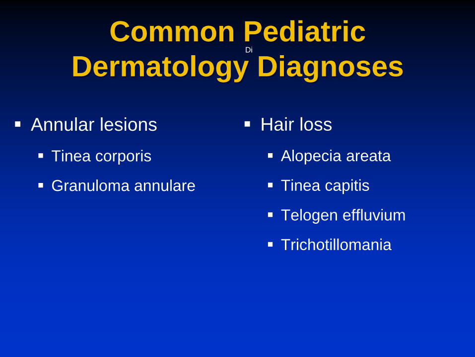
Annular lesions Tinea corporis
Granuloma annulare
Di
Hair loss Alopecia areata
Tinea capitis
Telogen effluvium
Trichotillomania
Classification of Vascular Lesions
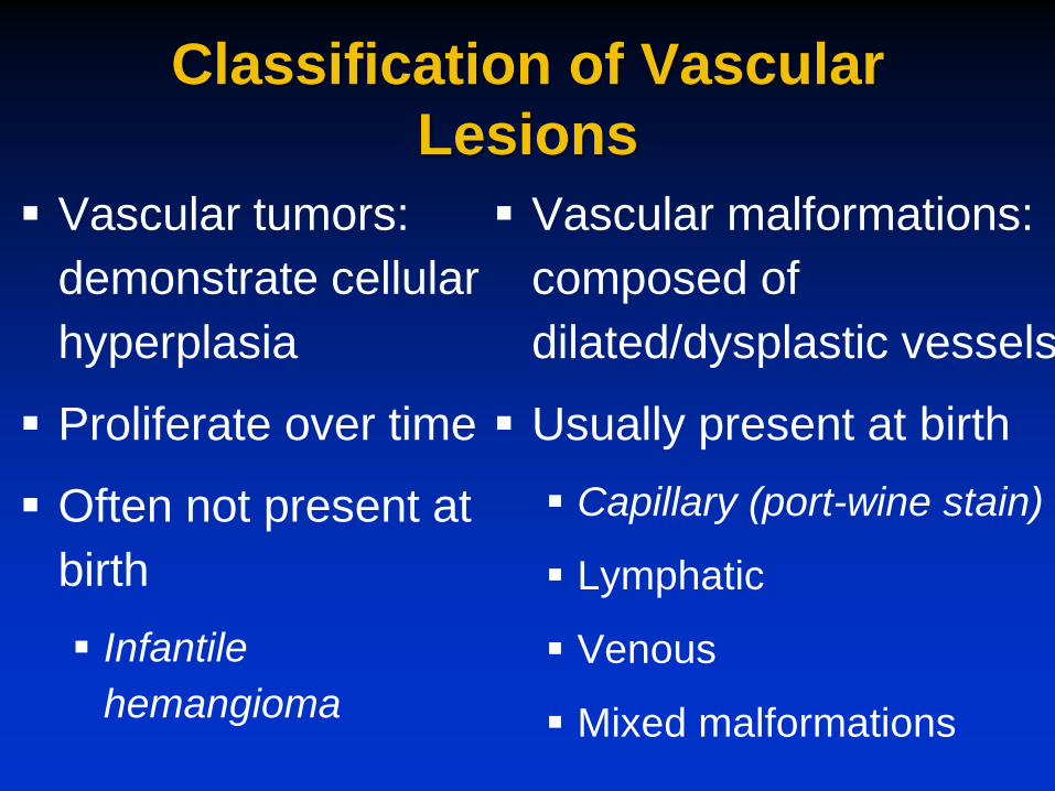
Vascular tumors: demonstrate cellular hyperplasia
Proliferate over time
Often not present at birth Infantile
hemangioma
Vascular malformations: composed of dilated/dysplastic vessels
Usually present at birth Capillary (port-wine stain)
Lymphatic
Venous
Mixed malformations
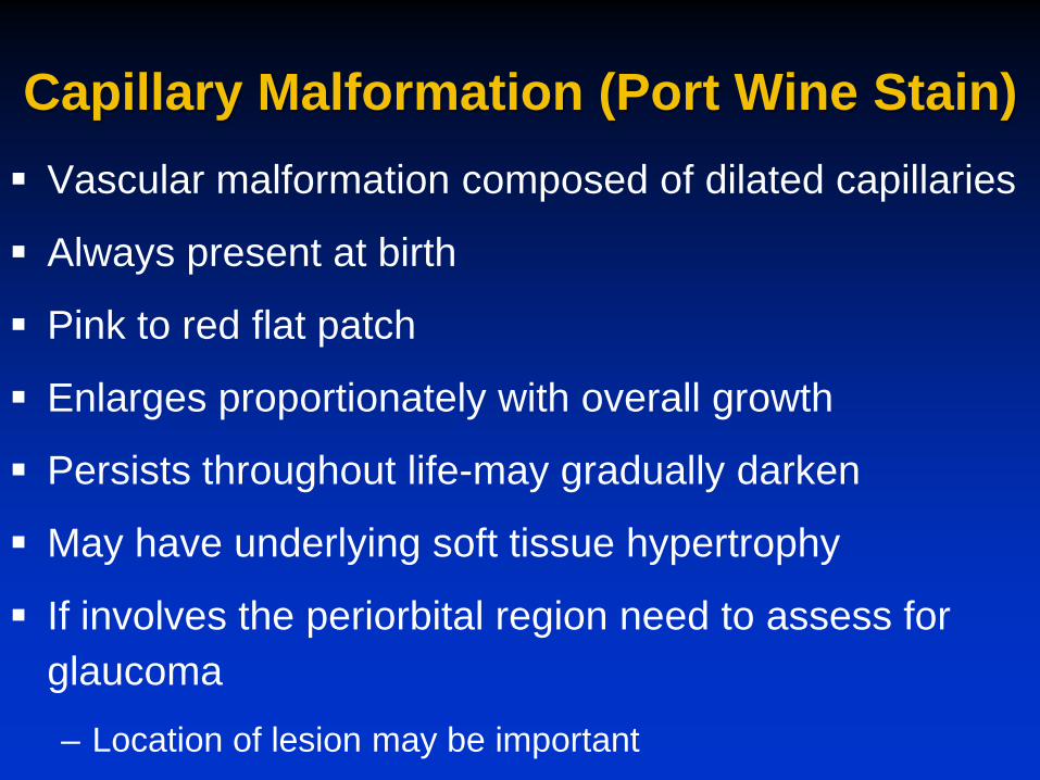
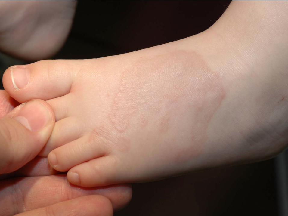
Capillary Malformation (Port Wine Stain) Vascular malformation composed of dilated capillaries
Always present at birth
Pink to red flat patch
Enlarges proportionately with overall growth
Persists throughout life-may gradually darken
May have underlying soft tissue hypertrophy
If involves the periorbital region need to assess for glaucoma – Location of lesion may be important

Sturge-Weber Syndrome Classic triad Facial capillary malformation (port-wine stain) in trigeminal
nerve (V1) distribution
Higher risk if bilateral V1 involvement
Ipsilateral eye findings: glaucoma
Leptomeningeal and brain anomalies: leptomeningeal vascular malformation, calcifications
May develop seizures and developmental delay
Ophthalmologic exam and possible brain MRI

Infantile Hemangiomas Common vascular birthmark Affects approximately 4-5% of all infants
–More common in females –Not usually present at birth
Tumor composed of proliferating endothelial cells Rapid growth phase occurs during the
first 2-6 months of life –Some high risk lesions may require
treatment during the proliferative phase Gradual regression over many years
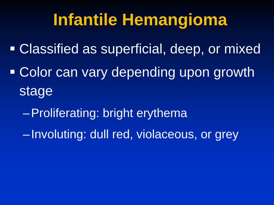
Infantile Hemangioma Classified as superficial, deep, or mixed
Color can vary depending upon growth stage –Proliferating: bright erythema
–Involuting: dull red, violaceous, or grey

Superficial Hemangioma
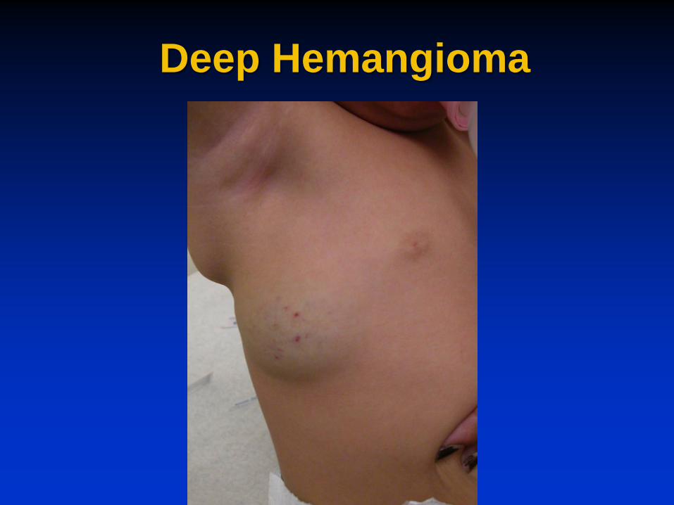
Deep Hemangioma

Mixed Hemangioma

Segmental Hemangiomas Newer classification system divides into
localized, segmental, or indeterminate
Segmental lesions defined as covering an “anatomic territory” – Can be linear/geometric
– Usually large and plaque-like
Lesion morphology may help predict outcome – Segmental lesions had poorer outcome
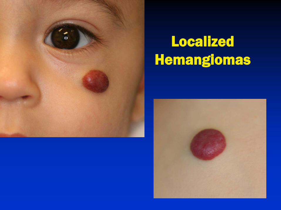
Localized Hemangiomas
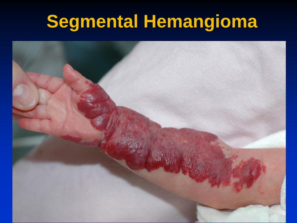
Segmental Hemangioma
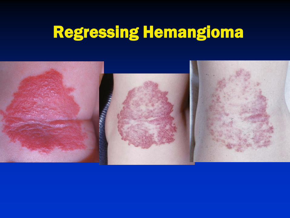
Regressing Hemangioma

Which to Worry About? Location Counts!
Segmental
Increased complications & ulceration
Large facial: PHACE syndrome
“Beard” distribution
Airway involvement
Periocular
Visual axis impairment
Multiple >5
Possible extracutaneous invovlement
Lumbosacral
Spinal dysraphism
Perineal & Perioral
Risk of ulceration
Pain & infection
Feeding issues
Disfiguring locations
Central face, nasal tip, perioral, & ear

PHACE Syndrome (OMIM 606519) Segmental facial hemangioma and ≥ 1
extracutaneous manifestation
Posterior fossa brain malformations Hemangioma- segmental facial or cervicofacial Arterial anomalies-cervical or cerebral arteries Cardiac defects-coarctation of aorta Eye abnormalities (S)ternal defects or supraumbilical raphe
Indications for Treatment Majority of hemangiomas do not require
treatment
Need for treatment depends upon – Functional compromise of vital structures
– Rate of growth
– Secondary complications Ulceration
– Location Risk of disfigurement

Active Non-Intervention A technique for small lesions with good
prognosis for spontaneous resolution
Actively discuss expectations with the parents
Close observation
Discuss possible complications – Ulceration
Patients with “high risk” hemangiomas should be followed closely during the early rapid growth period
Treatment of Infantile Hemangiomas Treatment may be required for life or function
threatening complications or risk of disfigurement
Treatment should ideally be instituted during the early proliferative stage
No FDA approved treatment for hemangiomas – Oral propranolol
– Topical timolol
– Oral steroids
– Intralesional steroids
– Surgery
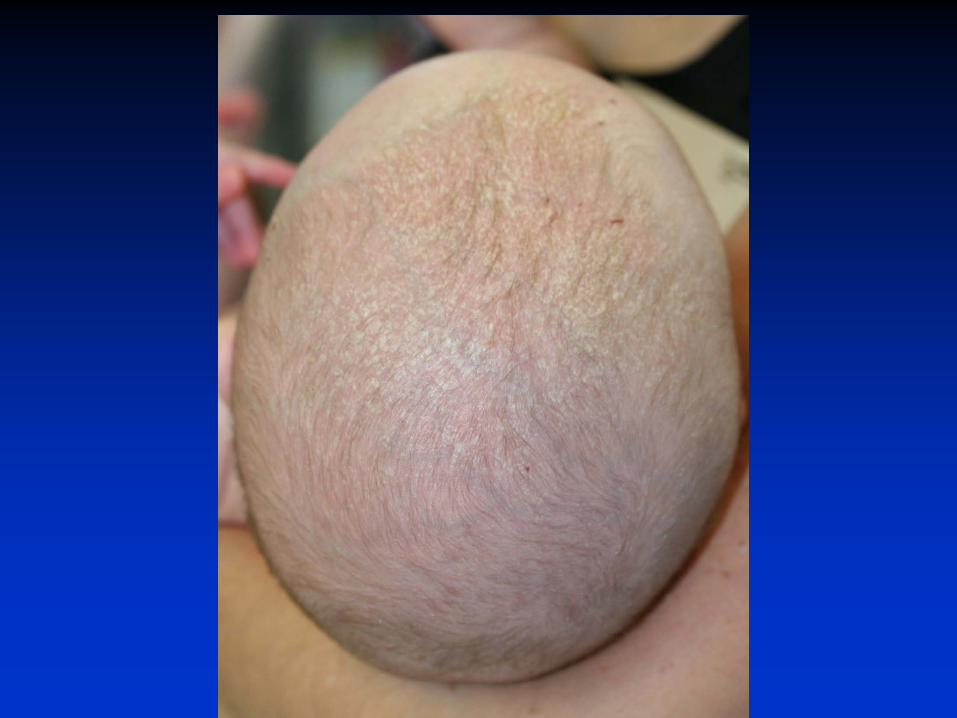
Seborrheic Dermatitis Commonly occurs in infants within the first
three months of life Also known as “cradle cap” May be associated with proliferation of
Malassezia species (yeast) Thick greasy scale on the scalp Greasy erythema and mild scaling behind the
ears, on the central face, flexural folds, & diaper area Usually non-pruritic & self limited
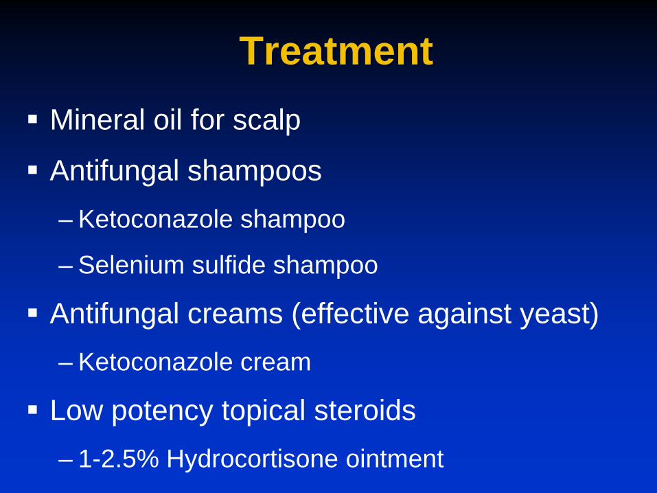
Treatment Mineral oil for scalp
Antifungal shampoos – Ketoconazole shampoo
– Selenium sulfide shampoo
Antifungal creams (effective against yeast) – Ketoconazole cream
Low potency topical steroids – 1-2.5% Hydrocortisone ointment

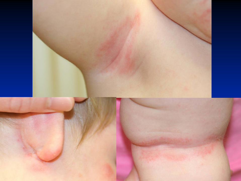
Diaper Dermatitis Common dermatologic problem in infancy Variants of diaper dermatitis
– Common Irritant contact diaper dermatitis Infectious-candida, staph, strep Seborrheic dermatitis
– Uncommon Psoriasis Zinc deficiency Histiocytosis
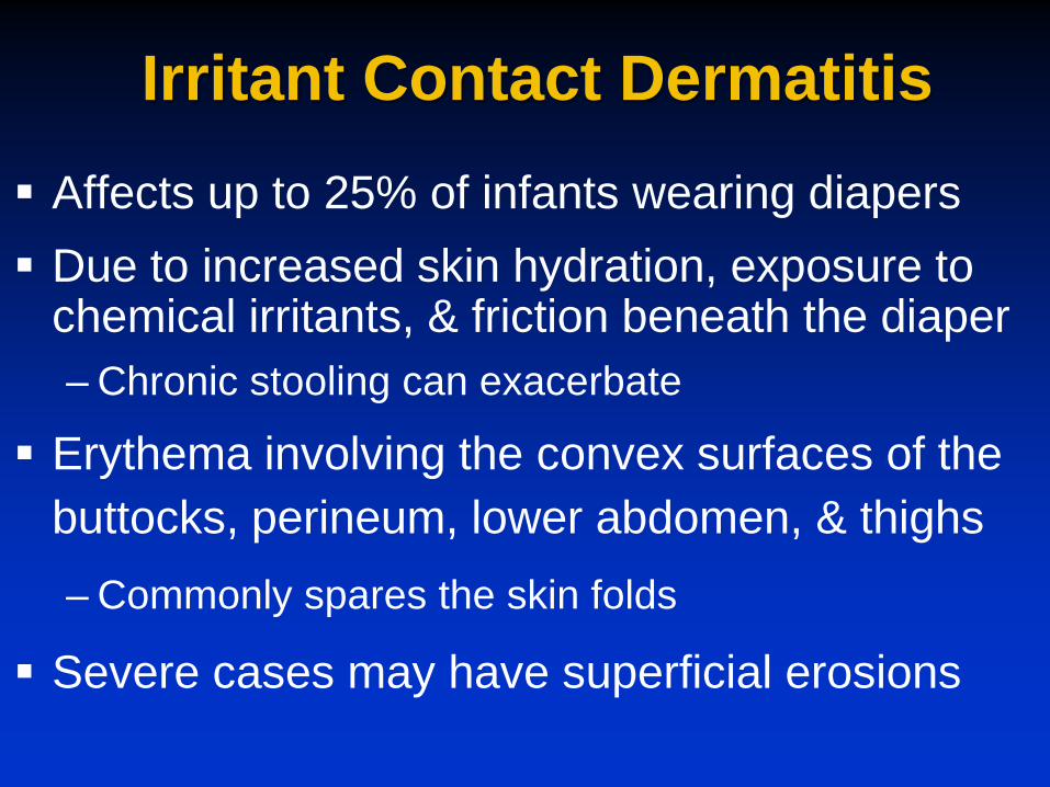
Irritant Contact Dermatitis Affects up to 25% of infants wearing diapers Due to increased skin hydration, exposure to
chemical irritants, & friction beneath the diaper – Chronic stooling can exacerbate
Erythema involving the convex surfaces of the buttocks, perineum, lower abdomen, & thighs – Commonly spares the skin folds
Severe cases may have superficial erosions

Treatment of Irritant Contact Dermatitis
Frequent diaper changes Gentle cleansing Topical barriers ointments/cream applied
thickly – Zinc oxide – White petrolatum

Treatment of Infectious Diaper Dermatitis
Candidal diaper dermatitis – Antifungal cream (effective for yeast) or ointment Azoles (clotrimazole, miconazole, ketoconazole)
Impetigo – Topical or oral antibiotics
Perianal Strep – Oral antibiotic
All types benefit from topical barriers .

Atopic Dermatitis Also known as eczema or “the itch that
rashes” Affects at least 10-15% of children &
adolescents Chronic inflammatory skin disease Characterized by
– Xerosis – Pruritus – Recurrent skin lesions in a specific distribution
Usually strong family history of “atopy”

Atopic Dermatitis Atopic dermatitis is often the first manifestation of
atopy
– Approximately 80% of children with atopic dermatitis will develop asthma or allergic rhinitis
Onset usually within the first 5 years of life
– 60% of cases begin by 1 year of age
– 90% of cases begin by 5 years of age
Prevalence decreases with age, though persistent or recurrent disease is common

Phases of Atopic Dermatitis
Infantile phase (1 month-18 months)
Childhood phase (18 months-puberty)
Adolescent and adult phase
Infantile Phase Features
Xerosis and evidence of pruritus
Dermatitis begins on the cheeks or scalp
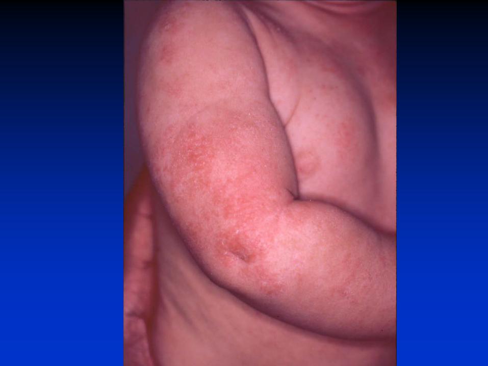
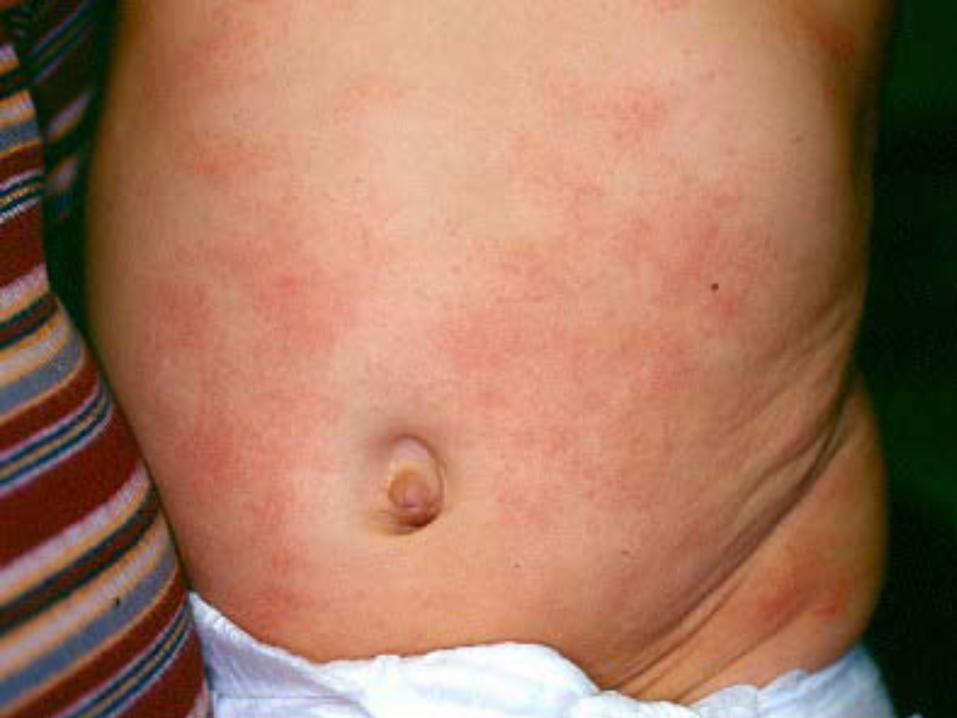
Eventually involves the trunk and extensor extremities
Diaper area usually spared
“Rubbing” of face

©2013 Children's Mercy. All Rights Reserved. 09/13


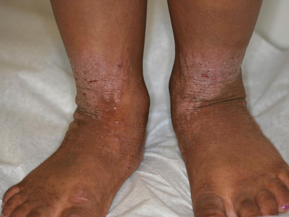
Childhood/Adolescent Phase Features
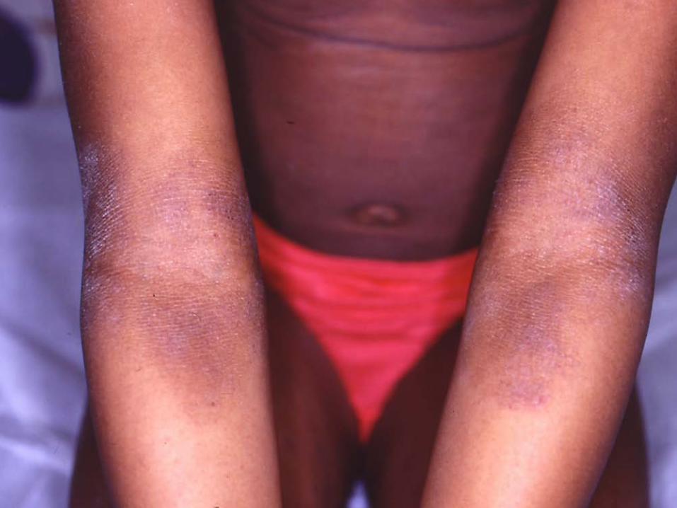
Chronic dermatitis Lichenification and excoriation Flexural surfaces of neck, arms,
wrists, ankles, and legs Complaints of pruritus


Atopic Dermatitis Treatment Appropriate skin care regimen Eliminate or avoid triggering agents Treat
–Active inflammation (red, rough areas) –Secondary infection –Pruritus
Extensive patient & family education
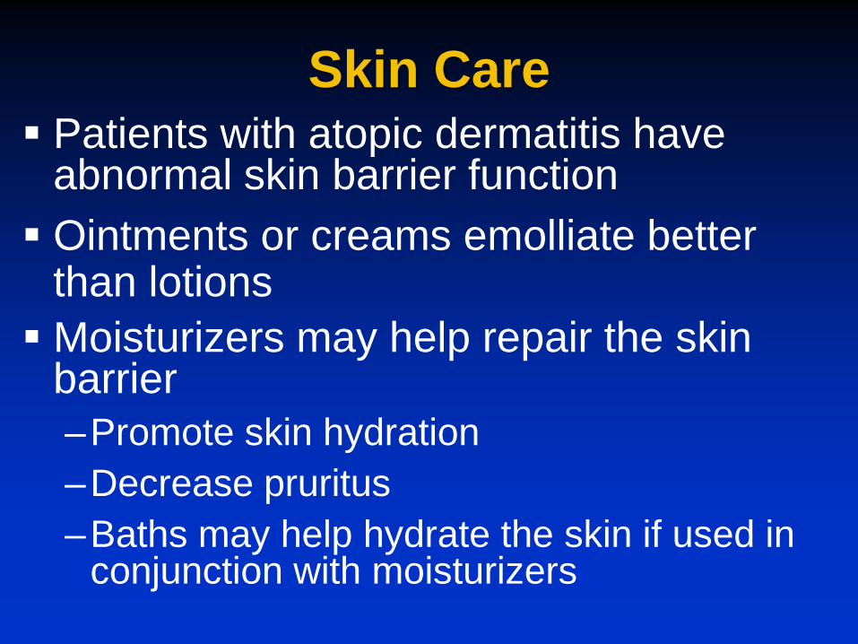
Skin Care Patients with atopic dermatitis have
abnormal skin barrier function Ointments or creams emolliate better
than lotions Moisturizers may help repair the skin
barrier –Promote skin hydration –Decrease pruritus –Baths may help hydrate the skin if used in
conjunction with moisturizers
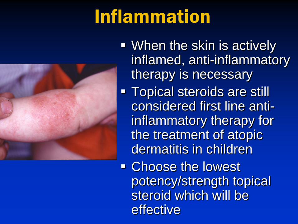
Inflammation When the skin is actively
inflamed, anti-inflammatory therapy is necessary Topical steroids are still
considered first line anti-inflammatory therapy for the treatment of atopic dermatitis in children Choose the lowest
potency/strength topical steroid which will be effective

Topical Steroids Topical steroids should be used no more
than twice daily Applied in combination with an emollient As inflammation subsides, attempt to
decrease the strength/potency of topical steroid and/or frequency of use When inflammation recurs, restart
topical steroid

Associated Co-morbidity of Atopic Dermatitis
Children with atopic dermatitis have notable differences in sleep –Greater difficulty falling asleep –Frequent night awakening –Daytime sleepiness, behavior problems
Psychosocial effects –Quality of life

Infections and Atopic Dermatitis
Patients with atopic dermatitis are more susceptible to cutaneous viral & bacterial infections
–Herpes simplex (eczema herpeticum)
–Molluscum contagiosum
–Human papilloma virus (warts)
–Staph aureus (impetiginization)

Eczema Herpeticum Usually due to generalized type 1
Herpes simplex infection in patients with underlying atopic dermatitis Can masquerade as a severe sudden
flare of atopic dermatitis or secondary bacterial infection Multiple superficial vesicles that evolve
to form punched out erosions

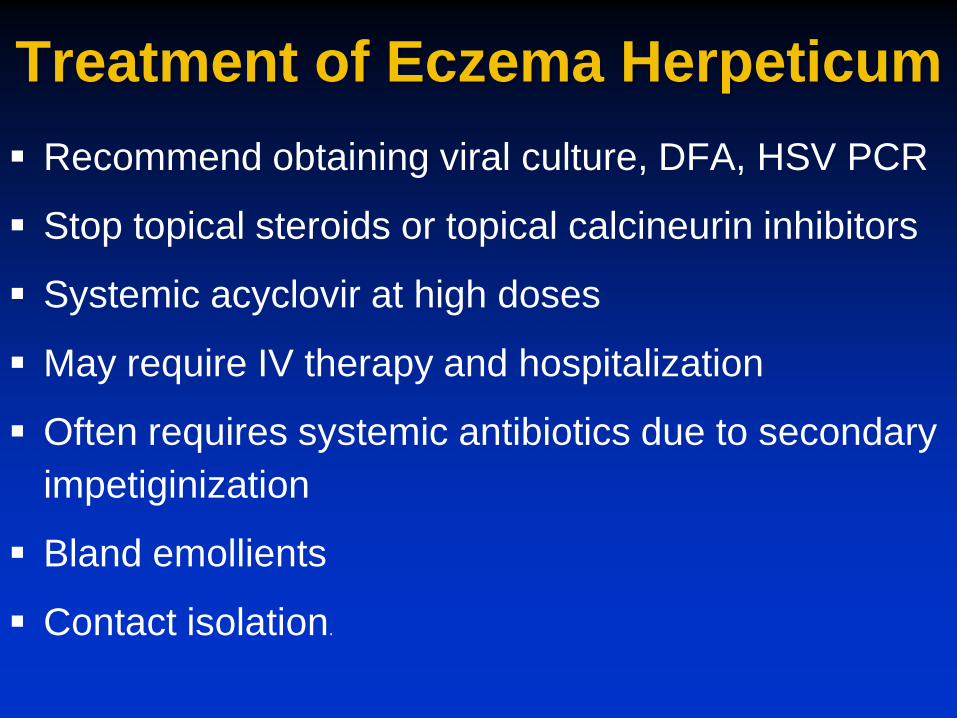
Treatment of Eczema Herpeticum Recommend obtaining viral culture, DFA, HSV PCR
Stop topical steroids or topical calcineurin inhibitors
Systemic acyclovir at high doses
May require IV therapy and hospitalization
Often requires systemic antibiotics due to secondary impetiginization
Bland emollients
Contact isolation.

Bacterial Infections 90% of patients with atopic dermatitis
are colonized with Staph aureus Staph aureus has increased adherence
to the skin of atopics Presence of Staph aureus can be
associated with an exacerbation of atopic dermatitis –Oral antibiotics are often necessary to
treat secondary infection
Impetigo
Most common bacterial skin infection in children
Predominant organisms: Staph aureus (most common) & Strep pyogenes (GAS)
More common in hot humid climates
May occur at sites of trauma
Can spread by direct skin contact
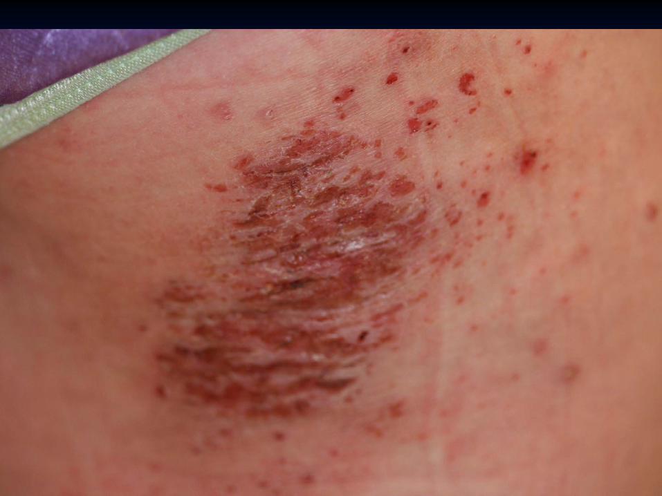
Non-bullous Impetigo Non-bullous impetigo-70% of cases
Begins as erythematous macules and papules which develop into pustules
Eventually pustules rupture leaving erosions covered by honey-colored crust
Associated pruritus or pain
Common sites: perinasal, perioral, & extremities


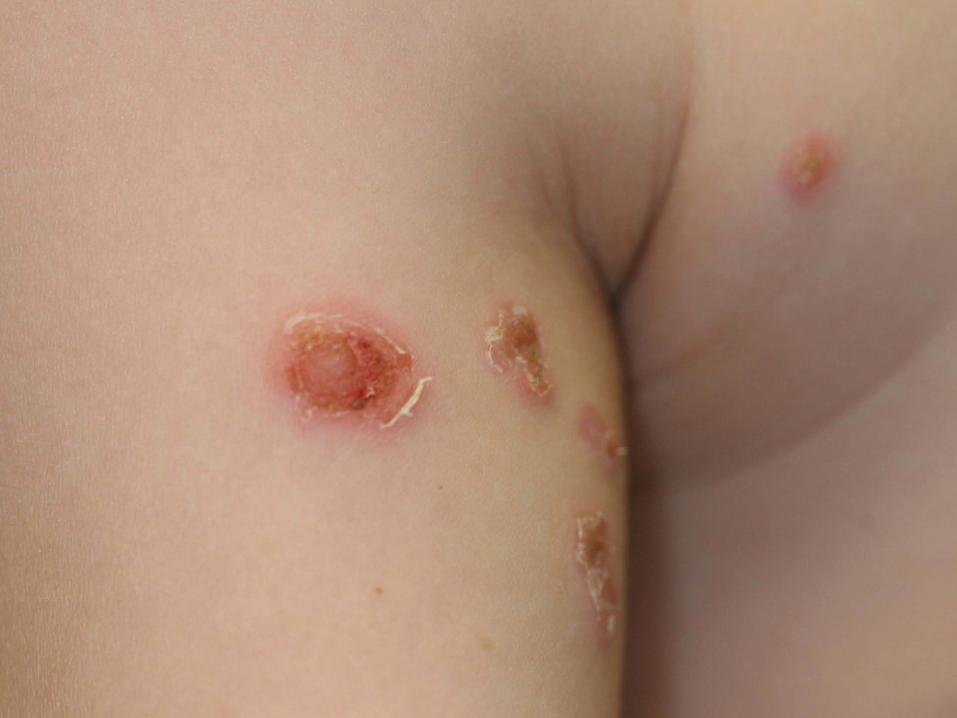
Bullous Impetigo Bullous impetigo-30% of cases
Flaccid blisters with cloudy fluid –Rupture easily leaving shallow erosions and
well demarcated collarettes of scale
Staph aureus present in blister fluid –Releases exfoliative toxin that leads to blister
formation
Obtain culture from blister fluid


Work-up & Treatment of Impetigo For small isolated areas-topical antibiotic Systemic antibiotics are often necessary for
larger areas of involvement or bullous impetigo – Cephalexin – Dicloxacillin
Concern of resistant organisms (MRSA) – Bacterial wound cultures identify antibiotic
susceptibilities – Clindamycin or Trimethoprim sulfamethoxasole may be
options
Local skin care

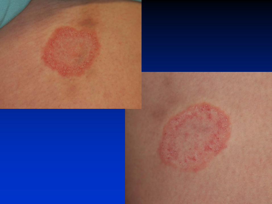
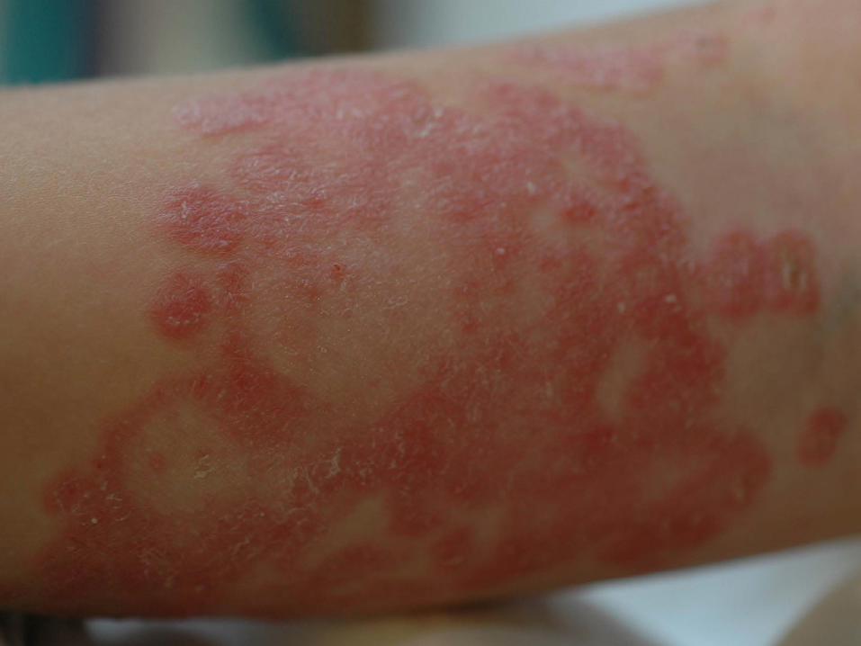
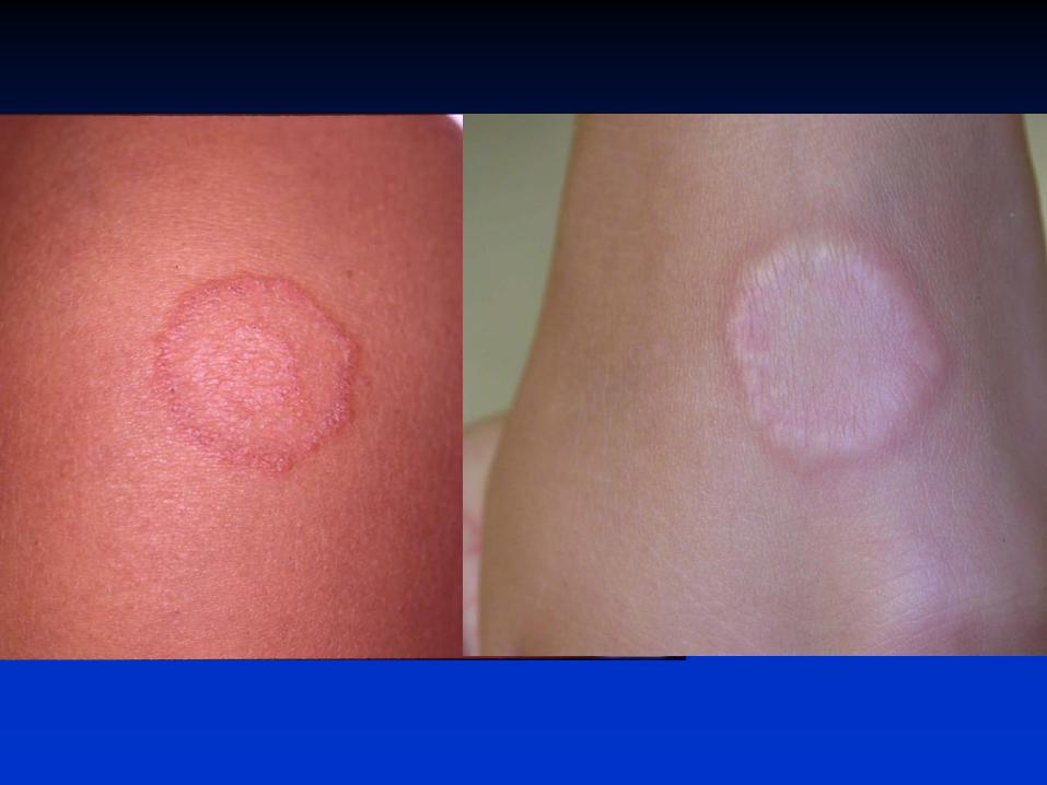
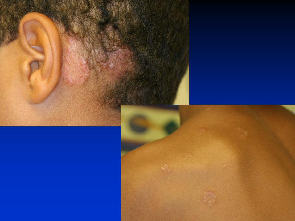
Tinea Corporis Well demarcated, annular,
erythematous scaly plaques
May have central clearing
May have inflammatory papules or pustules in the advancing edge
Usually pruritic
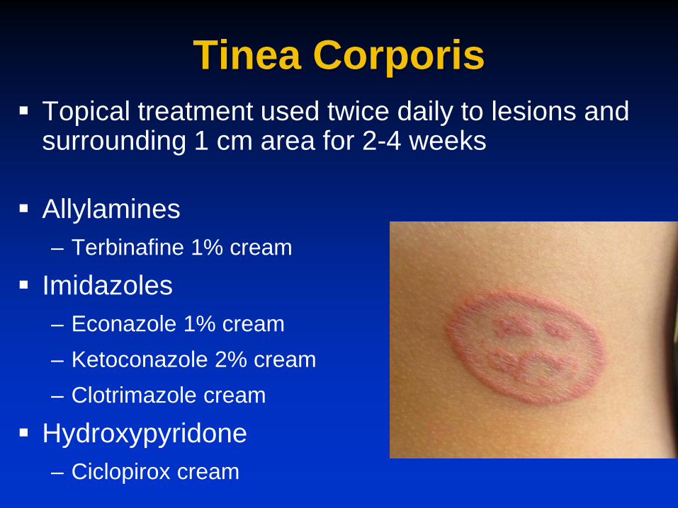
Tinea Corporis Topical treatment used twice daily to lesions and
surrounding 1 cm area for 2-4 weeks
Allylamines – Terbinafine 1% cream
Imidazoles – Econazole 1% cream – Ketoconazole 2% cream – Clotrimazole cream
Hydroxypyridone – Ciclopirox cream

Tinea Corporis Systemic therapy
– Indicated for diffuse infection, not responsive to topical therapy
– Immunocompromised patients – Always needed to treat tinea capitis
Griseofulvin Itraconazole Terbinafine Fluconazole

Granuloma Annulare Skin colored subcutaneous papules or nodules
often grouped in a ring configuration – No scale! (often misdiagnosed as ring worm) – Borders may be elevated
Most commonly located on the feet, ankles, shins, and dorsal hands Usually asymptomatic Enlarge over time with central clearing
– Size range from 0.5 cm to 3-5 cm

Granuloma Annulare
Unknown etiology
Histology is diagnostic
Usually resolve spontaneously over many months to years
Limited therapeutic options

Alopecia Areata Acquired non-scarring alopecia (bald spots) Cause is unknown, but autoimmune basis is
hypothesized Males=females 20% of all cases occur in children Family history of alopecia areata is common Commonly seen in families with autoimmune
diseases
– Vitiligo, thyroid disease, rheumatoid arthritis, diabetes

Alopecia Areata Hair loss in circumscribed areas
–May have several patchy oval or round areas
Frontal, parietal areas commonly affected
No underlying skin changes (no scale, erythema, or pustules)
Usually asymptomatic


Alopecia Areata Prognosis:
– Spontaneous remission is common with limited patchy hair loss if <1 year duration
– 1/3 will have future episodes – ~10% will have chronic course – Worse prognosis if more diffuse involvement upon
initial presentation Support Group and Information
– National Alopecia Areata Foundation www.naaf.org

Alopecia Areata Treatment Treatment options: (not FDA approved)
–Active nonintervention –Supportive psychotherapy –Wigs/hair bands Locks of Love www.locksoflove.org
–Topical steroids – Intralesional steroids –Contact sensitization Squaric acid, Anthralin

Alopecia Areata Differential diagnosis includes
–Tinea capitis –Telogen effluvium –Trichotillomania –Traction alopecia

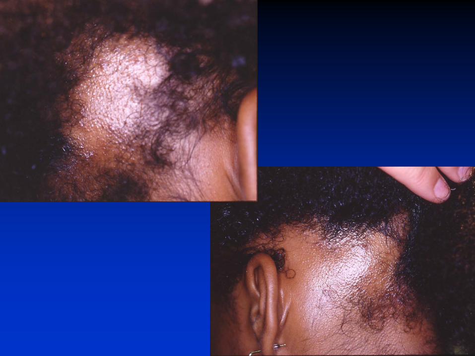
Tinea Capitis Trichophyton tonsurans is the most
common dermatophyte to cause tinea capitis in the United States Humans are the main reservoir
–More common in African Americans –Most common in 3-7 year olds
“Classic clinical triad” –Scalp scaling, alopecia, & cervical
adenopathy

Clinical Features Seborrheic type:
– Diffuse scaling/dandruff, may have subtle hair loss
“Black dot” type: – Patches of hair loss with broken hairs at follicular
orifice
Inflammatory type: – Pustules, abscesses, or kerions Higher risk of scarring


Tinea Capitis Treatment Requires systemic treatment Griseofulvin
– Gold standard – Good safety profile – Due to resistance, dosing may need to be higher
than recommended on the package insert for 6-8 weeks
– Absorption dependent on dietary fat intake Terbinafine
– Another possible option with shorter treatment duration

Telogen Effluvium Acquired hair thinning (can be diffuse) Rapid conversion of scalp hairs
Growing phase Resting phase (>25%) Normally: 85-90% is growing (anagen) 10-15% is resting (telogen) Acute stressful events act as trigger No areas of focal alopecia, scale, or erythema May develop several months after a high fever,
illness, surgery, traumatic or stressful event

Telogen Effluvium Diagnosis:
– History of proceeding event
– Clinical exam
– Consider obtaining CBC, iron studies, thyroid studies
Treatment: – Reassurance & time

Trichotillomania
Self induced hair loss resulting from pulling, rubbing, or twisting Individual often denies
pulling hair Preadolescence is most
common age of onset Hairs of varying lengths
often in an unusual pattern Scalp>eyelash>eyebrow
Trichotillomania Treatment
–Psychiatric referral
–Cognitive behavioral therapy by an experienced therapist
–Medications Antidepressants
References Enjolras O et al. Vascular tumors and vascular malformations, new issues.
Adv Dermatol 1998;13:375-423.
Hook KP. Cutaneous vascular anomalies in the neonatal period. Semin Perinatol 2013;37:40-48.
Piram M et al. Sturge-weber syndrome in patients with facial port-wine stain. Pediatr Dermatol 2012;29(1):32-37.
Chen TS et al. Infantile hemangiomas. Pediatircs 2013;131:99-108.
Kilcline C et al. Infantile hemangiomas: how common are they? Pediatr Dermatol 2008;25(2):168-173.
Tollefson MM et al. Early photographs of infantile hemangiomas: what parents’ photographs tell us. Pediatrics 2012;130:e314-320.

References Orlow S et al. Increased risk of symptomatic hemangiomas of the airway in
association with cutaneous hemangiomas in a “beard” distribution. J Pediatr 1997;131:643-648.
Ceisler EJ et al. Periocular hemangiomas: what every pediatrician should know. Pediatr Dermatol 2004;21(1):1-9.
Haggstrom AN et al. Prospective study of infantile hemangiomas: clinical characteristics predicting complications and treatment.Pediatrics 2006;118:882-887.
Horii KA et al. A prospective study of the frequency of hepatic hemangiomas in infants with multiple cutaneous infantile hemangiomas, Pediatr Dermatol 2011;28(3):245-253.
Schumacher WE et al. Spinal dysraphism associated with the cutaneous lumbosacral hemangioma: a neuroradiological review Pediatr Radiol 2012;42(3):315-320.
Metry D et al. Consensus statement on diagnostic criteria for PHACE syndrome. Pediatrics 2009;124:1447-1456.
Leaute-Labreze C et al. Propranolol for severe hemangiomas of infancy. N Engl J Med 2008;358(24):2649-2651.
References Marqueling AL et al. Propranolol and infantile hemangiomas four years later:
a systematic review. Pediatr Dermatol 2013;30(2):182-191. Drolet BA et al. Initiation and use of propranolol for infantile hemangiomas.
Pediatrics 2013;131:128-140.
Kwon EM et al. Retrospective review of adverse effects from propranolol in infants. JAMA Dermatol 2013;149(4):484-485.
Schwartz RA et al. Seborrheic dermatitis: an overview. Am Fam Physician 2006;74:125-130.
Poindexter GB et al. Therapies for pediatric seborrheic dermatitis. Pediatr Ann 2009;38(6):333-338.
Ravanfar P et al. Diaper dermatitis. Curr Opin Pediatr 2012;24:474-479.
Eichenfield et al. Guidelines of care for the management of atopic dermatitis. J Am Acad Dermatol 2014;70:338-351.
Bieber T. Atopic dermatitis. N Eng J Med 2008;358(14):1483-1494.
References Eichenfield LF et al. Consensus conference on pediatric atopic dermatitis. J
Am Acad Dermatol 2003;49:1088-1095. Ring J et al. Guidelines for treatment of atopic eczema Part I. JEADV
2012;26:1045-1060. Schneider L et al. Atopic dermatitis: a practice parameter update 2012. J
Allergy Clin Immunol 2013;131:295-299. Krakowski AC et al. Management of atopic dermatitis in pediatric
population. Pediatrics 2008;122;4:812-824. Luca NJ et al. Eczema herpeticum in children: clinical features and factors
predictive of hospitalization. J Pediatr 2012;161:671-675. Aronson PL et al. Delayed acyclovir and outcomes of children hospitalized
with eczema herpeticum. Pediatrics 2011;128:1161-1167.
Andrews MD. Common tinea infections in children. Am Fam Physician 2008;77(10):1415-1420.
Kelly BP. Superficial fungal infections. Pediatr Rev 2012;33(4):e22-e37.
References Cyr PR. Diagnosis and management of granuloma annulare. Am Fam
Physician 2006;74(10):1729-1734.
Dahl MW. Granuloma annulare: long term follow-up. Arch Dermatol 2007;143(7):946-947.
Gupta AK et al. Meta-analysis of randomized, controlled trials comparing particular doses of griseofulvin and terbinafine for the treatment of tinea capitis. Pediatr Dermatol 2012;30(1):1-6.
Bedocs LA et al. Adolescent hair loss. Curr Opin Pediatr 2008;20:431-435. Tay YK et al. Trichotillomania in childhood. Pediatrics 2004 113:e494-e498. Shah KN et al. Factitial dermatoses in children. Curr Opin Pediatr
2006;18:403-409.