common pediatric respiratory emergencies · pdf filecommon pediatric respiratory emergencies...
TRANSCRIPT
Common PediatricRespiratoryEmergencies
Joseph Choi, MDa,*, Gary L. Lee, MD, CCFP-EM, FRCPCb
KEYWORDS
� Pediatric � Asthma � Bronchiolitis � Croup � Pneumonia
Acute respiratory distress is one of the most common reasons why parents bring theirchildren to the emergency department (ED). Severity can range from mild, self-limitingillness to life-threatening disease. This article reviews the 4 most common of theseconditions, namely asthma, croup, bronchiolitis, and pneumonia, to update the readeron the current state of evidence in the assessment and treatment of these conditions.
ASTHMA IN CHILDREN
Asthma is a chronic inflammatory condition of the airways leading to episodicwheezing, coughing, chest tightness, and shortness of breath. It is a common condi-tion, with the highest prevalence occurring between the ages of 5 and 17 years.Asthma afflicted 7.0 million children in this age group in the United States in 2008.1
It led to 1.7 million ED visits in 2006, and children younger than 15 years accountedfor 33% of those with a discharge diagnosis of asthma while this age group only repre-sents 20% of the general population.1
Asthma is the most activity-limiting condition in children and accounts for 14.4million lost school days. It is an expensive disease, with an annual burden of $15.6billion in direct health care costs and $5.1 billion in indirect health care costs andlost productivity, for a total annual sum of $20.7 billion.1
Pathophysiology
Asthma is a chronic disease of the lower airways punctuated with episodic acuteexacerbations. The clinical manifestations are caused by airway hyperresponsivenessto stimuli that are generally innocuous, leading to constriction of bronchial smooth
The authors have nothing to disclose.a McGill University FRCP Emergency Medicine Residency Program, Royal Victoria Hospital, 687Pine Avenue West, Room A4.62, Montreal, Quebec, Canada H3A 1A1b Department of Emergency Medicine, Montreal Children’s Hospital, Montreal GeneralHospital, McGill University, 1650 Cedar Avenue, Montreal, Quebec, Canada H3G 1A4* Corresponding author.E-mail address: [email protected]
Emerg Med Clin N Am 30 (2012) 529–563doi:10.1016/j.emc.2011.10.009 emed.theclinics.com0733-8627/12/$ – see front matter � 2012 Elsevier Inc. All rights reserved.

Choi & Lee530
muscle (bronchospasm), the major cause of wheezing during an asthma exacerbation.Airway inflammation and edema in response to these stimuli further narrows theairway and restricts ventilation.2,3
These physiologic changes on the cellular level can occur through IgE-mediatedpathways (allergen-triggered asthma)4 and non–IgE-mediated pathways (asthma inresponse to nonsteroidal anti-inflammatory drugs,5 certain other drugs, exercise, andcold temperatures).3 Both pathways lead to a release of various cytokines and chemo-kines from inflammatory cells, which promote furthermigration and activation of inflam-matory cells in the lower airways, thus perpetuating the cycle.3
Diagnosis
The diagnosis of asthma is particularly challenging in the pediatric population. Youngchildren usually cannot be cooperative enough to undergo formal pulmonary functiontesting, which is the gold standard in the diagnosis of asthma. Thus they often must bediagnosed clinically.Many first-time wheezers also present to the ED. Most instances of wheezing in
young children presenting to the ED are solely related to upper respiratory infections(URI) causing inflammation of the lower airways rather than true asthma.6 The majorityof early wheezers do not go on to develop asthma in later childhood or adulthood.6,7
This distinction is an important one, as it can affect the efficacy of certain therapeuticoptions.8 Clues that increase the likelihood that the wheezing is due to asthma includethe frequency of episodes (more than once a month), triggers (exercise, allergens,tobacco smoke), prolonged respiratory symptoms in the setting of URI (symptomslasting more than 10 days suggest a viral trigger of asthma), personal or family historyof atopy or asthma, and a history of a good and rapid response to bronchodilatortherapy.3,6,9
Other historical features of the patient that may aid in predicting the severity of theasthma exacerbation include the frequency and compliance in using asthma medica-tions at home, previous hospital visits for asthma exacerbations (requiring admissionto the ward or intensive care unit [ICU]), and severity of asthma exacerbations(requiring intubation). Social attributes of the patient and caregivers, such as the abilityto purchase medications and comply to their use, a household environment free ofknown or suspected asthma triggers, and ability to obtain follow-up and accessmedical services in the event of another exacerbation, have important discharge plan-ning implications and should be elicited early.2,3,7,9
The physical examination may reveal any combination of the classic constellation ofsymptoms in the acute asthma exacerbation, which includes wheezing, cough, chesttightness, tachypnea, respiratory distress, intercostal indrawing, and accessorymuscle use.3,7,9 Of interest, the use of the scalene muscles and suprasternalretractions have the highest interrater reliability and correlation with asthma severity.10
Clinical asthma assessment tools, such as the Pediatric Respiratory AssessmentMeasure (PRAM; Table 1)10,11 and the Pediatric Asthma Severity Score (PASS),12
have been independently shown to be predictive in discriminating a patient’s lengthof stay in the hospital and admission.10,12 The strength of these two scales is thatthey include preschool-aged children, in comparison with older severity scales suchas the Pulmonary Index and the Pulmonary Score, which are only validated in older,school-aged children.10–12 A recent head-to-head comparison of the two scoresshowed very similar performance in their ability to predict a prolonged stay (>6 hours)and/or admission when taken at triage.13 However, a repeat score taken 90 minutesafter treatment showed that the PRAM score was more responsive and predictive
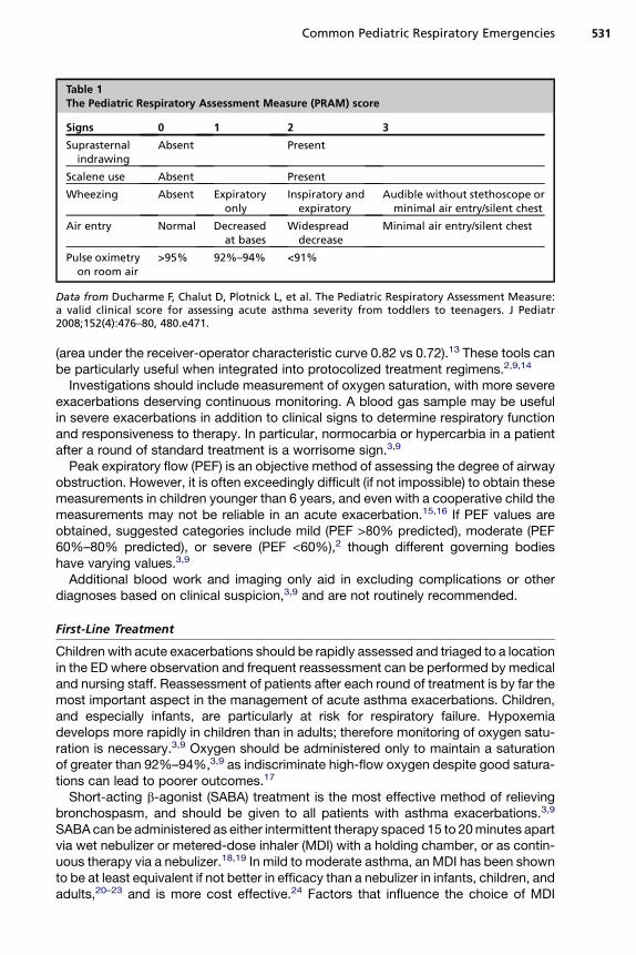
Table 1The Pediatric Respiratory Assessment Measure (PRAM) score
Signs 0 1 2 3
Suprasternalindrawing
Absent Present
Scalene use Absent Present
Wheezing Absent Expiratoryonly
Inspiratory andexpiratory
Audible without stethoscope orminimal air entry/silent chest
Air entry Normal Decreasedat bases
Widespreaddecrease
Minimal air entry/silent chest
Pulse oximetryon room air
>95% 92%–94% <91%
Data from Ducharme F, Chalut D, Plotnick L, et al. The Pediatric Respiratory Assessment Measure:a valid clinical score for assessing acute asthma severity from toddlers to teenagers. J Pediatr2008;152(4):476–80, 480.e471.
Common Pediatric Respiratory Emergencies 531
(area under the receiver-operator characteristic curve 0.82 vs 0.72).13 These tools canbe particularly useful when integrated into protocolized treatment regimens.2,9,14
Investigations should include measurement of oxygen saturation, with more severeexacerbations deserving continuous monitoring. A blood gas sample may be usefulin severe exacerbations in addition to clinical signs to determine respiratory functionand responsiveness to therapy. In particular, normocarbia or hypercarbia in a patientafter a round of standard treatment is a worrisome sign.3,9
Peak expiratory flow (PEF) is an objective method of assessing the degree of airwayobstruction. However, it is often exceedingly difficult (if not impossible) to obtain thesemeasurements in children younger than 6 years, and even with a cooperative child themeasurements may not be reliable in an acute exacerbation.15,16 If PEF values areobtained, suggested categories include mild (PEF >80% predicted), moderate (PEF60%–80% predicted), or severe (PEF <60%),2 though different governing bodieshave varying values.3,9
Additional blood work and imaging only aid in excluding complications or otherdiagnoses based on clinical suspicion,3,9 and are not routinely recommended.
First-Line Treatment
Children with acute exacerbations should be rapidly assessed and triaged to a locationin the ED where observation and frequent reassessment can be performed by medicaland nursing staff. Reassessment of patients after each round of treatment is by far themost important aspect in the management of acute asthma exacerbations. Children,and especially infants, are particularly at risk for respiratory failure. Hypoxemiadevelops more rapidly in children than in adults; therefore monitoring of oxygen satu-ration is necessary.3,9 Oxygen should be administered only to maintain a saturationof greater than 92%–94%,3,9 as indiscriminate high-flow oxygen despite good satura-tions can lead to poorer outcomes.17
Short-acting b-agonist (SABA) treatment is the most effective method of relievingbronchospasm, and should be given to all patients with asthma exacerbations.3,9
SABA can be administered as either intermittent therapy spaced 15 to 20minutes apartvia wet nebulizer or metered-dose inhaler (MDI) with a holding chamber, or as contin-uous therapy via a nebulizer.18,19 In mild to moderate asthma, an MDI has been shownto be at least equivalent if not better in efficacy than a nebulizer in infants, children, andadults,20–23 and is more cost effective.24 Factors that influence the choice of MDI

Choi & Lee532
versus nebulizer include patient cooperation, response to treatment via MDI, andseverity of the exacerbation. Salbutamol (albuterol, Ventolin) 2 to 4 puffs, can be givenevery 15 minutes via MDI with chamber, waiting 5 tidal volume breaths betweenpuffs.3,9 More puffs can be administered (up to 10) in more resistant exacerbations.Salbutamol can also be given at 0.15 to 0.3 mg/kg via nebulizer, with a minimum of2.5 mg and maximum of 5 mg per nebulized mask. This volume is then diluted withnormal saline for a total of 5 mL of fluid per nebulized mask. The most severe exacer-bations may benefit from continuous therapy19 with a nebulizer driven by oxygen ifhypoxia is also present.3,9,17 With continuous nebulization, salbutamol is recommen-ded to be given at 0.5 mg/kg/h with the hourly dose not exceeding 10 to 15 mg/h.3
Levosalbutamol ((R)-salbutamol, also known as levalbuterol) is the pure (R)-enan-tiomer of the salbutamol molecule. In a typical salbutamol preparation, there isa 50:50 mixture of the (S)- and (R)-enantiomers, and it is the (R)-enantiomer thatprovides the vast majority of the bronchodilating effects, due to its 100-fold higheraffinity for the b2-adrenergic receptor. The selectivity of levosalbutamol theoreticallymaximizes the bronchodilating effects while minimizing systemic side effects suchas tachycardia25 and hypokalemia.26 Small trials have shown mixed results, withsome trials showing benefit in pulmonary function,27,28 reduction in hospital admissionrates,29 and reduced side effects26,30; whereas other studies have shown no differ-ence.30,31 Levosalbutamol is considerably more expensive than the conventionalracemic mixture, and current guidelines do not recommend using one over the other.The dose of levosalbutamol is half that of salbutamol.Ipratropium bromide (Atrovent) has been shown to be an effective adjunct in
moderate to severe asthma in addition to inhaled b-agonists.32,33 It is a muscarinicacetylcholine receptor blocker, which produces bronchodilation via smooth musclerelaxation. Ipratropium bromide can be given via MDI (4–8 puffs every 15–20 minutes)or nebulizer (0.25–0.5 mg, combined in the same nebulizer as the b-agonist).3,9 Treat-ment can be tapered as the patient improves clinically.Systemic corticosteroids (SCS) have been shown to decrease the need for hospital
admission from the EDwhen given early34,35 and to decrease the length of stay.36 SCSshould be considered in all but the mildest exacerbations.3,9,34 However, there isevidence that shows administration of steroids to children with wheezing triggeredby a URI and with no other history suggesting asthma provides no benefit in time todischarge, admission rate, morbidity, or mortality.8 The efficacy of oral versus intrave-nous (IV) SCS has been shown to be equivalent in pediatric asthma exacerbations.37,38
Parenteral SCS should be reserved for those who cannot tolerate oral steroids or haveintestinal issues that would affect its absorption.3,9 Oral prednisone or prednisolone, 1to 2 mg/kg, should be given once daily, with a maximum dose of 60 mg/dfor 3 to 5 days.2,3 Studies suggest that a 2-day course of oral dexamethasone (dosedat 0.6 mg/kg daily, maximum of 16 mg) is as effective (measured by symptoms scores,admission rates, and 10-day relapse rate) and well tolerated (rates of nausea and vom-iting) as a 5-day course of oral prednisone in adults39 and children.40,41 Another studyeven suggests that a single dose is non-inferior to a 5-day course of prednisone.42
Intramuscular (IM) injection of depot steroids, such as dexamethasone acetate, hasalso been shown in small studies to be as effective as a 5-day course of predni-sone.43,44 IV steroids can be given as methylprednisolone, 2 mg/kg/d in two divideddoses.3 Inhaled corticosteroids (ICS) are not currently recommended as a replacementfor SCS for the treatment of acute asthma exacerbations presenting to the ED,3,9
because of the lack of efficacy when used alone.34,35,45–48 Recent evidence suggeststhat the addition of inhaled budesonide to standard therapy including SCS does notimprove outcomes.49

Common Pediatric Respiratory Emergencies 533
Second-Line Treatments
In children with severe or life-threatening asthma, the aforementioned therapies maynot be sufficient. It is crucial to recognize refractory asthma and to treat it aggressively.Magnesium sulfate is a safe drug with few side effects, which has been shown to
have bronchodilating effects.50–52 Its use has not been shown to be beneficial inmild to moderate asthma, but has demonstrated a reduction in admission rates forsevere asthma, with minimal side effects.53,54 A single IV dose of 25 to 75 mg/kg(not exceeding 2 g) can be given over 2 hours.3,9 Inhaled magnesium sulfate hasbeen shown in small studies to improve expiratory flow measures when used in addi-tion to inhaled b-agonists in severe asthma exacerbations,55,56 but confers no benefitin mild to moderate episodes.57 A Cochrane review showed that nebulized magne-sium sulfate provided significant improvement in pulmonary function tests anda nonsignificant trend to decreased hospital admission rates in severe asthma exac-erbations, but no statistically significant difference when included in all severities ofasthma attacks.58 Inhaled magnesium sulfate is given as the diluent in place of normalsaline (usually 2.5 mL of a 250 mmol/L solution) combined with salbutamol and ipra-tropium bromide in the same nebulized mask.Oral leukotriene receptor antagonists (LTRA) such as montelukast (Singulair) have
been shown to decrease symptoms of mild to moderate asthma exacerbations,59,60
but their role in severe asthma is unknown because of their slow onset of action.61
In moderate to severe asthma there is some evidence in the adult literature that IVadministration may be effective,62 but there are no corresponding studies done in chil-dren. The oral dosage of montelukast in children is 4 to 10 mg orally once per day.Heliox is a blend of helium and oxygen, the use of which is based on the principle of
increased ventilation into the lower airspaces in asthma, due to its low viscosity.63
Some studies have shown modest benefit63–65 whereas others show no benefit.66 Ithas a level D recommendation as the driver of nebulized salbutamol in severeasthma,3 mainly because of its lack of side effects.Routine antibiotics are not recommended unless there is a suspicion of pneumonia
(fever, purulent sputum) or bacterial sinusitis.2,3,9 Mucolytics and sedation are notrecommended.2,3,9
Status Asthmaticus and Imminent Respiratory Failure
One of the most terrifying prospects for an emergency physician is the sight of a childwith asthma not responding to treatment and heading toward respiratory failure. Signsinclude increasing somnolence, tiring of breathing muscles, cyanosis, and a silentchest. Heralds of impending cardiac arrest are bradycardia, severe hypoxia, andhypercapnia.If not already performed, IV access should be obtained and any hypovolemia should
be corrected. This action is also taken to prevent hypotension that the induction drugsmay cause during rapid sequence intubation and positive ventilation.IV b-agonists have not been shown to be beneficial in severe exacerbations, and
carry significant side effects.3,67 Likewise, aminophylline has not been recommended,due to its considerable toxicity and lack of clear benefit,68 though a study has shownsome effect in children with life-threatening asthma already receiving maximum dosesof conventional therapy.69 However, in the setting of a status asthmaticus in extremiswith no response to other therapies, guidelines still endorse consideration of their useas a last resort.2,3,9
Temporizing measures can include a trial of noninvasive positive-pressure ventila-tion (NPPV). There are small studies analyzing the ability of NPPV to avoid intubation
Choi & Lee534
and improve outcomes in children with status asthmaticus,70–75 and a Cochranereview showed a trend toward benefit.76 If the patient is unable to tolerate NPPVand continues to deteriorate, endotracheal intubation with mechanical ventilation isnecessary.3,9 Once the decision has been made, the most experienced physicianshould make the attempt, as asthmatic patients are often difficult to intubate anddesaturate quickly. Traditionally the induction agent of choice is ketamine (1–2 mg/kg IV) because of its mild bronchodilating properties,77 though the clinical significanceof this bronchodilation is questionable,77,78 and several trials using ketamine infusionas an adjunct in status asthmaticus have shown no benefit.78,79 Ketamine can alsoincrease oral and airway secretions80 and trigger laryngospasm, and should beused in conjunction with a paralytic agent to counteract this possibility.81 Otherchoices for induction include etomidate (0.3 mg/kg IV) and propofol (1.5–3 mg/kgIV). The paralytic agent of choice depends on patient characteristics, the presenceof contraindications to particular agents, and physician preference and experience.A ventilator strategy that has shown benefit is one of permissive hypercapnea.3,82–86
With this strategy, PCO2 is allowed to reach up to 70 mm Hg, providing high FiO2concentrations to maintain saturations greater than 92%, and manipulating ventilatorsettings (such as the prolonging the expiratory time to allow for complete exhalation)to minimize pressures and avoid barotrauma and other complications. Bicarbonatecan be given to correct severe acidosis.87
Disposition
The decision to hospitalize children with severe asthma depends on the severity ofthe exacerbation and its response to ED therapy. Those who present with PEFless than 25%, or have a PEF less than 40% or significant symptoms after therapy,should be admitted for continued treatment and observation.2,3 Unstable homesituations or predicted poor compliance and follow-up are also indications foradmission.2,3,9
Children receiving treatment in the ED should be monitored for at least 1 hour aftertheir last round of treatment to ensure resolution of symptoms.2,3,9 In those able toprovide PEF measures, a general value of greater than 70% to 80% of expected valueis acceptable for discharge. Those with a PEF of 40% to 69% with minimal symptomscan also be discharged if they have good follow-up and demonstrate good compli-ance, and if medical attention is readily accessible.2,3
Follow-up with a primary care provider or asthma specialist should be arrangedwithin a month of discharge from the ED to reassess medications and treatmentplans,2,3 as this has been shown to improve outcomes and decrease ED visits.88,89
The appointment should be scheduled before discharge from the ED, as this hasbeen shown to increase compliance.90,91
Medications to be continued after discharge include inhaled salbutamol and oralsteroids as already described, with no need for a tapering dose.2,3,9 Current guidelinesstate that the initiation of ICS should be considered in those with moderate to severeexacerbations who were not previously on ICS.2,3,9 Patients should be given a 1- to 2-month supply, as this has been shown to reduce the number of exacerbations and EDvisits.46,92,93 ICS should be continued if previously prescribed. Ipratropium bromidehas not been shown to be of benefit after discharge from the ED.Discharge home with a peak flowmeter is also recommended for children older than
5 years,3 especially in those who do not perceive mild symptoms well or have recur-rent severe exacerbations.94
Children to be discharged should be provided a written action plan (WAP) that delin-eates clearly discharge medications, instructions in proper inhaler and peak flow
Common Pediatric Respiratory Emergencies 535
meter technique, follow-up appointments, and warning signs that indicate a need toreturn to the ED.2,3
CROUP
Croup is a common cause of stridor in the young child. Croup is characterized bya harsh inspiratory stridor and a hoarse cough that is often described as barky orresembling a seal, secondary to upper airway inflammation and edema. Althoughusually benign and self-limiting, it can cause significant respiratory distress requiringintubation.Croup is the most common cause of stridor in young children older than 6 months. It
peaks between 6 and 36 months of life. At 2 years of age, 5% of all children will havehad croup.95 In a 14-year observational study in Ontario, Canada, its incidence seemsto have a biennial mid-autumn peak and an annual summer trough, with boys beingaffected 1.5 times as often as girls.96
Pathophysiology
There has been much confusion in the use of the term croup. It has been used todescribe different disease entities in which stridor and hoarse cough are the predom-inant symptoms,95 such as spasmodic croup, laryngotracheobronchitis (LTB), laryng-otracheobronchopneumonia (LTBP), bacterial tracheitis, and diphtheria. In this reviewcroup specifically refers to laryngotracheitis, as the other entities have differentpresentations, treatment options, and prognoses.Croup is commonly caused by parainfluenza virus (PIV)-1.97 PIV-2 and PIV-3 are
also implicated in croup, with type 2 causing a milder form and type 3 causinga more severe form. Other viruses that can lead to croup include influenza, respiratorysyncytial virus (RSV), rhinoviruses, enteroviruses, and measles, among others.95
Viral infection often starts via inoculation of the nares and pharynx, which leadsto typical URI symptoms of low-grade fever, coryza, and rhinorrhea. The infectionthen spreads down to the larynx and subglottic area, causing cough and inflammationand edema of the upper airway and leading to varying degrees of obstruction. Accord-ing to Poiseuille’s equation, resistance to flow is inversely proportional to radius to thefourth power. Therefore even slight decreases in diameter can cause significant resis-tance, especially in the already tiny airways of the young child. The lower airways areusually not affected in PIV-associated croup, though RSV and influenza can causelower respiratory symptoms.
Diagnosis
The constellation of a barky cough, hoarseness, and stridor is common in manydiseases, and differentiation between them is of utmost importance, as treatmentand potential complications vary widely.The onset and progression of symptoms leading to the ED visit should be explored.
The history of a preceding URI in the last day or two is often elicited. In addition tocough and stridor, the child is often febrile. Combined with other features of thehistory, a suddenly stridorous child without fever or URI should raise a suspicion offoreign body aspiration or angioneurotic edema. An immunization and travel historyshould be elicited, since an unimmunized child is at higher risk of developing laryngealdiphtheria from Corynebacterium diphtheriae infection. Pharyngitis and dysphagia arefeatures that are uncommon with croup, and may suggest retropharyngeal abscess,peritonsillar abscess, epiglottitis, or diphtheria. Finally, caution is required when diag-nosing croup in a stridorous child younger than 6 months, due to the important
Choi & Lee536
differential diagnosis in this age group. The differential includes laryngomalacia (themost common cause, and a self-limited condition that 90% grow out of by 12–18months),98 vocal cord paralysis,99 papillomatosis,100 congenital causes (such ashemangiomas,101 laryngeal webs,100 and neurofibromas102), and iatrogenic causes(such as subglottic stenosis following intubation for prematurity,103 or vocal cordparalysis caused by laryngeal nerve damage from thoracic or cardiac procedures104).The child should otherwise appear well; a toxic-looking child should be investigated
for alternative diagnoses such as bacterial tracheitis (which can be a complicationof croup), LTB, LTBP, or epiglottitis. There should be no signs of lower airway involve-ment such as wheezing or crackles, which may suggest an alternative diagnosis. Ifhemangiomas are noted on the child, especially above the clavicles, a subglottichemangioma should be considered along with historical features such as a lack ofURI symptoms. A throat examination should identify pharyngeal causes of stridoreasily, though caution should be exercised if the presentation is suspicious for epiglot-titis. The epiglottis, if visualized, should be normal.The diagnosis of croup remains a clinical one, with additional testing being useful in
ruling out other differential diagnoses. Complete blood cell counts (CBC) may showa mildly elevated white cell count in croup, whereas it is usually markedly elevatedor depressed in bacterial infections of the upper airway with increased neutrophilsand band forms. Posterior-anterior neck radiographs in croup may show subglotticnarrowing (steeple sign) with a smooth tracheal contour. Irregularity of this contoursuggests bacterial tracheitis and other diagnoses.95 Lower respiratory symptomsshould be investigated with a chest radiograph (CXR) to assess for a possible pneu-monia. A lateral neck radiograph showing a thickened mass at the level of theepiglottis (thumbprint sign) suggests epiglottitis.105 Foreign bodies may appear onplain films, depending on the object.
Classification
Different classification scales based on physical examination findings have beendevised. The Westley Croup Score106 is the most widely known and used, althoughit is used more commonly in research protocols and less frequently in clinical prac-tice.107 Key criteria used in this score include level of consciousness, stridor, air entry,cyanosis, and chest wall retractions.95,106,107
A simplified classification, based from the original Westley Croup Score, has beensuggested by several investigators.95,107 Mild croup is defined by an absenceof stridor at rest, minimal respiratory distress, and occasional cough. Moderate crouphas stridor at rest and increased amount of respiratory distress, but behavior andmental status are normal. Severe croup has significant respiratory distress and mentalstatus changes, with increasing somnolence and decreasing air entry signifyingimpending respiratory failure.
Treatment
As with all patients, the ABCs (Airway, Breathing, Circulation) must be prioritized.Patients with signs of impending respiratory failure should be intubated with anendotracheal tube 0.5 to 1 mm smaller than the expected size. Oxygen shouldbe delivered to maintain oxygen saturation greater than 92% to 94%. Wherepossible, the child should be kept calm to decrease respiratory distress andimprove airway dynamics.The mainstays of pharmacotherapy in the ED management of croup are corticoste-
roids and nebulized epinephrine. Dexamethasone remains the corticosteroid of choiceover prednisolone through its ability to decrease return visits and admissions.108 In
Common Pediatric Respiratory Emergencies 537
a recent Cochrane review, dexamethasone was shown to reduce symptoms in the ED,decrease length of stay, and result in fewer return visits.109 Dexamethasone is given asa single dose of 0.6 mg/kg by mouth/IM/IV (oral is preferred, though parenteral routeshave been shown to be equally effective110) to a maximum of 10 mg. There are severalstudies that show lower doses of dexamethasone (0.15–0.3 mg/kg) may be equallyeffective.111–113 Inhaled budesonide can be used if available (2 mg via nebulizer) andhas been shown to be similar in efficacy to dexamethasone,109,114,115 though avail-ability, cost, and convenience makes dexamethasone a more attractive option. Theredoes not appear to be any additional benefit from combining oral and inhaled steroidsin the setting of croup.116 Corticosteroids should be considered in all severities ofcroup.Nebulized epinephrine is used in moderate to severe croup, and has been shown to
be highly efficacious in reducing symptom scores at 30 minutes after treatment andtime spent in the ED.117 However, the natural history of the disease is unchanged,and thus it is important to monitor children after epinephrine treatment for reboundreactions.106,118 Despite the theoretical benefits of L-epinephrine over racemicepinephrine, studies did not show a benefit of choosing one over the other.117
L-Epinephrine is given as 5 mL of a 1:1000 solution (racemic epinephrine is given as0.5 mL of a 2.25% solution in 2.5 mL of normal saline) delivered via nebulizer every15 minutes to effect. Although serious cardiac complications from epinephrine treat-ment are exceedingly rare, it is prudent to put children requiring multiple treatments oncontinuous cardiac monitoring.Although cold, humid air anecdotally has been thought to improve croup symptoms,
recent trials did not show this benefit in the ED setting.119–121 A study evaluating theuse of Heliox showed that it may be as effective as nebulized racemic epinephrinein moderate to severe croup.122 Heliox also demonstrated a nonstatistically significanttrend toward improvement in croup scores when combined with epinephrine andsteroids.123 However, a Cochrane review showed no significant difference in croupscores when Heliox was added to conventional therapy, and therefore did notroutinely recommend its use at this time.124 Antibiotics should be saved for suspectedbacterial complications such as bacterial tracheitis or LTBP. Sedatives and antitus-sives are not indicated.95,107
Disposition
Most cases of croup are mild to moderate and respond well to steroid with orwithout nebulized epinephrine therapy, and the vast majority of patients are dis-charged home. Children with mild symptoms (ie, no stridor at rest) can be safelysent home. Patients receiving epinephrine should be observed for 4 hours to watchfor any rebound phenomenon. Children who require multiple epinephrine dosesshould be admitted for observation. ICU admission may be required in cases ofsevere croup that fail to respond to treatment. On discharge, it is prudent to informthe parents that symptoms of croup usually peak between days 2 to 3.
BRONCHIOLITIS
Bronchiolitis is the most common lower respiratory tract condition in children youngerthan 2 years, and is the leading cause of hospitalization of infants. Its most commoncause, RSV, is ubiquitous worldwide, affecting nearly all children by the age of 2, oftencausing dyspnea in its tiniest of victims with the potential to cause respiratory failureand death. Significant literature and practice guidelines have been published to guidepractitioners. Active research in the field is ongoing, and even small improvements intherapy or resource use can make a major impact given the burden of disease.

Choi & Lee538
Definition and Epidemiology
Bronchiolitis is typically caused by viral infection, characterized by bronchiolarinflammation in children usually younger than 2 years. Children from age 2 to 5years with similar symptoms rarely have bronchiolitis, as asthma, recurrent viralwheeze, or pneumonia are more likely diagnoses. Wheezing is the hallmark of theAmerican definition of bronchiolitis, where British and Australian definitions includeinspiratory crackles as part of the diagnosis.125,126 Most studies and guidelinesdefine bronchiolitis as the first episode of wheezing or crackles. As the presenta-tions of bronchiolitis, viral-associated wheeze, and asthma may appear similar,attention to the features of patients becomes very important given differing patho-physiology and responses to treatment.125
RSV continues to be the most common cause, responsible for approximately 50%to 80% of cases. The epidemiology of bronchiolitis follows closely that of RSV. Astrongly seasonal virus, RSV causes outbreaks most commonly between Novemberand March in the northern hemisphere and between May and September in thesouthern hemisphere. Up to half of infected children younger than 2 years will havelower respiratory tract symptoms, whereas older children and adults are more likelyto have upper tract disease only. Peak age for bronchiolitis is 2 to 6 months old,with most cases occurring before age 9 months. Reinfection with RSV is common,given poor postinfection immunity.Recent studies demonstrate an increasing role and frequency of other viruses
causing bronchiolitis, including human metapneumovirus (10%–20%), human bocavi-rus, rhinovirus, parainfluenza, adenovirus, influenza, and coronavirus.127–129 Althoughrhinovirus and humanmetapneumovirus infection has been shown to be generally lesssevere than RSV (Group A strains causing more severe disease than Group B strains),etiologic diagnosis is often not known in the ED and does not currently affect EDmanagement.127,130
Bronchiolitis-associated hospitalizations have more than doubled over recentdecades, likely attributable to several factors including increased spread of viralillnesses from daycare exposures, greater awareness of hospitalization criteria,routine oximetry use, and marginally larger numbers of higher-risk childrenafflicted because of higher survival rates of at-risk populations.131 Associatedmortalities for the condition fortunately have fallen from an alarming 4500 annuallyin the United States in 1985 to 390 in 1999.132 Although a significant portion ofthe decrease in mortality is likely attributable to proper recognition and admissionof high-risk infants and improvements in supportive care, a continuing challengefor emergency room providers remains predicting those children in true need ofhospitalization as opposed to those requiring appropriate supportive outpatientcare.
Clinical Presentation and Course
Nasopharyngeal viral invasion usually causes rhinorrhea and coryza. As infectionspreads via inhalation and direct spread of the virus toward the lower respiratorytract, a dry wheezy cough develops. Low-grade fever (<39�C) may often occur. Ahigh (>40�C) or prolonged fever is unusual, and should prompt consideration forcoexistent infection. Lower respiratory symptoms typically start on the second tothird day of symptoms and peak within the third to fifth day. Viral invasion proceedsdistally, leading to the classic changes of bronchiolar inflammation, edema,increased mucus production, bronchospasm, and necrosis and sloughing of epithe-lial cells.125,126 This process leads to bronchiolar airway obstruction, atelectasis,

Common Pediatric Respiratory Emergencies 539
and hyperinflation. Tachypnea, increased respiratory effort, and wheezing becomecommon reasons for ED presentation. Patients may or may not have audiblecrackles on examination.The peak in symptom severity may be an important consideration with respect to ED
disposition decisions and parental education. Although symptoms may improvesubstantially after 7 days, it is not uncommon for postbronchiolitic symptoms suchas dry cough and wheeze to continue on to the second and third week for almosthalf of children, with up to 9% extending beyond 4 weeks.133,134 This aspect is impor-tant to address with respect to parental anticipatory guidance and management ofparental expectations. Education on duration may potentially affect the frequency ofotherwise unwarranted return ED visits.133,134
The differential diagnosis of wheeze in the infant and young child must be consid-ered, which includes asthma, episodic viral wheeze, bronchitis, pneumonia, conges-tive heart failure, gastroesophageal reflux, and foreign body aspiration. Patients notmatching the traditional features of acute viral bronchiolitis may require diagnostictesting and management appropriate for the presumptive diagnosis.For most patients, bronchiolitis will be a mild self-limited disease and will resolve
without notable sequelae. Significant complications may occur in up to 10% to 20%of patients and may include dehydration, hypoxemia, coinfection, apneic periods,and respiratory failure. Rates of admission for bronchiolitis cases assessed in theED range between 19% and 45% in some studies.127 The duration of hospitalizationis usually between 2 and 4 days for the average patient. Children more severelyafflicted manifest with markedly increased respiratory effort, and rarely with alteredmental status, sepsis syndrome, and cyanosis. Feeding difficulties and sleepingdisturbances may occur secondary to the respiratory symptoms or occult hypoxemia,and are thus important additional markers of severity that should be inquired about inthe ED.130 It is crucial to recognize significant feeding difficulties, whichmay be harbin-gers of early respiratory failure. Approximately 3% of hospital admissions will requireICU admission135 and as many as 1.5% may require assisted ventilation.136 Althoughthe mortality rate has recently remained stable, the potential lethality of the illness willalways remain a key concern.A common question of both parents and clinicians concerns future risk of asthma in
children and infants presenting with bronchiolitis. Literature and expert opinionsuggest that viral lower respiratory tract infections do not cause asthma, but that chil-dren affected remain at higher risk of future episodes of non-atopic wheezing as wellas asthma exacerbations.137–139
Assessment and Management
The diagnosis of bronchiolitis can be made clinically in most children with classicsigns and symptoms. Given that a very significant proportion of bronchiolitis casesare mild, they resolve with supportive care alone. Routine diagnostic tests such asCXR, viral cultures, and blood tests have rarely been shown to have an impact onthe clinical course, and often add unnecessary and unjustified expense.Multiple studies and guidelines have illustrated the low yield of CXR in the setting of
bronchiolitis, particularly in the mildly ill child without risk factors. Moreover, routineCXR has been shown to increase the rate of unnecessary antibiotic prescriptionwithout improving outcomes.140 Schuh and colleagues141 reported on 265 cases ofchildren with a clinical diagnosis of likely bronchiolitis who underwent CXR. Of these,92.8% showed airway disease and another 6.9% showed airway and airspacedisease, both patterns consistent with viral bronchiolitis. In only 0.75% of cases didCXR reveal lobar consolidation warranting antibiotics. This study concluded that the

Choi & Lee540
risk of airspace disease was particularly low in patients with O2 saturation greater than92% and only mild to moderate respiratory distress, and that CXR was unwarranted inthis patient subgroup. Another study of 270 children found that the subset of patientswith focal findings on CXR were more likely to present with fever, have temperaturehigher than 38.4�C in the ED, and were 4 times more likely to present with focalcrackles on examination.142 Settings whereby CXR has been shown to be more likelyof value include prolonged or unusually high fevers, significant hypoxemia (<90%),previous cardiopulmonary disease, need for ICU admission or mechanical ventilation,and atypical cases.Although rapid RSV identification is available in many centers and offers sensitivities
up to of 90%, the added value over clinical diagnosis for patients well enough fordischarge remains questionable, and therefore should not be routinely ordered. Forthose patients requiring admission, nasopharyngeal fluid testing may be of value. Insome situations, testing of febrile young infants for RSV may decrease the extent ofseptic workup that would otherwise be undertaken. Rapid influenza testing may bewarranted if treatment of a positive result would be indicated.A CBC is not routinely recommended in the setting of bronchiolitis. Although often
taken in patients being admitted, the use of an elevated white blood cell count(WBC) to predict bacterial superinfection in this setting has been shown to be unreli-able. A study of 1920 patients with confirmed RSV infection showed that the WBCwas unable to distinguish between those with and without serious bacterialinfection.143 WBC can also be significantly elevated in the setting of adenovirusinfections.144
Coinfection should be considered in cases with unexpectedly high or prolongedfevers. Otitis media has been reported to be the most common coinfection withRSV, in the range of 53% to 62%.130 Although the possibility of RSV etiology ispossible in individual cases, bacterial isolation has been shown to be extremelycommon in this scenario and should be treated as such. The literature has alsoaddressed the issue of the extent of workup necessary in infants 1 to 90 days oldwith a clinical diagnosis of bronchiolitis. It has been shown that the incidence ofserious bacterial illness (SBI) is lower compared with controls without the diagnosisof bronchiolitis,145 though a full septic workup is still recommended for patientsyounger than 1 month. In two case series of 42 and 187 children having blood, urine,and cerebrospinal fluid cultures sent despite the diagnosis of bronchiolitis, no cases ofsepticemia or meningitis were found and the incidence of urinary tract infection (UTI) inthe latter study was 2%. One study of 282 infants younger than 60 days with bronchio-litis showed a 1.5% incidence of SBI including 3 UTI, 1 pneumococcal bacteremia,and 1 meningitis; however, the clinical presentations included shock, apnea/cyanosis,hypothermia, and resolving pneumonia.146 These studies together suggest that theyield of a full septic workup in this scenario is very low, including the yield of urineculture, and that antibiotics should be used sparingly and with clear indications.147
One notable caveat to this is that several studies have documented significantly higherrates of bacterial coinfection (mainly bacterial pneumonia) in bronchiolitic patients illenough to require admission to the ICU,148,149 so further testing in this population islikely warranted.
Treatment
Despite several studies and Cochrane reviews on the possible interventions to treatbronchiolitis, there has been little impact on the overall course and outcome in patientspresenting to the ED.150–156 Supportive care measures such as suctioning, supple-mental oxygen, hydration, and respiratory monitoring remain the current cornerstones
Common Pediatric Respiratory Emergencies 541
of treatment. Nasal suctioning is an easy, useful adjunctive treatment that mayimprove respiratory status, given the significant secretions in many younger infantswho are obligate nose breathers; it is recommended before feeds.157 No evidencecurrently exists to support deeper airway suctioning. Oxygen should be administeredfor saturation less than 90% according to American Academy of Pediatrics Guide-lines,130 and is an option in those with saturations of 90% to 94% and moderate signsof respiratory distress.157,158 Non-invasive positive-pressure ventilation or intubationis indicated for those rare cases presenting with respiratory failure. IV hydration isnecessary for those with dehydration and feeding difficulty. Attention to the risk ofsyndrome of inappropriate antidiuretic hormone secretion (SIADH) is necessary. Chestphysiotherapy has been well studied in the inpatient setting and has not been shownto improve short-term clinical scores or hospital course, and is thereforediscouraged.130,157–159
The benefit of bronchodilators such as epinephrine and salbutamol is controversial.A recent Cochrane review of more than 1912 infants showed no significant effect ofbronchodilators on O2 saturation, hospital admission rate, length of stay, and diseaseduration.160 There was a small improvement in clinical scores in the outpatient studies;however, the clinical significance of this is questionable. Another systematic reviewincluded 48 trials using bronchodilators (and/or steroids).151 Only epinephrine usewas found to decrease hospital admissions on day 1 by 33% compared with placebo(relative risk [RR] of 0.67 with 95% confidence interval [CI] of 0.50–0.89). This figuretranslates into a number needed to treat of 15 for epinephrine to avoid 1 hospitalization(95% CI 10–45). However, a 2004 Cochrane review found the benefit of nebulizedepinephrine in inpatients to be unproven.150 Future studies will likely clarify the roleof nebulized epinephrine ED treatments.One cost estimate of bronchodilator use in 2003 in the United States for admitted
patients was $37.5 million,160 suggesting overutilization of these often temporizingtreatments. Side effects such as tachycardia, agitation, hyperactivity, flushing, pro-longed cough and tremor, and decreased oxygen saturation160 are reported, althoughsignificant morbidity from their use is rare. As a result, routine use of bronchodilators inthe setting of clear bronchiolitis is discouraged in all guidelines.130,157–159
However, because of the challenges in distinguishing bronchiolitis from asthma andepisodic viral wheeze, an initial trial of bronchodilators remains an option.130,158,159 Upto 25% of patients may respond to bronchodilators,130 although the positive placeboresponse has been estimated at up to 43%.161 Clinical scores such as the RespiratoryDistress Assessment Index or Respiratory Assessment Change Score130,162 beforeand after bronchodilator treatment is recommended.Anticholinergics such as ipratropium bromide have been studied, often in conjunc-
tion with b-agonists, and have been shown not to add any benefit in the setting ofbronchiolitis.153
In examining the role of steroids in the management of bronchiolitis, a 2010Cochrane systematic review154 of 17 controlled studies concluded no effect ofsystemic or inhaled glucocorticoids on the rate of admissions or length of hospital-ization. Furthermore, a large, multicenter, randomized controlled trial of 600 patientsin the United States showed no benefit from steroid treatment, including subgroupanalysis of those with a personal or family history of atopy or asthma.163 Thisfinding refutes the theory proposed by some that a small asthmatic subgroupmay benefit.In a much debated study, the Pediatric Emergency Research Canada (PERC) group
published a multicenter ED study of a series of 800 patients who were treated withnebulized epinephrine, dexamethasone, neither, or both, and outcome measures

Choi & Lee542
such as risk of hospitalization at 1 week were documented.164 Unexpectedly, thecombined treatment of inhaled epinephrine and dexamethasone demonstrateda significantly reduced admission rate versus placebo at day 7 (RR 0.65, 95% CI0.44–0.95). Improvements in breathing and feeding were noted on subsequentdays, without harmful outcomes. However, given the high doses of dexamethasoneused (1 mg/kg day 1, 0.6 mg/kg days 2–6) and its potential side effects, lack of efficacyof dexamethasone alone, and the lack of other supporting studies, this treatmentoption needs further validation. Evidence for treatment synergy as well as rationalefor potential efficacy via targeting various aspects of the pathophysiology of thedisease exists.126
Nebulized hypertonic (3%) saline has emerged over the last decade as a potentiallypromising inpatient therapy for bronchiolitis. Given its proposed actions on improvingclearance of mucus and cellular debris, initial studies in the setting of cystic fibrosisshowed some benefit.165 Multiple studies and a 2010 Cochrane review155 haverevealed that nebulized hypertonic saline consistently improves post-inhalation clin-ical scores and decreases length of stay in hospital by approximately 25% or 0.94days.166 In contrast to the inpatient data whereby nebulized hypertonic saline is givenat intervals of every 8 hours or less over several days, ED studies using 1 to 3 consec-utive doses with a bronchodilator have not to date shown statistically significantimprovement in short-term clinical scores, nor decreased hospitalization rates(although trends to these outcomes are suggested and decreased severity is foundat 24–72 hours).135,151,155,162,166,167 Nebulized hypertonic saline may need to be givenat adequate intervals, dose, and duration for a significant treatment benefit to be seen.It may also be possible that ED initiation followed by continuation of treatment maylead to later benefits in hospital. Further research is currently under way to betterestablish and clarify the roles of nebulized hypertonic saline in both the outpatientand inpatient settings.Small studies looking at the effect of Heliox have demonstrated some benefits in
improving short-term respiratory distress scores, but without clear reductions in theneed for intubation or length of ICU stay.156,168 The use of ventilatory support withpositive-pressure ventilation has also been examined in small studies showingimproved carbon dioxide clearance and clinical scores, but without a clear decreasein the need for intubation.135,161
Given that bacterial superinfection is rare in bronchiolitis, antibiotics should only beadministered in the setting of proven or highly likely bacterial illness or in suspectedsepsis in the unusual toxic child with bronchiolitis. Although pertussis and atypicalpneumonias such asMycoplasma and Chlamydophilamay mimic bronchiolitis, signif-icant suspicion for these entities should exist before empiric treatment is considered.Use of ribavirin should be restricted to immunodeficient children with severe illness,
given the lack of evidence in other scenarios.130,158,159 Surfactant treatment may playa role in young infants requiring ICU care or intubation, although its current role in theED is not clearly defined.135
Given the highly contagious nature of RSV through direct contact and contact withfomites, the importance of hand washing with soap or alcohol-based solutions mustbe observed by ED personnel and must be stressed to parents. Exposure tosecond-hand smoke has been found to be a risk factor RSV infection, whereas breast-feeding has been shown to be protective. Palivizumab (Synagis), a monoclonal anti-body against RSV given preventatively in 5 monthly doses at the start of the RSVseason, may be prescribed for infants at high risk. However, cost effectivenesscompared with isolation strategies for those infants at high risk of severe diseasehas not been well studied, and calls its routine use into question.
Common Pediatric Respiratory Emergencies 543
Disposition
Admission ideally should be reserved for those at high risk of morbidity and mortalityor in whom admission allows for (1) necessary physical observation in the likelihood ofserious deterioration requiring immediate medical interventions, and (2) provision ofmedical interventions and treatment that are not available or practical in the outpatientsetting.Many attempts to define admission criteria are present in the literature. Because
bronchiolitis typically worsens and peaks after 3 to 5 days, the natural history of thedisease should be taken into account in determining disposition.Mansbach and colleagues169 published in 2008 a multicenter cohort study to iden-
tify factors associated with safe discharge from the ED in order to create a low-riskmodel. These factors include:
1. Age �2 months2. No history of intubation3. Eczema4. Respiratory rate less than normal for age5. Mild retractions6. Initial O2 saturation �94%7. Few treatments with b-agonists or epinephrine in the first hour8. Adequate fluid intake.
Infants at high risk of apnea are currently considered a high-risk group and requireadmission for monitoring. Willwerth and colleagues170 reviewed admitted bronchiolitisinfants over a 5-year period and retrospectively identified a set of risk criteria that pre-dicted the occurrence of apnea in all of the 19 of 691 (2.7%) infants who developedapnea in hospital. These criteria were: (1) born at full term but chronologic age lessthan 1 month; (2) born preterm at less than 37 weeks but chronologic age less than48 weeks post conception; or (3) witnessed prior apnea event by parents or a clinicianbefore admission.In an attempt to identify predictors of bounce-back visits for worsening bronchiolitis
within 2 weeks of ED discharge, Norwood and colleagues171 reviewed 121 of 717patients with unscheduled return visits, and identified age younger than 2 months,male sex, and history of previous hospitalization as risk factors. Identifying patientsat higher risk of return visits or deterioration may be useful to stratify those patientsand families in need of the highest level of discharge counseling and instruction,and follow-up with a primary care provider in the following days.Assessment of physical signs is required to determine need for admission.
However, it must be noted that no single physical examination parameter (exceptperhaps oxygen saturation) has been found to be strongly predictive of severedisease. Studies have instead emphasized constellations of signs and symptoms.A study in 2011 identified 5 predictors of admission. These factors were then assim-
ilated into a clinical scoring system in which each was equally weighted with 1 point172:
1. Duration of symptoms less than 5 days2. Respiratory rate 50 breaths/min or more3. Heart rate 155 beats/min or more4. Oxygen saturation less than 97%5. Age less than 18 weeks.
The study suggests that patients with scores of 3 or more may require admissionor careful monitoring, and scores could be used in conjunction with assessment

Choi & Lee544
of other high-risk factors. This study, while providing promising conclusions, has yet tobe validated.
PNEUMONIA IN CHILDREN
Pediatric pneumonia is the number one cause of mortality in children worldwide, withan annual incidence of more than 150 million cases per year. More children die ofpneumonia than of diarrheal illnesses, malaria, and AIDS,173 making it a significantglobal health care concern. In developed countries pneumonia in children, althoughsignificantly less likely to cause mortality, remains a common ED presentation.
Assessment
The assessment of a child for pneumonia can be challenging for numerous reasons:
1. Significant overlap in presentation may occur with other common respiratoryconditions such as bronchiolitis, asthma, and bronchitis, among others
2. Significant overlap in the presentation between viral, bacterial, and atypical patho-gens makes definitive diagnosis challenging given lack of readily available, precise,rapid, noninvasive etiologic testing in the ED setting
3. The extent to which diagnostic testing may truly aid and change the clinical courseis often not known
4. An important minority of cases may be caused by atypical pathogens requiringspecific testing and treatment beyond standard empiric treatments.
Clinical Presentation
Although the classic presenting symptoms of fever, productive cough, dyspnea andchest pain can assist the ED practitioner in recognizing pneumonia, children mayhave less typical presentations that require a higher level of suspicion anddiscernment.Neonates and infants may simply present with lethargy, poor feeding, or irritability.Atypical pneumonias may present with predominantly upper respiratory tract orsystemic symptoms such as malaise, headache, vomiting, or rash. Neck pain and,rarely, meningismus may be a manifestation of an upper lobe consolidative process.Abdominal pain is a not uncommon presenting symptom of pneumonia in children,although usually cough, fever, or other symptoms of a URI are present.174 Feverwithout a localizable sourcemay be a less common presentation, but is well described.The World Health Organization guidelines advocate measurement of respiratory
rate as an important initial guide to suspecting pneumonia. These guidelines suggestthresholds of RR greater than 60 in infants younger than 2 months, RR greater than 50in infants 2 to 12 months old, RR greater than 40 in children 1 to 5 years old, and RRgreater than 30 in children older than 5 years175,176 as a simple tool that may helpdetect as many as 50% to 80% of pneumonias. Respiratory rates should be countedfor a full minute when the child is calm. However, studies show the specificity oftachypnea in developed countries is reportedly low in infants under 6 months (39%)compared with older children up to age 5 years (67%).177 Tachypnea beyond age-specific limits was found in 61% of children under 2 years old and in 26% of thosepatients older than 2 years in another study of radiographically confirmed pneu-monia,178 underscoring the limits of using tachypnea alone to predict pneumonia.Other physical findings associated with pneumonia in infants and younger children
include nasal flaring, grunting, and retractions, although their specificity for pneumoniais low in infants and younger children, who are more likely to have bronchiolitis. Theprevalence of classic pneumonia signs such as crackles (49%), decreased breathsounds (58%), and fever (88%) were documented in a 2008 study of 101 cases of

Common Pediatric Respiratory Emergencies 545
community-acquired pneumonia (CAP).178 Wheeze may occasionally occur in typicalbacterial pneumonia (reported in the literature at 4.9%), but is more likely associatedwith viral lower respiratory tract infection andMycoplasma (atypical) pneumonia (up to30%).179 Absence of all respiratory findings, especially without fever or cough, makespneumonia highly unlikely, though occult pneumonia (defined as pneumonia in theabsence of any noted respiratory and auscultatory findings on examination) has anincidence of 5% to 6%,176 with absence of auscultatory findings in up to 30%.180
For the emergency physician, a careful assessment of presenting signs is crucial todetermine the likelihood of diagnosis as well as appropriate treatment and disposition.These signs include hypoxemia, abnormal respiratory rates (respiratory rate >70 orapnea in infants under 2 months, or respiratory rate >50 in children ages 1–4), signsof significantly increased work of breathing, inability to maintain oral hydration, clinicalsigns of dehydration, signs of sepsis/shock/toxicity, and high fever.181 Significantlydecreased breath sounds or dullness at a lung base may suggest a dense consolida-tion or effusion/empyema. Features associated with empyema described in a Finnishstudy included pain on abdominal palpation and tachypnea with greater duration offever before admission.182 In contrast to the adult literature, severity scores andindices (eg, CURB-65 score, Pneumonia Severity Index, and so forth) have not todate been validated for use in pediatric pneumonia.
Etiology
Most pediatric pneumonia seen in the ED is caused by respiratory viruses, typicalbacterial agents (primarily Streptococcus pneumoniae), or atypical bacterial agents(Mycoplasma and Chlamydophila). Awareness and knowledge of rare but “criticallycausal” agents is important to properly diagnose and treat these unusual causes,which include tuberculosis, pertussis, Legionella, coronavirus/severe acute respira-tory syndrome (SARS), H1N1 influenza, hantavirus, varicella, measles, fungi, andpotential bioterrorist agents such as anthrax and plague. Workup and treatmentbeyond the standard empiric approach would be required and is beyond the scopeof this review.The single most important predictor of the causative agent for pneumonia is age.
Neonatal pneumonia (<1month) is unique, as it has a significant incidence ofmaternallytransmitted organisms such as group B Streptococcus, gram-negative organisms(Escherichia coli, Haemophilus influenzae), Listeria monocytogenes, anaerobes, andoccasionally herpes simplex virus and cytomegalovirus. In the age group between 2and 12 weeks an organism unique to this age range is Chlamydophila trachomatis,which causes an afebrile pneumonitis syndrome characterized by a well-appearingchild presenting with cough, tachypnea, and interstitial infiltrates on CXR. The inci-dence of this disease has declined significantly with prenatal screening for this verti-cally transmitted organism from mothers. Other bacteria commonly seen in the first3 to 4months includeH influenzae (nontypable or Type B),Moraxella catarrhalis, Strep-tococcus pyogenes, Staphylococcus aureus and, rarely, Bordetella pertussis.In younger children, especially under the age of 2 years, viral causes predominate.
RSV is the most common, followed by parainfluenza, adenovirus, rhinovirus, humanmetapneumovirus, and influenza viruses. Bacterial causes, mainly S pneumoniae, iscommon, especially in children who require hospitalization. Between the age of 2and 5 years, the incidence of pneumococcal and atypical organisms rises. Inschool-aged children, there is a greater increase in the incidence of Mycoplasmaand Chlamydophila pneumonia, while S pneumoniae remains the most commonbacterial cause. Viral agents remain an important cause in this age group. Tubercu-losis accounts for a very small percentage of pediatric CAP.

Choi & Lee546
Classic pneumococcal pneumonia presents with sudden onset of high fever andrigors with a productive cough, focal pleuritic chest pain, mild to moderate systemictoxicity (lethargy, malaise, nausea, vomiting), and focal chest findings on examination.Unfortunately, only a small minority of patients who have a confirmed pneumococcalpneumonia present with these findings. Initial ED presentations may be subtle or atyp-ical. WBC and C-reactive protein (CRP) may be helpful in more severe disease but arenonspecific for milder cases.144 CXR is significantly limited given that pneumococcusmay present as a bronchopneumonia instead of lobar infiltrate.Features that may be more common in pneumonia caused byMycoplasma or Chla-
mydophila include a more insidious onset with constitutional symptoms of malaise,myalgias, pharyngitis, headache, low-grade fever, and a dry cough that progressivelyworsens. Bullous myringitis and rashes such as erythema nodosum or a generalizedmaculopapular eruption occasionally occur with Mycoplasma infection.Viral pneumonias are most common in those younger than 5 years, often presenting
in the fall or winter, and usually associated with a viral prodrome such as coryza, phar-yngitis, low-grade fever, and dry cough. Viral pneumonias account for the majority ofpneumonias in children younger than 2 years in North America. Radiographically, peri-hilar predominance with peribronchial thickening, hyperinflation, and interstitialinvolvement is most often seen; however, lobar infiltrates can also be seen.183 Coin-fection with viral and bacterial agents is fairly common, especially in those underthe age of 5 years, often ranging from 20% to 35% in many studies.178,184
Staphylococcal pneumonia is rare but is associated with more severe illness, espe-cially in the setting of viral coinfection. Reports suggest a rising incidence with theincreased prevalence of methicillin-resistant Staphylococcus aureus (MRSA) in thecommunity and in hospital settings.185 Risk factors for staphylococcal pneumoniainclude younger age (<1 year old), complicated CXR appearance (effusion/empyema,cavitating or necrotizing infiltrate, lung abscess), toxic appearance, known MRSAcontacts or history of community-acquired MRSA, and viral coinfection (notably influ-enza A).186,187 Presentation in the ED with hemoptysis, hypotension, and leukopeniashould also heighten suspicion for the organism.186
Investigations and Workup
Well-appearing children with a clinical diagnosis of pneumonia require no specificworkup, as outcome with empiric therapy is very favorable. Circumstances thatwarrant more rigorous efforts to identify a specific etiologic agent include:
1. The ill child requiring intensive care (ie, those with evidence of hypotension, sepsis,shock, hypoxemia <92%, hypovolemia, respiratory distress, altered mental status)or age <3 months (at higher risk of hypoxemia, apnea, and mortality than olderchildren)175,188
2. Significant comorbidities including immunocompromise, underlying cardiopulmo-nary disease (eg, cystic fibrosis) or neuromuscular/neurologic impairment
3. Evidence of complicated pneumonia on CXR (significant effusion/empyema, pneu-matocele, lung abscess)
4. Patients failing treatment, having persistent fever or prolonged clinical courses, orshowing clinical deterioration189
5. Suspected resistant microbes or rare etiologies (eg, varicella, SARS, fungi,tuberculosis)189
6. Unexplained community outbreaks caused by an unclear organism.

Common Pediatric Respiratory Emergencies 547
Knowledge of local epidemiologic data is important to recognize the possibility ofcommunity outbreaks or rising antibiotic resistance patterns. The recent outbreaksof SARS in 2002 to 2003 and of H1N1 in 2008 to 2009 serve as poignant remindersthat vigilance is key, and stringent infection control precautions are crucial to mini-mizing the spread of disease. Appropriate contact, droplet, and airborne precautionsmust be used. Household contact counseling, testing and treatment may be indicatedin outbreaks of serious respiratory infections.Clinical diagnosis of pneumonia in children is challenging, and physician judgment
based on physical findings alone has been shown to have limited predictive power. Ina study of 2071 children undergoing CXR in a pediatric ED for suspicion of pneumonia,7% showed definite pneumonia while 15% showed definite or probable pneu-monia.190 Among the group judged by clinicians to have a high likelihood of pneu-monia (>75%), CXR revealed definite consolidation in 30.6% and definite orprobable consolidation in 52.8%. In the low-likelihood (<5% clinician suspicion) cate-gory, 4.3% showed definite and 10.0% showed definite or probable pneumonia.Another study in 2007 showed a prevalence rate of only 41.2% of positive or equivocalpneumonia in patients with clinical signs of pneumonia.176 Although not addressingthe issue of sensitivity of CXR to the diagnosis, these studies highlight the potentiallimitations of diagnosis and treatment decisions based on purely clinical grounds.Many studies use radiographic criteria as the gold standard in the diagnosis of
pneumonia, but evidence suggests that CXR lacks sensitivity in making the diagnosis,and lacks specificity in differentiating potential causes.191 CXR has been shown tohave limited specificity in differentiating typical bacterial from atypical bacterial andviral pneumonias. Although lobar consolidation and effusion are more commonlyseen with typical bacterial causes, atypical agents such as Mycoplasma may presentwith lobar infiltrates as well as the more classic interstitial pattern and hilar adenop-athy.181 In 2002, Virkki and colleagues183 evaluated 254 cases of CAP in whichetiology was determined in 85%, and compared with CXR findings. Although theclassic alveolar/lobar pattern was significantly correlated with a bacterial cause in78% (P<.001), the interstitial pattern traditionally associated with atypical bacteriaand viral infections was less specific, with 50% caused by bacteria (typical and atyp-ical) and 50% caused by viruses. Variability among guidelines regarding the necessityof CXR exists. It has been shown that antimicrobial choice as well as overall outcomeis not affected with the use of CXR in children 2 months to 5 years of age with milddisease.140 As a result, they should not be considered mandatory in these cases.Although CXR is often ordered in the large number of children with wheezing seen in
EDs, the incidence of radiographic pneumonia is very low (3.7%–4.9%) unless fever(>38�C), abdominal pain, or significant hypoxemia (<92%) is present.179 Moreover,atelectasis may often mimic early consolidation. In those under the age of 2 yearswith wheezing and crackles, viral bronchiolitis is the most likely diagnosis, with bacte-rial superinfection being very rare in mild cases.Ultrasonography has recently emerged as a valuable tool in the diagnosis of pneu-
monia. Most studies in the adult literature on lung ultrasonography suggest highersensitivity than CXR, better delineation of complications (such as loculated effusion,empyema, abscess, necrosis, and pneumatocele), and rapid performance times(usually less than 5 minutes).192,193 Ultrasonography in children has compared veryfavorably with computed tomography (CT) scanning with respect to diagnosis, compli-cations, and guidance of thoracocentesis, without radiation exposure and risk.194
Furthermore, ultrasonography is less likely to require patient sedation in comparisonwith CT scanning. Given these advantages, ED clinicians should be aware of itsevolving role.

Choi & Lee548
CBC and differential can sometimes be helpful in suggesting a bacterial cause ofpneumonia. A European study reported that S pneumoniae pneumonia was morecommonly associated with WBC of greater than 15,000 to 20,000/mm3, anda mean of 25,000/mm3 if there was also a concomitant bacteremia.144 However, inva-sive viral disease such as adenovirus or even influenza may cause similar WBC eleva-tions, so interpretation should be done with caution in consideration of the overallclinical picture. Acute phase reactants such as erythrocyte sedimentation rate andCRP have shown suboptimal utility as sole determinants to distinguish bacterialfrom viral causes, due to limited specificities.181,194 Although a procalcitonin levelgreater than 1.0 ng/mL was helpful in distinguishing bacterial from viral CAP in a recentstudy,178 it was unable to distinguish pneumococcal from atypical bacterialpneumonia.Blood cultures can be highly specific for the etiologic organism and can identify
antimicrobial sensitivity patterns important to later care, but play little to no role ininitial ED management. Two ED-based studies have shown that bacteremia occursin only 2% to 3% of patients with radiographic CAP.195,196 Shah and colleagues195
reported higher yields of up to 13% in a subset of patients with complicatedpneumonia. Due to the overall low yield, the use of blood cultures should be individ-ualized to patients with suspected bacteremia, more ill patients, or patients withcomplications.Bacterial serology from serum or urine samples is currently neither widely available
nor practical in most EDs. Sensitivity and specificity is low, therefore testing is not rec-ommended by most recent guidelines.175,181,189,194,197 Cold agglutinin testing,although easy to perform, was shown to have a positive predictive value of only70%.181Mycoplasma IgM detection via enzyme-linked immunosorbent assay is sensi-tive and may be considered in children older than 2 years.189 Urine for pneumococcalantigen has been shown to lack high specificity in children. Legionella antigen testingin the urine may be considered for more severe cases requiring admission to the ICUor when clinical suspicion is high. Nasopharyngeal testing for pertussis and skintesting for tuberculosis should be done when these entities are suspected. Furthertesting should be based on clinical suspicion.Rapid viral testing is now available in many settings, and can allow early diagnosis of
influenza and RSV. Early identification allows treatment decisions with antivirals (whichare rarely indicated), infectious precaution advice, and isolation as inpatients. Disad-vantages include cost and low benefit/expense ratio in cases where clinical diagnosisis already clear and the necessity of obtaining specific etiologic diagnosis is low. Theoverall utility is generally low and should not be routine.198 Testing may be consideredin those with severe illness warranting hospitalization.Sputum for Gram stain and culture may be feasible for older school-aged children
capable of producing more reliable sputum specimens, and may be done for thoserequiring hospitalization for severe disease.194,199 Limitations include frequent poor-quality specimens (especially in younger children) and difficulties in culturing organ-isms such as Mycoplasma, Chlamydophila, Legionella, Moraxella, and tuberculosis.Nasopharyngeal and throat cultures have poor reliability in predicting the etiology ofpneumonia and are not recommended.
Treatment
Treatment regimens in suspected bacterial CAP take into account the age of the child,treatment setting, severity, special circumstances in each case, and local patterns oforganisms and antibiotic susceptibilities.175,181,189,194,197,199 Consideration for with-holding antibiotics should be given for those non-ill children with clinical presentations

Table 2Empiric antibiotic therapy for suspected bacterial community-acquired pneumonia
Age Outpatient Treatment Inpatient Treatment
Neonate Not recommended > Admit Ampicillin 50–200 mg/kg/d IV div q6–12 h plus
Cefotaxime 150–200 mg/kg/d IVdiv q 6 h–q 8 h
If ill, add gentamycin 7.5 mg/kg/dIV div q 8 h
If HSV likely, add acyclovir 500 mg/m2/dose IV q 8 h
1–4 months If afebrile pneumonitisClarithromycin 15 mg/kg/d PO divBIDa or
Erythromycin 40 mg/kg/d PO div q 6 h
Amoxicillin 90 mg/kg/d PO divBID–TIDb,c
If febrile or hypoxic > Admit
If afebrile pneumonitisClarithromycin 15 mg/kg/d PO/IV div
BIDa orErythromycin 40 mg/kg/d PO or
20 mg/kg/d IV div q 6 h
If febrile, cefotaxime 150–200 mg/kg/dIV div q 8 hor cefuroxime IV 150 mg/kg/d
IV div q 8 h
If ill or MRSA suspected,add vancomycin 40–60 mg/kg/
d IV div q 6–8 h orclindamycin 40 mg/kg/d IV div q 6–8 h
If MSSA suspected,cloxacillin 150–200 mg/kg/d IV
div q 6 h
4 monthsto 5 years
Amoxicillin 90 mg/kg/d PO divBID–TIDb,c
If atypical suspected addMacrolide PO (doses as above)
Second-line alternatives:Amoxicillin-clavulanic acid90 mg/kg/d PO div BID or TIDb,c
Cefuroxime axetil 30 mg/kg/d PO divBID
Ceftriaxone 50–100 mg/kg/d IVdiv q 12–24 h or
Cefotaxime 150–200 mg/kg/d IV divq 8 h or
Cefuroxime 150 mg/kg/d IV divq 8 h
If Streptococcus pneumoniaelikely,Ampicillin 150–200 mg/kg/d IV
div q 8 h–q 6 h
If severely ill,add vancomycin 40–60 mg/kg/d
IV div q 6–8 h orcloxacillin 150–200 mg/kg/d IV
div q 6 hor if pleural effusion,clindamycin 40 mg/kg/d IV div
q 6–8 h
If atypical suspected: Macrolide IVor PO (doses as above)a
(continued on next page )
Common Pediatric Respiratory Emergencies 549
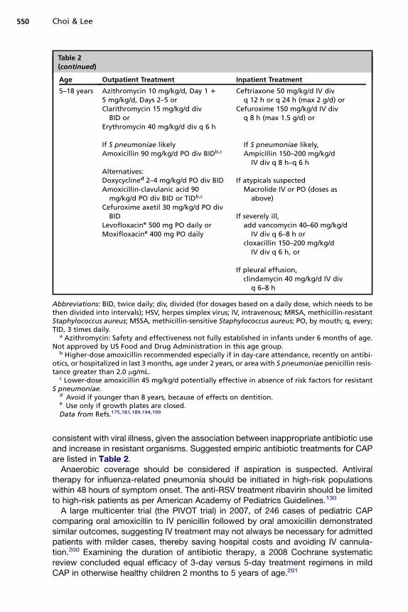
Table 2(continued)
Age Outpatient Treatment Inpatient Treatment
5–18 years Azithromycin 10 mg/kg/d, Day 1 1
5 mg/kg/d, Days 2–5 orClarithromycin 15 mg/kg/d div
BID orErythromycin 40 mg/kg/d div q 6 h
If S pneumoniae likelyAmoxicillin 90 mg/kg/d PO div BIDb,c
Alternatives:Doxycyclined 2–4 mg/kg/d PO div BIDAmoxicillin-clavulanic acid 90
mg/kg/d PO div BID or TIDb,c
Cefuroxime axetil 30 mg/kg/d PO divBID
Levofloxacine 500 mg PO daily orMoxifloxacine 400 mg PO daily
Ceftriaxone 50 mg/kg/d IV divq 12 h or q 24 h (max 2 g/d) or
Cefuroxime 150 mg/kg/d IV divq 8 h (max 1.5 g/d) or
If S pneumoniae likely,Ampicillin 150–200 mg/kg/d
IV div q 8 h–q 6 h
If atypicals suspectedMacrolide IV or PO (doses as
above)
If severely ill,add vancomycin 40–60 mg/kg/d
IV div q 6–8 h orcloxacillin 150–200 mg/kg/d
IV div q 6 h, or
If pleural effusion,clindamycin 40 mg/kg/d IV div
q 6–8 h
Abbreviations: BID, twice daily; div, divided (for dosages based on a daily dose, which needs to bethen divided into intervals); HSV, herpes simplex virus; IV, intravenous; MRSA, methicillin-resistantStaphylococcus aureus; MSSA, methicillin-sensitive Staphylococcus aureus; PO, by mouth; q, every;TID, 3 times daily.
a Azithromycin: Safety and effectiveness not fully established in infants under 6 months of age.Not approved by US Food and Drug Administration in this age group.
b Higher-dose amoxicillin recommended especially if in day-care attendance, recently on antibi-otics, or hospitalized in last 3 months, age under 2 years, or area with S pneumoniae penicillin resis-tance greater than 2.0 mg/mL.
c Lower-dose amoxicillin 45 mg/kg/d potentially effective in absence of risk factors for resistantS pneumoniae.
d Avoid if younger than 8 years, because of effects on dentition.e Use only if growth plates are closed.Data from Refs.175,181,189,194,199
Choi & Lee550
consistent with viral illness, given the association between inappropriate antibiotic useand increase in resistant organisms. Suggested empiric antibiotic treatments for CAPare listed in Table 2.Anaerobic coverage should be considered if aspiration is suspected. Antiviral
therapy for influenza-related pneumonia should be initiated in high-risk populationswithin 48 hours of symptom onset. The anti-RSV treatment ribavirin should be limitedto high-risk patients as per American Academy of Pediatrics Guidelines.130
A large multicenter trial (the PIVOT trial) in 2007, of 246 cases of pediatric CAPcomparing oral amoxicillin to IV penicillin followed by oral amoxicillin demonstratedsimilar outcomes, suggesting IV treatment may not always be necessary for admittedpatients with milder cases, thereby saving hospital costs and avoiding IV cannula-tion.200 Examining the duration of antibiotic therapy, a 2008 Cochrane systematicreview concluded equal efficacy of 3-day versus 5-day treatment regimens in mildCAP in otherwise healthy children 2 months to 5 years of age.201
551Common Pediatric Respiratory Emergencies
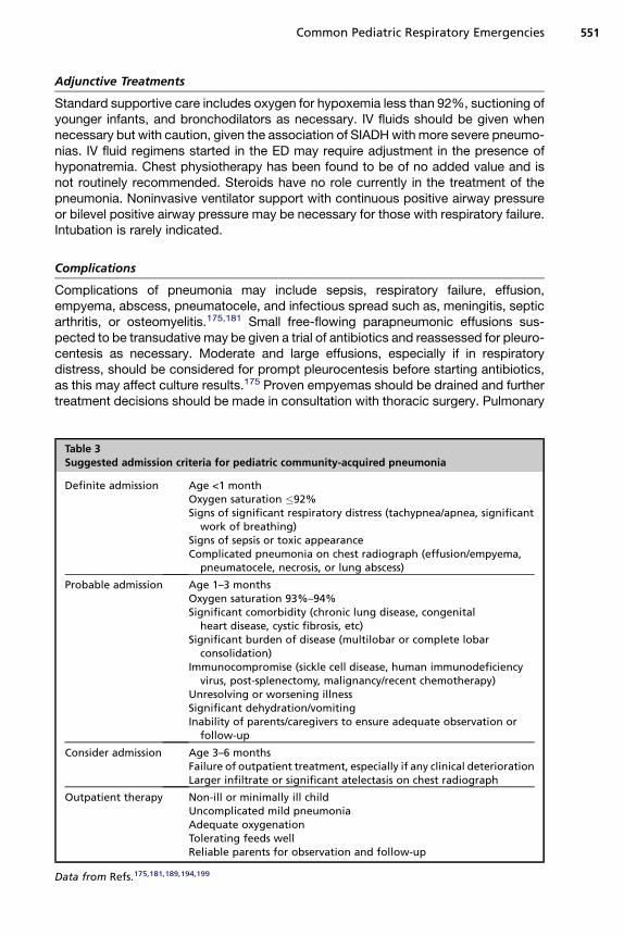
Adjunctive Treatments
Standard supportive care includes oxygen for hypoxemia less than 92%, suctioning ofyounger infants, and bronchodilators as necessary. IV fluids should be given whennecessary but with caution, given the association of SIADHwith more severe pneumo-nias. IV fluid regimens started in the ED may require adjustment in the presence ofhyponatremia. Chest physiotherapy has been found to be of no added value and isnot routinely recommended. Steroids have no role currently in the treatment of thepneumonia. Noninvasive ventilator support with continuous positive airway pressureor bilevel positive airway pressure may be necessary for those with respiratory failure.Intubation is rarely indicated.
Complications
Complications of pneumonia may include sepsis, respiratory failure, effusion,empyema, abscess, pneumatocele, and infectious spread such as, meningitis, septicarthritis, or osteomyelitis.175,181 Small free-flowing parapneumonic effusions sus-pected to be transudative may be given a trial of antibiotics and reassessed for pleuro-centesis as necessary. Moderate and large effusions, especially if in respiratorydistress, should be considered for prompt pleurocentesis before starting antibiotics,as this may affect culture results.175 Proven empyemas should be drained and furthertreatment decisions should be made in consultation with thoracic surgery. Pulmonary
Table 3Suggested admission criteria for pediatric community-acquired pneumonia
Definite admission Age <1 monthOxygen saturation �92%Signs of significant respiratory distress (tachypnea/apnea, significant
work of breathing)Signs of sepsis or toxic appearanceComplicated pneumonia on chest radiograph (effusion/empyema,
pneumatocele, necrosis, or lung abscess)
Probable admission Age 1–3 monthsOxygen saturation 93%–94%Significant comorbidity (chronic lung disease, congenital
heart disease, cystic fibrosis, etc)Significant burden of disease (multilobar or complete lobar
consolidation)Immunocompromise (sickle cell disease, human immunodeficiency
virus, post-splenectomy, malignancy/recent chemotherapy)Unresolving or worsening illnessSignificant dehydration/vomitingInability of parents/caregivers to ensure adequate observation or
follow-up
Consider admission Age 3–6 monthsFailure of outpatient treatment, especially if any clinical deteriorationLarger infiltrate or significant atelectasis on chest radiograph
Outpatient therapy Non-ill or minimally ill childUncomplicated mild pneumoniaAdequate oxygenationTolerating feeds wellReliable parents for observation and follow-up
Data from Refs.175,181,189,194,199

Choi & Lee552
and infectious disease specialist involvement may be warranted for cases of unresolv-ing infiltrates, abscesses, pneumatoceles, or effusions.
Admission Criteria/Predictors of Poor Outcome
Worldwide, significantly at-risk groups include neonates, human immunodeficiencyvirus (HIV)-related pneumonia (3–8 times higher case fatality rates compared withnon-HIV infected), and severely malnourished children.188 Significant hypoxemia isalso associated with a 2- to 5-fold increase in risk of death.202 Predictors of antibiotictreatment failure include young age, immunocompromised state (eg, HIV or malnutri-tion), presence of empyema, prior antibiotic use, poor adherence to treatment, andantibiotic resistance.188
Admission criteria based on published guidelines and literature reviews have beenproposed, though they have not been vigorously studied. Such criteria should be usedwith clinical judgment while taking into account modifying factors. Suggested admis-sion criteria are listed in Table 3.Other options for less ill or complicated patients include admission to a short-stay or
observation unit for children in whom rapid clinical response is expected, or once-dailyIV or IM antibiotics such as ceftriaxone with daily follow-up by an intensive ambulatorycare service, as exists in some major hospitals.
Follow-Up
Forty-eight-hour follow-up is strongly recommended for all children diagnosed withpneumonia,174 especially if any signs of deterioration occur. The World Health Orga-nization definition of clinical improvement requires “slower breathing, less fever, eatingbetter.”202 In children with clinical improvement, follow-up radiography is suggested incases of complicated pneumonia, round pneumonia, congenital abnormalities, lobarcollapse, complicated courses, or persistent clinical abnormalities.181,189 A recentradiographic follow-up study demonstrated that residual or new changes on CXRcould be seen in 30% of cases after 3 to 7 weeks but did not affect management.203
SUMMARY
Common respiratory illnesses including asthma, croup, bronchiolitis, and pneumoniawill undoubtedly continue to make up the bulk of pediatric respiratory problems pre-senting to EDs. Given their potential to cause life-threatening illness, emergency clini-cians will need to continue to maintain and update their knowledge on currentrecommendations for these illnesses. Underuse of some key therapies and overutili-zation of unproven therapies and low-utility diagnostic tests are several importantareas that can be improved through improved education and knowledge of publishedclinical practice guidelines.
REFERENCES
1. American Lung Association, Epidemiology and Statistics Unit. Trends in asthmamorbidity and mortality. Washington, DC. Available at: http://www.lungusa.org/finding-cures/our-research/trend-reports/asthma-trend-report.pdf. AccessedApril 13, 2011.
2. Global Initiative for Asthma. Global strategy for asthma management andprevention. Bethesda (MD): Global Initiative for Asthma; 2010.
3. National Heart, Lung, and Blood Institute (US). Expert panel report 3: guidelinesfor the diagnosis and management of asthma. Bethesda (MD): National Heart,
Common Pediatric Respiratory Emergencies 553
Lung, and Blood Institute, U.S. Department of Health and Human Services,National Institutes of Health; 2007.
4. Busse WW, Lemanske RF. Asthma. N Engl J Med 2001;344(5):350–62.5. Stevenson D, Szczeklik A. Clinical and pathologic perspectives on aspirin sensi-
tivity and asthma. J Allergy Clin Immunol 2006;118(4):773–86 [quiz: 787–8].6. Global Initiative for Asthma. Global strategy for the diagnosis and management
of asthma in children 5 years and younger. Bethesda (MD): Global Initiative forAsthma; 2009.
7. Brand PL, Baraldi E, Bisgaard H, et al. Definition, assessment and treatment ofwheezing disorders in preschool children: an evidence-based approach. EurRespir J 2008;32(4):1096–110.
8. Panickar J, Lakhanpaul M, Lambert P, et al. Oral prednisolone for preschoolchildren with acute virus-induced wheezing. N Engl J Med 2009;360(4):329–38.
9. British Thoracic Society and the Scottish Intercollegiate Guidelines Network.British guideline on the management of asthma. London, England: The BritishThoracic Society; 2009.
10. Ducharme F, Chalut D, Plotnick L, et al. The pediatric respiratory assessmentmeasure: a valid clinical score for assessing acute asthma severity fromtoddlers to teenagers. J Pediatr 2008;152(4):476–80, 480.e471.
11. Chalut DS, Ducharme FM, Davis GM. The Preschool Respiratory AssessmentMeasure (PRAM): a responsive index of acute asthma severity. J Pediatr2000;137(6):762–8.
12. Gorelick M, Stevens M, Schultz T, et al. Performance of a novel clinical score,the Pediatric Asthma Severity Score (PASS), in the evaluation of acute asthma.Acad Emerg Med 2004;11(1):10–8.
13. Gouin S, Robidas I, Gravel J, et al. Prospective evaluation of two clinical scoresfor acute asthma in children 18 months to 7 years of age. Acad Emerg Med2010;17(6):598–603.
14. Goldberg R, Chan L, Haley P, et al. Critical pathway for the emergency depart-ment management of acute asthma: effect on resource utilization. Ann EmergMed 1998;31(5):562–7.
15. Eid N, Yandell B, Howell L, et al. Can peak expiratory flow predict airflowobstruction in children with asthma? Pediatrics 2000;105(2):354–8.
16. Gorelick M, Stevens M, Schultz T, et al. Difficulty in obtaining peak expiratory flowmeasurements in childrenwith acute asthma. Pediatr EmergCare 2004;20(1):22–6.
17. Rodrigo G, Rodriquez Verde M, Peregalli V, et al. Effects of short-term 28% and100% oxygen on PaCO2 and peak expiratory flow rate in acute asthma:a randomized trial. Chest 2003;124(4):1312–7.
18. Camargo CA, Spooner CH, Rowe BH. Continuous versus intermittent beta-agonists in the treatment of acute asthma. Cochrane Database Syst Rev2003;4:CD001115.
19. Papo MC, Frank J, Thompson AE. A prospective, randomized study of contin-uous versus intermittent nebulized albuterol for severe status asthmaticus inchildren. Crit Care Med 1993;21(10):1479–86.
20. Cates CCJ, Bara A, Crilly JA, et al. Holding chambers versus nebulisers forbeta-agonist treatment of acute asthma. Cochrane Database Syst Rev 2003;3:CD000052.
21. Closa RM, Ceballos JM, Gmez-Pap A, et al. Efficacy of bronchodilators admin-istered by nebulizers versus spacer devices in infants with acute wheezing. Pe-diatr Pulmonol 1998;26(5):344–8.

Choi & Lee554
22. Wildhaber JH, Devadason SG, Hayden MJ, et al. Aerosol delivery to wheezyinfants: a comparison between a nebulizer and two small volume spacers. Pe-diatr Pulmonol 1997;23(3):212–6.
23. Rubilar L, Castro Rodriguez JA, Girardi G. Randomized trial of salbutamol viametered-dose inhaler with spacer versus nebulizer for acute wheezing in chil-dren less than 2 years of age. Pediatr Pulmonol 2000;29(4):264–9.
24. Doan Q, Shefrin A, Johnson D. Cost-effectiveness of metered-dose inhalers forasthma exacerbations in the pediatric emergency department. Pediatrics 2011;127(5):e1105–11 peds.2010-2963.
25. Asmus MJ, Hendeles L. Levalbuterol nebulizer solution: is it worth five times thecost of albuterol? Pharmacotherapy 2000;20(2):123–9.
26. Milgrom H, Skoner DP, Bensch G, et al. Low-dose levalbuterol in children withasthma: safety and efficacy in comparison with placebo and racemic albuterol.J Allergy Clin Immunol 2001;108(6):938–45.
27. Nelson HS, Bensch G, Pleskow WW, et al. Improved bronchodilation with leval-buterol compared with racemic albuterol in patients with asthma. J Allergy ClinImmunol 1998;102(6):943–52.
28. Gawchik SM, Saccar CL, Noonan M, et al. The safety and efficacy of nebulizedlevalbuterol compared with racemic albuterol and placebo in the treatment ofasthma in pediatric patients. J Allergy Clin Immunol 1999;103(4):615–21.
29. Carl J, Myers T, Kirchner HL, et al. Comparison of racemic albuterol and leval-buterol for treatment of acute asthma. J Pediatr 2003;143(6):731–6.
30. Tripp K, McVicar W, Nair P, et al. A cumulative dose study of levalbuterol andracemic albuterol administered by hydrofluoroalkane-134a metered-doseinhaler in asthmatic subjects. J Allergy Clin Immunol 2008;122(3):544–9.
31. Lam S, Chen J. Changes in heart rate associated with nebulized racemic albu-terol and levalbuterol in intensive care patients. Am J Health Syst Pharm 2003;60(19):1971–5.
32. Plotnick LH, Ducharme FM. Combined inhaled anticholinergics and beta2-agonists for initial treatment of acute asthma in children. Cochrane DatabaseSyst Rev 2000;4:CD000060.
33. Rodrigo GJ, Castro Rodriguez JA. Anticholinergics in the treatment of childrenand adults with acute asthma: a systematic review with meta-analysis. Thorax2005;60(9):740–6.
34. Rowe BH, Edmonds ML, Spooner CH, et al. Corticosteroid therapy for acuteasthma. Respir Med 2004;98(4):275–84.
35. Fiel S, Vincken W. Systemic corticosteroid therapy for acute asthma exacerba-tions. J Asthma 2006;43(5):321–31.
36. Smith M, Iqbal Shaikh Mohammed SI, Rowe Brian H, et al. Corticosteroids forhospitalised children with acute asthma. Cochrane Database Syst Rev 2003;1.Available at: http://www.mrw.interscience.wiley.com/cochrane/clsysrev/articles/CD002886/frame.html. Accessed May 1, 2011.
37. Becker JM, Arora A, Scarfone RJ, et al. Oral versus intravenous corticosteroidsin children hospitalized with asthma. J Allergy Clin Immunol 1999;103(4):586–90.
38. Barnett PL, Caputo GL, Baskin M, et al. Intravenous versus oral corticosteroidsin the management of acute asthma in children. Ann Emerg Med 1997;29(2):212–7.
39. Kravitz J, Dominici P, Ufberg J, et al. Two days of dexamethasone versus 5 daysof prednisone in the treatment of acute asthma: a randomized controlled trial.Ann Emerg Med 2011;58(2):200–4.

Common Pediatric Respiratory Emergencies 555
40. Greenberg R, Kerby G, Roosevelt G. A comparison of oral dexamethasone withoral prednisone in pediatric asthma exacerbations treated in the emergencydepartment. Clin Pediatr 2008;47(8):817–23.
41. Shefrin R. Use of dexamethasone and prednisone in acute asthma exacerba-tions in pediatric patients. Can Fam Physician 2009;55(7):704–6.
42. Altamimi M. Single-dose oral dexamethasone in the emergency management ofchildren with exacerbations of mild to moderate asthma. Pediatr Emerg Care2006;22(12):786–93.
43. Gordon S, Tompkins T, Dayan P. Randomized trial of single-dose intramusculardexamethasone compared with prednisolone for children with acute asthma.Pediatr Emerg Care 2007;23(8):521–7.
44. Gries DM, Moffitt DR, Pulos E, et al. A single dose of intramuscularly adminis-tered dexamethasone acetate is as effective as oral prednisone to treat asthmaexacerbations in young children. J Pediatr 2000;136(3):298–303.
45. FitzGerald JM,BeckerA, SearsMR, et al. Doubling thedoseof budesonide versusmaintenance treatment in asthma exacerbations. Thorax 2004;59(7):550–6.
46. Edmonds ML, Camargo CA, Pollack CV, et al. Early use of inhaled corticoste-roids in the emergency department treatment of acute asthma. Cochrane Data-base Syst Rev 2003;3:CD002308.
47. Schuh S, Dick P, Stephens D, et al. High-dose inhaled fluticasone does notreplace oral prednisolone in children with mild to moderate acute asthma. Pedi-atrics 2006;118(2):644–50.
48. Schuh S, Reisman J, Alshehri M, et al. A comparison of inhaled fluticasone andoral prednisone for children with severe acute asthma. N Engl J Med 2000;343(10):689–94.
49. Upham BD, Mollen CJ, Scarfone RJ, et al. Nebulized budesonide added tostandard pediatric emergency department treatment of acute asthma:a randomized, double-blind trial. Acad Emerg Med 2011;18(7):665–73.
50. Spivey WH, Skobeloff EM, Levin RM. Effect of magnesium chloride on rabbitbronchial smooth muscle. Ann Emerg Med 1990;19(10):1107–12.
51. Ciarallo L, Sauer AH, Shannon MW. Intravenous magnesium therapy formoderate to severe pediatric asthma: results of a randomized, placebo-controlled trial. J Pediatr 1996;129(6):809–14.
52. Noppen M, Vanmaele L, Impens N, et al. Bronchodilating effect of intravenousmagnesium sulfate in acute severe bronchial asthma. Chest 1990;97(2):373–6.
53. Rowe BH, Bretzlaff JA, Bourdon C, et al. Intravenous magnesium sulfate treat-ment for acute asthma in the emergency department: a systematic review ofthe literature. Ann Emerg Med 2000;36(3):181–90.
54. Rowe BH, Bretzlaff JA, Bourdon C, et al. Magnesium sulfate for treating exacer-bations of acute asthma in the emergency department. Cochrane DatabaseSyst Rev 2000;2:CD001490.
55. Gallegos-Solorzano MC, Perez-Padilla R, Hernandez-Zenteno RJ. Usefulness ofinhaled magnesium sulfate in the coadjuvant management of severe asthmacrisis in an emergency department. Pulm Pharmacol Ther 2010;23(5):432–7.
56. Hughes R, Goldkorn A, Masoli M, et al. Use of isotonic nebulised magnesiumsulphate as an adjuvant to salbutamol in treatment of severe asthma in adults:randomised placebo-controlled trial. Lancet 2003;361(9375):2114–7.
57. Bessmertny O, DiGregorio RV, Cohen H, et al. A randomized clinical trial ofnebulized magnesium sulfate in addition to albuterol in the treatment of acutemild-to-moderate asthma exacerbations in adults. Ann Emerg Med 2002;39(6):585–91.
Choi & Lee556
58. Blitz M, Blitz S, Beasely R, et al. Inhaled magnesium sulfate in the treatment ofacute asthma. Cochrane Database Syst Rev 2005;4. Available at: http://www.mrw.interscience.wiley.com/cochrane/clsysrev/articles/CD003898/frame.html.Accessed May 1, 2011.
59. Harmanci K, Bakirtas A, Turktas I, et al. Oral montelukast treatment of preschool-aged children with acute asthma. Ann Allergy Asthma Immunol 2006;96(5):731–5.
60. Nelson K, Smith S, Trinkaus K, et al. Pilot study of oral montelukast added tostandard therapy for acute asthma exacerbations in children aged 6 to 14 years.Pediatr Emerg Care 2008;24(1):21–7.
61. Dockhorn RJ, Baumgartner RA, Leff JA, et al. Comparison of the effects of intrave-nous and oral montelukast on airway function: a double blind, placebo controlled,three period, crossover study in asthmatic patients. Thorax 2000;55(4):260–5.
62. Camargo C, Smithline H, Malice MP, et al. A randomized controlled trial of intra-venous montelukast in acute asthma. Am J Respir Crit Care Med 2003;167(4):528–33.
63. Gupta VK, Cheifetz IM. Heliox administration in the pediatric intensive care unit:an evidence-based review. Pediatr Crit Care Med 2005;6(2):204–11.
64. Kim I, Phrampus E, Venkataraman S, et al. Helium/oxygen-driven albuterolnebulization in the treatment of children with moderate to severe asthma exac-erbations: a randomized, controlled trial. Pediatrics 2005;116(5):1127–33.
65. Lee D, Hsu CW, Lee H, et al. Beneficial effects of albuterol therapy driven by he-liox versus by oxygen in severe asthma exacerbation. Acad Emerg Med 2005;12(9):820–7.
66. Rivera M, Kim T, Stewart G, et al. Albuterol nebulized in heliox in the initial EDtreatment of pediatric asthma: a blinded, randomized controlled trial. Am JEmerg Med 2006;24(1):38–42.
67. Travers A, Jones AP, Kelly K, et al. Intravenous beta2-agonists for acute asthmain the emergency department. Cochrane Database Syst Rev 2001;2:CD002988:
68. Mitra A, Bassler D, Goodman K, et al. Intravenous aminophylline for acutesevere asthma in children over two years receiving inhaled bronchodilators. Co-chrane Database Syst Rev 2005;2:CD001276.
69. Ream RS, Loftis LL, Albers GM, et al. Efficacy of IV theophylline in children withsevere status asthmaticus. Chest 2001;119(5):1480–8.
70. Akingbola O, Simakajornboon N, Hadley E Jr, et al. Noninvasive positive-pressure ventilation in pediatric status asthmaticus. Pediatr Crit Care Med2002;3(2):181–4.
71. Bernet V, Hug M, Frey B. Predictive factors for the success of noninvasive maskventilation in infants and children with acute respiratory failure. Pediatr Crit CareMed 2005;6(6):660–4.
72. Carroll C, Schramm C. Noninvasive positive pressure ventilation for the treat-ment of status asthmaticus in children. Ann Allergy Asthma Immunol 2006;96(3):454–9.
73. Essouri S, Chevret L, Durand P, et al. Noninvasive positive pressure ventilation:five years of experience in a pediatric intensive care unit. Pediatr Crit Care Med2006;7(4):329–34.
74. Padman R, Lawless ST, Kettrick RG. Noninvasive ventilation via bilevel positiveairway pressure support in pediatric practice. Crit Care Med 1998;26(1):169–73.
75. Thill P, McGuire J, Baden H, et al. Noninvasive positive-pressure ventilation inchildren with lower airway obstruction. Pediatr Crit Care Med 2004;5(4):337–42.
Common Pediatric Respiratory Emergencies 557
76. Ram FS, Wellington S, Rowe B, et al. Non-invasive positive pressure ventilationfor treatment of respiratory failure due to severe acute exacerbations of asthma.Cochrane Database Syst Rev 2005;3:CD004360.
77. Brown RH, Wagner EM. Mechanisms of bronchoprotection by anesthetic induc-tion agents: propofol versus ketamine. Anesthesiology 1999;90(3):822–8.
78. Allen J, Macias C. The efficacy of ketamine in pediatric emergency departmentpatients who present with acute severe asthma. Ann Emerg Med 2005;46(1):43–50.
79. Howton JC, Rose J, Duffy S, et al. Randomized, double-blind, placebo-controlled trial of intravenous ketamine in acute asthma. Ann Emerg Med1996;27(2):170–5.
80. Green S, Roback M, Krauss B. Anticholinergics and ketamine sedation in chil-dren: a secondary analysis of atropine versus glycopyrrolate. Acad EmergMed 2010;17(2):157–62.
81. Green SM, Roback MG, Kennedy RM, et al. Clinical practice guideline for emer-gency department ketamine dissociative sedation: 2011 update. Ann EmergMed 2011;57(5):449–61.
82. Bellomo R, McLaughlin P, Tai E, et al. Asthma requiring mechanical ventilation. Alow morbidity approach. Chest 1994;105(3):891–6.
83. Cox RG, Barker GA, Bohn DJ. Efficacy, results, and complications of mechan-ical ventilation in children with status asthmaticus. Pediatr Pulmonol 1991;11(2):120–6.
84. Darioli R, Perret C. Mechanical controlled hypoventilation in status asthmaticus.Am Rev Respir Dis 1984;129(3):385–7.
85. Dworkin G, Kattan M. Mechanical ventilation for status asthmaticus in children.J Pediatr 1989;114(4):545–9.
86. Hickling KG, Walsh J, Henderson S, et al. Low mortality rate in adult respiratorydistress syndrome using low-volume, pressure-limited ventilation with permis-sive hypercapnia: a prospective study. Crit Care Med 1994;22(10):1568–78.
87. Menitove SM, Goldring RM. Combined ventilator and bicarbonate strategy in themanagement of status asthmaticus. Am J Med 1983;74(5):898–901.
88. Sin D, Bell N, Svenson L, et al. The impact of follow-up physician visits on emer-gency readmissions for patients with asthma and chronic obstructive pulmonarydisease: a population-based study. Am J Med 2002;112(2):120–5.
89. Zeiger RS, Heller S, Mellon MH, et al. Facilitated referral to asthma specialistreduces relapses in asthma emergency room visits. J Allergy Clin Immunol1991;87(6):1160–8.
90. Zorc J, Scarfone R, Li Y, et al. Scheduled follow-up after a pediatric emergencydepartment visit for asthma: a randomized trial. Pediatrics 2003;111(3):495–502.
91. Baren J, Boudreaux E, Brenner B, et al. Randomized controlled trial of emer-gency department interventions to improve primary care follow-up for patientswith acute asthma. Chest 2006;129(2):257–65.
92. Sin D, Man SF. Low-dose inhaled corticosteroid therapy and risk of emergencydepartment visits for asthma. Arch Intern Med 2002;162(14):1591–5.
93. Edmonds M, Brenner Barry E, Camargo Carlos A, et al. Inhaled steroids foracute asthma following emergency department discharge. Cochrane DatabaseSyst Rev 2000;3. Available at: http://www.mrw.interscience.wiley.com/cochrane/clsysrev/articles/CD002316/frame.html. Accessed May 1, 2011.
94. Cowie RL, Revitt SG, Underwood MF, et al. The effect of a peak flow-basedaction plan in the prevention of exacerbations of asthma. Chest 1997;112(6):1534–8.

Choi & Lee558
95. Cherry J. Clinical practice. Croup. N Engl J Med 2008;358(4):384–91.96. Segal A, Crighton E, Moineddin R, et al. Croup hospitalizations in Ontario: a 14-
year time-series analysis. Pediatrics 2005;116(1):51–5.97. Peltola V, Heikkinen T, Ruuskanen O. Clinical courses of croup caused by influ-
enza and parainfluenza viruses. Pediatr Infect Dis J 2002;21(1):76–8.98. Groblewski J, Shah R, Zalzal G. Microdebrider-assisted supraglottoplasty for
laryngomalacia. Ann Otol Rhinol Laryngol 2009;118(8):592–7.99. Antony R, Al Rawas A, Irwin M. Stridor in a newborn. CMAJ 2005;173(6):601–2.
100. Mancuso RF. Stridor in neonates. Pediatr Clin North Am 1996;43(6):1339–56.101. Koplewitz B, Springer C, Slasky B, et al. CT of hemangiomas of the upper
airways in children. AJR Am J Roentgenol 2005;184(2):663–70.102. Rahbar R, Litrovnik B, Vargas S, et al. The biology and management of laryngeal
neurofibroma. Arch Otolaryngol Head Neck Surg 2004;130(12):1400–6.103. Durden F, Sobol S. Balloon laryngoplasty as a primary treatment for subglottic
stenosis. Arch Otolaryngol Head Neck Surg 2007;133(8):772–5.104. Spanos W, Brookes J, Smith M, et al. Unilateral vocal fold paralysis in premature
infants after ligation of patent ductus arteriosus: vascular clip versus suture liga-ture. Ann Otol Rhinol Laryngol 2009;118(10):750–3.
105. Strife JL. Upper airway and tracheal obstruction in infants and children. RadiolClin North Am 1988;26(2):309–22.
106. Westley CR, Cotton EK, Brooks JG. Nebulized racemic epinephrine by IPPB forthe treatment of croup: a double-blind study. Am J Dis Child 1978;132(5):484–7.
107. Bjornson CL, Johnson DW. Croup—treatment update. Pediatr Emerg Care 2005;21(12):863–70 [quiz: 871–3].
108. Sparrow A, Geelhoed G. Prednisolone versus dexamethasone in croup: a rand-omised equivalence trial. Arch Dis Child 2006;91(7):580–3.
109. Russell K, Liang Y, O’Gorman K, et al. Glucocorticoids for croup. CochraneDatabase Syst Rev 2011;1:CD001955.
110. Cetinkaya F, Tfeki B, Kutluk G. A comparison of nebulized budesonide, andintramuscular, and oral dexamethasone for treatment of croup. Int J Pediatr Oto-rhinolaryngol 2004;68(4):453–6.
111. GeelhoedGC,MacdonaldWB.Oraldexamethasone in the treatmentof croup: 0.15mg/kg versus 0.3 mg/kg versus 0.6 mg/kg. Pediatr Pulmonol 1995;20(6):362–8.
112. Dobrovoljac M, Geelhoed G. 27 years of croup: an update highlighting theeffectiveness of 0.15 mg/kg of dexamethasone. Emerg Med Australas 2009;21(4):309–14.
113. Chub Uppakarn S, Sangsupawanich P. A randomized comparison of dexameth-asone 0.15 mg/kg versus 0.6 mg/kg for the treatment of moderate to severecroup. Int J Pediatr Otorhinolaryngol 2007;71(3):473–7.
114. Duman M, Ozdemir D, Atasever S. Nebulised L-epinephrine and steroid combi-nation in the treatment of moderate to severe croup. Clin Drug Investig 2005;25(3):183–9.
115. Klassen TP, Craig WR, Moher D, et al. Nebulized budesonide and oral dexa-methasone for treatment of croup: a randomized controlled trial. JAMA 1998;279(20):1629–32.
116. Geelhoed GC. Budesonide offers no advantage when added to oral dexameth-asone in the treatment of croup. Pediatr Emerg Care 2005;21(6):359–62.
117. Bjornson C, Russell Kelly F, Vandermeer B, et al. Nebulized epinephrine for croupin children. Cochrane Database Syst Rev 2011;2. Available at: http://www.mrw.interscience.wiley.com/cochrane/clsysrev/articles/CD006619/frame.html. Ac-cessed May 1, 2011.
Common Pediatric Respiratory Emergencies 559
118. Taussig LM, Castro O, Beaudry PH, et al. Treatment of laryngotracheobronchitis(croup). Use of intermittent positive-pressure breathing and racemic epineph-rine. Am J Dis Child 1975;129(7):790–3.
119. Scolnik D, Coates A, Stephens D, et al. Controlled delivery of high vs lowhumidity vs mist therapy for croup in emergency departments: a randomizedcontrolled trial. JAMA 2006;295(11):1274–80.
120. NetoG,KentabO,KlassenT, et al. A randomizedcontrolled trial ofmist in theacutetreatment of moderate croup. Acad Emerg Med 2002;9(9):873–9.
121. Moore M, Little P. Humidified air inhalation for treating croup. CochraneDatabase Syst Rev 2006;(3). Available at: http://www.mrw.interscience.wiley.com/cochrane/clsysrev/articles/CD002870/frame.html. Accessed May 1, 2011.
122. Weber JE, Chudnofsky CR, Younger JG, et al. A randomized comparison ofhelium-oxygen mixture (Heliox) and racemic epinephrine for the treatmentof moderate to severe croup. Pediatrics 2001;107(6):E96.
123. Terregino CA, Nairn SJ, Chansky ME, et al. The effect of heliox on croup: a pilotstudy. Acad Emerg Med 1998;5(11):1130–3.
124. Vorwerk C, Coats T. Heliox for croup in children. Cochrane Database Syst Rev2010;2:CD006822.
125. Everard M. Acute bronchiolitis and croup. Pediatr Clin North Am 2009;56(1):119–33.
126. Ducharme FM. Management of acute bronchiolitis. BMJ 2011;342:d1658.127. Mansbach JM, McAdam AJ, Clark S, et al. Prospective multicenter study of the
viral etiology of bronchiolitis in the emergency department. Acad Emerg Med2008;15(2):111–8.
128. Midulla F, Scagnolari C, Bonci E, et al. Respiratory syncytial virus, human boca-virus and rhinovirus bronchiolitis in infants. Arch Dis Child 2010;95(1):35–41.
129. Miron D, Srugo I, Kra-Oz Z, et al. Sole pathogen in acute bronchiolitis—is therea role for other organisms apart from RSV? Pediatr Infect Dis J 2010;2010(29):e7–10.
130. American Academy of Pediatrics Subcommittee on Diagnosis and Managementof Bronchiolitis. Diagnosis and management of bronchiolitis. Pediatrics 2006;118(4):1774–93.
131. Shay DK, Holman RC, Newman RD, et al. Bronchiolitis-associated hospitaliza-tions among US children, 1980-1996. JAMA 1999;282(15):1440–6.
132. Shay DK, Holman RC, Roosevelt GE, et al. Bronchiolitis-associated mortalityand estimates of respiratory syncytial virus-associated deaths among US chil-dren, 1979-1997. J Infect Dis 2001;183(1):16–22.
133. Swingler GH, Hussey GD, Zwarenstein M. Duration of illness in ambulatory chil-dren diagnosed with bronchiolitis. Arch Pediatr Adolesc Med 2000;154(10):997–1000.
134. Petruzella FD, Gorelick MH. Duration of illness in infants with bronchiolitis eval-uated in the emergency department. Pediatrics 2010;126(2):285–90.
135. Bialy L, FoisyM, SmithM, et al. TheCochrane library and the treatment of bronchio-litis in children: an overview of reviews. Evid Base Child Health 2011;6(1):258–75.
136. Goutzamanis J. Bronchiolitis. In: Frank LR, Jobe KA, editors. Admission &discharge decisions in emergency medicine. Philadelphia: Hanley & Belfus,Inc; 2002. p. 246–50.
137. Everard M. What link between early respiratory viral infections and atopicasthma? Lancet 1999;354(9178):527–8.
138. Gern JE. Viral respiratory infection and the link to asthma. Pediatr Infect Dis J2008;27(10):S97–103.

Choi & Lee560
139. Stein RT, Sherrill D, Morgan WJ, et al. Respiratory syncytial virus in early lifeand risk of wheeze and allergy by age 13 years. Lancet 1999;354(9178):541–5.
140. Swingler GH, Hussey GD, Zwarenstein M. Randomised controlled trial of clinicaloutcome after chest radiograph in ambulatory acute lower-respiratory infectionin children. Lancet 1998;351(9100):404–8.
141. Schuh S, Lalani A, Allen U, et al. Evaluation of the utility of radiography in acutebronchiolitis. J Pediatr 2007;150(4):429–33.
142. Mahabee-Gittens EM, Bachman DT, Shapiro ED, et al. Chest radiographs in thepediatric emergency department for children �18 months of age with wheezing.Clin Pediatr 1999;38(7):395–9.
143. Purcell K, Fergie J. Lack of usefulness of an abnormal white blood cell count forpredicting a concurrent serious bacterial infection in infants and young childrenhospitalized with respiratory syncytial virus lower respiratory tract infection. Pe-diatr Infect Dis J 2007;2007(26):311–5.
144. Peltola V, Mertsola J, Ruuskanen O. Comparison of total white blood cell countand serum C-reactive protein levels in confirmed bacterial and viral infections.J Pediatr 2006;149(5):721–4.
145. Liebelt EL, Qi K, Harvey K. Diagnostic testing for serious bacterial infections ininfants aged 90 days or younger with bronchiolitis. Arch Pediatr Adolesc Med1999;153(5):525–30.
146. Antanow J, Hansen K, McKinstry CA. Sepsis evaluations in hospitalized infantswith bronchiolitis. Pediatr Infect Dis J 1998;17(3):231–6.
147. Thorburn K, Harigopal S, Reddy V, et al. High incidence of pulmonary bacterialco-infection in children with severe respiratory syncytial virus (RSV) bronchioli-tis. Thorax 2006;61(7):611–5.
148. Bilavsky E, Shouval D, Yarden-Bilavsky H. A prospective study of the risk forserious bacterial infections in hospitalized febrile infants with or without bron-chiolitis. Pediatr Infect Dis J 2008;27(3):269–82.
149. Duttweiler L, Nadal D, Frey B. Pulmonary and systemic bacterial co-infections insevere RSV bronchiolitis. Arch Dis Child 2004;89(12):1155–7.
150. Hartling L, Bialy Liza M, Vandermeer B, et al. Epinephrine for bronchiolitis. CochraneDatabase Syst Rev 2011;(6). Available at: http://www.mrw.interscience.wiley.com/cochrane/clsysrev/articles/CD003123/frame.html. Accessed April 13, 2011.
151. Hartling L, Fernandes RM, Bialy L, et al. Steroids and bronchodilators for acutebronchiolitis in the first two years of life: systematic review and meta-analysis.BMJ 2011;342:d1714.
152. Chavasse Richard JPG, Seddon P, Bara A, et al. Short acting beta2-agonists forrecurrent wheeze in children under two years of age. Cochrane Database SystRev 2002;2. Available at: http://www.mrw.interscience.wiley.com/cochrane/clsysrev/articles/CD002873/frame.html. Accessed April 13, 2011.
153. Everard M, Bara A, Kurian M, et al. Anticholinergic drugs for wheeze in childrenunder the age of two years. Cochrane Database Syst Rev 2005;3. Available at:http://www.mrw.interscience.wiley.com/cochrane/clsysrev/articles/CD001279/frame.html. Accessed April 13, 2011.
154. Fernandes Ricardo M, Bialy Liza M, Vandermeer B, et al. Glucocorticoids foracute viral bronchiolitis in infants and young children. Cochrane DatabaseSyst Rev 2010;10. Available at: http://www.mrw.interscience.wiley.com/cochrane/clsysrev/articles/CD004878/frame.html. Accessed April 13, 2011.
155. Zhang L, Mendoza-Sassi Raul A, Wainwright C, et al. Nebulized hypertonicsaline solution for acute bronchiolitis in infants. Cochrane Database Syst Rev

Common Pediatric Respiratory Emergencies 561
2008;4. Available at: http://www.mrw.interscience.wiley.com/cochrane/clsysrev/articles/CD006458/frame.html. Accessed April 13, 2011.
156. Liet JM,Ducruet T,GuptaV, et al. Heliox inhalation therapy forbronchiolitis in infants.Cochrane Database Syst Rev 2010;4. Available at: http://www.mrw.interscience.wiley.com/cochrane/clsysrev/articles/CD006915/frame.html. Accessed April 13,2011.
157. Bronchiolitis Guideline Team, Cincinatti Children’s Hospital Medical Team.Evidenced-based care guideline for management of bronchiolitis in infants 1year of age or less with a first time epidose. Cincinnatti (OH): Bronchiolitis Pedi-atric Evidence-Based Care Guidelines, Cincinnatti Children’s Hospital MedicalCenter. Available at: http://www.cincinnatichildrens.org/service/j/anderseon-center/evidence-based-care/bronchiolitis/. Accessed October 27, 2011.
158. Turner T, WIlkinson F, Harris C. Evidence-based guideline for the managementof bronchiolitis. Aust Fam Physician 2008;37(6):6–13.
159. Bronchiolitis in children—a national clinical guideline. Scottish IntercollegiateGuidelines Network; 2006. Available at: http://www.sign.ac.uk/pdf/sign91.pdf.Accessed April 13, 2011.
160. Gadomski Anne M, Brower M. Bronchodilators for bronchiolitis. Cochrane Data-base Syst Rev 2010;12. Available at: http://www.mrw.interscience.wiley.com/cochrane/clsysrev/articles/CD001266/frame.html. Accessed April 13, 2011.
161. Seiden JA, Scarfone RJ. Bronchiolitis: an evidence-based approach to manage-ment. Clin Pediatr Emerg Med 2009;10(2):75–81.
162. Grewal S, Ali S, McConnell DW, et al. A randomized trial of nebulized 3% hyper-tonic saline with epinephrine in the treatment of acute bronchiolitis in the emer-gency department. Arch Pediatr Adolesc Med 2009;163(11):1007–12.
163. Corneli HM, Zorc JJ, Mahajan P, et al. A multicenter, randomized, controlled trialof dexamethasone for bronchiolitis. N Engl J Med 2007;357(4):331–9.
164. Plint AC, Johnson DW, Patel H, et al. Epinephrine and dexamethasone in chil-dren with bronchiolitis. N Engl J Med 2009;360(20):2079–89.
165. Wark P, McDonald Vanessa M. Nebulised hypertonic saline for cystic fibrosis. Co-chrane Database Syst Rev 2009;2. Available at: http://www.mrw.interscience.wiley.com/cochrane/clsysrev/articles/CD001506/frame.html. Accessed April 13, 2011.
166. Chaudhry K, Sinert R. Is nebulized hypertonic saline solution an effective treat-ment for bronchiolitis in infants? Ann Emerg Med 2010;55(1):120–2.
167. Kuzik BA, Flavin MP, Kent S, et al. Effect of inhaled hypertonic saline on hospitaladmission rate in children with viral bronchiolitis: a randomized trial. CJEM 2010;12(6):477.
168. Kim IK, Corcoran T. Recent developments in heliox therapy for asthma andbronchiolitis. Clin Pediatr Emerg Med 2009;10(2):68–74.
169. Mansbach JM, Clark S, Christopher NC, et al. Prospective multicenter study ofbronchiolitis: predicting safe discharges from the emergency department. Pedi-atrics 2008;121(4):680–8.
170. Willwerth BM, Harper MB, Greenes DS. Identifying hospitalized infants who havebronchiolitis and are at high risk for apnea. Ann Emerg Med 2006;48(4):441–7.
171. Norwood A, Mansbach JM, Clark S, et al. Prospective multicenter study of bron-chiolitis: predictors of an unscheduled visit after discharge from the emergencydepartment. Acad Emerg Med 2010;17(4):376–82.
172. Marlais M, Evans J, Abrahamson E. Clinical predictors of admission in infantswith acute bronchiolitis. Arch Dis Child 2011;96(7):648–52.
173. Black RE, Cousens S, Johnson HL, et al. Global, regional, and national causes ofchild mortality in 2008: a systematic analysis. Lancet 2010;375(9730):1969–87.

Choi & Lee562
174. Homier V, Bellevance C, Xhignesse M. Prevalence of pneumonia in childrenunder 12 years of age who undergo abdominal radiography in the emergencydepartment. CJEM 2007;9(5):347–51.
175. Evidence-Based (EB) Clinical Decision Support Team & Community-AcquiredPneumonia Content Expert Team - Texas Children’s Hospital. Community-acquired pneumonia (CAP) clinical guideline. Available at: http://www.bcm.edu/web/pediatrics/documents/rp_archive_21.pdf. AccessedOctober 27, 2011.
176. Murphy CG, Van De Pol AC, Harper MB, et al. Clinical predictors of occult pneu-monia in the febrile child. Acad Emerg Med 2007;14(3):243–9.
177. Shah S, Bachur R, Kim D, et al. Lack of predictive value of tachypnea in thediagnosis of pneumonia in children. Pediatr Infect Dis J 2010;29:406–9.
178. Korppi M, Don M, Valent F, et al. The value of clinical features in differentiatingbetween viral, pneumococcal and atypical bacterial pneumonia in children.Acta Paediatr 2008;97(7):943–7.
179. Mathews B, Shah S, Cleveland RH, et al. Clinical predictors of pneumoniaamong children with wheezing. Pediatrics 2009;124(1):e29–36.
180. Korppi M. Antibiotic therapy for pneumonia in the pediatric population. PediatrHealth 2007;1(1):77.
181. British Thoracic Society. BTS guidelines for the management of community-acquired pneumonia in childhood. Thorax 2002;57(Suppl 1):i1–24.
182. Lahti E, Peltola V, Virkki R, et al. Development of parapneumonic empyema inchildren. Acta Paediatr 2007;96(11):1686–92.
183. Virkki R, Juven T, Rikalainen H, et al. Differentiation of bacterial and viral pneu-monia in children. Thorax 2002;57(5):438–41.
184. Michelow IC, Olsen K, Lozano J, et al. Epidemiology and clinical characteristicsof community-acquired pneumonia in hospitalized children. Pediatrics 2004;113(4):701–7.
185. Carrillo-Marquez M, Hulten K, Hammerman W, et al. Staphylococcus aureuspneumonia in children in the era of community-acquired methicillin-resistanceat Texas Children’s Hospital. Pediatr Infect Dis J 2011;30(7):545–50.
186. Wallin T, Hern H, Frazee B. Community-acquired methicillin-resistant Staphylo-coccus aureus. Emerg Med Clin North Am 2008;26:431–55.
187. Liu C, Bayer A, Cosgrove SE, et al. Clinical practice guidelines by the InfectiousDiseases Society of America for the treatment of methicillin-resistant Staphylo-coccus aureus infections in adults and children: executive summary. Clin InfectDis 2011;52(3):285–92.
188. Graham S, English M, Hazir T, et al. Challenges to improving case managementof childhood pneumonia at health facilities in resource-limited settings. BullWorld Health Organ 2008;86(5):349–55.
189. Clinical Practice Guidelines Group - Toward Optimized Practice (TOP) Program.Guideline for the diagnosis and management of community acquired pneu-monia: pediatric - 2008 Update. Available at: http://www.topalbertadoctors.org/cpgs.php?sid=15&cpg_cats=61. Accessed April 26, 2011.
190. Newman M, Scully K, Kim D. Physician assessment of the likelihood of pneu-monia in pediatric emergency department. Pediatr Emerg Care 2010;26(11):817–22.
191. Lynch T, Bialy L, Kellner J. A systematic review on the diagnosis of pediatricbacterial pneumonia: when gold is bronze. PLoS One 2010;5(8):1–7.
192. Cortellaro F, Colombo S, Coen D, et al. Lung ultrasound is an accurate diag-nostic tool for the diagnosis of pneumonia in the emergency department. EmergMed J 2010. [Epub ahead of print].

Common Pediatric Respiratory Emergencies 563
193. Kurian J, Levin TL, Han BK, et al. Comparison of ultrasound and CT in the eval-uation of pneumonia complicated by parapneumonic effusion in children. Am JRoentgenol 2009;193(6):1648–54.
194. Community Acquired Pneumonia Guideline Team, Cincinnati Children’s HospitalMedical Center. Evidence-based care guideline for medical management ofcommunity acquired pneumonia in children 60 days to 17 years of age. Avail-able at: http://www.cincinnatichildrens.org/service/j/anderson-center/evidence-based-care/community-acquired-pneumonia/. Accessed October 27, 2011.
195. Shah S, Dugan M, Bell L, et al. Blood cultures in the emergency departmentevaluation of childhood pneumonia. Pediatr Infect Dis J 2011;30:475–9.
196. Hickey RW, Bowman MJ, Smith GA. Utility of blood cultures in pediatric patientsfound to have pneumonia in the emergency department. Ann Emerg Med 1996;27(6):721–5.
197. Jadavji T, Law B. A practical guide for the diagnosis and treatment of pediatricpneumonia. CMAJ 1997;156(5):S703.
198. Wilde JA. Rapid diagnostic testing for the identification of respiratory agents inthe emergency department. Clin Pediatr Emerg Med 2002;3(3):181–90.
199. Bradley JS, Byington CL, Shah SS, et al. The management of community-acquired pneumonia in infants and children older than 3 months of age: clinicalpractice guidelines by the Pediatric Infectious Diseases Society and the Infec-tious Diseases Society of America. Clin Infect Dis 2011;53(7):e25–76.
200. Atkinson M, Lakhanpaul M, Smyth A, et al. Comparison of oral amoxicillin andintravenous benzyl penicillin for community acquired pneumonia in children(PIVOT trial): a multicentre pragmatic randomised controlled equivalence trial.Thorax 2007;62(12):1102–6.
201. Haider Batool A, Lassi Zohra S, Bhutta Zulfiqar A. Short-course versus long-course antibiotic therapy for non-severe community-acquired pneumonia inchildren aged 2 months to 59 months. Cochrane Database Syst Rev 2008;2.Available at: http://www.mrw.interscience.wiley.com/cochrane/clsysrev/articles/CD005976/frame.html. Accessed June 12, 2011.
202. Ayieko P, English M. Case management of childhood pneumonia in developingcountries. Pediatr Infect Dis J 2007;26(5):432–40.
203. Virkki R, Juven T, Mertsola J, et al. Radiographic follow-up of pneumonia in chil-dren. Pediatr Pulmonol 2005;40(3):223–7.