community structure of subsurface biofilms in the thermal
TRANSCRIPT
APPLIED AND ENVIRONMENTAL MICROBIOLOGY, Sept. 2010, p. 5902–5910 Vol. 76, No. 170099-2240/10/$12.00 doi:10.1128/AEM.00647-10Copyright © 2010, American Society for Microbiology. All Rights Reserved.
Community Structure of Subsurface Biofilms in the Thermal SulfidicCaves of Acquasanta Terme, Italy�†
D. S. Jones,1 D. J. Tobler,1‡ I. Schaperdoth,1 M. Mainiero,2 and J. L. Macalady1*Pennsylvania State University, Department of Geosciences, University Park, Pennsylvania 16802,1 and
Studio Geologico Mainiero, Via Francesco Podesti 8, 60122 Ancona, Italy2
Received 15 March 2010/Accepted 7 July 2010
We performed a microbial community analysis of biofilms inhabiting thermal (35 to 50°C) waters more than 60 mbelow the ground surface near Acquasanta Terme, Italy. The groundwater hosting the biofilms has 400 to 830 �Msulfide, <10 �M O2, pH of 6.3 to 6.7, and specific conductivity of 8,500 to 10,500 �S/cm. Based on the results of 16SrRNA gene cloning and fluorescent in situ hybridization (FISH), the biofilms have low species richness, andlithoautotrophic (or possibly mixotrophic) Gamma- and Epsilonproteobacteria are the principle biofilm architects.Deltaproteobacteria sequences retrieved from the biofilms have <90% 16S rRNA similarity to their closest relativesin public databases and may represent novel sulfate-reducing bacteria. The Acquasanta biofilms share few speciesin common with Frasassi cave biofilms (13°C, 80 km distant) but have a similar community structure, withrepresentatives in the same major clades. The ecological success of Sulfurovumales-group Epsilonproteobacteria in theAcquasanta biofilms is consistent with previous observations of their dominance in sulfidic cave waters withturbulent water flow and high dissolved sulfide/oxygen ratios.
Despite rapid progress in the past decade, the deep subsurfaceremains one of the least explored microbial habitats on earth.Recent studies illustrate the presence of significant spatial hetero-geneity (13, 53) and the strong influence of mineralogy and fluidflow on subsurface microbial biodiversity (9, 16, 31, 61). Dataobtained by drilling are complemented by an increasing numberof studies that exploit subsurface passages navigable by humans(17). These subsurface passages include caves (14, 46) and mines(22, 32, 47, 52, 57). Approximately 10% of known caves (49) andperhaps more (33) are formed where reduced, sulfidic ground-waters interact with oxidized water descending from surface en-vironments. Limestone dissolution in these groundwater mixingzones results in deep caves that receive few organic inputs fromthe surface. Due to the presence of both sulfide and oxidantswhere the groundwaters mix, lithoautotrophic microorganismsthrive and supply the primary productivity for food chains thatmay include invertebrate and vertebrate animals (11, 23). Interestin these isolated terrestrial chemosynthetic microbial communi-ties is fueled by their potential as model systems for microbialbiogeography and as analogs for oxygen-poor, sulfur-rich envi-ronments prevalent early in Earth history or on other planets.
The sulfidic caves studied by microbiologists to date includeLower Kane Cave in Wyoming (15), Cueva de Villa Luz in Mex-ico (5, 23), Movile Cave in Romania (7, 28), Parker Cave inKentucky (4), and the Frasassi caves in Italy (38, 39). The averagewater temperatures of previously studied sulfidic caves rangefrom 12 to 28°C. In contrast, the groundwater in the GrottaNuova di Rio Garrafo and associated caves near Acquasanta
Terme (Italy) reaches temperatures up to 50°C (M. Mainiero,unpublished results). The Acquasanta caves host conspicuous mi-crobial biofilms that have not been previously investigated, pre-senting an opportunity to compare subsurface environments withsimilar energy resources but large differences in temperature.
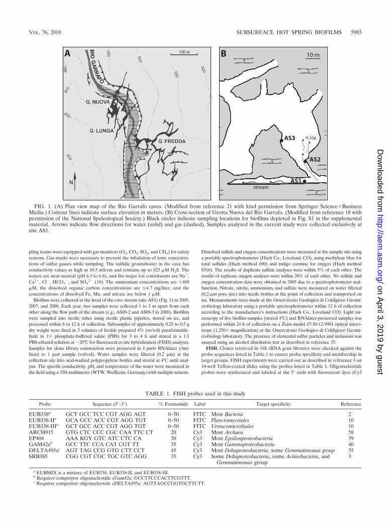
A reconnaissance of the Acquasanta caves (Fig. 1) showed thatthey contain biofilm types also reported in other sulfidic caves,including viscous snottites on walls above the water table (see Fig.S1a, b, and e in the supplemental material), reddish clay-richdeposits (“ragu”) similar to those on walls in Cueva de Villa Luz(see Fig. S1e in the supplemental material), and white biofilmscovering sediments below the water table (see Fig. S1d and f inthe supplemental material). As in Cueva de Villa Luz, subaerialsurfaces in areas with high sulfur gas concentrations are coveredwith elemental sulfur deposits (see Fig. S1c in the supplementalmaterial). Here, we describe the diversity and community struc-ture of biofilms in the thermal Acquasanta groundwater. In ad-dition, we investigate whether the Acquasanta stream biofilmshave a structure consistent with a simple ecological niche modeldeveloped for sulfur-oxidizing clades inhabiting nonthermal sul-fidic caves (37). The niche model considers aqueous sulfide andoxygen concentrations and hydrodynamic shear and suggests thatin turbulently flowing (high shear) waters with high sulfide/oxygenratios, Epsilonproteobacteria should outcompete filamentousGammaproteobacteria, such as Beggiatoa and Thiothrix. We findthat Acquasanta cave stream biofilms share very few phylotypes incommon with other sulfidic caves studied to date and are domi-nated by Epsilonproteobacteria as predicted by the niche model.
MATERIALS AND METHODS
Site description and field geochemistry. Grotta Nuova di Rio Garrafo (Fig. 1)is located approximately 2 km south of Acquasanta Terme, Italy (13°25�E,42o45�N). The cave entrance is 15 m above the western bank of the Rio Garrafo(Garrafo River), and the sulfidic water table in the cave is approximately 50 mbelow the level of the perched river. Grotta Nuova di Rio Garrafo contains morethan 1 km of passages with strong vertical development in marly Scaglia Rossalimestone (18, 21). The water table is accessible via vertical caving routes. Sam-
* Corresponding author. Mailing address: Department of Geo-sciences, Pennsylvania State University, University Park, PA 16802.Phone: (814) 865-6330. Fax: (814) 863-7823. E-mail: [email protected].
‡ Present address: Department of Geographical and Earth Sciences,University of Glasgow, Glasgow G12 8QQ, United Kingdom.
† Supplemental material for this article may be found at http://aem.asm.org/.
� Published ahead of print on 16 July 2010.
5902
on April 3, 2019 by guest
http://aem.asm
.org/D
ownloaded from
pling teams were equipped with gas monitors (O2, CO2, SO2, and CH4) for safetyreasons. Gas masks were necessary to prevent the inhalation of toxic concentra-tions of sulfur gasses while sampling. The sulfidic groundwater in the cave hasconductivity values as high as 10.5 mS/cm and contains up to 825 �M H2S. Thewaters are near-neutral (pH 6.3 to 6.8), and the major ion constituents are Na�,Ca2�, Cl�, HCO3
�, and SO42� (18). The ammonium concentrations are �400
�M, the dissolved organic carbon concentrations are �4.7 mg/liter, and theconcentrations of dissolved Fe, Mn, and nitrate are below 1 �M.
Biofilms were collected at the head of the cave stream (site AS1) (Fig. 1) in 2005,2007, and 2008. Each year, two samples were collected 1 to 3 m apart from eachother along the flow path of the stream (e.g., AS08-2 and AS08-3 in 2008). Biofilmswere sampled into sterile tubes using sterile plastic pipettes, stored on ice, andprocessed within 8 to 12 h of collection. Subsamples of approximately 0.25 to 0.5 gdry weight were fixed in 3 volumes of freshly prepared 4% (wt/vol) paraformalde-hyde in 1� phosphate-buffered saline (PBS) for 3 to 4 h and stored in a 1:1PBS-ethanol solution at �20°C for fluorescent in situ hybridization (FISH) analyses.Samples for clone library construction were preserved in 4 parts RNAlater (Am-bion) to 1 part sample (vol/vol). Water samples were filtered (0.2 �m) at thecollection site into acid-washed polypropylene bottles and stored at 4°C until anal-ysis. The specific conductivity, pH, and temperature of the water were measured inthe field using a 350i multimeter (WTW, Weilheim, Germany) with multiple sensors.
Dissolved sulfide and oxygen concentrations were measured at the sample site usinga portable spectrophotometer (Hach Co., Loveland, CO), using methylene blue fortotal sulfides (Hach method 690) and indigo carmine for oxygen (Hach method8316). The results of duplicate sulfide analyses were within 5% of each other. Theresults of replicate oxygen analyses were within 20% of each other. No sulfide andoxygen concentration data were obtained in 2005 due to a spectrophotometer mal-function. Nitrate, nitrite, ammonium, and sulfate were measured on water filtered(0.2-�m pore size) into sterile bottles at the point of collection and transported onice. Measurements were made at the Osservatorio Geologico di Coldigioco Geomi-crobiology laboratory using a portable spectrophotometer within 12 h of collectionaccording to the manufacturer’s instructions (Hach Co., Loveland CO). Light mi-croscopy of live biofilm samples (stored 4°C) and RNAlater-preserved samples wasperformed within 24 h of collection on a Zeiss model 47-30-12-9901 optical micro-scope (1,250� magnification) at the Osservatorio Geologico di Coldigioco Geomi-crobiology laboratory. The presence of elemental sulfur particles and inclusions wasassayed using an alcohol dissolution test as described in reference 35.
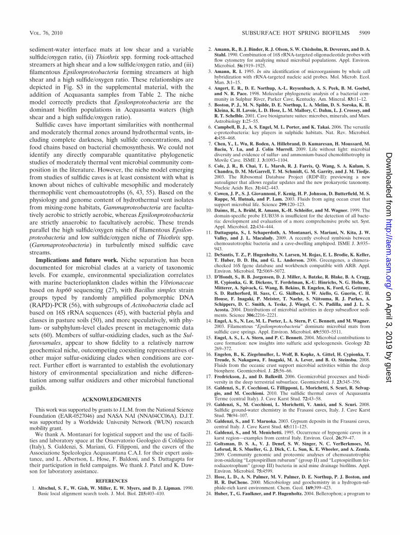
FISH. Clones retrieved in 16S rRNA gene libraries were checked against theprobe sequences listed in Table 1 to ensure probe specificity and membership intarget groups. FISH experiments were carried out as described in reference 3 on10-well Teflon-coated slides using the probes listed in Table 1. Oligonucleotideprobes were synthesized and labeled at the 5� ends with fluorescent dyes (Cy3
FIG. 1. (A) Plan view map of the Rio Garrafo caves. (Modified from reference 21 with kind permission from Springer Science�BusinessMedia.) Contour lines indicate surface elevation in meters. (B) Cross section of Grotta Nuova del Rio Garrafo. (Modified from reference 18 withpermission of the National Speleological Society.) Black circles indicate sampling locations for biofilms depicted in Fig. S1 in the supplementalmaterial. Arrows indicate flow directions for water (solid) and gas (dashed). Samples analyzed in the current study were collected exclusively atsite AS1.
TABLE 1. FISH probes used in this study
Probe Sequence (5�–3�) % Formamide Label Target specificity Reference
EUB338a GCT GCC TCC CGT AGG AGT 0–50 FITC Most Bacteria 2EUB338-IIa GCA GCC ACC CGT AGG TGT 0–50 FITC Planctomycetales 10EUB338-IIIa GCT GCC ACC CGT AGG TGT 0–50 FITC Verrucomicrobiales 10ARCH915 GTG CTC CCC CGC CAA TTC CT 20 Cy3 Most Archaea 58EP404 AAA KGY GTC ATC CTC CA 30 Cy3 Most Epsilonproteobacteria 39GAM42ab GCC TTC CCA CAT CGT TT 35 Cy3 Most Gammaproteobacteria 40DELTA495ac AGT TAG CCG GTG CTT CCT 45 Cy3 Most Deltaproteobacteria, some Gemmatimonas group 35SRB385 CGG CGT CGC TGC GTC AGG 35 Cy3 Some Deltaproteobacteria, some Actinobacteria, and
Gemmatimonas group3
a EUBMIX is a mixture of EUB338, EUB338-II, and EUB338-III.b Requires competitor oligonucleotide cGam42a: GCCTTCCCACTTCGTTT.c Requires competitor oligonucleotide cDELTA495a: AGTTAGCCGGTGCTTCTT.
VOL. 76, 2010 SUBSURFACE HOT SPRING BIOFILMS 5903
on April 3, 2019 by guest
http://aem.asm
.org/D
ownloaded from

and fluorescein isothiocyanate [FITC]) at Sigma-Genosys (United States). Cellswere stained after hybridization with 4�,6�-diamidino-2-phenylindole (DAPI),mounted with Vectashield (Vectashield Laboratories, United States) and viewedon a Nikon E800 epifluorescence microscope. Images were collected with aNikon charge-coupled device (CCD) camera using NIS Elements AR 2.30 imageanalysis software. The fluorescence coming from DNA probes in Fig. S2 in thesupplemental material is shown in false color, and an autolevel function wasapplied to each image shown in the figure. For population estimates based onFISH, the biofilm matrix was easily disrupted by shaking the sample tube. Atleast 3 slide wells were examined for each probe combination. At least 10 imageswere collected for each sample and probe combination, taking care to representthe sample variability, and a total DAPI-stained area of at least 3 � 104 mm2
(equivalent to the area of 5 � 104 E. coli cells) was analyzed in each case. Thepresence of cells ranging from small rods to large filaments precluded simplecounting that does not take into account differences in biomass between the celltypes. A comparison between visual area estimates and area counts obtainedusing the object count tool of NIS Elements AR 2.30 image analysis softwareindicated that visual estimation gave the same results within error in less time.Since semiquantitative area counts are sufficient to support the conclusionsobtained in this study, a visual estimation approach was employed.
Clone library construction. Environmental DNA was extracted, amplified, andcloned using archaeal- and bacterial-domain-specific primers as described inreference 37. Briefly, environmental DNA was extracted from approximately0.5 g of biofilm using phenol-chloroform extraction. Each 50-�l PCR mixturecontained environmental DNA template (1 to 150 ng), 1.25 U Ex Taq DNApolymerase (TaKaRa Bio, Inc., Shiga, Japan), 0.2 mM each deoxynucleosidetriphosphate (dNTP), 1� PCR buffer, 0.2 �M 1492r universal reverse primer,and 0.2 �M 27f bacterial domain primer (34). Thermal cycling was as follows:initial denaturation for 5 min at 94°C and 25 cycles of 94°C for 1 min, 50°C for25 s, and 72°C for 2 min, followed by a final elongation at 72°C for 20 min. Forarchaeal PCRs, the primers used were 21f (5�-TTCCGGTTGATCCYGCCGGA) and 977r (5�-YCCGGCGTTGAMTCCAATT), and thermal cycling wasperformed as described above except that the annealing temperature was 58°C.PCR products were cloned into the pCR4-TOPO plasmid and used to transformchemically competent OneShot MACH1 T1 Escherichia coli cells (TOPO TAcloning kit, Invitrogen, Carlsbad, CA). The plasmid inserts were screened usingcolony PCR with M13 primers. Colony PCR products of the correct size werepurified using a QIAquick PCR purification kit (Qiagen, Inc., United States).
Sequencing and phylogenetic analyses. Clones were sequenced at the PennState University Biotechnology Center with Sanger technology (ABI Hitachi3730XL DNA analyzer with BigDye fluorescent terminator chemistry) using T3and T7 plasmid-specific primers. Sequences were assembled with Phred basecalling using CodonCode Aligner version 1.2.4 (CodonCode Corp., UnitedStates) and manually checked for ambiguities. The nearly full-length gene se-quences (all �1,400 bp) were compared against sequences in public databasesusing BLAST (1) and submitted to the online analyses CHIMERA_CHECKversion 2.7 (8) and Bellerophon 3 (24). Putative chimeras were excluded fromsubsequent analyses. Sequences were aligned to the 7,682-character Hugenholtzalignment using the NAST aligner at greengenes (12). NAST-aligned sequenceswere loaded into an ARB database (36) containing over 230,000 full-lengthsequences. Alignments were edited manually in ARB using the ARB_Edit4sequence editor. Only sequences longer than 1,350 bp were used for phylogeneticanalyses, with the exception of short Gammaproteobacteria clones from ParkerCave’s Sulfur River (4). The analyses included the top BLAST hits, the top fiveclosest relatives in the ARB database, and representatives of major divisions
within each of the targeted groups (e.g., Sulfurovumales), with an emphasis onsulfidic cave sequences. The alignments were trimmed so that all sequences wereof equal length (the final alignment lengths were 1,153, 1,420, and 1,257 positionsfor analyses of Sulfurovumales, Thiofaba/Thiovirga, and Deltaproteobacteriaclades, respectively), and nucleotide positions with less than 50% base pairconservation were masked (calculated separately from all sequences in eachanalysis, exclusive of outgroups). Shorter-length sequences (Parker Cave clonesAF047617 and AF047623) were included in phylogenetic analyses with theirmissing data coded as missing and treated according to PAUP* defaults for eachanalysis type (59). Maximum-likelihood, maximum-parsimony, and neighbor-joining analyses were performed using PAUP* version 4b10 (59). Maximum-likelihood analyses used the general time-reversible (GTR) substitution modelwith gamma-distributed among-site variation, at least five random-addition-se-quence replicates, and tree bisection-reconnection (TBR) branch swapping. Basefrequency, substitution rates, and shape parameter were estimated from the data.Maximum-parsimony bootstrap analyses (2,000 replicates) were performed viaheuristic search with 100 random-addition-sequence replicates and TBR branchswapping. Neighbor-joining bootstrap analyses (2,000 replicates) were per-formed with a Jukes-Cantor (JC)-corrected distance matrix.
Diversity analyses. Rarefaction analyses were calculated based on the fre-quency of operational taxonomic units (OTUs) defined by the program DOTUR,version 1.53 (54). OTUs were defined at 98% sequence similarity, using theDOTUR “furthest neighbor” algorithm, based on ARB distance matrices.
Nucleotide sequence accession numbers. The 16S rRNA gene sequences de-termined in this study were submitted to GenBank and were assigned accessionnumbers GU390708 to GU390880.
RESULTS
Field observations and geochemistry. Water and biofilm sam-ples were collected at site AS1 (Fig. 1) in 2005, 2007, and 2008.The stream at site AS1 was fast flowing and turbulent for theentire length of the passage, and nearly every surface in thechannel was covered with biofilms of “streamer” morphology (seeFig. S1d and f in the supplemental material). Other biofilm mor-phologies were rare and limited in areal extent. The streamerswere 1 to 2 cm in length on average (see Fig. S1d and f in thesupplemental material) and covered both fine gray sediment andexposed limestone surfaces underwater. Microscopic examinationof both live and RNAlater-preserved streamers within 24 h ofcollection revealed that none of the cells contained intracellularsulfur inclusions. All biofilm samples had abundant elementalsulfur particles outside the cells in the biofilm matrix. The stream-ers collected in 2007 and 2008 were dominated by filamentousbacteria, with rod-shaped and coccoid cells also present. Filamen-tous bacteria were less common in the 2005 samples (AS05-2 andAS05-3), which were dominated by long thin rods.
The geochemical data for waters collected at sample site AS1(Table 2) reflected changes in the degree of dilution of the ther-mal groundwater by surface water recharge (18). The conductivity
TABLE 2. FISH cell area abundances and geochemical data for Acquasanta cave stream biofilm samples
Sample Datecollected
Percent hybridization by each FISH probeaTemp
oC pH Sp. cond.b
(mS/cm)SO4
2�
(mM)O2 (aq)(�M)
H2S (aq)(�M)
H2S (g)c
(ppm)EUBMIX EP404 GAM42a DELTA495a SRB385
AS05-3 18/08/2001 ����� ���� ���� �� �� 40.4 6.37 8.4 NM NM NM NMAS05-2 18/08/2001 ����� ���� ���� �� NM 40.4 6.37 8.4 NM NM NM NMAS07-6 31/05/2003 ����� ���� ���� �� NM 44.1 6.38 10.6 9.92 3.06 801 100AS07-7 31/05/2003 ����� ���� ���� �� �� 44.1 6.38 10.6 9.92 3.06 801 100AS08-2 13/06/2004 ����� ����� �� �� �� 42.7 6.31 10.5 12.71 6.69 415 15AS08-3 13/06/2004 ����� ���� ��� �� NM 42.7 6.31 10.5 12.71 6.69 415 15
a Estimated proportion of total cells. ND, none detected; �, �3%; ��, 3 to 15%; ���, 15 to 35%; ����, 35 to 75%; ����, 75 to 100%; NM, not measured.No ARCH915-positive cells were detected.
b Sp. cond., specific conductance.c Measured 1.5 m above the cave stream. g, gas.
5904 JONES ET AL. APPL. ENVIRON. MICROBIOL.
on April 3, 2019 by guest
http://aem.asm
.org/D
ownloaded from

was substantially lower in 2005 than in 2007 and 2008. The con-ductivity and major ion concentrations were similar for samplescollected in 2007 and 2008, but the hydrogen sulfide concentra-tions in both the stream and the cave atmosphere were muchhigher in 2007 (Table 2). These differences in geochemistry werenot accompanied by any noticeable changes in the density ormacroscopic morphology of the microbial streamers covering thestream sediments.
16S rRNA clone libraries. Attempts to amplify archaeal 16SrRNA genes from the biofilm samples were unsuccessful,whereas positive-control DNA yielded archaeal PCR productsof the expected length in the same PCR runs (data not shown).A total of 174 nearly full-length bacterial 16S rRNA genesequences were retrieved from samples AS05-3 (80 clones) andAS07-7 (94 clones) (Fig. 2A). The results of rarefaction anal-
yses (Fig. 2B) suggested that the major bacterial populationspresent in the biofilms have been adequately sampled. Themost abundant phylotype in both libraries fell within the Sul-furovumales clade in the Epsilonproteobacteria. Sulfurovumalesclones (Fig. 3) constituted 40% and 67% of libraries AS05-3and AS07-7, respectively. Most of the Sulfurovumales se-quences were 98% similar to clones AS053-B2 and AS077-B27(Fig. 3) and formed a clade with a single Frasassi cave clone.The AS05-3 library also included several clones (6.3%) of asecond Sulfurovumales phylotype. The second most abun-dant phylotype in both libraries (10.6% in AS05-3 and26.3% in AS07-7) fell within a clade containing the isolates“Candidatus Thiobacillus baregensis” and Thiofaba tepi-diphila in the Gammaproteobacteria (Fig. 4). The Deltapro-teobacteria sequences in the libraries were very distantly
FIG. 2. (A) Taxonomic composition of two 16S rRNA clone libraries from Acquasanta stream biofilms. Both biofilms were collected at site AS1(see Fig. 1B). (B) Rarefaction of 16S rRNA clone libraries constructed from Acquasanta and Frasassi cave stream biofilms. OTUs are defined at98% sequence identity. Frasassi clone libraries are from reference 37, except for library PC05-LKA (J. L. Macalady, unpublished data).
VOL. 76, 2010 SUBSURFACE HOT SPRING BIOFILMS 5905
on April 3, 2019 by guest
http://aem.asm
.org/D
ownloaded from

related (�90%) to sequences in public databases (Fig. 5).Both libraries contained representatives of Bacteroidetes,Elusimicrobia, and MVP-15, in addition to rare phylotypes(Fig. 2). The bacterial diversity in both samples was verylow, even compared with that of sulfur-oxidizing biofilmsfrom other sulfidic cave environments (Fig. 2).
Epifluorescence microscopy. Fluorescent in situ hybridiza-tion (FISH) was used to evaluate the relative abundances ofmajor microbial populations in the biofilms (Table 2). TheFISH data were consistent with the clone libraries, suggestinglittle PCR or DNA extraction bias for major populations. Nohybridization to archaeal-domain-specific probe ARCH915was detected. More than 95% of DAPI-stained cells hybridizedwith the bacterial-domain-specific probe EUBMIX in all sam-ples. The EUBMIX-negative populations were small cocci thatdid not appear to hybridize with any probes used in this study(see Fig. S2 in the supplemental material). No nucleated cells(i.e., protists or fungi) were observed. Large holdfast structuresuniting many filaments, such as those observed for Thiothrixspp. or filamentous Epsilonproteobacteria “rosettes” in Frasassicave streamers (37, 39), were not observed in the Acquasantabiofilms.
Samples AS07-6, AS07-7, AS08-2, and AS08-3 were domi-nated by EP404-positive filaments (Table 2; also see Fig. S2 inthe supplemental material). Samples AS05-2 and AS05-3 con-
tained long, thin EP404-positive rods, as well as a populationof large EP404-positive cocci (see Fig. S2 in the supplementalmaterial). GAM42a-positive cells were present in all samplesand were short rods with uniform morphology across samples(see Fig. S2 in the supplemental material). A newly designedFISH probe developed to identify close relatives of “Ca. Thio-bacillus baregensis” in biofilms from other caves hybridized tothe same set of short rods visualized using GAM42a (data notshown). Consistent with the results of FISH experiments,Gammaproteobacteria sequences retrieved in both Acquasantaclone libraries consisted of a single phylotype closely related to“Ca. Thiobacillus baregensis” (Fig. 4). FISH experiments usingSRB385 and Delta495 probes yielded similar area estimatesand cell morphologies in all samples. SRB385- and Delta495-positive cells were predominantly large rods, with smaller pop-ulations of smaller rods and cocci (see Fig. S2 in the supple-mental material).
DISCUSSION
Biofilm community composition. Based on a full-cyclerRNA approach, Acquasanta stream biofilms are dominatedby Sulfurovumales (Epsilonproteobacteria) populations, alongwith important and sometimes equally large populations ofGammaproteobacteria related to “Ca. Thiobacillus baregensis”
FIG. 3. Maximum-likelihood phylogram of 16S rRNA gene sequences from the Sulfurovumales clade (Epsilonproteobacteria). The number ofclones represented by each Acquasanta phylotype (98% nucleotide similarity) is indicated in parentheses. Neighbor-joining (left) and maximum-parsimony (right) bootstrap values greater than 50 are shown for each node.
5906 JONES ET AL. APPL. ENVIRON. MICROBIOL.
on April 3, 2019 by guest
http://aem.asm
.org/D
ownloaded from

(Table 2 and Fig. 2; also see Fig. S2 in the supplementalmaterial). Minor populations include Deltaproteobacteria, Bac-teroidetes, Spirochaetales, Elusimicrobia, MVP-15, and raretaxa. Although archaea were not detected using FISH or PCR,small cells that did not hybridize with either archaeal or bac-terial domain-specific probes made up 5% of the communityand may prove to be archaeal cells.
The Sulfurovumales (Fig. 3) are a monophyletic clade withfew isolates and large numbers of environmental sequencesand have been retrieved from sulfidic caves and springs world-wide (6). Based on the results of FISH experiments, the mostabundant Epsilonproteobacteria populations are filamentous in2007 and 2008 biofilms and nonfilamentous in 2005 biofilms.Cells in these populations have indistinguishable morphologies(long thin rods) but different arrangements (see Fig. S2 in thesupplemental material). Because the most abundant Epsilon-proteobacteria clones are nearly genetically identical in samplesfrom 2005 and 2007 (Fig. 3, clones AS053-B2 and AS077-B27),we hypothesize that they represent highly related species ca-pable of both filamentous and nonfilamentous habits. We rec-ognize that this hypothesis is speculative and remains to betested using strain-specific probes. Filamentous Sulfurovumaleshave not been cultured or investigated using metagenomics todate, and thus, there are few unassailable constraints on theirmetabolism. The genome of Sulfurovum sp. strain NBC37-1,distantly related to Acquasanta clones (�91.5% similarity) buttheir closest cultivated relative, has recently been sequenced(44). Sulfurovum sp. NBC37-1 is a lithoautotrophic hydrother-mal vent symbiont that can use hydrogen and reduced sulfurspecies as electron donors. Based on the predominance of
sulfur-based lithotrophic metabolisms in hydrothermal ventEpsilonproteobacteria (6, 43) and the importance of Epsilon-proteobacteria in terrestrial chemosynthetic ecosystems wherereduced sulfur species contribute the bulk of the availablechemical energy (37, 51), it is likely that Sulfurovumales clonesretrieved from Acquasanta biofilms will prove to be sulfur-oxidizing lithoautotrophs, sulfur-reducing lithoautotrophs, orpossibly, mixotrophs that can reduce sulfur using organic com-pounds, depending on environmental conditions.
The Gammaproteobacteria clones from Acquasanta clonelibraries belong to a single phylotype represented by clonesAS053-B125 and AS077-B32 (Fig. 4). Related isolates includethe sulfur-oxidizing lithoautotrophs “Ca. Thiobacillus baregen-sis” (25, 26), Thiofaba tepidiphila (42), and Thiovirga sulfuroxy-dans (29, 30). Representatives of the clade defined by theseisolates (Fig. 4) are important constituents of stream biofilmsin other sulfidic caves. For example, close relatives of “Ca.Thiobacillus baregensis” were found in six out of six Frasassistream biofilm clone libraries (37). Clones in this clade havealso been retrieved from Parker Cave in Kentucky (4) andMovile Cave in Romania (7). Thus, both the metabolisms ofcultivated relatives and the distribution of related clones in theenvironment suggest that Acquasanta “Ca. Thiobacillus bar-egensis” relatives are sulfur-oxidizing lithoautotrophs.
The Deltaproteobacteria 16S rRNA clones are very distantlyrelated (�90%) to publicly available sequences, including en-vironmental clones (Fig. 5). With the exception of the AS053-B137 phylotype (3 clones), the clones belong to a large envi-ronmental clone group with no cultivated representatives. Thenearest relatives of these phylotypes are from marine sediment
FIG. 4. Maximum-likelihood phylogram of 16S rRNA gene sequences from the Thiofaba/Thiovirga clade in the Gammaproteobacteria. Thenumber of clones represented by each Acquasanta phylotype is indicated in parentheses. Neighbor-joining (left) and maximum-parsimony (right)bootstrap values greater than 50 are shown for each node.
VOL. 76, 2010 SUBSURFACE HOT SPRING BIOFILMS 5907
on April 3, 2019 by guest
http://aem.asm
.org/D
ownloaded from

and methane seep environments, including Eel River sedi-ments (“Eel-2” clade) (48), where sulfate reduction is an im-portant process. Acquasanta Deltaproteobacteria may thus rep-resent novel sulfate-reducing bacteria. However, because oftheir dissimilarity to characterized isolates, inferences abouttheir metabolism remain somewhat speculative. It is interest-ing to note that none of the diverse Deltaproteobacteria clonesfrom the Frasassi cave system fall within the Acquasanta en-vironmental clone groups. Conversely, Acquasanta clones arenot represented in the Desulfocapsa clade, which contains thevast majority of Frasassi Deltaproteobacteria clones (37, 39).
The Acquasanta caves and the cooler Frasassi caves located�80 km away provide an interesting geochemical and physicalcontext for comparing microbial communities. The hydrogensulfide in Acquasanta and Frassasi cave waters is thought tohave a similar source, namely, partial reduction of sulfate ingypsum-bearing evaporite rocks in the underlying Triassic Bu-rano Formation (18, 20). The conductivity of the sulfidic waterat Acquasanta is somewhat higher than at Frasassi (�2,000versus �9,000 �S/cm), although dissolved ions are present insimilar ratios (18, 19). The temperature of the sulfidic water inthe Frasassi cave system is 13 to 14°C, compared to 35 to 50°Cat Acquasanta.
The most abundant Acquasanta clones belong to clades alsocontaining Frasassi phylotypes (i.e., Sulfurovumales in theEpsilonproteobacteria and “Ca. Thiobacillus baregensis” rela-
tives in the Gammaproteobacteria) (37). However, the Acqua-santa phylotypes are distinct (Fig. 3 and 4), and Frasassi clonesare often more closely related to phylotypes from geographi-cally distant sulfidic caves, such as Movile Cave (Romania),Parker Cave (Kentucky), and Lower Kane Caves (Wyoming),than to clones from the geographically nearby Acquasantacaves. Phylogeographical patterns can arise via several unre-lated mechanisms, including selective pressures imposed by theenvironment, dispersal limitations, and chance historical oc-currences, such as colonization events (41). Because the streamwaters in the Acquasanta caves have a significantly highertemperature than previously studied sulfidic cave waters, it isworth considering whether environmental selection based ontemperature could explain the phylogeographical pattern weobserve. However, clades within the Sulfurovumales containingFrasassi and other nonthermal cave clones also contain se-quences from hot sulfidic springs, suggesting that temperaturealone cannot account for the large genetic distances betweenFrasassi and Acquasanta phylotypes.
Sulfur oxidizer niches. We previously proposed a simpleniche model for sulfur-oxidizing bacteria in cave streams of thenonthermal Frasassi cave system (37). In the model, two nichedimensions (hydrodynamic shear and aqueous sulfide/oxygenratio) controlled the biofilm population structures. The modeldescribed the distribution of three major biofilm types namedafter their dominant populations: (i) Beggiatoa spp. forming
FIG. 5. Maximum-likelihood phylogram of 16S rRNA gene sequences from the Deltaproteobacteria. The number of clones represented by eachAcquasanta phylotype is indicated in parentheses. Neighbor-joining (left) and maximum-parsimony (right) bootstrap values greater than 50 areshown for each node. The tree includes the 5 nearest neighbors in public sequence databases for each Acquasanta phylotype.
5908 JONES ET AL. APPL. ENVIRON. MICROBIOL.
on April 3, 2019 by guest
http://aem.asm
.org/D
ownloaded from
sediment-water interface mats at low shear and a variablesulfide/oxygen ratio, (ii) Thiothrix spp. forming rock-attachedstreamers at high shear and a low sulfide/oxygen ratio, and (iii)filamentous Epsilonproteobacteria forming streamers at highshear and a high sulfide/oxygen ratio. These relationships aredepicted in Fig. S3 in the supplemental material, with theaddition of Acquasanta samples from Table 2. The nichemodel correctly predicts that Epsilonproteobacteria are thedominant biofilm populations in Acquasanta waters (highshear and a high sulfide/oxygen ratio).
Sulfidic caves have important similarities with nonthermaland moderately thermal zones around hydrothermal vents, in-cluding complete darkness, high sulfide concentrations, andfood chains based on bacterial chemosynthesis. We could notidentify any directly comparable quantitative phylogeneticstudies of moderately thermal vent microbial community com-position in the literature. However, the niche model emergingfrom studies of sulfidic caves is at least consistent with what isknown about niches of cultivable mesophilic and moderatelythermophilic vent chemoautotrophs (6, 43, 55). Based on thephysiology and genome content of hydrothermal vent isolatesfrom mixing-zone habitats, Gammaproteobacteria are faculta-tively aerobic to strictly aerobic, whereas Epsilonproteobacteriaare strictly anaerobic to facultatively aerobic. These trendsparallel the high sulfide/oxygen niche of filamentous Epsilon-proteobacteria and low sulfide/oxygen niche of Thiothrix spp.(Gammaproteobacteria) in turbulently mixed sulfidic cavestreams.
Implications and future work. Niche separation has beendocumented for microbial clades at a variety of taxonomiclevels. For example, environmental specialization correlateswith marine bacterioplankton clades within the Vibrionaceaebased on hsp60 sequencing (27), with Bacillus simplex straingroups typed by randomly amplified polymorphic DNA(RAPD)-PCR (56), with subgroups of Actinobacteria clade acIbased on 16S rRNA sequences (45), with bacterial phyla andclasses in pasture soils (50), and more speculatively, with phy-lum- or subphylum-level clades present in metagenomic datasets (60). Members of sulfur-oxidizing clades, such as the Sul-furovumales, appear to show fidelity to a relatively narrowgeochemical niche, outcompeting coexisting representatives ofother major sulfur-oxidizing clades when conditions are cor-rect. Further effort is warranted to establish the evolutionaryhistory of environmental specialization and niche differen-tiation among sulfur oxidizers and other microbial functionalguilds.
ACKNOWLEDGMENTS
This work was supported by grants to J.L.M. from the National ScienceFoundation (EAR-0527046) and NASA NAI (NNA04CC06A). D.J.T.was supported by a Worldwide University Network (WUN) researchmobility grant.
We thank A. Montanari for logistical support and the use of facili-ties and laboratory space at the Osservatorio Geologico di Coldigioco(Italy), S. Galdenzi, S. Mariani, G. Filipponi, and the cavers of theAssociazione Speleologica Acquasantana C.A.I. for their expert assis-tance, and L. Albertson, L. Hose, F. Baldoni, and S. Dattagupta fortheir participation in field campaigns. We thank J. Patel and K. Daw-son for laboratory assistance.
REFERENCES
1. Altschul, S. F., W. Gish, W. Miller, E. W. Myers, and D. J. Lipman. 1990.Basic local alignment search tools. J. Mol. Biol. 215:403–410.
2. Amann, R., B. J. Binder, R. J. Olson, S. W. Chisholm, R. Devereux, and D. A.Stahl. 1990. Combination of 16S rRNA-targeted oligonucleotide probes withflow cytometry for analyzing mixed microbial populations. Appl. Environ.Microbiol. 56:1919–1925.
3. Amann, R. I. 1995. In situ identification of microorganisms by whole cellhybridization with rRNA-targeted nucleic acid probes. Mol. Microb. Ecol.Man. 3:1–15.
4. Angert, E. R., D. E. Northup, A.-L. Reysenbach, A. S. Peek, B. M. Goebel,and N. R. Pace. 1998. Molecular phylogenetic analysis of a bacterial com-munity in Sulphur River, Parker Cave, Kentucky. Am. Mineral. 83:11–12.
5. Boston, P. J., M. N. Spilde, D. E. Northup, L. A. Melim, D. S. Soroka, K. H.Kleina, K. H. Lavoie, L. D. Hose, L. M. Mallory, C. Dahm, L. J. Crossey, andR. T. Schelble. 2001. Cave biosignature suites: microbes, minerals, and Mars.Astrobiology 1:25–55.
6. Campbell, B. J., A. S. Engel, M. L. Porter, and K. Takai. 2006. The versatilee-proteobacteria: key players in sulphidic habitats. Nat. Rev. Microbiol.4:458–468.
7. Chen, Y., L. Wu, R. Boden, A. Hillebrand, D. Kumaresan, H. Moussard, M.Baciu, Y. Lu, and J. Colin Murrell. 2009. Life without light: microbialdiversity and evidence of sulfur- and ammonium-based chemolithotrophy inMovile Cave. ISME J. 3:1093–1104.
8. Cole, J. R., B. Chai, T. L. Marsh, R. J. Farris, Q. Wang, S. A. Kulam, S.Chandra, D. M. McGarrell, T. M. Schmidt, G. M. Garrity, and J. M. Tiedje.2003. The Ribosomal Database Project (RDP-II): previewing a newautoaligner that allows regular updates and the new prokaryotic taxonomy.Nucleic Acids Res. 31:442–443.
9. Cowen, J. P., S. J. Giovannoni, F. Kenig, H. P. Johnson, D. Butterfield, M. S.Rappe, M. Hutnak, and P. Lam. 2003. Fluids from aging ocean crust thatsupport microbial life. Science 299:120–123.
10. Daims, H., A. Bruhl, R. Amann, K.-H. Schleifer, and M. Wagner. 1999. Thedomain-specific probe EUB338 is insufficient for the detection of all bacte-ria: development and evaluation of a more comprehensive probe set. Syst.Appl. Microbiol. 22:434–444.
11. Dattagupta, S., I. Schaperdoth, A. Montanari, S. Mariani, N. Kita, J. W.Valley, and J. L. Macalady. 2009. A recently evolved symbiosis betweenchemoautotrophic bacteria and a cave-dwelling amphipod. ISME J. 3:935–943.
12. DeSantis, T. Z., P. Hugenholtz, N. Larsen, M. Rojas, E. L. Brodie, K. Keller,T. Huber, D. D. Hu, and G. L. Anderson. 2006. Greengenes, a chimera-checked 16S fgene database and workbench compatible with ARB. Appl.Environ. Microbiol. 72:5069–5072.
13. D’Hondt, S., B. B. Jorgensen, D. J. Miller, A. Batzke, R. Blake, B. A. Cragg,H. Cypionka, G. R. Dickens, T. Ferdelman, K.-U. Hinrichs, N. G. Holm, R.Mitterer, A. Spivack, G. Wang, B. Bekins, B. Engelen, K. Ford, G. Gettemy,S. D. Rutherford, H. Sass, C. G. Skilbeck, I. W. Aiello, G. Guerin, C. H.House, F. Inagaki, P. Meister, T. Naehr, S. Niitsuma, R. J. Parkes, A.Schippers, D. C. Smith, A. Teske, J. Wiegel, C. N. Padilla, and J. L. S.Acosta. 2004. Distributions of microbial activities in deep subseafloor sedi-ments. Science 306:2216–2221.
14. Engel, A. S., N. Lee, M. L. Porter, L. A. Stern, P. C. Bennett, and M. Wagner.2003. Filamentous “Epsilonproteobacteria” dominate microbial mats fromsulfidic cave springs. Appl. Environ. Microbiol. 69:5503–5511.
15. Engel, A. S., L. A. Stern, and P. C. Bennett. 2004. Microbial contributions tocave formation: new insights into sulfuric acid speleogenesis. Geology 32:269–372.
16. Engelen, B., K. Ziegelmuller, L. Wolf, B. Kopke, A. Gittel, H. Cypionka, T.Treude, S. Nakagawa, F. Inagaki, M. A. Lever, and B. O. Steinsbu. 2008.Fluids from the oceanic crust support microbial activities within the deepbiosphere. Geomicrobiol. J. 25:56–66.
17. Fredrickson, J., and D. Balkwill. 2006. Geomicrobial processes and biodi-versity in the deep terrestrial subsurface. Geomicrobiol. J. 23:345–356.
18. Galdenzi, S., F. Cocchioni, G. Fillipponi, L. Morichetti, S. Scuri, R. Selvag-gio, and M. Cocchioni. 2010. The sulfidic thermal caves of AcquasantaTerme (central Italy). J. Cave Karst Stud. 72:43–58.
19. Galdenzi, S., M. Cocchioni, L. Morichetti, V. Amici, and S. Scuri. 2008.Sulfidic ground-water chemistry in the Frasassi caves, Italy. J. Cave KarstStud. 70:94–107.
20. Galdenzi, S., and T. Maruoka. 2003. Gypsum deposits in the Frasassi caves,central Italy. J. Cave Karst Stud. 65:111–125.
21. Galdenzi, S., and M. Menichetti. 1995. Occurrence of hypogenic caves in akarst region—examples from central Italy. Environ. Geol. 26:39–47.
22. Goltsman, D. S. A., V. J. Denef, S. W. Singer, N. C. VerBerkmoes, M.Lefsrud, R. S. Mueller, G. J. Dick, C. L. Sun, K. E. Wheeler, and A. Zemla.2009. Community genomic and proteomic analyses of chemoautotrophiciron-oxidizing “Leptospirillum rubarum” (group II) and “Leptospirillum fer-rodiazotrophum” (group III) bacteria in acid mine drainage biofilms. Appl.Environ. Microbiol. 75:4599.
23. Hose, L. D., A. N. Palmer, M. V. Palmer, D. E. Northup, P. J. Boston, andH. R. DuChene. 2000. Microbiology and geochemistry in a hydrogen-sul-phide-rich karst environment. Chem. Geol. 169:399–423.
24. Huber, T., G. Faulkner, and P. Hugenholtz. 2004. Bellerophon; a program to
VOL. 76, 2010 SUBSURFACE HOT SPRING BIOFILMS 5909
on April 3, 2019 by guest
http://aem.asm
.org/D
ownloaded from

detect chimeric sequences in multiple sequence alignments. Bioinformatics20:2317–2319.
25. Hubert, H. 1996. Sulfoxidizing bacteria from thermal water of Bareges(France): nature, metabolism, biological activities. Ph.D. thesis. MuseumNational d’Histoire Naturelle, Paris, France.
26. Hubert, H. 1997. Thermal waters of Barege: water microbiology. Eurobiolo-giste 31:43–51.
27. Hunt, D. E., L. A. David, D. Gevers, S. P. Preheim, E. J. Alm, and M. F. Polz.2008. Resource partitioning and sympatric differentiation among closelyrelated bacterioplankton. Science 320:1081–1085.
28. Hutchens, E., S. Radajewski, M. G. Dumont, I. R. McDonald, and J. C.Murrell. 2004. Analysis of methanotrophic bacteria in Movile Cave by stableisotope probing. Environ. Microbiol. 6:111–120.
29. Ito, T., K. Sugita, and S. Okabe. 2004. Isolation, characterization, and in situdetection of a novel chemolithoautotrophic sulfur-oxidizing bacterium inwastewater biofilms growing under microaerophilic conditions. Appl. Envi-ron. Microbiol. 70:3122–3129.
30. Ito, T., K. Sugita, I. Yumoto, Y. Nodasaka, and S. Okabe. 2005. Thiovirgasulfuroxydans gen. nov., sp. nov., a chemolithoautotrophic sulfur-oxidizingbacterium isolated from a microaerobic waste-water biofilm. Int. J. Syst.Evol. Microbiol. 55:1059–1064.
31. Jorgensen, B. B., and A. Boetius. 2007. Feast and famine—microbial life inthe deep-sea bed. Nat. Rev. Microbiol. 5:770–781.
32. Kieft, T. L., S. M. McCuddy, T. C. Onstott, M. Davidson, L.-H. Lin, B.Mislowack, L. Pratt, E. Boice, B. S. Lollar, J. Lippmann-Pipke, S. M.Pfiffner, T. J. Phelps, T. Gihring, D. Moser, and A. van Heerden. 2005.Geochemically generated, energy-rich substrates and indigenous microor-ganisms in deep, ancient groundwater. Geomicrobiol. J. 22:325–335.
33. Klimchouk, A. B. 2007. Hypogene speleogenesis: hydrogeological and mor-phogenetic perspective, vol. 1. National Cave And Karst Research Institute,Carlsbad, NM.
34. Lane, D. J. 1991. 16S/23S rRNA sequencing, p. 115–175. In E. Stackebrandtand M. Goodfellow (ed.), Nucleic acid techniques in bacterial systematics.Wiley, New York, NY.
35. Loy, A., A. Lehner, N. Lee, J. Adamczyk, H. Meier, J. Ernst, K.-H. Schleifer,and M. Wagner. 2002. Oligonucleotide microarray for 16S rRNA gene-baseddetection of all recognized lineages of sulfate-reducing prokaryotes in theenvironment. Appl. Environ. Microbiol. 68:5064–5081.
36. Ludwig, W., O. Strunk, R. Westram, L. Richter, H. Meier, Yadhukumar, A.Buchner, T. Lai, S. Steppi, G. Jobb, W. Forster, I. Brettske, S. Gerber, A. W.Ginhart, O. Gross, S. Grumann, S. Hermann, R. Jost, A. Konig, T. Liss, R.Lussmann, M. May, B. Nonhoff, B. Reichel, R. Strehlow, A. Stamatakis, N.Stuckmann, A. Vilbig, M. Lenke, T. Ludwig, A. Bode, and K.-H. Schleifer.2004. ARB: a software environment for sequence data. Nucleic Acids Res.32:1363–1371.
37. Macalady, J. L., S. Dattagupta, I. Schaperdoth, D. S. Jones, G. K. Druschel,and D. Eastman. 2008. Niche differentiation among sulfur-oxidizing bacte-rial populations in cave waters. ISME J. 2:590–601.
38. Macalady, J. L., D. S. Jones, and E. H. Lyon. 2007. Extremely acidic, pen-dulous microbial biofilms from the Frasassi cave system, Italy. Environ.Microbiol. 9:1402–1414.
39. Macalady, J. L., E. H. Lyon, B. Koffman, L. K. Albertson, K. Meyer, S.Galdenzi, and S. Mariani. 2006. Dominant microbial populations in lime-stone-corroding stream biofilms, Frasassi cave system, Italy. Appl. Environ.Microbiol. 72:5596–5609.
40. Manz, W., R. Amann, W. Ludwig, M. Wagner, and K.-H. Schleifer. 1992.Phylogenetic oligodeoxynucleotide probes for the major subclasses of Pro-teobacteria: problems and solutions. Syst. Appl. Microbiol. 15:593–600.
41. Martiny, J. B. H., B. J. M. Bohannan, J. H. Brown, R. K. Colwell, J. A.Fuhrman, J. L. Green, M. C. Horner-Devine, M. Kane, J. A. Krumins, andC. R. Kuske. 2006. Microbial biogeography: putting microorganisms on themap. Nat. Rev. Microbiol. 4:102–112.
42. Mori, K., and K.-i. Suzuki. 2008. Thiofaba tepidiphila gen. nov., sp. nov., anovel obligately chemolithoautotrophic, sulfur-oxidizing bacterium of theGammaproteobacteria isolated from a hot spring. Int. J. Syst. Evol. Micro-biol. 58:1885–1891.
43. Nakagawa, S., and K. Takai. 2008. Deep-sea vent chemoautotrophs:diversity, biochemistry and ecological significance. FEMS Microbiol.Ecol. 65:1–14.
44. Nakagawa, S., Y. Takaki, S. Shimamura, A.-L. Reysenbach, K. Takai, and K.Horikoshi. 2007. Deep-sea vent epsilonproteobacterial genomes provide in-sights into emergence of pathogens. Proc. Natl. Acad. Sci. U. S. A. 104:12146–12150.
45. Newton, R. J., S. E. Jones, M. R. Helmus, and K. D. McMahon. 2007.Phylogenetic ecology of the freshwater Actinobacteria acI lineage. Appl.Environ. Microbiol. 73:7169–7176.
46. Northup, D. E., S. M. Barns, L. E. Yu, M. N. Spilde, R. T. Schelble, K. E.Dano, L. J. Crossey, C. A. Connolly, P. J. Boston, D. O. Natvig, and C. N.Dahm. 2003. Diverse microbial communities inhabiting ferromanganese de-posits in Lechuguilla and Spider Caves. Environ. Microbiol. 5:1071–1086.
47. Onstott, T., D. McGown, C. Bakermans, T. Ruskeeniemi, L. Ahonen, J.Telling, B. Soffientino, S. Pfiffner, B. Sherwood-Lollar, S. Frape, R. Stotler,E. Johnson, T. Vishnivetskaya, R. Rothmel, and L. Pratt. 2009. Microbialcommunities in subpermafrost saline fracture water at the Lupin Au Mine,Nunavut, Canada. Microb. Ecol. 58:786–807.
48. Orphan, V. J., K. U. Hinrichs, W. Ussler III, C. K. Paull, L. T. Taylor, S. P.Sylva, J. M. Hayes, and E. F. Delong. 2001. Comparative analysis of meth-ane-oxidizing archaea and sulfate-reducing bacteria in anoxic marine sedi-ments. Appl. Environ. Microbiol. 67:1922–1934.
49. Palmer, A. N. 1991. Origin and morphology of limestone caves. Bull. Geol.Soc. Am. 103:1.
50. Philippot, L., D. Bru, N. P. A. Saby, J. Cuhel, D. Arrouays, M. Simek, and S.Hallin. 2009. Spatial patterns of bacterial taxa in nature reflect ecologicaltraits of deep branches of the 16S rRNA bacterial tree. Environ. Microbiol.11:3096–3104.
51. Porter, M. L., and A. S. Engel. 2008. Diversity of uncultured Epsilonpro-teobacteria from terrestrial sulfidic caves and springs. Appl. Environ. Micro-biol. 74:4973–4977.
52. Sahl, J. W., R. Schmidt, E. D. Swanner, K. W. Mandernack, A. S. Templeton,T. L. Kieft, R. L. Smith, W. E. Sanford, R. L. Callaghan, J. B. Mitton, andJ. R. Spear. 2008. Subsurface microbial diversity in deep-granitic-fracturewater in Colorado. Appl. Environ. Microbiol. 74:143–152.
53. Santelli, C. M., B. N. Orcutt, E. Banning, W. Bach, C. L. Moyer, M. L. Sogin,H. Staudigel, and K. J. Edwards. 2008. Abundance and diversity of microbiallife in ocean crust. Nature 453:653–656.
54. Schloss, P. D., and J. Handelsman. 2005. Introducing DOTUR, a computerprogram for defining operational taxonomic units and estimating speciesrichness. Appl. Environ. Microbiol. 71:1501–1506.
55. Sievert, S. M., K. M. Scott, M. G. Klotz, P. S. G. Chain, L. J. Hauser,J. Hemp, M. Hugler, M. Land, A. Lapidus, F. W. Larimer, S. Lucas, S. A.Malfatti, F. Meyer, I. T. Paulsen, Q. Ren, J. Simon, and the USF GenomicsClass. 2008. Genome of the epsilonproteobacterial chemolithoautotrophSulfurimonas denitrificans. Appl. Environ. Microbiol. 74:1145–1156.
56. Sikorski, J., and E. Nevo. 2005. Adaptation and incipient sympatric specia-tion of Bacillus simplex under microclimatic contrast at Evolution CanyonsI and II, Israel. Proc. Natl. Acad. Sci. U. S. A. 102:15924–15929.
57. Spear, J. R., H. A. Barton, C. E. Robertson, C. A. Francis, and N. R. Pace.2007. Microbial community biofabrics in a geothermal mine adit. Appl.Environ. Microbiol. 73:6172–6180.
58. Stahl, D. A., and R. Amann. 1991. Development and application of nucleicacid probes, p. 205–248. In E. Stackebrandt and M. Goodfellow (ed.), Nu-cleic acid techniques in bacterial systematics. John Wiley & Sons Ltd., Chich-ester, England.
59. Swofford, D. L. 2000. PAUP*: phylogenetic analysis using parsimony andother methods (software). Sinauer Associates, Sunderland, MA.
60. von Mering, C., P. Hugenholtz, J. Raes, S. G. Tringe, T. Doerks, L. J. Jensen,N. Ward, and P. Bork. 2007. Quantitative phylogenetic assessment of mi-crobial communities in diverse environments. Science 315:1126–1130.
61. Wong, D., J. M. Suflita, J. P. McKinley, and L. R. Krumholz. 2004. Impactof clay minerals on sulfate-reducing activity in aquifers. Microb. Ecol. 47:80–86.
5910 JONES ET AL. APPL. ENVIRON. MICROBIOL.
on April 3, 2019 by guest
http://aem.asm
.org/D
ownloaded from