comparative analysis of retina structure … (18) 503-518.pdf · la capa plexiforme interna, y el...
TRANSCRIPT
503
ORNITOLOGIA NEOTROPICAL 18: 503–518, 2007© The Neotropical Ornithological Society
COMPARATIVE ANALYSIS OF RETINA STRUCTURE AND PHOTOPIC ELECTRORETINOGRAMS IN DEVELOPING
ALTRICIAL PIGEONS (COLUMBA LIVIA) AND PRECOCIAL JAPANESE QUAILS (COTURNIX COTURNIX JAPONICA)
Luz Marina Rojas1, Makerys A. Mitchell1, Yleana M. Ramírez1, & Raymond McNeil2
1Instituto de Investigaciones en Biomedicina y Ciencias Aplicadas (IIBCA), Universidad de Oriente, Cumaná, Sucre, Venezuela. E-mail: [email protected]
2Département de Sciences biologiques, Université de Montréal, Montréal, Québec H3C 3J7, Canada. E-mail: [email protected]
Resumen. – Análisis comparativo de la estructura retiniana y electroretinogramas en la paloma,artricial, y la codorniz, precocial, en desarrollo. – Electroretinogramas (ERG) fueron obtenidos encondición fotópica de pichones y adultos de la paloma (Columba livia), altricial-nidícola, y de la codorniz(Coturnix japonica), precocial-nidífuga; luego, las retinas fueron procesadas para análisis histológicos(microscopía electrónica), con el objetivo de analizar la madurez retiniana en función de la edad, utilizandopalomas y codornices de 0-, 7-, 15-, 21- y adultos de 75 días de nacidos. Las respuestas de los ERG fueroncomparadas basadas en la amplitud de las ondas a y b obtenidas estimulando los ojos con flashes de lumi-nancia decreciente (0,0; -0,2; -0,4; -0,6 y -0,8 unidades logarítmicas; intensidad máxima: 3,31 cd-s/m2). Laspalomas de 0 y 7 días de edad no mostraron una respuesta medible al ERG. Las respuestas comenzaron enpalomas de 15 días e incrementaron su amplitud con la edad. En la codorniz, estas respuestas se observa-ron en pichones de 0 día. Sin embargo, los ERGs de las codornices adultas (75 días) decrecieron en ampli-tud al compararlos con los de las codornices de 21 días. Las palomas no presentaron fotorreceptores alnacer; no obstante, las palomas de 15 días mostraron fotorreceptores plenamente desarrollados. En lacodorniz recién nacida (0 día de edad), los fotorreceptores se encontraban totalmente desarrollados, distin-guiéndose fácilmente entre conos y bastones. En la paloma, las capas nuclear y plexiforme interna fueronmás delgadas que en la codorniz y el número de células ganglionares menor. La falta de respuesta al ERGy la falta de fotorreceptores en las palomas de 0 día de edad muestran que, al contrario de las codornices,las palomas son ciegas al nacer. La alta densidad de células en la capa nuclear interna, el mayor espesor dela capa plexiforme interna, y el gran número de células ganglionares son indicaciones de que la codorniztiene una mejor agudeza visual que la paloma.
Abstract. – Photopic electroretinograms (ERGs) were obtained of nestlings and adults of altricial-nidi-colous Common Pigeons (Columba livia), and chicks and adults of precocial-nidifugous Japanese Quails(Coturnix japonica); thereafter the retinas were processed for histological analysis (electron microscopy) inorder to test retinal maturity as a function of age, using 0-, 7-, 15-, 21- and adult 75-day old (after hatching)pigeons and quails. ERG responses were compared based on the a- and b-wave amplitudes followingflashes of decreasing luminance (0.0, -0.2, -0.4, -0-6 and -0.8 log units; maximal intensity: 3.31 cd-s/m2).Hatching and 7-day pigeons presented no measurable ERG response. The responses started at 15 days andincreased in amplitude with age. Quail chicks started to show measurable responses at the 0-day stage.However, the ERGs of adult (75-day) quails decreased compared to those of 21-day old chicks. Pigeonhatchlings (0-day stage) had no photoreceptor; however, fully developed photoreceptors were present in
504
ROJAS ET AL.
15-day old birds. On the other hand, in quail chicks, fully developed and easily distinguished rods andcones were present at hatching. The inner nuclear and inner plexiform layers averaged thinner and thenumber of ganglion cells lower in pigeons than in the quails. The lack of ERG response and of photore-ceptors in pigeon hatchlings shows that, contrary to precocial-nidifugous quail chicks, they are blind. Thehigh density of cells in the inner nuclear layer, the high number of ganglion, and the higher thickness of theinner plexiform layer are indications of better visual acuity for the quails, compared to the pigeons. Accepted5 July 2007.
Key words: Electroretinogram, retina, Common Pigeon, Japanese Quail, Columba livia, Coturnix japonica.
INTRODUCTION
One striking feature of postnatal growth inbirds is the dichotomy between precocial andaltricial development (Ricklefs 1983). Somehatchlings like those of songbirds, woodpeck-ers, hummingbirds, pigeons, parrots, are help-less and depend entirely on their parents;others like those of ducks, shorebirds, quails,grouses, are mobile and able to find theirfood by themselves (Gill 1994). The terms“altricial” and “precocial” refer to theextremes of the spectrum of increasing matu-rity at hatching and decreasing dependenceon parental care (Gill 1994). Altricial birds arenaked, blind, and immobile when they hatchand thus are completely dependent on theirparents; they appear to have hatched prema-turely. Precocial birds, on the contrary, arewell-developed chicks, usually covered withfuzzy down; they can feed themselves, runabout, and regulate their body temperaturesoon after they hatch (Gill 1994). Their brainsare quite large compared with those of altri-cial nestlings (Gill 1994). Precocial birds laylarger eggs than do altricial ones of the samesize, with 30–40% yolk, compared to 15–27%(Gill 1994). Incubation period is longer inprecocial birds than in altricial ones. Altricialbirds grow about three to four times morerapidly than precocial birds of the same bodysize (Ricklefs 1979a, Starck 1993).
Birds, except the kiwis (Apteryx sp.) arethe more highly visually dependent animals ofall vertebrates (Martin 1990, Martin et al.
2007). Many aspects of their adaptation totheir environment and their survival dependon precise and subtle visual discrimination(Hodos 1993). Behaviors such as foraging,territory and nest defense, mate selection, ori-entation, homing and navigation depend on awell developed and highly sensitive visual sys-tem (Hodos 1993). As shown by Ricklefs(1983), the central nervous system, in particu-lar the motor and visual systems, are morehighly developed at the time of hatching inprecocial than in altricial birds.
The retinas of most mammals are incom-pletely developed at birth and, during the firstweek of life, maturation proceeds rapidly; onthe contrary, in some non-mammalian verte-brates, retinal maturation occurs during theincubation period and, at birth, retinal prop-erties are fully developed (Bagnoli et al. 1985).In Common Pigeon (Columba livia) hatchlings,eyes are usually closed. Sometimes the lidsmay open but vision seems non-functional.Generally, eyelid opening occurs at about 2–5days after hatching (Heaton & Harth et al.1974) and photoreceptors are lacking. Bagnoliet al. (1985) have shown that photosensitivelamellae in the outer photoreceptors seg-ments and a few synapses in the outer plexi-form layer of the retina can be seen at thetime the first electroretinograms (ERGs) canbe recorded, i.e., at 4–6 days post hatching; incontrast, numerous synapses are alreadypresent in the inner plexiform layer whenphotoreceptor lamellae have yet to appear.The chicks of the Japanese Quail (Coturnix
505
VISION IN ALTRICIAL AND PRECOCIAL BIRDS
japonica) have their lids open and their retinasalready have all their layers at birth. In thechicken (Gallus gallus), the segregation of theouter and inner plexiform layers begins onday-6, and is completed on day-14 of embry-onic development (Meller & Tatzlaff 1976,Spence & Robson 1989). The first synapsesappear in the inner plexiform layer on day-13of the embryonic development, but appear inthe outer plexiform layer only by day-17 ofthe incubation period (Hering & Kröger1996). Yamada et al. (1998) consider that thevisual system of the Japanese Quail is fullyestablished at 30 days of age.
The ontogeny of visual function in birdscan be reflected by the papillary light reflex.The onset of the papillary reflex was reportedas taking place before hatching, i.e., around67–70%, 84%, and 87% of total incubationperiod in precocial (ducks and quails), semi-precocial (chicken) and altricial species (Com-mon Pigeon), respectively, and around the 7th
day after hatching in the altricial CommonGrackle (Quiscalus quiscula) (Heaton 1971,1973; Heaton & Harth 1974). ERG inresponse to flash stimuli is often used forviewing the ontogeny of the retinal functionin vertebrates (see Bagnoli et al. 1985).
In spite of many studies dealing with thevisual system of birds, as far as we know, thereis no comparative study of the structure andfunction of the retina of developing post-hatch altricial and precocial birds, from thetime of birth to the adult age. The presentstudy was conducted to compare the ontog-eny of the retinal structure and function ofaltricial-nidicolous Common Pigeons and pre-cocial-nidifugous Japanese Quails, and to cor-relate their electrophysiological responseswith a morphological analysis of their retina,using post-hatch individuals of both sexesranging in age from newly hatched to 75-dayold adults. The study of retinal function waslimited to photopic conditions taken the factthat both species are strictly diurnal birds, and
that recording ERG in scotopic, in addition tophotopic, conditions would have lengthenedthe experimental protocol and demanded toomuch from so small animals such as newlyborn nestlings and chicks.
METHODS
In order to test retinal maturity as a functionof age, we used 0-, 7-, 15-, 21- and 75-day old(after hatching) pigeons and quails, hereafterreferred to as P0-Q0, P7–Q7, etc. The birdswere obtained from farms. They were main-tained in laboratory until reaching therequired age.
ERG recording. The electroretinogram (ERG)is the recording of electrical potentials pro-duced by the retina in response to a light stim-ulus, and which can be recorded at a distance,i.e., at the cornea (Ikeda 1993). A typical ERGconsists of two waves which arise in differentlayers of the retina, reflecting light-evokedpotentials generated by different retinal cells.The first one (a-wave), negative, is generatedmainly by the photoreceptors; the second one(b-wave), positive, takes origin in the innernuclear layer (Armington 1974). The wave-form of the ERG and its components exhibitchanges depending on the intensity and wave-length of the stimulating flash, as well as thestate of retinal adaptation (i.e., photopic,scotopic), and thus can be used to comparethe retinal sensitivity of different animal spe-cies.
The number of nestlings or chicks usedfor ERG recording varied between 8 and 10for each age class. ERGs were recorded in adark room with the use of a LKC EPIC-2000visual electrodiagnostic system (LKC Tech-nologies Inc., Gaithersburg, MD), whichincludes a 41-cm diameter Ganzfeld full fieldstimulator (LKC Ganzfeld-2503B), using amethod previously reported (Rojas et al. 1997,1999a, 1999b). The birds were anesthetized
506
ROJAS ET AL.
with a 1:1 mixture of ketamine-xylazine(0.0044 cc/kg injected in the pectoral mus-cle), and immobilized on a home-maderecording holder with the head kept inside the
Ganzfeld and the left eye maintained openupward. The left eyelids and nictitating mem-brane were kept retracted with a speculum,the cornea was anesthetized with 0.5% propa-
Japanese Quail Common Pigeon
100 µV30 ms
100 µV30 ms
100 µV30 ms
100 µV30 ms
Day-0
Day-7
Day-15
Day-21
Day-75
Day-0
Day-7
Day-15
Day-21
Day-75
0,0
-0,2
-0,4
-0,6
-0,8
b
a0,0
-0,2
-0,4
-0,6
-0,8
0,0
-0,2
-0,4
-0,6
-0,8
0,0
-0,2
-0,4
-0,6
-0,8
b
a
0,0
-0,2-0,4
-0,6
0,0
-0,2-0,4
-0,6
0,0
-0,2-0,4
-0,6
0,0-0,2
-0,4-0,6
0,0-0,2
-0,4-0,6
0,0-0,2
-0,4-0,6
0,0
-0,2
-0,4-0,6
-0,8
0,0
-0,2
-0,4-0,6
-0,8
0,0
-0,2
-0,4-0,6
-0,8
b
a0,0
-0,2
-0,4
-0,6-0,8
UL b
a
b
a0,0
-0,2
-0,4
-0,6-0,8
UL
0,0
-0,2
-0,4
-0,6
-0,8
0,0
-0,2
-0,4
-0,6
-0,8
0,0
-0,2
-0,4
-0,6
-0,8
0,0
-0,2
-0,4
-0,6
-0,8
0,0
-0,2
-0,4
-0,6
-0,8
0,0
-0,2
-0,4
-0,6
-0,8
0,0
-0,2
-0,4
-0,6
0,0
-0,2
-0,4
-0,6
0,0
-0,2
-0,4
-0,6
0,0-0,2-0,4-0,6
0,0-0,2-0,4-0,6
0,0-0,2-0,4-0,6
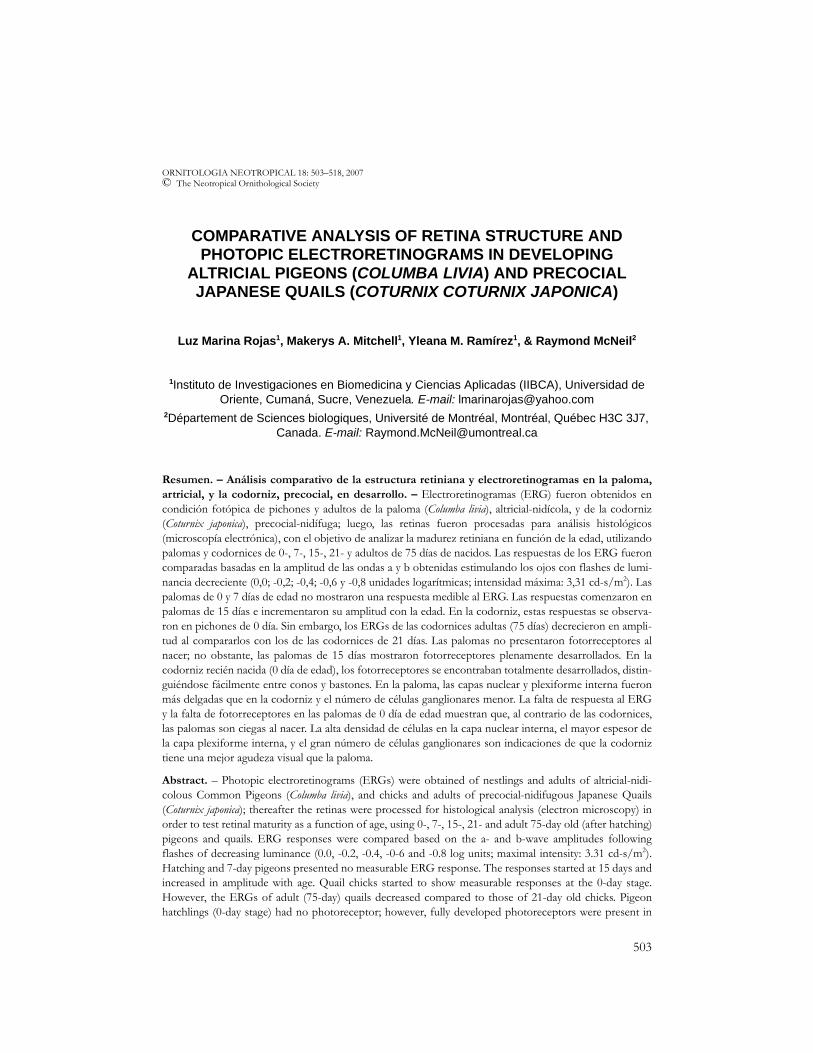
FIG. 1. Representative ERG responses of 0-, 7-, 15-, 21- and 75-day (after hatching) Common Pigeonsand Japanese Quails obtained under photopic conditions. Nomenclature: a = peak of the a-wave; b =peak of the b-wave. The figures on the left represent light intensity values (Log units).
507
VISION IN ALTRICIAL AND PRECOCIAL BIRDS
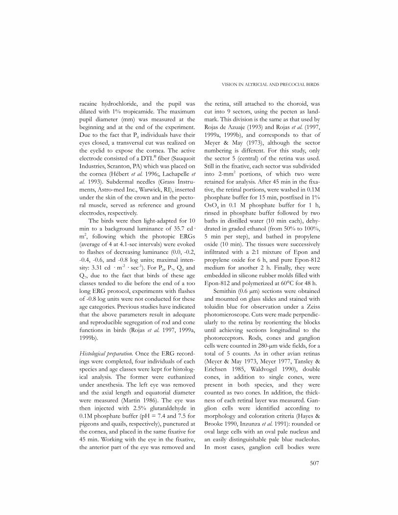
racaine hydrochloride, and the pupil wasdilated with 1% tropicamide. The maximumpupil diameter (mm) was measured at thebeginning and at the end of the experiment.Due to the fact that P0 individuals have theireyes closed, a transversal cut was realized onthe eyelid to expose the cornea. The activeelectrode consisted of a DTL® fiber (SauquoitIndustries, Scranton, PA) which was placed onthe cornea (Hébert et al. 1996;, Lachapelle etal. 1993). Subdermal needles (Grass Instru-ments, Astro-med Inc., Warwick, RI), insertedunder the skin of the crown and in the pecto-ral muscle, served as reference and groundelectrodes, respectively.
The birds were then light-adapted for 10min to a background luminance of 35.7 cd·m2, following which the photopic ERGs(average of 4 at 4.1-sec intervals) were evokedto flashes of decreasing luminance (0.0, -0.2,-0.4, -0.6, and -0.8 log units; maximal inten-sity: 3.31 cd · m-2 · sec-1). For P0, P7, Q0 andQ7, due to the fact that birds of these ageclasses tended to die before the end of a toolong ERG protocol, experiments with flashesof -0.8 log units were not conducted for theseage categories. Previous studies have indicatedthat the above parameters result in adequateand reproducible segregation of rod and conefunctions in birds (Rojas et al. 1997, 1999a,1999b).
Histological preparation. Once the ERG record-ings were completed, four individuals of eachspecies and age classes were kept for histolog-ical analysis. The former were euthanizedunder anesthesia. The left eye was removedand the axial length and equatorial diameterwere measured (Martin 1986). The eye wasthen injected with 2.5% glutaraldehyde in0.1M phosphate buffer (pH = 7.4 and 7.5 forpigeons and quails, respectively), punctured atthe cornea, and placed in the same fixative for45 min. Working with the eye in the fixative,the anterior part of the eye was removed and
the retina, still attached to the choroid, wascut into 9 sectors, using the pecten as land-mark. This division is the same as that used byRojas de Azuaje (1993) and Rojas et al. (1997,1999a, 1999b), and corresponds to that ofMeyer & May (1973), although the sectornumbering is different. For this study, onlythe sector 5 (central) of the retina was used.Still in the fixative, each sector was subdividedinto 2-mm2 portions, of which two wereretained for analysis. After 45 min in the fixa-tive, the retinal portions, were washed in 0.1Mphosphate buffer for 15 min, postfixed in 1%OsO4 in 0.1 M phosphate buffer for 1 h,rinsed in phosphate buffer followed by twobaths in distilled water (10 min each), dehy-drated in graded ethanol (from 50% to 100%,5 min per step), and bathed in propyleneoxide (10 min). The tissues were successivelyinfiltrated with a 2:1 mixture of Epon andpropylene oxide for 6 h, and pure Epon-812medium for another 2 h. Finally, they wereembedded in silicone rubber molds filled withEpon-812 and polymerized at 60°C for 48 h.
Semithin (0.6 µm) sections were obtainedand mounted on glass slides and stained withtoluidin blue for observation under a Zeissphotomicroscope. Cuts were made perpendic-ularly to the retina by reorienting the blocksuntil achieving sections longitudinal to thephotoreceptors. Rods, cones and ganglioncells were counted in 280-µm wide fields, for atotal of 5 counts. As in other avian retinas(Meyer & May 1973, Meyer 1977, Tansley &Erichsen 1985, Waldvogel 1990), doublecones, in addition to single cones, werepresent in both species, and they werecounted as two cones. In addition, the thick-ness of each retinal layer was measured. Gan-glion cells were identified according tomorphology and coloration criteria (Hayes &Brooke 1990, Inzunza et al. 1991): rounded oroval large cells with an oval pale nucleus andan easily distinguishable pale blue nucleolus.In most cases, ganglion cell bodies were
508
ROJAS ET AL.
arranged side by side in a 1-cell thicklayer, but in the specialized thickened areas(e.g., central retina), they occurred in two orthree layers. Displaced amacrine cells, on theother hand, appeared as small pale stainedbodies lying next to the inner plexiformlayer.
Additionally, for histological analysis ofcellular components of the retina, thin (70
µm) sections were cut and stained with uranileacetate and lead citrate. Histological observa-tions were made from microphotographsobtained with the use of a Hitachi H-600transmission electron microscope.
Data analysis. Results were analyzed by usingthe means ± 95% confidence intervals, con-ventional two-way variance analysis
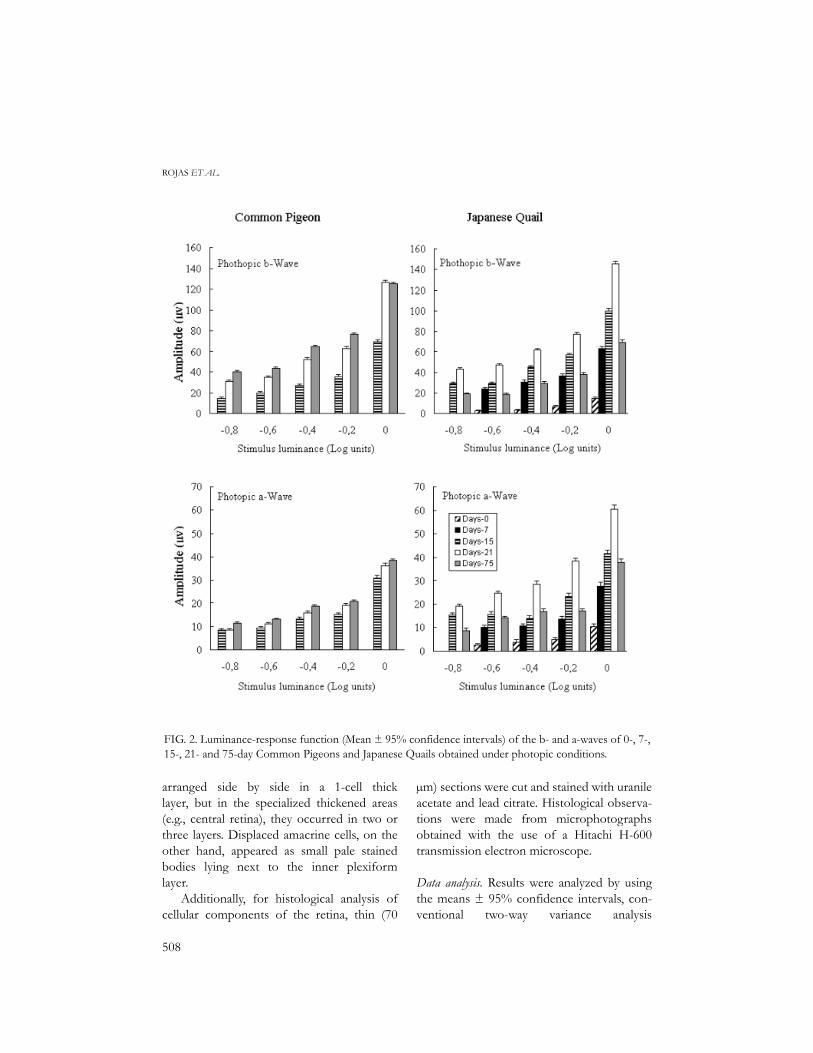
FIG. 2. Luminance-response function (Mean ± 95% confidence intervals) of the b- and a-waves of 0-, 7-,15-, 21- and 75-day Common Pigeons and Japanese Quails obtained under photopic conditions.
509
VISION IN ALTRICIAL AND PRECOCIAL BIRDS
(ANOVA), and Duncan a posteriori tests (Sokal& Rohlf 1979) for within and between groupcomparisons of the different variables for
morphometric parameters and photopic ERGrecordings (a- and b-wave amplitudes),evoked to flashes of 0.0 log units from which
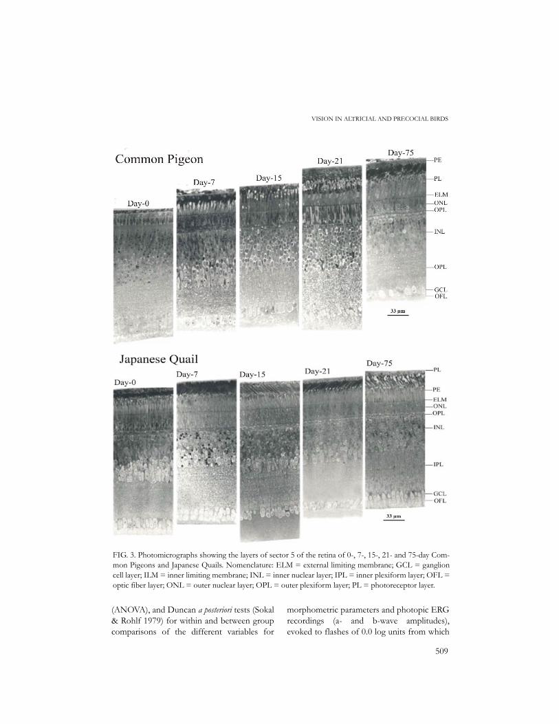
FIG. 3. Photomicrographs showing the layers of sector 5 of the retina of 0-, 7-, 15-, 21- and 75-day Com-mon Pigeons and Japanese Quails. Nomenclature: ELM = external limiting membrane; GCL = ganglioncell layer; ILM = inner limiting membrane; INL = inner nuclear layer; IPL = inner plexiform layer; OFL =optic fiber layer; ONL = outer nuclear layer; OPL = outer plexiform layer; PL = photoreceptor layer.
510
ROJAS ET AL.
luminance-response function curves weregenerated.
RESULTS
Analysis of ERG recordings included themeasurements of photopic a- and b-waveamplitudes for which luminance-responsefunction histograms were generated (mean ±95% confidence intervals). The analysis ofmorphological measurements also includedcalculation of the means (± 95% confidenceintervals) of dilated pupil diameter, cell densi-ties (rods, cones and ganglion cells), thicknessof each retinal layer as well as the rod:coneratios for each species.
Electroretinography. Representative ERGsobtained in photopic conditions for 0-, 7-, 15,21- and 75-day old pigeons and quails are pre-sented in Figure 1. They differ both in ampli-tude and shape between age classes of bothspecies. The luminance-response functiongenerated from amplitude measurements aregraphically represented in Figure 2 for b- anda-waves.
As seen in Figure 2, 0- and 7-day oldpigeon nestlings produced no ERG responseat all. However, starting with 15-day old indi-viduals, photopic b- and a-waves increasedprogressively in amplitude, both with theintensity of luminance stimulus and the age ofindividuals (Fig. 2), except for the b-wave of
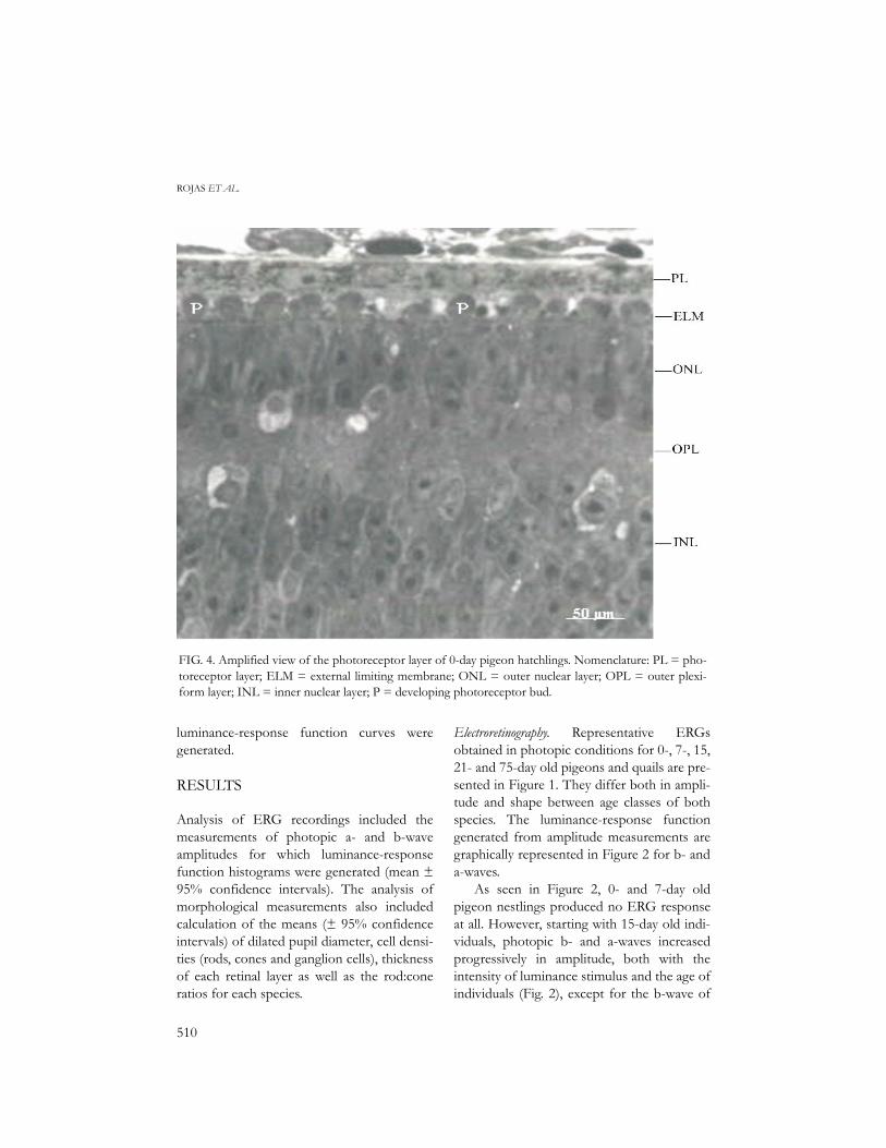
FIG. 4. Amplified view of the photoreceptor layer of 0-day pigeon hatchlings. Nomenclature: PL = pho-toreceptor layer; ELM = external limiting membrane; ONL = outer nuclear layer; OPL = outer plexi-form layer; INL = inner nuclear layer; P = developing photoreceptor bud.
511
VISION IN ALTRICIAL AND PRECOCIAL BIRDS
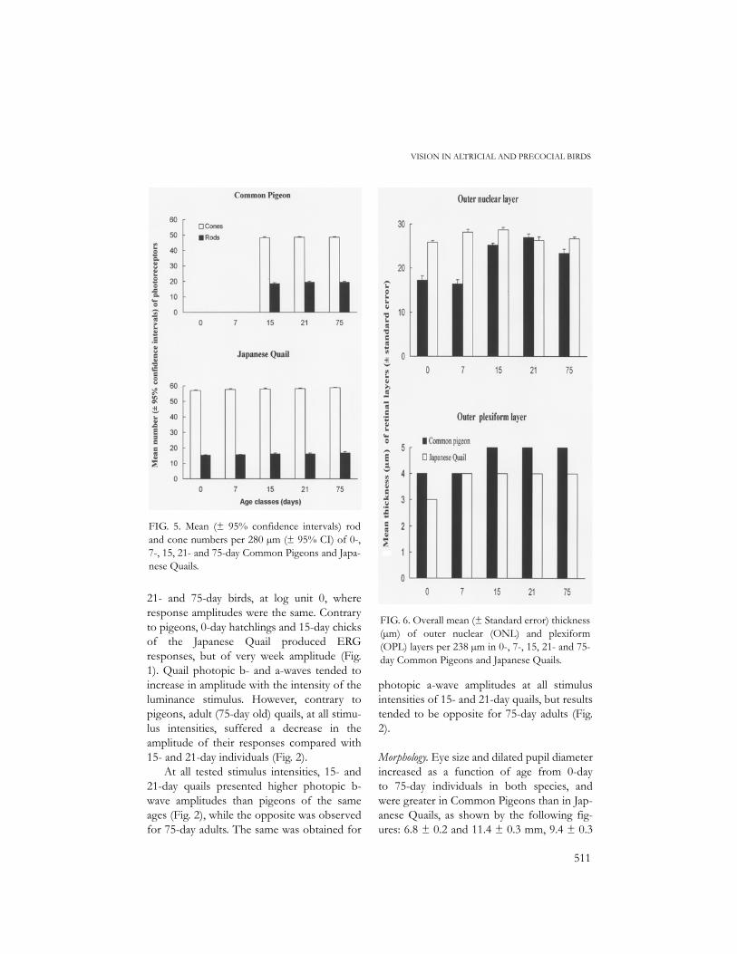
21- and 75-day birds, at log unit 0, whereresponse amplitudes were the same. Contraryto pigeons, 0-day hatchlings and 15-day chicksof the Japanese Quail produced ERGresponses, but of very week amplitude (Fig.1). Quail photopic b- and a-waves tended toincrease in amplitude with the intensity of theluminance stimulus. However, contrary topigeons, adult (75-day old) quails, at all stimu-lus intensities, suffered a decrease in theamplitude of their responses compared with15- and 21-day individuals (Fig. 2).
At all tested stimulus intensities, 15- and21-day quails presented higher photopic b-wave amplitudes than pigeons of the sameages (Fig. 2), while the opposite was observedfor 75-day adults. The same was obtained for
photopic a-wave amplitudes at all stimulusintensities of 15- and 21-day quails, but resultstended to be opposite for 75-day adults (Fig.2).
Morphology. Eye size and dilated pupil diameterincreased as a function of age from 0-dayto 75-day individuals in both species, andwere greater in Common Pigeons than in Jap-anese Quails, as shown by the following fig-ures: 6.8 ± 0.2 and 11.4 ± 0.3 mm, 9.4 ± 0.3
FIG. 5. Mean (± 95% confidence intervals) rodand cone numbers per 280 µm (± 95% CI) of 0-,7-, 15, 21- and 75-day Common Pigeons and Japa-nese Quails.
FIG. 6. Overall mean (± Standard error) thickness(µm) of outer nuclear (ONL) and plexiform(OPL) layers per 238 µm in 0-, 7-, 15, 21- and 75-day Common Pigeons and Japanese Quails.

512
ROJAS ET AL.
and 14.5 ± 0.2 mm, and 3.0 ± 0.0 and 4.8 ±0.1 mm for axial length, equatorial diameterand dilated pupil diameter in 0-day hatchlingsand 75-day Common Pigeons, respectively,compared to 5.2 ± 0.4 and 8.4 ± 0.5 mm, 7.1± 0.3 and 10.2 ± 0.4 mm, and 2.0 ± 0.0 and2.9 ± 20.2 mm for the same parameters in 0-day chicks and 75-day Japanese Quails.
Photomicrographs showing the principallayers of sector 5 (central) of the retina ofeach species and age class are presented inFigure 3. At hatching, Japanese Quails chickshad fully developed retinal layers, includingthe photoreceptor layer, with typical rods andcones (Fig. 3). On the contrary, in 0-daypigeon hatchlings, photoreceptors were lack-ing (Fig. 3). Small buds were present but theycorresponded to developing photoreceptors(Fig. 4). Progress in developing photorecep-tors was observed in 7-day nestlings, but therods and cones (with 2-µm oil droplets) canbe considered as immature compared tothose of 15-day pigeons. Excepting 0- and 7-
day pigeons (lack of rods and cones), rod andcone numbers were very stable from one ageclass to another in both species (Fig. 5); rodsand cones per 280µm in the central sectorwere ca. 19.0 and 48.5, and 16.0 and 58.0,resulting in rod:cone ratios of 0.4:1.0 and0.3:1.0 for the pigeons and quails, respectively.Thus, compared to Common Pigeons, Japa-nese Quails had more cones and fewer rods.Fully grown oil droplets in the cones of bothspecies varied in diameter between 3 and 4µm.
The outer nuclear and plexiform layers inpigeons (measuring from 13 to 20 µm and 4µm, respectively) were thinner in 0-dayhatchlings than in older individuals; in thequails, the outer nuclear layer, varying inthickness from 24 to 32 µm, showed only lit-tle variation as a function of age classes, butthe outer plexiform layer of 0-day chicks wasthinner (3 µm) than that of older chicks (5µm) (Fig. 6). In 7-day individuals of both spe-cies, the outer nuclear layer showed the pres-
FIG. 7. Ultrastructure of the outer nuclear layers of 7-day Common Pigeons and Japanese Quails.Nomenclature: N = nucleus; n = nucleolus; SF = synaptic foot.
513
VISION IN ALTRICIAL AND PRECOCIAL BIRDS
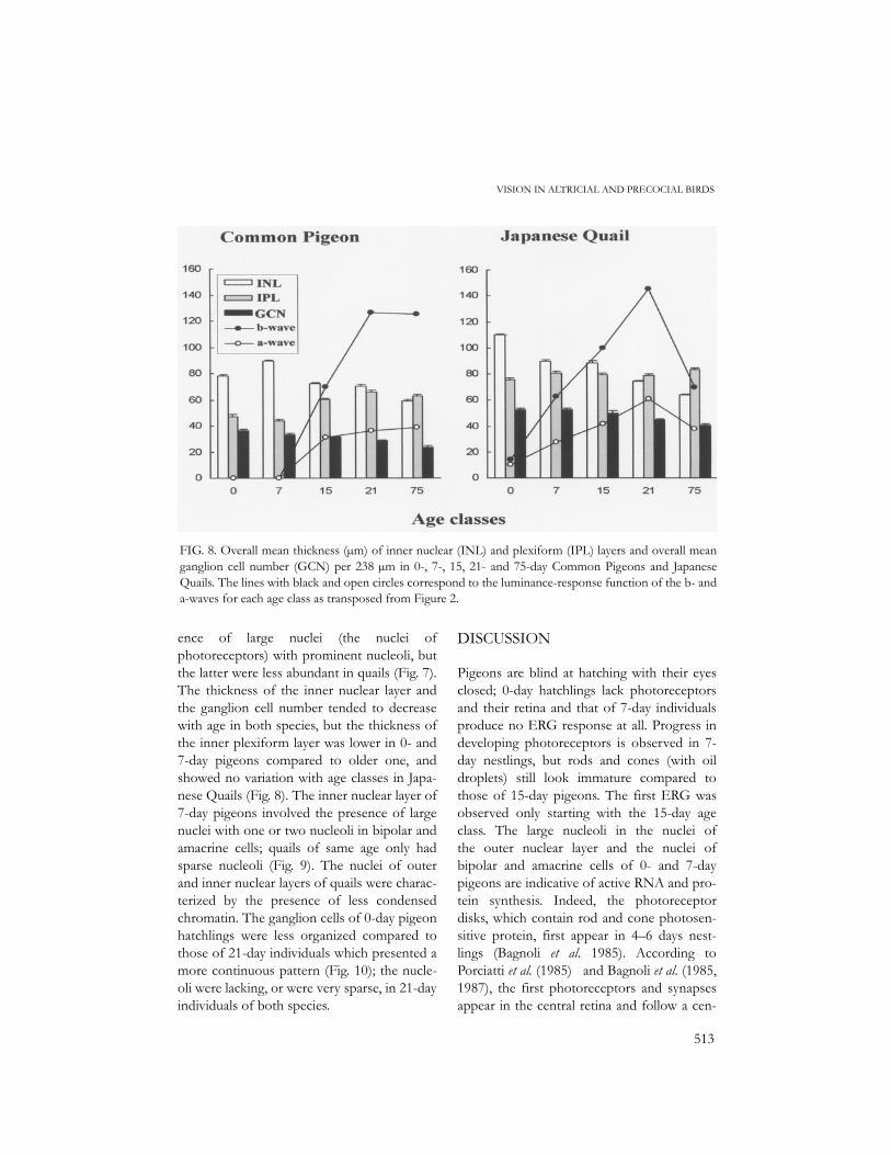
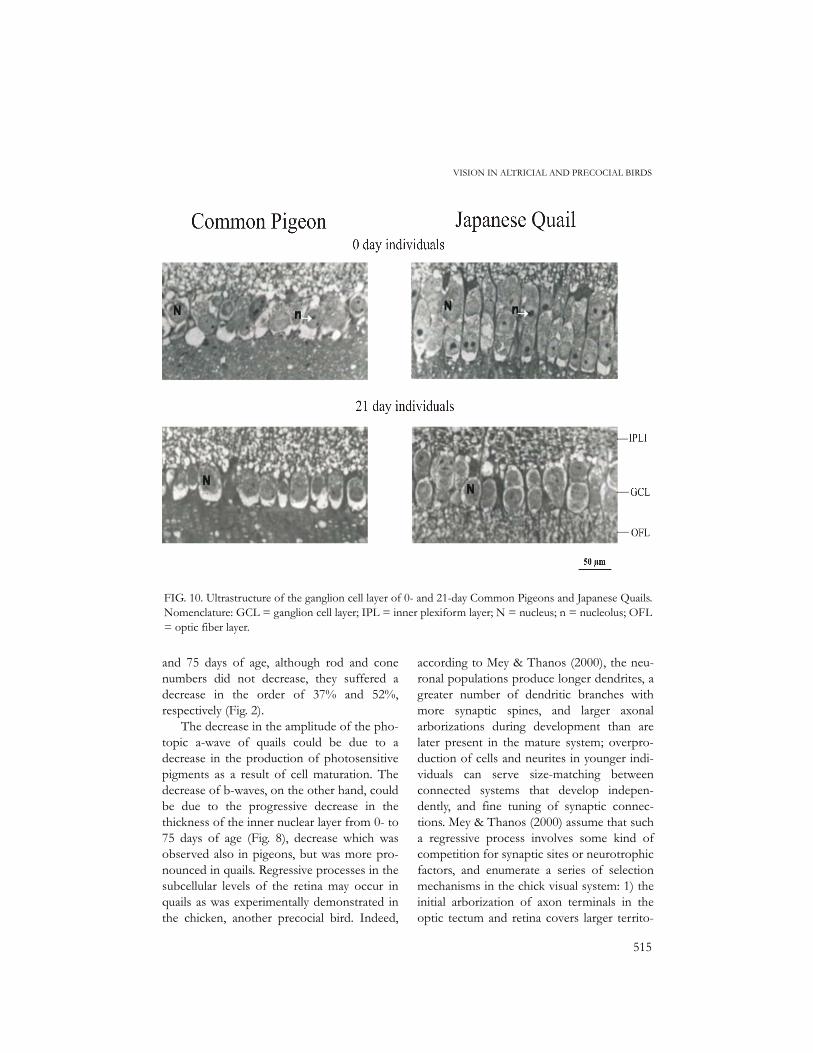
ence of large nuclei (the nuclei ofphotoreceptors) with prominent nucleoli, butthe latter were less abundant in quails (Fig. 7).The thickness of the inner nuclear layer andthe ganglion cell number tended to decreasewith age in both species, but the thickness ofthe inner plexiform layer was lower in 0- and7-day pigeons compared to older one, andshowed no variation with age classes in Japa-nese Quails (Fig. 8). The inner nuclear layer of7-day pigeons involved the presence of largenuclei with one or two nucleoli in bipolar andamacrine cells; quails of same age only hadsparse nucleoli (Fig. 9). The nuclei of outerand inner nuclear layers of quails were charac-terized by the presence of less condensedchromatin. The ganglion cells of 0-day pigeonhatchlings were less organized compared tothose of 21-day individuals which presented amore continuous pattern (Fig. 10); the nucle-oli were lacking, or were very sparse, in 21-dayindividuals of both species.
DISCUSSION
Pigeons are blind at hatching with their eyesclosed; 0-day hatchlings lack photoreceptorsand their retina and that of 7-day individualsproduce no ERG response at all. Progress indeveloping photoreceptors is observed in 7-day nestlings, but rods and cones (with oildroplets) still look immature compared tothose of 15-day pigeons. The first ERG wasobserved only starting with the 15-day ageclass. The large nucleoli in the nuclei ofthe outer nuclear layer and the nuclei ofbipolar and amacrine cells of 0- and 7-daypigeons are indicative of active RNA and pro-tein synthesis. Indeed, the photoreceptordisks, which contain rod and cone photosen-sitive protein, first appear in 4–6 days nest-lings (Bagnoli et al. 1985). According toPorciatti et al. (1985) and Bagnoli et al. (1985,1987), the first photoreceptors and synapsesappear in the central retina and follow a cen-
FIG. 8. Overall mean thickness (µm) of inner nuclear (INL) and plexiform (IPL) layers and overall meanganglion cell number (GCN) per 238 µm in 0-, 7-, 15, 21- and 75-day Common Pigeons and JapaneseQuails. The lines with black and open circles correspond to the luminance-response function of the b- anda-waves for each age class as transposed from Figure 2.

514
ROJAS ET AL.
tro-peripheral progression. On the first dayafter hatching, pigeons have various synapsesin the inner plexiform layer and only a fewones in the outer plexiform layer. The innerplexiform layer is made of the dendrites andneurites of bipolar and amacrine cells, whilethe outer plexiform layer is made of horizon-tal bipolar cells and photoreceptors. Accord-ing to the same authors, the outer plexiformlayer is formed at the same time as photore-ceptor disks. The results of this study showthat 0-day pigeon hatchlings already have thetwo plexiform layers, but synapses are proba-bly inactive because of the lack of photore-ceptors.
Hatching quails, on the contrary, haveopen lids and fully grown retinas with alltheir layers, and produce ERG responses,although of low amplitude. The presence ofless condensed chromatin in the nuclei oftheir outer and inner nuclear layers indicates
that protein synthesis largely took placebefore hatching. In the chicken, the segrega-tion of the outer and inner plexiform layersbegins on day-6, and ends around day-14 ofembryonic life (Meller & Tetzlaff 1976,Spence & Robson 1989). The first synapsesare found in the inner plexiform layer on day-13 of embryonic life, but appear between thephotoreceptors and the bipolar cells of theouter plexiform layer beginning only on day-17 of prehatching life (Hering & Kröger1996).
From the age of 15 days, the number ofrods and cones and their ratios remained thesame in the pigeons, but their photopic a-and b-waves continued increasing in ampli-tude until the age of 21 days, and remainedrelatively stable thereafter (Fig. 2). In thequails, on the contrary, the photopic a- andb-waves increased in amplitude up to the ageof 21 days, but thereafter, between the 21
FIG. 9. Ultrastructure of the inner nuclear layers of 7-day Common Pigeons and Japanese Quails.Nomenclature: N = nucleus; n = nucleolus; SF = synaptic foot.
515
VISION IN ALTRICIAL AND PRECOCIAL BIRDS
and 75 days of age, although rod and conenumbers did not decrease, they suffered adecrease in the order of 37% and 52%,respectively (Fig. 2).
The decrease in the amplitude of the pho-topic a-wave of quails could be due to adecrease in the production of photosensitivepigments as a result of cell maturation. Thedecrease of b-waves, on the other hand, couldbe due to the progressive decrease in thethickness of the inner nuclear layer from 0- to75 days of age (Fig. 8), decrease which wasobserved also in pigeons, but was more pro-nounced in quails. Regressive processes in thesubcellular levels of the retina may occur inquails as was experimentally demonstrated inthe chicken, another precocial bird. Indeed,
according to Mey & Thanos (2000), the neu-ronal populations produce longer dendrites, agreater number of dendritic branches withmore synaptic spines, and larger axonalarborizations during development than arelater present in the mature system; overpro-duction of cells and neurites in younger indi-viduals can serve size-matching betweenconnected systems that develop indepen-dently, and fine tuning of synaptic connec-tions. Mey & Thanos (2000) assume that sucha regressive process involves some kind ofcompetition for synaptic sites or neurotrophicfactors, and enumerate a series of selectionmechanisms in the chick visual system: 1) theinitial arborization of axon terminals in theoptic tectum and retina covers larger territo-
FIG. 10. Ultrastructure of the ganglion cell layer of 0- and 21-day Common Pigeons and Japanese Quails.Nomenclature: GCL = ganglion cell layer; IPL = inner plexiform layer; N = nucleus; n = nucleolus; OFL= optic fiber layer.

516
ROJAS ET AL.
ries because produce more cells and dendritesthan necessary, implying that non-functionalneurites are eliminated by a selection process;2) cell death and removal of axon collateralsaffect preferentially the neurons whose axonshave grown to an inappropriate target; 3)excessive branches which degenerate lateroften have failed to reach appropriate sites inthe tissue and have not become functional; 4)after a depletion of ganglion cells, only sur-vive those neurons which have larger sizesand more dendritic branches, and that againprovides circumstantial evidence for competi-tion among these neurons for synaptic input.It thus appears that posthatching retinaldevelopment in quails, including the degener-ation of many neurons of the inner plexiformlayer, could be slow and may continue forsome time after the 21th day of age, allowingthe remaining neurons to produce more den-drites and neurites, thus increasing or main-taining the thickness of the inner plexiformlayer. A decrease in the functionality of rodsand cones between the 21th and the 75th daysof age could also be a factor responsible forthe decrease in b-wave amplitude. Indeed,Yamada et al. (1998) also reported a decreasein the quail retina thickness until the 30 daysof posthatching age. During the same ageinterval, they observed a decrease in the gan-glion and inner nuclear cell density. Theyobserved a notable decrease in the innernuclear and plexiform layers particularlybetween the 20th and the 30th days of age.However, in the present study, the inner plex-iform layer did not suffer any decrease (seeFig. 8).
In the quails, the number of ganglion cellsalso suffered a progressive decrease frombirth to the 75 days of age. In 12- to 16-dayembryos, some 30-40% of ganglion cellsdegenerate (Hughes & McLoon 1979). Such areduction in the ganglion cell number wouldnot affect too much visual acuity in quails.Visual acuity is depending both on cones and
ganglion cells, i.e., both structures are respon-sible for the detection of movement and finedetails (Dowling 1987, Hodos et al. 1991,McIlwain 1996). Quails have more cones andganglion cells than pigeons; this may allowthem finer vision compared to pigeons. Inquails, ganglion cells are more abundant in thecentral region of the retina and they graduallydecrease in number from the center towardsthe periphery of the retina (Ikushima et al.1986). In pigeons, ganglion cells are moreabundant in the central and dorso-temporalareas (Binggeli & Paule 1969).
Posthatching growth of altricial birds isthree or four times faster than that of preco-cial ones (Ricklefs 1979b). For example, thealtricial European Starling (Sturnus vulgaris)grows four times faster than the semipreco-cial Common Tern (Sterna hirundo), and twoand half times faster that the precocial Japa-nese Quail (Ricklefs 1979b). Precocial birdshave, at birth, a relatively larger brain givingthem the capacity to forage, run about, andregulate their body temperature soon afterthey hatch, but their growth rate is very slow,resulting in a relatively smaller brain in adults;the opposite is observed in altricial birds(Starck 1993).
The main facts shown in this study arethat ERG responses are lacking in 0-dayhatchlings and 7-day nestlings of the Com-mon Pigeon; beginning with 21-day nestlings,they reach maximal intensity and remain rela-tively stable or increase only slightly thereaf-ter. In the Japanese Quail, on the other hand,measurable ERG responses take place in 0-day chicks and increase in amplitude until theage of 21 days, but decrease some timebetween 21 and 75 days of age. This is inagreement with the morphological features ofthe retinas, in particular with the lack of pho-toreceptors in 0- and 7-day pigeons, and withthe presence of fully developed and easily dis-tinguishable rods and cones in 0-day quailhatchlings.
517
VISION IN ALTRICIAL AND PRECOCIAL BIRDS
ACKNOWLEDGMENTS
This study was supported by research grantsof Consejo de Investigación de la Universidadde Oriente, Instituto de Investigaciones yCiencias Applicadas de la UDO (IIBCA-UDO), Natural Sciences and EngineeringResearch Council, and Université de Mont-real. The authors thank the personal ofIIBCA-UDO for assistance y laboratorywork. The experiments reported in this paperwere conducted in accordance to the guide-lines established by the Canadian Council onAnimal Care (1994).
REFERENCES
Armington, J. 1974. The electroretinogram. Aca-demic Press, New York, New York.
Bagnoli, P., V. Porciatti, A. Lanfranchi, & C. Bedini.1985. Developing pigeon retina: Light-evokedresponses and ultrastructure of outer segmentsand synapses. J. Comp. Neurol. 235: 384–394.
Bagnoli, P., V. Porciatti, G. Fontanesi, & L. Sebas-tiani. 1987. Morphological and functionalchanges in the retinotectal system of the pigeonduring the early posthatching period. J. Comp.Neurol. 256: 400–411.
Binggeli, R., & W. Paule. 1969. The pigeon retina:Quantitative aspects of the optic nerve andganglion cell layer. J. Comp. Neurol. 137: 118.
Canadian Council on Animal Care. 1994. Guide tothe care and use of experimental animals. Cana-dian Council on Animal Care,Ottawa, Ontario.
Dowling, J. 198. The retina. An approachable partof the brain. Harvard Univ. Press. Cambridge,Massachusetts.
Gill, F. 1994. Ornithology. W H Freeman andCompany, New York, New York.
Hahmann, U., & O Güntürkün. 1993. The visualacuity for the lateral visual field of the pigeon(Columba livia). Vision Res. 33: 1659–1664.
Hayes, B.P., & M. de L. Brooke. 1990. Retinal gan-glion cell distribution and behaviour in procel-lariiform seabirds. Vision Res. 30: 1277–1289.
Heaton, M. B. 1971. Ontogeny of vision in thePeking Duck (Anas platyrhynchos): The papillarylight reflex as a means for investigating visual
onset and development in avian embryos. Dev.Psychobiol. 4: 313–332.
Heaton, M. B. 1973. Early visual function in bob-white and Japanese Quail embryos as reflectedby papillary reflex. J. Comp. Physiol. Psychol.84: 134–139.
Heaton, M. B., & M.S. Harth. 1974. Developingvisual function in the pigeon embryo with com-parative reference to other avian species. J.Comp. Physiol. Psychol. 86: 151–156.
Hébert, M., P. Lachapelle, & M. Dumont. 1996.Reproducibility of electroretinograms recordedwith DTL electrodes. Doc. Ophthalmol. 91:333–342.
Hering, H., & S. Kröger. 1996. Formation of syn-aptic specializations in the inner plexiform layerof the developing chick retina. J. Comp. Neurol.375: 393–405.
Hodos, W. 1993. The visual capabilities of birds.Pp. 64–76 in Zeigler, H. P., & H. Bischof.(eds.). Vision, brain and behavior. BradfordBook, London, UK.
Hodos, W., R. Miller, & K. Fite. 1991. Age-depen-dent changes in visual acuity and retinal mor-phology in pigeons. Vision Res. 31: 669–677.
Hughes, W., & S. McLoon. 1979. Ganglion celldeath during normal retinal development in thechick: Comparisons with cell death induced byearly target field destruction. Exp. Neurol. 66:587–601.
Ikeda, H. 1993. Clinical electroretinography. Pp.115–139 in Halliday, A. M. (ed). Evoked poten-tials in clinical testing. Churchill Livingstone,New York, New York.
Ikushima, M., M. Watanabe, & H. Ito. 1986. Distri-bution and morphology of retinal ganglion cellin the Japanese Quail. Brain Res. 376: 320–334.
Inzunza, O., H. Bravo, R. L. Smith, & M. Angel.1991. Topography and morphology of retinalganglion cells in falconiforms: A study on pred-atory and carrion-eating birds. Anat. Rec. 229:271–277.
Lachapelle, P., J. Benoît, J. M. Little, & B.Lachapelle. 1993. Recording the oscillatorypotentials with the DTL electrode. Doc. Oph-thalmol. 83: 119–130.
Martin, G. 1990. Birds by night. Poyser, London,UK.
Martin, G. R. 1986. The eye of a passeriform bird,
518
ROJAS ET AL.
the European Starling (Sturnus vulgaris): eyemovement amplitude, visual fields and sche-matic optics. J. Comp Physiol. A 159: 545–557.
Martin, G. R., K. J. Wilson, J. M. Wild, S. Parsons,M. F. Kubke, & J. Corfield. 2007. Kiwi foregovision in the guidance of their nocturnal activi-ties. PLoS ONE 2(2): e198. doi:10.1371/jour-nal.pone.0000198.
McIlwain, J. 1996. An introduction to the biologyof vision. Cambridge Univ. Press. Cambridge,UK.
Meller, K., & W. Tetzlaff. 1976. Scanning electronmicroscopic studies on the development of thechick retina. Cell Tissue Res. 170: 145–159.
Mey, J., & S. Thanos. 2000. Development of thevisual system of the chick: I. Cell differentia-tion and histogenesis. Brain Res. Rev. 2: 343–379.
Meyer, D.B. 1977. The avian eye and its adapta-tions. Pp. 549–611 in Crescitelli, F. (ed). Thevisual system in vertebrates. Vol VII/5.Springer Verlag, Berlin, Germany.
Meyer, D., & C. May. 1973. The topographical dis-tribution of rod and cones in the adult chickenretina. Exp. Eyes. Res. 17: 347–355.
Porciatti, V., P. Bagnoli, A. Lanfranchi, & C. Bedini.1985. Interactions between photoreceptors andpigmented epithelium in developing pigeonretina: an electrophysiological and ultrastruc-tural study. Doc. Ophthalmol. 60: 413–419.
Ricklefs, R. 1979a. Patterns of growth in birds. V. Acomparative study of development in the Star-ling, common Tern, and Japanese Quail. Auk96: 10–30.
Ricklefs, R. 1979b. Adaptation, constraint, andcompromise in avian postnatal development.Biol. Rev. 54: 269–290.
Ricklefs, R. 1983. Avian postnatal development.Pp. 1–83 in Farner, D. S, J. R. King, & K. C,
Parkes (eds.). Avian biology. Volume VII. Aca-demic Press. New York, New York.
Rojas de Azuaje, L., S. Tai, & R. McNeil. 1993.Comparison of rod/cone ratio in three speciesof shorebirds having different nocturnal forag-ing strategies. Auk 110: 141–145.
Rojas, L., R. McNeil, T. Cabana, & P. Lachapelle.1997. Diurnal and nocturnal visual function intwo tactile foraging waterbirds: The Americanwhite ibis and the black skimmer. Condor 99:191–200.
Rojas, L., R. McNeil, T. Cabana, & P. Lachapelle.1999a. Diurnal and nocturnal visual capabilitiesin shorebirds as a function of their feedingstrategies. Brain Behav. Evol. 53: 29–43.
Rojas, L., R. McNeil, T. Cabana, & P. Lachapelle.1999b. Behavioral, morphological and physio-logical correlates of diurnal and nocturnalvision in selected wading bird species. BrainBehav. Evol. 53: 227–242.
Sokal, R., & J. Rohlf. 1979. Biometría. Ediciones H.Blumé, Madrid, Spain.
Spence, S., & J. Robson. 1989. An autoradiographicanalysis of neurogenesis in the chick retina invitro and in vivo. Neuroscience 32: 801–812.
Starck, J. M. 1993. Evolution of avian ontogenies.Pp. 275–366 in Power, D. M. (ed.). CurrentOrnithology. Volume 10. Plenum Press. NewYork, New York.
Tansley, K., & J. R. Erichsen. 1985. Vision. Pp.623–629 in Campbell B., & E. Lack (ed.). A dic-tionary of birds. Poyser, Calton, UK.
Waldvogel, J. A. 1990. The birds eye view. Am. Sci.78: 342–353.
Yamada, M., A. Goto, & S. Sugita. 1998. Morpho-metric analyses of the growth in the visualorgan and tectum of the Japanese Quail(Coturnix japonica) after hatching. Anim. Sci.Technol. 69: 941–949.