comparative genomic analysis of toxin-negative strains of
TRANSCRIPT

RESEARCH ARTICLE Open Access
Comparative genomic analysis of toxin-negative strains of Clostridium difficile fromhumans and animals with symptoms ofgastrointestinal diseasePiklu Roy Chowdhury1,2*, Matthew DeMaere1, Toni Chapman2, Paul Worden1, Ian G. Charles1,3,Aaron E. Darling1 and Steven P. Djordjevic1*
Abstract
Background: Clostridium difficile infections (CDI) are a significant health problem to humans and food animals.Clostridial toxins ToxA and ToxB encoded by genes tcdA and tcdB are located on a pathogenicity locus knownas the PaLoc and are the major virulence factors of C. difficile. While toxin-negative strains of C. difficile areoften isolated from faeces of animals and patients suffering from CDI, they are not considered to play a rolein disease. Toxin-negative strains of C. difficile have been used successfully to treat recurring CDI but theirpropensity to acquire the PaLoc via lateral gene transfer and express clinically relevant levels of toxins hasreinforced the need to characterise them genetically. In addition, further studies that examine the pathogenicpotential of toxin-negative strains of C. difficile and the frequency by which toxin-negative strains may acquirethe PaLoc are needed.
Results: We undertook a comparative genomic analysis of five Australian toxin-negative isolates of C. difficilethat lack tcdA, tcdB and both binary toxin genes cdtA and cdtB that were recovered from humans and farmanimals with symptoms of gastrointestinal disease. Our analyses show that the five C. difficile isolates clusterclosely with virulent toxigenic strains of C. difficile belonging to the same sequence type (ST) and havevirulence gene profiles akin to those in toxigenic strains. Furthermore, phage acquisition appears to haveplayed a key role in the evolution of C. difficile.
Conclusions: Our results are consistent with the C. difficile global population structure comprising six cladeseach containing both toxin-positive and toxin-negative strains. Our data also suggests that toxin-negativestrains of C. difficile encode a repertoire of putative virulence factors that are similar to those found intoxigenic strains of C. difficile, raising the possibility that acquisition of PaLoc by toxin-negative strains posesa threat to human health. Studies in appropriate animal models are needed to examine the pathogenicpotential of toxin-negative strains of C. difficile and to determine the frequency by which toxin-negativestrains may acquire the PaLoc.
Keywords: Clostridium difficile, Toxin-negative isolates, Comparative genomics, CDI, Zoonosis
* Correspondence: [email protected];[email protected] ithree institute, University of Technology Sydney, Sydney 2007, AustraliaFull list of author information is available at the end of the article
© 2016 Roy Chowdhury et al. Open Access This article is distributed under the terms of the Creative Commons Attribution4.0 International License (http://creativecommons.org/licenses/by/4.0/), which permits unrestricted use, distribution, andreproduction in any medium, provided you give appropriate credit to the original author(s) and the source, provide a link tothe Creative Commons license, and indicate if changes were made. The Creative Commons Public Domain Dedication waiver(http://creativecommons.org/publicdomain/zero/1.0/) applies to the data made available in this article, unless otherwise stated.
Roy Chowdhury et al. BMC Microbiology (2016) 16:41 DOI 10.1186/s12866-016-0653-3

BackgroundClostridium difficile is a Gram-positive pathogen thathas emerged to become one of the leading causes of in-fectious diarrhoea in adult humans, securing its inclu-sion in the ESCAPE group of pathogens [1–4]. C.difficile infections range from being asymptomatic tocausing mild or severe diarrhoea and occasionally life-threatening conditions such as pseudomembranous col-itis and toxic megacolon [1, 5]. However, community-acquired C. difficile infection is being reported withincreasing frequency [6] and C. difficile is also emergingas a pathogen in animals particularly cattle, pigs andhorses [5, 7–10]. Molecular epidemiological studiesshow that infections in humans and animals can sharethe same ribotype or multilocus sequence type (ST) [11]suggesting that pathogenic C. difficile may trafficbetween humans and animals, although further studiesare needed to confirm these linkages.C. difficile is a genetically diverse and globally dis-
persed species [11–16] having a clonal structure com-prising six major clades (clades 1, 2, 3, 4, 5 and C-I).Clade C-I is the most phylogenetically divergent cladeand may represent of a new subspecies of C. difficile[17]. Clade C-I typically comprise toxin-negative strainsof C. difficile [17] but toxigenic variants that reside inClade C-I have recently been described [18]. Representa-tives from most clades have been associated with CDI inhumans and comprise toxigenic strains with A+/B+, A−/B+
toxin types [11, 14, 17, 19–22]. Non-toxigenic strains of C.difficile are represented in all six clades [11].Toxin expression is considered mandatory for the de-
velopment of C. difficile disease [23, 24]. Two large clos-tridial toxins known as toxins A (308 kDa) and B(260 kDa) encoded by tcdA and tcdB and the genes im-plicated in regulating their expression (tcdC, tcdE andtcdR) reside on a 19.6-kb pathogenicity locus known asthe PaLoc [25, 26]. The PaLoc is replaced by 115/75 basepair non-coding region in toxin negative strains of C.difficile [27]. Approximately 20 % of C. difficile strainsexpress a third toxin, known as the binary toxin (CDT)[28]. Genes encoding binary toxin (cdtA and cdtB) and aregulator gene (cdtR) are usually located on a locus(CdtLoc) that is physically separated from the PaLoc. Arecent study described six toxin-negative (A-/B-) isolatesof C. difficile that were positive for CDT from patientwith symptoms of CDI [28].Assays that detect toxin genes or the products of their
expression dominate laboratory-based tests used to diag-nose infections caused by C. difficile [29, 30]. Diagnostictests that target tcd genes underestimate the frequencyof detection of toxin-negative strains (including thosethat express binary toxin) in C. difficile disease and assuch, their role in disease is poorly understood. Phylo-genetic studies show that toxin-negative strains of C.
difficile cluster tightly with toxin-positive isolates be-longing to the same ST [17] suggesting that presenceand absence of the PaLoc may be one of the majordefining features that differentiate toxin-negative strainsfrom toxin producing strains of C. difficile. Notably, oralbacteriotherapy with toxin-negative strains or theirspores has been used successfully to treat patientsundergoing long-term antibiotic regimes and preventcolonisation by toxigenic strains of C. difficile [31–33].The utility of this therapeutic strategy is supported byprevious studies in hamsters which showed that expos-ure of the gastrointestinal tract to toxin-negative C. diffi-cile strains prevented colonisation by toxin-positivestrains [34, 35]. Interestingly, challenge studies in ham-sters have shown that toxin-negative strains can effect-ively colonise the gut [36, 37] suggesting that toxinproduction may be of little consequence in determiningthe success of colonisation of the gastrointestinal tract.Notably, the toxin-negative strain CD1342 (tcdA−, tcdB−,cdtA− and cdtB−) was reported to elicit an innateimmune response in the caecum resulting in neutrophilinfiltration, damage to epithelial mucosa and localisedhaemorrhagic congestion [36]. These findings suggestthat virulence factors are carried by C. difficile inaddition to the known toxins that can induce hostpathology.Studies of toxin-negative C. difficile strains have fo-
cused on the characterisation of functional binarytoxins and their roles in pathogenesis [28, 33, 38].The binary toxins cdtA and cdtB have adenosine di-phosphate ribosyltransferase activity but their capacityto induce symptoms of C. difficile infection remainsunclear [39–42]. Several adhesins, ECM-binding pro-teins, proteases, motility proteins, hydrolytic enzymesand other surface-associated proteins have been de-scribed in C. difficile and these factors are likely tocontribute significantly to the establishment, progres-sion and severity of C. difficile disease [11, 43].Therefore, further studies are needed to examine thepathogenic potential of toxin-negative strains of C.difficile and to determine the frequency at whichtoxin-negative strains may acquire the PaLoc and ex-press toxins.Studies that seek to understand the evolutionary his-
tory of the PaLoc highlight the complex nature of themultiple clade-specific acquisitions that have occurredafter clonal expansion of each clade in populations of C.difficile [17]. Those studies report homologous and site-specific recombination events as having played an im-portant role in the loss and gain of the PaLoc [17]. ThePaLoc is proposed to be a mobile element that cantransfer to toxin-negative strains rendering the recipientwith the ability to produce clinically relevant concentra-tions of ToxA and ToxB [44]. Toxin-negative strains are
Roy Chowdhury et al. BMC Microbiology (2016) 16:41 Page 2 of 13

purported to be ancestral to modern C. difficile but lat-eral genetic events complicate phylogenetic interpret-ation and alternate hypotheses have been proposed [17].Genomic studies incorporating a greater diversity oftoxin-negative strains of C. difficile are needed to shedlight on their potential to cause disease.
MethodsIsolation and culture of Clostridium difficileAll C. difficile isolates analysed in this study (P29, 5.3,19.3, 22.1, H3) were obtained from watery diarrhoeastool samples from their respective hosts (Additional file1: Table S1). The porcine and equine C. difficile isolatesanalysed in this study were sourced in 2008 from differ-ent geographical locations in New South Wales,Australia. The porcine isolate P29 was isolated from astool sample submitted by the veterinarian attending apiglet with severe but non-fatal diarrhoea. The equineisolate H3 was isolated from a live neonatal foal suffer-ing from non-fatal watery diarrhoea. Stool samples weretested with PCR targeting major ETEC virulence genes[45] and common viruses known to cause diarrhoea inneonatal animals and were plated on blood agar platesto select for enteric pathogens. The stool specimenswere initially tested for Escherichia coli, Clostridiumperfringens and C. difficile using species-specific PCRprimers [46]. Briefly, DNA was extracted from 500 μl ofstool sample using a FastDNA spin kit (QBiogene,California, USA) and used as a template for PCR usingprimers specific for C. difficile and C. perfringens 16SrDNA [46], tcdA and tcdB genes (see below) and for E.coli [45]. To enrich for C. difficile 100 μl of each faecalsample was added to 10 ml cooked meat medium(TM0102 Oxoid Australia) and incubated anaerobicallyat 37 °C for 24 h using the anoxomat system (MARTMicrobiology B.B., The Netherlands).Two hundred μl of culture samples that tested positive
for C. difficile by PCR were transferred (from cookedmeat media enrichment broth) into an Eppendorf tubeand centrifuged (10,000 rpm, 5 min). The pellet wasresuspended in 1 ml of absolute ethanol (roomtemperature, 2 h with periodic inversions), harvested bycentrifugation (10,000 rpm, 5 min), resuspended in brainheart infusion broth (100 μl) and plated onto C. difficileselective agar (CC-BHIA + Taurocholate, PP2362 OxoidAustralia). Plates were incubated under anaerobic condi-tions at 37 °C for 24 h. Colonies morphologically repre-senting C. difficile from each plate were selected andsub-cultured onto CC-BHIA + Taurocholate until purecultures were achieved.
DNA extractionFor routine PCR, template DNA was extracted withChelex (BIO-RAD) from 2 ml brain heart infusion broth
cultures grown under anaerobic conditions at 37 °C for48 h. Briefly, cell pellets were obtained by centrifuging(10,000 rpm for 5 min) 200 μl aliquots of liquid culture,washed 2 × with 500 μl of sterile water and resuspended in200 μl of 6 % Chelex solution made in Tris-EDTA buffer(pH 7.5). The samples were incubated at 56 °C for 20 min,vortexed for 10 s and incubated at 100 °C for 8 min. Afterincubation, the sample was immediately transferred to ice.One aliquot was stored at 4 °C for routine PCR tests whilethe other aliquots were archived at −20 °C.Sequencing-quality genomic DNA was prepared from
2 ml brain heart infusion broth culture of isolates grownunder anaerobic conditions at 37 °C for 48 h. The over-night culture was harvested by centrifugation(10,000 rpm for 10 min), washed in sterile PBS and re-suspended in 180 μl of lysis buffer comprising20 mM Tris-HCl, pH 8.0, 2 mM EDTA, 1.2 % TritonX-100 and lysozyme (20 mg ml−1) and incubated for45 min at 37 °C. DNA was isolated using a DNeasy®Blood and Tissue Kit (Qiagen) by adhering to themanufacturer’s instructions for the extraction of DNAfrom Gram-positive bacteria.
PCR conditionsC. difficile specific 16S rDNA primers, C.diff-F: 5′-TTGAGCGATTTACTTCGGTAAAGA-3′ and C.diff-R:5′-CCATCCTGTACTGGCTCACCT-3′ were used foridentification and confirmation of C. difficile in enrich-ment as well as pure cultures. The presence of the tpigene (encoding Triose Phosphate Isomerase), tcdA gene(encoding Toxin A) and tcdB gene (encoding Toxin B)were tested using previously published primer pairs. Con-ditions for PCR were as described previously [22] withminor modifications. Briefly, PCR was carried out in 25 μlvolumes containing 2 μl of Chelex extracted DNA, 2.5 μlof 10 × PCR buffer, 1.5 mM of MgCl2, 1 mM of eachdATP, dGTP, dCTP and dDTP (Bioline, Australia), 0.5 μMof each primer and 1 U of BioRad Taq polymerase (Bio-line, Australia). PCR cycling conditions consisted of aninitial denaturation cycle (2 min, 95 °C) followed by 30 cy-cles of denaturation (94 °C, 1 min), annealing (55 °C,1 min) and extension (72 °C, 2 min). The cycling processwas completed with a final extension of 72 °C for 5 min.
Whole genome sequencing, data assembly andphylogenetic analysisSequencing was performed at the Next GenerationSequencing facility within the ithree institute at the Univer-sity of Technology Sydney using a bench top IlluminaMiSeq®sequencer and MiSeq V3 chemistry. Genomic DNA stocksshipped to the sequencing facility at concentrations between1.8 and of 3.7 ng μl−1 were used as template for the prepar-ation of sequencing libraries. The genomes were sequencedand assembled de novo using published protocols [47]. Raw
Roy Chowdhury et al. BMC Microbiology (2016) 16:41 Page 3 of 13
data and assembled genome sequences were submitted inGenBank under the following Bio-project numbers, 5.3:PRJNA232267, 19.3: PRJNA239262, 22.1: PRJNA239264,P29: PRJNA239265 andH3: PRJNA238844.PhyloSift was used to conduct a phylogenetic analysis
of the five C. difficile genomes (P29, 5.3, 19.3, 22.1, H3)with nine closed C. difficile genomes including strainsM120, CF5, M68, 2007855, BI1, CD196, R20291,ATCC43255 and CD630 available in the NCBI genomedatabase on the 18th of December 2014 [48]. FigTreeversion 1.4.0 (http://tree.bio.ed.ac.uk/software/figtree/)was used to draw phylogenetic trees. Genome sequencesof C. perfringens (ATCC13124), Clostridium botulinum(ATCC19397) and Clostridium tetani (E88) were in-cluded as outgroups in the analysis. To improve visualresolution of the evolutionary distances between test andreference strains of C. difficile the final figure was gener-ated without the out-groups.For reference-genome based phylogenetic inference,
raw Illumina reads from all taxa were mapped to a singlereference (strain CD630) using BWA-MEM (ver0.7.9a)(Li unpublished, github commit: 3efc33160c) and con-sensus sequences generated using the samtools/bcftools(ver0.1.19-96b5f2294a) tool-chain [49]. The complete setof consensus sequences were combined into a multiplesequence alignment. 1,216,986 alignment columns con-taining unresolved nucleotides (N) were removed usingMothur (ver1.33.3) [50]. A total of 3,073,266 (72 %)polymorphic and non-polymorphic sites were retainedfor further analysis. The inclusion of invariant sites hasbeen demonstrated to improve accuracy of whole gen-ome phylogeny [51]. Maximum likelihood phylogeneticinference was employed using RAxML (ver8.0.20) [52]with the following options: raxmlHPC-PTHREADS-SSE3 -T 40 -f a -x 2136841 -p 1486312 -N autoMRE -mGTRCAT. Inference was carried out under a generaltime reversible (GTR) substitution model with an infin-ite mixture model for substitutional heterogeneity(CAT), following the suggestion of the RAxML userguide for datasets of this size. The CAT approximationhas been previously demonstrated to be an accurate andhighly efficient alternative to Gamma-distributed rateheterogeneity on data sets with many taxa (73 – 1663)[53, 54]. Confidence in each clade of the Maximum Likeli-hood tree was estimated using the rapid bootstrap proced-ure [55] with automatic extended majority-rule criterion(100 bootstraps) and the resulting tree and bootstrap con-fidence estimates were visualized with FigTree version1.4.0 (http://tree.bio.ed.ac.uk/software/figtree/).
Multi locus sequence typing and comparative genomicanalysisThe online version of C. difficile PubMLST database(http://pubmlst.org/cdifficile/) was used to sequence
type the isolates from the assembled genome sequences.The database was also exploited to locate certain genesof interest.The online version of the RAST annotation server
(http://rast.nmpdr.org/) [56] was used to annotate thegenomes. The Classic RAST annotation scheme and Fig-FAM release 70 were used to predict genes (5.3 = RAST-ID 6666666.71923, 19.3 = RAST-ID 6666666.71924, H3= RAST-ID 6666666.72094, P29 = RAST-ID 1440056.4and 22.1 = RAST-ID 6666666.72093). Amino acid se-quences corresponding to translated peptide products ofall open reading frames predicted by RAST [57] fromeach of the five genomes were used in the ‘all Vs all’homology search protocol deposited in the github re-pository as cRBLH (https://github.com/cerebis/crblh/tree/v0.1). The protocol included clustering of predictedpeptide sequences using a modified reciprocal best hitmethod, where simplicity was favoured for the apparentadvantage in identifying orthogroups [58]. The all vs. allhomology search was carried out with LAST [59–61]using runtime parameters (−T 1 -f 0 -e 100). The besthits were used to generate a directed graph with genesas vertices and best hits as edges. Unidirectional linksbetween any two nodes were then pruned. Sets ofdisconnected subgraphs were then analysed for weakintra-cluster linkages, which likely represented overlapbetween partially homologous protein clusters. Eachsubgraph was subjected to modularity optimisation [62]and further decomposed until modularity scores ofconstituent elements fell below a given threshold(0.2). The nodes of the resulting subgraphs were thenwritten out as protein clusters. Singletons defined asnodes without a single edge to any other weredeemed unique/isolated genes.Whole genome comparisons were performed using
Mauve version 2.3.1 [63, 64] and iterative BLASTn ana-lysis (http://blast.ncbi.nlm.nih.gov/Blast.cgi). Inter-isolateregions of interest identified from genome-wide compar-isons using Mauve, BLASTp and protein clustering ana-lyses were analysed further using iterative BLASTn andBLASTp searches. Figures of comparative genomic ana-lysis, including comparisons of the PaLoc, were com-piled using locally downloaded version of EasyFigversion 2.1 [65].The genome of the epidemic C. difficile CD630
strain was used in whole genome BLASTp analysis(in RAST) with our test C. difficile genomes to iden-tify genes that have been correlated with pathogen-icity. All genes deemed as candidate alternativevirulence genes or genes for which the products couldpotentially confer pathogenic traits were individuallyinterrogated using BLASTp and setting amino acidalignment cut off set to 100 % of input query se-quence to avoid any data extrapolation.
Roy Chowdhury et al. BMC Microbiology (2016) 16:41 Page 4 of 13
ResultsToxin-negative C. difficile from animals and humans withclinical diseaseThe original stool samples and primary enrichment cul-tures of the stool samples tested negative for C. difficiletoxins A and B. PCR assays using DNA from enrichmentbroths tested negative for enteric (other than C. difficile)and viral pathogens associated with neonatal diarrhoea.The porcine faecal sample was negative for the entero-toxigenic E. coli genes STa, STb and LT and C. perfrin-gens and the disease symptomology did not correlatewith viral disease as diagnosed by the attending veterin-arian. Similarly, the foal sample was tested for E. coli,Salmonella enterica and rotavirus and none were de-tected. While gastrointestinal disease was most likely as-sociated with the presence of toxin-negative C. difficilewe cannot rule out the possibility that disease wascaused by unculturable/unknown pathogens present inthe gastrointestinal tract of these animals. Toxin-negative human C. difficile isolates 5.3, 19.3 and 22.1were collected in the course of routine diagnostic testsfor C. difficile-associated diarrhoea in patients presentingtypical symptoms of the disease at a gastrointestinalclinic in Sydney, Australia in 2008.Interrogation of the C. difficile PubMLST database
confirmed that none of the toxin-negative isolates in ourcohort (P29, H3, 5.3, 19.3, 22.1) had homologs of theknown C. difficile PubMLST toxin genes (tcdA, tcdB,cdtA and cdtB) confirming our initial diagnostic PCRdata for toxin A and B genes (Additional file 1: TableS1). Isolates 5.3 (ST15), P29 (ST109) and H3 (ST29)were distinct from each other and from ST types ofAustralian isolates included in a recent phylogeneticstudy of C. difficile (Additional file 1: Table S1) [17].
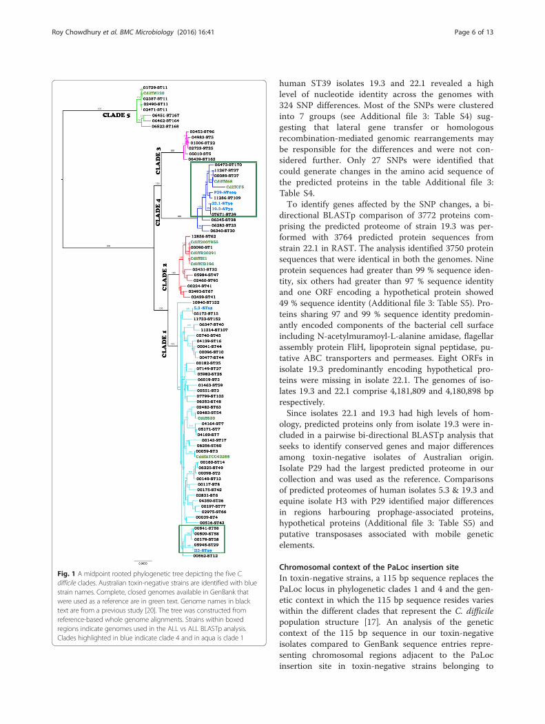
Phylogenetic analysis of toxin-negative isolates of C.difficileA study of the evolution of the C difficile pathogenicitylocus (PaLoc) identified an extremely divergent cladeC-I that exclusively comprised toxin-negative isolatespredominantly of Australian origin [17]. A maximum-likelihood phylogenetic tree using a reference-based,whole genome alignment protocol (see methods sec-tion for protocol details) that incorporates both variantand invariant sites of the C. difficile genome sequenceswas used to verify the ancestry of our toxin-negativeisolates. Our approach uses approximately 72 % of theC. difficile genome for the analysis, considerably morethan what was used in the original study [17]. All 73genomes and the reference genome CD630 used in theprevious study [17] as well as additional closed ge-nomes of C. difficile (strains 2007855, ATCC43255,BI1, CF5, M68, M120 and R20291 from the GenBankdatabase) were used in our initial phylogenetic
analysis. A preliminary phylogenetic tree (Additionalfile 2: Figure S12) revealed that our genome-basedphylogeny was largely congruent with that described inan earlier study [11] and clearly indicated that none ofthe five genomes that were the subject of our studyclustered within the divergent clade C-I.Strains that resided within clade C-I showed greater
than 5 % sequence divergence from the reference gen-ome at homologous sites. Clades that have divergedmore than 5 % from the reference genome can be poorlyresolved in workflows based on Illumina read mappingto the reference [51]. Given this and the uncertain an-cestry of the isolates included in clade C-I in the Dingleet al. [11] study, members of clade C-I were excludedfrom the subsequent analysis based on a cohort of 86strains depicted in Fig. 1. The branching order of the fiveclades in our phylogenetic tree was congruent with thatreported earlier [17] with identical clustering of strainsin the different sub-clades within each of the five clades.The five toxin-negative isolates from our study clusteredwith strains in Clades 1 (C. difficile 5.3, and H3) and 4(C. difficile 19.3, 22.1 and P29) that are known to con-tain toxin-negative strains [17]. Our toxin-negative iso-lates (sourced both from animal and human sources)also clustered with strains of the same sequence typethat included both toxin-positive and toxin-negativestrains isolated from human clinical specimens in anearlier study [17].Our genome sequences were assembled with a de novo
assembler using A5 [48]. Prior to conducting a detailedanalysis of the toxin-negative C. difficile isolates we iden-tified the closest reference genome for tiling genomicscaffolds. A preliminary phylogeny generated usingPhyloSift and FastTree (Additional file 2: Figure S2) indi-cated that C. difficile strain CF5 (toxin-positive ST86)was the most appropriate reference to order genomicscaffolds of isolates 19.3 (ST39), 22.1 (ST39) and P29(ST109) while C. difficile strain 630 (toxin-positive, ST54(PCR ribotype 012) was appropriate to order genomicscaffolds of isolates 5.3 (ST15) and H3 (ST29). StrainCF5 was isolated from a patient in Belgium in 1995while CD630 is a highly virulent, multiple antibiotic re-sistant strain of C. difficile that caused pseudomembran-ous colitis in a human patient and later caused anepidemic of C. difficile infection in a Swiss hospital wardin 1982. All down-stream analyses of the genomespresented in this study were performed on genomic as-semblies with scaffolds ordered to match the referencegenomes.
Homology based functional similarity in the toxin-negative isolatesInitially a Progressive Mauve alignment performed(Additional file 2: Figure S3) on genome sequences of
Roy Chowdhury et al. BMC Microbiology (2016) 16:41 Page 5 of 13
human ST39 isolates 19.3 and 22.1 revealed a highlevel of nucleotide identity across the genomes with324 SNP differences. Most of the SNPs were clusteredinto 7 groups (see Additional file 3: Table S4) sug-gesting that lateral gene transfer or homologousrecombination-mediated genomic rearrangements maybe responsible for the differences and were not con-sidered further. Only 27 SNPs were identified thatcould generate changes in the amino acid sequence ofthe predicted proteins in the table Additional file 3:Table S4.To identify genes affected by the SNP changes, a bi-
directional BLASTp comparison of 3772 proteins com-prising the predicted proteome of strain 19.3 was per-formed with 3764 predicted protein sequences fromstrain 22.1 in RAST. The analysis identified 3750 proteinsequences that were identical in both the genomes. Nineprotein sequences had greater than 99 % sequence iden-tity, six others had greater than 97 % sequence identityand one ORF encoding a hypothetical protein showed49 % sequence identity (Additional file 3: Table S5). Pro-teins sharing 97 and 99 % sequence identity predomin-antly encoded components of the bacterial cell surfaceincluding N-acetylmuramoyl-L-alanine amidase, flagellarassembly protein FliH, lipoprotein signal peptidase, pu-tative ABC transporters and permeases. Eight ORFs inisolate 19.3 predominantly encoding hypothetical pro-teins were missing in isolate 22.1. The genomes of iso-lates 19.3 and 22.1 comprise 4,181,809 and 4,180,898 bprespectively.Since isolates 22.1 and 19.3 had high levels of hom-
ology, predicted proteins only from isolate 19.3 were in-cluded in a pairwise bi-directional BLASTp analysis thatseeks to identify conserved genes and major differencesamong toxin-negative isolates of Australian origin.Isolate P29 had the largest predicted proteome in ourcollection and was used as the reference. Comparisonsof predicted proteomes of human isolates 5.3 & 19.3 andequine isolate H3 with P29 identified major differencesin regions harbouring prophage-associated proteins,hypothetical proteins (Additional file 3: Table S5) andputative transposases associated with mobile geneticelements.
Chromosomal context of the PaLoc insertion siteIn toxin-negative strains, a 115 bp sequence replaces thePaLoc locus in phylogenetic clades 1 and 4 and the gen-etic context in which the 115 bp sequence resides varieswithin the different clades that represent the C. difficilepopulation structure [17]. An analysis of the geneticcontext of the 115 bp sequence in our toxin-negativeisolates compared to GenBank sequence entries repre-senting chromosomal regions adjacent to the PaLocinsertion site in toxin-negative strains belonging to
Fig. 1 A midpoint rooted phylogenetic tree depicting the five C.difficile clades. Australian toxin-negative strains are identified with bluestrain names. Complete, closed genomes available in GenBank thatwere used as a reference are in green text. Genome names in blacktext are from a previous study [20]. The tree was constructed fromreference-based whole genome alignments. Strains within boxedregions indicate genomes used in the ALL vs ALL BLASTp analysis.Clades highlighted in blue indicate clade 4 and in aqua is clade 1
Roy Chowdhury et al. BMC Microbiology (2016) 16:41 Page 6 of 13

Clades 1 (GenBank Accession no HG002393) and 4(GenBank Accession no HG002391) is depicted in Fig. 2.We identified several SNPs within the 115 bp-conservedfragment and an 80 nt long insertion in strains 5.3 andH3 downstream of the gene designated CD06642 in thereference genomes (Fig. 2). In addition, we identified a68 nt tandem repeat of the sequence adjacent to the80 nt insertion site in strain H3 (see Additional file 2:Figure S7). Within members of clade 4, porcine isolateP29 had a deletion of the hypothetical gene seen in thereference region while human isolates 19.3 and 22.1 hada significant decrease in the nucleotide identity in hypo-thetical genes in the reference genome. Consistent withobservations reported earlier, these differences indicateongoing micro-evolutionary events within the locus thatflanks the PaLoc insertion site [17].
Comparative BLASTp analyses of toxin-negative strains toidentify functional similarities between groups of isolatesAn all versus all BLASTp based protein-clustering ana-lysis was used to identify the putative core proteome ofa subset of Clade 1 strains comprising five genomes andClade 4 strains comprising 10 genomes (see boxed re-gion in Fig. 1). To maintain uniformity in the input dataused in the analysis, raw reads representing each of thegenomes from an earlier study [17] were reassembledand annotated using the protocols that were used to as-semble and annotate the five toxin-negative Australianisolates of C. difficile reported in this study, as describedabove. The assembly statistics and a comparison of theassembly outputs are presented in Additional file 3:Table S8. On an average, RAST predicted 3700 proteinsper genome and these were included in the protein
Fig. 2 Genetic context of the PaLoc in Australian toxin-negative strains of C. difficile. a Clade 1 prototype OxI_WB2011 (HG002393). b Clade 4prototype Oxa464a (HG002391)
Roy Chowdhury et al. BMC Microbiology (2016) 16:41 Page 7 of 13

clustering analysis. A bit score cut off of 50 was used tocluster homologous protein sequences. An overview ofthe data is presented in Table 1.Ten clade 4 genomes boxed in Fig. 1 shared 3357 pro-
teins (Table 1). Isolate P29 had the highest number (299)of unshared/unique proteins within the clade 4 cohortand most of the 299 proteins were phage-related(Additional file 3: Table S9). Some of the unique pro-teins clustered together in the same scaffold indicat-ing lateral movement of phage-associated genomicDNA. C. difficile isolates 19.3 and 22.1 carried eightand seven unique proteins respectively. The handfulof unshared proteins in 19.3 and 22.1 were attributedto mobile genetic elements or were designated to en-code proteins of unknown function. The five C. difficilestrains within clade 1 shared 3323 proteins. Equine isolateH3 had 111 unique proteins, most of which were phagerelated or hypothetical with some clustered in single scaf-folds (Additional file 3: Table S9).We queried the viral and prophage database in Gen-
Bank with the genome sequences of all five toxin-negative isolates using PHAST [66]. The database con-firmed the presence of several regions contain phage
DNA in each of the five genomes in Clade 1 (see Add-itional file 2: Figure S10 and Additional file 4: TableS11). Table 2 lists a summary of the complete prophagesequences (PHAST scores > 110 and over) identified inthe five Australian toxin-negative isolates. Notably,isolates 19.3 and 22.1 returned identical phage profiles(Additional file 2: Figure S10). Both genomes carried anidentical and complete 56.8 kb phage that is a close rela-tive of phiC2, a 56.5 kb phage first identified in C. diffi-cile strain CD242 [67]. phiC2 is one of the firstcompletely sequenced temperate phages in C. difficileand regions of phiC2 are present in the majority of C.difficile genomes of clinical origin [68]. We also identifieda phage in isolates P29 and H3 that carries sequenceidentity with phiC2. The prophage in P29 was larger(97.4 kb) than the original phiC2 sequence (Additionalfile 2: Figure S10). Isolate P29 also carries two othercomplete prophage sequences. One of these, prophageregion 9 has significant sequence identity to the C. diffi-cile temperate bacteriophage phiCD6356 that belongs tothe Siphoviridae family [69]. The Siphoviridae family pro-phage identified in P29 is 52.8 kb (Additional file 4:Table S11) and is considerably larger than the first reportof this phage sequence at 37.6 kb [69]. Isolate H3 alsocarries an identical variant of phage phiCD6356 (Add-itional file 2: Figure S12) comprising 41.1 kb. Evidence ofother complete prophage genomes is listed in Table 2.Comparative BLASTp analysis of the four genomes (19.3,5.3, H3 and P29) also aligned to the phage search proto-col and confirmed the data generated by PHAST ana-lyses, reinforcing the observation that the majordifferences in our Australian toxin-negative C. difficileisolates have a prophage origin.Comparative BLASTp analysis of isolates 19.3, 5.3, H3
and P29 identified a 119.3 kb region on contig 11 in P29(Additional file 5: Table S6). An all versus all proteinclustering analysis also identified a subset of unique pro-teins on contig 11 of the P29 genome but not in the 10human C. difficile genomes within clade 4 (Fig. 1).Equine isolate H3 was not included in this analysis as itwas on a different clade. BLASTn analysis of the119.3 kb region against the C. difficile genome databasein GenBank identified similarity at the DNA level toparts of C. difficile strain CD630 indicating a phage-mediated lateral movement of parts of the genome ofCD630 into the genomes of isolates H3 and P29.
Homology based functional prediction of Putative C.difficile virulence factors implicated in host colonizationA homology-based functional prediction analysis of pro-teins that have been experimentally verified, or predictedto play a role in the colonization of C. difficile strainCD630 with homologous proteins in the genomes of thetoxin-negative isolates in our study is shown in Table 3
Table 1 Summary of protein clustering results within thedifferent sub-clades containing the five toxin negative isolatesincluded in this study. Summary of protein clusters within thedifferent sub-clades of C. difficile
Clade 1
Genome names No of predicted proteins
Core genome 3323
Total number of unique peptides in:
C. difficile C0000509 45
C. difficile C0000541 38
C. difficile C0000562 176
C. difficile C00005945 212
C. difficile H3 111
Clade 4:
Core genome 3357
Total number of unique peptides in:
C. difficile 19.3 8
C. difficile 22.1 7
C. difficile C00000089 33
C. difficile C000011286 86
C. difficile C00006473 84
C. difficile C00007671 35
C. difficile C00011267 60
C. difficile CF5 30
C. difficile M68 19
C. difficile P29 299
Roy Chowdhury et al. BMC Microbiology (2016) 16:41 Page 8 of 13

[70, 71]. Most of the proteins i had near perfect (98–100 %) protein sequence identity including proteinsencoded by spo0A, which serves as a positive regula-tor for genes required in spore germination andgroEL, a chaperone that also functions as an adhesin[72]. Several serine-proteases and other metallopro-teases which may contribute to the disease develop-ment process [70] were also highly conserved.However, some membrane associated proteins thathave been shown to play significant roles in the dis-ease development process had lower alignmentscores including SlpA, a surface layer protein that isproposed to facilitate host cell adherence [73] andFliC, an adhesin necessary for the colonization ofgut epithelium [74].
DiscussionC. difficile colonisation in humans is age dependent.While asymptomatic carriage is common in infants lessthan three years of age it is rare in adults [75]. As such,infants can be a major reservoir of both pathogenic andtoxin-negative strains in a community setting [75]. Weisolated five toxin-negative isolates of C. difficile includ-ing three from humans (22.1, 19.3, 5.3), one from a pig(P29) and one (H3) from a horse all showing symptomsof gastrointestinal disease. Despite efforts to identify C.difficile toxin genes or toxin gene products in the stoolsamples during the course of the isolation of thesestrains, none were detected. Phylogenetic studies showedthat human ST39 isolates 19.3 and 22.1 and porcine
ST109 isolate P29 grouped with clinical human toxigenicstrains of ST39 and ST109 respectively in Clade 4.Furthermore, human ST15 isolate 5.3 and equine ST29isolate H3 grouped with human clinical toxigenic strainswith ST15 and ST29 respectively in Clade 1. Compara-tive genome analyses showed that our toxin-negativeisolates displayed virulence gene profiles akin to thoseidentified in toxigenic strains. The animals from whichsamples were collected in this study exhibited gastro-intestinal disease and we were unable to attribute thesesymptoms to the presence of toxin-positive strains of C.difficile. Given the mobility of the PaLoc [44] and evi-dence that the acquisition or loss of the PaLoc viarecombination [17] has occurred multiple times dur-ing the evolution of the five major clades of C. diffi-cile [11, 17], our data reinforces calls to includetoxin-negative strains in genomic epidemiologicalstudies of C. difficile [17, 36] and to better character-ise asymptomatic carriage of closely related Clostridiain gut microbiome surveys such as the HumanMicrobiome Project and MetaHIT, both in humansand close animal contacts.Our reference-based whole genome alignment and
phylogeny analyses support the global population struc-ture of C. difficile as described by Dingle et al. in 2014[14, 17, 19]. Each clade has been shown previously tohave representatives of both toxin-positive and toxin-negative strains [11]. Our toxin-negative isolates (19.3,22.1, 5.3, P29, H3) belonged to STs that are distinct fromthose reported in an earlier study [17]. The role of toxins
Table 2 Summary of Phage related regions identified by PHAST in the 5 genomes. Phage sequences identified in this study
PHAST regionidentifier
Length ofprophage
PHASTscore
No ofpredicted CDS
Relative positionon genome
Predicted Phage GCcontent
Location on Genomicscaffolds
C difficile P29 genome
Region_4 97.4Kb 150 126 1732045-1829500 PHAGE_Clostr_CDMH1_NC_024144 28.7 27.1, 30.1, 40.1, 34.1, 36,1,5.1, 47.1 and 16.1
Region_8 113.6Kb 150 101 3773336-3886941 PHAGE_Geobac_virus_E2_NC_009552 40.8 5.1, 19.1 and 22.1
Region_9 52.8Kb 140 65 4233532-4286418 PHAGE_Clostr_phiCD6356_NC_015262 29.9 8.1, 26.1, 41.1 and 42.1
Region_10 21.3Kb 100 22 4290994-4312349 PHAGE_Clostr_phiC2_NC_009231 36.4 In over 20 very smallscaffolds
C. difficile H3 genome
Region_2 41.1Kb 150 63 925289-966449 PHAGE_Clostr_phiCD6356_NC_015262 28.5 22.1 and 31.1
Region_3 50.1Kb 110 66 1114600-1164714 PHAGE_Clostr_phiC2_NC_009231 28.1 18.1 and 5.1
Region_6 31.5Kb 110 47 4070500-4102080 PHAGE_Clostr_phiC2_NC_009231 29.4 26.1, 27.1, 28.1, 32.1, 33.1
C. difficile 5.3 genome
Region_3 57.9Kb 140 87 1555612-1613526 PHAGE_Clostr_phiC2_NC_009231 28.6 1.1
Region_4 45.1Kb 150 49 1741883-1787019 PHAGE_Clostr_phiSM101_NC_008265 27.2 1.1
C.difficle 19.3 genome
Region_3 56.8Kb 140 74 1700180-1757059 PHAGE_Clostr_phiC2_NC_009231 28.8 1.1
C. difficile 22.1 genome:
Region_3 56.8Kb 140 74 1703858-1760737 PHAGE_Clostr_phiC2_NC_009231 28.8 3.1
Roy Chowdhury et al. BMC Microbiology (2016) 16:41 Page 9 of 13

Table 3 Proteins derived from C. difficile CD630 that are predicted to play a role in pathogenesisSelected gene and product C. difficile 630
locus tagExperimentalVerification
RAST Annotation identifiers
In C. difficile 5.3 (% identity) In C. difficile 19.3 (% identity) In C. difficile H3 (% identity) In Cc difficile P29 (% identity)
Flagellin C gene fliC CD630_02390 yes, RNAseq fig|6666666.71923.peg.3142 (86) fig|6666666.71924.peg.3067 (71)* fig|6666666.72094.peg.3176 (87) fig|1440056.4.peg.3191 (97)
Flagellin D gene fliD CD630_02370 no fig|6666666.71923.peg.3140 (88) fig|6666666.71924.peg.3065 (61) fig|6666666.72094.peg.3174 (88) fig|1440056.4.peg.3193 (98)
Precursor S-layer protein gene slpA CD630_27930 yes, proteome fig|6666666.71923.peg.2081 (43)* fig|6666666.71924.peg.3725 (59) * fig|6666666.72094.peg.2466 (58)* fig|1440056.4.peg.2665 (54)*
Stage 0 Sporulation gene spoA CD630_12140 yes, proteome fig|6666666.71923.peg.158 (100) fig|6666666.71924.peg.1561 (99) fig|6666666.72094.peg.2932 (99) fig|1440056.4.peg.240 (99)
Fibrinectin binding proten encodingfbpA gene
CD630_25920 no fig|6666666.71923.peg.2930 (99) fig|6666666.71924.peg.2028 (98) fig|6666666.72094.peg.3740 (99) fig|1440056.4.peg.41 (98)
GroEL encoding gene groL CD630_01940 yes, proteome fig|6666666.71923.peg.3095 (100) fig|6666666.71924.peg.2995 (99) fig|6666666.72094.peg.3129 (100) fig|1440056.4.peg.3232 (99)
Cell surface protein cwp66 CD630_27890 no fig|6666666.71923.peg.2085 (60) fig|6666666.71924.peg.3721 (78) fig|6666666.72094.peg.2462 (77) fig|1440056.4.peg.2669 (79)
Protease cwp84 CD630_27870 no fig|6666666.71923.peg.2087 (98) fig|6666666.71924.peg.3719 (99) fig|6666666.72094.peg.2460 (99) fig|1440056.4.peg.2671 (99)
Adhesin (LPXTG) CD630_28310 no fig|6666666.71923.peg.2041 (99) fig|6666666.71924.peg.3767 (94) fig|6666666.72094.peg.2504 (98) fig|1440056.4.peg.2625 (94)
Cell wall binding protein encoding cwp2 CD630_27910 yes, proteome fig|6666666.71923.peg.2083 (98) fig|6666666.71924.peg.3723 (99) fig|6666666.72094.peg.2464 (99) fig|1440056.4.peg.2667 (99)
Cell wall binding protein encoding cwp12 CD630_27940 no fig|6666666.71923.peg.2080 (65)* fig|6666666.71924.peg.3726 (98) fig|6666666.72094.peg.2467 (95) fig|1440056.4.peg.2664 (94)
Cell wall binding protein encoding cwp11 CD630_27950 yes, proteome fig|6666666.71923.peg.2079 (98) fig|6666666.71924.peg.3727 (99) fig|6666666.72094.peg.2468 (99) fig|1440056.4.peg.2663 (99)
Cell wall binding protein encoding cwp9 CD630_27980 no fig|6666666.71923.peg.2076 (99) fig|6666666.71924.peg.3730 (99) fig|6666666.72094.peg.2471 (99) fig|1440056.4.peg.2660 (99)
Cell wall hydrolase (LPXTG) CD630_01830 no fig|6666666.71923.peg.3084 (97)* fig|6666666.71924.peg.2984 (99) fig|6666666.72094.peg.3118 (100) fig|1440056.4.peg.3243 (99)
Cell wall binding protein encoding cwp25gene
CD630_08440 no fig|6666666.71923.peg.2189 (100) fig|6666666.71924.peg.3292 (97) fig|6666666.72094.peg.2128 (99) fig|1440056.4.peg.522 (97)
N-acetylmuramoyl-L-analini amidaseencoding cwp16
CD630_10350 no fig|6666666.71923.peg.1 (99) fig|6666666.71924.peg.3716 (65)* fig|6666666.72094.peg.1495 (99) fig|1440056.4.peg.1845 (98)
Cell wall hydrolase encoding gene(invasin)
CD630_27680 no fig|6666666.71923.peg.2107 (99) fig|6666666.71924.peg.3700 (99) fig|6666666.72094.peg.2441 (99) fig|1440056.4.peg.2690 (99)
Polysaccharide de-acetylase CD630_15220 yes, RNAseqand proteome
fig|6666666.71923.peg.489 (100) fig|6666666.71924.peg.291 (99) fig|6666666.72094.peg.3592 (100) fig|1440056.4.peg.1197 (99)
LmbE-like deacetylase encoding gene CD630_27900 no fig|6666666.71923.peg.2084 (93) fig|6666666.71924.peg.3722 (100) fig|6666666.72094.peg.2463 (100) fig|1440056.4.peg.2668 (97)
Invasin/Sh3 domain containing surfaceprotein
CD630_11350 no fig|6666666.71923.peg.77 (100) fig|6666666.71924.peg.1641 (98) fig|6666666.72094.peg.2849 (99) fig|1440056.4.peg.320 (98)
Cell wall hydrolase/Invasin associatedprotein
CD630_24020 fig|6666666.71923.peg.2730 (100) fig|6666666.71924.peg.2779 (98) fig|6666666.72094.peg.2094 (99) fig|1440056.4.peg.3691 (99)
Autolysin acd gene homolog/mannosyl-glycoprotein endo neta N acetylglucosamine
CD630_13040 no fig|6666666.71923.peg.256 (100) fig|6666666.71924.peg.1460 (98) fig|6666666.72094.peg.3031 (99) fig|1440056.4.peg.158 (98)
Protease/Serine protease, HrtA family CD630_32840 no fig|6666666.71923.peg.1608 (100) fig|6666666.71924.peg.3459 (99) fig|6666666.72094.peg.64 (100) fig|1440056.4.peg.2801 (99)
Intracellular serine protease CD630_32540 no fig|6666666.71923.peg.1638 (100) fig|6666666.71924.peg.3426 (99) fig|6666666.72094.peg.94 (99) fig|1440056.4.peg.2834 (99)
Protease/Subtilase family CD630_07030 no fig|6666666.71923.peg.2324 (100) fig|6666666.71924.peg.3155 (97) fig|6666666.72094.peg.2691 (100) fig|1440056.4.peg.1169 (97)
Ser-type protease/subtilisin-like serinegermination related protease
CD630_22470 yes, Massspectrometry
fig|6666666.71923.peg.1327 (99) fig|6666666.71924.peg.2513 (99) fig|6666666.72094.peg.1937 (99) fig|1440056.4.peg.1500 (98)
Serine protease precursor/Subtilinasesubfamily
CD630_20000 no fig|6666666.71923.peg.1085 (100) fig|6666666.71924.peg.2267 fig|6666666.72094.peg.1687 (99) fig|1440056.4.peg.2329 (99)
Membrane-associated zinc metalloprotease/M50 family peptidase
CD630_21290 no fig|6666666.71923.peg.1209 (100) fig|6666666.71924.peg.2404 (99) fig|6666666.72094.peg.1813 (100) fig|1440056.4.peg.1610 (100)
Zinc Protease/M16 family peptidase CD630_26610 yes, proteome fig|6666666.71923.peg.2996 (100) fig|6666666.71924.peg.626 (99) fig|6666666.72094.peg.3677 (100) fig|1440056.4.peg.438 (99)
* indicates gaps in alignment of amino acid sequences with reference, likely suggesting presence of inactive proteins or variants in the test genomes
RoyChow
dhuryet
al.BMCMicrobiology
(2016) 16:41 Page
10of
13

in C. difficile infection has been extensively studied butfactors that enable C. difficile to efficiently colonise thehuman gastrointestinal tract are relatively poorly under-stood and are not associated with genes encoded on thePaLoc. It is not known why some toxigenic strainsevolve into dominant hypervirulent clones. Thus, con-sidering the genetic diversity inherent within the phylo-genetic structure of C. difficile [14] a sub-population oftoxin-negative strains of C. difficile that are efficientcolonisers of the host gastrointestinal tract may read-ily acquire the PaLoc and evolve to become futurehypervirulent strains. Several proteins have been sug-gested to play crucial roles in the colonization ofgastrointestinal epithelium and disease progression[43, 71, 73, 74, 76–78]. A recent global proteomestudy of C. difficile strains CD630 and R20291 hasidentified numerous extracellular proteins from cul-ture supernatants that may contribute to the virulenceattributes of these strains [70].Our study reinforced the important role played by
phage in the evolution of C. difficile. While PHAST ana-lysis was useful for identifying phage sequences, the ana-lysis may not have identified the full extent of lysogenicphage because our draft genomes remain in multiplescaffolds. Although the complete sequence of phagephiC2 was identified in isolates 19.3 and 22.1 the regionsthat had significant homology with phiC2 in isolates P29and H3 were located on different scaffolds. We used ascaffold tiling approach against the closed genome of areference strain to create the input file for PHAST ana-lysis (PHAST converts the scaffolded genomes into aconcatenated artificial chromosome prior to predictingthe phage content) and as such it remains a possibilitythat the partial matches are a consequence of the datahandling process. Phage phiC2 is present in the majorityof human isolates of C. difficile [68]. However, we de-tected regions of phiC2 in strains P29 and H3 suggestingthat further studies are needed to address issues sur-rounding the association of phiC2 in C. difficile ofanimal origin. We also identified the C. difficile temper-ate bacteriophage phiCD6356 from the Siphoviridaefamily in isolates P29 and H3 but not in our human iso-lates of C. difficile. Genomes of bacteriophages belong-ing to the Siphoviridae family range in size from 14 to50 kb [79, 80] and this broad range may be a reflectionof the stringency governing the amount of DNA thatcan be packaged by phiCD6356. In addition to the ac-quisition of phage-associated genes, a 119.3-kb regionon contig 11 in isolate P29 was also identified in thecourse of this analysis. This region is unique to the P29genome and displayed significant DNA sequence identityto portions of the CD630 genome. It remains unknownif the 119.3-kb region exists in C. difficile strains of por-cine origin. Further analyses with greater numbers of
genomes from both human and animal sources are re-quired to conclusively address these questions.
ConclusionsOur studies reinforce calls to improve our understandingof the physiological conditions that promote lateraltransfer of the PaLoc in the gastrointestinal tract [44].This is important because the conditions that facilitatemovement of fragments of DNA carrying the PaLoc andtheir recombination into the chromosome are also con-ducive to the movement of conjugative transposons thatcarry antibiotic resistance genes and putative virulencefactors as independent genetic events [44].
Data accessibilityGenome sequences reported in this analysis were sub-mitted to GenBank and are available via the accessionnumbers provided. The bioinformatics softwares aremade available through the GitHub repository links.
Additional files
Additional file 1: Table S1. Isolation history, genomic assembly andinitial PCR screening results of the five isolates analysed in thismanuscript. (DOCX 36 kb)
Additional file 2: Figure S2. Preliminary phylogeny with referencegenomes to identify the most closely related reference genome for tilingof genomic scaffolds. Figure S3. Mauve alignments of genomes 19.3 and22.1. Figure S7. DNA sequence alignment of repetitive regions in PaLocof isolates in clade 1. Figure S10. Phage profiles in the genomes of thefive Australian toxin-negative C. difficile isolates included in this study.Figure S12. Preliminary Phylogenetic tree with all isolates included inDingle et al’s publication in 2011 [11]. Isolates that form clade C-I ishighlighted in blue. (DOCX 2963 kb)
Additional file 3: Table S4. SNPs identified by Mauve alignments ofgenomes of C. difficile strains 19.3 and 22.1. Table S5. BidirectionalBLASTp analysis of peptide sequences predicted from genomes of C.difficile strains 19.3 and 22.1 to identify amino acid changes caused bySNPs in the genomes. Table S8. Assembly Statistics downloaded fromshort read archives. Table S9. Unique proteins identified from the All VsAll BLASTp analysis of C. difficile genomes. (XLSX 632 kb)
Additional file 4: Table S11. Regions identified in each C. difficilegenome by PHAST. Sheet 1. C. difficile strain P29 genome, Sheet 2. C.difficile strain H3 genome, Sheet 3. C. difficile strain 5.3 genome, Sheet 4.C. difficile strain 19.3 genome. Sheet 5. C. difficile strain 22.1 genome.(XLSX 134 kb)
Additional file 5: Table S6. Sheet 1 = Comparative BLASTp analysis of 4genomes, Sheet 2 = Alignment of scaffold 11.1 of genome P29 against C.difficile CD630 genome. (XLSX 682 kb)
Competing interestsThe authors declare that they have no competing interests, includingfinancial competing interests.
Authors’ contributionPRC assembled the genome sequences, conceived, analysed and designedthe presentation of data in the manuscript, performed all comparativegenomic analysis and benchwork to confirm features identified from thecomparative genomic analysis and drafted the manuscript. AED and MDwrote data analysis software packages and scripts and assisted with datainterpretation. TC cultured the isolates, performed PCR analyses for toxingenes strains, ribotyping and fingerprinting assays and designed aspects the
Roy Chowdhury et al. BMC Microbiology (2016) 16:41 Page 11 of 13

project. PW constructed the libraries for genome sequencing. IGC assistedwith experimental design, data analysis and writing the manuscript. SPDconceived and designed aspects of the project, managed the project anddrafted the final manuscript. All authors read and approved the finalmanuscript.
AcknowledgementsThis work is a product of the ausgem partnership. The authors wish toacknowledge Prof Thomas Borody for kindly providing the human isolateincluded in this analysis.
Author details1The ithree institute, University of Technology Sydney, Sydney 2007, Australia.2NSW Department of Primary Industries, Elizabeth Macarthur AgriculturalInstitute, PMB 8, Camden, NSW 2570, Australia. 3Institute of Food Research,Norwich Research Park, Colney, Norwich NR4 7UA, UK.
Received: 29 October 2015 Accepted: 2 March 2016
References1. Karadsheh Z, Sule S. Fecal transplantation for the treatment of recurrent
clostridium difficile infection. N Am J Med Sci. 2013;5(6):339–43. PubmedCentral PMCID: 3731863.
2. Lessa FC, Mu Y, Bamberg WM, Beldavs ZG, Dumyati GK, Dunn JR, et al.Burden of Clostridium difficile infection in the United States. N Engl J Med.2015;372(9):825–34.
3. Peterson LR. Bad bugs, no drugs: no ESCAPE revisited. Clin Infect Dis. 2009;49(6):992–3.
4. Redelings MD, Sorvillo F, Mascola L. Increase in Clostridium difficile-relatedmortality rates, United States, 1999–2004. Emerg Infect Dis. 2007;13(9):1417–9. Pubmed Central PMCID: 2857309.
5. Rupnik M, Wilcox MH, Gerding DN. Clostridium difficile infection: newdevelopments in epidemiology and pathogenesis. Nat Rev Microbiol. 2009;7(7):526–36.
6. Khanna S, Pardi DS, Aronson SL, Kammer PP, Baddour LM. Outcomes incommunity-acquired Clostridium difficile infection. Aliment Pharmacol Ther.2012;35(5):613–8. Pubmed Central PMCID: 3293482.
7. Bauer MP, Kuijper EJ. Potential sources of Clostridium difficile in humaninfection. Infect Dis Clin North Am. 2015;29(1):29–35.
8. Songer JG, Anderson MA. Clostridium difficile: an important pathogen offood animals. Anaerobe. 2006;12(1):1–4.
9. Hensgens MP, Keessen EC, Squire MM, Riley TV, Koene MG, de Boer E, et al.Clostridium difficile infection in the community: a zoonotic disease? ClinMicrobiol Infect. 2012;18(7):635–45.
10. Goorhuis A, Debast SB, van Leengoed LA, Harmanus C, Notermans DW,Bergwerff AA, et al. Clostridium difficile PCR ribotype 078: an emergingstrain in humans and in pigs? J Clin Microbiol. 2008;46(3):1157. PubmedCentral PMCID: 2268365, author reply 8.
11. Dingle KE, Griffiths D, Didelot X, Evans J, Vaughan A, Kachrimanidou M, et al.Clinical Clostridium difficile: clonality and pathogenicity locus diversity. PLoSOne. 2011;6(5):e19993. Pubmed Central PMCID: 3098275.
12. Walk ST, Micic D, Jain R, Lo ES, Trivedi I, Liu EW, et al. Clostridium difficileribotype does not predict severe infection. Clin Infect Dis. 2012;55(12):1661–8. Pubmed Central PMCID: 3501335.
13. Cairns MD, Stabler RA, Shetty N, Wren BW. The continually evolvingClostridium difficile species. Future Microbiol. 2012;7(8):945–57.
14. Stabler RA, Dawson LF, Valiente E, Cairns MD, Martin MJ, Donahue EH, et al.Macro and micro diversity of Clostridium difficile isolates from diversesources and geographical locations. PLoS One. 2012;7(3):e31559. PubmedCentral PMCID: 3292544.
15. Behroozian AA, Chludzinski JP, Lo ES, Ewing SA, Waslawski S, Newton DW,et al. Detection of mixed populations of Clostridium difficile fromsymptomatic patients using capillary-based polymerase chain reactionribotyping. Infect Control Hosp Epidemiol. 2013;34(9):961–6. PubmedCentral PMCID: 4016961.
16. Waslawski S, Lo ES, Ewing SA, Young VB, Aronoff DM, Sharp SE, et al.Clostridium difficile ribotype diversity at six health care institutions in the UnitedStates. J Clin Microbiol. 2013;51(6):1938–41. Pubmed Central PMCID: 3716112.
17. Dingle KE, Elliott B, Robinson E, Griffiths D, Eyre DW, Stoesser N, et al.Evolutionary history of the Clostridium difficile pathogenicity locus. GenomeBiol Evol. 2014;6(1):36–52. Pubmed Central PMCID: 3914685.
18. Monot M, Eckert C, Lemire A, Hamiot A, Dubois T, Tessier C, et al.Clostridium difficile: New insights into the evolution of the pathogenicitylocus. Sci Rep. 2015;5:15023. Pubmed Central PMCID: 4597214.
19. Griffiths D, Fawley W, Kachrimanidou M, Bowden R, Crook DW, Fung R, et al.Multilocus sequence typing of Clostridium difficile. J Clin Microbiol. 2010;48(3):770–8. Pubmed Central PMCID: 2832416.
20. Lemee L, Bourgeois I, Ruffin E, Collignon A, Lemeland JF, Pons JL. Multilocussequence analysis and comparative evolution of virulence-associatedgenes and housekeeping genes of Clostridium difficile. Microbiology.2005;151(Pt 10):3171–80.
21. Lemee L, Dhalluin A, Pestel-Caron M, Lemeland JF, Pons JL. Multilocussequence typing analysis of human and animal Clostridium difficile isolatesof various toxigenic types. J Clin Microbiol. 2004;42(6):2609–17. PubmedCentral PMCID: 427854.
22. Lemee L, Dhalluin A, Testelin S, Mattrat MA, Maillard K, Lemeland JF, et al.Multiplex PCR targeting tpi (triose phosphate isomerase), tcdA (Toxin A),and tcdB (Toxin B) genes for toxigenic culture of Clostridium difficile. J ClinMicrobiol. 2004;42(12):5710–4. Pubmed Central PMCID: 535266.
23. Rupnik M. How to detect Clostridium difficile variant strains in a routinelaboratory. Clin Microbiol Infect. 2001;7(8):417–20.
24. Voth DE, Ballard JD. Clostridium difficile toxins: mechanism of action androle in disease. Clin Microbiol Rev. 2005;18(2):247–63. Pubmed CentralPMCID: 1082799.
25. Hundsberger T, Braun V, Weidmann M, Leukel P, Sauerborn M, vonEichel-Streiber C. Transcription analysis of the genes tcdA-E of thepathogenicity locus of Clostridium difficile. Eur J Biochem/FEBS. 1997;244(3):735–42.
26. Matamouros S, England P, Dupuy B. Clostridium difficile toxin expression isinhibited by the novel regulator TcdC. Mol Microbiol. 2007;64(5):1274–88.
27. Braun V, Hundsberger T, Leukel P, Sauerborn M, von Eichel-Streiber C.Definition of the single integration site of the pathogenicity locus inClostridium difficile. Gene. 1996;181(1–2):29–38.
28. Eckert C, Emirian A, Le Monnier A, Cathala L, De Montclos H, Goret J, et al.Prevalence and pathogenicity of binary toxin-positive Clostridium difficilestrains that do not produce toxins A and B. New Microbes New Infect. 2015;3:12–7. Pubmed Central PMCID: 4337936.
29. Collins DA, Elliott B, Riley TV. Molecular methods for detecting and typing ofClostridium difficile. Pathology. 2015;47(3):211–8.
30. Rupnik M, Brazier JS, Duerden BI, Grabnar M, Stubbs SL. Comparison oftoxinotyping and PCR ribotyping of Clostridium difficile strains anddescription of novel toxinotypes. Microbiology. 2001;147(Pt 2):439–47.
31. Villano SA, Seiberling M, Tatarowicz W, Monnot-Chase E, Gerding DN.Evaluation of an oral suspension of VP20621, spores of nontoxigenicClostridium difficile strain M3, in healthy subjects. Antimicrob AgentsChemother. 2012;56(10):5224–9. Pubmed Central PMCID: 3457387.
32. Nagaro KJ, Phillips ST, Cheknis AK, Sambol SP, Zukowski WE, Johnson S,et al. Nontoxigenic Clostridium difficile protects hamsters against challengewith historic and epidemic strains of toxigenic BI/NAP1/027 C. difficile.Antimicrob Agents Chemother. 2013;57(11):5266–70. Pubmed CentralPMCID: 3811292.
33. Natarajan M, Walk ST, Young VB, Aronoff DM. A clinical and epidemiologicalreview of non-toxigenic Clostridium difficile. Anaerobe. 2013;22:1–5.Pubmed Central PMCID: 3729612.
34. Seal D, Borriello SP, Barclay F, Welch A, Piper M, Bonnycastle M. Treatmentof relapsing Clostridium difficile diarrhoea by administration of a non-toxigenic strain. Eur J Clin Microbiol. 1987;6(1):51–3.
35. Wilson KH, Sheagren JN. Antagonism of toxigenic Clostridium difficile bynontoxigenic C. difficile. J Infect Dis. 1983;147(4):733–6.
36. Buckley AM, Spencer J, Maclellan LM, Candlish D, Irvine JJ, Douce GR.Susceptibility of hamsters to Clostridium difficile isolates of differingtoxinotype. PLoS One. 2013;8(5):e64121. Pubmed Central PMCID: 3660315.
37. Sambol SP, Merrigan MM, Tang JK, Johnson S, Gerding DN. Colonization forthe prevention of Clostridium difficile disease in hamsters. J Infect Dis. 2002;186(12):1781–9.
38. Hung YP, Lin HJ, Wu TC, Liu HC, Lee JC, Lee CI, et al. Risk factors of fecaltoxigenic or non-toxigenic Clostridium difficile colonization: impact of Toll-like receptor polymorphisms and prior antibiotic exposure. PLoS One. 2013;8(7):e69577. Pubmed Central PMCID: 3723847.
Roy Chowdhury et al. BMC Microbiology (2016) 16:41 Page 12 of 13
39. Gerding DN, Johnson S, Rupnik M, Aktories K. Clostridium difficile binarytoxin CDT: mechanism, epidemiology, and potential clinical importance.Gut Microbes. 2014;5(1):15–27. Pubmed Central PMCID: 4049931.
40. Geric B, Carman RJ, Rupnik M, Genheimer CW, Sambol SP, Lyerly DM, et al.Binary toxin-producing, large clostridial toxin-negative Clostridium difficilestrains are enterotoxic but do not cause disease in hamsters. J Infect Dis.2006;193(8):1143–50.
41. Bacci S, Molbak K, Kjeldsen MK, Olsen KE. Binary toxin and death afterClostridium difficile infection. Emerg Infect Dis. 2011;17(6):976–82. PubmedCentral PMCID: 3358205.
42. Barbut F, Decre D, Lalande V, Burghoffer B, Noussair L, Gigandon A, et al.Clinical features of Clostridium difficile-associated diarrhoea due to binarytoxin (actin-specific ADP-ribosyltransferase)-producing strains. J MedMicrobiol. 2005;54(Pt 2):181–5.
43. Barketi-Klai A, Monot M, Hoys S, Lambert-Bordes S, Kuehne SA, Minton N,et al. The flagellin FliC of Clostridium difficile is responsible for pleiotropicgene regulation during in vivo infection. PLoS One. 2014;9(5):e96876.Pubmed Central PMCID: 4026244.
44. Brouwer MS, Roberts AP, Hussain H, Williams RJ, Allan E, Mullany P. Horizontalgene transfer converts non-toxigenic Clostridium difficile strains into toxinproducers. Nat Commun. 2013;4:2601. Pubmed Central PMCID: 3826655.
45. Casey TA, Bosworth BT. Design and evaluation of a multiplex polymerasechain reaction assay for the simultaneous identification of genes for ninedifferent virulence factors associated with Escherichia coli that causediarrhea and edema disease in swine. J Vet Diagn Investig. 2009;21(1):25–30.
46. Rinttila T, Kassinen A, Malinen E, Krogius L, Palva A. Development of anextensive set of 16S rDNA-targeted primers for quantification of pathogenicand indigenous bacteria in faecal samples by real-time PCR. J ApplMicrobiol. 2004;97(6):1166–77.
47. Darling AE, Worden P, Chapman TA, Roy Chowdhury P, Charles IG,Djordjevic SP. The genome of Clostridium difficile 5.3. Gut pathogens. 2014;6(1):4. Pubmed Central PMCID: 4234979.
48. Darling AE, Jospin G, Lowe E, Matsen FA, Bik HM, Eisen JA. PhyloSift:phylogenetic analysis of genomes and metagenomes. PeerJ. 2014;2:e243.Pubmed Central PMCID: 3897386.
49. Li H, Handsaker B, Wysoker A, Fennell T, Ruan J, Homer N, et al. TheSequence Alignment/Map format and SAMtools. Bioinformatics. 2009;25(16):2078–9. Pubmed Central PMCID: 2723002.
50. Schloss PD, Westcott SL, Ryabin T, Hall JR, Hartmann M, Hollister EB, et al.Introducing mothur: open-source, platform-independent, community-supported software for describing and comparing microbial communities.Appl Environ Microbiol. 2009;75(23):7537–41. Pubmed Central PMCID:2786419.
51. Bertels F, Silander OK, Pachkov M, Rainey PB, van Nimwegen E. Automatedreconstruction of whole-genome phylogenies from short-sequence reads.Mol Biol Evol. 2014;31(5):1077–88. Pubmed Central PMCID: 3995342.
52. Stamatakis A. RAxML-VI-HPC: maximum likelihood-based phylogeneticanalyses with thousands of taxa and mixed models. Bioinformatics. 2006;22(21):2688–90.
53. Lartillot N, Philippe H. A Bayesian mixture model for across-siteheterogeneities in the amino-acid replacement process. Mol Biol Evol. 2004;21(6):1095–109.
54. Stamatakis A, editor. Phylogenetic models of rate heteroginity: a highperformance computing perspective. Parallel and Distributed ProcessingSymposium, 2006. Rhodes Island: IEEE; 2006.
55. Stamatakis A, Hoover P, Rougemont J. A rapid bootstrap algorithm for theRAxML Web servers. Syst Biol. 2008;57(5):758–71.
56. Aziz RK, Bartels D, Best AA, DeJongh M, Disz T, Edwards RA, et al. The RASTServer: rapid annotations using subsystems technology. BMC Genomics.2008;9:75. Pubmed Central PMCID: 2265698.
57. Overbeek R, Olson R, Pusch GD, Olsen GJ, Davis JJ, Disz T, et al. The SEEDand the Rapid Annotation of microbial genomes using SubsystemsTechnology (RAST). Nucleic Acids Res. 2014;42(Database issue):D206–14.Pubmed Central PMCID: 3965101.
58. Salichos L, Rokas A. Evaluating ortholog prediction algorithms in a yeastmodel clade. PLoS One. 2011;6(4):e18755. Pubmed Central PMCID: 3076445.
59. Frith MC, Hamada M, Horton P. Parameters for accurate genome alignment.BMC Bioinformatics. 2010;11:80. Pubmed Central PMCID: 2829014.
60. Frith MC, Wan R, Horton P. Incorporating sequence quality data intoalignment improves DNA read mapping. Nucleic Acids Res. 2010;38(7):e100.Pubmed Central PMCID: 2853142.
61. Kielbasa SM, Wan R, Sato K, Horton P, Frith MC. Adaptive seeds tamegenomic sequence comparison. Genome Res. 2011;21(3):487–93. PubmedCentral PMCID: 3044862.
62. Blodel VD, Guillaume JL, Lambiotte R, Lefebvre E. Fast unfolding ofcommunities in large networks. J Stat Mech. 2008;P10008:P10008.
63. Darling AE, Treangen TJ, Messeguer X, Perna NT. Analyzing patterns ofmicrobial evolution using the mauve genome alignment system. MethodsMol Biol. 2007;396:135–52.
64. Rissman AI, Mau B, Biehl BS, Darling AE, Glasner JD, Perna NT. Reorderingcontigs of draft genomes using the Mauve aligner. Bioinformatics. 2009;25(16):2071–3. Pubmed Central PMCID: 2723005.
65. Sullivan MJ, Petty NK, Beatson SA. Easyfig: a genome comparison visualizer.Bioinformatics. 2011;27(7):1009–10. Pubmed Central PMCID: 3065679.
66. Zhou Y, Liang Y, Lynch KH, Dennis JJ, Wishart DS. PHAST: a fast phagesearch tool. Nucleic Acids Res. 2011;39(Web Server issue):W347–52. PubmedCentral PMCID: 3125810.
67. Goh S, Chang BJ, Riley TV. Effect of phage infection on toxin production byClostridium difficile. J Med Microbiol. 2005;54(Pt 2):129–35.
68. Goh S, Ong PF, Song KP, Riley TV, Chang BJ. The complete genomesequence of Clostridium difficile phage phiC2 and comparisons tophiCD119 and inducible prophages of CD630. Microbiology. 2007;153(Pt 3):676–85.
69. Horgan M, O'Sullivan O, Coffey A, Fitzgerald GF, van Sinderen D,McAuliffe O, et al. Genome analysis of the Clostridium difficile phagePhiCD6356, a temperate phage of the Siphoviridae family. Gene. 2010;462(1–2):34–43.
70. Cafardi V, Biagini M, Martinelli M, Leuzzi R, Rubino JT, Cantini F, et al.Identification of a novel zinc metalloprotease through a global analysis ofClostridium difficile extracellular proteins. PLoS One. 2013;8(11):e81306.Pubmed Central PMCID: 3841139.
71. Pettit LJ, Browne HP, Yu L, Smits WK, Fagan RP, Barquist L, et al. Functionalgenomics reveals that Clostridium difficile Spo0A coordinates sporulation,virulence and metabolism. BMC Genomics. 2014;15:160. Pubmed CentralPMCID: 4028888.
72. Hennequin C, Porcheray F, Waligora-Dupriet A, Collignon A, Barc M,Bourlioux P, et al. GroEL (Hsp60) of Clostridium difficile is involved in celladherence. Microbiology. 2001;147(Pt 1):87–96.
73. Merrigan MM, Venugopal A, Roxas JL, Anwar F, Mallozzi MJ, Roxas BA, et al.Surface-layer protein A (SlpA) is a major contributor to host-cell adherenceof Clostridium difficile. PLoS One. 2013;8(11):e78404. Pubmed CentralPMCID: 3827033.
74. Baban ST, Kuehne SA, Barketi-Klai A, Cartman ST, Kelly ML, Hardie KR, et al.The role of flagella in Clostridium difficile pathogenesis: comparisonbetween a non-epidemic and an epidemic strain. PLoS One. 2013;8(9):e73026. Pubmed Central PMCID: 3781105.
75. Rousseau C, Poilane I, De Pontual L, Maherault AC, Le Monnier A,Collignon A. Clostridium difficile carriage in healthy infants in thecommunity: a potential reservoir for pathogenic strains. Clin Infect Dis.2012;55(9):1209–15.
76. Dawson LF, Valiente E, Faulds-Pain A, Donahue EH, Wren BW.Characterisation of Clostridium difficile biofilm formation, a role for Spo0A.PLoS One. 2012;7(12):e50527. Pubmed Central PMCID: 3517584.
77. Deakin LJ, Clare S, Fagan RP, Dawson LF, Pickard DJ, West MR, et al.The Clostridium difficile spo0A gene is a persistence and transmissionfactor. Infect Immun. 2012;80(8):2704–11. Pubmed Central PMCID:3434595.
78. Ethapa T, Leuzzi R, Ng YK, Baban ST, Adamo R, Kuehne SA, et al.Multiple factors modulate biofilm formation by the anaerobic pathogenClostridium difficile. J Bacteriol. 2013;195(3):545–55. Pubmed CentralPMCID: 3554014.
79. Petrovski S, Dyson ZA, Seviour RJ, Tillett D. Small but sufficient: theRhodococcus phage RRH1 has the smallest known Siphoviridae genome at14.2 kilobases. J Virol. 2012;86(1):358–63. Pubmed Central PMCID: 3255915.
80. Sekulovic O, Garneau JR, Neron A, Fortier LC. Characterization of temperatephages infecting Clostridium difficile isolates of human and animal origins.Appl Environ Microbiol. 2014;80(8):2555–63. Pubmed Central PMCID:3993186.
Roy Chowdhury et al. BMC Microbiology (2016) 16:41 Page 13 of 13