comparative genomics of a plant-pathogenic fungus ... · abstract pyrenophora tritici-repentis is a...
TRANSCRIPT
INVESTIGATION
Comparative Genomics of a Plant-Pathogenic Fungus,Pyrenophora tritici-repentis, Reveals Transduplicationand the Impact of Repeat Elements on Pathogenicityand Population DivergenceViola A. Manning,* Iovanna Pandelova,* Braham Dhillon,† Larry J. Wilhelm,*,‡ Stephen B. Goodwin,§
Aaron M. Berlin,** Melania Figueroa,*,†† Michael Freitag,‡‡,§§ James K. Hane,*** Bernard Henrissat,†††
Wade H. Holman,* Chinnappa D. Kodira,**,‡‡‡ Joel Martin,§§§ Richard P. Oliver,****Barbara Robbertse,†††† Wendy Schackwitz,§§§ David C. Schwartz,‡‡‡‡ Joseph W. Spatafora,*B. Gillian Turgeon,§§§§ Chandri Yandava,** Sarah Young,** Shiguo Zhou,‡‡‡‡ Qiandong Zeng,**Igor V. Grigoriev,§§§ Li-Jun Ma,**,*****,1,2 and Lynda M. Ciuffetti*,§§,1,2
*Department of Botany and Plant Pathology, Oregon State University, Corvallis, Oregon 97331, †Department of ForestSciences, University of British Columbia, Vancouver, British Columbia, Canada, V6T 1Z4, ‡Carbone/FergusonLaboratories, Division of Neuroscience, Oregon National Primate Research Center (ONPRC), Beaverton, Oregon 97006,§USDA–Agricultural Research Service, Purdue University, West Lafayette, Indiana 47907, **The Broad Institute,Cambridge, Massachusetts 02142, ††USDA-Agricultural Research Service, Forage Seed and Cereal Research Unit,Oregon State University, Corvallis, Oregon 97331, ‡‡Department of Biochemistry and Biophysics, Oregon StateUniversity, Corvallis, Oregon 97331, §§Center for Genome Research and Biocomputing, Oregon State University,Corvallis, Oregon 97331, ***Commonwealth Scientific and Industrial Research Organization2Plant Industry, Centre forEnvironment and Life Sciences, Floreat, Western Australia 6014, Australia, †††Architecture et Fonction desMacromolécules Biologiques, Aix-Marseille Université, Centre National de la Recherche Scientifique, 13288 Marseillecedex 9, France; ‡‡‡Roche 454, Branford, Connecticut 06405, §§§US DOE Joint Genome Institute, Walnut Creek,California 94598, ****Australian Centre for Necrotrophic Fungal Pathogens, Department of Environment andAgriculture, Curtin University, Bentley, Western Australia 6845, Australia, ††††National Center for BiotechnologyInformation, National Library of Medicine, National Institutes of Health, Department of Health and Human Services,Bethesda, Maryland 20894, ‡‡‡‡Laboratory for Molecular and Computational Genomics, Department of Chemistry,Laboratory of Genetics, UW Biotechnology Center, University of Wisconsin–Madison, Madison, Wisconsin 53706,§§§§Department of Plant Pathology and Plant-Microbe Biology, Cornell University, Ithaca, New York 14850, and*****Department of Biochemistry and Molecular Biology, University of Massachusetts, Amherst, Massachusetts 01003
ABSTRACT Pyrenophora tritici-repentis is a necrotrophic fungus causal to the disease tan spot of wheat,whose contribution to crop loss has increased significantly during the last few decades. Pathogenicity by thisfungus is attributed to the production of host-selective toxins (HST), which are recognized by their host ina genotype-specific manner. To better understand the mechanisms that have led to the increase in diseaseincidence related to this pathogen, we sequenced the genomes of three P. tritici-repentis isolates. A path-ogenic isolate that produces two known HSTs was used to assemble a reference nuclear genome of approx-imately 40 Mb composed of 11 chromosomes that encode 12,141 predicted genes. Comparison of thereference genome with those of a pathogenic isolate that produces a third HST, and a nonpathogenic isolate,showed the nonpathogen genome to be more diverged than those of the two pathogens. Examination ofgene-coding regions has provided candidate pathogen-specific proteins and revealed gene families that mayplay a role in a necrotrophic lifestyle. Analysis of transposable elements suggests that their presence in thegenome of pathogenic isolates contributes to the creation of novel genes, effector diversification, possiblehorizontal gene transfer events, identified copy number variation, and the first example of transduplication byDNA transposable elements in fungi. Overall, comparative analysis of these genomes provides evidence thatpathogenicity in this species arose through an influx of transposable elements, which created a geneticallyflexible landscape that can easily respond to environmental changes.
KEYWORDS
wheat (Triticumaestivum)
copy numbervariation
histone H3transduplication
ToxAToxBanastomosis
Volume 3 | January 2013 | 41

Changes in global and regional climates, the adoption of alternativeagricultural practices, and the global exchange of commodities oftenlead to changes in the profiles and ranges of pathogens that affect ourmost important agricultural food crops (Bailey 1996; Gange et al.2011; Pautasso et al. 2012; Rees and Platz 1992; Stukenbrock andMcDonald 2008). The disease tan spot of wheat, caused by the ne-crotrophic fungal pathogen Pyrenophora tritici-repentis (Died.)Drechs. (syn. P. trichostoma (Fr.) Fckl.), anamorph: Helminthospo-rium tritici-repentis (syn. Drechslera tritici-repentis (Died.) Shoem.(Ciuffetti and Tuori 1999, De Wolf et al. 1998, Strelkov and Lamari2003), was first detected in the United States in the 1940s (Barrus1942). Within a few decades, it was recognized as one of the fastest-spreading and economically important foliar diseases in major wheat-growing regions worldwide (Benslimane et al. 2011; Kohli et al. 1992;Murray and Brennan 2009; Postnikova and Khasanov 1998; Rees andPlatz, 1992). The increase in tan spot often has been attributed toshifts in agricultural practices that allow pathogen accumulation in thefield, which provides abundant inoculum to initiate disease cycles(Bailey 1996; Bockus and Claasen 1992; Bockus and Shroyer 1998;Brennan and Murray 1988; Ciuffetti et al. 1998; De Wolf et al. 1998;Rees and Platz, 1983; Summerall et al. 1988; Sutton and Vyn 1990).Most recently, it has been proposed that the increase in disease wasdue not only to changes in agricultural practices but also to theacquisition of a potent virulence/pathogenicity factor, ToxA, by hor-izontal gene transfer (HGT) (Friesen et al. 2006).
Symptoms produced by P. tritici-repentis (Ptr) are the result ofhost-selective toxins (HST) on toxin-sensitive cultivars (Ciuffettiet al. 2010; Lamari and Strelkov 2010; Strelkov and Lamari 2003).Currently there are three HSTs that have been described in the Ptr-wheat pathosystem, although others remain to be characterized (Aliet al. 2009; Andrie et al. 2007; Lepoint et al. 2010; Tuori et al. 1995).Unlike the majority of HSTs produced by fungi in the Pleosporalesthat are secondary metabolites (Markham and Hille 2001, Wolpertet al. 2002), at least two of the HSTs of Ptr are proteinaceous: PtrToxA (ToxA) and Ptr ToxB (ToxB) (Ballance et al. 1989; Ciuffettiet al. 1998; Martinez et al. 2001; Strelkov et al. 1999; Tomas et al. 1990;Tuori et al. 1995). Partial characterization of Ptr ToxC (ToxC) indi-cates that it is a low-molecular-weight, polar, nonionic compound(Effertz et al. 2002). These HSTs may be present singly or in combi-nation in a given isolate and the composition of these HSTs (or lack ofa HST) determines the eight currently characterized races of Ptr(Lamari et al. 2003; Strelkov and Lamari 2003). ToxA is the mostcommon HST in field isolates (Antoni et al. 2009; Ali and Francl2002; Ali et al. 2009; Ballance et al. 1989; Benslimane et al. 2011;Friesen et al. 2005; Lamari et al. 2005; Leisova et al. 2008; Lepoint
et al. 2010). The discovery of a highly similar ToxA gene in the wheatpathogen Stagonospora nodorum led to the hypothesis of a HGT eventof ToxA from S. nodorum to Ptr shortly before 1941 (Friesen et al.2006). However, the presence of ToxA in all possible combinationswith other HSTs produced by Ptr (Lamari et al. 2003), and the reportof ToxA in the closely related barley pathogen P. teres (Leisova-Svobodova et al., 2010a,b), though currently unable to confirm(L. Ciuffetti laboratory and B. McDonald laboratory, personal com-munications), suggests that the evolutionary history of ToxA may bemore complicated than previously believed.
Ptr is a homothallic fungus (Lepoint et al. 2010), yet populationsappear to be genetically diverse and isolates can have variable numbersand sizes of chromosomes (Aboukhaddour et al. 2009, 2011; Friesenet al. 2005; Leisova et al. 2008; Lepoint et al. 2010; Lichter et al. 2002;Moreno et al. 2008; Singh and Hughes 2006). Studies on populationgenetics have failed to find genetic groupings based on race or geo-graphic location, with a few notable exceptions. Some genetic markerscan be correlated with ToxA-containing isolates (Leisova-Svobodovaet al. 2010b; Lichter et al. 2002), which appear to be more genet-ically similar to each other than they are to ToxA-minus isolates(Aboukhaddour et al. 2011). Other studies have shown that there aredifferences between isolates that can cause disease on wheat and thosethat do not, other than the presence (or absence) of HSTs [for sim-plicity, we refer to those isolates that do not cause disease on wheat asnon-pathogenic (Aboukhaddour et al. 2009, 2011; Cao et al. 2009;Leisova-Svobodova et al. 2010b; Lepoint et al. 2010; Lichter et al. 2002)].These differences include larger genome sizes for those isolates thatcause disease (Aboukhaddour et al. 2009; Lichter et al. 2002; Martinezet al. 2004) and a tendency of nonpathogenic isolates to grouptogether (although these groups are not exclusive of pathogenic iso-lates) in analyses of molecular data obtained by IRAP [i.e., interre-trotransposon amplified polymorphism (Leisova-Svobodova et al.2010b)] analysis and MAT locus sequencing (Lepoint et al. 2010).
Genetic analyses have established a strong correlation betweentoxin production by Ptr and host sensitivity to the toxin and diseasesusceptibility to the pathogen, which underscores the “inverse” gene-for-gene nature of the Ptr-wheat pathosystem. In tan spot of wheat,virulence of the pathogen and disease susceptibility of the host appearto be the dominant factors (Effertz et al. 2002; Friesen and Faris 2004;Gamba et al. 1998; Lamari and Bernier 1991; Strelkov and Lamari2003; Wolpert et al. 2002). Consequently, just as investigations of“classical” gene-for-gene interactions contributed much to our under-standing of pathogen avirulence and host resistance (incompatibility),those relating to tan spot of wheat have considerable potential tocontribute to our understanding of pathogen virulence and host sus-ceptibility (compatibility). Indeed, understanding the Ptr pathosystemas an inverse gene-for-gene system has already led to insights into thecontribution of HSTs and other effectors to a necrotrophic lifestyle(Friesen and Faris 2010) in this and other necrotrophic pathogenicfungi.
In this study, we generated a high-quality assembly and annotatedreference genome of a pathogenic isolate (BFP-ToxAC) of P. tritici-repentis. To determine whether genome structure contributes to path-ogenicity, the genomes of two additional isolates, one pathogenic(DW7-ToxB) and one nonpathogenic on wheat (SD20-NP) were se-quenced and a comparative genome analysis was conducted. In addition,sequences of transcripts produced by the reference isolate (BFP-ToxAC), the sequenced nonpathogenic isolate (SD20-NP), and anadditional pathogenic isolate (SO3-P) were analyzed. We found thattransposable elements play a significant role in shaping the genomiclandscape of pathogenic isolates. Analyses also led to the discovery
Copyright © 2013 Manning et al.doi: 10.1534/g3.112.004044Manuscript received August 16, 2012; accepted for publication November 2, 2012This is an open-access article distributed under the terms of the CreativeCommons Attribution Unported License (http://creativecommons.org/licenses/by/3.0/), which permits unrestricted use, distribution, and reproduction in anymedium, provided the original work is properly cited.Supporting information is available online at http://www.g3journal.org/lookup/suppl/doi:10.1534/g3.112.004044/-/DC1NCBI Bioproject accession no. PRJNA29813.Data also are available at: http://www.broadinstitute.org/annotation/genome/pyrenophora_tritici_repentis and http://genome.jgi.doe.gov/Pyrtr1/Pyrtr1.home.html.1These authors contributed equally to this work.2Corresponding authors: Department of Botany and Plant Pathology, Oregon StateUniversity, Corvallis, OR 97331-8530. E-mail: [email protected]; and Department of Biochemistry and Molecular Biology, University ofMassachusetts Amherst, Amherst, MA 01003. E-mail: [email protected]
42 | V. A. Manning et al.

that fungi, like plants, contain transduplicating DNA transposons,capable of replicating nontransposable element domains and codingregions throughout the genome. The acquisition of genetic flexibilityand its role in pathogenicity as well as the identification of pathogen-specific genes will be discussed.
MATERIALS AND METHODS
Phylogenetic analysisPhylogenetic analyses were performed using the phylogenetic pipeline,Hal (Robbertse et al. 2006, 2011). To summarize in brief, orthologousclusters of proteins were estimated from 16 Ascomycota genomesusing the program MCL (Enright et al. 2002) across 13 inflationparameters (1.1, 1.2, 1.3, 1.4, 1.5, 1.7, 2.0, 2.5, 3.0, 3.5, 4.0, 4.5, and5.0). Orthologous clusters were then filtered to remove redundantclusters, clusters with more than one protein per genome, and clusterswith fewer than 15 genomes per cluster. This resulted in 2319 ortholo-gous clusters that were individually aligned using the programMUSCLE(Edgar 2004), with poorly aligned regions masked using GBlocks(Castresana 2000). The best model of amino acid substitution (Abascalet al. 2005) was estimated for the 100 longest post-GBlocks proteinalignments. These 100-protein alignments were concatenated into asingle super alignment, which contained 93,210 amino acid positions,and analyzed using RAxML (Stamatakis 2006) with the best model ofamino acid substitution assigned to each protein partition; branchsupport was estimated from 100 bootstrap partitions with the rapidbootstrap option in RAxML.
Genomic DNA isolation, reference genome sequencing,assembly, and optical mappingGenomic DNA from the three wheat-pathogenic isolates Pt-1C (BFP-ToxAC), SO3 (SO3-P), and DW7 (DW7-ToxB) and the nonpatho-genic isolate SD20 (SD20-NP) of Ptr (Supporting Information, TableS1) was purified via organic extraction in conjunction with a modifiedpolysaccharide precipitation method (Martinez et al. 2004). For opti-cal mapping, protoplasts were isolated as described previously (Andrieet al. 2007).
Whole-genome shotgun assemblies of BFP-ToxAC (6.93 X) weregenerated with Sanger technology. To compensate for the lack ofgenetic mapping information, a whole-genome optical map of thesequenced Ptr isolate was constructed using the optical mapping sys-tem (Dimalanta et al. 2004; Lin et al. 1999; Zhou et al. 2007, 2009) andthe restriction enzyme AflII in the Laboratory for Molecular andComputational Genomics, University of Wisconsin, Madison. Theassembled sequence scaffolds were ordered and oriented, and the gapswere estimated based on the alignment of the assembled sequencescaffolds to the optical maps. The optical linkage group maps canbe accessed at:
http://www.broadinstitute.org/annotation/genome/pyrenophora_tritici_repentis.3/MapsIndex.html. Genome sequence informationis downloadable from: http://www.broadinstitute.org/annotation/genome/pyrenophora_tritici_repentis
EST library preparation, mapping, and gene annotationGene annotation of the reference genome was facilitated by sequencesfrom four EST libraries: three normalized libraries of transcriptsproduced in liquid culture by BFP-ToxAC, SD20-NP, and anadditional pathogenic isolate, SO3-P, which produces an uncharac-terized toxin; and one subtracted and normalized library from BFP-ToxAC in planta. Conidia were produced as described previously(Andrie et al. 2007) and used for both liquid culture and plant
inoculations. For the libraries from liquid media, tissue from fourculture conditions was harvested for RNA extraction: mycelial tissuefrom conidial inoculations of 1/4 potato dextrose broth (Difco,Becton, Dickinson, and Company, Sparks, MD) or modified Friesmedium (Tomas and Bockus 1987) incubated for 48 hr at 25� in con-stant dark or constant light. For the in planta library, conidial inocu-lations were performed as described previously (Andrie et al. 2007).Plants were grown in a growth chamber set to 16 hr of light at 22�and 8 hr of darkness at 18�. Leaves were harvested 48 hr after inocu-lation and frozen in liquid nitrogen. All samples were stored at 280�.
RNA was isolated separately from each condition with the RNeasyPlant Mini Kit plus on-Column DNase Digestion with the RNase-FreeDNase Set following the manufacturer’s instructions (QIAGEN,Chatsworth, CA). RNA integrity was assayed using the RNA 6000nano LabChip kit on the Agilent Bioanalyzer 2100 (Agilent Technol-ogies, Inc., Palo Alto, CA) at the Central Services Lab, Center forGenome Research and Biocomputing at Oregon State University,and its quantity was determined by a Nanodrop spectrophotometer.For each library from liquid culture, equal amounts of RNA (1 mg)from the four conditions were pooled, and cDNA was synthesizedwith a Mint cDNA synthesis kit (Evrogen, Moscow, Russia) and nor-malized with a TRIMMER cDNA normalization kit (Evrogen). For thein planta library, total RNA was isolated from mock-inoculated (driver)and BFP-ToxAC-inoculated (target) wheat leaves, mRNA was isolatedusing Dynabeads oligo (dT)25 (Invitrogen, Grand Island, NY), andsubtraction was performed as described by Lönneborg et al. 1995.Subsequent cDNA synthesis and normalization of subtracted mRNAwere performed as described previously. All cDNA libraries werecloned into the pGEM-T Easy vector (Promega, Madison, WI) andtransformed into ElectroMAX DH10B T1 Phage Resistant Cells(Invitrogen) per the manufacturer’s instructions. Forward and re-verse sequences of 5000 plasmids from each library were obtainedand aligned to the genome using BLAT (Kent 2002). EST align-ments with 90% identity over 50% of the EST length with canonicalsplice junctions were considered valid EST alignments suitable forbuilding gene models.
Protein-encoding genes were annotated using a combination ofmanual curation, EST alignments, and ab initio gene predictions madeby FGENESH, FGENESH+ (http://linux1.softberry.com) and GENEID(http://genome.crg.es/software/geneid). In addition, protein-encodinggenes were predicted based on BLAST searches (E, 1e-10) of knowngenes available in public databases. HMMER searches also were per-formed using the Pfam library (Finn et al. 2010) to find Pfam domainson predicted protein sequences.
Illumina sequencingGenomic DNA of isolates DW7-ToxB and SD20-NP was randomlysheared into ~200-bp fragments using Covaris E210 (Covaris, Inc,Woburn, MA) according to the manufacturer’s recommendations andthe resulting fragments were used to create an Illumina library. Thislibrary was sequenced on Illumina GAII (Illumina, Inc., San Diego,CA) sequencers that generated 75-bp paired-end reads. These readswere aligned to the reference genome and putative SNPs and smallindels were called with maq-0.7.1 (Li et al. 2008) at default settings.K-mer analysis was performed with Tallymer (Kurtz et al. 2008).
To determine the presence/absence of features in the Illumina-sequenced genomes compared with the reference, read mapping wasperformed with SOAP2 (Li et al. 2009) using different mapping strin-gency criteria. The least stringent allowed reads with up to four mis-matches and repeats to be mapped; the mid-stringency allowed
Volume 3 January 2013 | Genome of P. tritici-repentis | 43

mapping of reads that contain up to four mismatches but no repeatmapping; and the highest stringency allowed mapping of only thosereads with exact matches. For each feature, data were normalized toreads per kilobase per million input reads and a range around themedian from each stringency mapping was used to determine singlecoverage for that stringency. When determining the presence or ab-sence of a repeat feature, we only used the least-stringent mappingcriteria. To determine the presence or absence of a putative gene, allstringencies were considered. De novo assemblies were performed inVelvet (Zerbino and Birney 2008). Absence of a particular feature wasconfirmed by BLAST (E, 1e-100 or, 1e-50 for SSP) to the de novoassembled contigs.
Comparative analysis of Pfam domains of selectedcereal pathogensProtein FASTA files were downloaded from the genome sequences of:Pyrenophora teres - NCBI (Ellwood et al. 2010); Stagonospora nodorum -Broad Institute [Annotation Update 2011-05-06 (Hane et al. 2007)];Mycosphaerella graminicola - Joint Genome Institute (Goodwin et al.2011); Colletotrichum (Glomerella) graminicola - Broad Institute; Fusa-rium graminearum - Broad Institute (Cuomo et al. 2007); and Pucciniagraminis - Broad Institute (Duplessis et al. 2011). A stringent Pfamdomain prediction (E , 1e-20) was performed for each organismwith HMMER3 (Finn et al. 2011). Pfam domains were placed intocategories relevant to plant pathogenesis and heat maps representingthe number of each Pfam present in each genome were generated.
Discovery and annotation of repetitive sequencesand transposable elementsInitial detection of repeat sequences was performed at the BroadInstitute with CrossMatch (http://www.genome.washington.edu/UWGC/analysistools/Swat.cfm). The genome sequence was searchedagainst itself, filtering for alignments longer than 200 bp with greaterthan 60% sequence similarity. Full-length transposable elements wereannotated using a combination of computational predictions based onBLAST analysis for transposase genes, and manual inspection.
For the de novo repeat analysis, repetitive elements were identifiedusing RECON (Bao and Eddy 2002). Output was parsed to includefamilies with 10 or more copies. Annotation of these families wasbased on BLAST analysis done with blastx against the NCBI ‘nr’database. When in doubt, based on the protein frame in blastx, blastpwas used. Inverted repeats were identified using ‘einverted’ in theEMBOSS package (Rice et al. 2000). Sequences from individual fam-ilies were aligned using MUSCLE (Edgar 2004) and each alignmentwas edited manually. Sequences identified by RECON constitute thecustom repeat library. This library accounted for 13.7% of thegenome’s repetitive fraction. The custom library was used to maskthe genome with RepeatMasker (http://www.repeatmasker.org). A to-tal of 16.7% of the genome was found to be repetitive. This estimationof repeat content may be a slight overestimation (by 0.8%) due to anoverlap between different repeats or nested insertions. Some addi-tional annotations were performed in Censor (Jurka 2000). Repeat-induced point mutation (RIP) analysis was performed on the manualalignment files with RIPCAL (Hane and Oliver 2008). Only thosefamilies that were .700 bp (98 families) were considered as candi-dates for RIP.
Gene family annotationAll pairwise comparisons for identity were performed in Needle(Needleman and Wunsch 1970). NRPS/PKS prediction: NRPSs were
predicted by searching the genome for Pfam domains that correspondto the conserved AMP-binding enzyme, condensation domain, andphosphopantetheine attachment sites. For PTRG_10433, which con-tained two gaps, several attempts to fill the first gap in the sequencewere not successful, but the second gap was filled in by polymerasechain reaction (PCR) amplification using primers NRPSF2 (59-AACAGCCGGAGAGAACACAT-39) and NRPSR2 (59-AGTCCTGCAGCTCTGACTTG-39) in a 50-mL reaction that contained 1.25 mMMgCl2, 0.0025 mM each dNTP, 2 mM primers, 10 ng of BFP-ToxACgenomic DNA, 0.25 mL of GoTaq of DNA polymerase, and 1X GoTaqFlexi buffer (Promega). Cycling was performed in a Mastercycler gra-dient machine (Eppendorf) with the following parameters: 94� for5 min, 30 cycles of 94� for 45 sec, 58� for 45 sec, 72� for 1 min 30sec, followed by 1 cycle at 72� for 7 min and a 4� hold. The PCRproduct was cloned into pGEM-T Easy vector and sequenced at theCentral Services Lab in the Center for Genome Research and Bio-computing, Oregon State University, Corvallis, OR. PKSs were pre-dicted by searching for Pfam domains that correspond to theconserved acyl transferase domain, beta-ketoacyl synthase N-terminaldomains, and phosphopantetheine attachments sites. Coding regionswere annotated manually. Putative clusters were predicted by manualinspection of the surrounding coding regions and, where possible, bycomparisons with regions containing the homologous NRPS/PKS inthe context of their genomes and known clusters.
CAZymes: Carbohydrate-active enzymes were identified bysearching against a library of catalytic and carbohydrate-bindingmodules of carbohydrate-active enzymes [(Cantarel et al. 2009); seealso www.cazy.org] using blastp. Additional data for comparison wasadapted from Amselem et al. 2011.
Secreted protein prediction: Our initial dataset pooled proteinspredicted to have a signal peptide [SignalP 3.0 (Dyrløv Bendtsen et al.2004)] with those predicted to have extracellular localization [WoLFPSORT (Horton et al. 2007)]. We then looked for the presence oftransmembrane domains (TM) in this dataset (TMHMM, http://www.cbs.dtu.dk/services/TMHMM/), and removed all proteins thathad .1 TM. Because it is sometimes difficult to differentiate TMsfrom signal peptides, those proteins that were predicted to have oneTM that began before aa 10 were retained in the data set. Conserveddomains within these proteins were predicted with a HMMER searchof the Pfam-A library with a cutoff of E , 1e-0.1 (Finn et al. 2010)and proteins were annotated with GO terms in Blast2GO (Conesaet al. 2005).
Conidial anastomosis and microscopyConidia production was induced as described previously (Andrie et al.2007). To test for conidial anastomosis, conidia were harvested 17 to24 hr after induction, placed on a slide, and examined using light orfluorescent microscopy. Of 22 Ptr isolates tested for the presence ofconidial anastomosis, 19 were positive (data not shown).
Green fluorescent protein (GFP) transformants of Ptr were de-scribed previously (Lorang et al. 2001). Fluorescent imaging of theGFP transformant was performed with a Leica DMRB epifluorescencemicroscope (Leica Microsystems, Wetzler, Germany) with filter setsfor visualization of sGFP (Chroma Technology Corp., Rockingham,VT; Endow GFP Bandpass Emission Set: HQ470/40x exciter, Q495LPdichroic, HQ525/50m emitter). For DAPI staining, conidia were fixedon slides in 70% ethanol for l22 hr at room temperature, stained with0.5 mg/mL DAPI in McIlvaine’s buffer, pH 4.4, for 30 min, andexamined with a Zeiss Universal microscope (filter set 48 77 02) withexcitation between 340-370nm.
44 | V. A. Manning et al.

PCR detection of a pathogen-specific secreted proteinTo screen additional isolates for the presence of the pathogen-specificsecreted protein, PTRG_11888, DNA was extracted from the isolateslisted in Table S1 with the FastDNA SPIN Kit (MP Biomedicals,Solon, OH) per the manufacturer’s instructions. PCR was performedusing GoTaq Flexi DNA polymerase (Promega). Primers usedwere PTRG_11888F2 (59-TTCGGCCTTGCTCTACATTT-39) andPTRG_11888R2 (59-AAGCCGTTGCATCTACGAGT-39) and the re-action components included 1· PCR buffer, 3.5 mM MgCl2, 0.8 mMdNTP, 0.5 mM of each primer, 0.25 U of GoTaq DNA polymerase and20 ng of DNA. PCR conditions were 95� for 5 min, 30 cycles each of95� for 1 min, 55� for 1 min, and 72� for 1 min, followed by a finalextension at 72� for 7 min. For expression screening, total RNA wasisolated as described for library preparation. First-strand cDNA syn-thesis was performed with the iScript cDNA Synthesis Kit (Bio-RadLaboratories, Hercules, CA) per the manufacturer’s instructions. Re-verse Transcriptase-PCR was performed using the same primers andreaction components described above.
Determination of colinear blocks surroundingthe 145-kb ToxA-containing regionA BLAST analysis of the coding regions of a 170-kb region ofsupercontig 4 (1.3821.55 M) with tblastn against the contigs ofthe assemblies of Cochliobolus heterostrophus isolates C4 and C5,Cochliobolus sativus, and S. nodorum indicated that the 59 and 39regions were present on a single scaffold in each organism and codingregions within the 145-kb ToxA-containing region were present oneither one (C. heterostrophus and C. sativus) or several scaffolds
(S. nodorum; data not shown). We then performed a blastn searchagainst the C. heterostrophus C4 assembly and the de novo assemblyof the Illumina-sequenced isolates, and a specific region of scaffold37 in C. heterostrophus, a single contig in the SD20-NP genome, andtwo contigs in the DW7-ToxB genome were identified as havingsequence that was present in these flanking regions. These sequenceswere aligned in progressiveMAUVE (Darling et al. 2010) and co-linear blocks were defined with default parameters.
RESULTS AND DISCUSSIONDothideomycetes is the most diverse class of the phylum Ascomycotaand includes a total of 14 orders that are distributed across two majorsubphyla, Pleosporomycetidae and Dothideomycetidae (Schoch et al.2009a, 2006). The ancestral ecology of the class is estimated to bea plant decomposer with multiple origins of diverse ecologies includ-ing plant pathogens, lichens, marine and rock-inhabiting fungi(Schoch et al. 2009a). Phylogenetic analyses of genome-scale data(Figure 1A) support the monophyly of Dothideomycetes and infera relatively close relationship with Eurotiomycetes. This finding isconsistent with the distribution of certain morphological and life his-tory characters including the formation of ascostromatic sporocarpsand the production of bitunicate asci (Schoch et al. 2009b, Spataforaet al. 2006).
Pyrenophora tritici-repentis (Ptr) is a plant pathogen in the orderPleosporales and family Pleosporaceae. Pleosporales contains numerousagronomically important plant pathogens including species of Cochlio-bolus, Leptosphaeria, and Stagonospora (Schoch et al. 2009a). Pyreno-phora contains important pathogens of cereal crops and individual
Figure 1 Phylogeny of Pyrenophora tritici-repentis and symptoms induced by the three sequenced isolates. RAxML maximum likelihood phy-logenetic tree (best tree -nL1505566.555282) inferred from a 100-protein superalignment comprising 93,210 amino acid positions (A). Subphyla(-mycotina) and classes (-mycetes) of the phylum Ascomycota are shown and numbers near nodes are bootstrap partitions. (B) ToxA and ToxCsymptoms induced by BFP-ToxAC (on Glenlea and 6B365, respectively; top 2 leaves), ToxB symptoms induced by DW7-ToxB (on 6B662; middleleaf), and the resistant reaction produced by the nonpathogenic SD20-NP (on Auburn; bottom leaf).
Volume 3 January 2013 | Genome of P. tritici-repentis | 45

species are typically associated with disease on only one host. However,these same pathogens have been isolated from a range of graminaceousspecies that may serve as an inoculum reservoir (De Wolf et al. 1998;Friesen et al. 2005; Liu et al. 2011; Medd et al. 2003; Postnikova andKhasanov 1998). Ptr is most associated with disease on wheat, but cancause lesions on barley, rye, and wild grasses (Ali and Francl, 2003;Krupinsky 1982). Interestingly, isolates identified as Ptr that appear tobe nonpathogenic on wheat (from this point referred to as non-path-ogenic) are often isolated from native grasses as well as wheat; whetherthis is due to sampling bias or actual host preference is currently un-known. All isolates for which sequencing data were acquired for thisstudy have been shown by weighted parsimony analysis of combinedITS, gpd, and MAT-2 HMG box sequence data to belong to the speciesP. tritici-repentis (Andrie et al. 2008).
Comparison of P. tritici-repentis genomes revealedgreater genetic divergence between the pathogenicand nonpathogenic isolates, suggestinggenetic segregation
Generating the reference genome for P. tritici-repentis: Wegenerated a high-quality genome assembly of the race 1 isolate, Pt-1C-BFP. Race 1 is the most prevalent race found in wheat-growingregions in which tan spot is a major disease, and much of themolecular genetics and applied research have been conducted on race1 isolates. Pt-1C-BFP is a subculture of the field isolate Pt-1C, fromwhich the first host-selective protein toxin, ToxA, and its coding genewere characterized (Ciuffetti et al. 1997; Tomas and Bockus, 1987;Tomas et al. 1990; Tuori et al. 1995). Race 1 isolates also produceToxC (Gamba et al. 1998; Lamari and Bernier 1991; Ballance et al.1989), which appears to be a low-molecular-weight molecule that islikely the product of either a nonribosomal peptide synthetase orpolyketide synthase modified in a biosynthetic pathway (Effertz et al.2002). ToxA and ToxC induce necrosis and chlorosis, respectively, ontoxin-sensitive cultivars (Figure 1B). In addition, data indicate thatthis isolate produces other uncharacterized proteinaceous toxins(Tuori et al. 1995). We will refer to this reference isolate as BFP-ToxAC.
The high-quality genome assembly of BFP-ToxAC presented herewas generated by Whole Genome Shotgun Sanger DNA sequencing.Sequence from the entire genome was generated from paired-endreads of plasmids (insert sizes of four and 10 kb), and fosmids (insertsize ~40 kb), and assembled with an improved version of Arachne[Table S2 (Jaffe et al. 2003)]. This genome assembly consisted of 47sequence scaffolds with a total length of 37.8 Mb and an N50 scaffoldlength of 1.99 Mb (Table 1). More than 96% of the sequence hadquality scores of at least 40 (i.e., one error for every 104 bases).
A whole-genome optical map of the sequenced isolate was con-structed, and the genome size is estimated to be ~40 Mb using theOptical Mapping System (Dimalanta et al. 2004; Lin et al. 1999;Zhou et al. 2007, 2009). This optical map consists of 11 opticalmap contigs, with an average of ~98X physical coverage, each ofwhich represents a single chromosome with clearly defined telomericends. Alignments were made between optical maps and the in silicomaps of the sequence scaffolds based on the restriction enzymerecognition sequence and the lengths of the restriction fragmentsusing map aligner software developed at the Broad Institute. Theoptical maps and sequence assemblies were highly congruentwith more than 96% of the assembled scaffolds aligned to theoptical map. The optical maps also allowed the assembled se-
quence scaffolds to be ordered and oriented (Table S3). Overall,more than 90% of the optical maps were covered by assembledsequence scaffolds.
Sequencing of additional P. tritici-repentis isolates from differentraces: The authors of several studies have shown that Ptr populationshave a high degree of genetic variability (Aboukhaddour et al. 2011;Friesen et al. 2005; Leisova et al. 2008; Leisova-Svobodova et al. 2010b;Moreno et al. 2008; Singh and Hughes, 2006; Pujol Vieira Dos Santoset al. 2002), and some suggest that there might be greater differencesbetween pathogenic and nonpathogenic isolates (Aboukhaddour et al.2011; Leisova-Svobodova et al. 2010b; Lepoint et al. 2010). Further-more, karyotyping of multiple isolates revealed high chromosomalplasticity, with a smaller predicted genome size for all nonpathogenicisolates compared to their pathogenic counterparts (Aboukhaddouret al. 2009; Lichter et al. 2002; Martinez et al. 2004). In an effort tounderstand the population structure and the evolution of pathogenicityof Ptr, we generated over 100X sequence coverage for a nonpathogenicrace 4 isolate (SD20-NP) and pathogenic race 5 isolate (DW7-ToxB)employing Illumina sequencing technology (Tables 2 and Table S4).No active HSTs have been reported in isolate SD20-NP, whereas DW7-ToxB produces at least one HST, ToxB, whose gene must be presentin multiple copies to produce significant levels of disease (Figure 1B)(Aboukhaddour et al. 2012; Amaike et al. 2008; Ciuffetti et al. 2010;Strelkov et al. 2002). Mapping of Illumina reads from these twoisolates to the reference genome assembly showed that fewer of thosefrom SD20-NP can be mapped to the reference genome (Figure 2,gray panel) compared with DW7-ToxB (85 vs. 93%, respectively).A prediction of single-nucleotide polymorphisms (SNPs) present in
n Table 1 Pyrenophora tritici-repentis genome assembly statistics
Assembly statistics BFP-ToxAC
Coverage 6.93 (6.19 q . 20)Assembly size 37.84 MbTotal contig length 37.21 MbScaffolds 47Scaffold N50 1.99 MbContigs 703Contig N50 115.53 KbQ40 96.51%Linkage groups 11GC content 50.98%Protein-coding genes 12,141tRNA genes 129
q, quality score.
n Table 2 Pyrenophora tritici-repentis Illumina sequencing statistics
Sequencing Statistics DW7-ToxB SD20-NP
Average depth 62X 111XMaximum depth 2894X 5623XNumber PE reads mapped 34,248,040 67,243,074PE reads mapped 93% 85%PE pairs mapped 98% 97%SNP found 86% 84%Homozygous SNP 7429 73,190SNPs/bp 5008 508Multiallellic SNP 1250 6648Small indels found 71% 71%Small indels 1843 4282Multiallellic indels 377 1660
SNP, single-nucleotide polymorphism; PE, paired-end.
46 | V. A. Manning et al.

each isolate compared with the reference revealed approximately10 times as many SNPs in SD20-NP compared with DW7-ToxB(Figure 2, top panel). In addition, mapping of SNP density (10-kbwindow) onto the reference genome shows uniformity in the distri-bution of elevated SNP density between SD20-NP (blue histogram,
top panel, Figure 2) and the reference. Taken together, these datasuggest that SD20-NP is more divergent from the pathogenic referenceisolate than is DW7-ToxB. Furthermore, the patterns of sequencevariation among these three sequenced isolates suggest a lack of geneticexchange between the pathogenic and nonpathogenic populations,
Figure 2 Mapping of sequence reads of resequenced isolates relative to the reference genome of Pyrenophora tritici-repentis. Schematicrepresents the reference genome scaffolds (gray bars with supercontig numbers) mapped to each chromosome as defined by the referenceoptical map. The border box reflects chromosome size as indicated next to the chromosome number and white spaces between the scaffoldsrepresent gaps in the sequence assemblies. Repeat density (green) in the reference isolate, and read (gray central panel) and SNP (top panel)densities of the Illumina-sequenced isolates (DW7-ToxB-red, SD20-NP-blue), were mapped per 10 kb of the high-quality genome assembly basedon the reference isolate (BFP-ToxAC) of P. tritici-repentis.
Volume 3 January 2013 | Genome of P. tritici-repentis | 47

although it is possible that the genome integration might be too smallto detect at the 10-kb window scale.
Increased content of transposable elements inpathogenic isolates of P. tritici-repentis may contributeto population divergenceA CrossMatch repeat analysis was performed on the reference genomeand indicated that approximately 16% of its content is repetitive DNA,and 81% of the repeat content is 95–100% identical (Figure 3), veryhigh numbers when compared with most other sequenced fungalgenomes. This finding suggests that repeat proliferations withinthe reference genome have amplified recently and that surveillance mech-anisms, such as RIP (Cambareri et al. 1989, Selker et al. 1987), areinactive, despite the presence of a Neurospora crassa rid homolog(PTRG_05633), known to be required for RIP (Freitag et al. 2002).High identity in a high percentage of repeats is similar to what wasdetected in the genome of Magnaporthe oryzae, which is able to un-dergo RIP, but apparently far less efficiently than N. crassa (Dean et al.2005). Examination of individual repeat families in Ptr for RIP-likemutations by RIPCAL (Hane and Oliver 2008) indicated that of the 98families that contain members of sufficient length to be targets of RIP,only 10 contain family members that show CpA to TpA bias in all
mutations present (Table S5), but not all family members contain thesetypes of mutations. Additionally, 12 families have one or two membersthat show slight CpA to TpA bias. These data are consistent withfindings that Ptr is most similar to fungi that do not use RIP asa genome defense mechanism (Clutterbuck 2011). Taken together,these data suggest that if RIP is functional in Ptr, the efficiency is low.
We can access the common repeats among the three sequencedgenomes if we allow the multiple placement of repetitive sequenceswhen mapping the reads from the additional sequenced isolates.Increased density of mapped reads from the Illumina-sequencedpathogenic isolate correlates well with the location of repeats in thereference, i.e., where there is a high density of repeats in the referencethere is a high density of reads from DW7-ToxB mapped to thegenome (Figure 2, gray panel vs. green histogram). There are far fewerregions of high coverage from SD20-NP and only a few cases in whichcoverage from SD20-NP is greater than coverage from DW7-ToxB.Mapping of ESTs from the reference and an additional pathogenicisolate, SO3-P (Andrie et al. 2007), resulted in a greater number ofmapping events than the number of input transcript reads, whichindicated that there are transcripts of repeats being produced in thepathogenic isolates that map multiple times to the reference genome(Table S6). In contrast, only 79% of the EST sequence reads fromSD20-NP mapped to the reference, supporting the hypothesis ofgreater divergence between pathogenic and non-pathogenic isolatesof Ptr. Together, these data indicate that the pathogenic isolates(the high-quality reference assembly of BFP-ToxAC, the Illumina-sequenced DW7-ToxB, and the ESTs from SO3-P) share a similarrepeat profile that is different from that in the non-pathogenic iso-late (the Illumina-sequenced SD20-NP).
A de novo repeat analysis of the reference genome assembly in-dicated that long terminal repeat (LTR) retrotransposons and DNAtransposons account for the largest percentages of repeat elements at40.5% and 36%, respectively (Table 3 and Figure S1A2C). Approxi-mately 1% of the repetitive sequences were MITE elements; one highlyconserved family, with. 200 members that are well distributed acrossthe genome, represents the majority of these elements. Several of therepeat families have members that are transcribed both during inculture and in planta growth of BFP-ToxAC, and many of theseelements are also transcribed in SO3-P, but far fewer are transcribedin SD20-NP (Table S5). In fact, the most abundant transcript presentin the in planta library belongs to a pathogen-specific short inter-spersed nuclear element that contains a putative tRNAser at its 59end. Although replication of this element has resulted in 25 copiesof a tRNAser with a GCT anti-codon, there is no evidence of serinecodon bias in the Ptr genome (Table S7). The genome of the protistparasite Entamoeba histolytica also contains expansions of tRNAs for
Figure 3 Repeat identity in the genome sequence of Pyrenophoratritici-repentis. The graph represents a comparison of repeat similaritiesbetween Neurospora crassa, Stagonospora nodorum, Magnaportheoryzae, and Pyrenophora tritici-repentis. An identity was assigned toeach repetitive sequence based on its similarity to other repeats inthe genome (naturally grouped into one repeat family). The numbershown on the y-axis is a collection of all repeats identified in the ge-nome sequences (minimal 400 bp, 50% identity) and binned into groupsaccording to the sequence identity present in that repeat family.
n Table 3 Repetitive content of Pyrenophora tritici-repentis isolates
BFP-ToxAC % Shared
Repeat Total Length, nt % of Repetitive % of Genome DW7-ToxB SD20-NP
DNA transposon 2,295,943 36.4 6.1 46.5 16.5MITEs 74,049 1.2 0.2 0.8 1.1LTR retrotransposon 2,553,686 40.5 6.7 28.1 20.6Non-LTR retrotransposon 405,556 6.4 1.1 7.2 26.3Unknown TEs 685,925 10.9 1.8 17.4 35.5Others (genes) 159,636 2.5 0.4 ND NDSimple repeats 84,534 1.3 0.2 ND NDLow complexity 50,671 0.8 0.1 ND NDTotal 6,310,000 16.7
Represent nt covered by custom repeat library. MITE, miniature inverted-repeat transposable element; LTR, long terminal repeat; TE, transposable element; ND, notdetermined.
48 | V. A. Manning et al.

which no correlation between copy number and codon usage could bemade (Loftus et al. 2005).
Determination of the shared repeat elements between SD20-NP/DW7-ToxB and the reference showed that the two most commontypes of repeat elements in BFP-ToxAC are also the most commonelements shared with the pathogenic isolate, DW7-ToxB (Table 3);46.5% and 28.1% of the reads that mapped to repeats in the referencerepresented shared DNA transposons and LTR retrotransposons, re-spectively. The five families in the reference genome to which the mostreads map are all DNA transposons, three with characteristics of thehAT (hobo-Activator-Tam3) superfamily and two with characteristicsof the Tc1/Mariner superfamily (Table S8); one showing high simi-larity toMolly from S. nodorum. In contrast, the largest class of sharedrepeats between the non-pathogenic and the reference isolate are thosethat are not well represented in the reference, the non-LTR retrotrans-posons (6.4% in the reference and 26.3% shared between isolates) andunknown elements (10.9% in the reference and 35.5% shared betweenisolates). Most repeat family reads from the non-pathogenic isolate arefrom non-LTR retrotransposons, similar to the Tad1 element of Blu-meria graminis, others are in novel, previously undescribed elements.
Clamped homogeneous electric field gel analysis had indicatedpreviously that the genome size of DW7-ToxB was greater than thatof SD20-NP (Martinez et al. 2004). In addition, Southern blot analysisof BFP-ToxAC and SD20-NP with probes from LTR retrotransposonsand DNA transposons indicated that these elements were present infar fewer copies or not at all in the non-pathogen when comparedwith the pathogen. The difference in repeat elements between thepathogenic and nonpathogenic isolates suggests that amplified trans-posable elements (TEs) are likely to be the major underlying cause forthe differences in genome size. K-mer analysis (Kurtz et al. 2008) ofIllumina reads from DW7-ToxB and SD20-NP confirmed the repet-itive nature of DW7-ToxB (Table S9). In a subsample of reads at 1Xdepth, 95% of 16-mers in SD20-NP were present in fewer than 10copies as opposed to 88% in DW7-ToxB. In addition, a greater per-centage of 16-mers are present 100 times or more in the DW7-ToxBreads vs. those from SD20-NP reads (1.68 vs. 0.01%, respectively). TEscan lead to chromosomal rearrangements, which can lead to reducedfertility in heterozygous progeny (Daboussi 1997; Delneri et al. 2003).Thus, the presence of TEs may accelerate the rate at which this in-compatibility occurs (Bennetzen 2000; Coghlan et al. 2005).
Although Ptr is a homothallic fungus, outcrossing (heterothalliccapability) of differentially marked antibiotic-resistant isolates in thelaboratory has been shown to occur at low frequency (L. Ciuffetti andJ. Gaventa, unpublished data; McCallum et al. 1994); however, theextent of outcrossing that occurs in nature remains unknown. In theabsence of outcrossing, nuclear transfer could take place throughhyphal fusion (data not shown) or conidial anastomosis (Figure 4).Conidial anastomosis has often been observed during conidiation ofsingle isolates of Ptr but has not yet been described between isolates.With or without evidence of outcrossing, the data obtained fromsequencing suggests a correlation between the genetic isolation of thepathogenic and nonpathogenic isolates and the expansion of TEs,most significantly, DNA transposons, which could potentially leadto speciation.
The P. tritici-repentis genome exemplifies thecontribution of transposable elements to pathogenicityand the creation of new genesTransposable elements are noted for their mobility within the genomeand their ability to amplify their copy number. TEs can lead to
changes in gene structure and regulation through various activities;for example, transposition, insertion, excision, and recombination(Daboussi, 1997, Daboussi and Capy 2003). In the Ptr reference ge-nome, we found evidence for the amplification of non-TE-encodinggenes, recombination of genes into new coding regions, which resultsin protein diversification, and association with putative HGT events,all attributable to the action of TEs. Additionally, there are examplesfor how TE-driven phenomena are directly impacting pathogenicity.Among the characterized repetitive sequences in the reference ge-nome, about 2.5% were annotated as functional genes. While a com-parative analysis of Pfam domains present in the Ptr genome withthose of other fungal wheat pathogens reveals a typical profile of anascomycete fungal genome (Figure S2A2C), at the same time, weobserved strong footprints of TEs and their clear contribution topathogenesis.
Transduplications create novel gene and dosage effects: TEs canincrease gene copy number through the acquisition of segments of thegenome and their subsequent movement to new locations. Althoughthese genes may be fragmented, they may provide material for newcomposite genes. One possible explanation for the expansion ofindividual domains and genes is “transduplication” (Juretic et al.2005), which refers to the process by which DNA transposable ele-ments acquire cellular genes or gene fragments between their terminalinverted repeats (TIR) and replicate them throughout the genome.Transduplication is well documented in plants, especially rice andArabidopsis, and it has been shown that expression of the replicatedgene fragments can alter cellular gene expression (Hanada et al. 2009;Hoen et al. 2006; Jiang et al. 2004). In addition, chimeras from genefragments within transduplicating transposons and a preference forincorporation of DNA-binding proteins and transcription factors havebeen documented (Hanada et al. 2009; Hoen et al. 2006). Comparedwith other TE-mediated gene duplication events, transduplicationspecifies the event in which a gene/gene fragment is integrated as partof the transposable element, which results in a greater possibility thatthe incorporated gene/gene fragment would be amplified throughreplication of the TE.
The first indication that transduplication might be occurring inPtr, and indeed the first indication for transduplication in fungi, was
Figure 4 Conidia of Pyrenophora tritici-repentis connected by anas-tomosis bridges. (A) Phase-contrast microscopy of conidial anastomo-sis in Ptr. (B) The same conidia shown in A stained with DAPI andvisualized under an ultraviolet filter to image nuclei; Note the loss ofnuclei from the upper cell. (C) Nomarski optics (D) and fluorescencemicroscopy of anastomosed conidia of Ptr expressing green fluores-cent protein. Arrows indicate the anastomosis bridges. Scale bars =approximately 5 mm.
Volume 3 January 2013 | Genome of P. tritici-repentis | 49

the presence of an unusually large number of histone H3-like globulardomains (PF00125, core histone H2A/H2B/H3/H4) in the referencegenome (Figure S2B). A typical filamentous fungal genome has two orthree such family members but no more (Hays et al. 2002). Exami-nation of the genomic regions that contain these domains resulted inthe discovery of two forms of a related repeat element that containsa hAT dimerization domain and carries additional open readingframes (ORFs) between their TIRs, one of which encodes a histoneH3-like protein, “H3L” (Figure 5). The longer, ~5.6-kb form of thetransduplication was present 10 times in the reference genome inalmost identical copies. It encodes a full-length hAT transposase geneand a central ORF of unknown function, as well as the H3L gene. Theshort, 2.3-kb form of the repeat, also highly similar in DNA sequence,likely arose from recombination of the transposon ORF and the cen-tral ORF at a ‘gactat’ sequence present in both, and was found in 16
copies in the reference genome. The result of this recombination eventis an in-frame chimeric protein that contains the N-terminus of thecentral ORF at its 59 end and the C-terminus of the transposon ORFat its 39 end and still retains the transposon hAT dimerization domain.Based on read-mapping data, both DW7-ToxB and SD20-NP containthe large, likely autonomous element, with a predicted 25 and fivecopies, respectively. However, only the two sequenced pathogens con-tain the recombinant, non-autonomous element, with an estimated 25copies in DW7-ToxB.
We assembled parts of the element from Illumina reads, and thesepartial sequences contain numerous point mutations when comparedwith the 26 almost identical H3L repeats (43 mutations in four partialcopies in SD20-NP and DW7-ToxB vs. 18 total mutations in 26complete copies in the reference genome; Figure S3; data not shown).Mapping of the Illumina reads to the short and long elements found
Figure 5 Transduplication of a histoneH3-like protein family in the genomeof Pyrenophora tritici-repentis. (A) Tworepresentatives of a transduplicatingfamily of DNA transposable elementsin the BFP-ToxAC reference genomeare shown with the characteristicmotifs of the elements indicated(brown box = terminal inverted repeats(TIR); green box = hAT transposoncoding regions; purple box = hAT di-merization motif; blue box = centralopen reading frame (ORF) coding re-gion; yellow box = histone H3 codingregion; black boundary box = full-length element). The large, likely au-tonomous element is approximately5.6 kb in length. Black lines betweenelements illustrate a pathogen-specificrecombination event that results ina smaller element of approximately2.3 kb. The graphs show coverage ofreads obtained from the pathogenicDW7-ToxB and nonpathogenic SD20-NP isolates. Arrows and lines belowthe element representation indicatealignments of ESTs detected in variouslibraries (arrows = poly(A) tails, lightgray = introns). (B) Alignment of novelhistone H3-like (H3L) proteins identi-fied in Ptr with bona fide histone H3variants from Ptr, S. nodorum, and N.crassa. Four variants of full-length H3Lexist in Ptr, all are different by a singleaa change (underlined; also see TableS9). Two changes in H3L alleles resultin frameshift mutations, yielding 59 and39 truncated versions of H3L. Most (21)copies are identical to PTRG_00559.1in amino acid and DNA sequence.Completely conserved residues areshown in black, similar residues ingreen and variable residues in gray.
50 | V. A. Manning et al.

in the reference genome also suggests that point mutations or indelsare present in the elements from DW7-ToxB (Figure 5A, Figure S3,and data not shown). Taken together, our analyses suggest that thisfamily of repeats was introduced in a precursor of the sequenced racesand has only relatively recently expanded in BFP-ToxAC as all 26 H3Lgenes are very similar. Although there is no evidence that the full-length transposon is mobile, ESTs for the H3-like protein are presentin the libraries from both pathogenic and non-pathogenic isolates andthe pathogenic isolate SO3-P produces transcripts that suggest that thechimeric protein is expressed (Table S5).
As a result of transduplication, there is a large expansion of H3-type proteins in the Ptr genome. One may wonder where these addi-tional H3Ls originated. Alignments to other H3 genes showed thatoverall similarity is greatest to bona fide H3 genes from Candidaspecies but the amino-terminal tail closely resembles that of H3 fromthe mosquito Culex quinquefasciatus (EDS45503.1), the protozoanCryptosporidium parvum (EAK87921.1), and the rabbit, Oryctolaguscuniculus (XP_002721691.1) (data not shown). As suggested previ-ously for the whole element, the 26 ORFs have extremely similarDNA sequences, as 12 are identical, six contain one silent mutation,two have two silent mutations, one has three silent mutations, threehave a single missense mutation each, and only two contain frameshiftmutations that result in one 59- and one 39-truncated H3 variant(Figure 5B and Table S10). Alignments to bona fide fungal H3 andcentromere-specific H3 variant (CenH3 or CenpA) showed that thenovel H3Ls belong to neither group (Figure 5B and Figure S4). TrueH3 proteins are extremely well conserved especially at their amino-terminal tails, which are subject to post-translational modifications,and their globular domains (Figure S4A). All 26 Ptr H3Ls have inser-tions in the amino-terminal tail and regions within conserved a-helicesin the histone fold domain are also variable, matching neither H3s(Figure S4A) nor CenH3 (Figure S4B) completely, suggesting that thesegenes, while conserved in Ptr race 1, may have undergone drift.
An expansion of core histone proteins also has occurred in theobligate phytopathogenic rust fungus, Puccinia graminis (Table S11),but it is likely that this has occurred through amplification of the H3-H4 gene cluster, because four of six copies are still linked, as is typ-ically the case in fungi (Hays et al. 2002). The H3L copies in P.graminis are not as similar as those in Ptr, nor are they linked toa potentially active transposon. It is possible, though unlikely, thattranscription of the H3L genes within the transduplications is impact-ing cellular gene expression, including perhaps expression of the bonafide H3. It also remains possible that translation of this H3 isoform isoccurring, although it is quite different from typical fungal H3 pro-teins. Because H3 is an essential component of nucleosomes it ispossible that altered expression levels or aberrant nucleosomes canalter chromatin in specific regions or generally; however, whether thisoccurs in Ptr remains to be determined. Although attempted, expres-sion of the Ptr transposon in a heterologous host has not been ob-served yet, but additional studies are underway (V. Manning, M.Freitag, and L. Ciuffetti, unpublished data).
To test for additional transduplication events in Ptr, we looked fornontransposon-associated Pfams in the repetitive DNA component ofthe genome (Table S12). We found an abundance of osmosensorytransporter coiled-coil domains (PF08946) and domains associatedwith nonribosomal peptide synthetases [AMP-binding enzyme(PF00501), condensation (PF00668), methyltransferase (PF08242),and phosphopantetheine attachment (PF00550) domains]. Indeed,the osmosensory transporter coiled-coil domains were present in whatappeared to be an ORF that had been sequestered between the TIRs ofa DNA transposon of the Tc1/Mariner superfamily and multiplied
across the genome (Figure S5). There are 46 members of this repeatfamily in the reference genome, 31 of which are almost identical andcontain full-length copies of both coding regions. Although this formof transduplication was not detected in the nonpathogenic isolateSD20-NP, the read alignment from the pathogenic isolate DW7-ToxBsuggested not only the existence of the same transduplicated form, butalso suggested a similar number of duplications. Several transcriptsfrom the culture libraries of the pathogens BFP-ToxAC and SO3-Pmap to this repeat family and, as expected, no transcript was found inthe culture library of the nonpathogen, isolate SD20-NP. Interestingly,coding sequences similar to both the transduced ORF and the trans-poson are present in tandem on a BAC from the apple pathotype ofAlternaria alternata (AM-BAC-13) that contains the genes necessaryfor the production of AM-toxin. Although the functional significanceof transduplication events in the Ptr genome has not yet been empir-ically validated, our observations demonstrate the genetic plasticityachieved through the expansion of TEs.
Horizontal transfer, duplication, and diversification—the birthplaceof virulence factors: Horizontal transfer of ToxA: The Ptr genomeproject revealed a wealth of evidence to suggest that amplification ofTEs in the genome created an evolutionary cradle that enables rapidchange and created new virulence factors. In fact, the ways in whichTEs can affect the structure and evolution of individual genes appearto be almost unlimited. Significantly, researchers have proposed thatTEs can move horizontally in eukaryotes and therefore can be sensi-tive indicators of the potential for HGT (Keeling and Palmer 2008).ToxA in Ptr appears to serve as a good example to illustrate the impactthat HGT can have on pathogenicity. ToxA is the proteinaceous HSTthat is produced by the majority of the virulent Ptr isolates collectedfrom wheat fields worldwide (Antoni et al. 2009; Benslimane et al.2011; Lamari and Strelkov, 2010; Lamari et al. 2005; Leisova-Svobodovaet al. 2010b; Shaukat and Francl 2002). ToxA is a potent pathogenicity/virulence factor, inducing rapid cell death in toxin-sensitive cultivars(Pandelova et al. 2012), and the mode of action is fairly well under-stood (Faris et al. 2010; Kwon et al. 1998; Manning and Ciuffetti, 2005;Manning et al. 2008, 2009; Pandelova et al. 2009, 2012; Rasmussenet al. 2004; Sarma et al. 2005; Vincent et al. 2011).
HGT of an 11-kb region of DNA that contains the ToxA geneflanked by two TEs has been suggested between Ptr and S. nodorum,two wheat-adapted species in the fungal order Pleosporales (Friesenet al. 2006). The sequence identity between the 11-kb regions is re-markably high at 98–100%, whereas the nucleotide sequence identityfor a typical housekeeping gene, glyceraldehyde 3-phosphate dehydro-genase, is 80% (Friesen et al. 2006). The ToxA genes from a diversecollection of S. nodorum isolates have been sequenced and there are atleast 13 haplotypes found in a global population. In contrast, a sam-pling of predominantly North American isolates of Ptr revealed fewerhaplotypes (Stukenbrock and McDonald, 2007; Tan et al. 2010). Thegreater diversity of SnToxA in the S. nodorum population has led tothe hypothesis that this species was the donor in the HGT event(Friesen et al. 2006), although other scenarios are still plausible.
Karyotype analyses of different races of Ptr indicate that ToxA canbe present on different chromosomes (Aboukhaddour et al. 2009;Lichter et al. 2002), which suggests it is either mobile within thegenome or has been introduced independently several times. In thereference genome assembly ToxA is located on Chr 6, which is com-prised of a single 2.788-Mb scaffold (scaffold 4). Mapping of the readsfrom the non-ToxA-containing isolates indicated that a ~145-kb re-gion containing ToxA, as well as several types of repeat elements and
Volume 3 January 2013 | Genome of P. tritici-repentis | 51
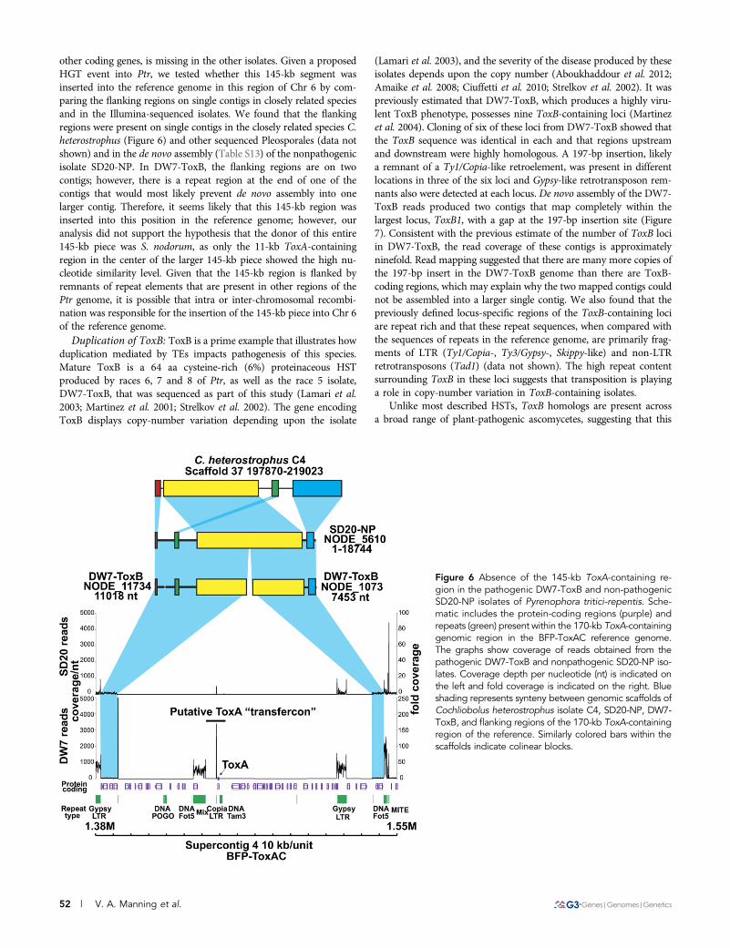
other coding genes, is missing in the other isolates. Given a proposedHGT event into Ptr, we tested whether this 145-kb segment wasinserted into the reference genome in this region of Chr 6 by com-paring the flanking regions on single contigs in closely related speciesand in the Illumina-sequenced isolates. We found that the flankingregions were present on single contigs in the closely related species C.heterostrophus (Figure 6) and other sequenced Pleosporales (data notshown) and in the de novo assembly (Table S13) of the nonpathogenicisolate SD20-NP. In DW7-ToxB, the flanking regions are on twocontigs; however, there is a repeat region at the end of one of thecontigs that would most likely prevent de novo assembly into onelarger contig. Therefore, it seems likely that this 145-kb region wasinserted into this position in the reference genome; however, ouranalysis did not support the hypothesis that the donor of this entire145-kb piece was S. nodorum, as only the 11-kb ToxA-containingregion in the center of the larger 145-kb piece showed the high nu-cleotide similarity level. Given that the 145-kb region is flanked byremnants of repeat elements that are present in other regions of thePtr genome, it is possible that intra or inter-chromosomal recombi-nation was responsible for the insertion of the 145-kb piece into Chr 6of the reference genome.
Duplication of ToxB: ToxB is a prime example that illustrates howduplication mediated by TEs impacts pathogenesis of this species.Mature ToxB is a 64 aa cysteine-rich (6%) proteinaceous HSTproduced by races 6, 7 and 8 of Ptr, as well as the race 5 isolate,DW7-ToxB, that was sequenced as part of this study (Lamari et al.2003; Martinez et al. 2001; Strelkov et al. 2002). The gene encodingToxB displays copy-number variation depending upon the isolate
(Lamari et al. 2003), and the severity of the disease produced by theseisolates depends upon the copy number (Aboukhaddour et al. 2012;Amaike et al. 2008; Ciuffetti et al. 2010; Strelkov et al. 2002). It waspreviously estimated that DW7-ToxB, which produces a highly viru-lent ToxB phenotype, possesses nine ToxB-containing loci (Martinezet al. 2004). Cloning of six of these loci from DW7-ToxB showed thatthe ToxB sequence was identical in each and that regions upstreamand downstream were highly homologous. A 197-bp insertion, likelya remnant of a Ty1/Copia-like retroelement, was present in differentlocations in three of the six loci and Gypsy-like retrotransposon rem-nants also were detected at each locus. De novo assembly of the DW7-ToxB reads produced two contigs that map completely within thelargest locus, ToxB1, with a gap at the 197-bp insertion site (Figure7). Consistent with the previous estimate of the number of ToxB lociin DW7-ToxB, the read coverage of these contigs is approximatelyninefold. Read mapping suggested that there are many more copies ofthe 197-bp insert in the DW7-ToxB genome than there are ToxB-coding regions, which may explain why the two mapped contigs couldnot be assembled into a larger single contig. We also found that thepreviously defined locus-specific regions of the ToxB-containing lociare repeat rich and that these repeat sequences, when compared withthe sequences of repeats in the reference genome, are primarily frag-ments of LTR (Ty1/Copia-, Ty3/Gypsy-, Skippy-like) and non-LTRretrotransposons (Tad1) (data not shown). The high repeat contentsurrounding ToxB in these loci suggests that transposition is playinga role in copy-number variation in ToxB-containing isolates.
Unlike most described HSTs, ToxB homologs are present acrossa broad range of plant-pathogenic ascomycetes, suggesting that this
Figure 6 Absence of the 145-kb ToxA-containing re-gion in the pathogenic DW7-ToxB and non-pathogenicSD20-NP isolates of Pyrenophora tritici-repentis. Sche-matic includes the protein-coding regions (purple) andrepeats (green) present within the 170-kb ToxA-containinggenomic region in the BFP-ToxAC reference genome.The graphs show coverage of reads obtained from thepathogenic DW7-ToxB and nonpathogenic SD20-NP iso-lates. Coverage depth per nucleotide (nt) is indicated onthe left and fold coverage is indicated on the right. Blueshading represents synteny between genomic scaffolds ofCochliobolus heterostrophus isolate C4, SD20-NP, DW7-ToxB, and flanking regions of the 170-kb ToxA-containingregion of the reference. Similarly colored bars within thescaffolds indicate colinear blocks.
52 | V. A. Manning et al.

gene may have arisen in an early ancestor of the Ascomycota (Andrieet al. 2008). However, not all ToxB homologs may be functional intheir host-pathogen interaction (Andrie and Ciuffetti 2011). For in-stance, the ToxB homolog in SD20-NP (Martinez et al. 2004), whichwe refer to as toxb, does not appear to encode a toxin active inwheat, most likely due to changes in amino acid sequence (Andrie andCiuffetti, 2011, Figueroa Betts et al. 2011). A single toxb-containingcontig, with approximately onefold coverage, was present in the denovo assembly of SD20-NP, consistent with the previous predictionof a single copy of toxb in this isolate (Martinez et al. 2004). The contigextends sequence information of the genomic region that containstoxb by approximately 10 fold (from ~2550 nt to .25,000 nt). Ablastn search with this SD20-NP contig against the reference genomerevealed a 10-kb stretch that is syntenic to a region of Chr 5, whichoptical mapping predicts to be 3.1 Mb. A previous study showed thattoxb was localized to a 3.1-Mb chromosome in SD20-NP (Martinez et al.2004); therefore, it is possible that the 3.1-Mb chromosomes of these twoisolates are homologous. Sequence data provide ample evidence thathighly mobile TEs around ToxB in pathogenic isolates provided a favor-able environment for expansion of an active toxin gene in which dosageplays an important role in virulence. In addition, it leads to speculationthat toxb might at one time have had a marginal function in pathogen-esis, but that the lack of TEs in the SD20-NP genome, and therefore, thelack of opportunity to expand, resulted in pseudogenization.
Duplication and diversification of secreted proteins: In addition toknown HSTs, other candidate pathogenicity-related proteins asso-ciated with TEs also are present. One example is a family of smallsecreted cysteine-rich proteins (Table S14) that is present as a resultof tandem duplication followed by diversification (PTRG_11771,PTRG_11772, PTRG_11773), and more recent duplication events(PTRG_11773 and PTRG_11346) in the sequenced pathogenic isolatesbut is not expanded and diverged in the sequenced non-pathogenicisolate. PTRG_11772 is the most diverged member of this family, yetthe cysteine residues in all of the family members are conserved.PTRG_11773 is expressed during infection, as ESTs were found inthe in planta expression library, suggesting that this protein may
be involved in pathogenesis. A blast search of the NCBI nonredun-dant database indicated that, with the exception of one homolog(FOXB_14349) from the Arabidopsis-infecting strain of F. oxyspo-rum (strain 5176), all other homologous proteins are present incereal pathogens that are able to colonize wheat, including P. teresf. teres (PTT_08876), Colletotrichum (Glomerella) graminicola(GLRG_11876, GLRG_08163, GLRG_08162, GLRG_04750), andMycosphaerella graminicola (MYCGRDRAFT_96981). Expansionof this protein family in Ptr and C. graminicola suggests that in-creased copy number of this gene may provide an advantage, whichmakes these genes compelling candidates for further studies of ad-aptation to a host-specific lifestyle.
Duplication and recombination creates pathogen-specificnonribosomal peptide synthetases: Nonribosomal peptide synthe-tases (NRPS) are mono- or multimodular enzymes that producediverse compounds with a multiplicity of functions (Schwarzer andMarahiel 2001). The most basic module contains an adenylationdomain (A) followed by a thiolation domain (T), which are oftenaccompanied by a condensation domain (C). Additional functions en-coded in a module could include epimerization and N-methyltransferaseactivities. Certain plant-pathogenic fungi produce NRPSs that areknown to be important for the biosynthesis of toxins involved inpathogenesis (Gardiner et al. 2004; Johnson et al. 2000; Walton2006). When we surveyed the Ptr reference genome we found thatseveral NRPS-coding genes (NPSs) were conserved between Ptr andother Ascomycota (Table 4). All Ptr isolates sequenced in this studycontain NPS 2, 4, 6, and 10 (nomenclature adopted from the NRPSsof Cochliobolus heterostrophus), which are thought to be moderatelyconserved in the Ascomycota (Lee et al. 2005; Turgeon et al. 2008).They also encode a NRPS (PTRG_12015) that is similar to theNRPSs required for synthesis of the histone deacetylase (HDAC)inhibitor apicidin in Fusarium incarnatum (APS1) and HC-toxin inCochliobolus carbonum (HTS1; Figure S6). In the reference genome,PTRG_12015 is present within a cluster (Table S15), and almost allof the proteins in the cluster have EST support, suggesting that thisis a functional biosynthetic cluster that is coordinately regulated.
Figure 7 Read mapping to andde novo assembly of ToxB- andtoxb-containing loci in the ge-nome of Pyrenophora tritici-repentis. Schematic of Illuminasequence reads (line graph) ofisolate DW7-ToxB mapped tothe ToxB1 locus (top: ToxB1 lo-cus; accession number:AY425480.1) and of SD20-NPmapped to the toxb locus (bot-tom: toxb locus; accession num-ber: AY083456.2). Coveragedepth per nt is indicated onthe left and fold coverage is in-dicated on the right. Straightlines above the graphs depictthe contigs present in the denovo assemblies of the Illu-mina-sequenced isolates. Thearrow on the contig above thetoxb locus shows how that con-tig extends beyond the locus.
Volume 3 January 2013 | Genome of P. tritici-repentis | 53

Homologs of 8 of the 11 proteins identified in the apicidin biosyn-thetic cluster (Jin et al. 2010) and four genes required for the pro-duction of HC-toxin (Walton 2006) are present in the Ptr cluster.
Two additional NRPSs that are conserved in the three Ptr isolates,PTRG_09101 and 09808, have homologs in the wheat pathogen S.nodorum, one of which appears to be present in a biosynthetic cluster(PTRG_09101); however, the function of these NRPSs is unknown.There are also two NRPS:PKS (polyketide synthase) hybrid moleculespresent in all isolates. PTRG_00649 encodes an incomplete NRPSmodule at its N-terminus and a PKS ketoacyl-synthase (KS) domainat its C-terminus. It is most closely related to MGG_07803 ofM. oryzaewith the exception that PTRG_00649 does not contain a full NRPSmodule. Upstream of this gene is a TE, which may have led to thetruncation of the NRPS module or could be the footprint from an HGTevent. Unlike PTRG_00649, PTRG_04244 encodes a 59 PKS moduleand a complete NRPS module at its 39 end. PTRG_04244 is surroundedby a putative biosynthetic cluster (Figure S7), and many of the genes inthis cluster have EST support. The PTRG_04244-containing clusterencodes several genes similar to those found in the genome of theascomycete Talaromyces stipitatus, a teleomorph of a potent humanpathogen, Penicillium marneffei (Cooper and Vanittanakom 2008).
The observation of complicated duplication, recombination anddomain swapping of Ptr NRPSs in the pathogenic isolates further
confirms that TEs play an important role in NRPS diversification inthis species (Figure 8). Many of the domains and modules that arebeing duplicated and recombined are related to those present in C.heterostrophus NRPS1 (ChNRPS1). For example PTRG_10128, whichis orthologous to the central module of ChNRPS1, is surrounded byTEs and remnants of TEs. The putative NRPS, PTRG_10437, is com-posed of one incomplete (T-C) and two complete modules (A-T-C)that contain domains that are related to the 39 end of ChNRPS1.Interestingly, the A domain of module 3 of ChNRPS1 and some ofthe modules present in PTRG_10437 are related to A domains in theNRPS necessary for the production of AM-toxin from A. alternata(Bushley and Turgeon 2010). The first incomplete module ofPTRG_10437 is . 95% similar to the T-C domains in the secondmodule, suggesting a fairly recent duplication. The A domains inmodules two and three do not show that level of conservation andthe C domain in the third module is more similar to the last C domainin ChNRPS1 than to any other C domain in the NRPSs of Ptr.
This complex level of duplication and domain swapping is evenmore evident in PTRG_10433, the largest NRPS (8,548 aa) in the Ptrgenome. This protein appears to be derived from PTRG_10437 and iscomposed of six complete modules (A-T-C) with an additional Adomain at the C-terminus. Two new A domains have been introducedinto this protein, one at its N-terminus and one into the fifth module,
n Table 4 Putative Pyrenophora tritici-repentis nonribosomal peptide synthetase genes
LocusBest Hita Cochliobolus
NPS (%id/sim)cRepeateddomainsd
Assoc.with TEe EST
Present (SNP)b
Org. Acc. (%id/sim)c Rec. DW7-ToxB SD20-NP
ConservedPTRG_08276f Ch AAX09984.1 (g ) Yes NPS2 (62/76) No No Yesh Yes (1) Yes (2)PTRG_01800f Ab AAP78735.1 (77/87) Yes NPS4 (72/84) No No Noh Yes (1) Yes (37)PTRG_01683f Cm ABI51982.1 (77/86) Yes NPS6 (76/86) No No Noh Yes (1) Yes (10)PTRG_00447f Ch AAX09992.1 (g ) Yes NPS10 (91/96) No No Yesh Yes (0) Yes (3)PTRG_03139 Ch AAX09994.1 (g ) Yes NPS12 (70/80)) No No No Yes (2) Yes (10)PTRG_09101f Sn XP_001791762.1 (72/80) Yes No No Yes Yes (1) Yes (39)PTRG_09808 Sn XP_001797376.1 (60/70) Yes No No No Yes (0) Yes (0)PTRG_12015f Fi ACZ66258.1 (39/56) Yes HTS1 (35/51) No No Yes Yes (2) Yes (18)
HybridsPTRG_00649 Mg XP_367899.2 (37/50) No No Yes No Yes (1) Yes (6)PTRG_04244f Pa XP_001905191.1 (49/66) No No No Yesh Yes (7) Yes (76)
NPS pathogen-specificdiversification
PTRG_10128 Ch AAX09983.1 (g ) No NPS1- w/o 59&39(61/70)
No Yes No Divergent No
PTRG_10433 Cg XP_001226467.1 (28/43) No Yes Yes Yesi Divergent NoPTRG_10437 Ptr 10433 (j) Yes Yes No No NoPTRG_11759 Ch AAX09988.1 (g) Yes NPS6 – w/o - 39
(73/84)No Yes No No No
PTRG_11809 Ptr 10433 (j) Yes Yes Yes No NoPTRG_11818 Ptr 11836 (j) No Yes Yes No NoPTRG_11836 Ptr 11818 (j) No Yes Yes No No
Ab, Alternaria brassicae; Acc., accession number; Cg, Chaetomium globosum; Ch, Cochliobolus heterostrophus; Cm, Cochliobolus miyabeanus; Fi, Fusariumincarnatum; Mg, Magnaporthe grisea; Org., organism; Pa, Podospora anserina; Rec., reciprocal best blast hit; Sn, Stagonospora nodorum; SNP, single-nucleotide polymorphism.aBest hit BlastP (Altschul et al. 1997) NCBI.
bSNP predicted in maq.
cComparison by Needle in EMBOSS (Needleman and Wunsch, 1970, Rice et al. 2000); % identity/similarity.
dAt least two domains in the protein share .96% similarity.
eAssociated with transposable elements (TE).
fPresent in putative cluster.
gSee Cochliobolus NPS column.
hOther members in cluster have EST support.
iP. Martinez and L. Ciuffetti, unpublished data.
jApparent duplication in P. tritici-repentis genome.
54 | V. A. Manning et al.

resulting in modules with unique A domains but practically identicalT and C domains. A new T, epimerization and C domain combinationhas been introduced, duplicated and recombined, in one case creatinga unique module. Reads from the pathogenic DW7-ToxB map to theseNPSs, but it is difficult to determine if the domains and modules arearranged in a similar manner due to the difficulty of assembling highlysimilar reads. The presence of a similarNPS in only the pathogenic isolatesis consistent with a previous study in which pathogen-specific probes wereisolated using a subtractive approach, and two of these probes containedsequences present in PTRG_10433 (Lichter et al. 2002). There is anotherlocus, PTRG_11809 that appears to have been derived from PTRG_10433;however, neither of the two modules is complete. Whether theNPSs in Ptrthat encode partial modules are functional may not be as important astheir role as reservoirs for NRPS diversification.
In addition to domain and module duplication contributing toNRPS diversification, the Ptr reference genome also contains duplica-tions (three copies) of an entire chromosomal region that contains notonly two NPS genes (PTRG_11818 and 11836, 97% identical at theamino acid level), but also cytochrome p450, amino transferase andmethyltransferase genes, although not all genes are equally maintainedin the duplication (Figure S8). The repetition of this region of thegenome and the loss of some genes are likely due to transpositionevents as there are TEs throughout the duplications. Although thefunction of this NRPS and the surrounding proteins present in thisregional duplication is not known, it has been shown in the applepathotype of A. alternata that gene duplication in the biosyntheticcluster responsible for the production of the HST, AM-toxin, is nec-essary for full pathogenicity (Harimoto et al. 2008).
Unique among the three sequenced isolates, the reference genomeproduces ToxC, which is hypothesized to be the product of a bio-synthetic cluster as opposed to a single gene (Effertz et al. 2002).Therefore, the BFP-ToxAC strain-specific NRPSs and/or their bio-synthetic cluster components are potential candidates for characteriz-ing this HST. Alternatively, ToxC and other currently uncharacterizedPtr toxins could be the products of polyketide synthases (PKS). ThePtr reference genome contains 21 putative PKS loci. However, most ofthese PKSs are conserved in all of the isolates sequenced (Table S15),thereby reducing the probability that one of them is responsible forthe production of the BFP-ToxAC-specific ToxC. (Table S16). Ptr isknown to produce anthraquinone-derived phytotoxins (Bouras andStrelkov, 2010; Bouras et al. 2009) and a biosynthetic cluster similar to
the emodin cluster of Aspergillus nidulans (Bok et al. 2009) was iden-tified in the reference genome. Also found are clusters related to thosein other fungi that produce resorcyclic-lactone-based compounds [i.e.,zealerone (Lysoe et al. 2009; Reeves et al. 2008)] and alternapyrone(Fujii et al. 2005). The PKS and accessory proteins necessary for melaninbiosynthesis (Kihara et al. 2008) were also identified (PTRG_03323,PTRG_03315 = T4HN, PTRG_04757 = SCD, and PTRG_03318 =CmrI). Although there is no evidence for duplication and recombi-nation events playing a role in PKS diversification in the pathogens,there are loci that have clearly been truncated when compared totheir putative homologs. Additionally, there are two other pathogen-specific PKSs, which are recent duplications and are 100% identical.So although TEs are not necessarily as important for PKS as com-pared with NRPS evolution in Ptr, copy-number variation may con-tribute to the amount of a product that can accumulate.
The P. tritici-repentis secretome provides insightsinto a necrotrophic lifestyleThe secretome of plant-pathogenic fungi plays an important role inbreaching host defenses, establishing and maintaining growth oncethe host has been penetrated and, in the case of a necrotrophicpathogen, killing host tissue to obtain nutrients by initiating cell deathcascades, often by taking advantage of the host plant immune system(Oliver and Solomon, 2010; Pandelova et al. 2009, 2012; Wolpert et al.2002). Therefore, to understand the diseases caused by these patho-gens in general, and necrotrophs in particular, it is imperative toanalyze the secreted component of the proteome. Although the pro-teinaceous HSTs of Ptr described in the previous section are undoubt-edly critical for pathogenesis and virulence, they are only two proteinsin what is likely an extensive repertoire of pathogenesis-related se-creted proteins important to the Ptr-wheat pathosystem. For the pur-poses of identifying a comprehensive set of putative secreted proteins,we produced a very inclusive secreted protein dataset, at the risk ofincluding some false-positives. Once defined, the secreted proteinswere annotated with conserved domains and GO ontology terms.
We predicted 1146 potential secreted proteins in the referencegenome of Ptr (Table S14) of which 317 are small (, 200 aa) and 69of those are cysteine-rich, characteristics typically shared among smalleffectors (Rep 2005; Stergiopoulos and De Wit 2009). One hundredten of these proteins had no BLAST hits in the nonredundant
Figure 8 NRPSs diversification in thegenome of Pyrenophora tritici-repentis.Schematic shows the modular architec-ture of Cochliobolus heterostrophusNRPS1 (black solid bar and associatedmodules) and related P. tritici-repentisPtNRPSs (gray solid bars and associ-ated modules). Brackets with straightarrows define modules in C. hetero-strophus NRPS1 that have homolo-gous and/or related regions presentin PtNRPSs. The module domains(adenylation, thiolation, condensation,methyltransferase, and epimerization),and the solid bars that represent theentire protein are drawn to scale. Thinlines between gray bars extend to theregion(s) of identity between proteinsand the curved arrow depicts a recentduplication event.
Volume 3 January 2013 | Genome of P. tritici-repentis | 55

database at the NCBI and we consider these to be Ptr-specific (TableS17). Comparison with the two Illumina-sequenced isolates (Figure9A) revealed that 1120 (98%) of the secreted proteins are sharedbetween all isolates, 12 proteins are shared only between the twopathogenic isolates, eight are found only in the reference race 1 isolate,BFP-ToxAC, and six are shared between the reference genome andthat of the nonpathogen. Of the eight BFP-ToxAC-specific secretedproteins, only two have informative annotations: the known HSTToxA (PTRG_04889), and a putative endochitinase (PTRG_04903),which is also encoded in the ToxA-containing 145-kb region of thereference genome that is absent in the other two sequenced isolates. Agood candidate effector among the eight BFP-ToxAC-specific secretedproteins is a small secreted protein (SSP) of 59 aa and 7% cys content(PTRG_10524). ESTs for three of the 12 pathogen-shared secretedproteins (PTRG_11888, PTRG_11773 and PTRG_12138) weredetected in the in planta expression library, suggesting their potentialcontribution to disease development. PCR screening of one of thesecandidate pathogenicity-related genes, PTRG_11888, among five ad-ditional pathogenic and two additional non-pathogenic isolates (TableS1), confirmed the association of this locus with pathogenic isolatesregardless of race (Figure 9B). Although its function has yet to bedetermined, this small (85 aa) cysteine-rich (14%) protein encodesa conserved 2Fe-2S ferredoxin, iron-sulfur binding site, indicatinga potential function in redox reactions. Among all of the secretedproteins in the reference protein data set there were three main cat-egories of abundant GO/PFAM terms: Cell wall degrading enzymesand lipases, proteins involved in oxidation-reduction reactions, andproteins involved in proteolysis (Table 5).
Cell wall-degrading enzymes: Similar to most plant-pathogenicfungi, Ptr encodes a high number and variety of cell wall-degradingenzymes (CWDEs) to degrade host cell walls during infection (TableS18). However, the number of CWDEs encoded in Ptr is greater thanthose encoded by the “stealth pathogen,” M. graminicola, anotherhemibiotrophic Dothideomycete wheat pathogen. The lifestyle of M.graminicola differs from that of Ptr in that it has a latent biotrophicphase before switching to necrotrophic symptoms (Goodwin et al.2011). In contrast, Ptr induces cell death rapidly in susceptible hosts.It is hypothesized that the reduction of CWDEs in M. graminicolaresults in the ability of the pathogen to escape detection by the host.Therefore, the presence of unique expansions and contractions ofCWDE families can provide information about the lifestyle of Ptr.
The cell walls of grasses are significantly different than those of dicotsas they contain, in addition to cellulose, a relatively high percentageof glucuronoarabinoxylans, mixed-linkage glucans, and a relatively
low percentage of xyloglucan, mannans and glucomannans, pectinand structural proteins (Vogel 2008). Consistent with a lifestyle asa wheat pathogen, we find that the Ptr genome encodes fewer pectin-degrading enzymes, including those in the polysaccharide lyase andGH (glycosyl hydrolase) families 28, 78, 88, 95, compared withthe dicot pathogens Verticillium dahliae (Klosterman et al. 2011)and F. oxysporum (Table S19) (Ma et al. 2010). Furthermore, thereare at least three putative mixed-linkage glucanases that wouldaid in cell wall degradation in grasses, PTRG_07078 (GH16) andPTRG_06274 (GH12), orthologs of Mlg1 and Mlg2 of the maizepathogen C. carbonum, and PTRG_03370 (GH16) (Kim et al.2001). PTRG_06274 is one of the loci represented in a GO enrich-ment analysis of the in planta EST library, suggesting its importancein the pathogenesis process (Table S20).
In the GO enrichment analysis, the endoglucanases represented thelargest group (six) of cellulose-degrading enzymes, with expression offour of these genes (PTRG_04829, 06274, 04285, 03392 and their fam-ilies GH45, GH12, GH5, and GH61/CBM1, respectively) found exclu-sively in the in planta EST library. The cellobiohydrolase/exoglucanasePTRG_09037 (GH7), the ortholog of Cel1 of C. carbonum (Sposatoet al. 1995) and CbhA of A. nidulans (Gielkens et al. 1999), had thegreatest number of transcripts detected in the in planta library of any ofthe secreted proteins. Glucuronoarabinoxylans is the major hemicellu-lose in grass cell walls and degradation of the xylose backbone requiresthe production of xylanases (Aro et al. 2005; De Vries and Visser2001). PTRG_08873, 09530, and 10207, orthologs of C. carbonumXyl1 (GH11), Xyl4 (GH10) and Xyl3 (GH11), respectively, weredetected in the in planta EST library with only PTRG_10207 detectedboth in planta and in culture. Xylanases, as well as other CWDEsappear to be functionally redundant in C. carbonum and it has beenhypothesized that the differential expression of these genes may reflecta need for redundancy (Apel-Birkhold andWalton 1996); this may alsobe true for Ptr where CWDEs also appear to be differentially expressed.
Unexpectedly, Ptr encodes fewer proteins in the CBM family ofCWDE in comparison to most of the other fungi examined in thisstudy, be they plant pathogenic or not, although not as few as in M.graminicola (Table S21). Most of this decrease is due to fewer proteinsin the CBM18 family, predicted to be fungal chitinases. Fungal chiti-nases can be grouped into three subgroups, sg A, sg B, and sg C, withsg C containing either CBM18 or CBM50 (LysM) domains (Hartlet al. 2012). Although functions of proteins in these families are notwell understood, expression studies of sg C genes in the mycoparasiticfungi Trichoderma virens and T. atroviride suggest roles in morpho-genetic development and exogenous chitin degradation (Gruber et al.2011; Gruber and Seidl-Seiboth 2012).
Figure 9 Predicted secreted proteins in thegenome sequence of Pyrenophora tritici-repentis. (A) Distribution of secreted proteinsin the reference Ptr isolate (BFP-ToxAC) andthe Illumina-sequenced pathogenic (DW7-ToxB) and nonpathogenic (SD20-NP) isolates.(B) PCR screen of pathogenic and nonpatho-genic isolates for the presence of the geneand transcript for PTRG_11888, a predictedputative secreted protein. Pathogenic iso-lates: BFP-ToxAC; ASC1; 86-124; D308;DW2; SO3-P; Non-pathogenic isolates:SD20-NP; 90-2; 98-31-2 (see also Table S1).
56 | V. A. Manning et al.

Proteins involved in detoxifying redox reactions—cytochrome p450s,berberine bridge enzymes, and other oxidative-stress-related proteins:Plant responses to invasion include the formation of reactive oxygenspecies, the production of secondary metabolites, and the secretion ofdefense-related proteins. Fungi must deal with all of these componentsif they are to survive. The secreted proteins of Ptr reflect the need toprotect itself from plant-produced compounds.
A surprising number of proteins (29) containing conservedcytochrome p450 domains (p450) were present in our secreted
protein dataset. This may be due to the retention of a subset ofproteins with one TM domain. When we remove those p450s thatcontain predicted TM domains, there are still 12 loci remaining in thedataset. The secretion of these p450s must be experimentally validated,but it suggests a possible method by which Ptr could persist within thehost prior to induction of cell death by neutralizing plant-producedanti-fungal compounds. The best example of a p450 involved in thistype of interaction is pisatin demethylase, produced by the pea path-ogen Nectria hematococca, which neutralizes the pea phytoalexin
n Table 5 The most abundant annotations in the predicted secretome of Pyrenophora tritici-repentis
Gene Ontology (GO) and Pfam Terms
GO ID/Accession No. GO Term/Accession Description No. Loci with Term
Molecular functionGO:0004553 Hydrolase activity, hydrolyzing O-glycosyl compounds 112GO:0050660 FAD binding 43GO:0020037 Heme binding 37GO:0004497 Monooxygenase activity 34GO:0009055 Electron carrier activity 32GO:0003676 Nucleic acid binding 18GO:0016614 Oxidoreductase activity, acting on CH-OH group of donors 18GO:0004601 Peroxidase activity 16GO:0004091 Carboxylesterase activity 16GO:0004180 Carboxypeptidase activity 15GO:0008270 Zinc ion binding 13GO:0005515 Protein binding 13GO:0016810 Hydrolase activity, acting on carbon-nitrogen (but not peptide) bonds 12GO:0030248 Cellulose binding 12GO:0004252 Serine-type endopeptidase activity 12GO:0016829 Lyase activity 12GO:0008237 Metallopeptidase activity 11GO:0016757 Transferase activity, transferring glycosyl groups 11
Biological processesGO:0055114 Oxidation reduction 65GO:0006508 Proteolysis 61GO:0006066 Alcohol metabolic process 24GO:0044267 Cellular protein metabolic process 18GO:0051704 Multiorganism process 16GO:0006629 Lipid metabolic process 16GO:0051591 Response to cAMP 14GO:0044262 Cellular carbohydrate metabolic process 14GO:0009308 Amine metabolic process 14GO:0034645 Cellular macromolecule biosynthetic process 13GO:0006139 Nucleobase, nucleoside, nucleotide and nucleic acid metabolic process 13GO:0043581 Mycelium development 12GO:0045493 Xylan catabolic process 11GO:0016998 Cell wall macromolecule catabolic process 11
Pfam domainsa
PF00067 Cytochrome p450 30PF03443 Glycosyl hydrolase family 61 26PF01565 FAD binding domain 25PF08031 Berberine and berberine-like 21PF00732 GMC oxidoreductase (binds FAD as a cofactor) 18PF05199 GMC oxidoreductase 16PF00187 Carbohydrate Binding Module 18 16PF00135 Carboxylesterase 15PF00082 Subtilase 11PF00734 Carbohydrate Binding Module 1 (cellulose-binding domain) 11PF04389 Peptidase Family M28 (metalloprotease) 10PF00657 GDSL family of esterases/lipases 10PF01083 Cutinase (cutin + H2O 4 cutin monomers) 10
Values represent the lowest node per branch (.10 sequences) of a directed acyclical graph of the hierarchical structure of GO terms.ae-value cutoff for Pfam analysis E , 0.1.
Volume 3 January 2013 | Genome of P. tritici-repentis | 57

pisatin (Maloney and VanEtten 1994). Other preformed secondarymetabolites (Imperato and Pandalai 2006), as well as phenolic com-pounds produced in response to Ptr (Dushnicky et al. 1998a,b;Pandelova et al. 2009, 2012), also might be a target for these p450s.
There are 21 proteins predicted in the Ptr secretome that containa combination of FAD-binding and berberine or berberine-likedomains. Flavoenzymes with this combination of domains are knownas berberine bridge enzymes (BBE), some of which are produced inresponse to plant pathogens, and have been shown to be importantfor the production of plant alkaloids (Dittrich and Kutchan 1991;Facchini et al. 1996) and carbohydrate oxidases (Carter and Thornburg2004; Custers et al. 2004), and the generation of hydrogen peroxide.BBEs also are over-represented in the secretome of the oomyceteP. infestans (Raffaele et al. 2010), where they may contribute to theproduction of reactive oxygen species and alkaloid modification duringP. infestans infection. TEs also are playing a role in the expansion ofBBE-family proteins in Ptr. A segment of the genome containingPTRG_11734, a BBE, and PTRG_11735 is surrounded by TEs anda duplication of this segment (96.4% identical at the nucleotide level),also surrounded by TEs, is present in the genome (PTRG_02303 =PTRG_11734 and PTRG_02304 = PTRG_11735).
Other dominant Pfams suggest that proteins important foroxidation-reduction reactions are predominant in the secretome;these include glucose2methanol2choline oxidoreductase domains(18 loci). Although these enzymes appear to have multiple catalyticfunctions (Cavener 1992), little is known about their impact in planthost-pathogen interactions. It has recently been proposed that a glu-cose2methanol2choline oxidoreductase produced by Glomerellacingulata plays a role in plant pathogenicity through the reductionof pathogen-induced quinones, which have anti-fungal activities,and phenoxy radicals produced for reinforcement of plant cells wallsduring pathogen attack (Sygmund et al. 2011).
SummaryThe era of genome sequencing has provided us with incredibleresources to answer questions and pose new hypotheses about whatcontributes to the pathogenicity of microorganisms and the hostresponses that govern resistance or susceptibility. Sequencing ofmultiple isolates of Ptr, including two pathogenic and one non-pathogenic isolate, has provided ample evidence that the establish-ment of pathogenicity in this system, and possibly host switchingfrom wild grasses (nonpathogens) to wheat (pathogens), likely startedwith structural changes to the genome. These changes were facilitatedby the influx of TEs that resulted in increased genetic plasticity andthereby likely also reduced the possibility of gene flow between path-ogenic and non-pathogenic isolates. In addition, TEs are probably oneof the major factors that contribute to the high genetic diversity of thepathogen population. Such dynamic changes enabled the introduc-tion, amplification, and diversification of various toxin genes andpathogenicity factors and created the complex race structure thatthis organism displays today. Many of these TEs appear to be active,which contributes to a genomic landscape in the pathogen popula-tion that is capable of rapid adaptation. These findings are consistentwith the “jump or die model” for the macroevolutionary persistenceof host-specialized filamentous fungi proposed by Raffaele andKamoun (2012). In this model, pathogen lineages with highly adapt-able genomes are better able to jump to new hosts than those withless flexible genomes, and therefore, are more likely to persist overevolutionary time when host extinction or complete resistance wouldotherwise result in pathogen extinction.
Highly adaptable genomes often have relatively high amounts ofrepetitive DNA (Raffaele and Kamoun 2012), which may result inexpansion of genome size; for instance, biotrophic- and oomycete-plant pathogens have some of the largest genome sizes and repetitiveDNA content (Haas et al. 2009; Spanu et al. 2010). Though the size ofthe Ptr genome and the amount of repetitive DNA falls into the rangeof that of other Dothideomycete-plant pathogens (Goodwin et al.2011; Hane et al. 2007; Rouxel et al. 2011), nonpathogenic Ptr isolatestend to have smaller genome sizes and the isolates sequenced in thisstudy indicate a higher repetitive DNA content in the pathogenicisolates. The expansion of TEs in the pathogenic Ptr isolates appearsto be an ongoing event, as members of a single repeat family oftenshare identical or very similar sequences. The low amount of typicalrepeat-induced point mutations in these highly similar repeat sequen-ces suggests that RIP, if occurring at all, is inefficient in Ptr. The lackof RIP has been postulated to be one of the drivers in the expansion ofgenome size in the powdery mildew fungi (Spanu et al. 2010) and mayalso explain genome expansion in pathogenic isolates of Ptr.
Many of the events that contribute to genome plasticity alsocontribute to changes in pathogen virulence. Recombination can leadto domain swapping between virulence proteins as has been shown inthe oomycete Crinkler family of modular proteins, which exhibitdomain swapping and chimera formation (Haas et al. 2009), and inthe NRPSs of Ptr. TE-mediated duplications can lead to copy numbervariation (CNV), which in Phytophthora sojae has been associatedwith silencing of effectors (Qutob et al. 2009) and in P. infestanshas been proposed to contribute to the assembly of novel genes (Jianget al. 2006). In Ptr, CNV of the HST gene, ToxB, correlates with isolatevirulence. Additional loci in the Ptr genome display CNV; however,their role in virulence has yet to be determined.
HGT is another mechanism that can alter the plant-pathogengenome landscape and contribute to virulence. Entire chromosomescan be transferred between fungi that alter pathogenicity or host-specificity (Coleman et al. 2009; Ma et al. 2010). Proposed gene trans-fer events that would alter virulence include the transfer of HSTs, as inthe case of ToxA in Ptr and AAL toxin in Alternaria alternata (Akagiet al. 2009). Whether genome flexibility is a pre-requisite for efficientHGT is currently unknown.
And what are the implications of transduplication in Ptr? Asidefrom changes normally associated with transposition, capture of genesand gene fragments and their deposition throughout the genomeincreases the chances of novel gene formation and may very wellplay important roles in cellular gene expression. Discovery of thisphenomenon in Ptr allows us to ask some interesting questions. Isthere selective pressure for the gene/gene fragments that are cap-tured; and if so, why? Do they play a role in development or path-ogenicity? Clearly, there is an increased copy number and expressionof a histone H3 variant, which might very well have a significantimpact on genome structure and function as histones are essentialcomponents of chromosomes. Could altered chromatin be in partresponsible for the recent amplification of transposable elements,segments of the genome, and the unique recombination events wesee in the pathogen? Can this unusual histone H3 provide a tool forbetter understanding of chromatin structure and function? Imple-mentation of novel strategies, computational and experimentalapproaches will be required to answer these fundamental questions.
ACKNOWLEDGMENTSWe thank Cedar Hesse and Jeffrey Kimbrel for helpful discussionabout bioinformatics and the Center of Genome Research and Bio-computing, Oregon State University, Corvallis, Oregon, for biocomputing
58 | V. A. Manning et al.

infrastructure. We thank K. Cook, P. Martinez, and S. Ottum formicroscopy. We also thank the Colletotrichum genome project (PIs-Lisa Vaillancourt, Michael Thon, and Marty Dickman) for sharingunpublished data. Funding for M. Figueroa was provided by theNational Science Foundation under a Minority Postdoctoral Re-search Fellowship (0706881) awarded in 2007. Sequencing of thereference strain was funded by the National Research Initiative ofthe U.S. Department of Agriculture Cooperative State Research,Education and Extension Service, grant number (2006-55600-16619),and reviewed through the U.S. Department of Agriculture/NationalScience Foundation Microbial Genome Sequencing Project. Illuminasequencing work was conducted by the U.S. Department of EnergyJoint Genome Institute and supported by the Office of Science ofthe U.S. Department of Energy (under Contract No. DE-AC02-05CH11231).
LITERATURE CITEDAbascal, F., R. Zardoya, and D. Posada, 2005 ProtTest: selection of best-fit
models of protein evolution. Bioinformatics 21: 2104–2105.Aboukhaddour, R., S. Cloutier, G. M. Ballance, and L. Lamari,
2009 Genome characterization of Pyrenophora tritici-repentis isolatesreveals high plasticity and independent chromosomal location of ToxAand ToxB. Mol. Plant Pathol. 10: 201–212.
Aboukhaddour, R., S. Cloutier, L. Lamari, and S. E. Strelkov, 2011 Simplesequence repeats and diversity of globally distributed populations ofPyrenophora tritici-repentis. Can. J. Plant Pathol. 33: 389–399.
Aboukhaddour, R., Y. M. Kim, and S. E. Strelkov, 2012 RNA-mediatedgene silencing of ToxB in Pyrenophora tritici-repentis. Mol. Plant Pathol.13: 318–326.
Akagi, Y., H. Akamatsu, H. Otani, and M. Kodama, 2009 Horizontalchromosome transfer, a mechanism for the evolution and differentiationof a plant-pathogenic fungus. Eukaryot. Cell 8: 1732–1738.
Ali, S., and L. J. Francl, 2002 Race structure of Pyrenophora tritici-repentisisolates obtained from wheat in South America. Plant Prot. Sci. 38: 302–304.
Ali, S., and L. J. Francl, 2003 Population race structure of Pyrenophoratritici-repentis prevalent on wheat and noncereal grasses in the GreatPlains. Plant Dis. 87: 418–422.
Ali, S., S. Gurung, and T. B. Adhikari, 2009 Identification and character-ization of novel isolates of Pyrenophora tritici-repentis from Arkansas.Plant Dis. 94: 229–235.
Altschul, S. F., T. L. Madden, A. A. Schaffer, J. Zhang, Z. Zhang et al.,1997 Gapped BLAST and PSI-BLAST: a new generation of proteindatabase search programs. Nucleic Acids Res. 25: 3389–3402.
Amaike, S., J. A. Ozga, U. Basu, and S. E. Strelkov, 2008 Quantification ofToxB gene expression and formation of appressoria by isolates of Pyre-nophora tritici-repentis differing in pathogenicity. Plant Pathol. 57: 623–633.
Amselem, J., C. A. Cuomo, J. L. Van Kan, M. Viaud, E. P. Benito et al.,2011 Genomic analysis of the necrotrophic fungal pathogens Sclerotiniasclerotiorum and Botrytis cinerea. PLoS Genet. 7: e1002230.
Andrie, R. M., and L. M. Ciuffetti, 2011 Pyrenophora bromi, causal agent ofbrownspot of bromegrass, expresses a gene encoding a protein with ho-mology and similar activity to Ptr ToxB, a host-selective toxin of wheat.Mol. Plant Microbe Interact. 24: 359–367.
Andrie, R. M., I. Pandelova, and L. M. Ciuffetti, 2007 A combination ofphenotypic and genotypic characterization strengthens Pyrenophora tri-tici-repentis race identification. Phytopathology 97: 694–701.
Andrie, R. M., C. L. Schoch, R. Hedges, J. W. Spatafora, and L. M. Ciuffetti,2008 Homologs of ToxB, a host-selective toxin gene from Pyrenophoratritici-repentis, are present in the genome of sister-species Pyrenophorabromi and other members of the Ascomycota. Fungal Genet. Biol. 45:363–377.
Antoni, E. A., K. Rybak, M. P. Tucker, J. K. Hane, P. S. Solomon et al.,2009 Ubiquity of ToxA and absence of ToxB in Australian populationsof Pyrenophora tritici-repentis. Australas. Plant Pathol. 39: 63–68.
Apel-Birkhold, P. C., and J. D. Walton, 1996 Cloning, disruption, andexpression of two endo-beta 1, 4-xylanase genes, XYL2 and XYL3, fromCochliobolus carbonum. Appl. Environ. Microbiol. 62: 4129–4135.
Aro, N., T. Pakula, and M. Penttila, 2005 Transcriptional regulation ofplant cell wall degradation by filamentous fungi. FEMS Microbiol. Rev.29: 719–739.
Bailey, K. L., 1996 Diseases under conservation tillage systems. Can. J. PlantSci. 76: 635–639.
Ballance, G. M., L. Lamari, and C. C. Bernier, 1989 Purification and char-acterization of a host-selective necrosis toxin from Pyrenophora tritici-repentis. Physiol. Mol. Plant Pathol. 35: 203–213.
Bao, Z., and S. R. Eddy, 2002 Automated de novo identification of repeatsequence families in sequenced genomes. Genome Res. 12: 1269–1276.
Barrus, M. F., 1942 Yellow-spot disease of wheat in New York. Plant Dis.Rep. 26: 246.
Bennetzen, J. L., 2000 Transposable element contributions to plant geneand genome evolution. Plant Mol. Biol. 42: 251–269.
Benslimane, H., L. Lamari, A. Benbelkacem, R. Sayoud, and Z. Bouznad,2011 Distribution of races of Pyrenophora tritici-repentis in Algeria andidentification of a new virulence type. Available at: http://www.fupress.net/index.php/pm/article/view/8746. Accessed: November 20, 2012.
Bockus, W. W., and M. M. Claasen, 1992 Effects of crop rotation andresidue management practices on severity of tan spot of winter wheat.Plant Dis. 76: 633–636.
Bockus, W. W., and J. P. Shroyer, 1998 The impact of reduced tillage onsoilborne plant pathogens. Annu. Rev. Phytopathol. 36: 485–500.
Bok, J. W., Y. M. Chiang, E. Szewczyk, Y. Reyes-Dominguez, A. D. Davidsonet al., 2009 Chromatin-level regulation of biosynthetic gene clusters.Nat. Chem. Biol. 5: 462–464.
Bouras, N., and S. E. Strelkov, 2010 Influence of carbon source on growthand mycotoxin production by isolates of Pyrenophora tritici-repentis fromwheat. Can. J. Microbiol. 56: 874–884.
Bouras, N., Y. M. Kim, and S. E. Strelkov, 2009 Influence of water activityand temperature on growth and mycotoxin production by isolates ofPyrenophora tritici-repentis from wheat. Int. J. Food Microbiol. 131: 251–255.
Brennan, J. P., and G. M. Murray, 1988 Australian wheat diseases—assessingtheir economic importance. Agricultural Sci. 1: 26–35.
Bushley, K. E., and B. G. Turgeon, 2010 Phylogenomics reveals subfamiliesof fungal nonribosomal peptide synthetases and their evolutionary rela-tionships. BMC Evol. Biol. 10: 26.
Cambareri, E. B., B. C. Jensen, E. Schabtach, and E. U. Selker, 1989 Repeat-induced G-C to A-T mutations in Neurospora. Science 244: 1571–1575.
Cantarel, B. L., P. M. Coutinho, C. Rancurel, T. Bernard, V. Lombard et al.,2009 The Carbohydrate-Active EnZymes database (CAZy): an expertresource for glycogenomics. Nucleic Acids Res. 37: D233–D238.
Cao, T., Y. M. Kim, N. N. Kav, and S. E. Strelkov, 2009 A proteomicevaluation of Pyrenophora tritici-repentis, causal agent of tan spot ofwheat, reveals major differences between virulent and avirulent isolates.Proteomics 9: 1177–1196.
Carter, C. J., and R. W. Thornburg, 2004 Tobacco nectarin V is a flavin-containing berberine bridge enzyme-like protein with glucose oxidaseactivity. Plant Physiol. 134: 460–469.
Castresana, J., 2000 Selection of conserved blocks from multiple alignmentsfor their use in phylogenetic analysis. Mol. Biol. Evol. 17: 540–552.
Cavener, D. R., 1992 GMC oxidoreductases. A newly defined family ofhomologous proteins with diverse catalytic activities. J. Mol. Biol. 223:811–814.
Ciuffetti, L. M., and R. P. Tuori, 1999 Advances in the characterization ofthe Pyrenophora tritici-repentis-wheat interaction. Phytopathology 89:444–449.
Ciuffetti, L. M., R. P. Tuori, and J. M. Gaventa, 1997 A single gene encodesa selective toxin causal to the development of tan spot of wheat. Plant Cell9: 135–144.
Volume 3 January 2013 | Genome of P. tritici-repentis | 59

Ciuffetti, L. M., L. J. Francl, G. M. Ballance, W. W. Bockus, L. Lamari et al.,1998 Standardization of toxin nomenclature in the Pyrenophora tritici-repentis/wheat interaction. Can. J. Plant Pathol. 20: 421–424.
Ciuffetti, L. M., V. A. Manning, I. Pandelova, M. F. Betts, and J. P. Martinez,2010 Host-selective toxins, Ptr ToxA and Ptr ToxB, as necrotrophiceffectors in the Pyrenophora tritici-repentis-wheat interaction. New Phy-tol. 187: 911–919.
Clutterbuck, A. J., 2011 Genomic evidence of repeat-induced point muta-tion (RIP) in filamentous ascomycetes. Fungal Genet. Biol. 48: 306–326.
Coghlan, A., E. E. Eichler, S. G. Oliver, A. H. Paterson, and L. Stein,2005 Chromosome evolution in eukaryotes: a multi-kingdom perspec-tive. Trends Genet. 21: 673–682.
Coleman, J. J., S. D. Rounsley, M. Rodriguez-Carres, A. Kuo, C. C. Wasmannet al., 2009 The genome of Nectria haematococca: contribution of su-pernumerary chromosomes to gene expansion. PLoS Genet. 5: e1000618.
Conesa, A., S. Götz, J. M. García-Gómez, J. Terol, M. Talón et al.,2005 Blast2GO: a universal tool for annotation, visualization andanalysis in functional genomics research. Bioinformatics 21: 3674–3676.
Cooper, C. R., and N. Vanittanakom, 2008 Insights into the pathogenicityof Penicillium marneffei. Future Microbiol. 3: 43–55.
Cuomo, C. A., U. Güldener, J.-R. Xu, F. Trail, B. G. Turgeon et al.,2007 The Fusarium graminearum genome reveals a link between lo-calized polymorphism and pathogen specialization. Science 317: 1400–1402.
Custers, J. H., S. J. Harrison, M. B. Sela-Buurlage, E. Van Deventer, W.Lageweg et al., 2004 Isolation and characterisation of a class of carbo-hydrate oxidases from higher plants, with a role in active defence. Plant J.39: 147–160.
Daboussi, M. J., 1997 Fungal transposable elements and genome evolution.Genetica 100: 253–260.
Daboussi, M. J., and P. Capy, 2003 Transposable elements in filamentousfungi. Annu. Rev. Microbiol. 57: 275–299.
Darling, A. E., B. Mau, and N. T. Perna, 2010 progressiveMauve: multiplegenome alignment with gene gain, loss and rearrangement. PLoS ONE 5:e11147.
De Vries, R. P., and J. Visser, 2001 Aspergillus enzymes involved in deg-radation of plant cell wall polysaccharides. Microbiol. Mol. Biol. Rev. 65:497–522.
De Wolf, E. D., R. J. Effertz, S. Ali, and L. J. Francl, 1998 Vistas of tan spotresearch. Can. J. Plant Pathol. 20: 349–444.
Dean, R. A., N. J. Talbot, D. J. Ebbole, M. L. Farman, T. K. Mitchell et al.,2005 The genome sequence of the rice blast fungusMagnaporthe grisea.Nature 434: 980–986.
Delneri, D., I. Colson, S. Grammenoudi, I. N. Roberts, E. J. Louis et al.,2003 Engineering evolution to study speciation in yeasts. Nature 422:68–72.
Dimalanta, E. T., A. Lim, R. Runnheim, C. Lamers, C. Churas et al., 2004 Amicrofluidic system for large DNA molecule arrays. Anal. Chem. 76:5293–5301.
Dittrich, H., and T. M. Kutchan, 1991 Molecular cloning, expression, andinduction of berberine bridge enzyme, an enzyme essential to the for-mation of benzophenanthridine alkaloids in the response of plants topathogenic attack. Proc. Natl. Acad. Sci. USA 88: 9969–9973.
Duplessis, S., C. A. Cuomo, Y.-C. Lin, A. Aerts, E. Tisserant et al.,2011 Obligate biotrophy features unraveled by the genomic analysis ofrust fungi. Proc. Natl. Acad. Sci. USA 108: 9166–9171.
Dushnicky, L. G., G. M. Ballance, M. J. Sumner, and A. W. Macgregor,1998a Detection of infection and host responses in susceptible and re-sistant wheat cultivars to a toxin-producing isolate of Pyrenophora tritici-repentis. Can. J. Plant Pathol. 20: 19–27.
Dushnicky, L. G., G. M. Ballance, M. J. Sumner, and A. W. Macgregor,1998b The role of lignification as a resistance mechanism in wheat toa toxin-producing isolate of Pyrenophora tritici-repentis. Can. J. PlantPathol. 20: 35–47.
Dyrløv Bendtsen, J., H. Nielsen, G. Von Heijne, and S. Brunak, 2004 Improvedprediction of signal peptides: SignalP 3.0. J. Mol. Biol. 340: 783–795.
Edgar, R. C., 2004 MUSCLE: multiple sequence alignment with high ac-curacy and high throughput. Nucleic Acids Res. 32: 1792–1797.
Effertz, R. J., S. W. Meinhardt, J. A. Anderson, J. G. Jordahl, and L. J. Francl,2002 Identification of a chlorosis-inducing toxin from Pyrenophoratritici-repentis and the chromosomal location of an insensitivity locus inwheat. Phytopathology 92: 527–533.
Ellwood, S. R., Z. Liu, R. A. Syme, Z. Lai, J. K. Hane et al., 2010 A firstgenome assembly of the barley fungal pathogen Pyrenophora teres f.teres. Genome Biol. 11: R109.
Enright, A. J., S. Van Dongen, and C. A. Ouzounis, 2002 An efficientalgorithm for large-scale protein families. Nucleic Acids Res. 30: 1575–1584.
Facchini, P. J., C. Penzes, A. G. Johnson, and D. Bull, 1996 Molecularcharacterization of berberine bridge enzyme genes from opium poppy.Plant Physiol. 112: 1669–1677.
Faris, J. D., Z. Zhang, H. Lu, S. Lu, L. Reddy et al., 2010 A unique wheatdisease resistance-like gene governs effector-triggered susceptibility tonecrotrophic pathogens. Proc. Natl. Acad. Sci. USA 107: 13544–13549.
Figueroa Betts, M., V. A. Manning, K. B. Cardwell, I. Pandelova, and L. M.Ciuffetti, 2011 The importance of the N-terminus for activity of PtrToxB, a chlorosis-inducing host-selective toxin produced by Pyrenophoratritici-repentis. Physiol. Mol. Plant Pathol. 75: 138–145.
Finn, R. D., J. Mistry, J. Tate, P. Coggill, A. Heger et al., 2010 The Pfamprotein families database. Nucleic Acids Res. 38: D211–D222.
Finn, R. D., J. Clements, and S. R. Eddy, 2011 HMMER web server: in-teractive sequence similarity searching. Nucleic Acids Res. 39: W29–W37.
Freitag, M., R. L. Williams, G. O. Kothe, and E. U. Selker, 2002 A cytosinemethyltransferase homologue is essential for repeat-induced point mu-tation in Neurospora crassa. Proc. Natl. Acad. Sci. USA 99: 8802–8807.
Friesen, T. L., and J. D. Faris, 2004 Molecular mapping of resistance toPyrenophora tritici-repentis race 5 and sensitivity to Ptr ToxB in wheat.Theor. Appl. Genet. 109: 464–471.
Friesen, T. L., and J. D. Faris, 2010 Characterization of the wheat-Stagonospora nodorum disease system: what is the molecular basis ofthis quantitative necrotrophic disease interaction. Can. J. Plant Pathol.32: 20–28.
Friesen, T. L., S. Ali, K. K. Klein, and J. B. Rasmussen, 2005 Populationgenetic analysis of a global collection of Pyrenophora tritici-repentis,causal agent of tan spot of wheat. Phytopathology 95: 1144–1150.
Friesen, T. L., E. H. Stukenbrock, Z. Liu, S. Meinhardt, H. Ling et al.,2006 Emergence of a new disease as a result of interspecific virulencegene transfer. Nat. Genet. 38: 953–956.
Fujii, I., N. Yoshida, S. Shimomaki, H. Oikawa, and Y. Ebizuka, 2005 Aniterative type I polyketide synthase PKSN catalyzes synthesis of the dec-aketide alternapyrone with regio-specific octa-methylation. Chem. Biol.12: 1301–1309.
Gamba, F. M., L. Lamari, and A. L. Brule-Babel, 1998 Inheritance of race-specific necrotic and chlorotic reactions induced by Pyrenophora tritici-repentis in hexaploid wheats. Can. J. Plant Pathol. 20: 401–407.
Gange, A. C., E. G. Gange, A. B. Mohammad, and L. Boddy, 2011 Hostshifts in fungi caused by climate change? Fungal Ecol. 4: 184–190.
Gardiner, D. M., A. J. Cozijnsen, L. M. Wilson, M. S. Pedras, and B. J.Howlett, 2004 The sirodesmin biosynthetic gene cluster of the plantpathogenic fungus Leptosphaeria maculans. Mol. Microbiol. 53: 1307–1318.
Gielkens, M. M., E. Dekkers, J. Visser, and L. H. De Graaff, 1999 Twocellobiohydrolase-encoding genes from Aspergillus niger require D-xyloseand the xylanolytic transcriptional activator XlnR for their expression.Appl. Environ. Microbiol. 65: 4340–4345.
Goodwin, S. B., S. B. M’barek, B. Dhillon, A. H. Wittenberg, C. F. Crane et al.,2011 Finished genome of the fungal wheat pathogen Mycosphaerellagraminicola reveals dispensome structure, chromosome plasticity, andstealth pathogenesis. PLoS Genet. 7: e1002070.
Gruber, S., and V. Seidl-Seiboth, 2012 Self vs. non-self: fungal cell walldegradation in Trichoderma. Microbiology 158: 26–34.
60 | V. A. Manning et al.

Gruber, S., C. P. Kubicek, and V. Seidl-Seiboth, 2011 Differential regulationof orthologous chitinase genes in mycoparasitic Trichoderma species.Appl. Environ. Microbiol. 77: 7217–7226.
Haas, B. J., S. Kamoun, M. C. Zody, R. H. Jiang, R. E. Handsaker et al.,2009 Genome sequence and analysis of the Irish potato famine patho-gen Phytophthora infestans. Nature 461: 393–398.
Hanada, K., V. Vallejo, K. Nobuta, R. K. Slotkin, D. Lisch et al., 2009 Thefunctional role of pack-MULEs in rice inferred from purifying selectionand expression profile. Plant Cell 21: 25–38.
Hane, J. K., and R. P. Oliver, 2008 RIPCAL: a tool for alignment-basedanalysis of repeat-induced point mutations in fungal genomic sequences.BMC Bioinformatics 9: 478.
Hane, J. K., R. G. Lowe, P. S. Solomon, K. C. Tan, C. L. Schoch et al.,2007 Dothideomycete-plant interactions illuminated by genome se-quencing and EST analysis of the wheat pathogen Stagonospora nodorum.Plant Cell 19: 3347–3368.
Harimoto, Y., T. Tanaka, M. Kodama, M. Yamamoto, H. Otani et al.,2008 Multiple copies of AMT2 are prerequisite for the apple pathotypeof Alternaria alternata to produce enough AM-toxin for expressingpathogenicity. J. of Genl. Plant Pathol. 74: 222–229.
Hartl, L., S. Zach, and V. Seidl-Seiboth, 2012 Fungal chitinases: diversity,mechanistic properties and biotechnological potential. Appl. Microbiol.Biotechnol. 93: 533–543.
Hays, S. M., J. Swanson, and E. U. Selker, 2002 Identification and charac-terization of the genes encoding the core histones and histone variants ofNeurospora crassa. Genetics 160: 961–973.
Hoen, D. R., K. C. Park, N. Elrouby, Z. Yu, N. Mohabir et al.,2006 Transposon-mediated expansion and diversification of a family ofULP-like genes. Mol. Biol. Evol. 23: 1254–1268.
Horton, P., K.-J. Park, T. Obayashi, N. Fujita, H. Harada et al., 2007 WoLFPSORT: protein localization predictor. Nucleic Acids Res. 35: W585–W587.
Imperato, F., and S. G. Pandalai, 2006 Phytochemistry: Advances in Re-search: 2006. Research Signpost, Kerala, India.
Jaffe, D. B., J. Butler, S. Gnerre, E. Mauceli, K. Lindblad-Toh et al.,2003 Whole-genome sequence assembly for mammalian genomes:Arachne 2. Genome Res. 13: 91–96.
Jiang, N., Z. Bao, X. Zhang, S. R. Eddy, and S. R. Wessler, 2004 Pack-MULEtransposable elements mediate gene evolution in plants. Nature 431: 569–573.
Jiang, R. H. Y., R. Weide, P. J. I. Van De Vondervoort, and F. Govers,2006 Amplification generates modular diversity at an avirulence locusin the pathogen Phytophthora. Genome Res. 16: 827–840.
Jin, J.-M., S. Lee, J. Lee, S.-R. Baek, J.-C. Kim et al., 2010 Functionalcharacterization and manipulation of the apicidin biosynthetic pathwayin Fusarium semitectum. Mol. Microbiol. 76: 456–466.
Johnson, R. D., L. Johnson, Y. Itoh, M. Kodama, H. Otani et al.,2000 Cloning and characterization of a cyclic peptide synthetase genefrom Alternaria alternata apple pathotype whose product is involved inAM-toxin synthesis and pathogenicity. Mol. Plant Microbe Interact. 13:742–753.
Juretic, N., D. R. Hoen, M. L. Huynh, P. M. Harrison, and T. E. Bureau,2005 The evolutionary fate of MULE-mediated duplications of hostgene fragments in rice. Genome Res. 15: 1292–1297.
Jurka, J., 2000 Repbase update: a database and an electronic journal ofrepetitive elements. Trends Genet. 16: 418–420.
Keeling, P. J., and J. D. Palmer, 2008 Horizontal gene transfer in eukaryoticevolution. Nat. Rev. Genet. 9: 605–618.
Kent, W. J., 2002 BLAT—the BLAST-like alignment tool. Genome Res. 12:656–664.
Kihara, J., A. Moriwaki, N. Tanaka, C. Tanaka, M. Ueno et al.,2008 Characterization of the BMR1 gene encoding a transcription fac-tor for melanin biosynthesis genes in the phytopathogenic fungus Bipo-laris oryzae. FEMS Microbiol. Lett. 281: 221–227.
Kim, H., J. H. Ahn, J. M. Gorlach, C. Caprari, J. S. Scott-Craig et al.,2001 Mutational analysis of beta-glucanase genes from the
plant-pathogenic fungus Cochliobolus carbonum. Mol. Plant MicrobeInteract. 14: 1436–1443.
Klosterman, S. J., K. V. Subbarao, S. Kang, P. Veronese, S. E. Gold et al.,2011 Comparative genomics yields insights into niche adaptation ofplant vascular wilt pathogens. PLoS Pathog. 7: e1002137.
Kohli, M., Y. Mehta, and M. Ackermann, 1992 Spread of tan spot in theSouthern Cone Region of South America, pp. 86–90 in Second Interna-tional Tan Spot Workshop, edited by L. J. Francl, J. Krupinsky, and M.McMullen. North Dakota State University, Fargo, ND.
Krupinsky, J. M., 1982 Observations on the host range of isolates of Pyr-enophora trichostoma. Can. J. Plant Pathol. 4: 42–46.
Kurtz, S., A. Narechania, J. C. Stein, and D. Ware, 2008 A new method tocompute K-mer frequencies and its application to annotate large repeti-tive plant genomes. BMC Genomics 9: 517.
Kwon, C. Y., J. B. Rasmussen, and S. W. Meinhardt, 1998 Activity of PtrToxA from Pyrenophora tritici-repentis requires host metabolism. Phys-iol. Mol. Plant Pathol. 52: 201–212.
Lamari, L., and C. C. Bernier, 1991 Genetics of tan necrosis and extensivechlorosis in tan spot of wheat caused by Pyrenophora tritici-repentis.Phytopathology 91: 1092–1095.
Lamari, L., and S. E. Strelkov, 2010 The wheat/Pyrenophora tritici-repentisinteraction: progress towards an understanding of tan spot disease. Can. J.Plant Pathol. 32: 4–10.
Lamari, L., S. E. Strelkov, A. Yahyaoui, J. Orabi, and R. B. Smith, 2003 Theidentification of two new races of Pyrenophora tritici-repentis from thehost center of diversity confirms a one-to-one relationship in tan spot ofwheat. Phytopathology 93: 391–396.
Lamari, L., S. E. Strelkov, A. Yahyaoui, M. Amedov, M. Saidov et al.,2005 Virulence of Pyrenophora tritici-repentis in the countries of theSilk Road. Can. J. Plant Pathol. 27: 383–388.
Lee, B.-N., S. Kroken, D. Y. T. Chou, B. Robbertse, O. C. Yoder et al.,2005 Functional analysis of all nonribosomal peptide synthetases inCochliobolus heterostrophus reveals a factor, NPS6, involved in virulenceand resistance to oxidative stress. Eukaryot. Cell 4: 545–555.
Leisova, L., A. Hanzalova, and L. Kucera, 2008 Genetic diversity of Pyre-nophora tritici-repentis isolates as revealed by AFLP analysis. J. PlantPathol. 90: 233–245.
Leisova-Svobodova, L., A. Hanzalova, and L. Kucera, 2010a Expansion andvariability of the Ptr ToxA gene in populations of Pyrenophora tritici-repentis and Pyrenophora teres. J. Plant Pathol. 92: 729–735.
Leisova-Svobodova, L., A. Hanzalova, and L. Kucera, 2010b The variabilityof a Pyrenophora tritici-repentis population as revealed by inter-retro-transposon amplified polymorphism with regard to the Ptr ToxA gene.Czech Mycol. 61: 125–128.
Lepoint, P., M.-E. Renard, A. Legreve, E. Duveiller, and H. Maraite,2010 Genetic diversity of the mating type and toxin production genes inPyrenophora tritici-repentis. Phytopathology 100: 474–483.
Li, H., J. Ruan, and R. Durbin, 2008 Mapping short DNA sequencing readsand calling variants using mapping quality scores. Genome Res. 18: 1851–1858.
Li, R., C. Yu, Y. Li, T. W. Lam, S. M. Yiu et al., 2009 SOAP2: an improvedultrafast tool for short read alignment. Bioinformatics 25: 1966–1967.
Lichter, A., J. M. Gaventa, and L. M. Ciuffetti, 2002 Chromosome-basedmolecular characterization of pathogenic and non-pathogenic wheatisolates of Pyrenophora tritici-repentis. Fungal Genet. Biol. 37: 180–189.
Lin, J., R. Qi, C. Aston, J. Jing, T. S. Anantharaman et al., 1999 Whole-genome shotgun optical mapping of Deinococcus radiodurans. Science285: 1558–1562.
Liu, Z., S. R. Ellwood, R. P. Oliver, and T. L. Friesen, 2011 Pyrenophorateres: profile of an increasingly damaging barley pathogen. Mol. PlantPathol. 12: 1–19.
Loftus, B., I. Anderson, R. Davies, U. C. M. Alsmark, J. Samuelson et al.,2005 The genome of the protist parasite Entamoeba histolytica. Nature433: 865–868.
Volume 3 January 2013 | Genome of P. tritici-repentis | 61

Lönneborg, A., P. Sharma, and P. Stougaard, 1995 Construction of sub-tractive cDNA library using magnetic beads and PCR. PCR MethodsAppl. 4: 168–176.
Lorang, J. M., R. P. Tuori, J. P. Martinez, T. L. Sawyer, R. S. Redman et al.,2001 Green fluorescent protein is lighting up fungal biology. Appl.Environ. Microbiol. 67: 1987–1994.
Lysoe, E., K. R. Bone, and S. S. Klemsdal, 2009 Real-time quantitativeexpression studies of the zearalenone biosynthetic gene cluster in Fusa-rium graminearum. Phytopathology 99: 176–184.
Ma, L. J., H. C. Van Der Does, K. A. Borkovich, J. J. Coleman, M. J. Daboussiet al., 2010 Comparative genomics reveals mobile pathogenicity chro-mosomes in Fusarium. Nature 464: 367–373.
Maloney, A. P., and H. D. VanEtten, 1994 A gene from the fungal plantpathogen Nectria haematococca that encodes the phytoalexin-detoxifyingenzyme pisatin demethylase defines a new cytochrome P450 family. Mol.Gen. Genet. 243: 506–514.
Manning, V. A., and L. M. Ciuffetti, 2005 Localization of Ptr ToxA Pro-duced by Pyrenophora tritici-repentis reveals protein import into wheatmesophyll cells. Plant Cell 17: 3203–3212.
Manning, V. A., S. M. Hamilton, P. A. Karplus, and L. M. Ciuffetti,2008 The Arg-Gly-Asp2containing, solvent-exposed loop of PtrToxA is required for internalization. Mol. Plant Microbe Interact. 21:315–325.
Manning, V. A., A. L. Chu, J. E. Steeves, T. J. Wolpert, and L. M. Ciuffetti,2009 A host-selective toxin of Pyrenophora tritici-repentis, Ptr ToxA,induces photosystem changes and reactive oxygen species accumulationin sensitive wheat. Mol. Plant Microbe Interact. 22: 665–676.
Markham, J. E., and J. Hille, 2001 Host-selective toxins as agents of celldeath in plant-fungus interactions. Mol. Plant Pathol. 2: 229–239.
Martinez, J. P., S. A. Ottum, S. Ali, L. J. Franci, and L. M. Ciuffetti,2001 Characterization of the ToxB gene from Pyrenophora tritici-repentis. Mol. Plant Microbe Interact. 14: 675–677.
Martinez, J. P., N. W. Oesch, and L. M. Ciuffetti, 2004 Characterization ofthe multiple-copy host-selective toxin gene, ToxB, in pathogenic andnonpathogenic isolates of Pyrenophora tritici-repentis. Mol. Plant Mi-crobe Interact. 17: 467–474.
McCallum, B. D., C. C. Bernier, and L. Lamari, 1994 Generation and uti-lization of chemical-resistant mutants in Pyrenophora tritici-repentis, thecausal agent of tan spot of wheat. Can. J. Bot. 72: 100–105.
Medd, R. W., G. M. Murray, and D. I. Pickering, 2003 Review of theepidemiology and economic importance of Pyrenophora semeniperda.Australas. Plant Pathol. 32: 539–550.
Moreno, M. V., S. A. Stenglein, P. A. Balatti, and A. E. Perello,2008 Pathogenic and molecular variability among isolates of Pyreno-phora tritici-repentis, causal agent of tan spot of wheat in Argentina. Eur.J. Plant Pathol. 122: 239–252.
Murray, G. M., and J. P. Brennan, 2009 Estimating disease losses to theAustralian wheat industry. Australas. Plant Pathol. 38: 558–570.
Needleman, S. B., and C. D. Wunsch, 1970 A general method applicable tothe search for similarities in the amino acid sequence of two proteins. J.Mol. Biol. 48: 443–453.
Oliver, R. P., and P. S. Solomon, 2010 New developments in pathogenicityand virulence of necrotrophs. Curr. Opin. Plant Biol. 13: 415–419.
Pandelova, I., M. F. Betts, V. A. Manning, L. J. Wilhelm, T. C. Mockler et al.,2009 Analysis of transcriptome changes induced by Ptr ToxA in wheatprovides insights into the mechanisms of plant susceptibility. Molec.Plant 2: 1067–1083.
Pandelova, I., M. Figueroa, L. J. Wilhelm, V. A. Manning, A. N. Mankaneyet al., 2012 Host-selective toxins of Pyrenophora tritici-repentis inducecommon responses associated with host susceptibility. PLoS ONE 7:e40240.
Pautasso, M., T. Döring, M. Garbelotto, L. Pellis, and M. Jeger,2012 Impacts of climate change on plant diseases—opinions andtrends. Eur. J. Plant Pathol. 133: 1–19.
Postnikova, E. N., and B. A. Khasanov, 1998 Tan spot in Central Asia, pp.107–113 in Helminthosporium Blights of Wheat: Spot Blotch and TanSpot, El Batan, Mexico, edited by E. Duveiller, H. J. Dubin, J. Reeves, A.
McNab. CIMMYT (International Maize and Wheat Improvement Cen-ter), Mexico, D.F., Mexico.
Pujol Vieira Dos Santos, A. M., A. T. Santos Matsumura, and S. T. Van DerSand, 2002 Intraspecific genetic diversity of Drechslera tritici-repentis asdetected by a random amplified polymorphic DNA analysis. Genet. Mol.Biol. 25: 243–250.
Qutob, D., J. Tedman-Jones, S. Dong, K. Kuflu, H. Pham et al., 2009 Copynumber variation and transcriptional polymorphisms of Phytophthorasojae RXLR effector genes Avr1a and Avr3a. PLoS ONE 4: e5066.
Raffaele, S., and S. Kamoun, 2012 Genome evolution in filamentous plantpathogens: why bigger can be better. Nat. Rev. Microbiol. 10: 417–430.
Raffaele, S., J. Win, L. M. Cano, and S. Kamoun, 2010 Analyses of genomearchitecture and gene expression reveal novel candidate virulence factorsin the secretome of Phytophthora infestans. BMC Genomics 11: 637.
Rasmussen, J. B., C. Y. Kwon, and S. W. Meinhardt, 2004 Requirement ofhost signaling mechanisms for the action of Ptr ToxA in wheat. Eur. J.Plant Pathol. 110: 333–335.
Rees, R. G., and G. J. Platz, 1983 Effects of yellow spot on wheat: com-parison of epidemics at different stages of crop development. Aust. J.Agric. Res. 31: 259–267.
Rees, R. G., and G. J. Platz, 1992 Tan spot and its control—some Aus-tralian experiences. pp. 1–9 in Second International Tan Spot Workshop,edited by L. J. Francl, J. Krupinsky, and M. McMullen. North DakotaState University, Fargo, ND.
Reeves, C. D., Z. Hu, R. Reid, and J. T. Kealey, 2008 Genes for the bio-synthesis of the fungal polyketides hypothemycin from Hypomyces sub-iculosus and radicicol from Pochonia chlamydosporia. Appl. Environ.Microbiol. 74: 5121–5129.
Rep, M., 2005 Small proteins of plant-pathogenic fungi secreted during hostcolonization. FEMS Microbiol. Lett. 253: 19–27.
Rice, P., I. Longden, and A. Bleasby, 2000 EMBOSS: the European Mo-lecular Biology Open Software Suite. Trends Genet. 16: 76–77.
Robbertse, B., J. B. Reeves, C. L. Schoch, and J. W. Spatafora, 2006 Aphylogenomic analysis of the Ascomycota. Fungal Genet. Biol. 43: 715–725.
Robbertse, B., R. J. Yoder, A. Boyd, J. Reeves, and J. W. Spatafora, 2011 Hal:an automated pipeline for phylogenetic analyses of genomic data. PLoSCurr. 3: RRN1213.
Rouxel, T., J. Grandaubert, J. K. Hane, C. Hoede, A. P. Van De Wouw et al.,2011 Effector diversification within compartments of the Leptosphaeriamaculans genome affected by repeat-induced point mutations. Nat.Commun. 2: 202.
Sarma, G. N., V. A. Manning, L. M. Ciuffetti, and P. A. Karplus,2005 Structure of Ptr ToxA: an RGD-containing host-selective toxinfrom Pyrenophora tritici-repentis. Plant Cell 17: 3190–3202.
Schoch, C. L., R. A. Shoemaker, K. A. Seifert, S. Hambleton, J. W. Spataforaet al., 2006 A multigene phylogeny of the Dothideomycetes using fournuclear loci. Mycologia 98: 1041–1052.
Schoch, C. L., P. W. Crous, J. Z. Groenewald, E. W. A. Boehm, T. I. Burgesset al., 2009a A class-wide phylogenetic assessment of Dothideomycetes.Studies Mycol. 64: 1–15S10.
Schoch, C. L., G. H. Sung, F. Lopez-Giraldez, J. P. Townsend, J. Miadlikowskaet al., 2009b The Ascomycota tree of life: a phylum-wide phylogenyclarifies the origin and evolution of fundamental reproductive and eco-logical traits. Syst. Biol. 58: 224–239.
Schwarzer, D., and M. A. Marahiel, 2001 Multimodular biocatalysts fornatural product assembly. Naturwissenschaften 88: 93–101.
Selker, E. U., E. B. Cambareri, B. C. Jensen, and K. R. Haack,1987 Rearrangement of duplicated DNA in specialized cells of Neu-rospora. Cell 51: 741–752.
Shaukat, A., and L. J. Francl, 2002 Race structure of Pyrenophora tritici-repentis isolates obtained from wheat in South America. Plant Prot. Sci.38: 302–304.
Singh, P. K., and G. R. Hughes, 2006 Genetic similarity among isolates ofPyrenophora tritici-repentis, causal agent of tan spot of wheat. J. Phyto-pathol. 154: 178–184.
62 | V. A. Manning et al.

Spanu, P. D., J. C. Abbott, J. Amselem, T. A. Burgis, D. M. Soanes et al.,2010 Genome expansion and gene loss in powdery mildew fungi revealtradeoffs in extreme parasitism. Science 330: 1543–1546.
Spatafora, J. W., G. H. Sung, D. Johnson, C. Hesse, B. O’rourke et al.,2006 A five-gene phylogeny of Pezizomycotina. Mycologia 98: 1018–1028.
Sposato, P., J. H. Ahn, and J. D. Walton, 1995 Characterization and dis-ruption of a gene in the maize pathogen Cochliobolus carbonum encodinga cellulase lacking a cellulose binding domain and hinge region. Mol.Plant Microbe Interact. 8: 602–609.
Stamatakis, A., 2006 RAxML-VI-HPC: maximum likelihood-based phylo-genetic analyses with thousands of taxa and mixed models. Bioinfor-matics 22: 2688–2690.
Stergiopoulos, I., and P. J. G. M. De Wit, 2009 Fungal effector proteins.Annu. Rev. Phytopathol. 47: 233–263.
Strelkov, S. E., and L. Lamari, 2003 Host–parasite interactions in tan spot[Pyrenophora tritici-repentis] of wheat. Can. J. Plant Pathol. 25: 339–349.
Strelkov, S. E., L. Lamari, and G. M. Ballance, 1999 Characterization ofa host-specific protein toxin (Ptr ToxB) from Pyrenophora tritici-repentis.Mol. Plant Microbe Interact. 12: 728–732.
Strelkov, S. E., L. Lamari, R. Sayoud, and R. B. Smith, 2002 Comparativevirulence of chlorosis-inducing races of Pyrenophora tritici-repentis. Can.J. Plant Pathol. 24: 29–35.
Stukenbrock, E. H., and B. A. McDonald, 2007 Geographical variation andpositive diversifying selection in the host-specific toxin SnToxA. Mol.Plant Pathol. 8: 321–332.
Stukenbrock, E. H., and B. A. McDonald, 2008 The origins of plantpathogens in agro-ecosystems. Annu. Rev. Phytopathol. 46: 75–100.
Summerall, B. A., T. A. Klein, and L. W. Burgess, 1988 Influence of stubble-management on yellow spot of wheat. Plant Prot. Q. 3: 12–13.
Sutton, J. C., and T. J. Vyn, 1990 Crop sequences and tillage practices inrelation to diseases of winter wheat in Ontario. Can. J. Plant Pathol. 12:358–368.
Sygmund, C., M. Klausberger, A. K. Felice, and R. Ludwig, 2011 Reductionof quinones and phenoxy radicals by extracellular glucose dehydrogenase
from Glomerella cingulata suggests a role in plant pathogenicity. Micro-biology 157: 3203–3212.
Tan, K. C., R. P. Oliver, P. S. Solomon, and C. S. Moffat,2010 Proteinaceous necrotrophic effectors in fungal virulence. Funct.Plant Biol. 37: 907–912.
Tomas, A., and W. W. Bockus, 1987 Cultivar-specific toxicity of the culturefiltrates of Pyrenophora tritici-repentis. Phytopathology 77: 1337–1340.
Tomas, A., G. H. Feng, G. R. Reeck, W. W. Bockus, and J. E. Leach,1990 Purification of a cultivar-specific toxin from Pyrenophora tritici-repentis, causal agent of tan spot of wheat. Mol. Plant Microbe Interact. 3:221–224.
Tuori, R. P., T. J. Wolpert, and L. M. Ciuffetti, 1995 Purification andimmunological characterization of toxic components from cultures ofPyrenophora tritici-repentis. Mol. Plant Microbe Interact. 8: 41–48.
Turgeon, B. G., S. Oide, and K. Bushley, 2008 Creating and screening Co-chliobolus heterostrophus non-ribosomal peptide synthetase mutants.Mycol. Res. 112: 200–206.
Vincent, D., L. A. Du Fall, A. Livk, U. Mathesius, R. J. Lipscombe et al.,2011 A functional genomics approach to dissect the mode of action ofthe Stagonospora nodorum effector protein SnToxA in wheat. Mol. PlantPathol. 13: 467–482.
Vogel, J., 2008 Unique aspects of the grass cell wall. Curr. Opin. Plant Biol.11: 301–307.
Walton, J. D., 2006 HC-toxin. Phytochemistry 67: 1406–1413.Wolpert, T. J., L. D. Dunkle, and L. M. Ciuffetti, 2002 Host-selective toxins
and avirulence determinants: what’s in a name? Annu. Rev. Phytopathol.40: 251–285.
Zerbino, D. R., and E. Birney, 2008 Velvet: algorithms for de novo shortread assembly using de Bruijn graphs. Genome Res. 18: 821–829.
Zhou, S., J. Herschleb, and D. C. Schwartz, 2007 A Single Molecule Systemfor Whole Genome Analysis. Elsevier, Amsterdam.
Zhou, S. G., F. S. Wei, J. Nguyen, M. Bechner, K. Potamousis et al., 2009 Asingle molecule scaffold for the maize genome. PLoS Genet. 5: e1000711.
Communicating editor: A. Rokas
Volume 3 January 2013 | Genome of P. tritici-repentis | 63