comparative study between ultrasound kelantan … · icu idiopathic calcium urolithiasis ivp...
TRANSCRIPT
COMPARATIVE STUDY BETWEEN ULTRASOUND & KUB AND IVU IN RENAL COLIC PATIENTS IN
KELANTAN
By
DR HARITH SHERIFUDDIN BIN ISMAIL
Dissertation Submitted In Partial Fulfillment Of The
Requirements For Degree Of Master Of Medicine
(Radiology;) ' ,
UNIVERSITI SAINS MALAYSIA 2001
To My wife Dayang Ramlah A bang Ali, daughters
Nur Alya Hanani & Nur Hanis and my mother for their encouragement and support.
II
Acknowledgements
The author would like to thank the following individuals for their valuable comments,
advice, guidance, help and support during the preparation of this dissertation.
o Assoc. Prof (Dr) Nurul Azman Ahmad Alias, my supervisor of this dissertation
and lecturer/radiologist.
o Assoc. Prof (Dr). Ibrahim Lutfi Shuaib, Head Department of Radiology,
lecturer/radiologist.
o All the lecturers/radiologist who directly or indirectly contribute their ideas and
comments.
o Dr. Syed Hatim Noor @ Nyi Nyi Naing, lecturer, Unit of Biostatistic and
Epidemiology, Department of Community Medicine.
o Dr. Md A riff A bas, Head Department of Radiology, Hospital Kota Bharu.
o Puan Zaini Mohd Noor, Penolong Pengarah (Rekod Perubatan), Hospital USM.
o Staffs in the record office, Hospital USM.
o Colleagues and all the staffs in the Department of Radiology, HUSM.
0 Colleagues and staffs in the Department of Radiology Hospital Kota Bharu.
II

CONTENTS
Acknowledgement
Contents
Lists ~f graphs. tables and.figures
Abbreviations
Abstract
Bahasa Melayu
English
Section One: Introduction
1. 1 Introduction
Section Two: Literature Review
2. 1 Literature Review
2. 1 .I How do stone Form?
2. 1. 1. 1 Dietary Risk Factors
2. I. I .2 Types of Stones
2. 1 .1.2.1 Calcium Stones
2.1.1.2.2 Uric Acid Stones
2.1.1.2.3 Struvite Stones
2. J .1.2.4 Cystine Stones
2.1.2 Diagnostic Approach
2.1.2.1 History And Physical Examination
2. 1 .2.2 Radiographic Imaging
IV
Page
iii
JV
VJ
JX
X
XHJ
5
5
8
10
11
13
14
15
16
16
17

2.1.2.2.1 Intravenous Urography
2.1.2.2.2 Kidney, Ureter and Bladder Radiograph
2.1.2.2.3 Ultrasound
Section Three: Aim & Objectives
3. I The Purpose of The Study
3.2 Specific Objectives
3.3 Null Hypothesis
Section Four: Material & Methods
4.0
4. I
Intravenous Urography
Ultrasound Examination
Section Five: Results
5.0 Results
5.1 Patient Identification Data
5.2 Ultrasound Findings
5.3 Kidney, Ureter And Bladder
Intravenous Urography 5.4
5.5 Correlation Between Ultrasound, KUB and IVU
Section Six: Discussion
Section Seven: Conclusion & Recommendation
Section Eight: References & Appendices
8.1
8.2
References
Appendix - Patients Data Entry
v
17
20
22
26
26
26
28
29
31
31
35
38
40
45
58
69
71
77

LISTS OF GRAPHS, TABLES AND FIGURES
Graphs
Graph 5.1: Histogram of age distribution
Graph 5.2: Frequency of subjects in different sex group
Graph 5.3: Frequency of subjects in different race
Graph 5.4: Distribution of cases in different inlands
in Kelantan
Page
31
32
32
33
Graph 5.5: Frequency of calculi detected by ultrasound 35
Graph 5.6: Frequency site of calculi noted by ultrasound 36
Graph 5.7: Frequency grade of hydronephrosis detected by US 37
Graph 5.8: Frequency of other findings noted by US 38
Graph 5.9: Frequency of calculi detected by KUB 39
Graph 5.10: Frequency site of calculi noted by KUB 39
Graph 5.11: Frequency of calculi detected by IVU 40
Graph 5.12: Frequency of site of calculi detected by IVU 41
Graph 5.13: Percentage of opaque and non-opaque calculi noted in IVU 41
Graph 5.14: Frequency of hydronephrosis detected by IVU 42
Graph 5.15: Frequency site of obstruction detected by IVU 43
Graph 5.16: Frequency of cases that showed delay and non-delay 44
features in IVU
Graph 5. 17: Frequency of other findings noted by IVU 44
VI
Tables Page
Table 2.1: Causes of ultrasonic false-positive results for renal obstruction 24
Table 5.1: Relation of renal calculi noted by US, KUB and IVU 34
with inlands
Table 5.2: Ultrasound Vs IVU in detecting renal calculi 45
Table 5.3: KUB Vs IVU in detecting renal calculi 46
Table 5.4: KUB & US Vs IVU in detecting renal calculi 47
Table 5.5: US Vs IVU in detecting hydronephrosis 47
Table 5.6: US Vs IVU in grading of hydronephrosis 48
Table 6.1: Sensitivity of US in detecting calculi done by various authors 64
Table 6.2: Sensitivity of KUB in detecting calculi done by various authors 66
Table 6.3: Sensitivity of KUB & US in detecting calculi done by various 67
authors.
Figures
Figure 5.1: Normal IVU 49
Figure 5.2: Preliminary X-ray showing radio-opaque calculi lateral to 50
right transverse process of L2
Figure 5.3: Grade I hydronephrosis with egg in a cup appearance 50
papillary necrosis and left ureter arising from the lower
pole. Malrotation of the left kidney.
Figure 5.4: Grade 2 hydronephrosis with calculi noted in the lower 51
pole calyx and proximal ureter
VII

Figure 5.5: Preliminary view of an IVU showing radio-opaque stone 52
at right lumbar region.
Figure 5.6: Four-hour post IV contrast of similar patient as fig 5, 53
showing no nephrogram phase of the right kidney
Figure 5.7: Ultrasound of similar patient as in figure 5.5 and 5.6 54
showing grade 3 hydronephrosis and calculi at the lower
pole
Figure 5.8: KUB showing a calculus at the left hemi-peJvis 55
Figure 5. 9: Similar patient as in figure 5.8 showing left hydroureter tiiJ 56
the level of obstruction at the left hemi-pelvis
Figure 5. I 0: Grade 3 hydronephrosis and hydroureter. 57
VIII

ABBREVIATIONS
CT Computed Tomography
ESWL Extracorporeal Shock Wave Lithotripsy
HKB Hospital Kota Bharu
HUSM Hospital Universiti Sains Malaysia
ICU Idiopathic Calcium Urolithiasis
IVP Intravenous Pyelogram
IVU Intravenous Urography
KUB Kidney Ureter Bladder
LT Lower Urinary Tract
PAR Plain Abdominal Roentgenogram
PTH Parathyroid Hormone
RTA Renal Tubular Acidosis
us Ultrasound
UT Upper Urinary Tract
UTI Urinary Tract Infection
IX

ABSTRAK
Tajuk: Kajian perbandingan an tara US & KUB dan IVU pada pesakit 'renal colic' di
Kelantan.
Latarbelakang: 'Intravenous urography' ialah satu penyiasatan radiologi di dalam
pesakit batu karang. Penggunaannya telah melibatkan peningkatan masa pemeriksaan dan
kos. Penyiasatan ini juga menggunakan radiasi dan kontras media yang mungkin
memudaratkan pesakit. 'Ultrasound' boleh digunakan untuk mengenal pasti batu karang
dan bengkak pada sistem ginjal. Gabungan antara 'ultrasound' dan 'KUB' boleh
digunakan untuk mengenal pasti batu karang pada saluran ginjal. Kajian ini telah
menunjukkan keberkesanan ultrasound dan KUB dalam siasatan pesakit buah pinggang.
Objektif dan Tatacara: Objektif kajian ini ialah untuk mengkaji keberkesanan US
bersama atau tanpa KUB bagi pesakit yang mengalami 'renal colic. Kebengkakan dan
komplikasi dari penyakit ini pada sistem ginjal turut dikaji. Umur, bangsa, jantina dan
taburan geografi pesakit turut dilaksanakan. Satu penyelidikan prospektif ke atas I 00
orang pesakit 'renal colic' telah dijalankan bermula pada I hb Julai I998 sehingga Julai
2000. Kajian ini telah dijalankan di Hospital Universiti Sains Malaysia (HUSM) dan
Hospital Kota Bharu (HKB). Penyediaan pesakit sebelum pemeriksaan IVU telah
dijalankan. Kesemua pesakit menggunakan kontras media yang bukan ionik dalam
penyiasatan IVU. Pesakit yang mempunyai sejarah Ielah dan alah kepada makanan I aut
diberi rawatan prednisolone sebelum penyiasatan IVU dijalankan. Tiada komplikasi
X

terhadap kontras media dikenalpasti. Radiografi biasa (KUB) merupakan filem kawalan
dalam pemeriksaan IVU. Ultrasound dijalankan sebaik sahaja pemeriksaan IVU selesai
atau selepas filem untuk pundi kencing diambil.
Keputusan: Batu karang telah dikenalpasti pada 61 orang pesakit yang menjalani
pemeriksaan ultrasound dan IVU. Sembi Ian pesakit menunjukkan negatif palsu
ultrasound dalam mengenalpasti batu karang. 'Intravenous urography~ membuktikan
bahawa batu karang dalam kes tersebut sebenarnya berada di dalam salur ginjal. Batu
karang telah dikenalpasti melalui ultrasound pada 6 orang pesakit, apabila IVU gaga)
membuktikannya. Ini disebabkan batu karang tersebut terlalu kecil untuk dikenalpasti
dalam IVU. Sensitiviti dan spesifisiti ultrasound dalam mengesan batu karang adalah
87.4% dan 80.0%. Seramai 65 orang pesakit telah menunjukkan positif batu karang
dalam penyiasatan IVU dan KUB. Negatif palsu KUB dikenalpasti dalam 5 kes yang
mana batu karang adalah 'radiolucent'. Tiada positif palsu dalam KUB. Sensitiviti dan
spesifisiti KUB dalam mengesan batu karang adalah 92.9% dan I 00.0%. Sensitiviti dan
spesifisiti yang tinggi menggunakan KUB disebabkan ia merupakan sebahagian dari
filem-filem IVU. Kecenderungan KUB dalam mengesan batu karang mungkin telah
berlaku. Gabungan diantara US dan KUB telah meningkatkan sensitiviti ultrasound
kepada 95.7% dalam mengesan batu karang. Spesifisitinya adalah sama. Ultrasound
menunjukkan sensitiviti yang tinggi dalam mengenalpasti bengkak pada ginjal iaitu
98.1% dan specificiti 86.7%. Terdapat hubungan signifikan di antara KUB, US dan IVU
dalam mengesan batu karang dan antara IVU dan US dalam mengesan bengkak pada
ginjal.
XI
Kesimpulan: Gunasama ultrasound dan KUB mengesan batu karang adalah lebih baik
berbanding ultrasound sahaja. Penggunaan KUB membantu ultrasound mengesan batu
karang pada salur ginjal. Tetapi pada masa yang sama, KUB tidak dapat mengenalpasti
batu karang yang 'radiolucent'. Ultrasound dan KUB saling membantu dalam keadaan
ini. 'Intravenous urography' masih merupakan penyiasatan yang utama untuk mengesan
punca dan paras obstruksi. Penggunaanya harus dihadkan kepada pesakit yang menjalani
rawatan ESWL dan pesakit apabila US dan KUB tidak diagnostik.
XII
ABSTRACT
Topic: Comparative study between Ultrasound & KUB and IVU in renal colic patients in
Kelantan.
Overview: Intravenous urography has been a standard investigative method in patients
with nephrolithiasis. Routine use of intravenous urography has shown to increase
examination time and cost. Besides, it requires ionizing radiation and contrast media that
carries a risk to the patient. Ultrasound can detect calculi and dilatation of the
peJvicalyceaJ system. Combination of KUB and US can significantly improve the
detection of calculi especially at the ureter. The study illustrates the effectiveness of
ultrasound and KUB in evaluating renal colic patients compared to IVU.
Objectives and Methodology: The specific objectives in this study are to assess the
usefulness of ultrasound with or without KUB in the evaluation of renal colic.
Pelvicalyceal system dilatations or any other lesions noted in IVU and US were also
evaluated. The age, sex, race and geographical distribution of patients were noted. A two
year prospective study was performed from I st July 1998 till July 2000 in 100 patients
who presented with renal colic. The study was conducted in Hospital USM and Hospital
Kota Bharu. Intravenous urography was performed with bowel preparation. Non-ionic
contrast media were used in all of the patients. Patients with history of asthma or allergic
to seafood were properly covered with prednisolone. No com pi ication noted in all of the
XIII

patients. A plain film (KUB) was done as part of IVU films. Ultrasound was performed
immediately after IVU or just after full bladder view film was taken.
Results: Calculi were noted in 61 patients both IVU and US. Nine patients showed false
negative of ultrasound in detecting calculi. Intravenous Urography showed that these
calculi were actually in the ureters. Six patients showed false positive for calculi in US.
The sensitivity and specificity of US in detecting calculi were 87.4% and 80.0%
respectively. Both KUB and IVU detected calculi in 65 patients. Five false negative
noted in KUB, which were actually radiolucent stones. No false positive noted in KUB.
The sensitivity and specificity of KUB in detecting calculi were 92.9% and I 00%
respectively. The high sensitivity and specificity of KUB were due to the fact that it was
done the control film for the IVU examination. A detection bias probably occurred.
Combinations of US & KUB in detecting calculi were significantly increased the
effectiveness of US in detecting calculi. The sensitivity was 95.7% and specificity
remains 80.0%. Ultrasound also showed high sensitivity in detecting hydronephrosis. The
sensitivity and specificity were 98.1% and 86.7% respectively. There were significant
correlations between KUB, US and IVU in detecting calculi and between IVU and US in
detecting hydronephrosis.
Conclusion: Ultrasound and KUB X-ray have significantly improved the detection of
calculi compared to ultrasound alone. Kidney, Ureter and Bladder X-ray (KUB) helps US
in the detection of calculi in the ureter. However. the drawback of KUB was it is unable
to detect radiolucent calculi. Ultrasound played a complementary role in this situation.
XIV

Intravenous urography still remains the gold standard method in evaluating the cause and
level of obstruction. Routine use of IVU should be reserved for patients undergoing
ESWL and for patients with persistent colic in whom US & KUB were not diagnostic.
XV
SECTION ONE
INTRODUCTION

in lesser-developed parts of the world tend to have bladder stones. Epidemiological data
(Benedict et al. 1 997) suggests that both climate and diet may play significant roles in the
pathogenesis of urolithiasis and these factors may help to explain the geographic
disparity. Several regional trends have been identified within the United States. The
prevalence of kidney stones increases as one travels from the West coast to the East
coast. as well from the North to the South. (Seftel et at 1990, Soucie et al, 1 996). These
findings suggest that the Southeast have the highest prevalence of stone disease.
Urinary calculus disease is a common problem encountered in clinical practice tn
Kelantan. the northernmost state on the east coast of Peninsular Malaysia. A study by
Lim et al ( 1986) in Hospital Universiti Sa ins Malaysia (HUSM) showed it accounted for
12.4% of general surgical admissions. The ratio of males to females in that study was
1.2: I. Two hundred and sixteen patients had surgery for urinary calculi in Kelantan. 136
in Hospital Kota Bharu and 80 in Hospital USM. The admission/operation ratio in HUSM
was 7.4: 1 for patients with UT stones. but I: I for those with L T stones. Multiple
admissions were common amongst patients with UT as opposed to L T stones.
In previously published study of extent and distribution of calculus disease in the
Peninsula Malaysia. Screenevason et al ( 1981) used the five yearly hospital returns to
examine the admission rate. In the last period of the study. between 1972-1976, he found
an incidence of 33.3 per I 00.000 for upper urinary tract (UT) calculi and 3.6 per 100.000
for lower urinary tract (L T) calculi in Kelantan.
2

in lesser-developed parts of the world tend to have bladder stones. Epidemiological data
(Benedict et al, J 997) suggests that both climate and diet may play significant roles in the
pathogenesis of urolithiasis and these factors may help to explain the geographic
disparity. Several regional trends have been identified within the United States. The
prevalence of kidney stones increases as one travels from the West coast to the East
coast, as well from the North to the South. (Seftel et al, I 990, Soucie et al, I 996). These
findings suggest that the Southeast have the highest prevalence of stone disease.
Urinary calculus disease is a common problem encountered in clinical practice in
Kelantan. the northernmost state on the east coast of Peninsular Malaysia. A study by
Lim et a) ( 1 986) in Hospital Universiti Sains Malaysia (HUSM) showed it accounted for
12.4% of general surgical admissions. The ratio of males to females in that study was
1.2: I. Two hundred and sixteen patients had surgery for urinary calculi in Kelantan, 136
in Hospital Kota Bharu and 80 in Hospital USM. The admission/operation ratio in HUSM
was 7.4: I for patients with UT stones, but I: I for those with L T stones. Multiple
admissions were common amongst patients with UT as opposed to LT stones.
In previously published study of extent and distribution of calculus disease in the
Peninsula Malaysia. Screenevason et al ( J 981) used the five yearly hospital returns to
examine the admission rate. In the last period of the study. between I 972- I 976, he found
an incidence of 33.3 per I 00,000 for upper urinary tract (UT) calculi and 3.6 per 100,000
for lower urinary tract (L T) calculi in Kelantan.
2

The latest statistics obtained also showed that calculi of the upper urinary tract are more
common than the lower urinary tract. From 1997 to 2000. there were 203 patients
admitted in HUSM for the upper urinary tract calculi and 95 for the lower urinary tract
calculi. In HUSM~ patients with renal colic are commonly seen at emergency department
and urology clinics. Pain relief is the initial treatment to these patients and evaluation of
the underlying cause is the ultimate aim. Kidney~ ureter and bladder (KUB) x-ray is
commonly requested; later proceed with US and/or IVU. From 1998 to 2000, 439 IVUs
had been performed. They were requested for various reasons such as to assess the
ureters in pelvic tumours and the investigation for the cause of back pain and haematuria.
The most common request was to evaluate the renal tract for renal cal cui i.
Intravenous urography is an important and reliable method of evaluating the urinary tract.
However, it is associated with complications and limitations. (Haddad et al, 1992). It can
cause death and mortality rate of 1.3:100,000 had been reported. (Hartman et al, 1982).
Ultrasound has been shown to be a sensitive method of evaluating patients with chronic
obstructions, bladder outlet obstruction~ urinary tract infection, renal failure, renal and
bladder neoplasm and renal transplants. It is now method of choice for preliminary
assessment and follow-up of several of these disorders. A major limitation of US in the
urinary system is the inability to depict the entire length of the ureter. Several reports
have assessed the value of US in evaluation of renal colic. The results were controversial.
In many centers. IVU is the primary radiological study for patients with renal colic. To
date. there are not many local studies to evaluate the usefulness of plain X-ray of kidney.
ureter and bladder (KUB ). ultrasound and IVU in renal colic patients. Therefore. it is felt
3

that the study will be useful to improve the cost effectiveness in the investigation of renal
colic patients.
4
SECTION TWO
LITERATURE REVIEW

2. 1 LITERATURE REVIEW
The radiologist is involved intimately in the diagnosis and treatment renal colic and
urinary tract obstruction. Developments in diagnostic imaging have elevated the
importance of the radiologist as a participant in the clinical evaluation of such disease. A
basic understanding of stones formation and the physiology of obstruction are critical if
the radiologist is to maximize the information available from the many imaging
techniques used in the assessment. The radiologist should exercise treatment options only
after assessing imaging studies and properly evaluating the physiologic characteristics of
obstruction.
2.1. 1 HOW DO STONES FORM?
About 75% of stones are calcium-based. consisting of calcium oxalate, calcium
phosphate or a mixture of oxalate and phosphate. Mixed stones have more than one
component. such as a uric acid nidus with aggregation of calcium. Another 1 0% of renal
stones are uric acid. 1% cystine-based and the remainder primarily struvite. In susceptible
patients. stone formation begins when urine is supersaturated with calcium. cystine. uric
acid. struvite or oxalate.
Two fundamental processes are involved in the pathogenesis of nephrolithiasis, namely.
supersaturation and nucleation. (Coe et al. 1992. Kupin et al. 1995. Trivedi et al. 1996).
5

Supersaturation occurs when the substances that make up the stone are found in large
volumes in the urine, when urine volume decreases and when the chemicals in urine that
inhibit stone formation decrease. In normal urine the concentration of calcium oxalate
salt is four times higher than its solubility. High rates of calcium and oxalate excretion
and low urinary volumes increase calcium oxalate supersaturation. Because citrate forms
a soluble complex with calcium, low urinary citrate excretion increases calcium oxalate
supersaturation. A urinary pH above 6.5 increases the proportion of divalent and trivalent
phosphate ions and therefore increases calcium phosphate supersaturation. Inhibitors of
crystallization include citrate, magnesium, pyrophosphate, nephrocalcin, uropontin and
Tamm-Horsfall mucoprotein. the latter three being protein synthesized in the kidney.
Nephrocalcin, an acidic glycoprotein that contains unusual amino acid y-carboxyglutamic
acid. inhibits calcium oxalate nucleation, growth and aggregation. Tamm-Horsfall
mucoprotein inhibits aggregation alone. Uropontin inhibits the growth of calcium
crystals. (Coe et at 1992, Trivedi et al, 1996). Urine citrate forms a soluble salt with
calcium that normally reduces free calcium ion levels appreciably; low urine citrate levels
from bowel. renal tubular acidosis. dietary and hereditary causes, can raise the level of
calcium oxalate supersaturation and promote the formation of stones.
Supersaturation creates stone crystals by causing ions in solution to combine with one
another into a solid phase. a process called nucleation. Calcium and oxalate ions can
orient themselves on the surfaces of another crystal. such as uric acid and such
heterogeneous nuclei may promote the formation of calcium oxalate stones. Disorders
that raise the level of supersaturation and promote heterogeneous nucleation are presently
6

accepted causes of nephrolithiasis. SmaJJ ( 1 to 4mm) calculi often have one smooth
convex face and one concave face that may have been attached to a renal papi IJa. The
composition of these stones is usuaJiy calcium oxalate monohydrate but stereo
microscopic and scanning electron microscope studies of these stones often demonstrate
a whitish Randall's plaque, composed of calcium phosphate, in the concave depression.
Calcified renal tubules have even been found. Renal calculi may originate with the intra
tubular precipitation of calcium phosphate, which becomes overgrown with calcium
oxalate monohydrate. A papiJiary tip calculus can detach and pass spontaneously as a
common ureteral stone or grow into a larger renal pelvic or calyceal stone. (Jenkins et al,
1992).
Epidemiological studies have been performed in an attempt to explain the regional
differences in the prevalence of kidney stones. It has been found that both ambient
temperature and sunlight index are independently associated with stone prevalence. The
higher the annual ambient temperature and sunlight index. the higher the prevalence of
stone disease. One proposed theory (Soucie et al, 1996) suggested that warmer climates
are associated with an increased frequency of dehydration; this. in turn. can cause both
the concentration and the acidity of urine to increase and subsequently promote stone
formation. Another theory (Soucie et al, 1996) suggests that sunlight exposure influences
the occurrence of calcium stones secondary to an increased level of 1 ,25-dihydroxy
vitamin D. Elevated levels of I ,25-dihydroxy vitamin D can enhance intestinal calcium
absorption. which can lead to hypercalciuria and stone formation. Hypercalciuria has
been found to be the most common risk factor for nephrolithiasis and elevated levels of
7
1.25-dihydroxy vitamin D have been found in some patients with hypercalciuria.
(Benedict et al, 1997).
2.1.1.1 DIETARY RISK FACTORS
The role of diet in the formation of stone disease has been and is being examined closely.
There is speculation that a high dietary protein intake may be associated with increased
risk of stone formation. (Seftel et al, 1990, Benedict et al, 1997). This was proposed
based on the historical observation that during the World War II, the incidence of stone
disease declined as meat products became scarce and dietary animal protein consumption
declined. It has also been observed that vegetarians have a significantly reduced
incidence of stones. (Benedict et al, 1997). Studies have shown that urinary calcium.
oxalate and uric acid levels are increased after ingestion of a protein load, conditions that
favour stone growth. However, it has yet to be shown that restriction of dietary protein
can produce a long lasting remission from stone disease. (Kupin et al, 1995, Benedict et
al. 1997).
The role of dietary sodium is evaluated. Increased urinary sodium is directly correlated
with increased urinary calcium excretion. There is no definitive evidence that patients
with recurrent calcium stones ingest a greater amount of dietary sodium than do controls.
It has been suggested that patients with recurrent nephrolithiasis might be more sensitive
to the calciuric effects of urinary sodium excretion than non-stone formers. (Benedict et
al. 1997).
8
Restriction of dietary calcium has long been a component of maintenance therapy for
recurrent calcium stone formers. However, Curhan et al ( 1993) actually showed an
apparent protective effect of high dietary effect of high dietary calcium intake. Thus, the
mechanism by which hypercalciuria occurs is presumed to influence by more factors than
simple excess calcium ingestion and gastrointestinal absorption.
Normal urinary oxalate levels are much lower than normal urinary calcium levels. Some
studies suggested that a mildly elevated urinary oxalate level has greater risk factor for
stone formation than mildly elevated urinary calcium. Sources of dietary oxalate include
tea. chocolate, spinach, peanuts, rhubarb. strawberries and pepper. (Seftel et al, 1990,
Kupin et al, I 995, Trivedi et al, I 996. Benedict et al, 1997). It is proposed that a small
increase in urinary oxalate has a more profound effect on crystal precipitation than a
comparable increase in urinary calcium. Historically, recurrent calcium oxalate stone
formers have been instructed to avoid oxalate-rich foods~ as it has been shown that
urinary oxalate excretion does indeed increase after ingestion of these items. (Kupin et al,
1995).
The role of fluid consumption play in the prevention of stone formation has been given
significant importance. Dietary fluid intakes need to be high enough to adequately dilute
salts that are prone to precipitate in the urinary tract. (Kupin et al. 1995). Ideally, the fluid
ingested to prevent supersaturation of salts should be water. Pak et al ( 1980) showed that
urinary dilution by ingestion of water reduced the urinary saturation of calcium
phosphate. calcium oxalate and monosodium urate. Increased fluid intake increases the
9
urinary output. lowering the concentration of the substances involved in stone formation.
While strict guidelines are not available, doubling the urinary output or a 24-hour urinary
output greater than 2 L is generally recommended to reduce new stone formation. In
actual practice, however, the beneficial effects of hydration may be seen with a much
smaJier increase in urinary volume. (Coe et al, I 988).
Certain medications can significantly alter the composition of the urine and may
predispose to stone formation. High doses of aspirin or probenecid are associated with
increased excretion of uric acid. Carbonic anhydrase inhibitors, such as Diamox
(acetazolamide) can cause a chronically alkaline urine and decreased urinary citrate,
predisposing to calcium calculus formation. Triamterene preparations (e.g. Dyazide) and
its metabolites have been identified in some urinary stones. Loop diuretics are well
known to promote hypercalciuria. (Benedict et al. 1997).
2.1.1.2 TYPES OF STONE
Understanding how to prevent and treat stone disease requiring basic knowledge of the
metabolic conditions that predispose to nephrolithiasis. There are many different types of
stones. the most common are caJcium stones (caJcium oxalate and calcium phosphate).
struvite (i.e. infection) stones, uric acid stones and cystine stones. It is important to
identify both the metabolic and environmental risk factors present in each patient.
Calcium containing stones the most common and 20-40o/o (Benedict et al. 1997) of
10

patients with recurrent calcium stones will have hypercalciuria. Approximately 20% will
have an identifiable underlying systemic disease as the cause. (Coe et al, 1992, Curhan et
al. 1993). The remainder of calcium stone patient's will be diagnosed with idiopathic
calcium urolithiasis (ICU).
2.1.1.2.1 CALCIUM STONES
Idiopathic calcium urolithiasis (ICU) is a diagnosis by exclusion. Thus, conditions that
are known to cause hypercalcaemia and/or hypercalciuria, such as primary
hyperparathyroidism, sarcoidosis, Cushing's syndrome, hyperthyroidism and
immobilization must be ruled out. Of all the aforementioned conditions, primary
hyperparathyroidism is the most common hypercalcemic condition associated with
urolithiasis and accounts for stone formation in 5% of patients. (Benedict et al, 1997).
Elevated levels of parathyroid hormone (PTH) cause increased bone resorption, renal
reabsorption and intestinal absorption of calcium, thus increasing serum calcium levels
and supersaturating the urine with calcium salts. (Seftel et al, 1990, Coe et al, 1992).
Patients are diagnosed with the finding of an elevated serum calcium level in conjunction
with an inappropriate elevated PTH level. Eighty percent of these patients were found to
have a single adenoma.
Many patients will have hypercalciuria without hypercalcaemia. Many of these patients
will have idiopathic hypercalciuria secondary to excessive gastrointestinal absorption.
11

However, hypercalciuria can also be secondary to administration of loop diuretics,
excessive sodium ingestion, and familial hypercalciuria or type I distal renal tubular
acidosis (RT A).
Seventy percent of patients with distal RTA wiJJ develop nephrolithiasis. The diagnosis
of a Type I RTA is made when there is a systemic acidosis and urine pH>6. These
patients are unable to lower their urine pH despite being systemicaJJy acidotic and have a
normal ability to reabsorb bicarbonate. Excessive serum acid is buffered by calcium from
bone. which is then excreted in the urine. This hypercalciuria, particularly in the setting
of alkaline urine (which favours calcium phosphate precipitation) and hypocitraturia
(citrate inhibits calcium phosphate crystal formation) favours the formation of stones.
Calcium can also complex with oxalate to form stones. Primary hyperoxaluria is an
extremely rare disorder with approximately 50% of patients dying of renal failure by the
age 20. (Benedict et a), 1997) This disease diagnosis should be considered in the very
young patient who has evidence of large calculi on plain abdominal films, a positive
family history for stone disease and elevated urinary oxalate level upon laboratory
testing. A somewhat more common oxalate disorder is enteric hyperoxaluria. Enteric
hyperoxaluria is most commonly associated with patients who have smaJJ bowel
disorders. Patients with inflammatory bowel disease or chronic pancreatitis or patients
post smaJJ bowel resection or jejunoi leal bypass can have problems with fat
malaborption. The increased intestinal fat binds calcium. thus less calcium is available to
bind oxalate present in the gut predisposing to hyperoxaluria and stone formation.
12
Patients with inflammatory bowel disease have been found to have 2-3% incidences of
nephrolithiasis, post ileal resection have a I 0% incidence of stone disease.
Calcium oxalate and calcium phosphate stones are black, gray or white; on x-ray films
they are small (<I em in diameter), dense, opaque and sharply circumscribed. (Coe et al,
1992)
2.1.1.2.2 URIC ACID STONES
Uric acid stones account for up to 5 to 10% of stones in the United States and most of
these patients do not have gout. (Pollack et al, I 978, Motola et al, 1990, Benedict et al,
1 997). There are three factors felt to be associated with uric acid stone formation: I)
hyperuricosuria, 2) persistently acid urine and 3) low urinary volume. Approximately
25o/o (Benedict et al, 1997) of patients with primary gout will develop uric acid stones
and those patients treated with uricosuric agents are at increased risk. Other disorders that
predispose to uric acid stone formation include myeloproliferative disorders (secondary
to increased cell turnover). particularly when undergoing chemotherapy. Patients with
atypically acid urine, e.g. patients with ileostomies or chronic diarrhea (in which fluids
and bicarbonate losses can be excessive), can also develop uric acid stones.
The average pH uric acid stones is often less than 5.5. making uric acid available to
promote crystal and stone formation. Indeed. the renal collecting tubules may become
13

plugged by uric acid crystals, causing acute renal failure. Uric acid stones are white or
orange. Uric acid gravel is orange but nearly transparent radiographically unless mixed
with calcium crystals or struvite. Uric acids stones are typically seen as filling defects on
intravenous pyelogram. (Coe et aL 1992)
2.1.1.2.3 STRUVITE STONES
Struvite stones. also called infection stones, are composed of magnesium ammonium
phosphate. Infection stones account for 15-20o/o (Ohkawa et aL 1992~ Benedict et aL
I 997) of all urinary stones. These stones are the consequence of infection of the urinary
tract with urease-producing bacteria, such as Proteus species. Ohkawa et al,( 1992)
studied the composition of urinary calculi related to urinary tract infection. Their study
revealed that the highest frequency of urinary tract infection was found in patients with
struvite stones and lowest in patients with calcium oxalate stones. Thirty five percent
were urease-producing organisms, consisting of I 0 strains of Proteus. Mirabilis and 7
other proteus species. Klebsiella, Pseudomonas and Staphylococci. (Seftel et al. 1990).
These organisms hydrolyze urea to produce ammonia. which then causes alkalinization of
the urine. Alkaline urine then promotes the precipitation of magnesium. ammonium and
phosphate. Struvite stones are found in the setting of other disease whereby urinary tract
infection has been superimposed. If the infection is silent and the stones allowed
growing. large staghorn calculi formed: these stones form a cast of the calyceal system
and cause complete obstruction requiring surgical intervention.
14
Struvite stones occur mainly in women. They can grow to very large size, filling the renal
pelvis as typical staghorn calculi. The stones seldom pass spontaneously; 25% are
discovered incidentally. If untreated, they result in loss of the affected kidney in 50%
(Kupin et al. 1995) of cases. Struvite stones demand urgent intervention. The stones are
amenable to lithotripsy, with percutaneous nephrolithotomy to extract stone particles.
Occasionally, open surgery is required. Struvite stones seemed gnarled and laminated on
radiographs, resembling ginger root. (Coe et al, 1992).
2.1.1.2.4 CYSTINE STONES
Cystine stones are extremely rare. Its occurrence is due to the inherited disorder
cystinuria. It is an autosomal recessive disorder in which proximal tubular and jejunal
transport is impaired for dibasic amino acids such as cystine, lysine, ornithine and
arginine. Hence, excessive amounts of all of these are excreted. However, clinical disease
is due solely to the poor urinary solubility of cystine. (Kupin et al. 1995). Because of the
heritable nature of the disease, cystine nephrolithiasis is usually diagnosed in children
and is an uncommon cause for first time stones in adults. Cystine stones are greenish
yellow and flecked with shiny crystallites. like mica. On X-ray films, they look like
homogeneous pieces of sculpted wax or soap. (Coe et al. 1 992).
] 5

2. I .2 DIAGNOSTIC APPROACH
2. I .2. I HISTORY AND PHYSICAL EXAMINATION
The initial step to diagnose nephrolithiasis is clinical suspicion. The diagnosis of
nephrolithiasis is not difficult in most patients. Classically, most patients initially present
with excruciating episodic pain. It begins suddenly and intensifies over a period of 15 to
30 minutes into a steady, unbearable pain that causes nausea and vomiting. (Coe et al,
1992). The pain, like the stone, often passes downward from the flank along a path that
curves anteriorly toward the groin. Urinary frequency and dysuria can occur as the stone
reaches the ureterovesical junction. When the stone passes into the bladder or moves in
the ureter to decompress the urinary system. the pain vanishes so abruptly.
Stones cause obstructive uropathy, especially if they are painless and therefore remain
undetected for long periods. Though common and disturbing for patients, bleeding has
little importance of its own. The most helpful piece of history is history of a previous
kidney stone. Stones also present ·'silently" as persistent or recurrent urinary tract
infections (UTI). A family history. dietary history and medication history should also be
taken.
In the absence of colic. the physical examination is usually unremarkable. When colic is
present. patient is usually restless and uncomfortable. Costo-vertebral angle tenderness or
diffuse abdominal tenderness may be present. however. there should be no signs of
16
peritoneal irritation. The physical examination is the initial screen for underlying occult
systemic disease, such as evidence of gouty arthritis or findings of malignancy. (Benedict
et aJ. 1997, Coe et al. 1 992).
2.1.2.2 RADIOGRAPHIC IMAGING
2. 1 .2.2.1 INTRA VENOUS UROGRAPHY
Intravenous urography (IVU) has an unrivaJJed position in imaging of acute renal colic
for over 50 years. This position is further strengthened by the progressive refinement of
contrast media. nephrotomographic techniques and film screen combinations.
Nevertheless, the inherent risks in the use of iodinated contrast media, especially the
ionic ones and the use of ionizing radiation. It remains the gold standard with 95%
sensitivity and specificity in detecting the cause and level of obstruction. (Mutazindwa et
aL 1996).
Intravenous urogram can delineate the exact location of a calculus within the
pyelocaliceal system or ureter. Renal calculi may be proven to be within the pyelocaliceal
system by at least two projections or by being outlined by the contrast materiaL resulting
in the appearance of a ~filling defect'. Even opaque renal calculi that are evident on a
plain abdominal radiograph may produce the picture of a filling defect if they are
relatively less dense than the excreted contrast material. Once the location within the
17
kidney is defined. they do not necessarily require any further investigation. Similarly,
after the administration of contrast material, it may become evident that the calcification
is within the kidney but not within the collecting system. This will be accompanied by
demonstration that the density is not obscured by the contrast material or by obi ique films
that the density is clearly outside the area defined by the contrast material.
The degree of obstruction associated with a ureteral calculus affects the findings on the
intravenous urogram and often the clinical management. In acute ureteral obstruction, an
obstructive nephrogram is often produced that is characterized by the presence of an
enlarged kidney that is increasingly dense opacified.
Renal function and anatomy may be ascertained after the injection of contrast material.
Although the study is not a test of renal blood flow and differential function, enough
information can be gained in most instances to make a decision regarding management of
a calculus or calculi. The intravenous urogram may suggest segmental problems
associated with obstruction from a calculus or other abnormalities related to anomalies of
position and development. A few examples are incompletely rotated kidneys, ectopic
kidneys. horseshoe kidneys and duplex systems. Renal cysts and other renal masses may
also be noted. All these factors will influence the surgical, endourologic or extracorporeal
approach to the management of a stone within the afflicted kidney or ureter.
Recently. IVU as a first line modality in this clinical set up has been challenged. and US
+ plain abdominal film (KUB) proposed as a replacement. Haddad et al ( 1992) noted in
18

his study that. US when combined with KUB, ROC curves yielded sensitivities of 94%
and 97% for two reviewers at a specificity of 90%. He concluded that US combined with
KUB radiography could replace IVU in initial evaluation. Middleton et al ( 1988) noted
that US has overall sensitivity of 96%. The ability of US to detect stone was independent
to location or patient size. His study concluded that US is an effective means for
detecting kidney stones in patients with suspected nephrolithiasis.
Dalla Palma et al ( 1993) in their prospective study suggested that in the hydrated patient
the combination of KUB plus US is a sensitive but not very specific screening test.
(sensitivity 95o/o, specificity 67o/o).
Mutazindwa et al ( 1996) showed that although intravenous urography is relatively more
expensive than US. it still remains the gold standard in investigation of renal colic. In
their experience, they suggested that an IVU should be conducted, whenever possible, in
acute renal colic and should be extended until the cause and site of obstruction are
established. This important strategy allowed prompt definitive clinical decisions on
management to be made. If the results are negative, the other differential diagnoses that
might explain the pain can be considered.
19
2. J .2.2.2 Kidney, Ureter and Bladder Radiograph.
A common practice for evaluating renal colic includes a plain abdominal roentgenogram
(PAR) or precisely a radiograph of Kidney, Ureter and Bladder (KUB). The use of KUB
in detecting renal stones dates back to 1897, when Swain published a case in which the
diagnosis of renal stone was made by abdominal roentgenogram. Subsequently, Swain~
Chappel and Chauvel (I 932) studied the radiopacity of renal stone after they were passed
or extracted. Based on the results of their studies, they reported that 90% of the urinary
stones cast radiologic shadows. Their results were supported by two other studies that
were limited to patients from urology clinics. This became the basis for the assumption
that more than 90% of renal stones are radiopaque and are visible on KUB. (Mutgi et aL
1991)
The KUB has become a routine preliminary test to evaluate renal colic and great reliance
has been placed on it. Major textbooks recommend using KUB routinely in diagnosing or
excluding renal stones and this has been incorporated into clinical teaching.
Unfortunately, the studies did not take into consideration referral bias. selection bias and
detection bias. They also did not consider the influence of prevalence of renal stones and
the predictive value of KUB in detecting or excluding renal stones. In emergency
department and primary care practice, a KUB is often used to exclude the presence of
kidney stones in patients with suspected renal colic: this test is often followed by an
intravenous pyelogram (IVP).
20
Roth et al ( 1985) chaHenged the validity of routine use of KUB in renal colic. Their study
suggested a relatively low sensitivity for KUB and poor predictive value in diagnosing
renal stones. Another study by Mutgi et al ( 1991) highlighted the low sensitivity and
specificity of KUB, which had 58% and 69% respectively. The positive predictive value
of KUB did not significantly improve the pretest probability at any given prevalence rates
of stone occurrence.
Chia et aJ ( 1995) showed that the sensitivity and specificity of KUB for picking up
urinary calculi were 82% and 72% respectively. This study gave slightly higher yield
than Mutgi et al. ( 1991 ). However, it reflected the inconsistence of results of KUB in
detecting stones. It was known that KUB would merely show opacifications along the
urinary tract and occasionally the renal outline in the better-prepared bowel. Although
90% of urinary calculi are radio-opaque, they may not be seen on the KUB. This may be
due to the fact that the stone is too small or obscured by adjacent structures. KUB has
limited value as the initial investigations of renal colic and cannot be regarded as the sole
investigation to determine the subsequent evaluation. Other explanations for poor
performance of KUB, which were well recognized include, ( 1) intestinal gas. (2)
overlapping bone shadows. (3) obesity, (4) other calcified structures, such as pelvic
phleboliths.
Ghali et al. ( 1998) noted that KUB had a sensitivity of 79o/o, a specificity of 77%.
positive predictive value of 87% and negative predictive value of 66°/o. These results
indicated that KUB was neither an excellent test in screening for stones, nor a poor one as
21

reported by some workers. It was a test of moderate value that cannot be relied upon
since one third of the patients who had no stones on KUB in their study turned out to
have stones in the final diagnosis.
2.1.2.2.3 ULTRASOUND
It has been suggested that ultrasound examination used as the first line of investigation of
patients with colic, thereby avoiding invasive. expensive and labour-intensive techniques.
Renal ultrasound can be readily performed in patients who are sensitive to contrast
agents. The examination is obviously independent of renal function, which is uniquely
helpful in severe renal failure where calculus disease maybe the cause of the obstructive
uropathy. (Edell et al, 1978) At our institution, HUSM, ultrasonography has been used
often as the preferred supplemental method for diagnosing and differentiating nonopaque
renal calculi from other filling defects within the collecting system. All calculi, regardless
of their chemical composition. has a characteristic appearance that consist of bright
echoes representing the stone and a relatively echo-free zone or acoustic shadow beyond
the stone that results from reflection of most of the sound wave by the stone. (Pollack et
al. 1978, Van Arsdalen et al. 1990). Sonographic evaluation of patients with urolithiasis
has progressively improved with technological advances. Many publications reflected the
considerable clinical observations of stones variable appearance with differing technique
factors. Sonography has been shown to be accurate in the evaluation of calyceal stones
larger than 5mm. (LeRoy et al. 1994). Unfortunately. most clinicians believed stones in
22

this position rarely caused signs or symptoms. It is less clear that sonographic detection
was rei iable for stones in the usual locations associated with symptoms or surgical
impact. such as within a nondilated renal pelvis, at the ureteropelvic junction or in the
upper or midureter. Stones in the lower ureter and at the ureterovesical junction are well
seen with sonography. A confident diagnosis of stone disease can be made in patients
with findings such as dilated collecting systems, definable calculi, elevated intrarenal
vascular resistance with spectral Doppler and a Jack of urine flow from the affected ureter
into the bladder. (LeRoy et al. 1994).
The ultrasound evaluation may also be beneficial to determine the degree of
hydronephrosis and to measure the thickness of the renal parenchyma, especially in cases
of chronic obstruction. (Van Arsdalen et al. 1990). The sensitivity of ultrasound for
detecting hydronephrosis is between 98 and 1 00%; (Laing et al, 1985, Scola et al. 1989)
it was considered a reliable screening technique for excluding obstruction. Its high
sensitivity. the rapidity and ease with which it can be performed and the avoidance of
ionizing radiation and contrast material make it an attractive screening modality for
patients with suspected renal obstruction.
Ellenbogen et al ( 1978) demonstrated that the sensitivity of ultrasound detecting
hydronephrosis was 98°/o and specificity of ultrasound detecting hydronephrosis was
7go;0
. Hence. ultrasonography was highly sensitive in detecting dilated kidneys but led to
a great number of false-positive diagnoses. The large number of false-positive diagnoses
ultimately led to a great deal of confusion. It was recognized that many causes of false-
23
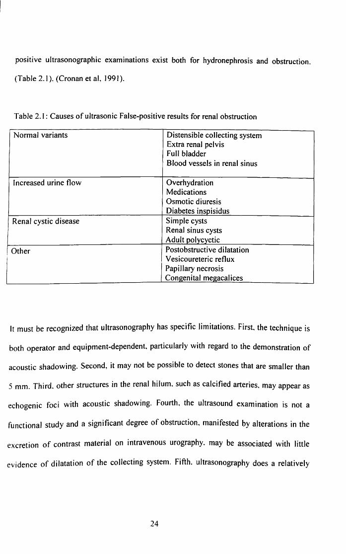
positive ultrasonographic examinations exist both for hydronephrosis and obstruction.
(Table 2.1 ). (Cronan et al, 1991 ).
Table 2.1: Causes of ultrasonic False-positive results for renal obstruction
Normal variants Distensible collecting system Extra renal pelvis FuJI bladder Blood vessels in renal sinus
Increased urine flow Overhydration Medications Osmotic diuresis Diabetes insoisidus
Renal cystic disease Simple cysts Renal sinus cysts Adult oolvcyctic
Other Postobstructive diJatation Vesicoureteric reflux Papillary necrosis Congenital megacalices
It must be recognized that ultrasonography has specific limitations. First. the technique is
both operator and equipment-dependent, particularly with regard to the demonstration of
acoustic shadowing. Second, it may not be possible to detect stones that are smaJJer than
5 mm. Third. other structures in the renal hilum. such as calcified arteries. may appear as
echogenic foci with acoustic shadowing. Fourth, the ultrasound examination is not a
functional study and a significant degree of obstruction, manifested by alterations in the
excretion of contrast material on intravenous urography. may be associated with little
evidence of dilatation of the collecting system. Fifth. ultrasonography does a relatively
24