comparative study of conventional staining …. vaisakhi, et al.pdf · strip of nitrocellulose acts...
TRANSCRIPT

Int.J.Curr.Microbiol.App.Sci (2017) 6(8): 395-409
395
Original Research Article https://doi.org/10.20546/ijcmas.2017.608.053
Comparative Study of Conventional Staining Techniques, Quantitative Buffy
Coat, Immunochromatography Methods and Molecular Methods
(Gene Amplification) for Diagnosis of Malaria
K.S. Vaisakhi1*
, Sujatha Kolla2 and Achut Rao
2
1Dr. V. R. K. Medical College, STAR Hospitals, Hyderabad, Telangana, India
2RVMIMS Medical College, Hyderabad, Telangana, India
*Corresponding author
A B S T R A C T
Introduction
Malaria is said to be the oldest disease in the
world, as from Egypt, Indonesia, and China.
Malaria means bad air. It is the one of the
most important parasitic disease of humans
affecting hundred and three endemic countries
with a population over 2.5 billion people and
causing between 1 and 3 million deaths every
year worldwide. There has been a resurgence
of the disease in many parts of the tropics.
In addition, there is continuing spread of drug
resistant malaria especially of Plasmodium
falciparum and Plasmodium vivax throughout
South Asia, Western Pacific Central and
South America. Due to this magnitude of drug
resistance in India, the incidence of
complicated forms of malaria increased.
There is a compelling reason to justify the
implementation of a rapid malaria diagnostic
test in field. Diagnosis must therefore be
immediate in order to provide proper
treatment and prevent complications. Many
rapid diagnostic methods have been
introduced by the laboratories which would
help in early diagnosis and thereby decrease
the morbidity in endemic countries.
The WHO33, 34, 35
has initiated a project term
as Role Back Malaria (RBM) to coordinate
global actions. In view of the above criteria,
International Journal of Current Microbiology and Applied Sciences ISSN: 2319-7706 Volume 6 Number 8 (2017) pp. 395-409 Journal homepage: http://www.ijcmas.com
The study showed that the immunochromatography assay tests were reliable and
easy to use. Blood samples were collected from 175 patients presenting with
typical features suggesting clinical malaria. These blood samples were subjected
to various test which included thin and thick smears stained with JSB, Giemsa,
Leishman, Fields stain and the other methods employed which included Arcidine
orange staining, Quantitative Buffy Coat methods, immunochromatography strips
– optimal and para HIT – f. Assay are used. Gene amplification technique
(Polymerase chain reaction) was used. However, RDTS are more suited for
investigator / health workers in situations where health services are different (or)
absent. RDT’s in conjunction with microscopy should improve diagnosis of
malaria, thus likely to contribute greatly to an effective control of malaria in
resource poor countries.
K e y w o r d s
Microscopy,
Quantitative buffy
coat, Immuno-
chromatography,
PCR.
Accepted:
04 June 2017
Available Online: 10 August 2017
Article Info

Int.J.Curr.Microbiol.App.Sci (2017) 6(8): 395-409
396
the present study titled “Comparative Study
of Conventional Staining Techniques,
Quantitative Buffy Coat,
immunochromatography Methods for
Diagnosis of Malaria” was carried out in the
Department of Microbiology at S. R. R. I. T.
C. D. (Sir Ronald Ross Institute of
Communicable Diseases), Nallakunta, under
Osmania Medical College, Hyderabad.
Aims of the study are to compare the results
of rapid diagnostic method with the
conventional microscopy as it is the gold
standard for diagnosis of malaria and to
determine the sensitivity and specificity of
various diagnostic methods and judge them
and their usage in diagnosis and early
treatment of the cases to prevent
complications.
Materials and Methods
A total of 200 cases of fever were diagnosed
by peripheral smear examination and blood
samples were collected from patients
attending S. R. R. I. T. C. D (Sir Ronald Ross
Institute of Communicable Diseases),
Nallakunta, under Osmania Medical College,
Hyderabad.
Smear
Scratch free slides, lancet, Microscope, Cedar
wood oil, Reagents for staining (Jaswant
Singh Bhattacharjee (JSB), Leishman stain,
Geimsa stain, Field’s stain. The procedure is
from Chaterjee K.D5, Cheesbrough
7 with the
techniques reference. Jaswant Singh
Bhattacharjee (JSB): In-house the stain has
been prepared following the preparation
protocol.
OptiMAL rapid malaria tests
Immunochromatography is based on the
capture of parasite antigen from peripheral
blood using monoclonal antibodies prepared
against malarial antigen target and conjugated
to either liposome containing selenium dye or
gold particle in a mobile phase. 2nd
and 3rd
capture monoclonal antibody applied to a
strip of nitrocellulose acts as an immobile
phase. The labelled antigen is to be captured
by the monoclonal antibody of the immobile
phase, producing coloured line.
The key enzyme regulating the energy
metabolism of malarial parasite produce by
sexual and asexual stages and is
immunologically and structural different from
host LDH. Only live parasites produce pLDH.
Sample material
Capillary blood collected by veni puncture.
Fresh blood collected by veni puncture.
Sample tubes containing anti-coagulant
(EDTA) heparin.
Kit contents: Dip sticks
Conjugate wells
Wash well holder
Buffer
Lancets
Disinfectant wads
These should be store at 2 -8°C.
Test procedure
The kit should be allowed to reach the room
temperature before use.
A dipstick from the container was taken and
patients’ identification was written on the
label. (Hold always the dipstick by the label,
do not touch the reaction field).

Int.J.Curr.Microbiol.App.Sci (2017) 6(8): 395-409
397
One conjugate was placed and washed well in
the holder.
One drop of buffer (approximately 20ml) was
dispensed into the conjugate well and 4 drops
(approximately 80ml) into the wash well.
Allowed to stand for 1 minute.
Aseptically the skin surface of the finger tip
was cleaned with the disinfectant load, let to
dry, the lateral part of the finger tip was
pricked with the lancet. The load and the
sterile lancet were discarded into a suitable
waste container.
While gently squeezing the tube, the open end
was immersed in the blood drop and then
gently the pressure was released to draw
blood into the pipette. When using whole
blood, blood from the collection tube was
drawn into the pipette.
One drop of blood (approximately 10µl) was
added into the conjugate well. Mixed gently
with the stirrer or the same pipette (upper
end). Allowed to stand for 1minute, (the
pipette was discarded into a suitable waste
container). The approximate dipstick was
placed vertically into the conjugate well and
let it in the well for 10 minutes (the blood
migrates towards the filter pad and the control
band will appear progressively).
The dipstick was transferred from the wash
well and the reactions were read.
The used dipstick was kept for future
reference and post treatment monitoring.
ParaHIT-f
The malaria antigen test contains a membrane
strip which is precoated with one monoclonal
antibody as two separate lines across a test
strip.
The monoclonal antibody (test line) is
specific to the histidine rich protein of P.
falciparum and another line which acts as
control has goat anti-mouse antibody.
Kit contains: Malaria antigen test kit
Test strip.
Assay buffer.
Specimen collection and storage
Finger prick (venous blood) using lancet.
Whole blood into collection tube (Containing
EDTA, citrate or heparin) by venipuncture.
Specimen not done immediately – refrigerated
at 2-8 degrees Celsius, not more than 3 days,
used within 3days.
Test procedure
Dispense 5ml of whole blood on the white
sample pad allow blood to absorb into the
sample pad for 60 seconds.
Dispense 300ul of assay buffer into a test
tube.
Allow it to for 10 minutes.
Read the test strip after the blood colour has
cleared 10 minutes.
Positive – presence of two colour bands
indicates positive result for P. falciparum and
control band.
Negative if no control band appears.
Invalid if only P. falciparum band appears.
Polymerase Chain Reaction: Gene
amplification
PCR involves denaturation of DNA to from
single DNA followed by annealing to allow
binding of complementary strands of DNA

Int.J.Curr.Microbiol.App.Sci (2017) 6(8): 395-409
398
fragments to 5 and 3 primers and finally
primer extension catalysed by thermostable
taq polymerase.
Each 1 parasite in 1 ml can be detected with
this test. Highly sensitive and specific,
requires equipment and trained personnel.
Venous blood is collected with EDTA and
stored at 4 degrees Celsius until test done.
Material: Extraction of DNA from infected
RBC’s
Infected erythrocytes
PBS (phosphate buffered saline).
5% saponin solution.
Lysis buffer (40mu tris HCL, 80mu EDTA,
2% SDS, 0. 1mg/ml, proteinase K).
The proteinase K was added just before using
the buffer.
Phenol equilibrated with 0. 1uTris HCL.
Chloroform (removal of phenol).
RNase (RNase-it is a ribonuclease cock tail).
3u sodium acetate.
Absolute ethanol (helps in precipitation of
DNA)
70% ethanol (wash DNA (remove of salt)).
TE Buffer (10mu Tris-HCL; 1mu EDTA).
Primer from MGW company was taken. The
primers for PCR procedure were chosen such
that 5 primer was plasmodium conserved
while the 3 primer were species specify.
Pf = A1 A1 (ATCAGCTTTTGATGTTAG
GGTATT) A1 291 (GCTTATATTTGTATC
TTTGAGC)
Pv = A1 A1 (ATCAGCTTTTGATGTTAGG
GTATT) A1 292 (TTCGCTTTTCATACTG
T)
Procedure
Centrifuge the infected RBC at 3000xg for 2
minutes each cells once in cold PBS.
Resuspended cells from and microfuge tube
(1. 7ml) in 1ml of PBS.
10ul of 5% of saponin (for a final conc. 0.
05%) was added and gently mixed.
Immediately centrifuge at 6000xg for 5
minutes. The supernatant was removed to the
25ul of lysis buffer and 75ul of distilled
water.
Incubated at 37 degrees Celsius for ~ 3 hours
with intermittent stirring by hand.
100ul of distilled H2O, then 200ul of phenol
was added. Mixed well and centrifuged at
12,000 rpm for 8 minutes.
Extracted with phenol and chloroform as
above.
The genomic DNA was precipitated by
adding 1/10 volume of sodium acetate and 2.
Five volume of absolute ethanol for a couple
of hours or overnight at -20 degree Celsius.
The DNA can be stored this way as well.
The precipitate was centrifuged for 30
minutes at 4 degrees Celsius, wash gently
once with 70% ethanol, dry in speed-vac and
gently resuspend the pellet in 25-100ul of
distilled water, depending on its size.
Int.J.Curr.Microbiol.App.Sci (2017) 6(8): 395-409
399
The DNA concentration was determined at
OD 260, 2ml of culture may yield ~2ug of
genomic DNA runs as a high molecule
weight, somewhat broad, band and is this
unsheared and free of DNA.
PCR is set up with conditions.
Hot start - 95-1
Denaturation, Annealing, Extension, Further
extenstion.
Agarose gel was set with 0. 8%.
4 gms of agar powder + 1 x TAE (Tris
Acetate EDTA) in 50 mi.
The mixture was placed in oven or heated for
2 minutes and set it in a tray with well stand
and it will set in ½ hr. Take out well stand and
the DNA ladder was loaded in the 1st well and
samples in other wells and the gel was run.
Results and Discussion
A total of 200 patients presenting with history
of fever attending malaria clinic at Sir Ronald
Ross Institute of Tropical and Communicable
disease, Hyderabad were studied.
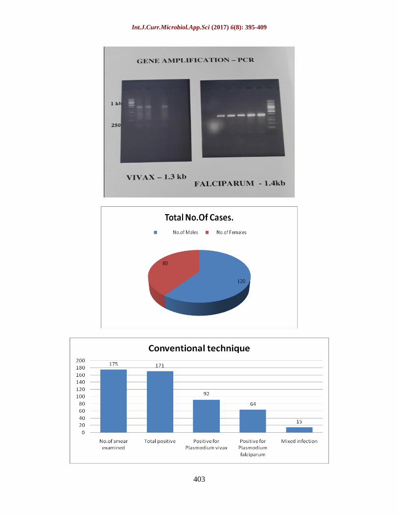
Among the patients there were 120 males, 80
females maximum number of cases were
falling in the age group ranging from 16yrs to
60 yrs with mean age 24 to 48yrs (Tables 2).
Blood samples were collected from 175
patients presenting with typical features
regarding clinical malaria were subjected to
various tests which included thin and thick
smears stained with Jaswant Singh
Bhattacharji, Giemsa, Leishman, Field’s stain
other methods employed for comparative
study includes Acridine orange staining,
Quantitative buffy coat method.
Immunochromatography strips -OptiMAL
and paraHIT –f assay are used. Gene
amplification technique (Polymerase chain
reaction) used (only 49 samples including I
sample a negative control).
Of 200 samples taken from study who have
been attending malaria clinic at S. R. R. I. T.
C. D. 175 samples have been diagnosed
clinically positive for malaria.
Demonstration of Malaria parasite in
peripheral blood smears stained by JSB
(Jaswant Singh Bhattacharjee) after thorough
microscopic examination, was taken as the
gold standard for definitive diagnosis of
malaria
Conventional technique
Park 24
of the 175 clinical positive cases
171(97. 7%) of samples have been diagnosed
by microscopy.
Out of which 92(53. 8%) were found to be
Plasmodium vivax 57(34. 7%) were found to
be Plasmodium falciparum and 15(8. 7%)
were mixed infection (both Pv and Pf were
identified) the sensitivity 97.7% specificity
92% positive and negative predictive values
are 98.8% and 85% respectively.
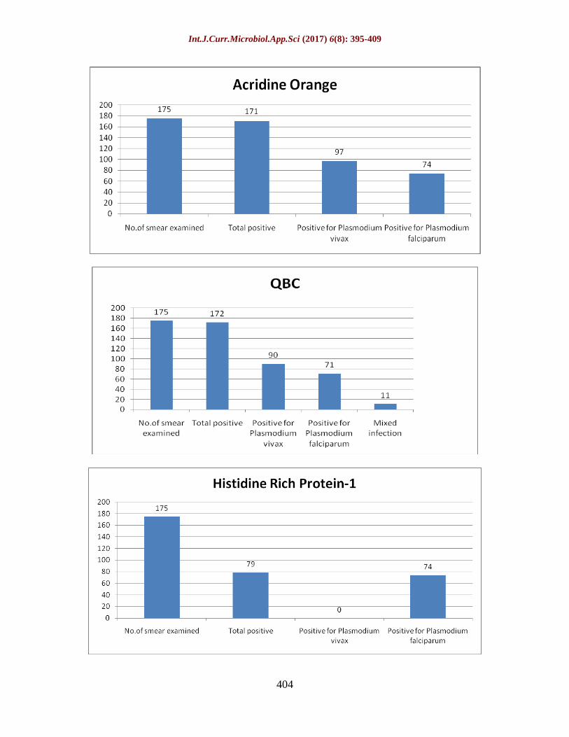
Acridine orange staining
Staining the blood smears with fluorescence
dyes is a widely used procedure and has been
recognised as a rapid and sensitive method.
This technique has been found to be as
sensitive as thick film preparation. This
method is good for rapid screening.
Of 171(97. 7%) positive smears, 97(56. 7)
were found to be Plasmodium vivax and 74
(43. 2%) were found to be Plasmodium
falciparum. By this method it is difficult to
segregate the mixed infection. The sensitivity
aggregated the mixed infection. The

Int.J.Curr.Microbiol.App.Sci (2017) 6(8): 395-409
400
sensitivity 97.7% and specificity 88% and
positive and negative predictive values are
98.2% and 84.6% respectively.
Quantitative Buffy Coat fluorescing method is
the more technically demanding and requires
specialised equipment to separate the cell
layers by centrifugation and good fluorescent
microscopy. The results are as following:
Out of 175 clinically positive cases, 172 (93.
2%) positive by QBC method, 90(54. 5%) of
which were found to be Plasmodium
falciparum and 11(6. 4%) were found to be
mixed infection (both Pv and Pf). The
sensitivity was 98. 2% and specificity 84%
and positive, negative predictive values are
97.7% and 87.5% respectively.
Immunochromatography assay
Histidine rich protein 2 detection kit
It was the first antigen to be used to develop a
rapid diagnostic test kit. The commercial kit
used to detect this antigen is paraHIT –f
(Span diagnostic Ltd).
Humar et al.,10,
and Pieroni et al., investigated
on ParaSIGHT f test specificity and
sensitivity.
Wongarichanalai et al.,30
, Shiff et al.,
Karbwang et al., assessed ICT and studied the
sensitivity and specificity of HRP-2 Kit.
74 (93.6%) samples were found to be positive
for Plasmodium falciparum. A total of 79
(45.1%) smear were confirmed for mixed
infection along Plasmodium falciparum. The
sensitivity and specificity are 93.6% and 92%
respectively. The positive predictive value is
97. 3% and negative predictive value is 82%.
OptiMAL (Parasite lactate dehydrogenase
detection kit)
This test can be of clinical relevance.
Different isomers of pLDH for each of four
plasmodium species infecting humans exist
and their detection constitutes a 2nd
approach
for rapid diagnostic test (RDT) development.
The commercially available kit optiMAL
(Flow Inc. Portland oreg) was used in this
study.
John et al.,13
and Hunt-Cooke et al.,11
confirmed the sensitivity and specificity of
optiMAL.
Palmer et al.,16, 23
, Fryauff et al.,6, Mills et
al.,18
, Jelinek et al.,12
, Tjitra et al., Eisen and
Saul worked on the sensitivity and specificity
of ICT test.
OptiMAL has showed 17.3 (98.8%) positive
cases of the clinically positive 175 cases. It
could identify a few cases which could not be
identified by microscopy due to low
parasitemia. 75 (43.3%) were Plasmodium
vivax, 98 (56.8%) were Plasmodium
falciparum. Mixed infection could not be
differentiated because the pan specific band
consists of isomers of pLDH of all the four
species. The sensitivity, specificity, positive,
negative and predictive values are 98.8%,
92%, 98.8% and 92% respectively.
Table.1 Field’s stain contain 2 solutions Solution - A Solution - B
Methylene blue – 0. 8 gms Eosin (yellow eosin) - 1gm.
Azure I (Azure B) – 0. 5 gms Disodium hydrogen phosphate – 5gms.
Disodium hydrogen phosphate – 5gms. Potassium dihydrogen phosphate – 6. 25gms
Potassium dihydrogen phosphate – 6.
25gms.
Distilled water 500ml.
Distilled water 500ml

Int.J.Curr.Microbiol.App.Sci (2017) 6(8): 395-409
401
Table.2 Total number of cases and gender variation Total cases No. of Males No. of Females
200 120 80
Table.3 Smear examination with JSB Method No. of smear
examined
Positive for
Plasmodium
vivax
Positive for
Plasmodium
falciparum
Mixed infection Total Positive
JSB 175 92(53. 8%) 64(37. 4%) 15(8. 7%) 171(97. 7%)
Table.4 Smear examination with Acridine Orange Method No. of smear examined Positive for
Plasmodium vivax
Positive for
Plasmodium
falciparum
Total positive
Acridine Orange 175 97(56. 7%) 74(43. 5%) 171(97. 7%)
Table.5 Smear examination with QBC Method No. of smear
examined
Positive for
Plasmodium vivax
Positive for
Plasmodium
falciparum
Mixed
infection
Total positive
QBC 175 90(54. 5%) 71(41. 2%) 11(6. 4%) 172(98. 2%)
Table.6 Smear examination with HRP-2 Method No. of smear
examined
Positive for
Plasmodium vivax
Positive for
Plasmodium
falciparum
Total positive
HRP-2 175 - 74(93. 6%) 79(49. 1%)
Table.7 Smear examination with OptiMAL Method No. of smear
examined
Positive for
Plasmodium vivax
Positive for
Plasmodium
falciparum
Total
positive
OptiMAL 175 75(43. 3%) 98(56. 6%) 173(98. 8%)
Table.8 Smear examination with PCR Method No. of smear
examined
Positive for
Plasmodium vivax
Positive for Plasmodium
falciparum
Total positive
PCR 175 87. 5(50%) 86. 5(49. 42%) 174(99. 42%)
Table.9 Comparative study of different detection methods Method Sensitivity Specificity Positive predictive Negative predictive
Microscopy 97. 7% 92% 98. 8% 85%
Acridine orange 97. 7% 88% 98. 2% 84. 6%
QBC 98. 2% 84% 97. 7% 87. 5%
ParaHIT-f 93. 6% 92% 97. 3% 82%
OptiMAL(pLDH) 98. 8% 92% 98. 8% 92%
PCR 98% 100% 99. 9% 96. 1%

Int.J.Curr.Microbiol.App.Sci (2017) 6(8): 395-409
402
Table.10 Studied by Sensitivity Specificity Predictive positive Values negative.
Gay F7
93. 8% 99. 8% 99. 3% 98. 3%
David J. K Purnomo
et al.,4
99. 6% 81. 7% - -
Oloo et al.,21
98% 84% 97% 98%
Wang et al.,32
87. 2% 95% - -
Gaye O et al.,8
100% 83. 6% 93. 4% 100%
Nandwani S et al.,20
97. 5% 100% - -
Present study 98. 2% 84% 97. 7% 87. 5%
Table.11 Studied by Sensitivity Specificity Predictive
positive
Values
negative.
Tarimo DS et al.,29
94. 1% 100% 100% 94%
Present study 97. 7% 88% 98. 2% 84. 6%
Table.12 Studied by Sensitivity Specificity Predictive
positive
Values negative.
Humar et al.,10
100% 98% 95. 4% 100%
N. Singh et al.,28
93% 92. 5% - -
Valecha N et al.,27, 28
98. 5% 97. 1% - -
Palmer CJ et al.,16, 23
94% 88% 88% 99%
Pinto MJW et al.,25, 26
100% 84. 5% - -
Jelinck H et al.,12
92. 5% 98. 5% - -
Wongchai S et al.,30
97. 2% 96. 3% 77. 8% 99. 6%
Guthman JD et al., 9
97% 88% - -
Pinto MJ et al.,25, 26
88% 100% - -
Present study 93. 6% 92% 97. 3% 8. 2%
Table.13 Studied by Sensitivity Specificity Predictive
positive
Values
negative.
C J Palmer et al.,16, 23
94% 88% 88% 99%
Mills CD et al.,18
88 – 90 % 96 – 97 % - -
Jelinck H et al.,12
88. 5% 99. 4%
Mason DD et al.,17
86. 2% 76. 9%
Valecha N et al.,27, 28
61. 8% 100% 100% 71. 8%
C J Palmer et al., 16, 23
98% 100% 100% 99%
Present study 98. 8% 92% 98. 8% 92%
Table.14 Comparison of PCR studies for P. falciparum Studied by Sensitivity Specificity Predictive
positive
Values
negative
Gaye O et al.,8
100% 72. 2% 89. 4% 100%
Trisophon W et al.,31
92% 100% - -
S Nandwani et al.,20
96. 8% 100% - -
Present study 98% 100% 99. 9% 96. 1%
Int.J.Curr.Microbiol.App.Sci (2017) 6(8): 395-409
403
Int.J.Curr.Microbiol.App.Sci (2017) 6(8): 395-409
404

Int.J.Curr.Microbiol.App.Sci (2017) 6(8): 395-409
405
Int.J.Curr.Microbiol.App.Sci (2017) 6(8): 395-409
406
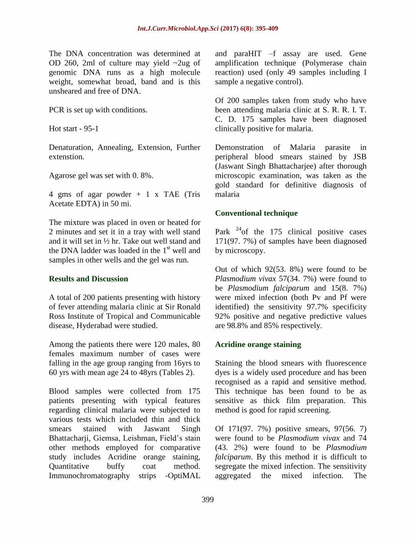
Polymerase chain reaction
In this study, 175 clinically positive cases
samples which are smear positive were
subjected to PCR analysis, one known
negative control was included.
Srinivasan et al.,19
studied that PCR detected
DNA from blood of the patients who were
suffering from malarial infection.
Quitana et al., studied the comparison of
microscopy and PCR. Tagger and Jensen
cultivate the parasite in vivo in the medium.
Of the 175 samples subjected to PCR analysis
87.5(50%) were Plasmodium vivax and 86.
5(49.4%) were Plasmodium falciparum. The
sensitivity was 98% specificity 100% and
positive, negative predictive values are 99.9%
and 96.1% respectively.
On comparison of the various methods used
in the study, the following results have been
observed.
Since the negative predictive value of a
specific indicator of the efficacy test. It was
found that pLDH optiMAL test showed the
highest negative predictive value indicating
that the test is most reliable in diagnosing this
disease. The resurgence of malaria has
renewed interest in developing not only
preventive measures, but also rapid diagnostic
techniques. Several methods have been
developed to supplement and replace the
conventional microscopic method of
diagnosis. The most promising new malarial
diagnostics are the immunochromatographic
assays for detecting HRP -2 and pLDH,
Quantitative Buffy Coat, Acridine orange and
Polymerase chain reaction. Microscopic
examination of blood films is accepted as
current universal gold standard for diagnosis
of malaria.
The blood film is still the only widely
available result against which the newer
methods for diagnosis of malaria can be
compared, even though microscopy continues
to be method of primary choice for
enumeration of parasites in blood film.
In this study the sensitivity, specificity,
positive predictive value, negative predictive
values of microscopy are 97.7%, 92%, 98.
8%, and 85% of which is taken us standard
comparable findings.
All the results of the present study are
compared with the conventional method.
Moody et al.,19
studied about the possibility
of replacing microscopy with ICT kits.
Comparison of quantitative buffy coat
results
Comparison of results of acridine orange
staining
In my study, both Quantitative Buffy Coat
and in house prepared Acridine Orange
staining were comparable and their sensitivity
and specificity, positive negative predictive
values were in concurrence.
The acridine orange staining technique was
useful for rapid screening but speciation was
difficult in few cases and mixed infections
could not be differentiated. The limitation of
the Acridine orange method is usage of
fluorescent microscope or paralens with UV
attachment.
Comparison of Histidine rich protein -2
The limitation of this method is as Histidine
rich protein -2 is produced only by the
Plasmodium falciparum only P. f can be
detected.

Int.J.Curr.Microbiol.App.Sci (2017) 6(8): 395-409
407
Comparison of parasite lactate
dehydrogenase enzyme
This test is more of clinical importance as this
test detects pLDH from the live parasites
only. The results of various studies mentioned
above concurrence with results of present
study. This kit could diagnose a few cases
which are not detected due to low parasitemia
by microscopy.
In this study only 175 cases have been
studied, out of which 1 negative sample was
taken as control. PCR has been claimed to the
theoretically capable of detecting the presence
of less than 5- 10 parasites in microlitre of
blood. This technique is labour intensive and
requires high level expertise and
standardisation. PCR analysis was many time
costlier, varying techniques used for
extraction of DNA and source of printer.
Hence PCR is now being used mostly for
research purposes is very few centres.
Malaria being an endemic diseases in India
and emerging disease in other countries in
response, the WHO launched the Roll Back
Malaria initiative, which has emphasized on
early detection of malaria, in which kit
methods have been given much importance
and which comparable to conventional
techniques.
Blood samples were collected from 175
patients presenting with typical features
suggesting clinical malaria. The study showed
that the immunochromatography assay tests
were reliable and easy to use. Thus, this RDT
is an appropriate test for the use in the field
by paramedical staff when laboratory
facilities are not available are thus likely to
contribute greatly to an effective control of
malaria in resource poor countries.
RDT’s in conjunction with microscopy should
improve diagnosis of malaria. However,
RDTS are more suited for investigator / health
workers in situations where health services
are different (or) absent.
In conclusion, this study investigated the use
of non – microscopic, rapid optiMAL test is
the best kit method. The performance of the
test was adequate and the results obtained
were correlated well with those obtained by
microscopy.
The advantages of optiMAL test when
compared with other methods are:
Availability of rapid results (10-15 mins).
Relative, simplicity compared with
microscopy.
Can be used by relatively in experienced
persons.
It is of clinical relevance.
Therefore, it is reasonable to consider future
use of RDT’s as an epidemiological too for
the rapid screening of malaria. Where
possible if microscopy and the RDT’s can
both be used together, the chance of missing
the diagnosis of malaria will be remote.
Hence it is suggested the RDT’s also may be
supplied to malaria clinics and their use is
encouraged.
Molecular techniques are sensitive, accurate
and specific but expensive and require expert
knowledge hence their practical application is
restricted in our country.
References
1. Chatterjee K. D. Text book of
parasitology 1987, 12th
edition 71-100.
2. Cheesebrough; District laboratory
practices in tropical countries 239-258.
Int.J.Curr.Microbiol.App.Sci (2017) 6(8): 395-409
408
3. Craig MH, Sharp B. L., Comparative
evaluation of four techniques for the
diagnosis of P. falciparum infections.
Tran R Soc. Trop. Med. Hyg. 1997; 91:
279-82.
4. David J. K. Purnomo and T. R. Jones,
1992 Diagnosis of malaria in the field
by fluorescence microscopy of QBC,
Capillary tubes. Trans. R. Soc. Trop.
Med. Hyg., 86: 35.
5. Cooke A. H., et al., 1992 use of the
flouruchrome benzothiocarboxypurine
in malaria diagnosis is an endemic area.
Trans. R Soc. Trop. Med. Hyg., 87: 549.
6. Fryauff D. Et al., 2000. Performance of
optimal assay for detection and
identification of malaria infection is
asymptomatic resident of Irian. Ann. J.
Trop. med. Hyg. 03: 139 – 145.
7. Gay F et al.,: Sante 1994; Evaluation of
the QBC system for the diagnosis of
malaria.
8. Gaye O et al., Parasite 1999; A
comparsion of thick smear, QBC, PCR
and path falciparum malaria test strip in
P. falciparium diagnosis.
9. Guthmann JP et al., Frans R, Soc. Trop.
Med. Hyg. 2002. Validity, reliability
and ease of use in the field of five rapid
test for the diagnosis of P. falciparum
malaria in Uganda.
10. Humar A, et al., 1997: Parasight
falciparium test compared with the
polymerase chain reaction and
microscopy for the diagnosis of
Plasmodium falciparum malaria in
travellers. Am. J. Trop. Med. Hyg 56: 47
– 48.
11. Hunt. Cooke, A:et al., Comparison of a
parasite lactate dehydrogenase – based
ICT antigen detection assay (optiMAL)
with microscopy for the detection of
malaria parasites in human samples.
Ann. J. Trop. Med. Hyg., 60: 20 -23.
12. Jelinek T, 2001. Evaluation of a dipstick
test for the rapid diagnosis of imported
malaria among patients presenting
within the network. Trop Net Europe.
Scand J. Infect Dis, 33: 752–754,
13. John’s et al., 1998. Evaluation of
optiMAL, a dipstick test for the
diagnosis of malaria. Ann. Trop. Med.
Parasitol., 92: 621 -622.
14. Srinivasan B. V et al., 2003.
Comparison between conventional and
QBG methods for diagnosis of malaria.
Indian J. Pathol Microbiol.,
15. Lee MA et al., 1999. A comparison of
antigen dipstick assays with polymerase
chain reaction (PCR) technique and
blood film examination in the rapid
diagnosis of malaria. Ann, Acad. Med.,
16. Makler, M. J., C. J. Palmer, A. L. Ager
1992. A review of practical techniques
for the diagnosis of malaria. Ann. Trop.
Med. Parasitol., 92: 419-433.
17. Mason DP et al., 2002, A comparison of
two rapid field immunochromatogenic
tests to expert microscopy in the
diagnosis of malaria. Acta Trop.
18. Mills CD et al., 1999 Evaluation of a
rapid and inexpensive dipstick assay for
the diagnosis of Plasmodium falciparum
malaria. Bull World Health Organ,
19. Moody A et al., Performance of the
optimal malaria antigen capture dipstick
for malaria diagnosis and treatment
monitoring at the hospital for tropical
diseases, London. British J. Haematol
2000; 109: 891-894.
20. Nandwani S, Mathur M, Rawat S.
Evaluation of the polymerase chain
reaction analysis for diagnosis of
falciparum malaria in Delhi, India.
ISMM., 2005; 23; 176-178.
21. Oloo AJ et al., 1994: Evaluation of
QBC method to detect malaria
infections in field surveys. East Afr.
Med. J.
22. Palmer C. L., et al., Evaluation of the
optimal test for rapid diagnosis of
Plasmodium vivax and Plasmodium
Int.J.Curr.Microbiol.App.Sci (2017) 6(8): 395-409
409
falciparum malaria. J. Clin. Microbial
1998; 38: 203-2006.
23. Palmer CJ. 2003; Multicentre study of
evaluate the optimal test for rapid
diagnosis of malaria in US Hospitals. J.
Clin. Microbial.
24. Park K, Text book of preventive and
social medicine, 18th
edition, Jabalpur
m/s Banarasidas Bhanot, 1997; Pp. 188-
201.
25. Pinto MJ et al., 1999. Rapid diagnosis
of falciparum malaria by detection of
Plasmodium falciparum HRP – 2
antigens. J. Assoc. Physician India.
26. Pinto MJW et al., Rapid diagnosis of
falciparum malaria by detection of
Plasmodium falciparum HRP- 2 Ag,
JAPI 1999; 47(11): 1076-1078.
27. Sharma VP, Valecha N, Diagnosis of
Malaria – Fam Med India, 1997; 1: 11-
5.
28. Singh N, Valecha N, Sharma V. P.,
Malaria diagnosis by field workers
using an immunochromatographic test.
Trans R. Soc. Trop. Hyg. 1997; 91: 396-
397.
29. Tarimo DS, 2001. Malaria diagnosis
and treatment under the stratergy of the
integrated management of childhood
illness relevance of lab support from the
rapid immunochromatographic tests of
ICT Malaria P. falciparum and
Plasmodium vivax and optiMAL. Ann.
Trop Med parasitol,
30. Thepsamarn P, Prayoolawongsa N,
Puksupa P, Puttoom P, Wongchai S, et
al., –the ICT Malaria at the Thia –
Myanmar border. South East Trop med
Public Health 1997; 28: 723-6.
31. Triasophon w, Raj Kulchai P, et al., A
highly sensitive, rapid and simple
polymerase chain reaction –based
method to detect human malaria (P.
falciparum and vivax) in blood samples.
Am. J. Trop. Med. Hyg. 1993; 87: 647-
8.
32. Wang X et al., Bull World Health
Organization 1996. Field evaluation of
the QBC technique for rapid diagnosis
of Vivax malaria.
33. WHO (1986). The Clinical
Management of Acute Malaria.
SEARO, SEA. Sr. NO. 9. New Delhi.
34. World Health Organization basic
malaria microscopy Geneva; 1991 part
IP. 17-68.
35. World Health Organization, 1999, New
perspectives, Malaria diagnosis report
of a joint WHO/USAID informal
consultation, October 25 – 27, 1999
Geneva WHO/MAL/2000/1091.
How to cite this article:
Vaisakhi, K.S., Sujatha Kolla and Achut Rao. 2017. Comparative Study of Conventional
Staining Techniques, Quantitative Buffy Coat, Immunochromatography Methods and
Molecular Methods (Gene Amplification) for Diagnosis of Malaria.
Int.J.Curr.Microbiol.App.Sci. 6(8): 395-409. doi: https://doi.org/10.20546/ijcmas.2017.608.053