comparative study of the effects of fetal bovine serum ... · serum versus horse serum on growth...
TRANSCRIPT

Franke et al. BMC Veterinary Research 2014, 10:119http://www.biomedcentral.com/1746-6148/10/119
METHODOLOGY ARTICLE Open Access
Comparative study of the effects of fetal bovineserum versus horse serum on growth anddifferentiation of primary equine bronchialfibroblastsJana Franke1, Vanessa Abs1, Claudia Zizzadoro2 and Getu Abraham1*
Abstract
Background: Airway fibroblasts have become a critical addition to all facets of structural lung tissue changes suchas in human asthma and chronic obstructive pulmonary disease, but little is known about their role in the equinerecurrent airway obstruction, a disease that resembles to the human asthma. Since the equine bronchial fibroblasts(EBF) have not been isolated and characterized yet, the use of defined medium was investigated.
Results: Primary EBF were cultured on non-collagen coated flasks without serum or in the presence of fetal bovineserum (FBS) or horse serum (HS) or in serum depleted medium. EBF cultured in serum-free basal media and thoseserum deprived were not able to proliferate and even exhibited considerable cell death. In media containing FBS orHS, proliferation of the cells was reproducible between different primary cultures and cells demonstrated expressionof vimentin. Large variations were found in the ability of FBS and HS to support growth and differentiation of EBFin monolayer culture. Indications of growth-promoting actions, increasing passage number as well as maintainingfibroblast morphology were found rather in FBS than in HS. EBF culturing in HS needed longer doubling andconfluence time. The protein content of the cell pellets was higher in EBF cultured in medium containing HS thanFBS. Alpha-smooth muscle actin seemed to be less expressed in EBF cultured in medium containing FBS than thosein HS.
Conclusions: In sum, serum addition to basal EBF medium enhanced EBF differentiation into myofibroblasts, andthese findings are useful to develop in vitro fibroblast culture models that mimic in vivo physiological processesand to study airway disease mechanisms and remodeling.
Keywords: Airways, Primary bronchial fibroblasts, Cell culture, In vitro, Serum types
BackgroundChronic airway diseases like human bronchial asthma andchronic obstructive pulmonary disease (COPD) and theequine recurrent airway obstruction (RAO) principallycharacterized by bronchial hyperreactivity and airflow ob-struction result from several factors including structuralalterations of the airway wall and cell function. Such air-way remodelling is a common feature of abnormal depos-ition of extracellular matrix (ECM) components in airway
* Correspondence: [email protected] of Pharmacology, Pharmacy and Toxicology, University of Leipzig,An den Tierkliniken 15, Leipzig 04103, GermanyFull list of author information is available at the end of the article
© 2014 Franke et al.; licensee BioMed CentralCommons Attribution License (http://creativecreproduction in any medium, provided the or
mesenchymal layer associated with airway wall thickness[1-3]. Airway fibroblasts and those cells differentiated intomyofibroblasts contribute to sub-epithelial fibrosis linkedto airway remodelling by producing ECM proteins such ascollagen, fibronectin and proteoglycans [4,5].There are several well characterized human fetal lung
fibroblast cell lines that are cultured in defined media andfor use as in vitro cell model to study airway diseases andremodeling, for example, HFL-1 [6], IMR-90 [7], HEL299,MCR-5 [8], WI-38 [9] and GM 06114 [10]. Culture ofprimary bronchial fibroblasts exists for human [11,12],mice [13] and rats [14]. However, there is currently neither
Ltd. This is an Open Access article distributed under the terms of the Creativeommons.org/licenses/by/2.0), which permits unrestricted use, distribution, andiginal work is properly credited.

Franke et al. BMC Veterinary Research 2014, 10:119 Page 2 of 9http://www.biomedcentral.com/1746-6148/10/119
description of equine adult primary airway fibroblast cul-tures nor there are such cell lines for this species.Not all cells have the same requirements for growth and
survival. With this regard, for successful growth, mainten-ance and expression of differentiated metabolic functionsof human or animal cells in vitro, either primary culturesor continuous cell lines, appropriate culture conditions arerequired that mimic the physiological conditions in vivoand situ. In fact, it is well known that serum represents afundamental source of nutrients, cytokines and adhesivemolecules necessary for in vitro cell growth, metabolismand to stimulate proliferation [15]. In serum-free medium,only the addition of growth factors could initiate mouselung fibroblast proliferation [16]. On the other hand, pro-liferating primary cardiac and dermal fibroblasts as well asestablished cell lines have been synchronized into a non-dividing G0-phase to cell cycle by serum withdrawal [17]which favour fibrotic processes or cause cell death [18,19].While significant advances are made to culture conditionsof permanent cell lines, current research is lacking thatcompares cultures of primary airway fibroblasts in differ-ent serum origins added to a standard culture medium orserum depleted medium, and how this may affect their dif-ferentiation. The sera that most widely used are bovineorigin (adult or new born or fetal origin); as well the horseserum has been seen as an alternative to provide growthfactors and hormones in modern cell biology. Indeed, al-most it is not known about the responses of primary air-way fibroblast cultures to horse serum factors.Aim of the present study was to develop a primary bron-
chial fibroblast culture technique and investigate the influ-ence of horse serum in comparison to FBS on cell viability,morphology and immunocytochemical characteristics, cellproliferation and α-smooth muscle actin expression (α-sma) to further understand the mechanism of peribron-chial fibrosis (airway remodelling) in the equine RAO.
ResultsEffect of serum absence and serum withdrawal onprimary EBF culturePrimary EBF cultured without serum presence in basicDMEM failed to attach and to proliferate and those at-tached, disappeared completely from the flask surfaceswithin 1-week of culture. Also, we tested whether conflu-ent EBF cultured in DMEM can be affected by serumwithdrawal. Serum withdrawal led to modification of EBFnumber in culture; they exhibited detachment within threedays of serum starvation (data not shown). Even theaddition of 10% FBS after 24 h did not significantly en-hance EBF attachment and proliferation.
Cell viability and morphology under FBS and HS influenceCell yield from digested bronchial tissue was consistent.Under both culture conditions, EBF were stained with
trypan blue and the percentage of viable cells was similar,usually > 95%. No significant evidence of cell necrosis orcell apoptosis was observed under inverted light micro-scopic analysis. With regard to cell morphology, EBF cul-tured in DMEM with 10% FBS appeared to be typicallyflattened and spindle-shaped (with a homogenous cyto-plasm) over several weeks of passages compared to EBFcultured in DMEM with 10% HS (Figure 1A). In mediumcontaining FBS, EBF were grown over the growth surfacewith only loose cell-cell-contact until reaching confluenceand then formed tight parallel lines which remained as typ-ical fibroblastic monolayer until passage 15. EBF betweenpassages 16 – 20 started to change their morphology: cellswere large, flat and more polygonal shaped, with a large,heterogeneous nucleus. At the same time, cell growth wasrapidly reduced and cell viability was diminished. In con-trast, EBF cultured in the presence of HS showed alteredmorphological changes within 2 days of culture; cells weresmall and more compact in shape combined with granula-like dark structures in the cytoplasm (Figure 1C). More-over, cells grew in clusters and chains and failed to reachconfluence within a week. This morphological behaviourof EBF was seen under this culture conditions until pas-sage 7, but thereafter, EBF failed to proliferate regularlyand decreased in their viability (passage 9). Under bothculture conditions, most EBF (>99%) were positive forvimentin, but with more characteristic filamentous struc-tures within the cytoplasm in medium containing FBS(Figure 1B) than in the presence of HS (Figure 1D).
Sera effects on cell population, proliferation and total proteinNumber of passages and proliferation differed significantlybetween the two culture conditions. By routinely passa-ging primary EBF (once a week), EBF maintained theirtypical morphological fibroblastic properties up to passage20 in culture medium containing fetal bovine serum,whereas EBF in horse serum did exhibit significantly lim-ited proliferation rate merely up to passage 9 (Figure 2A).Under both experimental conditions, growth curves ofEBF showed similar initial 24 h lag-phase. The populationdata began to exhibit significant differences between thetwo conditions until day 4 of EBF culture (Figure 2B). Celldoubling time was slow in the presence of horse serum,and here, it took significantly longer than in culturemedium containing FBS (Figure 2C). Under culture condi-tions in the presence of FBS, primary EBF reached conflu-ence on day 4 but not in the presence of HS. Indeed,significant differences between serum groups were notfound on day 6 and 8. Total protein content of the cellmonolayers did not reveal significant difference betweenthe two experimental conditions until day five (Figure 2D),while after day six the amount of total protein increasedunder HS condition and statistically differed from FBScondition, though cell number was the vice versa.
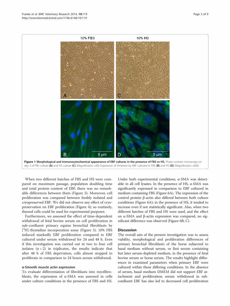
Figure 1 Morphological and immunocytochemical appearance of EBF cultures in the presence of FBS or HS. Phase contrast microscopy onday 3 of FBS culture (A) and HS culture (C) (Magnification: x40); Expression of Vimentin by EBF cultured in FBS (B) and HS (D) (Magnification: x400).
Franke et al. BMC Veterinary Research 2014, 10:119 Page 3 of 9http://www.biomedcentral.com/1746-6148/10/119
When two different batches of FBS and HS were com-pared on maximum passage, population doubling timeand total protein content of EBF, there was no remark-able differences between them (Figure 3). Moreover, cellproliferation was compared between freshly isolated andcryopreserved EBF. We did not observe any effect of cryo-preservation on EBF proliferation (Figure 4); so routinely,thawed cells could be used for experimental purposes.Furthermore, we assessed the effect of time-dependent
withdrawal of fetal bovine serum on cell proliferation insub-confluent primary equine bronchial fibroblasts by[3H]-thymidine incorporation assay (Figure 5). 10% FBSinduced markedly EBF proliferation compared to EBFcultured under serum withdrawal for 24 and 48 h. Evenif this investigation was carried out in two to four cellisolates (n = 2) in triplicates, the results indicate thatafter 48 h of FBS deprivation, cells almost stopped toproliferate in comparison to 24 hours serum withdrawal.
α-Smooth muscle actin expressionTo evaluate differentiation of fibroblasts into myofibro-blasts, the expression of α-SMA was assessed in cellsunder culture conditions in the presence of FBS and HS.
Under both experimental conditions, α-SMA was detect-able in all cell lysates. In the presence of HS, α-SMA wassignificantly expressed in comparison to EBF cultured inmedium containing FBS (Figure 6A). The expression of thecontrol protein β-actin also differed between both cultureconditions (Figure 6A); in the presence of HS, it tended toincrease even if not statistically significant. Also, when twodifferent batches of FBS and HS were used, and the effecton α-SMA and β-actin expression was compared, no sig-nificant difference was observed (Figure 6B, C).
DiscussionThe overall aim of the present investigation was to assessviability, morphological and proliferation differences ofprimary bronchial fibroblasts of the horse subjected tobasal medium without serum, or first serum containingbut later serum-depleted medium, in the presence of fetalbovine serum or horse serum. The results highlight differ-ences in examined parameters when primary EBF werecultured within these differing conditions. In the absenceof serum, basal medium DMEM did not support EBF at-tachment and proliferation; serum withdrawal in sub-confluent EBF has also led to decreased cell proliferation

Figure 2 Effect of FBS and HS on cell growth, doubling time, protein content and passage number. EBF were passaged every week andseeded in a density of 2 × 10⁴ cells/cm2. EBF cultured in 10% FBS keep proliferating over a longer period than in 10% HS (A). EBF were seededin 6-well-plates at a density of 2 × 10⁴ cells/cm2. Effect of HS and FBS on growth curve (B), population doubling takes longer in HS than in FBS(calculated between day 1 and day 3) (C) and effect of HS and FBS on total protein amount over 8 days (D). Data are presented as means ± SEM.(*p < 0.05, **p < 0.01, n = 10).
Franke et al. BMC Veterinary Research 2014, 10:119 Page 4 of 9http://www.biomedcentral.com/1746-6148/10/119
and attachment. The most important factors in signifi-cantly affecting cell growth and differentiation were thesera types added to basal DMEM. EBF cultures wereestablished from freshly isolated EBF (P0) or from frozenisolated EBF. Despite cryopreservation, the proliferationdata generated from frozen cells were similar to thosefrom freshly solated and further cultured EBF.After short-term trypsin digestion of peeled and minced
equine bronchial mucosa and from cultures of epithelialcell mixture in DMEM containing FBS, we could observea complete disappearance of epithelial cells within 1-weekand instead five days later a complete coverage of cultureflasks with viable primary equine bronchial fibroblasts.Thus, DMEM does not support epithelial cell growth des-pite the presence of serum. This isolation procedure is de-scribed, to our knowledge, the first time for these cells andresulted in reproducible large cell yields with typical mes-enchymal cell properties. EBF cultured in serum-freeDMEM were, however, not only unable to proliferate butalso were non-adherent after seven days in culture. From
this result, indeed, it is difficult to point out the proteinswhich are present in serum and absent in the basalmedium that are essential for primary EBF growth. Thereason why EBF disappeared seems to be, even if we didnot proof, the occurrence of apoptosis and necrosis. Thus,it can be argued that under serum-free conditions in ourcultures EBF-specific growth factors which are usuallypresent in serum are missing. But in many established per-manent cell line culture under serum-free conditions[20,21] the serum-free environment did support growthand differentiation with clonal growth as well as mass cul-ture (for review see [22]).Although recent efforts have shown that human mes-
enchymal stem cells and primary epithelial cells can beisolated and expanded long-term in serum-free medium[23-26], no published work had shown whether humanor animal primary airway fibroblasts are able to expandunder serum-free conditions for long-term in culture.The selective nature of serum-free media formulationscould be used in primary cultures of tissue explants by

Figure 3 Effect of FBS and HS batches on EBF passage number, population doubling and total protein content. Under standard cultureconditions as described in material and methods, cells were subjected to obtain (A) maximum passage number (n = 2), (B) population doubling(n = 5) and (C) total protein amount (n = 2). F1 and F2 represent FBS batches, and H1 and H2 the HS batches.
Franke et al. BMC Veterinary Research 2014, 10:119 Page 5 of 9http://www.biomedcentral.com/1746-6148/10/119
allowing growth suppression of other cell types whichmight quantitatively influence experiments with primarycells obtained from tumor tissues. Presumably, in ourcase, the serum-free condition should be optimized forenhanced primary fibroblast expansion by adding growthfactors which have to be further investigated. In sum,understanding of these processes is essential in under-standing the use of these cells as in vitro model and tostudy their role in airway remodelling during respiratorydisease mechanisms.The importance of serum supplementation for cell at-
tachment, growth and passaging of in vitro proliferatingcells [27] as well as the influence of serum deprivationon permanent cell line proliferation and cycle [28] are
Figure 4 Effect of cryopreservation on EBF passage number and popsubjected to obtain (A) maximum passage number (n = 3) and (B) populattreated as described under Figure 2.
well known. On the other hand, no data are available onthe effect of time–dependent serum withdrawal on pri-mary airway fibroblast proliferation and differentiation.Since serum components might affect cell stimulation,many studies in vitro are conducted in the absence ofserum, and cells can be deprived of serum for 1 – 8 days[29,30], and thus, the aim of the present study was to in-vestigate the response to serum withdrawal for 24 to48 hours in sub-confluent primary equine bronchial fibro-blasts which were first cultured in DMEM containing 10%FBS. In our study, we have clearly demonstrated a markedtime-dependent decrease in [3H]-thymidine incorporationin cells deprived of serum for 24 to 48 hours, suggestingdiscontinuation of proliferation in the absence of FBS.
ulation doubling. Under standard culture conditions, cells wereion doubling (n = 2). Thawed cells were used after 2 passages and

Figure 5 Effect of serum-deprivation on [3H]-thymidineincorporation. Sub-confluent EBF were subjected to serumdeprivation for 24 and 48 hours. Cell proliferation was determinedby [3H]-thymidine incorporation (n = 2).
Franke et al. BMC Veterinary Research 2014, 10:119 Page 6 of 9http://www.biomedcentral.com/1746-6148/10/119
After 72 hours of serum depletion many cells were floatingin the medium and the number of attached cells was de-creasing, indicating altering cell viability and morphology,in agreement with data found for cardiac fibroblasts [18].These findings show that EBF in primary cultures are notable to adapt to serum deprivation for a given time, pre-sumably sufficient serum growth factors are not available.Moreover, we have tested the effects of sera types on
proliferation, morphology, population doubling and via-bility of primary equine bronchial fibroblasts. The results
Figure 6 Sera effects on β-actin and α-SMA expression. Western blotsDMEM containing 10% FBS or HS. Densitometry of β-actin and α-SMA wasexpression than EBF cultured in FBS (n = 5). Serum batch of (B) FBS and (Cbatches, and H1 and H2 the HS batches. Data are presented as means ± SE
highlight striking differences in cell proliferation, morph-ology, passaging time and number as well as total proteinamount and α-SMA expression when EBF were culturedwithin these differing conditions. Both FBS and HS arenatural products, presumably, with varying concentrationsof growth factors within different batches; however, the ef-fects measured in our study were not dependent on batch/lot number. EBF grown in DMEM containing 10% FBScontinue to proliferate even after reaching confluence,whereas these same cells, when cultured in DMEM con-taining 10% HS, had relatively limited proliferation rate,longer population doubling time and somehow quite dif-ferent morphological features. Cells in HS were smaller,chain-forming and more compact in shape with dark cyto-plasmic granules which can be related to the occurrenceof abnormal protein accumulation than those in FBS, andthey failed to reach confluence. In concordance, sera typesaffect other various cell types in a similar way: bovine adi-pocytes and sheep skeletal muscle satellite cells proli-ferated rapidly, when FBS was supplemented to growthmedium instead of HS; thus, FBS-containing medium isoften used as growth medium in these cultures [31,32].On the contrary, equine chondrocytes could equally pro-liferate under both conditions [33]. Even if they are equinecells, it is noteworthy to find that EBF exhibit a preferencefor fetal bovine serum factors over the horse serum fac-tors, whereas the vice versa has been seen for neural cells
were performed on whole cell lysate from EBF cultured for 4 days incalculated by SynGene. (A) EBF cultured in HS show a higher α-SMA) HS (n = 2) did not affect α-SMA expression. F1 and F2 represent FBSM. (*p < 0.01).

Franke et al. BMC Veterinary Research 2014, 10:119 Page 7 of 9http://www.biomedcentral.com/1746-6148/10/119
[34]. Merely, early studies have proposed that horse serumis appropriate for neuronal cell cultures [34,35].Moreover, despite the short passaging time and limited
proliferation and population doubling rate, increased α-SMA expression was accompanied by higher proteinamount in the presence of horse serum than in bovineserum, suggesting that HS might stimulate fibroblast dif-ferentiation into myofibroblasts. It is well known thatmyofibroblasts are able to produce large amount of extra-cellular matrix (ECM) proteins, as well as growth factorsand cytokines and express cytoplasmic contractile struc-tures including α-SMA. Thus, it seems that the horseserum increases the protein content in EBF by increasingsynthesis of structural proteins, ECM and cytokines. In-deed, it is not yet known if differential expression of α-SMA and protein is functionally relevant.
ConclusionsFetal bovine serum favors fibroblastic morphology withenhanced proliferation rate, population doubling, passagenumber and triggers cell differentiation (as vimentin stain-ing showed), suggesting serum factors essential for theequine airway fibroblasts are available in the fetal bovineserum, whereas in the horse serum cells there were signsof degeneration or cell granularity. Moreover, we can con-clude that serum withdrawal for 48 hours rather decreaseEBF adaptation and enhance cell detachment than the24 hour FBS depletion and in the latter case cell viabilitywas though not altered, thus, EBF cultured in 10% FBSrepresent a good model allowing studying the response todrugs that influence cell proliferation and pathways of air-way remodeling in airway diseases.Study limitation: Animal serum is a complex mixture of
a large number and variety of components; therefore, it isdifficult to assess, at this stage, the significance of the obser-vation that horse serum inhibits the programmed progressof equine airway cells through the lineage of differentiationin cultures.
MethodsIsolation and culture of equine bronchial fibroblastsPrimary equine bronchial fibroblasts were cultured inDulbecco’s Modified Eagle Medium (DMEM) supple-mented with 200 Units/ml Penicillin, 0.2 mg/ml Strepto-mycin, 5 mg/ml Amphotericin B (PAA LaboratoriesGmbH, Pasching, Austria). Surfaces of culture flask werenot pretreated with adhesion supporting matrix like collagen.Bronchial segment tissue samples were obtained from
adult non-diseased slaughter horses of different breed,age and sex and slaughtered at local abattoirs (Freiberg,Germany). Primary equine bronchial fibroblasts (EBF) wereisolated from these tissues. Briefly, the bronchial mucosawas removed from the bronchi, washed, minced to about1-3 mm pieces, and 500 mg tissue were digested with
0.25% trypsin/EDTA (Sigma Aldrich, Deisehnhofen,Germany) in Hanks’ balanced salt solution (HBSS)(PAA Laboratories GmbH, Pasching, Austria) in 50-mlErlenmeyer glass flasks and incubated at 37°C for2 hours in humidified atmosphere of 5% CO2. Trypsi-nized samples were then filtered through sterile double-layered gauze and rinsed twice with ice-cold HBSS. Cellsuspension was then further sieved through sterilenylon cell strainers (mesh size: 40 μm) (BD Biosciences,Franklin Lakes, NJ) and centrifuged two times to re-move tissue debris.To evaluate the effect of sera on proliferation and differ-
entiation of primary equine bronchial fibroblasts, we usedat least two to three different lots of cell culture appropriatenot inactivated fetal bovine serum (FBS) (Gibco, Carlsbad,CA, USA) and horse serum (HS) (Sigma Aldrich,Deisehnhofen, Germany). Since it was not the objective ofthe study, we did not compare different serum products ofseveral companies. FBS is routinely used to culture othercell lines in our laboratory, and should be tested beforeuse; thus, after cell isolation, final cell pellets were first re-suspended and cultured in DMEM containing 10% fetalbovine serum (FBS) for at least up to two passages. There-after, EBF were trypsinized and sub-cultured under differ-ent conditions: a) in DMEM without serum, or b) first inDMEM containing FBS and then serum deprived, or c) inDMEM in the presence of 10% FBS or d) in DMEM in thepresence of 10% horse serum (HS). Cells were routinelypassaged every 7 days with replacement of medium every2 to 3 days. Unless and otherwise specified; cells wereroutinely cultured in 75 cm2 flasks (Greiner Bio-One,Frickenhausen, Germany).
Cryopreservation and thawing of EBFCertain density of isolated primary EBF were either pro-vided for direct culturing or frozen in 1 ml DMEM con-taining 20% FBS, 10% DMSO (Sigma Aldrich, Deisenhofen,Germany), penicillin, streptomycin and amphotericin B inliquid nitrogen until use. Also, some passages of EBF cul-tures were frozen. Cells were quickly thawed at 37°C inwater bath and subsequently transferred into culture flaskswith tempered culture medium (see above). Thawed cellswere used for our study after 2 passages.
Cell viability, morphology and immunostainingFor cell counting and viability testing, cells were trypsi-nized and washed with PBS and stained with trypan bluedye (Sigma-Aldrich, Deisenhofen, Germany). Cells exclud-ing the dye were counted using Neubauer cell chamber.Also, after 1-week of EBF culture in two conditions, i.e.,DMEM+ FBS or DMEM+HS, fibroblasts were trypsi-nized, centrifuged and an aliquot was re-suspended in PBSwith trypan blue.

Franke et al. BMC Veterinary Research 2014, 10:119 Page 8 of 9http://www.biomedcentral.com/1746-6148/10/119
EBF were plated into 75 cm2 flasks at a density of 2 ×10⁴ cells/cm2 and grown in 10 ml defined DMEM in thepresence of 10% FBS or HS. Cell morphology was evalu-ated under inverted light microscope (Olympus CKX41).Digital images were taken from cells at different daysand at possible confluence.Expression of intermediate filaments in primary cultured
EBF was evaluated using immunofluorescence stainingagainst vimentin using a primary mouse monoclonal anti-body and a secondary anti-mouse FITC-antibody (DakoDeutschland GmbH, Hamburg, Germany). EBF were cul-tured under the indicated culture conditions on glass coverslips (Carl Roth GmbH, Karlsruhe, Germany), washedtwice with PBS after medium removal, and fixed in ice-cold acetone for 5 min at -20°C and immunostained aspreviously described [36].
Cell proliferation assay and population doubling timeTo further assess the effects of sera types on cell prolifera-tion and population doubling, EBF between passage 3 and6 were transferred into 6-well plates (2 × 10⁶ cells/cm2)and cell numbers were determined manually after day 1, 2,3, 4, 6 and 8. Each time, the average from 2 wells wastaken. The mean cell number was logarithmically trans-formed and the linear regression slope was calculated toderive the doubling time (DT). Proliferation rate was alsodetermined in cryopreserved, thawed and cultured cells.
[3H]-thymidine assayFurthermore, cell proliferation was measured by [3H]-thymidine incorporation assay [11]. EBF (40 000 – 60 000cells/well) were seeded into 12-well plates, in DMEM inthe presence of 10% FBS, grown until 60% confluence, andfurther cultured in the presence of 10% FBS or serum-withdrawn for 24 or 48 hours. In all three settings, [3H]-thymidine (37 kBq/well) (Perkin Elmer, Waltham, MA)was added to the culture medium. 24 hours after incuba-tion of cells with [3H]-thymidine, cells were washed in ice-cold PBS and incorporated radioactivity was determinedby liquid scintillation counting (Beckman LS 6500 Scintil-lation Counter). Also, in thawed and cultured EBF, prolif-eration experiments were performed about 1-week afterthawing, to avoid a major increase in the number of cu-mulative population doublings compared to experimentsdone with fresh cells.
Cell growth determinationTo measure the effect of sera on cellular growth, totalprotein was measured at defined time points during cul-ture in 6-well plates by the colorimetric method [37]. Inbrief, EBF were rinsed twice in PBS and then trypsinized,centrifuged at 500 × g for 10 min. After removal of thesupernatant, cell pellets were resuspended in PBS andsonicated 4 rounds for 30 second. Crude cell lysates
were then diluted in 0.1 M potassium phosphate buffer(pH 7.4), incubated with copper in alkaline solution for10 minutes at room temperature. After addition of Folin(Merck, Darmstadt, Germany), reduction of copper wascompleted within 45 minutes and quantitative analysiscould be carried out by spectrophotometry at 660 nm(Beckman DU640 spectrophotometer, Beckman Coulter,Krefeld, Germany).
Western blotPrimary EBF grown in 6-well plates under different cultureconditions were harvested and suspended in sample buffer(containing 2% SDS, 25% [v/v] glycerol, 60 mM Tris–HCland 0.1% bromphenol blue, 14.4 mM β-mercaptoethanol,pH 6.8) and boiled for 5 min. Equal amounts of whole celllysate (2,5 × 10⁵ cells/ml) were subjected to SDS-PAGE;samples were separated on 12% acrylamide gel underreducing conditions and transferred to a nitrocellulosemembrane (Whatman GmbH, Dassel, Germany). Themembrane was then blocked in 3% BSA (PAA Laborator-ies) with TBST (20 mM Tris–HCl, pH 7.5, 150 mM NaCland 0.05% [v/v] Tween-20) for 1 h at room temperature.α-SMA was detected following an overnight incubation ofsamples with mouse monoclonal antibody against humananti-α-SMA (1:1000; Sigma-Aldrich) and β-actin was de-tected using mouse monoclonal antibody against humananti-β-actin (1:10,000; Sigma-Aldrich) in 3% BSA-TBST at4°C. Band visualisation was performed using alkaline phos-phatase conjugated secondary anti-mouse igG antibody(1:5000; Promega GmbH, Mannheim, Germany) over 1 hat room temperature. Enzyme activity was detected usingWestern Blue stabilised substrate for alkaline phosphate(Promega). The membranes were digitalized and quanti-tated by densitometry analysis (SynGene, Cambridge, UK).
Data and statistical analysisAll data are expressed as means ± SEM. Statistical signifi-cance of differences was evaluated by paired two-tailed stu-dent’s t-test using GraphPad Prism version 5.1 (GraphPadSoftware, San Diego, CA, USA). P < 0.05 was consideredsignificant.
Competing interestsAll authors declare that no Competing interests exist.
Authors’ contributionsJF, VA, CZ and GA designed the research and wrote the paper. JF and VAperformed the experiment and analyzed the data. JF analyzed the data andwrote the paper. GA analyzed the data, wrote and approved the paper. Allauthors read and approved the final manuscript.
AcknowledgmentsThis work was supported financially by Frankenförder ForschungsgesellschaftmbH (Berlin, Germany). We thank the abattoirs of Freiberg and Jena foraccess to fresh bronchial tissue samples. We also thank Martina Wieczorek fortechnical assistance.

Franke et al. BMC Veterinary Research 2014, 10:119 Page 9 of 9http://www.biomedcentral.com/1746-6148/10/119
Author details1Institute of Pharmacology, Pharmacy and Toxicology, University of Leipzig,An den Tierkliniken 15, Leipzig 04103, Germany. 2Division of VeterinaryPharmacology and Toxicology, Department of Veterinary Public Health,Faculty of Veterinary Medicine, University of Bari, Strada Prov.le perCasamassima, km 3, Valenzano, BA 70010, Italy.
Received: 31 August 2013 Accepted: 20 May 2014Published: 26 May 2014
References1. Jeffery PK: Remodeling in asthma and chronic obstructive lung disease.
Am J Respir Crit Care Med 2001, 164:S28–S38.2. An SS, Bai TR, Bates JH, Black JL, Brown RH, Brusasco V, Chitano P, Deng L,
Dowell M, Eidelman DH, Fabry B, Fairbank NJ, Ford LE, Fredberg JJ,Gerthoffer WT, Gilbert SH, Gosens R, Gunst SJ, Halayko AJ, Ingram RH, IrvinCG, James AL, Janssen LJ, King GG, Knight DA, Lauzon AM, Lakser OJ,Ludwig MS, Lutchen KR, Maksym GN, et al: Airway smooth muscledynamics: a common pathway of airway obstruction in asthma. EurRespir J 2007, 29:834–860.
3. Yamauchi K, Inoue H: Airway remodeling in asthma and irreversibleairflow limitation-ECM deposition in airway and possible therapy forremodeling. Allergol Int 2007, 56:321–329.
4. Fernandes DJ, Bonacci JV, Stewart AG: Extracellular matrix, integrins, andmesenchymal cell function in the airways. Curr Drug Targets 2006,7:567–577.
5. Brewster CE, Howarth PH, Djukanovic R, Wilson J, Holgate ST, Roche WR:Myofibroblasts and subepithelial fibrosis in bronchial asthma. Am J RespirCell Mol Biol 1990, 3:507–511.
6. Ichikawa T, Sugiura H, Koarai A, Yanagisawa S, Kanda M, Hayata A, FurukawaK, Akamatsu K, Hirano T, Nakanishi M, Matsunaga K, Minakata Y, Ichinose M:Peroxynitrite augments fibroblast-mediated tissue remodeling viamyofibroblast differentiation. Am J Physiol Lung Cell Mol Physiol 2008,295:L800–L808.
7. Kolodsick JE, Peters-Golden M, Larios J, Toews GB, Thannickal VJ, Moore BB:Prostaglandin E2 inhibits fibroblast to myofibroblast transition via E.prostanoid receptor 2 signaling and cyclic adenosine monophosphateelevation. Am J Respir Cell Mol Biol 2003, 29:537–544.
8. Haag S, Matthiesen S, Juergens UR, Racké K: Muscarinic receptors mediatestimulation of collagen synthesis in human lung fibroblasts. Eur Respir J2008, 32:555–562.
9. Liu X, Ostrom RS, Insel PA: cAMP-elevating agents and adenylyl cyclaseoverexpression promote an antifibrotic phenotype in pulmonaryfibroblasts. Am Physiol Cell Physiol 2004, 286:C1089–C1099.
10. Silvestri M, Fregonese L, Sabatini F, Dasic G, Rossi GA: Fluticasone andsalmeterol downregulate in vitro, fibroblast proliferation and ICAM-1 orH-CAM expression. Eur Respir J 2001, 18:139–145.
11. Matthiesen S, Bahulayan A, Kempkens S, Haag S, Fuhrmann M, Stichnote C,Juergens UR, Racké K: Muscarinic receptors mediate stimulation of humanlung fibroblast proliferation. Am J Respir Cell Mol Biol 2006, 35:621–627.
12. Jacques E, Semlali A, Boulet LP, Chakir J: AP-1 overexpression impairscorticosteroid inhibition of collagen production by fibroblasts isolatedfrom asthmatic subjects. Am J Physiol Lung Cell Mol Physiol 2010,299:L281–L287.
13. Sugiura H, Liu X, Duan F, Kawasaki S, Togo S, Kamio K, Wang XQ, Mao L,Ahn Y, Ertl RF, Bargar TW, Berro A, Casale TB, Rennard SI: Culturedlung fibroblasts from ovalbumin-challenged "asthmatic" mice differfunctionally from normal. Am J Respir Cell Mol Biol 2007, 37:424–430.
14. Warnken M, Haag S, Matthiesen S, Juergens UR, Racke K: Speciesdifferences in expression pattern of arginase isoenzymes and differentialeffects of arginase inhibition on collagen synthesis in human and ratpulmonary fibroblasts. Naunyn Schmied Arch Pharmacol 2010, 381:297–304.
15. Bettger WJ, McKeehan WL: Mechanisms of cellular nutrition. Physiol Rev1986, 66:1–35.
16. Kumar RK, O'Grady R, Li W, Smith LW, Rhodes GC: Primary culture of adultmouse lung fibroblasts in serum-free medium: responses to growthfactors. Exp Cell Res 1991, 193:398–404.
17. Iyer VR, Eisen MB, Ross DT, Schuler G, Moore T, Lee JC, Trent JM, Staudt LM,Hudson J, Boguski MS, Lashkari D, Shalon D, Botstein D, Brown PO: Thetranscriptional program in the response of human fibroblasts to serum.Science 1999, 283:83–87.
18. Leicht M, Marx G, Karbach D, Gekle M, Köhler T, Zimmer HG: Mechanism ofcell death of rat cardiac fibroblasts induced by serum depletion. Mol CellBiochem 2003, 251:119–126.
19. Simm A, Bertsch G, Frank H, Zimmermann U, Hoppe J: Cell death ofAKR-2B fibroblasts after serum removal: a process between apoptosisand necrosis. J Cell Sci 1997, 110:819–828.
20. Minotti S, Scicchitano BM, Nervi C, Scarpa S, Lucarelli M, Molinaro M, AdamoS: Vasopressin and insulin-like growth factors synergistically inducemyogenesis in serum-free medium. Cell Growth Differ 1998, 9:155–163.
21. Goto S, Miyazaki K, Funabiki T, Yasumitsu H: Serum-free culture conditionsfor analysis of secretory proteinases during myogenic differentiation ofmouse C2C12 myoblasts. Anal Biochem 1999, 272:135–142.
22. Barnes D, Sato G: Serum-free cell culture: a unifying approach. Cell 1980,22:649–655.
23. Agata H, Watanabe N, Ishii Y, Kubo N, Ohshima S, Yamazaki M, Tojo A,Kagami H: Feasibility and efficacy of bone tissue engineering usinghuman bone marrow stromal cells cultivated in serum-free conditions.Biochem Biophys Res Commun 2009, 382:353–358.
24. Chase LG, Lakshmipathy U, Solchaga LA, Rao MS, Vemuri MC: A novelserum-free medium for the expansion of human mesenchymal stemcells. Stem Cell Res Ther 2010, 1:8.
25. Shibeshi W, Abraham G, Kneuer C, Ellenberger C, Seeger J, Schoon HA,Ungemach FR: Isolation and culture of primary equine tracheal epithelialcells. In Vitro Cell Dev Biol Anim 2008, 44:179–184.
26. Gruenert DC, Finkbeiner WE, Widdicombe JH: Culture and transformationof human airway epithelial cells. Am J Physiol 1995, 268:L347–L360.
27. Honn KV, Singley JA, Chavin W: Fetal bovine serum: a multivariatestandard. Proc Soc Exp Biol Med 1975, 149:344–347.
28. Gos M, Miloszewska J, Swoboda P, Trembacz H, Skierski J, Janik P: Cellularquiescence induced by contact inhibition or serum withdrawal inC3H10T1/2 cells. Cell Prolif 2005, 38:107–116.
29. Hetzel M, Bachem M, Anders D, Trischler G, Faehling M: Different effects ofgrowth factors on proliferation and matrix production of normal andfibrotic human lung fibroblasts. Lung 2005, 183:225–237.
30. Chen M, Huang J, Yang X, Liu B, Zhang W, Huang L, Deng F, Ma J, Bai Y, LuR, Huang B, Gao Q, Zhuo Y, Ge J: Serum starvation induced cell cyclesynchronization facilitates human somatic cells reprogramming.PLoS One 2012, 7:e28203.
31. Fernyhough ME, Hausman GJ, Dodson MV: () Progeny fromdedifferentiated bovine adipocytes display protracted adipogenesis.Cells Tissues Organs (Print) 2008, 188:359–372.
32. Wu H, Ren Y, Li S, Wang W, Yuan J, Guo X, Liu D, Cang M: In vitro cultureand induced differentiation of sheep skeletal muscle satellite cells.Cell Biol Int 2012, 36:579–587.
33. Ahmed YA, Tatarczuch L, Pagel CN, Davies HM, Mirams M, Mackie EJ:Hypertrophy and physiological death of equine chondrocytes in vitro.Equine Vet J 2007, 39:546–552.
34. Fedoroff S, Hall C: Effect of horse serum on neural cell differentiation intissue culture. In Vitro 1979, 15:641–648.
35. Moonen G, Nelson PG: Some physiological properties of astrocytes inprimary cultures. In Dynamic Properties of Glial Cells. Edited by SchoffenielsE, Franck G, Hertz L, Towers DB. New York: Pergamon Press; 1978:389–393.
36. Abraham G, Zizzadoro C, Kacza J, Ellenberger C, Abs V, Franke J, Schoon HA,Seeger J, Tesfaigzi Y, Ungemach FR: Growth and differentiation of primaryand passaged equine bronchial epithelial cells under conventional andair-liquid-interface culture conditions. BMC Vet Res 2011, 7:26.
37. Lowry OH, Rosebrough NJ, Farr AL, Randall RJ: Protein measurement withthe Folin phenol reagent. J Biol Chem 1951, 193:265–275.
doi:10.1186/1746-6148-10-119Cite this article as: Franke et al.: Comparative study of the effects offetal bovine serum versus horse serum on growth and differentiation ofprimary equine bronchial fibroblasts. BMC Veterinary Research2014 10:119.