comparison of gene xpert and fluorescent...
TRANSCRIPT
COMPARISON OF GENE XPERT AND FLUORESCENT MICROSCOPY
FOR DIAGNOSIS PULMONARY TUBERCULOSIS IN HUMAN
IMMUNODEFICIENCY VIRUS INFECTED PATIENTS AT MNAZI
MMOJA HOSPITAL, ZANZIBAR
ABDALLA SAID MOHAMED
A DISSERTATION SUBMITTED IN PARTIAL FULFILLMENT OF THE
REQUIREMENTS FOR THE DEGREE OF MASTER OF SCIENCE IN
ENVIRONMETAL STUDIES (ENVIRONMENTAL HEALTH STREAM) OF
THE OPEN UNIVERSITY OF TANZANIA
2016

ii
CERTIFICATION
The undersigned certifies that he has read and hereby recommends for acceptance by
The Open University of Tanzania the dissertation titled “Assessment of Two
Techniques for Diagnosis Pulmonary Tuberculosis in HIV Patient at Mnazi Mmoja
Hospital Zanzibar” in partial fulfillment of the requirements for the degree of Master
of Science in Environmental Studies (Environmental Health Stream) of The Open
University of Tanzania.
……………………..………………
Prof. Emmanuel S.P. Kigadye
(Supervisor)
……………………………………
Date
iii
COPYRIGHT
No part of this Dissertation may be reproduced or transmitted in any form or by any
means electronics or mechanical including photocopying, recording or any
information storage and retrieval system without prior written permission of the
author or The Open University of Tanzania in that behalf.

iv
DECLARATION
I, Abdalla Said Mohamed, do hereby declare that this Dissertation is my own
original work and has not been presented, and will not be presented, to any other
university for a similar or any other degree award.
…………………………
Signature
………………………
Date

v
DEDICATION
This piece of work is dedicated to my beloved wife Asma O. Mwinyi, my two sons
Tirmidhy and Junaid as well as my daughter Rumaisar.

vi
ACKNOWLEGEMENT
I would like to thank Prof. Emmanuel S. P. Kigadye (supervisor) for his
encouragement and advice on how to develop this Dissertation whose experience,
constructive criticism, generous support and encouragement gave me energy,
strength and self-confidence to finish this study. Dr. Saria (course coordinator), for
his recommendation for title setting of this study. Dr. Ahmed and Dr. Ali of Zanzibar
Integrated HIV, TB and Leprosy Program, for their information about HIV and TB
in Zanzibar. All staff of Microbiology department at Mnazi Mmoja Hospital for their
support in this study. All staff of CTC and TB clinic at Mnazi Mmoja Hospital for
their support in specimen collection. I would like to extend my gratitude to Ministry
of Health Zanzibar and Health Research Unity Zanzibar. Also I sincerely express my
gratitude to Dr. Jamala, Director of Mnazi Mmoja Hospital Zanzibar and Miss
Fauzia Ameir, the Laboratory manager at Mnazi Mmoja Hospital Zanzibar. Thankful
to my wife and my children for their patience and love during this study. Lastly but
not least thanks to Mr. Hussein and Mr. Kibwana who worked hard in data analysis
assistance.

vii
ABSTRACT
HIV associated TB is not easy to make diagnosis and is linked with extreme disease
and death. The spread of MDR-TB, together with increasing harmful of HIV
infection and inadequate availability of quick examination instrument have lead to
cause disappointment of global TB control. The intention of the study was to
evaluate Gene Xpert MTB/RIF technique and smear auramine LED FM technique
for detection of pulmonary tuberculosis (PTB) in people living with HIV at Mnazi
Mmoja Hospital Zanzibar. The study was experimental type of design that involved
laboratory analysis of sputum specimens for determination of Mycobacterium
tuberculosis as well as rifampicin resistance for HIV patients. The specimens were
processed in Gene Xpert MTB/RIF and smear auramine LED FM. The sum of 246
patients sputum specimens were analyzed for the existence of Mycobacterium
tuberculosis by means of Gene Xpert MTB/RIF and LED FM techniques. The results
showed that 169 (68.7%) spot samples (sample I) and 169 (68.7%) morning samples
(sample II) were positive for Gene Xpert MTB/RIF technique. Then 112 (45.5%)
spot samples (sample I) and 118 (48.0%) morning samples (sample II) were smear
positive (LED FM). The sensitivity of LED FM in spot samples and morning
samples were 66.3% and 69.8% respectively. Among 169 positive TB cases 2
patients were found with rifampicin resistance. The outcome of this study indicated
that the performance of Gene Xpert MTB/RIF machine is more accuracy than LED
FM in diagnosis of PTB in HIV patients suspected with TB. Gene Xpert MTB/RIF
may be significant as a single quick method for PTB case finding in HIV patients
suspected with active TB.

viii
TABLE OF CONTENTS
CERTIFICATION ..................................................................................................... ii
COPYRIGHT ............................................................................................................ iii
DECLARATION ....................................................................................................... iv
DEDICATION ............................................................................................................ v
ACKNOWLEGEMENT ........................................................................................... vi
ABSTRACT .............................................................................................................. vii
LIST OF TABLES ................................................................................................... xii
LIST OF FIGURES ................................................................................................ xiii
LIST OF APPENDICES ........................................................................................ xiv
LIST OF ABBREVEATIONS ................................................................................ xv
CHAPTER ONE ........................................................................................................ 1
1.1 Background ...................................................................................................... 1
1.2 Statement of Research Problem ....................................................................... 6
1.3 Objectives ......................................................................................................... 7
1.3.1 General Objective ............................................................................................. 7
1.3.2 Specific Objective ............................................................................................ 7
1.4 Hypotheses ....................................................................................................... 8
1.5 Significance of the Study ................................................................................. 8
CHAPTER TWO ....................................................................................................... 9
LITERATURE REVIEW .......................................................................................... 9
2.1 Common Knowledge Concerning Tuberculosis .............................................. 9
ix
2.1.1 Spread of Tuberculosis ..................................................................................... 9
2.2 Prevalence of Mycobacterium tuberculosis ................................................... 10
2.3 Prevalence of MDR TB .................................................................................. 13
2.4 Current Status of TB Diagnosis ..................................................................... 14
2.5 Delays in Diagnosis of TB ............................................................................. 14
2.6 Xpert MTB/RIF Diagnosis ............................................................................. 16
2.7 Smear Auramine FM Diagnosis ..................................................................... 17
2.8 Importance of Accurate and Rapid Diagnosis of TB and Drug Resistant ..... 17
2.9 Environmental Control in TB Diagnosis ........................................................ 18
CHAPTER THREE ................................................................................................. 20
MATERIALS AND METHODS ............................................................................ 20
3.1 Study Design .................................................................................................. 20
3.2 Study Area ...................................................................................................... 20
3.3 Study Population ............................................................................................ 24
3.4 Sample Size .................................................................................................... 24
3.5 Specimen Collection ...................................................................................... 25
3.6 Sample Transport ........................................................................................... 26
3.7 Materials and Equipments .............................................................................. 26
3.7.1 For Gene Xpert Technique ............................................................................. 26
3.7.1.1 Diagnosis of Sputum Samples by GeneXpert MTB/RIF Technique ........... 27
3.7.1.2 Comparison Between GeneXpert MTB/RIF and LED FM Techniques ...... 27
3.7.1.3 Investigate of MDR-TB in the Study Area .................................................. 27
3.7.2 For Auramine Staining Technique ................................................................ 27
3.7.2.1 Diagnosis of Sputum Samples by LED FM Technique ............................... 29

x
3.8 Examination ................................................................................................... 29
3.9 Permission and Ethical Consideration ............................................................ 29
3.10 Quality Control ............................................................................................... 30
3.11 Data Analysis ................................................................................................. 30
3.12 Limitation ....................................................................................................... 31
CHAPTER FOUR .................................................................................................... 32
RESULTS ................................................................................................................. 32
4.1 Characteristics of Study Participants .............................................................. 32
4.2 Evaluation of the Accuracy of Gene Xpert MTB/RIF and LED FM
for Diagnosis of PTB ...................................................................................... 34
4.3 Sensitivity and Specificity of Smear Auramine FM for PTB Diagnosis
in the Study Area ............................................................................................ 35
4.4 Occurrence of MDR-TB in the Study Area ................................................... 37
CHAPTER FIVE ...................................................................................................... 37
DISCUSSION ........................................................................................................... 37
5.1 Characteristics of Study Participants .............................................................. 37
5.2 Evaluation of the Accuracy of Gene Xpert MTB/RIF and LED FM for
Diagnosis of PTB ........................................................................................... 38
5.3 Sensitivity and Specificity of Smear Auramine LED FM
for PTB Diagnosis in the Study Area ............................................................. 40
5.4 Occurrence of MDR-TB in the Study Area ................................................... 42
CHAPTER SIX ........................................................................................................ 43
CONCLUSIONS AND RECOMMENDATIONS ................................................. 43
6.1 Conclusions .................................................................................................... 43

xi
6.2 Recommendation ............................................................................................ 44
REFERENCES ......................................................................................................... 45
APPENDICES .......................................................................................................... 53
xii
LIST OF TABLES
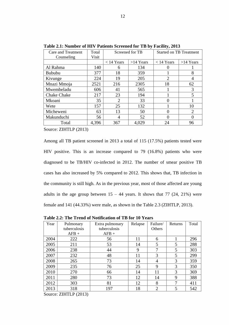
Table 2.1: Number of HIV Patients Screened for TB by Facility, 2013 .................. 12
Table 2.2: The Trend of Notification of TB for 10 Years ........................................ 12
Table 2.3: Distribution of New Sputum Smear Positive (SSP)
Cases by Age and Sex for the Year 2013 ................................................ 13
Table 2.4: Trend of MDR TB in Zanzibar for the Past Six Years ............................. 14
table 2.5: Zanzibar Health Centers which have LED FM ....................................... 14
table 2.6: Positive Results Utilizing LED FM and Xpert MTB/RIF Assay ............ 16
Table 4.1: Patients Age Groups and Sex .................................................................... 32
Table 4.2: Evaluation between Gene Xpert and LED FM in Positivity Results ........ 34
Table 4.3: Smear Auramine LED FM Results ........................................................... 35
Table 4.4: Gene Xpert Results for MTB/RIF Detection ............................................ 37

xiii
LIST OF FIGURES
Figure 2.1: Sputum Samples Processed in Class II Biological Safety Cabinet ....... 19
Figure 3.1: Map of Zanzibar Stone Town showing Location of Mnazi Mmoja
Hospital where Sputum was Collected ................................................. 21
Figure 3.2: Map of Unguja Island showing TB Diagnosis Centres ......................... 23
Figure 3.3: Gene Xpert MTB/RIF Machine ............................................................. 26
Figure 3.4: Fluorescent Microscope ......................................................................... 29
Figure 4.1: Distribution of Patients in Districts of Unguja Island ........................... 33
Figure 4.2: Distribution of Patients in Regions of Unguja Island ........................... 34
Figure 4.3: Distribution of Smear Positivity of Spot Sputum Samples ................... 36
Figure 4.4: Distribution of Smear Positivity of Morning Sputum Samples ............ 36

xiv
LIST OF APPENDICES
Appendix 1: Consent Form ....................................................................................... 53
Appendix 2: SOP for Auramine Staining Fluorescent Microscopy .......................... 56
Appendix 3: SOP for Sample Processing for Diagnosis and Rifampicin
Resistance in Gene Xpert MTB/ Rif ..................................................... 59

xv
LIST OF ABBREVEATIONS
AFB Acid Fast Bacillus
ART Antiretroviral Therapy
BSCs Biological Safety Cabinets
CD4 Clusters of Differentiation
Cfu Colon form unit
CI Confidence Interval
CNS Central Nervous System
CPT Co-trimoxazole Preentive Therapy
CTC Care and Treatment Counseling
DNA Deoxyribonucleic Acid
DST Drug Susceptibility Test
ELISA Enzyme Linked Immunosorbent Assay
EQA External Quality Assurance
EPTB Extra Pulmonary Tuberculosis
FM Fluorescent Microscopy
HEPA High Efficiency Particulate Air
HIV Human Immunodeficiency Virus
IPT Isoniazid Preventive Therapy
LED Light Emitting Diode
MDR-TB Multidrug Resistant Tuberculosis
MMH Mnazi Mmoja Hospital
MTB Mycobacterium tuberculosis

xvi
NAAT Nucleic-acid Amplification Test
OUT Open University of Tanzania
PHCU Primary health care
PTB Pulmonary Tuberculosis
RIF Rifampicin
rt-PCR real time Polymerase Chain Reaction
SPO Standard operating procedure
SPSS Statistical Package for Social Science
SR Sample Treatment Reagent
TB Tuberculosis
USA United States of America
UVGI Ultraviolet germicidal irradiation
WHO World Health Organization
XDR-TB Extensively Drug-Resistant Tuberculosis
Xpert GeneXpert
ZIHTLP Zanzibar Integrated HIV, TB and Leprosy Program
ZN Ziehl Neelsen

1
CHAPTER ONE
INTRODUCTION
1.1 Background
Tuberculosis is still a main public health problem in the world. WHO estimate that
between 2000 – 2020, about one billion people will be newly infected, 200 million
will become sick, as well as 35 million people will die from TB. Currently, HIV
infection is known as the greatest risk factor for development of latent TB infection
to active TB. Co-infection HIV and TB particularly in combination with drug
resistance have caused outbreaks of increasing death rate. Mycobacterium
tuberculosis is passed in airborne particles (droplet nuclei) generated when patients
with pulmonary tuberculosis cough. These particles, 1 – 5 µm in size, are kept
suspended by normal air current. Infection occurs when a susceptible person inhales
the droplet nuclei. Once in the alveoli, the organisms are engulfed by alveolar
macrophages. Normally, the cell mediated immunity of a host act to resist the
multiplication and spread of MTB. However, some MTB can still be viable but
inactive for several years after the early infection. The clinical features of PTB are
cough, weight loss, night sweats, low grade fever, dyspnoea and chest pain (Murray,
2003).
Global prevalence of TB in 2013 was 159 per 100,000 populations (WHO, 2014). In
the year 2013 nine million people who were infected with TB in the world, more
than half (56%) were in the South-East Asia and Western Pacific Regions. One
quarter was from Africa Region, which also had the maximum morbidity and
mortality rates in relation to population. India and China, both accounted for 24%
2
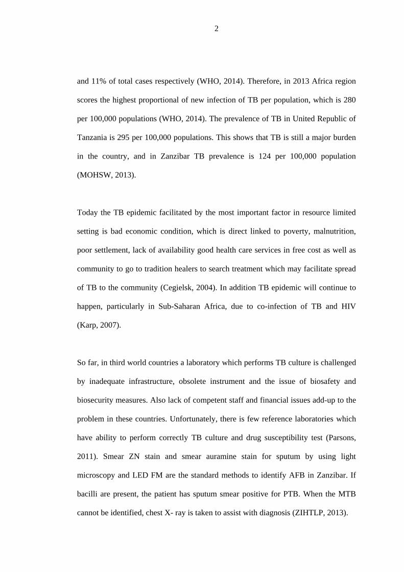
and 11% of total cases respectively (WHO, 2014). Therefore, in 2013 Africa region
scores the highest proportional of new infection of TB per population, which is 280
per 100,000 populations (WHO, 2014). The prevalence of TB in United Republic of
Tanzania is 295 per 100,000 populations. This shows that TB is still a major burden
in the country, and in Zanzibar TB prevalence is 124 per 100,000 population
(MOHSW, 2013).
Today the TB epidemic facilitated by the most important factor in resource limited
setting is bad economic condition, which is direct linked to poverty, malnutrition,
poor settlement, lack of availability good health care services in free cost as well as
community to go to tradition healers to search treatment which may facilitate spread
of TB to the community (Cegielsk, 2004). In addition TB epidemic will continue to
happen, particularly in Sub-Saharan Africa, due to co-infection of TB and HIV
(Karp, 2007).
So far, in third world countries a laboratory which performs TB culture is challenged
by inadequate infrastructure, obsolete instrument and the issue of biosafety and
biosecurity measures. Also lack of competent staff and financial issues add-up to the
problem in these countries. Unfortunately, there is few reference laboratories which
have ability to perform correctly TB culture and drug susceptibility test (Parsons,
2011). Smear ZN stain and smear auramine stain for sputum by using light
microscopy and LED FM are the standard methods to identify AFB in Zanzibar. If
bacilli are present, the patient has sputum smear positive for PTB. When the MTB
cannot be identified, chest X- ray is taken to assist with diagnosis (ZIHTLP, 2013).
3
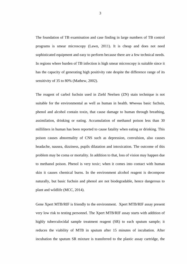
The foundation of TB examination and case finding in large numbers of TB control
programs is smear microscopy (Lawn, 2011). It is cheap and does not need
sophisticated equipment and easy to perform because there are a few technical needs.
In regions where burden of TB infection is high smear microscopy is suitable since it
has the capacity of generating high positivity rate despite the difference range of its
sensitivity of 35 to 80% (Mathew, 2002).
The reagent of carbol fuchsin used in Ziehl Neelsen (ZN) stain technique is not
suitable for the environmental as well as human in health. Whereas basic fuchsin,
phenol and alcohol contain toxin, that cause damage to human through breathing,
assimilation, drinking or eating. Accumulation of methanol poison less than 30
milliliters in human has been reported to cause fatality when eating or drinking. This
poison causes abnormality of CNS such as depression, convulsion, also causes
headache, nausea, dizziness, pupils dilatation and intoxication. The outcome of this
problem may be coma or mortality. In addition to that, loss of vision may happen due
to methanol poison. Phenol is very toxic; when it comes into contact with human
skin it causes chemical burns. In the environment alcohol reagent is decompose
naturally, but basic fuchsin and phenol are not biodegradable, hence dangerous to
plant and wildlife (MCC, 2014).
Gene Xpert MTB/RIF is friendly to the environment. Xpert MTB/RIF assay present
very low risk to testing personnel. The Xpert MTB/RIF assay starts with addition of
highly tuberculocidal sample treatment reagent (SR) to each sputum sample; it
reduces the viability of MTB in sputum after 15 minutes of incubation. After
incubation the sputum SR mixture is transferred to the plastic assay cartridge, the

4
cartridge lid is closed and pressed in Xpert instrument. The remainder of the assay is
performed within the closed cartridge. The use of closed cartridge system further
reduces biohazard risk by performing sample processing in an aerosol resistant
enclosure (Banada, 2010).
Health care sites which located in peripheral for the time being have a challenge
concerning availability of a quick method for diagnosis of active TB infection.
Therefore, patients with co-infection TB and HIV or active TB cases in endemic
areas miss laboratory diagnosis and are treated clinically with improper drugs. Hence
forth, facilitate TB infection to spread in society (Horries, 2010).
Success of accurate diagnosis of TB infection in people living with HIV is not very
easy compared to immunocompetent people (Sterling, 2010). An HIV patient
infected with TB infection tends to have very small number of TB organisms, that‟s
why the accuracy of sputum smear microscopy is limited (Sterling, 2010; Getahun,
2010). Besides patients suffering with advanced immunocompromise are highly
susceptible to infection with TB. Rapid and accurate diagnosis of TB is very
important in HIV patients in order to initiate early treatment and thus decrease death
related to TB (Carriquiry, 2012).
The verification of TB and MDR-TB in the laboratory is useful in ensuring that
people who develop sign and symptoms are properly examined and immediately
provided with effective treatment. In 2013, out of 4.6 million cases (new and relapse)
PTB reported in the world, 2.8 million were identified positive to MTB by all
methods recommended by WHO (WHO, 2014). Identification of TB without
5
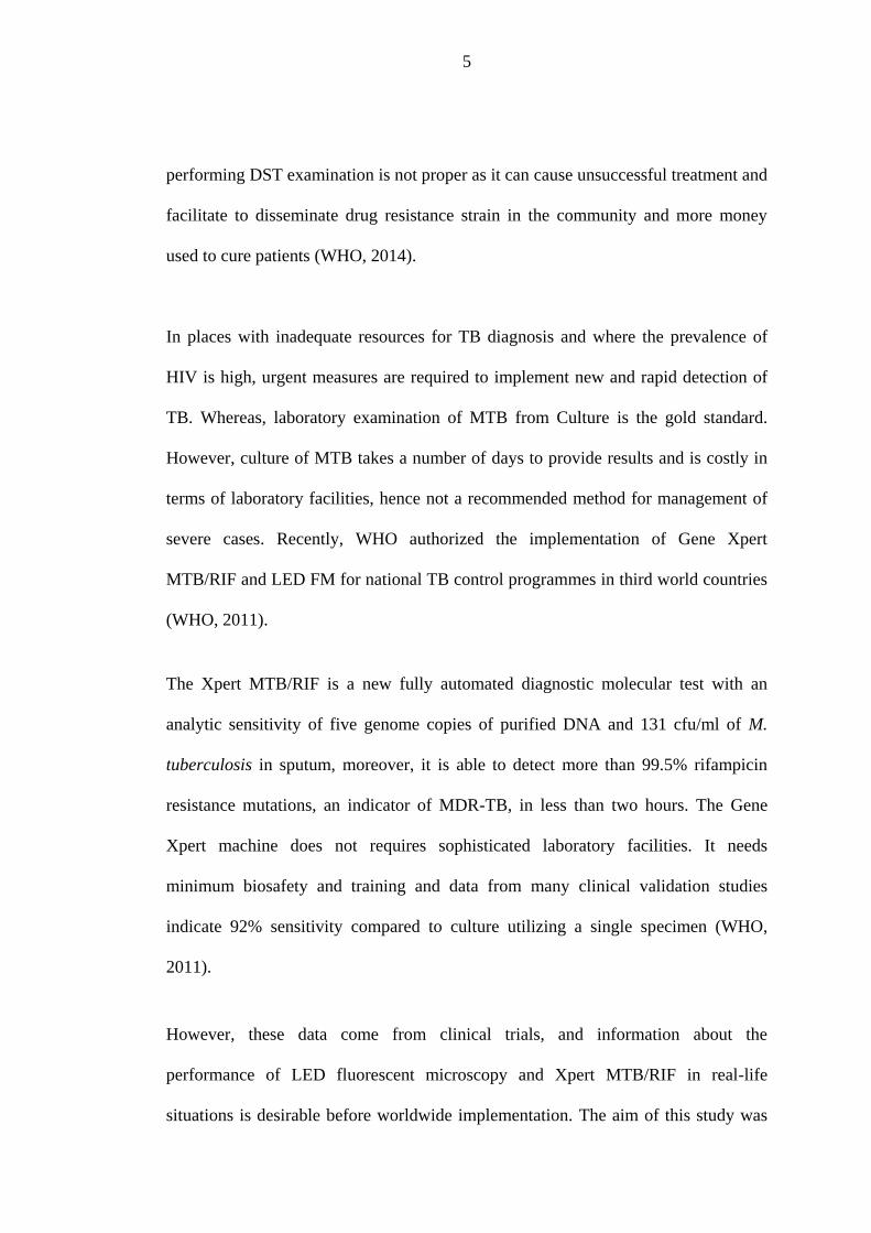
performing DST examination is not proper as it can cause unsuccessful treatment and
facilitate to disseminate drug resistance strain in the community and more money
used to cure patients (WHO, 2014).
In places with inadequate resources for TB diagnosis and where the prevalence of
HIV is high, urgent measures are required to implement new and rapid detection of
TB. Whereas, laboratory examination of MTB from Culture is the gold standard.
However, culture of MTB takes a number of days to provide results and is costly in
terms of laboratory facilities, hence not a recommended method for management of
severe cases. Recently, WHO authorized the implementation of Gene Xpert
MTB/RIF and LED FM for national TB control programmes in third world countries
(WHO, 2011).
The Xpert MTB/RIF is a new fully automated diagnostic molecular test with an
analytic sensitivity of five genome copies of purified DNA and 131 cfu/ml of M.
tuberculosis in sputum, moreover, it is able to detect more than 99.5% rifampicin
resistance mutations, an indicator of MDR-TB, in less than two hours. The Gene
Xpert machine does not requires sophisticated laboratory facilities. It needs
minimum biosafety and training and data from many clinical validation studies
indicate 92% sensitivity compared to culture utilizing a single specimen (WHO,
2011).
However, these data come from clinical trials, and information about the
performance of LED fluorescent microscopy and Xpert MTB/RIF in real-life
situations is desirable before worldwide implementation. The aim of this study was

6
assess the two techniques (i.e., Gene Xpert and LED FM technique) for detection of
PTB in HIV patients.
1.2 Statement of Research Problem
Environmental factors which facilitate the possibility of the threat for MTB
dissemination to increase are contact to TB in small closed area, insufficient
ventilation which provides infectious TB droplets, unable to follow safety procedure
effectively during samples handling, improper decontamination of medical
instruments and recirculation of infectious TB droplets in the air (CDC, 2005).
Repeated visit for collection of spot and morning sputum samples causes the problem
of patients dropout in health care facilities. More patients faced a challenge to get
fare transport for submission of samples to heath care facilities and compilation of
result. In fact the sensitivity of smear microscopy depend on the quality of samples
which has been collected, quality of stain reagents and slide smear prepared, that is
why there are variation range of 20% to 85% concerning sensitivity (Steingart,
2007). In addition, sputum smear microscopy loss its sensitivity for diagnosis of HIV
patients infected with TB and for children due to small number of MTB in the
patients (Fox, 1999).
The HIV infection leads to decreased number of cell mediated immunity, this
situation not only lead to boost the TB disease but also change the way of clinical
diagnosis of TB infection. When HIV infection continues to develop the number of
CD4 T- lymphocytes decrease and facilitate to stimulate the growth and replication
of MTB. CD4 T- lymphocytes play the role for immune response against infection.
7
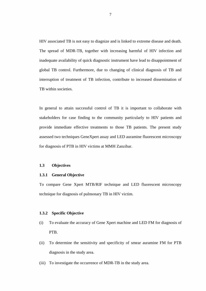
HIV associated TB is not easy to diagnize and is linked to extreme disease and death.
The spread of MDR-TB, together with increasing harmful of HIV infection and
inadequate availability of quick diagnostic instrument have lead to disappointment of
global TB control. Furthermore, due to changing of clinical diagnosis of TB and
interruption of treatment of TB infection, contribute to increased dissemination of
TB within societies.
In general to attain successful control of TB it is important to collaborate with
stakeholders for case finding to the community particularly to HIV patients and
provide immediate effective treatments to those TB patients. The present study
assessed two techniques GeneXpert assay and LED auramine fluorescent microscopy
for diagnosis of PTB in HIV victims at MMH Zanzibar.
1.3 Objectives
1.3.1 General Objective
To compare Gene Xpert MTB/RIF technique and LED fluorescent microscopy
technique for diagnosis of pulmonary TB in HIV victim.
1.3.2 Specific Objective
(i) To evaluate the accuracy of Gene Xpert machine and LED FM for diagnosis of
PTB.
(ii) To determine the sensitivity and specificity of smear auramine FM for PTB
diagnosis in the study area.
(iii) To investigate the occurrence of MDR-TB in the study area.

8
1.4 Hypotheses
(i) Gene Xpert MTB/RIF is more accuracy than LED FM in diagnosis of PTB.
(ii) Smear auramine FM is less sensitivity but has high specificity for PTB
diagnosis.
(iii) The prevalence of MDR TB is less in the study area.
1.5 Significance of the Study
The issue of safe environment in diagnosis of MTB in laboratory is very vital. The
use of engineering controls (For example BSC and area exposure to air) and personal
protective equipment (like oxygen mask) can help to protect laboratory personnel
against exposure with TB infection in linked with breathing of infectious droplets.
On the other hand, the major significant concept in the testing area is to diminish the
threat of generating aerosols within and outside the work environment so that to
decrease the danger of exposure with infectious agents.
There is a lack of accurate and rapid diagnosis technique for TB in many health
centers in Zanzibar. Definitely this study will provide useful and valuable
information to policy makers about accurate and rapid diagnosis technique for TB.
Determination of accurate and rapid diagnosis technique for TB could help to reduce
death by providing proper treatment to those patients suffering with HIV and TB co-
infection as well as to keep environment well against contamination with aerosol
when processing sputum. Then the importance of this study was to find rapid and
accurate technique for diagnosis of PTB in patients suffering with HIV and TB co-
infection.
9
CHAPTER TWO
LITERATURE REVIEW
2.1 Common Knowledge Concerning Tuberculosis
Tuberculosis is a persistence infectious illness caused mostly by Mycobacterium
tuberculosis (and rarely by Mycobacterium bovis or Mycobacterium africanum).
Another name of these organism is known as acid fast bacilli (AFB) because they are
decolorized by acid alcohol. The lungs are affected when organisms penetrate the
body through breathing. Normally there are two kinds of tuberculosis: first PTB
which damage the respiratory organs, this type of disease is common to the
community. The second is extra pulmonary tuberculosis (EPT) is the infection that
damage other organ than the respiratory organ such as joint, lymph nodes, spine,
brain, pleural and genital urinary tract (TURT-MOH, 2006).
2.1.1 Spread of Tuberculosis
TB is a communicable disease; the route of infection is person to person contact
through inhalation. The individual who is affected by PTB can transmit the
infectious droplets when coughing, talking or sneezing into the recirculated air. The
exposure of this facilitated by environmental factors such as small closed area and
insufficient ventilation provide infectious TB droplets. These infectious droplets can
be eradicated in the building by providing well ventilation. TB organisms die within
minutes when exposed to direct sunlight; however they are alive in a dark area for 24
to 48 hrs. Health individual staying together with a patient confirmed PTB positive in
a small room that lack good ventilation for a long time is susceptible to TB infection.
The danger of dissemination of TB from individual with negative smear PTB is low.
10
Smoking and silicosis boost the vulnerability to disease. However, facilities which
have nice ventilation dissolve the mass of infectious agents (TURT-MOH, 2006).
The threat of development infection to active TB disease depends on the condition of
the immune system. Normal people with HIV negative are able to resist TB disease
to about 90% even though they acquire infectious agent of TB due to the active
immune response. Only tuberculin skin test may be indicating positivity of TB
infection to immunocompetent individual. The condition of immunocompromise
stimulates the dormant bacilli to be active and cause TB disease to an individual
because the cells associated with immune response become weak and fail to fight
against infectious agents (TURT-MOH, 2006).
Currently, HIV infection is the source which cause immune suppression and lead to
reactivation of TB in Tanzania. Co-infection TB and HIV individuals have 29 – 30
times chance of progression to active TB disease than HIV negative individuals.
Also there are other conditions such as recurrent infection of any kind, malnutrition
and diabetes mellitus may cause reactivation of the TB infection (TURT-MOH,
2006).
2.2 Prevalence of Mycobacterium tuberculosis
In 2013, about 1.1 million (13%) of the 9.0 million people who were infected with
TB disease globally were HIV positive. The African region accounted for 78% of the
estimated number of HIV positive incident TB cases. Since 2004 the mortality rate of
people with co-infection HIV and TB has been decreasing. Nevertheless, in 2013 the
mortality of HIV related with TB was still 360,000 in the world (WHO, 2014).

11
Although majority of the TB morbidity and mortality arise among men, also the
number of TB cases among women is high. About 3.3 million cases of TB and
51,000 TB mortality among women, and about 550,000 TB cases and mortality
80,000 among children happened in the year 2013. The high number of deaths would
be preventable, if the individuals may arrive at health facilities for diagnosis and
appropriate treatment, this would decrease the TB mortality rate.
In fact the first line drugs for TB available at health facilities for decades may heal
TB disease about 90% (WHO, 2014). Zanzibar is estimated to have 7,200 people
living with HIV, this account for the prevalence of HIV to about 0.6%. HIV infected
clients attending care and treatment services are screened for TB at each encounter.
These include patient on ART, Isoniazid Preventive Therapy (IPT) or stopped IPT,
adult and children, male and female, with exception of those on TB therapy.
Percentage of patients screened for TB has increased from 97.1% in 2011 and 2012
to 99% in 2013. Table 2.1 shows the number of HIV patient who were screened for
TB out of those who received care during the period and those started on TB
treatment (ZIHTLP, 2013). Currently there are two sites providing comprehensive
TB/HIV activities (Mnazi Mmoja and Chake Chake Hospitals).
TB patients who are diagnosed as HIV positive are treated at TB clinic and receive
ARV drugs until they finish TB treatment when they are referred to care and
treatment counseling. A total of 71 patients received ART at TB/HIV under one roof
clinic in 2013 (ZIHTLP, 2013).
12
Table 2.1: Number of HIV Patients Screened for TB by Facility, 2013
Care and Treatment
Counseling
Total
Visit
Screened for TB Started on TB Treatment
< 14 Years >14 Years < 14 Years >14 Years
Al Rahma 140 6 134 0 1
Bububu 377 18 359 1 8
Kivunge 224 19 205 2 4
Mnazi Mmoja 2521 216 2305 18 62
Mwembeladu 606 41 565 1 3
Chake Chake 217 23 194 1 5
Mkoani 35 2 33 0 1
Wete 157 25 132 1 10
Micheweni 63 13 50 0 2
Makunduchi 56 4 52 0 0
Total 4,396 367 4,029 24 96
Source: ZIHTLP (2013)
Among all TB patient screened in 2013 a total of 115 (17.5%) patients tested were
HIV positive. This is an increase compared to 79 (16.8%) patients who were
diagnosed to be TB/HIV co-infected in 2012. The number of smear positive TB
cases has also increased by 5% compared to 2012. This shows that, TB infection in
the community is still high. As in the previous year, most of those affected are young
adults in the age group between 15 – 44 years. It shows that 77 (24, 21%) were
female and 141 (44.33%) were male, as shown in the Table 2.3 (ZIHTLP, 2013).
Table 2.2: The Trend of Notification of TB for 10 Years
Year
Pulmonary
tuberculosis
AFB +
Extra pulmonary
tuberculosis
AFB +
Relapse Failure/
Others
Returns Total
2004 222 56 11 6 1 296
2005 211 53 14 5 5 288
2006 238 44 9 7 5 303
2007 232 48 11 3 5 299
2008 265 73 14 4 3 359
2009 235 76 25 9 3 350
2010 270 66 14 11 3 369
2011 280 73 12 14 9 388
2012 303 81 12 8 7 411
2013 318 197 18 2 5 542
Source: ZIHTLP (2013)

13
Table 2.3: Distribution of New Sputum Smear Positive (SSP) Cases by Age and
Sex for the Year 2013
Age groups 0 – 4 5 – 14 15–24 25–34 35–44 45–54 55–64 65 + Total
Male (SSP) 0 6 41 58 42 34 14 14 209
Female (SSP) 0 5 27 30 20 15 5 7 109
Total 0 11 68 88 62 49 19 21 318
Source: ZIHTLP (2013)
2.3 Prevalence of MDR TB
Multidrug resistant tuberculosis (MDR-TB) is defined as tuberculosis resistant to at
least isoniazid and rifampicin, the most powerful anti-TB drugs. Globally, an
estimated 3.5% of new cases and 20.5% of previous treated cases have MDR-TB. In
2013, there were an estimated 480,000 new cases of MDR-TB worldwide, and
approximately 210,000 deaths from MDR-TB. Among patients with PTB who were
notified in 2013, an estimated 300,000 had MDR-TB. More than half of these
patients were in India, China and Russian federation (WHO, 2014).
Between 2009 and the end of June 2014, nearly 90,000 people with MDR-TB were
detected through the expand TB project, which has established capacity to detect
drug resistant TB using line probe assay, liquid culture and Xpert MTB/RIF in 27
low and middle income countries (WHO, 2014).
In Zanzibar epidemiological data for MDR-TB prevalence is low, the reasons for low
prevalence of MRD-TB may be lack of accurate diagnosis for detecting MDR-TB
from patients. Also community is not aware about MDR-TB. The equipment, which
detects the indicator for MDR-TB, is available only at MMH, which was
commissioned in 2013. The trend of MDR TB as shown in Table 2.4 (ZIHTLP,
2014).

14
Table 2.4: Trend of MDR TB in Zanzibar for the Past Six Years
Serial number Year Unguja Pemba
1. 2009 0 1
2. 2010 2 0
3. 2011 0 1
4. 2012 0 0
5. 2013 0 1
6. 2014 2 0
Source: ZIHTLP (2014)
2.4 Current Status of TB Diagnosis
In Zanzibar a total of diagnostic centers are 51. Unguja have 32 and Pemba 19
centers. The health centers in unguja which has LED FM are 7 and 25 centers has
ordinary light microscopy for the diagnosis of MTB. Also in Pemba 8 centers has
LED FM and 11 has ordinary light microscopy. Only one centre (i.e Mnazi Mmoja
Hospital) has Gene Xpert MTB/RIF (ZIHTLP, 2014).
Table 2.5: Zanzibar Health Centers which have LED FM
UNGUJA PEMBA
Mnazi Mmoja Hospital Public health laboratory
Kivunge Cottage Hospital Bogoa PHCU
Makunduchi Cottage Hospital Vitongoji Cottage Hospital
Rahaleo PHCU Abdulla Mzee Hospital
Kiomba mvua PHCU Konde PHCU
Fuoni PHCU Chakechake Hospital
Jambiani PHCU Wete Hospital
Micheweni Cottage Hospital
Source: ZIHTLP (2014)
2.5 Delays in Diagnosis of TB
Diagnosis of tuberculosis (TB) is often delayed in resource-limited settings. Factors
contributing to the delayed diagnosis of TB include: 1) patient delays, 2) health-

15
system delays and, 3) delays inherent to the conventional TB diagnostic process
(Storla, 2008 and Sreeramareddy, 2009). Of these, delays related to the low
sensitivity of sputum AFB smear microscopy, have been identified as the most
significant contributor to the total diagnostic delay times experienced by TB patients
(Millen, .2008). The consequences of delayed TB diagnosis and treatment include
increased TB-related morbidity, increased mortality, and continued TB transmission
(Greenaway, 2002; a Golub, 2006). In countries with a high burden of HIV, HIV
infection has both reduced the sensitivity of smear microscopy contributing to further
delays in TB diagnosis, while simultaneously increasing the urgency in which a rapid
TB diagnosis is needed.
Two multi-centre studies: the first study using rapid molecular detection of TB and
rifampicin resistance and the second study using feasibility, diagnostic accuracy and
effectiveness of decentralized of Xpert MTB/RIF test for diagnosis of TB and MDR
have provided strong evidence that Xpert MTB/RIF (Cepheid, Sunnyvale, CA,
USA), the first commercially available, automated, real-time NAAT for MTB, could
reduce diagnostic delay (Boehme, 2010 and Boehme, 2011). Xpert MTB/RIF was
found to be highly accurate (including 70% sensitivity in AFB smear-negative
patients) and led to more rapid diagnosis and shorter time-to-TB treatment initiation
compared to sputum smear microscopy (Boehme, 2010 and Boehme, 2011). Xpert
MTB/RIF was also simple to perform and required minimal technician training.
However, the clinical impact of more rapid TB diagnosis via Xpert MTB/RIF has not
been adequately evaluated. Reducing diagnostic delays may be of particular
importance in populations at high risk of early mortality.

16
2.6 Xpert MTB/RIF Diagnosis
In the study performed in South Africa with HIV-infected patients, the Xpert
MTB/RIF increased case detection for TB by 45% compared to FM (Lawn, 2011).
WHO recommended the use of Xpert MTB/RIF for diagnosis of HIV associated PTB
(Golub, 2006). However, the cost of Xpert MTB/RIF can be too high for some
resource limited settings. In the study which was conducted in a District Hospital in
India concerning the Xpert MTB/RIF compared with LED FM the following results
were obtained as shown in Table 2.6 (Alvares-Uria, 2012).
Table 2.6: Positive Results Utilizing LED FM and Xpert MTB/RIF Assay
Sample Tota
l
Smear positive X-pert positive Absolute
difference
Ratio
X-pert/LED
# # % (95% CI) # % (95% CI) % (95%
CI)
(95% CI)
Sputum 166 106 63.9 (56.2 to
70.6)
124 74.7 (67.5 to
80.8)
10.8 (5.2 to
16.5)
1.17 (1.08 to
1.26)
Source: Alvares-Uria (2012)
In setting with low prevalence of MDR TB where it is possible to obtain two
additional early morning sputum samples in different days, LED FM could be used
with not much loss of sensitivity compared to the Xpert MTB/RIF assay for
diagnosis of HIV infected patients with suspicion of PTB (Alvares-Uria, 2012).
WHO approved the Xpert MTB/RIF assay for sputum based rapid diagnosis of PTB
and MDR TB. The assay can be used to accurately measure the MTB load beyond
the detection limit of 131 organisms per mL in an in-vitro suspension (Helb, 2010).
The Xpert MTB/RIF assay in patients with suspected TB and newly diagnosis case
TB has been evaluated in several studies (Theron, 2011).
17
2.7 Smear Auramine FM Diagnosis
Researchers are also looking at improving the accuracy of smear microscopy, which
remain the first line diagnostic test for MTB for much of the world for foreseeable
future. LED FM is more sensitive than standard microscopy (Smart, 2012). Since
2012, LED FM has been introduced in about 200 medical college microscopy centers
in India. Given better sensitivity (10% more) of LED FM over ZN microscopy, it
may be possible that LED FM in spot specimens performed better in detecting AFB
and the difference in performance between spot and early morning specimen may be
lower (Cuevas, 2011).
After systematic review and meta analysis of available data, WHO reported that in
comparison with direct ZN (standard) microscopy. LED FM was statistically
significantly more sensitive by 6% (95% CI, 0.1 – 13%), with no appreciable loss in
specificity, and LED FM was 5% (95% CI, 0 – 11%) more sensitive and 1% (95%
CI, 0.7 – 3%) more specific than Conventional FM (WHO, 2011).
2.8 Importance of Accurate and Rapid Diagnosis of TB and Drug Resistant
Modelling studies have estimate that 400,000 lives could be saved each year with the
introduction of an accurate, rapid and widely available TB diagnostic system with
sensitivity greater than 85% for both smear positive and smear negative cases and
97% specificity (Malicotti, 2014).
The ability to rapid and accurately detect drug resistance in MTB clinical specimens
is essential for appropriate treatment to be initiated in patients suffering from TB and
for prevention of further spread of drug resistant strains. This is of paramount

18
importance for TB control of MDR-TB at national and global level (Drobniewsk,
2013).
2.9 Environmental Control in TB Diagnosis
In the diagnosis of MTB in laboratory environmental controls is very important so
that to avoid the transmission and decrease the concentration of infectious aerosol in
ambient air. Environmental controls include the following technologies to removal or
inactivate MTB: Local exhaust ventilation, general ventilation, high efficient
particulate air (HEPA) filtration, and ultraviolet germicidal irradiation (UVGI).
These controls help to stop the transmission and decrease the concentration of
airborne infectious agents. Environmental controls are the second line of defense in
the TB infection control program, and they work in harmony with administrative
controls. The reduction of exposure can be facilitated through the effective use of
environmental controls at the source of exposure (e.g., coughing patient or laboratory
specimen) or in the general work place environment. General ventilation can be used
to dilute the air and remove air contaminants and to control air flow patterns in room
or in health care setting. Air cleaning technologies include HEPA filtration to reduce
the concentration of MTB droplet nuclei and UVGI to kill or inactivate the
microorganism so that they no longer pose a risk of infection (CDC, 2005).
Class II BSCs in Figure 2.1 provide personnel protection (containment), product
protection (a virtually particle free work area to help minimize contamination of
culture or other products), and environmental protection (helps prevent
contamination of laboratory, the building and the community). Characteristics that
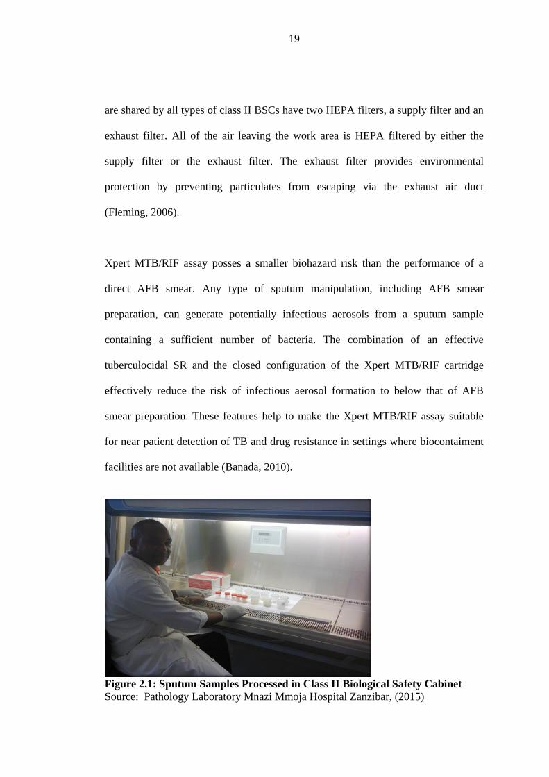
19
are shared by all types of class II BSCs have two HEPA filters, a supply filter and an
exhaust filter. All of the air leaving the work area is HEPA filtered by either the
supply filter or the exhaust filter. The exhaust filter provides environmental
protection by preventing particulates from escaping via the exhaust air duct
(Fleming, 2006).
Xpert MTB/RIF assay posses a smaller biohazard risk than the performance of a
direct AFB smear. Any type of sputum manipulation, including AFB smear
preparation, can generate potentially infectious aerosols from a sputum sample
containing a sufficient number of bacteria. The combination of an effective
tuberculocidal SR and the closed configuration of the Xpert MTB/RIF cartridge
effectively reduce the risk of infectious aerosol formation to below that of AFB
smear preparation. These features help to make the Xpert MTB/RIF assay suitable
for near patient detection of TB and drug resistance in settings where biocontaiment
facilities are not available (Banada, 2010).
Figure 2.1: Sputum Samples Processed in Class II Biological Safety Cabinet
Source: Pathology Laboratory Mnazi Mmoja Hospital Zanzibar, (2015)

20
CHAPTER THREE
MATERIALS AND METHODS
3.1 Study Design
The study was experimental type of design that involved laboratory analysis of
sputum samples for determination of AFB and rifampicin resistance for the HIV
patients attending CTC at Mnazi Mmoja Hospital from all districts of Unguja Island.
3.2 Study Area
The study was done at Mnazi Mmoja Referral Hospital in Zanzibar specifically at
CTC where the samples were collected and then laboratory test conducted at TB
section in microbiology department. The study was conducted from 5th
April to 10th
August 2015. Therefore, this study in Zanzibar provides a base line data to other
studies.
Mnazi Mmoja is the main referral hospital in Zanzibar. The hospital itself is located
at Urban west region in the Stone Town, the historic centre of Zanzibar City. It
comprises of outpatient clinic, specialized clinics and several wards for the inpatient
services. Although termed as referral hospital, basic outpatient services are also
provided to the nearby communities.
The hospital contains 450 beds. The specialty departments include paediatrics,
surgery, internal medicine, acupuncture, physiotherapy, occupational
therapy, obstetrics and gynaecology, maternity services, dental and eye
(ophthalomology). The hospital is fairly well-staffed with doctors, nurses, mid-
21
wives, nursing students and a number of foreign medical students. Regularly foreign
doctors also work at the hospital, which is the product of medical exchange
programmes between Zanzibar and other countries such as Cuba, China, Egypt,
Russia and Sweden.
This hospital has a good laboratory with well-equipped equipment. This equipment
includes; CD4 count machines, automatic tissue processor, ELISA reader, teach
microscopes and genexpert machine. The laboratory has six departments which are
microbiology, parasitology, clinical chemistry, haematology, histopathology and
blood transfusion. The laboratory is three stars ranked and has about 55 staffs.
Among these; they have one pathologist, four laboratory scientists, twenty one
technologists, twenty three technicians and six laboratory assistants.
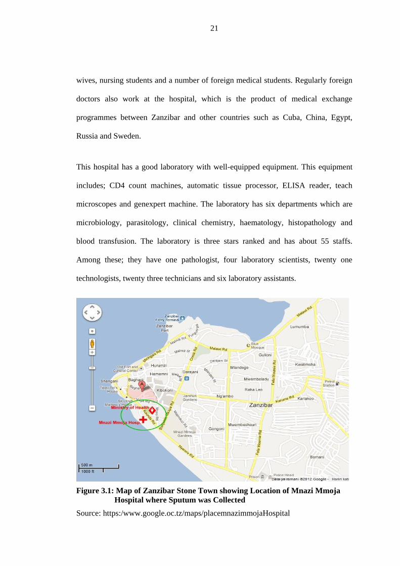
Figure 3.1: Map of Zanzibar Stone Town showing Location of Mnazi Mmoja
Hospital where Sputum was Collected
Source: https:/www.google.oc.tz/maps/placemnazimmojaHospital

22
Zanzibar consists of two sisters Islands named Unguja (Zanzibar) and Pemba, the
land area of the two Islands is approximately 2,332 square kilometers. Zanzibar is
1,464 square kilometers. The population of Zanzibar is approximately 1.303,569
based on 2012 National census.
It is located about 35km off the cost of Dar es Salaam (commercial capital city of
Tanzania), between 39 degree longitudinal and 6 degree latitude south of equator.
Zanzibar Island has 5 regions; Unguja Island has 3 regions named Urban west, North
and South. Pemba Island has 2 regions named North and south.

23
Figure 3.2: Map of Unguja Island showing TB Diagnosis Centres
Source: www.faysafaris.com/Tanzania Maps.htm

24
3.3 Study Population
Study population included all HIV patients male and female suspected with TB
attending CTC and TB clinic at Mnazi Mmoja Hospital. All agreed and signed the
informed consent form for the study purpose.
(a) Inclusion criteria of patient
(i) All HIV patients suspects with TB overt 18 years
(ii) Must have capability of producing two quality sputum samples
(iii) Cough duration must be two weeks or more
(b) Exclusion criteria patient
(i) HIV patient(s) less than 18 years old were not allowed to participate in the
study.
(ii) HIV patient(s) who were not able to produce quality sputum was not
involved.
(iii) HIV patient(s) with less than two weeks of coughing episodes were
excluded.
3.4 Sample Size
The formula for calculating sample size was available in (htpp://www.caribvet. 13th
February, 2015). The prevalence was determined by regarding the number of PTB
cases during a specified time period in the study area. The size of the population of
PTB patients occurred in study area. The prevalence was then calculated by dividing
the number of PTB incidence during specified time period by the size of population
of HIV patients, the result is expressed as a percentage. The total numbers of PTB

25
cases reported in Zanzibar between 2009 – 2013 were 1506 and the total numbers of
the population were 7200. The prevalence was calculated as shown 1506/7200 = 0.2
(20%). Therefore the prevalence of PTB was 0.2. In this study 246 HIV patients
male and female attended CTC at MMH were examined. Those HIV patients
suspected with PTB were used in the study to represent the population. The numbers
of sputum samples collected are shown below.
n= Z²1-α/2 P(1-P)
e²
Where n= number to sample
Z²= (1.96)² for 95% confidence (i.e. α= 0.05)
P= 0.2
e= maximum tolerable error for the prevalence estimation 0.05
From formula above n = (1.96)² x 0.2(1 – 0.2)
(0.05)²
n = 246
3.5 Specimen Collection
Two sputum samples were requested from all HIV patients suspects with TB
according to inclusion and exclusion criteria mentioned above. One on-the-spot
sputum sample at the time of the patient's first visit, and an early morning sputum
sample taken by the patient at home on the day following the initial visit. Patients
were instructed on production of good quality sputum samples. Biological safety
cabinet was used when specimens were assessed macroscopically to inspect the
quality and quantity and a record was made.
26
3.6 Sample Transport
All sputum samples were transported to the laboratory in clean containers, and
transported in cooler box after collection. Sample were labeled by necessary
information including name and number of patient, number of sample either 1 or 2,
date of collection.
3.7 Materials and Equipments
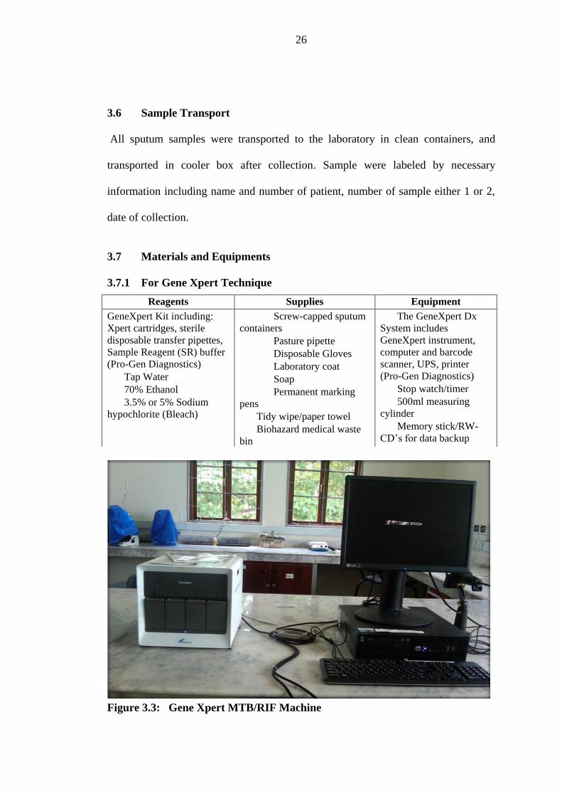
3.7.1 For Gene Xpert Technique
Figure 3.3: Gene Xpert MTB/RIF Machine
Reagents Supplies Equipment
GeneXpert Kit including:
Xpert cartridges, sterile
disposable transfer pipettes,
Sample Reagent (SR) buffer
(Pro-Gen Diagnostics)
Tap Water
70% Ethanol
3.5% or 5% Sodium
hypochlorite (Bleach)
Screw-capped sputum
containers
Pasture pipette
Disposable Gloves
Laboratory coat
Soap
Permanent marking
pens
Tidy wipe/paper towel
Biohazard medical waste
bin
The GeneXpert Dx
System includes
GeneXpert instrument,
computer and barcode
scanner, UPS, printer
(Pro-Gen Diagnostics)
Stop watch/timer
500ml measuring
cylinder
Memory stick/RW-
CD‟s for data backup

27
Source: Pathology Laboratory Mnazi Mmoja Hospital (2014)
3.7.1.1 Diagnosis of Sputum Samples by GeneXpert MTB/RIF Technique
GeneXpert machine was easy to perform, gave results within 2 hours of sample
processed and there was no operation difficult during its usage. This is automated
method; both spot and morning sputum samples were processed in this machine to
identify the presence of MTB in the sputum. Processed of sputum in GeneXpert
MTB/RIF assay (see appendix 3)
3.7.1.2 Comparison Between GeneXpert MTB/RIF and LED FM Techniques
Comparisons of these two techniques were done after sputum samples examined.
The results of sputum samples generated from GeneXpert were compared with the
results yielded by LED FM technique
3.7.1.3 Investigate of MDR-TB in the Study Area
GeneXpert MTB/RIF machine is an automated machine which used to detect MTB
and rifampicin resistance. The rifampicin resistance is an indicator of MDR-TB. All
sputum samples collected were done in GeneXpert MTB/RIF machine, processed of
sputum (see appendix 3), all results yielded were analyzed to identify if there were a
results of rifampicin resistance. In fact machine itself generate this results.
3.7.2 For Auramine Staining Technique
Requirements for performing this technique were as follows:
(i) Timer
(ii) Fluorescent microscope
(iii) Bunsen burner or alcohol lamp
(iv) Slide rack to support slides over the sink
28
(v) Slide rack for drying stained slides
(vi) Sink with water
(vii) Forceps
(viii) Diamond pencil
(ix) Waste bin
(x) Positive and negative control slides
(xi) Paper filters 32.0 cm in size
(xii) Gloves
(xiii) Slides
(xiv) 0.1% of auramine
(xv) 0.5% of acid-alcohol
(xvi) 0.5 of potassium permanganate or 0.3 of methylene blue

29
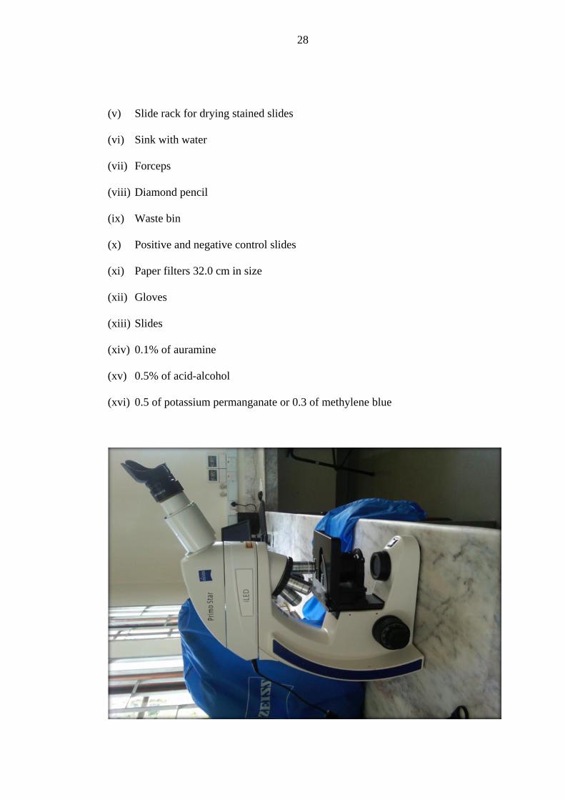
Figure 3.4: Fluorescent Microscope
Source: Pathology Laboratory Mnazi Mmoja Hospital Zanzibar (2014)
3.7.2.1 Diagnosis of Sputum Samples by LED FM Technique
All spot and morning sputum samples were processed. An appropriate portion of the
sputum samples (from the most purulent portion) was collected with an applicator
stick or a wire loop and spread on the labelled side of the microscope slide. The
slides were air dried and then heat-fixed. All smears were stained by the FM
technique (See appendix 2).
3.8 Examination
All slides were examined blindly by FM at 20 - 25 X objectives to scan the smear
and 40X objectives for confirming AFB. The AFB was graded according to the
following scheme:
N of AFB Report
No AFB/1 length Negative
1-29 AFB/1 length Record exact number
1 - 9 AFB/ fields on average 1+
10-100 AFB/field on average 2+
> 100 AFB/ field on average 3+
3.9 Permission and Ethical Consideration
Ethical clearance was sought and obtained from the concerned authorities for sample
collection which is the Ethical Committee of the Ministry of Health Zanzibar. During
the study, the purpose, procedures of the study, measures taken to ensure

30
confidentiality of the participants, the voluntary nature of the study and applicability
of findings were explained to participants. Participants were assured that their
participation in the study is voluntary and they are free to withdraw without any
negative impact in their treatment at the clinic. Written informed consent was sought
and obtained from the participants, since the study dealt with human and involved
collection of sample direct from human the consensus of participants was necessary.
3.10 Quality Control
Both positive and negative slides were used for internal quality control during
samples processed.
3.11 Data Analysis
All data analysis was performed using SPSS version 20 software. Comparison of
performance of Gene Xpert MTB/RIF and LED FM were performed. Sensitivity,
specificity and Pearson chi-square were performed to compare two diagnostic
techniques. A p value (< 0.05) was considered significant.
Objective Analytical Tool
To compare the diagnosis accuracy
of Gene Xpert MTB/RIF and LED
FM among TB suspect
Pearson chi-square test used to determine
association between the Gene Xpert
MTB/RIF and LED FM techniques using p
value (< 0.05) level of significant.
To evaluate the sensitivity and
specificity of smear auramine FM to
TB diagnosis in the study area.
Pearson chi-square test used to determine
the different of two sides of Gene Xpert
and LED FM techniques to generate
positive and negative results of TB using p
value (< 0.05) level of significant.

31
To determine the prevalence of
MDR-TB in the study area
Pearson chi-square test used to calculate
the prevalence value of MDR-TB for those
TB positive
3.12 Limitation
The limitation of this study was 10 patients failed to collect morning sample (second
sample), so in this case were removed from the study because they did not adhered to
the inclusion criteria. Also some of the clients refused to provide their telephone
number because they thought of stigmatization.

32
CHAPTER FOUR
RESULTS
4.1 Characteristics of Study Participants
The age of study participants ranged between 18 – 80 years and majority of them
were females 136 (55.3%) as shown in Table 4.1. A high proportional of the patients
were between ages of 31 to 40 years which account 66(26.8%). Majority of study
participants lived in Urban district of Unguja (40.7%) which is located in Urban
West region while high number of patients were living (80.9%) as shown in Figure
4.1 and Figure 4.2.
Table 4.1: Patients Age Groups and Sex
Age
group
(years)
Patient gender GeneXpert MTB/RIF technique results
Male N(%) Female
N(%)
Total
patients
Total patients
tuberculosis
positive N(%)
Positive
tuberculosis
Male N(%)
Positive
tuberculosis
Female N(%)
< 20 9(3.7%) 14(5.7%) 23(9.3%) 11(6.5%) 2(1.2%) 9(5.3%)
21 - 30 25(10.2%) 33(13.4%) 58(23.6%) 44(26.0%) 23(13.6%) 21(12.4%)
31 - 40 27(11.0%) 39(15.9%) 66(26.8%) 44(26.0%) 20(11.8%) 24(14.2%)
41 - 50 26(10.6%) 32(13.0%) 58(23.6%) 41(24.3%) 21(12.4%) 20(11.8%)
51 - 60 16(6.5%) 11(4.5%) 27(11.0%) 18(10.7%) 12(7.1%) 6(3.6%)
> 61 7(2.8%) 7(2.8%) 14(5.7%) 11(6.5%) 7(4.1%) 4(2.4%)
Total 110(44.7%) 136(55.3%) 246(100%) 169(100%) 85(50.3%) 84(49.7%)
Among the male and female patients, the least proportional were in the age group
above 61 years as shown in Table 4.1. Among 246 confirmed seropositive
HIV/AIDS patients examined, 169 were pulmonary tuberculosis positive for Gene
Xpert MTB/RIF technique. Among the HIV/AIDS patients that were positive of

33
pulmonary tuberculosis 85 (50.3%) were males and 84 (49.7%) were females as
shown in Table 4.1.
Figure 4.1: Distribution of Patients in Districts of Unguja Island
34
Figure 4.2: Distribution of Patients in Regions of Unguja Island
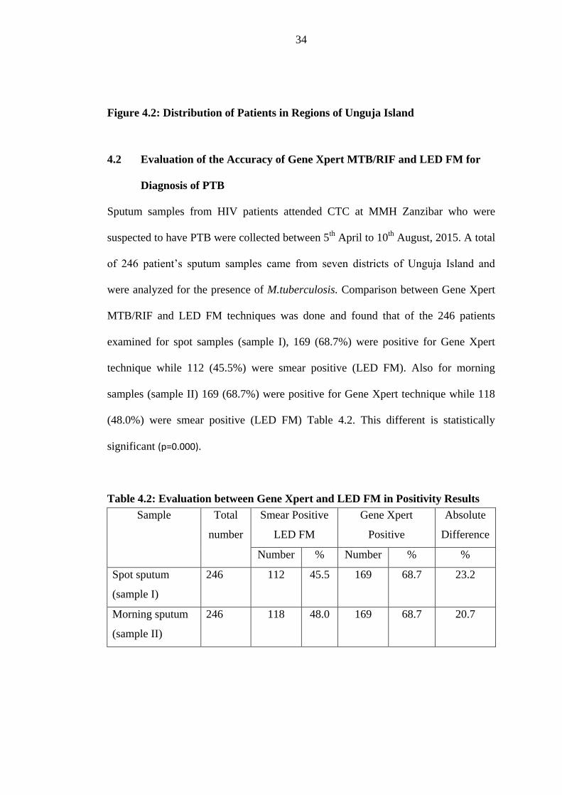
4.2 Evaluation of the Accuracy of Gene Xpert MTB/RIF and LED FM for
Diagnosis of PTB
Sputum samples from HIV patients attended CTC at MMH Zanzibar who were
suspected to have PTB were collected between 5th
April to 10th
August, 2015. A total
of 246 patient‟s sputum samples came from seven districts of Unguja Island and
were analyzed for the presence of M.tuberculosis. Comparison between Gene Xpert
MTB/RIF and LED FM techniques was done and found that of the 246 patients
examined for spot samples (sample I), 169 (68.7%) were positive for Gene Xpert
technique while 112 (45.5%) were smear positive (LED FM). Also for morning
samples (sample II) 169 (68.7%) were positive for Gene Xpert technique while 118
(48.0%) were smear positive (LED FM) Table 4.2. This different is statistically
significant (p=0.000).
Table 4.2: Evaluation between Gene Xpert and LED FM in Positivity Results
Sample Total
number
Smear Positive
LED FM
Gene Xpert
Positive
Absolute
Difference
Number % Number % %
Spot sputum
(sample I)
246 112 45.5 169 68.7 23.2
Morning sputum
(sample II)
246 118 48.0 169 68.7 20.7

35
4.3 Sensitivity and Specificity of Smear Auramine FM for PTB Diagnosis in
the Study Area
All 246 sputum samples collected were processed by Gene Xpert, as well as smear
auramine FM in order to evaluate sensitivity and specificity of LED FM. Among 169
spot and morning samples which were positive in Gene Xpert processed by smear
auramine. The sensitivity of LED FM in spot and morning samples were 66.3% and
69.8% respectively. Also the specificity for both samples was 100% as shown in
Table 4.3. In Figure 4.3 and 4.4 indicate percentage of grades AFB positivity for
both samples in smear examination. This difference is statistical significance of
(p=0.02)
Table 4.3: Smear Auramine LED FM Results
Fluorescent microscope Spot samples (sample I) Morning sample (sample II)
Sensitivity 66.3% 69.8%
Specificity 100% 100%
Number of true positive 112 (66.3%) 118 (69.8%)
Number of false positive 0 (0%) 0 (0%)
Number of true negative 77 (31.3%) 77 (31.3%)
Number of false negative 57 (33.7%) 51 (30.2%)

36
Figure 4.3: Distribution of Smear Positivity of Spot Sputum Samples
Figure 4.4: Distribution of Smear Positivity of Morning Sputum Samples
37
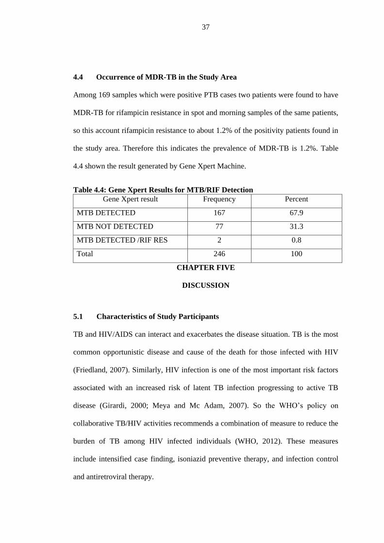
4.4 Occurrence of MDR-TB in the Study Area
Among 169 samples which were positive PTB cases two patients were found to have
MDR-TB for rifampicin resistance in spot and morning samples of the same patients,
so this account rifampicin resistance to about 1.2% of the positivity patients found in
the study area. Therefore this indicates the prevalence of MDR-TB is 1.2%. Table
4.4 shown the result generated by Gene Xpert Machine.
Table 4.4: Gene Xpert Results for MTB/RIF Detection
Gene Xpert result Frequency Percent
MTB DETECTED 167 67.9
MTB NOT DETECTED 77 31.3
MTB DETECTED /RIF RES 2 0.8
Total 246 100
CHAPTER FIVE
DISCUSSION
5.1 Characteristics of Study Participants
TB and HIV/AIDS can interact and exacerbates the disease situation. TB is the most
common opportunistic disease and cause of the death for those infected with HIV
(Friedland, 2007). Similarly, HIV infection is one of the most important risk factors
associated with an increased risk of latent TB infection progressing to active TB
disease (Girardi, 2000; Meya and Mc Adam, 2007). So the WHO‟s policy on
collaborative TB/HIV activities recommends a combination of measure to reduce the
burden of TB among HIV infected individuals (WHO, 2012). These measures
include intensified case finding, isoniazid preventive therapy, and infection control
and antiretroviral therapy.
38
Among the 169 HIV/AIDS patients having TB in this study, 85 (50.3%) were male
and 84 (49.7%) were female and these agree with previous reports indicating higher
prevalence in male than females (Homes, 1988; Abeld, 2002). In our study, the age
distribution reveals the majority of TB/HIV patients were 31 to 40 years old (26.8%),
followed by 21 to 30 years old (23.6%), which represent the most sexually active age
group and correlates with the study done by (Gyar, 2014).
5.2 Evaluation of the Accuracy of Gene Xpert MTB/RIF and LED FM for
Diagnosis of PTB
In low income countries, TB diagnosis and treatment initiation is based on smear
microscopy results. In our study, Gene Xpert MTB/RIF out performed smear
microscopy for MTB detection in almost one third of the patients. The present study
has several important finding: first controlled comprehensive clinical validation
study of the diagnostic accuracy of LED FM in a patient suspected of having active
PTB from HIV patient. Second, the study shows that LED FM has a low sensitivity
and high specificity. Thirdly, the study shows that Gene Xpert MTB/RIF is more
accuracy than LED FM. Fourthly, Gene Xpert assay was positive in 57 spot and 51
morning samples which were smear negative in LED FM and were classified as
having PTB. Lastly, the Gene Xpert MTB/RIF was easy to perform, gave results
within two hours of sample processing and there were no operational difficulties
during its usage.
In the comparison between Gene Xpert MTB/RIF and smear auramine LED FM
results, it was noted that Gene Xpert MTB/RIF increase the number of TB positive

39
results by 23.2% compared to smear auramine LED FM. The finding of this study
does not concur with the study of (Alvares-Uria, 2012) who reported Gene Xpert
MTB/RIF increase the number of positivity by 10.8% compared to LED FM in India.
Also our finding does not concur with the study performed in South Africa with HIV
infected patients that reported that Gene Xpert MTB/RIF increase case detection for
TB by 45% compared to LED FM (Lawn, 2011).
The different between three studies may be explained by the difference in
populations of the studies and in the way the sputum specimens were collected. In
the South African study, two sputum samples were collected in a single visit to out
patient clinics before the initiation of antiretroviral treatment and regardless of
symptoms (Lawn, 2011). In the Indian study, they studied patient with high
suspicious of active tuberculosis infection, and all patients were admitted to the
Hospital (Alvares-Uria, 2012).
In this study, I examined patients with signs and symptoms of tuberculosis infection,
and two sputum specimens were collected in a two visit to the CTC and TB clinics.In
the present study no patient was found to have a false positive result in smear
auramine LED FM. The 77 samples which were negative in Gene Xpert MTB/RIF
were also processed in smear auramine LED FM, and also revealed negative results.
One important limitation of the Gene Xpert MTB/RIF assay is that it can process a
maximum of four samples every two hours, so it may not be suitable for a busy
laboratories receiving large number of sample in resource limited setting (Alvares-

40
Uria, 2012). The prompt results provided by Gene Xpert MTB/RIF would allow a
timely diagnosis and prompt initiation of TB treatment. As extensively reported in
the medical literature, the benefit of rapid treatment initiation of TB in HIV co-
infected patients could improve individual prognosis and reduce overall TB disease
transmission (Sterling, 2010).
My data suggest that smear negative TB patients could benefit from the new assay of
Gene Xpert MTB/RIF assay in those areas where no culture is available. The Gene
Xpert MTB/RIF assay could lead to avoid loss of patients and treatment delay in
those TB suspect with a negative smear result. This could lead to the reduction of
infection and improvements in TB control in the society.
5.3 Sensitivity and Specificity of Smear Auramine LED FM for PTB
Diagnosis in the Study Area
The AFB staining method is the most common and supportive method in diagnosis
of TB infection in resource limited settings. Although AFB staining method is a
much cheaper, simple, and rapid method but also possess a low specificity and
variable sensitivity (Reid, 2009). Current recommendation for the control of
tuberculosis emphasizes early case detection so as to follow treatment of patients and
there by limit the transmission of the bacilli. The main stay for its control is the rapid
and accurate identification of the infected individuals (Prasanthi, 2005).
In this study smear LED FM have shown higher specificity of 100% so this is similar
to another study carried out in South Africa which showed specificity above 95 % by

41
(Lawn, 2011). Also in the present study sensitivity of smear LED FM was 69.8%,
this does not agree with a study conducted in Uganda by (Yoon, 2012) that showed
42% sensitivity.
Tuberculosis control, especially in TB/HIV endemic areas with poor resources, is
hampered by lack of sensitivity and specificity of sputum smear microscopy which is
often the only diagnosis method in place. In December 2010, WHO endorsed the
Gene Xpert MTB/RIF for wide spread use. Among other indications the assay has a
strong recommendation as initial diagnostic test in individual with suspected HIV
associated TB (WHO, 2010). HIV infected patients are to have smear negative
sputum samples. This could be confirmed by our data that showed that 33.7% of spot
samples and 30.2% of morning samples of negative smears were positive when
processed by Gene Xpert MTB/RIF. In the present study indicated that smear LED
FM has a lower sensitivity compared to the Gene Xpert MTB/RIF assay in diagnosis
of HIV patients affected by PTB.
Co-infection with TB/HIV leads to many challenges in both the diagnosis and
treatment of TB. With increase in rates of drug resistance tuberculosis, including
multidrug resistant tuberculosis (MDR-TB) and extensively drug resistant
tuberculosis (XDR-TB) mortality has increased. Sputum smear microscopy in HIV
infected patients are not highly sensitive therefore newer diagnostic test are urgently
required that are cheaper, sensitive, specific and easy to perform and use in remote
and resource constrained settings (Solomon, 2013). Treatment is very important in
prevention of TB and/or HIV which can only be done with a very effective

42
diagnostic tool. Thus an effective diagnostic tool is very crucial in diagnosing TB in
early stage and in HIV patients to reduce mortality and spread of infection.
5.4 Occurrence of MDR-TB in the Study Area
Available data on the prevalence of XDR-TB and MDR-TB in Tanzania are still low
owing to improved case management. Although the levels of anti-tuberculosis drug
resistance in the country are still low, the need for continuous monitoring has always
been emphasized (Chonde, 2010, and Range, 2012). The most effective strategies for
limiting further spread of drug resistance tuberculosis include rapid detection of drug
resistance followed by prompt and effective therapy of each case. Routine
surveillance linked to patient care, represents the best approach to monitor drug
resistance (Hoza, 2015).
The present study has demonstrated a significant increase of positive results when
comparing Gene Xpert MTB/RIF with LED FM when performing both tests in the
same sputum sample. In this study two (1.2%) out of 169 samples with valid
rifampicin resistant were found. This indicates that the prevalence of MDR-TB is
1.2% in Zanzibar. This results do not concur with the study of (Alvares-Uria, 2012)
who reported six (4.8%) out of 124 specimens in India. Also the results do not
concur with the study of (Hoza, 2015) conducted in Tanga, Tanzania Mainland that
showed the prevalence of MDR-TB to rifampicin resistant was 6.3%. The reasons for
the difference between the prevalence in Tanzania Mainland and Zanzibar could be
that in Tanzania Mainland more study have been conducted to find the prevalence of
MDR-TB compared to Zanzibar. Also population difference between Zanzibar and
43
Tanzania Mainland could lead to higher prevalence of MDR-TB in Mainland than
Zanzibar.
CHAPTER SIX
CONCLUSIONS AND RECOMMENDATIONS
6.1 Conclusions
The results of this study indicated that Gene Xpert MTB/RIF is more accuracy than
LED FM in diagnosis of PTB in HIV patients suspected with TB. It is usefulness to
detecting sputum smear negative patients needs further examination. The sensitivity
of LED FM in diagnosis of PTB in HIV patient suspected with TB is lower and their
specificity is higher. In our study, active case finding using the Gene Xpert
MTB/RIF analysis has increased active PTB case detection among HIV infected
patients.
44
The study has showed that spot and morning sputum samples of the same patients
yielded the same results when examined in Gene Xpert MTB/RIF, therefore Gene
Xpert MTB/RIF may be useful as a sole rapid test for PTB case detection in HIV
patients suspected with TB, particularly in settings lacking capacity for liquid
culture. Lastly the prevalence of MDR-TB rifampicin resistance is lower in Zanzibar
it might be in the cause of increasing.
6.2 Recommendation
A single spot or single morning sputum sample is enough to examine PTB from HIV
infected patients by using Gene Xpert MTB/RIF. This will lead to release the
patients‟ results on the same day of the sample collection and will eliminate patients
drop out as well as decrease costing of the patients to return at a health facility to
submit the second sputum sample and to get the results at the following day. Policy
makers should procure and disseminate the Gene Xpert MTB/RIF machine at least to
the district Hospitals so that to increase case detection of PTB from the HIV infected
patients in order to provide early treatment to patients who have active PTB cases
and to protect environmental pollution.
Also the study recommends expansion of MDR-TB surveillance to cover all patients
with history of previous treatment for TB, all patients with infectious TB (sputum
smear positive TB patients), contacts of known MDR-TB cases and all HIV positive
TB patients. HIV positive patients should be screen for TB. Lastly the use of faster
MDR-TB diagnostic methods such as Gene Xpert MTB/RIF to detect drug resistance
45
within two hours should be considered for screening among HIV positive TB and
among sputum smear positive TB cases that are likely to transmit MDR-TB.
REFERENCES
Abeld, F. H., (2002). Stop T Blight Poverty. The Newsletter of the global partnership
Movement to Stop Tuberculosis, Oxfam Programme Policy, London, UK.
Alvares-Uria, G., Azcona, J. M, Middle, M., Naik P. K., Redd, S., and Redd R.,
(2012). Rapid Diagnosis of Pulmonary and Extrapulmonary Tuberculosis in
HIV- Infected Patients. Comparison of LED FM and Gene Xpert MTB/RIF
Assay in a District Hospital in India. Tuberculosis research and treatment, New
Delhi, India.

46
Banada, P., Sivasubraman, S. K., Brakemore, R., Boehme, C., Perkins, M. D.,
Fennelly, K., and Alland, D., (2010). Contaiment of Bioaerosol Infection Risk
by the Xpert MTB/RIF Assay and its Applicability to Point of Care Settings.
Journal of Clinical Microbiology, 48 (10) 3551 – 3557.
Boehme, C. C., Nabeta, P., Hillemann, D., Nicol, M. P., and Shenai, S., (2010).
Rapid Molecular Detection of Tuberculosis and Rifampin Resistance. New
England Journal of Medicine, 363 (11) 1005–1015.
Boehme, C. C., Nicol M. P., Nabeta, P., Michael J. S, and Gotuzzo E, (2011).
Feasibility, Diagnostic Accuracy, and Effectiveness of Decentralised Use of
the Xpert MTB/RIF Test for Diagnosis of Tuberculosis and Multidrug
Resistance: A Multicentre Implementation Study. Lancet, 377(9776) 1495–
1505.
Carriquiry G, Otero, L, González-Lagos, E., Zamudio, C;, Sánchez, E., (2012). A
Diagnostic Accuracy Study of Xpert®MTB/RIF in HIV-Positive Patients with
High Clinical Suspicion of Pulmonary Tuberculosis in Lima, Peru.
CDC, (2005). Guidelines for Preventing the Transmission of MTB in Health- Care
Setting. Morbidity and Mortality Weekly Report, 54 (17) 6,8,60.
Cegielsk, J. P., McMurray, D. N., (2004). The Relationship Between Malnutrition
and Tuberculosis: Evidence From Studies in Humans and Experimental
Animals. International journal tuberculosis lung disease, 8(3) 286- 298.
Chonde, T. M., Basra, D., Mfinanga, S. G. M., Range, N., Lwilla, L., and Shirima, R.
P., (2010). National Anti-Tuberculosis Drug Resistance Study in Tanzania.
International journal tuberculosis lung disease, 14(2), 967-972.

47
Cuevas, L. E., AL-Sonbolo, N., Yassin, M. A., and Arbide, I., (2011). LED
Fluorescence Microscopy for the Diagnosis of Pulmonary Tuberculosis: A
Multi-Country Cross-Section Evaluation. PLOS Medicine, 8 (7), 123 - 135
Drobniewsk, F., Papaventsis, D., Bang, D., Nikolayevskyy, V., and Dahile, U.,
(2013). Technical Report European Reference Laboratory Network for TB
Expert Opinion on Use of Rapid Molecular Assay for Diagnosis of TB and
Detection of Drug Resistance. European centre for disease prevention and
control. New York, USA.
Fleming, D. O, and Hunt, D. L, (2006). Biological Safety, Principles and Practices.
4th
Edition, New York: American Society for Microbiology Press.
Fox, W., Ellard, G. A., and Mitchison, D. A., (1999). Studies on the Treatment of TB
Undertaken by the British Medical Research Council TB Unit, 1946 – 1986,
With Relevant Subsequent Publication. International journal tuberculosis lung
disease, 10(3), S231- S279.
Friedland, G., Churchyard, G. J., and Nardell, E., (2007). Tuberculosis and HIV Co-
Infection: Current State of Knowledge and Research Priorities. Journal
infectious disease, 2(1), 45 0 57.
Getahun, H., Gunneberg, C., Granich, R., and Nunn, P., (2010). HIV Infection-
Associated Tuberculosis: The Epidemiology and the Response. Clinical
Infectious Disease. Oxyfod journal, 50 (3) S201–S207.
Girardi, E., Raviglione, M. C., Antonucci, G., Godfrey-Fausset, P., and Ippolito, G.,
(2000). Impact of HIV Epidemic on the Spread of Other Disease: The Case of
Tuberculosis. Aids 14. Michigan, USA.

48
Golub, J. E., Bur, S., Cronin, W. A., Gange, S., and Baruch, N., (2006). Delayed
Tuberculosis Diagnosis and Tuberculosis Transmission. The International
journal of Tuberculosis and lung disease, 10(1), 24–30.
Greenaway, C., Menzies, D., Fanning, A., Grewal, R., and Yuan, L., (2002). Delay
in Diagnosis Among Hospitalized Patients with Active Tuberculosis–
Predictors and Outcomes. American journal of respiratory and clinical care
medicine, 165 (7), 927–933.
Gyar, S. D., Dauda, E., and Reuben, C. R., (2014). Prevalence of Tuberculosis in
HIV/AIDS Patients in Lafia, Central Nigeria. International journal of current
microbiology and applied science, 3(6), 831-838.
Helb, D., Jones, M., and Story, E., (2010). Rapid Detection of Mycobacterium
Tuberculosis and Rifampin Resistance by Use of on Demand, Near Patient
Technology. Journal clinical microbial, 48 (1), 229-37.
Homes, C. F., Hauster, H., and Num, P., (1988). A Review of Sex Difference in the
Epidemiology of Tuberculosis. The International journal of Tuberculosis and
lung disease, 2(1) 96-104.
Horries, A. D., Zachariah, R., Corbett, E. L., Lawn, S. D., Santos, E.T., (2010). The
HIV Associated Tuberculosis Epidemic – When Will We Act? The Lancet,
3(1), 1906 – 1919.
Hoza, A. S., Mfinanga, S. G. M., and Konig, B., (2015). Anti-Tuberculosis Drug
Resistance in Tanga, Tanzania: A Cross Sectional Facility Base Prevalence
Among Pulmonary Tuberculosis Patients. Asian pacific journal of tropical
medicine, 8 (11), 907 – 913.

49
Karp, C. L., and Auwaerter, P. G., (2007). Co-infection with HIV and Tropical
Infectious Desease. Protozoal Pathogens. Clinical infectious disease, 45(9),
1208- 1213.
Lawn, S. D., and Nicol, M. P., (2011). Xpert MTB/RIF Assay: Development,
Evaluation and Implementation of a New Rapid Molecular Diagnostic for
Tuberculosis and Rifampicin Resistance. Europe Pubmed Central ,Future
microbial, 6(9), 1067–1082.
Lawn, S. D., Brooks, S. V., Kranzer, K., Nicol, M. P.,Whitelaw, A., Vogt, M., and
Woor, R. (2011). “Screening for HIV-Associated Tuberculosis and Rifampicin
Resistance Before Antiretroviral Therapy Using the Xpert MTB/RIF Assay: A
Prospective Study.” PLOS medicine, 8 (7), e 1001067.
Malicotti, P., Bua, A., and Zanetti, S., (2014). Cost – Effectiveness in the Diagnosis
of Tuberculosis: Choice in Developing Countries. The journal of infection in
developing countries, 8 (01) 024 – 038.
Mathew, P., Kuo, Y. H., Vazirani, B., Eng, R. H., and Weinstein, M. P., (2002). Are
Three Sputum Acid-Fast Bacillus Smears Necessary for Discontinuing
Tuberculosis Isolation? Journal clinical microbial, 40 (9), 3482–3484.
(MCC) Medical Chemical Corporation, (2014). Material Safety Data Sheet for
Carbol Fuchsin, Ziehl – Neelsen. Cairo, Misri.
Meya, D. B., and McAdam, K. P., (2007). The Tuberculosis Pandemic: An Old
Problem Seeking New Solution. Journal international medicine, 26(1), 309-
329.
50
Millen, S. J., Uys, P. W., Hargrove J., van Helden, P. D., and Williams, B. G.,
(2008). The Effect of Diagnostic Delays on the Drop-Out Rate and the Total
Delay to Diagnosis of Tuberculosis. Plos one, 3(4) e1933 1941.
(MOHSW) Ministry of Health and Social Welfare, (2013). The First National TB
Prevalence Survey in the United Republic of Tanzania Final Report Dar es
Salaam, Tanzania.
Murray, P. R., Bara, A. J., Jorgensen, J. H., Pfaller, M. A., and Yollan, R. H., (2003).
Manual of Clinical Microbiology 8th
Edition. New York: Wiley & Sons
Parsons, L. M., Somoskovi, A., Guturrez, C., Lee, E., Paramasivan, C. N., Abimiku,
A., Spector, S., Roscigno, et al (2011). Laboratory Diagnosis of Tuberculosis
in Resource Poor Countries: Challenges and Opportunities. American Society
for Microbiology, 24 (2), 314 – 350.
Prasanthi, K., Kumara, A. R., (2005). Efficacy of Fluorochrome Stain in the
Diagnosis of Pulmonary Tuberculosis Co-Infected with HIV. Indian journal of
medicine and microbial, 23(3), 179-185.
Range, N., Friis, H., Mfaume, S., Magnussen, P., Chungalucha, J., and Kilala, A.,
(2012). Anti-Tuberculosis Drug Resistance Pattern Among Pulmonary
Tuberculosis Patients with or Without HIV Infection in Mwanza, Tanzania.
Tanzania journal health research, 143), 1 – 9.
Reid, M. J., and Shah, N. S., (2009). Approaches to Tuberculosis Screening and
Diagnosis in People with HIV in Resource Limited Settings. The Lancet
infectious disease, 9 (3), 173-184.
Solomon, S., Balakrishnan, P., Vignesh, R., Waldrop, G., Solomon, S. S., and
Murugavel, K.G., (2013). A Rapid and Low Cost Microscopic Observation,

51
Drug Susceptibility Assay for Detecting Tuberculosis and MDR-TB Among
Individuals Affected by HIV in South India. Indian journal of medicine and
microbial, 31(10), 130-137.
Sreeramareddy, C. T., Panduru, K. V., Menten, J., and Van den, E. J., (2009). Time
Delays in Diagnosis of Pulmonary Tuberculosis: A Systematic Review of
Literature. 9(9), 67 – 81.
Steingart, K. R., Ramsay, A., and Pai, M., (2007). Optimizing Sputum Smear
Microscopy for the Diagnosis of Pulmonary Tuberculosis. Expert Review of
Anti-Infective Therapy, 5(3), 327- 331.
Sterling, T. R., Pham, P. A., and Chaisson, R. E., (2010). HIV Infection-Related
Tuberculosis: Clinical Manifestations and Treatment. Oxyford journal clinical
infectious disease, 50 (3), S223–S230.
Storla, D. G., Yimer, S., Bjune, G. A., (2008). A Systematic Review of Delay in the
Diagnosis and Treatment of Tuberculosis. Biomedical central public health,
8(1), 15 – 32.
Smart, T., (2012). New Laboratory Tests to Enhance TB diagnosis: Microscopy,
LAM and Xpert MTB/RIF Used as Drug Resistance Test. Cape Town, South
Africa.
Theron, G., Peter, J., and Van Zyl, S.R., (2011). Evaluation of the Xpert MTB/RIF
Assay for Diagnosis of Pulmonary Tuberculosis in High HIV Prevalence
Setting. American journal of respiratory and critical care medicine, 184(1)
132-40.

52
(TURT-MOH) The United Republic of Tanzania Ministry of Health, (2006). Manual
of the National Tuberculosis and Leprosy Programme in Tanzania. 5th
Edition
Dar es Salaam, Tanzania.
World Health Organization, (2012). WHO Policy on Collaboration TB/HIV
Activities: Guidelines for National Programmes and Other Stakeholders.
Geneva: WHO Press.
World Health Organization, (2014). Global Tuberculosis Report, Geneva,
Switzerland.
World Health Organization, (2011). Rapid Implementation of the Xpert MTB/RIF
Diagnostic Test, Technical and Operational „How-to‟ Practical Consideration,
New York, USA.
World Health Organization, (2011). Fluorescent LED Microscopy for Diagnosis of
TB. Policy Statement Geneva, Switzerland
World Health Organization, (2010). Roadmap for Rolling Out Xpert MTB/RIF for
Rapid Diagnosis Tuberculosis and MDR- TB. Retrieved on 2nd
March, 2016
from: www.who.int.
Yoon, C., Cattamachi, A., Davis, J. L., Worodria, W., and Den, B. S., (2012). Impact
of Xpert MTB/RIF Testing on Tuberculosis Management and Outcome in
Hospitalize Patient in Uganda. Kampala, Uganda.
(ZIHTLP) Zanzibar Integrated HIV, Tuberculosis and Leprosy Program ,(2013).
Annual Report, Zanzibar, Tanzania.
(ZIHTLP) Zanzibar Integrated HIV, Tuberculosis and Leprosy Program, (2014).
Data Base. Zanzibar, Tanzania.

53
APPENDICES
Appendix 1: Consent Form
STUDY TITLE: ASSESSMENT OF TWO TECHNIQUES FOR DIAGNOSIS
PULMONARY TUBERCULOSIS IN HIV PATIENT AT MNAZI MMOJA
HOSPITAL ZANZIBAR: GENE XPERT AND FLUORESCENT
MICROSCOPY

54
You are being asked for your consent to participate in a study that involves
tuberculosis suspect in HIV patients in this hospital. We are asking for your consent
to participate in a medical research study .The study is done by Abdalla Said
Mohamed , the Master‟s student of environment Health at Open University of
Tanzania. I will explain the study to you and will ask if you are willing to participate.
Take your time to make your decision about participating. You may discuss your
decision with your family and friends and with your health care team. If you have
any questions, you may ask the researcher.
Purpose of the study
To compare Gene Xpert MTB/RIF and LED fluorescent microscopy for diagnosis of
pulmonary TB in HIV patients. This study is not funded by any institution. None of
the researchers have potential financial gain from the study.
Study procedure
If you take part in this research study, we will ask you to give us the specimen of
sputum. This is an activity that is part of the services provided by the tuberculosis
clinic.
Procedure for specimen donation
If you agree to participate in this study, the following will happen:
Your sputum will be tested and its result will be coded to safeguard their
privacy per protocol.
There will be no direct benefit to you
There will be no human genetic tests performed to your samples.

55
Withdraw from the study
You can decide to withdraw from the study at any time. You will tell the
researcher if you are thinking about stopping or decide to stop. Notify the
researcher at MMH (Abdalla S. Mohamed) in writing at Mnazi Mmoja Hospital,
P.O.Box 236, Zanzibar, Tanzania or by Mobile: +255 (0)777 435441, and he will
destroy any your identifiable. He will tell you how to stop your participation
safely.
Risk and Benefits for participating in this study
Testing the specimens will not cause any risk to you.
There will be no direct benefit to you from participating in this study. You may
choose not to participate. You will receive the same care whether you participate in
the study or not.
Private and confidentiality
We will do our best to make sure that the results kept private.
Costs
You will not be charged for any of the study activities.
Payments for your participation
You will not be paid for taking part in this study.
Participant’s right
Taking part in this study is your choice. You may choose either to take part or not to
take part in the study. If you decide to take part in this study, you may leave the
study at any time. No matter what decision you make, there will be no penalty to

56
you and you will not lose any of your regular benefits. Leaving the study will not
affect your medical care. You can still get your medical care from the health
facilities in Zanzibar as well as the whole Tanzania.
Consent
You have been given copies of this consent form.
Participation in research is voluntary
You have the right to decline to participate or to withdraw at any point in this study
without penalty or loss of benefits to which you are otherwise entitled.
If you wish to participate in this study, you should sign below.
________________ ________________ ___________________________
Date Participant's name Participant's Signature for Consent
________________ ________________ ___________________
Date Person Obtaining Consent Signature
Appendix 2: SOP for Auramine Staining Fluorescent Microscopy
1. Scope
This procedure applies to all laboratory personnel who perform auramine staining to
diagnose microscopically tuberculosis.
2. Principle

57
Sputum smears are stained with auramine which stains Mycobacterium tuberculosis
in greenish yellow against a dark background.
3. Specimen type
Sputum
4. Material and equipment
- Timer
- Fluorescent microscope
- Bunsen burner or alcohol lamp
- Slide rack to support slides over the sink
- Slide rack for drying stained slides
- Sink with water
- Forceps
- Diamond pencil
- Waste bin
- Positive and negative control slides
- Paper filters 32.0 cm in size
- Gloves
- 0.1% of auramine
- 0.5% of acid-alcohol
- 0.5 of patassium permanganate or 0.3 of methylene blue
5. Safety precaution
- Specimen may contain live potentially infectious agents that hazardous to your
health Observe Standard Universal Precautions.

58
- Ensure that the work surface is kept clean, free from clutter and other infectious
substances to avoid contamination.
- Spills should be immediately disinfected with appropriate disinfectant solution.
6. Quality control
- Positive and negative control slides must be used
- Ensure that reagents are not expired
7. Calibration
Ensure that the fluorescent microscope is regularly maintained
8. Activity description
- Wear appropriate PPE
- Gather necessary materials and reagents
- Filter auramine solution prior to use
- Put the slides on the staining rack, smear facing upward
- Cover all the slides with the filtered auramine
- Leave the stain on slides for minimum of 20 minutes
- Gently rinse each slide with water
- Till each slide off excess water
- Cover the slides with acid-alcohol and leave on for 1-2 minutes
- Gently rinse each slide with water and do not splash other slides
- Till each slide off excess water
- Cover each slide with potassium permanganate or methylene blue for 1 minute
- Gently rinse each slide with water and do not splash other slides
- Till each slide off excess water
- Air dry away from direct sunlight. Do not examine slides until they have dried
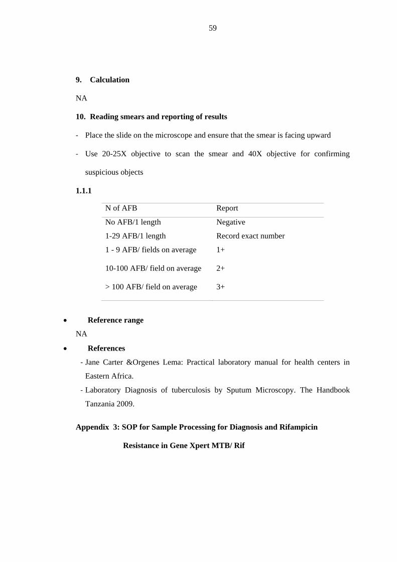
59
9. Calculation
NA
10. Reading smears and reporting of results
- Place the slide on the microscope and ensure that the smear is facing upward
- Use 20-25X objective to scan the smear and 40X objective for confirming
suspicious objects
1.1.1
N of AFB Report
No AFB/1 length Negative
1-29 AFB/1 length Record exact number
1 - 9 AFB/ fields on average 1+
10-100 AFB/ field on average 2+
> 100 AFB/ field on average 3+
Reference range
NA
References
- Jane Carter &Orgenes Lema: Practical laboratory manual for health centers in
Eastern Africa.
- Laboratory Diagnosis of tuberculosis by Sputum Microscopy. The Handbook
Tanzania 2009.
Appendix 3: SOP for Sample Processing for Diagnosis and Rifampicin
Resistance in Gene Xpert MTB/ Rif

60
1.0.Title
Standard Operating Procedures for TB diagnosis and Rif resistance using the Xpert
MTB/Rif on sputum
2.0 Objective and Scope
This SOP describes how to use the Gene Xpert instrument and how to perform the
Xpert MTB/Rif assay. To ensure that all trained health care providers are capable of
performing the Xpert MTB/RIF assay for diagnosing TB in sputum specimens
directly in a primary health care facility, according to the manufacturer‟s
instructions. Standard operating procedure for sample processing for Gene Xpert
used in detection of mycobacterium and Rifampicin testing.
2.1 Principle
The GeneXpert (Cepheid) is a closed, self-contained platform for the extraction,
amplification and detection of Mycobacterium tuberculosis (Mtb) complex from
unprocessed samples. The GeneXpert system is able to generate a result within 2
hours.
The Xpert MTB/RIF assay allows for the rapid detection of Mtb and Rifampicin
(Rif) resistance by combining automated extraction, amplification and detection on a
single system. Rif is one of the first line anti-TB drugs and is also a surrogate marker
for multi-drug resistant TB (MDR-TB). The assay amplifies a portion of the
“rifampicin resistance determining region” of the rpoB gene, the most common site
for Rif mutations, in real-time, using two sets of primers. Fluorescent probes are then

61
used to differentiate between wild-type and mutant strains so that if one or more
probes do not bind, this indicates the presence of a mutation and therefore Rif
resistance.
A sample processing control (SPC) consisting of spores from Bacillus globigii, is
included in the assay as an internal control to ensure adequate processing of the
sample as well as to monitor the presence of PCR inhibitors. A probe check control
(PCC) verifies reagent rehydration, PCR tube filling in the cartridge, probe integrity
and dye stability.
3.0 Safety and environment
Refer to Health Laboratory Safety and Waste Management Manual for safety
considerations
• The GeneXpert system should be installed on a solid, even base
• Always wear a lab coat and disposable gloves
• All sample processing should take place under a BioSafety Cabinet Class II hood
OR an open, well-ventilated area.
• Treat all samples as potentially infectious
• If liquid containing potentially infectious agent is spilled, clean affected area with
3.5% hypochloride
• Dispose of all waste in a biohazard medical waste bin
4.0 Procedure
Materials

62
Reagents Supplies Equipment
GeneXpert Kit including:
Xpert cartridges, sterile
disposable transfer
pipettes, Sample Reagent
(SR) buffer (Pro-Gen
Diagnostics)
Tap Water
70% Ethanol
3.5% or 5% Sodium
hypochlorite (Bleach)
Screw-capped
sputum containers
Pasture pipette
Disposable Gloves
Laboratory coat
Soap
Permanent
marking pens
Tidy wipe/paper towel
Biohazard medical
waste bin
The GeneXpert Dx
System includes
GeneXpert instrument,
computer and barcode
scanner, UPS, printer
(Pro-Gen Diagnostics)
Stop watch/timer
500ml measuring
cylinder
Memory stick/RW-
CD‟s for data backup
NOTE: Always wear gloves and laboratory coat when processing sputum specimens
and Sample preparation should take place in a Biosafety Cabinet Class II if available
OR a designated open, well ventilated work area in the laboratory!
5.0 Collection of sputum specimen
Explain the specimen collection procedure to the patient
Provide patient with the screw-capped container.
Instruct patient to cough deeply and spit up sputum into the screw-capped
sputum container.
Instruct patient to seal the specimen container tightly.
Ensure the sputum in the container is sent to the laboratory.
Label each specimen immediately with identifying information – patient name,
date and time of collection
Log the sputum specimen in laboratory log book/computer system

63
6.0 Sample preparation
1. For each of the sample: unscrew the lid of sputum collection container
2. Add directly in the collection container 2 volumes of the sample reagents to 1
volume of sample.
3. Replace the lid, and shake vigorously 10 – 20 times.(Note: one back and forth
movement is a single shake)
4. Incubate at room temperature: After 10 minutes of incubation, shake ( or vortex)
the specimen vigorously 10 – 20 times.
5. After 5 minutes of incubation. Sample should be perfectly fluid before being
processed, with no visible clumps of sputum. If still viscous, wait 5 – 10 further
minutes before processing it in the cartridge.
NOTE: The unprocessed sputum specimen can be stored at 4⁰C for 4-10days
7.0 Cartridge preparation
1. Label the cartridge with the sample ID by writing on the left or right side of the
cartridge or affix ID label.
2. Open the cartridge ,pipette at least 2 ml with the plastic transfer pipette from the
collection container to the cartridge, then close the cartridge lid.
Note: Do not put the label on the lid of the cartridge or obstruct the existing 2d
barcode on the cartridge,
8.0 References
Operators Manual for the GeneXpert system (Cepheid).
Or contact: Pro-Gen Diagnostics on 011 467 751