comparisonofradiosensitizingeffectsofthemammalian...
TRANSCRIPT

Comparison of radiosensitizing effects of the mammaliantarget of rapamycin inhibitor CCI-779 to cisplatin inexperimental models of head and neck squamouscell carcinoma
Oleksandr Ekshyyan,1,5 Youhua Rong,1,5
Xiaohua Rong,1,5 Kavita M. Pattani,1
Fleurette Abreo,2 Gloria Caldito,3
John Kai Siung Chang,4 Federico Ampil,4
Jonathan Glass,5 and Cherie-Ann O. Nathan1,5
Departments of 1Otolaryngology-Head and Neck Surgery,2Pathology, 3Biometry, and 4Radiation Oncology and5Feist-Weiller Cancer Center, Louisiana StateUniversity Health Sciences Center,Shreveport, Louisiana
AbstractTo determine if the mammalian target of rapamycin(mTOR) inhibitor CCI-779 can sensitize head and necksquamous cell carcinoma (HNSCC) to radiotherapy (XRT)and compare the radiosensitizing effects to cisplatin withits known considerable toxicity. Radiosensitizing effectsof CCI-779 were assayed on HNSCC cell lines in vitro.CCI-779 (5 mg/kg), cisplatin (1 mg/kg), and XRT (2 Gy)alone and in combination were evaluated for antitumoractivity in mice bearing FaDu and SCC40 xenografts.Effects of CCI-779 on radiation-induced activation ofthe Akt/mTOR pathway were analyzed. Although CCI-779 did not sensitize HNSCC cells to ionizing radiationin vitro, combination of CCI-779 and XRT significantlyaugmented the in vivo tumor growth-inhibitory effects ofXRT and CCI-779 (P< 0.05). In addition, CCI-779 + XRTsuppressed tumor growth more effectively than cisplatin+ XRT (P < 0.05). CCI-779 + XRT significantlyimproved survival compared with XRT alone in both cis-platin-sensitive FaDu (P < 0.01) and cisplatin-resistant
SCC40 (P<0.05) xenograft mice. Therewere no addition-al benefits of adding cisplatin to CCI-779 + XRT. CCI-779significantly attenuated irradiation-induced up-regulationof the mTOR pathway, increased apoptosis and displayedpotent antiangiogenic activity in FaDu xenografts thatwas further enhanced by its combination with XRT (P <0.05), which may explain the mechanism of its selectiveradiosensitizing effects in vivo and not in vitro. Antitumoractivity of XRT was enhanced when combined with CCI-779 in HNSCC xenograft model. CCI-779 + XRT showedantitumor activity superior to conventional chemoradiother-apy with cisplatin. These results pave the way for clinicaltrials using molecular targeted therapy with CCI-779 incombination with XRT for HNSCC treatment. [Mol CancerTher 2009;8(8):2255–65]
IntroductionHead and neck cancer is one of the six most frequent can-cers worldwide (1). Squamous cell carcinoma of the headand neck (HNSCC) remains a challenge because of the highlocoregional recurrence rates, ∼50% in advanced-stagedisease (2, 3). The combination of organ preservation cis-platin-based chemoradiotherapy is now used as standardtreatment (4). However, the incidence of severe acute ad-verse effects was higher in the group receiving cisplatin-based chemoradiotherapy than in the radiotherapy (XRT)group alone (77% versus 34%; ref. 4). Cisplatin displaysconsiderable toxicity causing significant morbidity as a re-sult of dysphagia, prolonged use of percutaneous endo-scopic gastrostomy tube, and tracheostomy tubes (5).Additionally, many patients acquire resistance to cisplatinduring therapy, suggesting a need for other treatmentstrategies (6).The use of molecularly targeted agents in combination
with XRT is a promising strategy. Cetuximab, an epidermalgrowth factor receptor monoclonal antibody, has beenapproved by the Food and Drug Administration for thetreatment of HNSCC patients. The addition of cetuximabto XRT is beneficial in a small fraction of HNSCC patients(10-15%), and some patients acquire cetuximab resistance(7–9). The presence of mutant epidermal growth factorreceptor (EGFRvIII) detected in 42% of HNSCC tumorsmight contribute to the limited clinical response. EGFR-vIII-overexpressing cells and xenografts were more resistantto cetuximab treatment compared with cells and xenograftsoverexpressing wild-type epidermal growth factor receptor(10). Moreover, in models of non–small cell lung cancer andcolon cancer, persistent activation of the phosphatidylinositol
Received 12/15/08; revised 6/3/09; accepted 6/7/09; publishedOnlineFirst 7/21/09.
Grant support: National Cancer Institute grant R01 CA 102363 (C-A.O.Nathan) and Feist-Weiller Cancer Center intramural grant.
The costs of publication of this article were defrayed in part by thepayment of page charges. This article must therefore be hereby markedadvertisement in accordance with 18 U.S.C. Section 1734 solely toindicate this fact.
Note: Supplementary material for this article is available at MolecularCancer Therapeutics Online (http://mct.aacrjournals.org/).
Requests for reprints: Cherie-Ann O. Nathan, Department ofOtolaryngology-Head and Neck Surgery, Louisiana State University HealthSciences Center, 1501 Kings Highway, Shreveport, LA 71130-3932.Phone: 318-675-6262; Fax: 318-675-6260. E-mail: [email protected]
Copyright © 2009 American Association for Cancer Research.
doi:10.1158/1535-7163.MCT-08-1184
Mol Cancer Ther 2009;8(8). August 2009
2255
on May 20, 2018. © 2009 American Association for Cancer Research. mct.aacrjournals.org Downloaded from
Published OnlineFirst July 22, 2009; DOI: 10.1158/1535-7163.MCT-08-1184

3-kinase (PI3K)/Akt/mammalian target of rapamycin(mTOR) pathway was associated with resistance to anti–epidermal growth factor receptor drugs, including cetuximab(11, 12). HNSCC is characterized by persistent activation ofthe Akt/mTOR pathway that leads to phosphorylation ofp70 S6 kinase and 4E-binding protein 1 (4E-BP1; ref. 13).The antiapoptotic role of Akt/mTOR accounts not only forits transforming potential but also for the resistance of cancercells to the action of chemotherapeutic agents and ionizingradiation (14).The PI3K/Akt/mTOR kinase pathway is a central regu-
lator of cell metabolism, proliferation, and survival and isup-regulated in many tumors, including HNSCC (15–17).Although XRT initiates cytotoxic cellular mechanismsthrough the induction of DNA damage and activation ofthe proapoptotic ASK1/JNK pathway, ionizing radiationalso activates the antiapoptotic PI3K/Akt/mTOR pathway.For example, ionizing radiation activates mTOR signalingin breast cancer cells (18) and T lymphocytes (19). Rapa-mycin analogues, such as CCI-779 and RAD001, effectivelyinhibit signal transduction downstream of mTOR, attenu-ating cap-dependent translation of many pro-oncogenicand prosurvival factors, suggesting that rapamycin analo-gues can be potentially used as radiosensitizing agents.mTOR inhibitors are well tolerated by patients with mild,manageable, and reversible toxicities (20–23). The radio-sensitizing effects of mTOR inhibitors have been shownin breast and prostate cancer cell models (24, 25) and inglioma xenografts in mice (26). Radiosensitizing propertiesof mTOR inhibitors in HNSCC have not been elucidated.Moreover, antitumoral effects of mTOR inhibitors com-bined with radiation therapy have not been comparedwith conventional cisplatin-based chemoradiotherapy inany other organ system.The goal of this study is to determine if the mTOR inhib-
itor CCI-779 can sensitize HNSCC to XRT in both in vitroand in vivo models and to compare the radiosensitizing ef-fects of CCI-779 to cisplatin. In addition, the effects of radi-ation on proapoptotic and antiapoptotic pathways inHNSCC are elucidated.
Materials and MethodsCell Lines
The HNSCC cell line FaDu (derived from a hypopharyn-geal SCC) was obtained from the American Type CultureCollection. SCC40 (tongue cancer) and SCC66 (establishedfrom locally advanced primary cancer of the floor of mouth)were kindly provided by Dr. Susanne Gollin and PCI-15a(pyriform sinus cancer) was provided by Dr. Theresa L.Whiteside (both from the University of Pittsburgh GraduateSchool of Public Health). All cell lines were maintained inMEM (Sigma-Aldrich) supplemented with 10% bovine calfserum, nonessential amino acids (Life Technologies), and100 units penicillin with 100 μg streptomycin. Cells weregrown in monolayers and maintained in humidified 5%CO2 atmosphere at 37°C. Chemicals were obtained from
Sigma-Aldrich, except CCI-779, which was provided byWyeth-Ayerst Research.
Radiation Cell Survival Clonogenic Assay
Radiation sensitivities of the HNSCC cell lines were de-termined by measuring colony formation after cells wereexposed to ionizing radiation. Exponentially growing cellswere pretreated with vehicle (DMSO) or CCI-779 at concen-trations of 10 or 100 ng/mL or cisplatin at concentrations of0.5 or 1.5 μmol/L for 24 h. The cells were then washed, sus-pended at a density of 2 × 10⁵/mL, and irradiated withescalating doses of radiation from 0 to 8 Gy using a 137Csγ-ray source (J.L. Shepard and Associates). After irradiation,cells were serially diluted and resuspended in fresh growthmedium without CCI-779 or cisplatin. Cells were seeded intriplicate at various densities dependent on plating efficien-cy in 60 mm culture dishes to achieve ∼50 to 100 coloniesper dish. The colonies were allowed to grow for 14 days andstained with gentian violet (1% gentian violet, 70% ethanol,5% formaldehyde), and colonies, defined as >50 cells, werecounted. The relative surviving fraction was determined bydividing the plating efficiency of the irradiated cells by theplating efficiency of the unirradiated control in at least threeindependent experiments. A Student's t test was used tocompare differences in clonogenic survival between treat-ment groups.
Proliferation and Vascular Endothelial Growth Factor
ELISA Assay
The cytotoxic effect of cisplatin treatment on FaDu andSCC40 cells was evaluated using the CellTiter 96 AQueouscell proliferation assay according to the manufacturer'sinstructions (Promega). Sensitivities of the cell lines to cis-platin were expressed as IC50 values. The growth-inhibitoryeffect of CCI-779 on HNSCC in vitro was tested in our lab-oratory previously (27).The effects of CCI-779 treatment and ionizing irradiation
on vascular endothelial growth factor (VEGF) expression inFaDu and SCC40 cells was determined with the ELISA com-mercial kit (R&D Systems) as described previously (27).
S.c. HNSCC Xenograft Model
In the xenograft model, BALB/c nu/nu mice (Harlan)were injected s.c. with 1 × 10⁶ FaDu or SCC40 cells.These two cell lines were chosen because of their relativedifferences in sensitivity to cisplatin. Animals werehoused in a barrier facility and maintained on a normaldiet ad libitum. All studies were conducted in compliancewith the Louisiana State University Health Sciences Cen-ter Institutional Animal Care and Use Committee guide-lines.Tumor volume [(length × width2) / 2] was determined
with a digital caliper.The flow diagram of the in vivo studies is shown in
Supplementary Fig. S1. To evaluate the early effects ofCCI-779 and radiation therapy on the Akt/mTOR path-way and proapoptotic factors, a group of mice were in-jected with FaDu cells. When tumors reached ∼300 to600 mm3, mice were divided randomly into severalgroups with three to four mice per group and treated ei-ther with vehicle, 5 mg/kg CCI-779, targeted radiation at
Effects of CCI-779 with XRT Compared to Cisplatin
Mol Cancer Ther 2009;8(8). August 2009
2256
on May 20, 2018. © 2009 American Association for Cancer Research. mct.aacrjournals.org Downloaded from
Published OnlineFirst July 22, 2009; DOI: 10.1158/1535-7163.MCT-08-1184

a single 2 Gy fraction, or combined CCI-779 and radia-tion. Tumors were harvested at 0, 1, 2, and 6 h after treat-ment, snap frozen in liquid nitrogen, and protein wasextracted at a later time.To determine prolonged effects of CCI-779 and XRT on
the mTOR pathway, six mice from each group of theFaDu xenograft experiment described below were ran-domly selected to be sacrificed after 3 weeks of treatmentand before the survival arm of the study to assay tumorsfor the mechanisms of antitumor action of different treat-ment modalities.Antitumor activities of CCI-779, cisplatin, and XRT alone
and in combination were evaluated in mice injected s.c.with FaDu or SCC40 cells. When tumors reached ∼40mm3 (defined as day 0), mice with established xenograftswere stratified by tumor volume and randomized into ex-perimental groups as follows: (a) control, vehicle only; (b)5 mg/kg CCI-779 i.p. five times a week; (c) 1 mg/kg cisplatini.p. twice a week before irradiation; (d) CCI-779 + cisplatin;(e) XRT at 2 Gy fractions twice a week for 3 weeks; (f) cis-platin + XRT; (g) CCI-779 + XRT; and (h) CCI-779 + cisplatin+ XRT. CCI-779 was prepared in 4% ethanol, 5.2% Tween 80,and 5.2% polyethylene glycol 400 and administered i.p. Thedose of 5 mg/kg CCI-779 was chosen, as it was the lowestdose tested in nude mice in our previous study (27). Cisplat-in was prepared in sterile 0.9% saline, stored at 4°C, andprotected from light until use. The dose of 1 mg/kg of cis-platin i.p. was chosen as a well-tolerated dose with signifi-cant antitumor efficacy against human oral squamouscarcinoma xenografts in nude mice (28). Maximum tolerat-ed dose of cisplatin for nude mice is ∼2.9 mg/kg (28). BothCCI-779 and cisplatin were injected 1 h before XRT. The ra-diation groups received targeted irradiation delivered by aSL1 Plus linear accelerator with 6 MV photons (Elekta) at2 Gy fractions. Each mouse was immobilized in a plastictube during XRT. A sheet of 1-cm-thick bolus (MedTec)was overlaid on animals covering the entire irradiated areato ensure that tumors receive a maximum dose of 6 MVphoton. All mice were treated for 3 weeks with 11 to 12 miceper group for the FaDu xenograft experiment and 5 to 6mice per group for the SCC40 xenograft experiment.Tumors were measured twice a week. Xenograft tumor vo-lumes were compared statistically on day 12 (FaDu) or day17 (SCC40), the last day when all mice were still alive. Inboth experiments, some mice in the control and cisplatinalone groups were sacrificed due to severe tumor burdenbefore the 3-week treatment was completed. The Kruskal-Wallis one-way ANOVA test was used to determine signif-icant differences among the treatment groups. When anoverall significant difference was indicated by the test,pairwise comparisons among the treatment groups weredone using the Wilcoxon rank-sum test to determine whichpairwise difference contributed to the overall significantdifference.As a surrogate marker of toxicity, body weight was mea-
sured two to three times a week for the duration of the ex-periment without any differences observed between treatedand control animals (data not shown).
Immunohistochemical Assessment of Intratumoral
Microvessel Density with CD31
FaDu xenograft tumors from the mice treated for 3 weekswere fixed overnight with 10% phosphate-buffered forma-lin, rinsed twice with 50% ethanol, and embedded in paraf-fin. Tissue sections (5 μm) were deparaffinized and stainedwith CD31 antibody to determine microvessel counts. CD31(PECAM-1) antibody was obtained from Santa Cruz Bio-technology and used at 1:300 dilution. Using low-powermagnification, the regions containing the most intense areaof vascularization were chosen for counting in each of thetumors. Individual microvessels were counted using a×400 field (×40 objective lens and ×10 ocular lens). Anystained endothelial cells that were clearly separate in ap-pearance were counted as individual vessels. Six randomfields within the areas of intense vascularization wereviewed and counted at ×400. Results were expressed asthe average number of microvessels per field. To evaluatethe differences between treatment groups, one-way ANOVAand Tukey's multiple comparison tests were done.
Terminal Deoxynucleotidyl Transferase–Mediated
dUTP Nick End Labeling Assay
Apoptosis was assayed by detecting terminal deoxynu-cleotidyl transferase–mediated dUTP nick end labeling(TUNEL) in paraffin sections using a commercially avail-able kit (In situ Cell Death Detection Kit; Roche) and doneaccording to the manufacturer's instructions. Slides weremounted with Vectashield mounting medium containing4′,6-diamidino-2-phenylindole (Vector Laboratories).TUNEL labeling was observed using a Zeiss Axioplanfluorescence microscope (Carl Zeiss). Images were ac-quired using AxioVision software and an AxioCam MRmdigital camera (Zeiss). The number of TUNEL-positivenuclei and total number of nuclei (4′,6-diamidino-2-pheny-lindole positive) per field of view were counted automati-cally after the threshold was set manually and thisprocedure was repeated in at least five fields from nonne-crotic regions of each tumor section. Percent of TUNEL-positive nuclei was counted separately for each imageand averaged for the sample.
Survival Study
In the survival arm of the xenograft study, treatment wasdiscontinued after 3 weeks and mice were followed untilone of the following end points were reached: xenograft vol-ume of 2,000 mm3, mice lost >15% of their body weight, ormaximum time point of 60 days after the treatment initia-tion. Kaplan-Meier curves in combination with log-rank testwere used for survival analysis.
Western Blot Analysis of Treated Cells and Tumors
Protein was extracted directly from cells and tumor tis-sues from the established tumor model (∼5 mg) using lysisbuffer [20 mmol/L Tris-HCl (pH 7.5), 150 mmol/L NaCl,1 mmol/L Na2EDTA, 1 mmol/L EGTA, 1% Triton X-100,2.5 mmol/L sodium pyrophosphate, 1 mmol/L β-glycero-phosphate, 1 mmol/L Na3VO4; Cell Signaling] containing1× protease inhibitor cocktail (Roche Molecular Biochem-icals). Western blot analysis was done according to previ-ously published laboratory protocol (16). The following
Molecular Cancer Therapeutics
Mol Cancer Ther 2009;8(8). August 2009
2257
on May 20, 2018. © 2009 American Association for Cancer Research. mct.aacrjournals.org Downloaded from
Published OnlineFirst July 22, 2009; DOI: 10.1158/1535-7163.MCT-08-1184

antibodies were used: anti–4E-BP1, anti–S6 ribosomal pro-tein, anti–phospho–S6 ribosomal protein (Ser235/Ser236),anti–phospho–4E-BP1 (Ser65), anti-Akt, anti–phospho-Akt(Ser473), anti–phospho-Akt (Thr308), anti-Bad, anti–phos-pho-Bad (Ser136), anti–β-actin, and anti–poly(ADP-ribose)polymerase (PARP). All antibodies were obtained from CellSignaling, except PARP antibody obtained from Santa CruzBiotechnology.
ResultsmTOR Inhibition Does Not Radiosensitize HNSCC Cell
Lines In vitroTo determine whether CCI-779 sensitizes HNSCC cell
lines to radiation therapy, clonogenic assays were done
(Fig. 1). In vehicle-treated samples, the surviving fractionswere 0.68 ± 0.06, 0.64 ± 0.12, 0.75 ± 0.07, and 0.46 ± 0.05after 2 Gy of irradiation for FaDu, SCC40, PCI 15a, andSCC66, respectively. There was no significant change inthe surviving fractions of cells in the presence of CCI-779 at all doses tested compared with controls for allHNSCC cell lines (Fig. 1; Supplementary Fig. S2). The re-sults show that neither 10 nor 100 ng/mL CCI-779 hadany significant effects on the sensitivity of the HNSCCcell lines to radiation. CCI-779 treatment also had no sig-nificant effect on plating efficiency in all three HNSCCcell lines.We also tested the radiosensitizing effects of cisplatin on
FaDu, SCC40, and PCI 15a cell lines (Fig. 1). Cisplatin treat-ment had significant radiosensitizing effects on FaDu (P <0.05 for 0.5 μmol/L cisplatin at 4 and 8 Gy and for 1.5μmol/L cisplatin at exposure to all levels of radiation test-ed). Cisplatin treatment decreased plating efficiency ofFaDu by 41.6 ± 14.4% and 96.9 ± 1.3% at 0.5 and 1.5μmol/L, respectively. In the PCI 15a cell line, only a higherdose of cisplatin (1.5 μmol/L) had significant radiosensitiz-ing effects. Cisplatin treatment decreased plating efficiencyof PCI 15a cells by 14.6 ± 1.9% and 76.5 ± 2.6% at doses of0.5 and 1.5 μmol/L, respectively. On the contrary, cisplatindid not sensitize SCC40 cells to radiation and affected plat-ing efficiency of SCC40 at 1.5 μmol/L but not 0.5 μmol/Lwith a decrease of plating efficiency of only 37.9 ± 7.8% at1.5 μmol/L. The results of the proliferation assay confirmedthat FaDu is more sensitive to cisplatin with an IC50 of 3.9 ±0.4 μmol/L, whereas SCC40 is a relatively cisplatin-resistantcell line with an IC50 of 7.6 ± 0.8 μmol/L (P < 0.01). Based ontheir differential sensitivities to cisplatin, these two cell lineswere selected for the in vivo studies.
mTOR Inhibition Enhances XRT Efficacy in HNSCC
Xenografts and Improves Survival
The effects of 3-week CCI-779 treatment on the sensitivityof FaDu (cisplatin-sensitive) and SCC40 (cisplatin-resistant)xenografts to radiation were compared with cisplatin(Table 1). On day 0, when mice were stratified into experi-mental groups, tumor volumes averaged∼40 mm3 and werenot significantly different between groups. Tumor growthcurves for FaDu and SCC40 xenografts are shown inFig. 2A and B. CCI-779 treatment alone, even at the low doseof 5 mg/kg, displayed remarkable inhibition of tumorgrowth comparable with the effect of XRT alone in bothHNSCC xenografts. The addition of CCI-779 to radiationtherapy further enhanced radiation-induced tumor regres-sion (P < 0.05 versus CCI-779 or XRT alone). CCI-779 +XRT suppressed xenograft growth significantly more thancisplatin + XRT for both cisplatin-sensitive FaDu and cisplat-in-resistant SCC40 cell lines (P < 0.05). Notably, the additionof cisplatin to CCI-779 + XRT had no additional benefit com-pared with CCI-779 + XRT.The Kaplan-Meier survival curves for the FaDu xenograft
survival study are shown in Fig. 2C. The median survivaltime of the control group was only 19 days (Table 2). CCI-779 significantly extended survival time compared withcontrol (35 days; P < 0.05), which was comparable with
Figure 1. mTOR inhibition does not radiosensitize HNSCC cells in vitro.HNSCC cells were pretreated with 10 or 100 ng/mL CCI-779 or with0.5 or 1.5 μmol/L cisplatin for 24 h, irradiated with the indicated dosesof γ-radiation, and seeded in drug-free medium. Colonies were countedafter 14 d. Mean ± SD number of colonies shown for at least three experi-ments. *, P < 0.05 versus vehicle-treated control; **, P < 0.01 versusvehicle-treated control, significant radiosensitizing effects.
Effects of CCI-779 with XRT Compared to Cisplatin
Mol Cancer Ther 2009;8(8). August 2009
2258
on May 20, 2018. © 2009 American Association for Cancer Research. mct.aacrjournals.org Downloaded from
Published OnlineFirst July 22, 2009; DOI: 10.1158/1535-7163.MCT-08-1184

median survival time in the XRT alone group (37 days). Acombination of CCI-779 with XRT significantly increasedsurvival of FaDu xenograft mice (P < 0.05 versus CCI-779 or XRT alone). Notably, CCI-779 alone prolonged themedian survival time by 16 days compared with controland radiation therapy, which prolonged the median sur-vival time by 18 days compared with the control group.However, the combination of CCI-779 + XRT prolongedthe median survival time by 37 days compared with con-trol, which is greater than the sum (16 + 18 = 34) of single-modality treatments, suggesting a synergistic effect ofcombined treatment on survival in FaDu xenograft mice.Mice bearing the relatively cisplatin-sensitive FaDu xeno-grafts treated with the combination of CCI-779 + XRThad a median survival time >56 days, which representedan improved survival compared with XRT alone that, inturn, was as effective only as conventional chemora-diotherapy, that is, cisplatin + XRT with a median survivaltime of 47.5 days. There was no additional benefit of com-bining both cisplatin and CCI-779 with XRT (median sur-vival time of 52 days).The Kaplan-Meier survival curves for mice with the cis-
platin-resistant SCC40 xenografts are shown in Fig. 2D.The median survival time of the control group in theSCC40 survival study was 27 days (Table 2). The combina-tion of cisplatin with XRT (median survival time of 38 days)failed to significantly improve survival of mice bearing therelatively cisplatin-resistant SCC40 xenograft tumors com-pared with control. However, all CCI-779–based treatmentmodalities significantly improved survival of SCC40 xeno-graft mice compared with either control, XRT alone, orchemoradiotherapy with cisplatin groups (P < 0.05). Themedian survival time for groups treated with CCI-779 was>49 days (Table 2).
Effects of Ionizing Radiation and CCI-779 on
Proapoptotic and Prosurvival Factors in HNSCC Cells
and Tumors
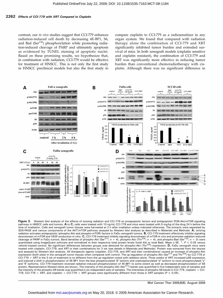
The early effects of a single treatment of ionizing radia-tion (2 Gy) and CCI-779 (5 mg/kg in vivo or 24 h treatmentof CCI-779 at 10 ng/mL in vitro) on the Akt/mTOR path-way were analyzed. Western blot analyses of tumors fromirradiated FaDu xenografts showed that ionizing radiationactivates antiapoptotic, prosurvival Akt/mTOR signaling.Although Akt phosphorylation at Ser473 was up-regulatedafter irradiation, phospho-Akt (Thr308) expression did notchange (Fig. 3A). Western blot analysis of irradiated FaDuand SCC40 cells revealed that, in contrast to the in vivo data,radiation had no effect on Akt phosphorylation in vitro(Fig. 3B). We tested the effect of ionizing radiation (2 Gy)and CCI-779 treatment on downstream targets of mTOR,4E-BP1, phospho-S6 ribosomal protein, and apoptotic mar-kers. CCI-779 treatment effectively inhibited phosphoryla-tion of S6 ribosomal protein at Ser235/Ser236 (a physiologicdownstream target of p70 S6 kinase; Fig. 3B and C) and4E-BP1 in HNSCC cell cultures and FaDu xenograft tumorsas evidenced by a shift of the more phosphorylated “δ” and“γ” isoforms and to the less phosphorylated “β” isoformand nonphosphorylated “α” isoform after Western blotanalysis of total 4E-BP1 (Fig. 3C). Treatment of FaDu andSCC40 cells with CCI-779 significantly inhibited VEGFsecretion in the medium when compared with the controlor irradiated samples (Fig. 3B). CCI-779 treatment signifi-cantly inhibited expression of phospho-Bad (Ser136) in FaDuxenograft tissues (P < 0.05; Fig. 3C), which facilitates apo-ptosis. We found that PARP cleavage, a crucial early markerof apoptosis, was up-regulated in FaDu xenograft tumors at6 h after irradiation (increased from 6% in control to 29%at 6 h after irradiation; Fig. 3C). CCI-779 treatment alone
Table 1. Mean ± SE tumor volume by treatment group and by time point for the established tumor models
FaDu xenografts Day 0 Day 7 Day 12 Day 14 Day 20
Control 37.5 ± 7.5 500.9 ± 104.7 1,444.9 ± 294.0* 1,316.0 ± 258.7 1,622.31 ± 397.8Cisplatin 38.6 ± 6.1 302.3 ± 120.9 896.4 ± 270.1* 879.2 ± 272.6 883.9 ± 353.5CCI-779 41.8 ± 7.5 107.3 ± 25.2 478.5 ± 65.4 508.1 ± 69.9 650.6 ± 80.0Cisplatin + CCI-779 40.7 ± 7.9 188.8 ± 88.1 340.0 ± 106.2 522.5 ± 157.3 511.7 ± 163.1XRT 39.0 ± 6.2 196.0 ± 40.0 467.1 ± 93.5 683.4 ± 158.7 844.4 ± 157.2Cisplatin + XRT 39.9 ± 7.9 249.1 ± 104.9 386.5 ± 149.0 465.9 ± 192.2 550.2 ± 218.1CCI-779 + XRT 40.5 ± 11.2 93.1 ± 36.4 132.7 ± 36.5 130.1 ± 41.3 209.4 ± 55.5Cisplatin + CCI-779 + XRT 39.8 ± 10.2 162.7 ± 73.6 198.2 ± 76.7 243.6 ± 110.9 218.5 ± 73.5
SCC40 xenografts Day 0 Day 7 Day 14 Day 17 Day 20
Control 37.2 ± 8.0 172.1 ± 45.0 722.2 ± 252.2 911.3 ± 323.7* 1,034.2 ± 340.93Cisplatin 37.3 ± 11.5 233.6 ± 70.2 745.0 ± 266.1 952.7 ± 295.9* 1,136.2 ± 274.2CCI-779 37.4 ± 7.5 101.0 ± 35.1 166.9 ± 95.3 202.4 ± 119.6 267.4 ± 161.2Cisplatin + CCI-779 37.2 ± 8.8 61.9 ± 15.5 115.3 ± 56.3 77.7 ± 39.3 86.0 ± 52.1XRT 37.3 ± 7.7 124.5 ± 15.6 294.9 ± 54.7 373.4 ± 72.7 583.1 ± 149.1Cisplatin + XRT 37.2 ± 8.1 120.0 ± 17.4 199.6 ± 52.4 252.0 ± 77.3 340.0 ± 89.8CCI-779 + XRT 37.4 ± 11.4 80.1 ± 17.1 81.6 ± 22.5 86.4 ± 21.4 58.5 ± 11.1Cisplatin + CCI-779 + XRT 37.3 ± 11.8 47.8 ± 9.9 49.4 ± 10.8 78.8 ± 37.0 49.5 ± 11.9
*The last day when all mice in the group were alive.
Molecular Cancer Therapeutics
Mol Cancer Ther 2009;8(8). August 2009
2259
on May 20, 2018. © 2009 American Association for Cancer Research. mct.aacrjournals.org Downloaded from
Published OnlineFirst July 22, 2009; DOI: 10.1158/1535-7163.MCT-08-1184

increased the percentage of cleaved PARP to 21% andsubstantially augmented radiation-induced cleavage ofPARP to 40%, which also occurred at an earlier time point,that is, 2 h after irradiation (Fig. 3C), suggesting that thedrug can facilitate apoptotic cell death caused by radiationin tumor tissues and therefore serve as a radiosensitizingagent.CCI-779 treatment up-regulated Akt phosphorylation at
Ser473 in both in vitro (Fig. 3B) and in vivo (Fig. 3C and D)settings, indicating that, in HNSCC experimental models,mTOR inhibition is disrupting the negative feedback loopthat suppresses PI3K/Akt signaling when S6K is phosphor-ylated. The changes in Akt (Thr308) phosphorylation afterCCI-779 were not significant (Fig. 3B and C). Although ion-izing radiation treatment did not up-regulate Akt phosphor-ylation in our in vitro studies (Fig. 3B), it caused an increasein phospho-Akt (Ser473) expression in xenograft tissue as
early as 2 h after irradiation (Fig. 3A, C, and D). Moreover,after 3 weeks of treatment, all three therapeutic agents (cis-platin, CCI-779, and XRT and their combinations) caused anincrease of phospho-Akt expression (both sites) in the xeno-graft tumor tissues when compared with control (Fig. 3D).Importantly, CCI-779 treatment did not further exacerbatecisplatin- and XRT-induced up-regulation of Akt phosphor-ylation at Ser473 and Thr308.The effects of prolonged 3-week treatment with CCI-779,
cisplatin, and XRTon mTOR signaling were examined in theFaDu xenograft model. Although we did not observe radi-ation-induced up-regulation of 4E-BP1 and S6 ribosomalprotein at early time points after irradiation (Fig. 3C),3 weeks of XRT (6 fractions of 2 Gy) resulted in a 2-foldincreased expression of pS6 and promoted phosphoryla-tion of 4E-BP1 as evidenced by a shift from less phosphor-ylated to more phosphorylated isoforms (Fig. 3D). In
Figure 2. Effects of XRT, CCI-779, and cisplatin and their combinations on tumor growth and survival. A, tumor growth-inhibitory effects of varioustreatment modalities in FaDu xenografts. Nude mice bearing FaDu xenografts were treated with either 5 mg/kg CCI-779 i.p. 5 d/wk, 1 mg/kg cisplatin (Cis)i.p. twice a week, radiation (XRT) to the xenograft at 2 Gy fractions twice a week, or the indicated combinations for 3 wk. Tumor growth curves arepresented as average ± SE tumor volumes. B, tumor growth-inhibitory effects of various treatment modalities in SCC40 xenografts. Treatment groupsare the same as in A. C and D, Kaplan-Meier curves of survival comparison between control and treated FaDu and SCC40 xenograft mice. Mice weresacrificed due to significant tumor burden (>2,000 mm3), weight loss >15%, or at maximum time point of 60 d after the treatment initiation. In bothxenograft survival studies, CCI-779 significantly improved survival compared with control (P < 0.05) and combination of CCI-779 with XRT significantlyimproved survival compared with XRT alone (P < 0.05).
Effects of CCI-779 with XRT Compared to Cisplatin
Mol Cancer Ther 2009;8(8). August 2009
2260
on May 20, 2018. © 2009 American Association for Cancer Research. mct.aacrjournals.org Downloaded from
Published OnlineFirst July 22, 2009; DOI: 10.1158/1535-7163.MCT-08-1184

contrast, CCI-779 decreased pS6 and caused a shift of 4E-BP1 phosphorylated bands to the unphosphorylated andless phosphorylated isoforms and significantly attenuatedradiation-induced up-regulation of these mTOR down-stream factors (P < 0.05).
Effect of CCI-779 on Vascularization and Apoptosis of
the FaDu Xenografts
The absence of radiation sensitizing effects noted in thein vitro group but not in the in vivo groups where CCI-779augmented the tumor growth-inhibitory effects of XRT ledus to evaluate the effects of CCI-779 on tumor-stromal inter-actions. To examine these interactions, we assessed the ef-fects of CCI-779 on angiogenesis by counting the numberof blood vessels in CD31-stained sections of xenografttumors following 3 weeks of treatment. At ×400 magnifica-tion, the average counts per field were as follows: 30.4 ± 5.8(mean ± SD) blood vessels for control tumors, 26.3 ± 4.7 forradiation alone, and 25.2 ± 1.4 for chemoradiotherapy withcisplatin (Fig. 4). XRTand chemoradiotherapy with cisplatindid not reduce the number of vessels significantly. Treat-ment with CCI-779 alone significantly reduced the intratu-moral microvessel density compared with control (17.6 ± 2.3vessels per field; P < 0.001 versus control and P < 0.05 ver-sus irradiation alone). The combination of CCI-779 withXRT even further enhanced the antiangiogenic effects ofCCI-779 (10.8 ± 2.3 vessels per field; P < 0.001 versus XRTalone; P < 0.05 versus CCI-779 alone).Apoptosis was assessed by TUNEL staining in xenograft
tumor tissues at early times after irradiation (2 h) and CCI-779 treatment (24 h) and following 3 weeks of treatment.Xenograft tumor tissues from vehicle-treated mice in theearly effects study had 1.52 ± 0.27% (mean ± SE) of the apo-ptotic TUNEL-positive nuclei per field. CCI-779 (24 h), XRT(2 h), and CCI-779 + XRT treatment significantly increasedthe percentage of apoptotic nuclei to 3.48 ± 0.09%, 3.41 ±0.21%, and 3.65 ± 0.33% correspondingly (P < 0.05 versuscontrol). Three weeks of treatment further increased theapoptosis in the FaDu xenograft tissues (Fig. 4, bottom). Theaverage percentage of apoptotic nuclei per field were asfollows: 2.16 ± 0.45% for control tumors, 4.41 ± 0.57% forCCI-779 alone, 6.48 ± 0.46% for XRT, and 9.3 ± 0.27% forchemoradiotherapy with cisplatin (Fig. 4). The combination
of CCI-779 and XRT augmented the percentage of apoptoticnuclei to 11.34 ± 0.92% (P < 0.001 versus XRT alone or CCI-779 alone; difference versus cisplatin + XRT is not significant).Representative photomicrographs of TUNEL-stained tumorsections from each treatment group are shown in Fig. 4.
DiscussionThe growth-inhibitory properties of mTOR inhibitors onvarious types of cancers have been well studied (29–32).We showed that CCI-779 is an effective agent againstHNSCC in both in vitro and in vivo studies (27). The combi-nation of mTOR inhibitors with cytotoxic treatments, suchas radiation therapy or chemotherapy, might potentiatethe antitumoral effects of mTOR inhibition (33). Hence, wewanted to elucidate the efficacy of using CCI-779 in combi-nation with XRT in preclinical HNSCC models and in com-parison with cisplatin. The efficacy of combining XRT withmTOR inhibitors have been studied in breast and prostatecancer cell models (24, 25) and murine glioma xenografts(26). However, the effects of CCI-779 and XRT have not beenstudied in HNSCC and have never been compared with cis-platin and XRT, which is a very commonly used radiosensi-tizer in several organ systems.In radiation therapy, activation of cytotoxic cellular me-
chanisms, such as induction of DNA damage and up-regulation of proapoptotic ASK1/JNK pathway, in tumorsis favorable. Additionally, irradiation further enhances theantiapoptotic PI3K/Akt/mTOR pathway, which is alreadyup-regulated in many types of cancer, including HNSCC.In our experiments, Akt (at Ser473) and mTOR were acti-vated in FaDu xenografts after irradiation (Fig. 3A). Rapa-mycin analogues, such as CCI-779 and RAD001, effectivelyinhibit signal transduction downstream of mTOR, attenu-ating cap-dependent translation of many pro-oncogenicand prosurvival factors, which may explain their radiosen-sitizing effects.We found that doses of CCI-779 that significantly inhibit
mTOR signaling have a growth-inhibitory effect and im-portantly cause significant inhibition of VEGF productionin the cells and did not sensitize HNSCC cell lines toradiation in vitro as evidenced by clonogenic assays. In
Table 2. Median survival time and survival rate in the treatment groups
Group FaDu xenograft mice SCC40 xenograft mice
n Survivalrate (%)
Median survival time(95% CI), d
n Survival rate (%) Median survival time(95% CI), d
Control 5 0.0 19 (15-28) 6 0.0 27 (20-34)Cisplatin 5 20.0 23 (13-∞) 5 0.0 22 (16-46)CCI-779 6 0.0 35 (28-40) 5 40.0 52 (34-∞)Cisplatin + CCI-779 6 16.7 28 (28-37) 6 40.0 55 (39-∞)XRT 6 0.0 37 (33-40) 6 16.7 35 (34-39)Cisplatin + XRT 6 33.3 47.5 (26-∞) 6 0.0 38 (36-43)CCI-779 + XRT 6 66.7 >56 (56-∞) 6 16.7 49 (46-57)Cisplatin + CCI-779 + XRT 6 16.7 52 (42-58) 6 66.7 >50 (50-∞)
Molecular Cancer Therapeutics
Mol Cancer Ther 2009;8(8). August 2009
2261
on May 20, 2018. © 2009 American Association for Cancer Research. mct.aacrjournals.org Downloaded from
Published OnlineFirst July 22, 2009; DOI: 10.1158/1535-7163.MCT-08-1184
contrast, our in vivo studies suggest that CCI-779 enhancesradiation-induced cell death by decreasing 4E-BP1, S6,and Bad (Ser136) phosphorylation while promoting radia-tion-induced cleavage of PARP and ultimately apoptosisas evidenced by TUNEL staining of apoptotic nuclei.Based on these promising results, we hypothesize that,in combination with radiation, CCI-779 would be effectivefor treatment of HNSCC. This is not only the first studyin HNSCC preclinical models but also the first study to
compare cisplatin to CCI-779 as a radiosensitizer in anyorgan system. We found that compared with radiationtherapy alone the combination of CCI-779 and XRTsignificantly inhibited tumor burden and extended sur-vival of mice. In both xenograft models (cisplatin sensitiveand cisplatin resistant), the combination of CCI-779 andXRT was significantly more effective in reducing tumorburden than conventional chemoradiotherapy with cis-platin. Although there was no significant difference in
Figure 3. Western blot analysis of the effects of ionizing radiation and CCI-779 on proapoptotic factors and antiapoptotic PI3K/Akt/mTOR signalingpathway in HNSCC cells and tumors. A to C, cells were treated with 10 ng/mL CCI-779 and mice were treated with 5 mg/kg of the drug 24 h before thetime of irradiation. Cells and xenograft tumor tissues were harvested at 2 h after irradiation unless indicated otherwise. The extracts were separated bySDS-PAGE and various components of the AKT/mTOR pathway assayed by Western blot analysis as described in Materials and Methods. A, ionizingradiation activates antiapoptotic (phospho-Akt and phospho-mTOR) factors in FaDu xenograft tumors. B, CCI-779 treatment effectively inhibits signalingdownstream of mTOR and VEGF production in vitro. C, CCI-779 treatment inhibits signaling downstream of mTOR in vivo and promotes radiation-inducedcleavage of PARP in FaDu tumors. Band densities of phospho-Akt (Ser473; n = 4), phospho-Akt (Thr308; n = 4), and phospho-Bad (Ser136; n = 3) werequantitated using ImageQuant software and normalized to their respective total protein levels (total Akt or total Bad). Mean ± SE. *, P < 0.05 versusvehicle-treated control. No significant differences between groups was detected for phospho-Akt (Thr308) expression. D, FaDu xenograft mice weretreated with cisplatin, CCI-779, and XRT or their combinations for 3 wk (see details in Materials and Methods). Protein was extracted from the tissuesand assayed by Western blot analysis. All therapeutic agents (cisplatin, CCI-779, and XRT and their combinations) caused an increase of phospho-Aktexpression (both sites) in the xenograft tumor tissues when compared with control. The up-regulation of phospho-Akt (Ser473 and Thr308) by CCI-779 orCCI-779 + XRT in the 3 wk of treatment is no different from the up-regulation noted with radiation alone. Three weeks of XRT increased pS6 expressionand induced a shift in phosphorylation of 4E-BP1 from the less phosphorylated “β” isoform and unphosphorylated “α” isoform to more phosphorylated “γ”and “δ” isoforms. CCI-779 treatment reversed radiation-induced phosphorylation of 4E-BP1 to some extent as well as decreased phosphorylation of S6protein. Representative Western blots are shown. The intensity of the phospho-Akt (Ser473) bands was quantified in five independent sets of samples andthe intensity of the phospho-S6 bands was quantified in six independent sets of samples. The intensities of phospho-S6 bands in CCI-779, cisplatin + CCI-779, CCI-779 + XRT, and cisplatin + CCI-779 + XRT groups were significantly different from those in XRT samples (P < 0.05).
Effects of CCI-779 with XRT Compared to Cisplatin
Mol Cancer Ther 2009;8(8). August 2009
2262
on May 20, 2018. © 2009 American Association for Cancer Research. mct.aacrjournals.org Downloaded from
Published OnlineFirst July 22, 2009; DOI: 10.1158/1535-7163.MCT-08-1184

survival of cisplatin-sensitive FaDu mice when comparedwith CCI-779, in the cisplatin-resistant SCC40 xenografts,CCI-779 and XRT combined significantly exceeded surviv-al of mice treated with cisplatin and XRT. Also, there wasno additional benefit when all three treatments (cisplatin,CCI-779, and XRT) were combined, which is important indesigning clinical trials where novel therapeutics typicallyare added to standard of care that already cause signifi-cant toxicity. The potent antitumoral efficacy of CCI-779as a single agent (>60% of tumor growth inhibition com-pared with control) precluded deciphering the exact modeof interaction (synergism, additive, and antagonism) be-tween CCI-779 and XRT. Importantly, the addition ofCCI-779 to XRT markedly enhanced the effects of radia-tion on tumor burden. Combined CCI-779 + XRT treat-ment inhibited tumor growth by >90% compared withcontrol as well as more than doubled the survival benefitof mice compared with XRT alone in both xenograftmodels. Importantly, the effects of combined CCI-779 +XRT treatment on angiogenesis and apoptosis in xeno-graft tissues exceeded the sum of those by the drug orXRT alone.Head and neck cancer is not the only type of cancer in
which dissonant results are observed when examining theradiosensitizing properties of mTOR inhibitors in an in vitroversus in vivo model. For example, in a glioma model, therewas no increased radiosensitivity with the mTOR inhibitorRAD001 treatment in vitro, whereas the drug significantlyenhanced radiation-induced regression of glioma xenografts(26). Conversely, in breast cancer cell lines, radiation sensi-tization was observed with RAD001 treatment in vitro (24).In our study, Akt phosphorylation was affected differentlyby ionizing radiation in cell cultures compared with a xeno-
graft model. Akt phosphorylation was not up-regulatedafter irradiation of FaDu and SCC40 cell cultures. It isknown that ionizing radiation effectively induces phosphor-ylation of Akt at Ser473 in the vascular endothelium withinminutes of irradiation (34), which may explain the observedincrease of phospho-Akt (Ser473) levels in the FaDu xeno-grafts that could most likely occur in tumor vascular endo-thelium. Radiation-induced activation of mTOR signalingwas absent in glioma GL261 cells and mTOR inhibitorsdid not sensitize them to radiation (26) similarly to whatwe observed in the HNSCC model. Radiation-induced up-regulation of Akt/mTOR signaling was observed in breastcancer cells (24) and human umbilical vein endothelial cells(26). mTOR inhibition attenuated radiation-induced up-regulation of Akt/mTOR signaling and sensitized breastcancer and HUVEC cells to radiation in vitro (24, 26). Also,mTOR inhibition sensitized vascular endothelium to radia-tion injury in vivo (26). mTOR inhibition reduces microves-sel density in tumor tissues by decreasing VEGF production(35–37). Our results show that CCI-779 significantly inhib-ited vascularization in the HNSCC xenografts. We haveshown that treatment of HNSCC cells with CCI-779 de-creases VEGF production that is consistent with antiangio-genic effects of the drug. Evidence suggests that the level ofVEGF might not only affect angiogenesis and radiation re-sistance of endothelial cells but also may have an effect onradiation resistance of tumor cells, including HNSCC (38).Although CCI-779 treatment decreased VEGF productionin HNSCC cells in vitro, it did not have an effect on radia-tion sensitivity, suggesting that a decrease in VEGF levelsalone is not sufficient for radiation sensitization of HNSCCcells. We found that CCI-779 treatment up-regulated Aktphosphorylation (Ser473) in vitro and in vivo, indicating
Figure 4. CCI-779 reducesmicrovessel density and increasesapoptosis in FaDu xenografts.Mice were treated with vehicle,5 mg/kg CCI-779 5 d/wk (i.p.),targeted irradiation (XRT) at 2 Gyfractions twice a week, a combi-nation of CCI-779 and XRT, anda combination of 1 mg/kg cisplatintwice a week (i.p.) and XRT for aperiod of 3 wk. For vessel densityquantification, tumor sectionswere stained with anti-CD31 anti-body and CD31+ microvesselswere counted at ×400 magnifica-tion. Representative sections fromeach treatment group are shownat ×200 magnification. To assessapoptosis, tumor sections weredeparaffinized, labeled usingTUNEL assay kit, and mountedusing medium containing 4′,6-diamidino-2-phenylindole and thepercentage of apoptotic (TUNEL-positive) nuclei was estimated inat least five fields not located innecrotic regions.
Molecular Cancer Therapeutics
Mol Cancer Ther 2009;8(8). August 2009
2263
on May 20, 2018. © 2009 American Association for Cancer Research. mct.aacrjournals.org Downloaded from
Published OnlineFirst July 22, 2009; DOI: 10.1158/1535-7163.MCT-08-1184
that in HNSCC experimental models mTOR inhibition isdisrupting the negative feedback loop that suppresses Aktphosphorylation (39, 40). However, the increase in phos-pho-Akt noted with CCI-779 and XRTwas not significantlydifferent from the increase in phospho-Akt noted with allother treatments, that is, cisplatin, XRT, and the combina-tion. Additionally, this increase did not affect the potentantitumoral effects of CCI-779 or its radioenhancing effects.In our study, CCI-779 showed potent antitumor activity
against cisplatin-sensitive as well as cisplatin-resistant celllines. Our data suggest that CCI-779 can be as effective as cis-platin in combinationwith XRT for the treatment of head andneck cancer. Because CCI-779 is a relatively well-tolerateddrug with mainly manageable and reversible dermatologicside effects, it is a promising therapy that can potentiallyreplace cisplatin with its known considerable toxicity.Because the PI3K/Akt/mTOR pathway in HNSCC is acritical regulator of multiple downstream effectors includ-ing angiogenesis, mitosis, and apoptosis, targeting thispathway during radiation therapy may be a key factorto blocking multiple other pathways enhancing the antitu-moral action of XRT.
Disclosure of Potential Conflicts of Interest
C-A.O. Nathan: Wyeth-Ayerst funded clinical trial in Louisiana StateUniversity Health Sciences Center-Shreveport. No other potential con-flicts of interest were disclosed.
Acknowledgments
We thank Cheryl Clark for help in the preparation of the article.Wyeth-Ayerst Research, manufacturer of CCI-779, had no role in
the study design, collection, analysis, interpretation of data, writing ordecision to submit the manuscript for publication.
References
1. Canto M, Devesa S. Oral cavity and pharynx cancer incidence rates inthe United States, 1975-1998. Oral Oncol 2002;38:610–7.
2. Jesse R, Sugarbaker E. Squamous cell carcinoma of the oropharynx:why we fail. Am J Surg 1976;132:435–8.
3. Howell G, Grandis J. Molecular mediators of metastasis in head andneck squamous cell carcinoma. Neck 2005;27:710–7.
4. Cooper J, Pajak T, Forastiere A, et al. Postoperative concurrent radio-therapy and chemotherapy for high-risk squamous-cell carcinoma of thehead and neck. N Engl J Med 2004;350:1937–44.
5. Ahmed K, Samant S, Vieira F. Gastrostomy tubes in patients with ad-vanced head and neck cancer. Laryngoscope 2005;115:44–7.
6. Lefebvre J. Current clinical outcomes demand new treatment optionsfor SCCHN. Ann Oncol 2005;16:vi7–12.
7. Thariat J, Yildirim G, Mason K, et al. Combination of radiotherapy withEGFR antagonists for head and neck carcinoma. Int J Clin Oncol 2007;12:99–110.
8. Bianco R, Shin I, Ritter C, et al. Loss of PTEN/MMAC1/TEP in EGFreceptor-expressing tumor cells counteracts the antitumor action of EGFRtyrosine kinase inhibitors. Oncogene 2003;22:2812–22.
9. Wheeler D, Huang S, Kruser T, et al. Mechanisms of acquired resistanceto cetuximab: role of HER (ErbB) family members. Oncogene 2008;27:3944–56.
10. Sok J, Coppelli F, Thomas S, et al. Mutant epidermal growth factorreceptor (EGFRvIII) contributes to head and neck cancer growth and resis-tance to EGFR targeting. Clin Cancer Res 2006;12:5064–73.
11. Bianco R, Garofalo S, Rosa R, et al. Inhibition of mTOR pathway byeverolimus cooperates with EGFR inhibitors in human tumours sensitiveand resistant to anti-EGFR drugs. Br J Cancer 2008;98:923–30.
12. Janmaat M, Kruyt F, Rodriguez J, Giaccone G. Response to epidermalgrowth factor receptor inhibitors in non-small cell lung cancer cells: limitedantiproliferative effects and absence of apoptosis associated with persis-tent activity of extracellular signal-regulated kinase or Akt kinase path-ways. Clin Cancer Res 2003;9:2316–26.
13. Amornphimoltham P, Patel V, Sodhi A, et al. Mammalian target ofrapamycin, a molecular target in squamous cell carcinomas of the headand neck. Cancer Res 2005;65:9953–61.
14. Datta S, Brunet A, Greenberg M. Cellular survival: a play in three Akts.Genes Dev 1999;13:2905–27.
15. Molinolo A, Hewitt S, Amornphimoltham P, et al. Dissecting the Akt/mammalian target of rapamycin signaling network: emerging results fromthe head and neck cancer tissue array initiative. Clin Cancer Res 2007;13:4964–73.
16. Nathan C, Amirghahar N, Abreo F, et al. Overexpressed eIF4E isfunctionally active in surgical margins of head and neck cancer patientsvia activation of the Akt/mammalian target of rapamycin pathway. ClinCancer Res 2004;10:5820–7.
17. Amornphimoltham P, Sriuranpong V, Patel V, et al. Persistent acti-vation of the Akt pathway in head and neck squamous cell carcinoma:a potential target for UCN-01. Clin Cancer Res 2004;10:4029–37.
18. Sunavala-Dossabhoy G, Fowler M, Benedetti AD. Translation of theradioresistance kinase TLK1B is induced by γ-irradiation through activationof mTOR and phosphorylation of 4E-BP1. BMC Mol Biol 2004;5:1.
19. Reits E, Hodge J, Herberts C, et al. Radiation modulates the peptiderepertoire, enhances MHC class I expression, and induces successful anti-tumor immunotherapy. J Exp Med 2006;203:1259–71.
20. Chan S, Scheulen M, Johnston S, et al. Phase II study of temsirolimus(CCI-779), a novel inhibitor of mTOR, in heavily pretreated patients withlocally advanced or metastatic breast cancer. J Clin Oncol 2005;23:5314–22.
21. Hidalgo M, Buckner J, Erlichman C, et al. A phase I and pharmacoki-netic study of temsirolimus (CCI-779) administered intravenously daily for5 days every 2 weeks to patients with advanced cancer. Clin Cancer Res2006;12:5755–63.
22. Raymond E, Alexandre J, Faivre S, et al. Safety and pharmacokineticsof escalated doses of weekly intravenous infusion of CCI-779, a novelmTOR inhibitor, in patients with cancer. J Clin Oncol 2004;22:2336–47.
23. Sarkaria J, Schwingler P, Schild S, et al. Phase I trial of sirolimus com-bined with radiation and cisplatin in non-small cell lung cancer. J ThoracOncol 2007;2:751–7.
24. Albert J, Kim K, Cao C, Lu B. Targeting the Akt/mammalian target ofrapamycin pathway for radiosensitization of breast cancer. Mol CancerTher 2006;5:1183–9.
25. Cao C, Subhawong T, Albert J, et al. Inhibition of mammalian target ofrapamycin or apoptotic pathway induces autophagy and radiosensitizesPTEN null prostate cancer cells. Cancer Res 2006;66:10040–7.
26. Shinohara E, Cao C, Niermann K, et al. Enhanced radiation damage oftumor vasculature by mTOR inhibitors. Oncogene 2005;24:5414–22.
27. Nathan C, Amirghahari N, Rong X, et al. Mammalian target of rapamy-cin inhibitors as possible adjuvant therapy for microscopic residual diseasein head and neck squamous cell cancer. Cancer Res 2007;67:2160–8.
28. Shalinsky D, Bischoff E, Gregory M, et al. Enhanced antitumor effi-cacy of cisplatin in combination with ALRT1057 (9-cis retinoic acid) inhuman oral squamous carcinoma xenografts in nude mice. Clin CancerRes 1996;2:511–20.
29. Dudkin L, Dilling M, Cheshire P, et al. Biochemical correlates of mTORinhibition by the rapamycin ester CCI-779 and tumor growth inhibition.Clin Cancer Res 2001;7:1758–64.
30. Mabuchi S, Altomare D, Cheung M, et al. RAD001 inhibits humanovarian cancer cell proliferation, enhances cisplatin-induced apoptosis,and prolongs survival in an ovarian cancer model. Clin Cancer Res 2007;13:4261–70.
31. Wu L, Birle D, Tannock I. Effects of the mammalian target of rapamy-cin inhibitor CCI-779 used alone or with chemotherapy on human prostatecancer cells and xenografts. Cancer Res 2005;65:2825–31.
32. Zitzmann K, Toni ED, Brand S, et al. The novel mTOR inhibitorRAD001 (everolimus) induces antiproliferative effects in human pancreaticneuroendocrine tumor cells. Neuroendocrinology 2007;85:54–60.
Effects of CCI-779 with XRT Compared to Cisplatin
Mol Cancer Ther 2009;8(8). August 2009
2264
on May 20, 2018. © 2009 American Association for Cancer Research. mct.aacrjournals.org Downloaded from
Published OnlineFirst July 22, 2009; DOI: 10.1158/1535-7163.MCT-08-1184
33. Dancey J, Chen H. Strategies for optimizing combinations of molecu-larly targeted anticancer agents. Nat Rev Drug Discov 2006;5:649–59.
34. Tan J, Geng L, Yazlovitskaya E, Hallahan D. Protein kinase B/Akt-dependent phosphorylation of glycogen synthase kinase-3β in irradiatedvascular endothelium. Cancer Res 2006;66:2320–7.
35. Bufalo DD, Ciuffreda L, Trisciuoglio D, et al. Antiangiogenic potentialof the mammalian target of rapamycin inhibitor temsirolimus. Cancer Res2006;66:5549–54.
36. Guba M, Breitenbuch Pv, Steinbauer M, et al. Rapamycin inhibits pri-mary and metastatic tumor growth by antiangiogenesis: involvement ofvascular endothelial growth factor. Nat Med 2002;8:128–35.
37. Wan X, Shen N, Mendoza A, Khanna C, Helman L. CCI-779 inhibits
rhabdomyosarcoma xenograft growth by an antiangiogenic mechanismlinked to the targeting of mTOR/Hif-1α/VEGF signaling. Neoplasia 2006;8:394–401.
38. Brieger J, Kattwinkel J, Berres M, Gosepath J, Mann W. Impact ofvascular endothelial growth factor release on radiation resistance. OncolRep 2007;18:1597–601.
39. Sun S, Rosenberg L, Wang X, et al. Activation of Akt and eIF4E sur-vival pathways by rapamycin-mediated mammalian target of rapamycininhibition. Cancer Res 2005;65:7052–8.
40. O'Reilly K, Rojo F, She Q, et al. mTOR inhibition induces upstreamreceptor tyrosine kinase signaling and activates Akt. Cancer Res 2006;66:1500–8.
Molecular Cancer Therapeutics
Mol Cancer Ther 2009;8(8). August 2009
2265
on May 20, 2018. © 2009 American Association for Cancer Research. mct.aacrjournals.org Downloaded from
Published OnlineFirst July 22, 2009; DOI: 10.1158/1535-7163.MCT-08-1184

2009;8:2255-2265. Published OnlineFirst July 22, 2009.Mol Cancer Ther Oleksandr Ekshyyan, Youhua Rong, Xiaohua Rong, et al. carcinomaexperimental models of head and neck squamous celltarget of rapamycin inhibitor CCI-779 to cisplatin in Comparison of radiosensitizing effects of the mammalian
Updated version
10.1158/1535-7163.MCT-08-1184doi:
Access the most recent version of this article at:
Material
Supplementary
http://mct.aacrjournals.org/content/suppl/2009/08/19/1535-7163.MCT-08-1184.DC1
Access the most recent supplemental material at:
Cited articles
http://mct.aacrjournals.org/content/8/8/2255.full#ref-list-1
This article cites 40 articles, 22 of which you can access for free at:
Citing articles
http://mct.aacrjournals.org/content/8/8/2255.full#related-urls
This article has been cited by 4 HighWire-hosted articles. Access the articles at:
E-mail alerts related to this article or journal.Sign up to receive free email-alerts
Subscriptions
Reprints and
To order reprints of this article or to subscribe to the journal, contact the AACR Publications
Permissions
Rightslink site. (CCC)Click on "Request Permissions" which will take you to the Copyright Clearance Center's
.http://mct.aacrjournals.org/content/8/8/2255To request permission to re-use all or part of this article, use this link
on May 20, 2018. © 2009 American Association for Cancer Research. mct.aacrjournals.org Downloaded from
Published OnlineFirst July 22, 2009; DOI: 10.1158/1535-7163.MCT-08-1184