complement fixation by rheumatoid...
TRANSCRIPT
Complement Fixation by Rheumatoid Factor
KiYoAmI TANIMOTO, NEIL R. COOPER,JOHNS. JOHNSON,andJoHN H. VAUGHAN
From the Rheumatology Division and the Department of ExperimentalPathology, Scripps Clinic and Research Foundation, La Jolla, California 92037
A B S T R A C T The capacity for fixation and activationof hemolytic complement by polyclonal IgM rheumatoidfactors (RF) isolated from sera of patients with rheu-matoid arthritis and monoclonal IgM-RF isolated fromthe cryoprecipitates of patients with IgM-IgG mixedcryoglobulinemia was examined. RF mixed with aggre-gated, -reduced, and alkylated human IgG (Agg-R/A-IgG) in the fluid phase failed to significantly reducethe level of total hemolytic complement, CHuo, or ofindividual complement components, Cl, C2, C3, and C5.However, sheep erythrocytes (SRC) coated with Agg-R/A-IgG or with reduced and alkylated rabbit IgGanti-SRC antibody were hemolyzed by complement inthe presence of polyclonal IgM-RF. Human and guineapig complement worked equally well. The degree ofhemolysis was in direct proportion to the hemagglutina-tion titer of the RF against the same coated cells. Mono-clonal IgM-RF, normal human IgM, and purifiedWaldenstrdm macroglobulins without antiglobulin ac-tivity were all inert.
Hemolysis of coated SRC by RF and complementwas inhibited by prior treatment of the complementsource with chelating agents, hydrazine, cobra venomfactor, specific antisera to Clq, C4, C5, C6, or C8, orby heating at 560C for 30 min. Purified radiolabeledC4, C3, and C8 included in the complement sourcewere bound to hemolysed SRC in direct proportion tothe degree of hemolysis. These data indicate that poly-clonal IgM-RF fix and activate complement via theclassic pathway.
The system described for assessing complement fixa-tion by isolated RF is readily adaptable to use withwhole human serum.
This work was presented in part at the 57th annualmeeting of the Federation of American Societies for Ex-perimental Biology at Atlantic City, N. J., 1973 (1973. Fed.Proc. 32: 959. (Abstr. 4153.).
Received for publication 28 February 1974 and in revisedform 26 August 1974.
INTRODUCTION
Complement participates in several biological reactionsincluding immune cytolysis, phagocytosis, immune ad-herence, and chemotaxis (reviewed in references 1, 2).The role of complement in the pathogenesis of anumber of human diseases including rheumatoid ar-thritis is not clear. The similarity of vascular lesionsin rheumatoid arthritis (3) with those of the immunecomplex disease, experimental serum sickness (4), sug-gests that complement might play a role in the patho-genesis of rheumatoid vasculitis. The role of rheumatoidfactor (RF) ' in rheumatoid arthritis is also unclear.Patients with highest titers of RF appear to be afflictedwith more severe disease (5, 6), and in experimentalanimals RF appear to augment tissue damage (7, 8).Both RF and complement have been identified in thelesions of rheumatoid arthritis (9, 10). Crucial to theelucidation of the role of RF and complement in thepossible pathogenesis of rheumatoid arthritis is thequestion of whether complement with its potential forproducing inflammation and alterations in vascularpermeability is activated by RF. Although a number ofrecent studies (11-15) indicate that RF probably doesfix complement under special conditions, other studies(14, 16-21) suggest that RF may, in fact, interferewith complement fixation under other circumstances.Because of uncertainty about the ability of RF to ini-tiate complement fixation, we have employed a differentapproach based on lysis of RF-sensitized cells by com-
'Abbreviations used in this paper: Agg-R/A-IgG, aggre-gated, reduced, and alkylated human IgG; EGTA, ethylene-glycol-bis- (amino ethyl) -tetraacetic acid; GPC, guinea pigcomplement; HuC, human complement; NHS, normal hu-man serum; PBS, 0.15 M NaCl buffered to pH 7.4 withsodium phosphate; R/A-IgG, reduced and alkylated humanIgG; R/A-Hem, reduced and alkylated rabbit IgG-hemoly-sin; REA, SRC coated with R/A-Hem; RF, rheumatoidfactor; SRC, sheep erythrocytes; tHEA, tanned SRCcoatedwith Agg-R/A-IgG; V0, void volume.
The Journal of Clinical Investigation Volume 55 March 1975 347-445 437
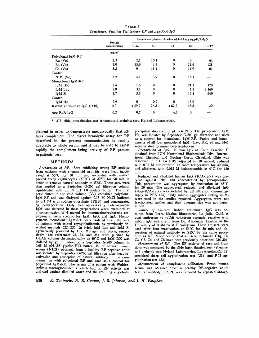
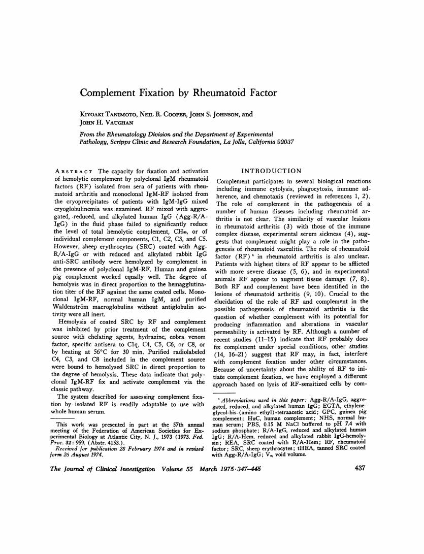
TABLE I
Complement Fixation Test between RFand Agg-R/A-IgG
Percent complement fixation with 0.2 mg Agg-R/A-IgGProtein
concentration CHbo C I C2 C3 LFT*
mg/miPolyclonal IgM-RF
Ra (Vo) 2.3 2.1 10.1 0 0 64Ba (Vo) 2.0 15.9 4.3 0 22.6 128Ca (Vo) 3.5 0 15.1 0 14.9 64
ControlNHS (Vo) 3.2 4.1 13.9 0 16.1
Monoclonal IgM-RFIgM DK 2.4 1.5 0 0 24.7 320IgM Lay 2.9 3.1 0 0 4.1 2,560IgM Si 2.7 5.3 0 0 12.4 640
ControlIgM He 2.9 0 0.9 0 13.9
Rabbit antihuman IgG (1:10) 6.7 >59.2 76.3 >67.2 18.3 20
Agg-R/A-IgG 0.2 0.7 0 4.2 0
* LFT, slide latex fixation test (rheumatoid arthritis test, Hyland Laboratories).
plement in order to demonstrate unequivocally that RFtixes complement. The direct hemolytic assay for RFdescribed in the present communication is readilyadaptable to whole serum, and it may be used to assessrapidly the complement-fixing activity of RF presentin patients' sera.
METHODSPreparation of RF. Sera exhibiting strong RF activity
from patients with rheumatoid arthritis were heat inacti-vated at 560C for 30 min and incubated with washedpacked sheep erythrocytes (SRC) at 370C for 90 min inorder to remove natural antibody to SRC. These sera werethen applied to a Sephadex G-200 gel filtration columnequilibrated with 0.1 N pH 4.0 acetate buffer. The firstpeak eluted in the void volume (V.) contained polyclonalIgM-RF and was dialyzed against 0.15 M NaCl bufferedto pH 7.4 with sodium phosphate (PBS) and concentratedby pervaporation. Only electrophoretically heterogeneousIgM was detected in these preparations when examined ata concentration of 4 mg/ml by immunoelectrophoresis em-ploying antisera specific for IgM, IgG, and IgA. Homo-geneous monoclonal IgM-RF were isolated from the seraof patients with mixed cryoglobulinemia by previously de-scribed methods (22, 23). In brief, IgM Lay and IgM Si(generously provided by Drs. Metzger and Stone, respec-tively; see references 22, 24, and 25) were purified byDEAEcolumn chromatography at 40°C and IgM DK wasisolated by gel filtration on a Sephadex G-200 column in0.05 M pH 2.5 glycine-HCl buffer. V. of normal humanserum (NHS) obtained from a healthy RF-negative adultwas isolated by Sephadex G-200 gel filtration after heat in-activation and absorption of natural antibody in the samemanner as with polyclonal RF and used as a control forpolyclonal IgM-RF. The serum of a patient with Walden-str6m's macroglobulinemia which had no RF activity wasdialyzed against distilled water and the resulting euglobulin
precipitate dissolved in pH 7.4 PBS. The paraprotein, IgMHe, was isolated by Sephadex G-200 gel filtration and usedas a control for monoclonal IgM-RF. Purity and homo-geneity of all four monoclonal IgM (Lay, DK, Si, and He)were verified by immunoelectrophoresis.
Preparation of IgG. Human IgG as Cohn Fraction IIobtained from ICN Nutritional Biochemicals Div., Interna-tional Chemical and Nuclear Corp., Cleveland, Ohio wasdissolved in pH 7.4 PBS adjusted to 10 mg/ml, reducedwith 0.01 M dithiothreitol at room temperature for 30 min,and alkylated with 0.015 M iodoacetamide at 0°C for 120min.
Reduced and alkylated human IgG (R/A-IgG) was dia-lyzed against PBS and concentrated by pervaporation.This preparation was aggregated by incubation at 63°Cfor 20 min. The aggregated, reduced, and alkylated IgG(Agg-R/A-IgG) was isolated by gel filtration chromatog-raphy in PBS (23). Only soluble aggregates eluting in V.were used in the studies reported. Aggregates were notfractionated further and their average size was not deter-mined.
Source of antisera. Rabbit antihuman IgG was ob-tained from Terra Marine Bioresearch, La Jolla, Calif. Agoat antiserum to rabbit colostrum strongly reactive withrabbit IgG was a gift from Dr. Alexander Lawton of theUniversity of Alabama at Birmingham. These antisera wereused after heat inactivation at 56°C for 30 min and ab-sorption of natural antibody to SRC by the same proce-dure as RF. Monospecific goat antisera to human Clq, C4,C3, C5, C6, and C8 have been previously described (26-30).
Measurement of RF. The RF activity of sera and frac-tions was measured by the slide latex fixation test (rheuma-toid arthritis test, Hyland Laboratories, LosAngeles,Calif.),sensitized sheep cell agglutination test (31), and F II ag-glutination test (32).
Measurement of complement utilization. Fresh humanserum was obtained from a healthy RF-negative adult.Natural antibody to SRC was removed by repeated absorp-
438 K. Tanimoto, N. R. Cooper, 1. S. Johnson, and J. H. Vaughan
100-
80-
'Z
* 60-
40-
20-
Pelyctemal IgMEFIVoI i-.-Hermai Mcmai Serum INNS, TVl o---o
1:50 1:200 1:800 1:3200R/A.IgG Hemolysin
FIGURE 1 SRC coated with R/A-Hem were incubated withconstant amounts of two different IgM-RF preparations(@*-) (Ra and Ba, see Table I). Percent hemolysis isplotted versus the dilution of R/A-IgG used to coat thecells. IgM prepared from RF-negative NHS (---- -0) isthe control. Cell lysis at 1: 50 R/A-IgG hemolysin is non-specific due to residual unreduced hemolysin. At 1:200R/A-IgG, lysis by IgM-RF is specific.
tion with washed packed SRC at 0C. Lyophilized guineapig serum purchased from Hyland Laboratories was re-constituted and absorbed with SRC as above. These ab-sorbed human and guinea pig sera were stored in smallaliquots at - 70'C until they were used as the humancomplement (HuC) or guinea pig complement (GPC)source.
Rabbit anti-SRC hemolysin, purchased from BBL, Divi-sion of Becton, Dickinson and Company, Cockeysville, Md.,was separated into IgM and IgG fractions by SephadexG-200 gel filtration. SRC were sensitized with IgM-hemo-lysin at four times the optimal sensitizing dose (IgM-EA)(33).
Testing for complement fixation in the fluid phase by RF(V.) was done by adding 0.5 ml of each RF preparationand 200 jig Agg-R/A-IgG to HuC or GPCdiluted in 0.15M Veronal-buffered saline containing 0.1% gelatin andoptimal Ca-+ and Mg-+ (GVB++) and incubating at 370Cfor 60 min. The mixture was stored in convenient aliquotsat - 70'C. Residual CH68 was measured by a modificationof Mayer's methods using IgM-EA in a total reactionvolume of 1.5 ml (34). C1, C2, and C3 hemolytic activitieswere determined by published methods (34-36).
Hemolysis of sensitized SRC by RF preparations andcomplement utilized SRC sensitized with reduced and alky-lated rabbit IgG-hemolysin (REA), or tanned SRC coatedwith Agg-R/A-IgG (tHEA). For preparation of the for-mer cells, IgG-hemolysin isolated by gel filtration was re-duced and alkylated (R/A-Hem) in order to diminish itscomplement-fixing activity. The hemagglutination titer ofthis preparation was 1: 40. SRCwere sensitized in the samemanner as with nonreduced and alkylated hemolysin. Forpreparation of tanned cells, washed SRC were suspendedin pH 7.2 PBS and treated with 1: 80,000 diluted tannicacid by a modification of Boyden's method (37). After tan-
nic acid treatment, the cells were suspended to 2% (4 X 10⁸/ml) in pH 6.4 PBS and reacted with an equal volume of200 Ag/ml of Agg-R/A-IgG.
For assessing complement-mediated hemolysis by RF, 0.2ml of each RF preparation was incubated with REA ortHEA (2 x 10⁸/ml) at 370C for 60 min and at 0C for3 h and the cells were washed three times with GVB++.Serial dilutions of the absorbed HuC and GPC sourceswere incubated with the sensitized cells at 370C for 60 minand the degree of hemolysis was assessed by spectrophoto-metric measurement (412 nm) of hemoglobin released intothe supernate.
Radiolabeled complement uptake. Purified HuC compo-nents C3, C4, and C8 were prepared by published methods(26-28), trace labeled with '1I by the chloramine-T method(38) without loss of activity, and used for quantitative cell
binding studies (39). SRC sensitized with R/A-Hem werereacted with the RF preparations as described in the pre-vious section. To a 1% suspension of these cells was added0.1 ml of a 1: 5 dilution of HuC containing 1-6 X 10⁶ cpmof 'I-labeled C3 (approximately 20 uig), C4 (approxi-mately 5 jig), or C8 (approximately 5 gig). The suspensionwas incubated at 370C for 60 min, centrifuged, and an ali-quot of the supernate removed for determination of thedegree of hemolysis. SRC stromata were washed three timeswith PBS by centrifugation at 20,000 rpm in a Spinco 40Srotor (Beckman Instruments, Inc., Spinco Div., Palo Alto,Calif.). Stroma-bound radioactivity was determined in awell-type gammascintillation spectrometer.
Miscellaneous. All chemicals were of reagent grade. Thesources and use of cobra venom and hydrazine have beenpreviously cited (40 43).
RESULTSEffect of RF on complement in free solution. Rheu-
matoid arthritis preparations were incubated with Agg-
100-
80-
E -
840-
2
Pelyclemal 1gM-IF *-.- HeS
\a-
1:2 1:4 1:8 1:16 1:32RF Dilution
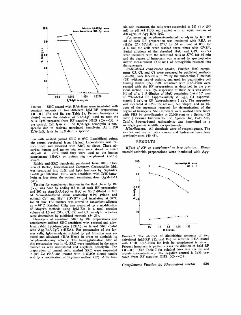
FIGURE 2 The abilities of diminishing amounts of two
polyclonal IgM-RF (Ra and Ba) to sensitize REA coatedwith 1: 500 R/A-Hem for lysis by complement is shown.Percent hemolysis is plotted versus the dilution of IgM-RF(0-*). (See Table I for original latex fixation test andprotein concentrations.) The negative control is IgM pre-pared from RF-negative NHS (-- -- 0).
Complement Fixation by Rheumatoid Factor 439
II
x
N
xN
--O-- ----
S 0---
-- --*-- --
o----- --O- --
100
80
;so-._
E 60-
40
20
100-
so-80
._LI; 60-
40-
20-
HuC0
0
0
0
a
Pelyclnal 1gM-IF .InIS
monoclonal glgM-F a
Nemi F meonclonal 1gM-HF oGoat autirabbit 1gC A
IT ,
GPC 0
* -
,,
U. U~~~
O 2 4 6 8RF Titer (2')
10
FIGURE 3 Percent hemolysis of REA incubated with vari-ous IgM (V.) preparations and two sources of comple-ment is compared with agglutination titer (2") measuredby these same cells. Percent hemolysis produced by HuCand GPC correlates directly with agglutination titer forpolyclonal IgM-RF (0) (r, = 0.992, P < 0.01 for HuCand r = 0.882, P < 0.01 for GPG). Non-RF monoclonalIgM (Ol) and normal human IgM (0) neither aggluti-nated nor sensitized REA for lysis by either complementsource. Monoclonal IgM-RF (U) did not trigger hemoly-sis. Goat anti-rabbit IgG (A) was weakly agglutinating butstrongly hemolytic with both complement sources.
R/A-IgG (see Methods) following which residualcomplement (CHso, C1, C2, and C3) was titrated. Thestudies shown in Table I employed undiluted humanserum as a complement source. Identical results wereobtained with dilute complement. None of the mono-clonal and polyclonal RF significantly reduced CHsoor Cl, C2, or C3 activities. One polyclonal RF (Ra)and one monoclonal IgM RF (Lay) were also ex-amined at 40C with results identical to those at 37°C.Fixation by rabbit antihuman IgG, the positive con-trol, was observed.
Lysis of SRC coated with R/A-Hem. In contrastto these results which do not show significant comple-ment fixation by RF in free solution, some of the RFpreparations readily induced lysis of SRC coated withR/A-IgG hemolysin in the presence of complement.SRC coated with various dilutions of R/A-Hem wereincubated with constant amounts of two different poly-clonal RF preparations and washed and incubated with
complement. Fig. 1 shows that the two polyclonal RFpreparations were able to sensitize REA for lysis bycomplement. The IgM (V.) prepared from NHS alsosensitized the cells for lysis, but only in higher dilu-tion due presumably to residual nonreduced hemolysin.At a 1: 200 dilution of R/A-IgG, greater than 80%lysis of the cells sensitized with polyclonal RF wasobserved, while the control gave less than 5% lysis. Insubsequent experiments, REA were prepared with a1: 500 dilution of R/A-Hem.
The effect of diminishing amounts of two polyclonalRF on the ability to sensitize REA for lysis is shownin Fig. 2. Significant hemolysis, 50% and 75%, re-spectively, was observed even at a 1: 32 dilution ofthese two preparations (original sensitized sheep cellagglutination test titers of Ra and Ba preparations are1: 256 and 1: 128). Since the slope of decrement ofhemolysis in this and subsequent experiments (see Figs.3 and 5) indicate the hemolytic end point would havebeen at 1: 256 or 1: 512 RF dilution, these resultsindicate even small amounts of RF are able to sensitizecells coated with R/A-Hem for lysis.
Hemolytic activities of seven polyclonal and threemonoclonal IgM-RF preparations were studied in thissystem. Hemagglutinating activity of the RF on thesame sensitized cells was also determined. All seven
100-
80- Pelyclemal 1gMlRF
,,,60-
40-
20-
200pg/ml 25pg/ml 6.25pg/mi 1.5625pg/miAgg-R/A*IgG
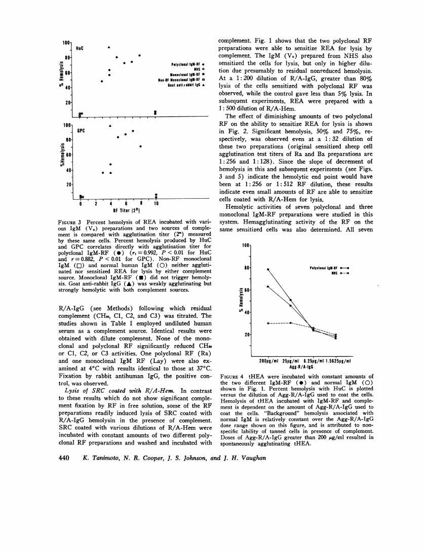
FIGURE 4 tHEA were incubated with constant amounts ofthe two different IgM-RF (0) and normal IgM (0)shown in Fig. 1. Percent hemolysis with HuC is plottedversus the dilution of Agg-R/A-IgG used to coat the cells.Hemolysis of tHEA incubated with IgM-RF and comple-ment is dependent on the amount of Agg-R/A-IgG used tocoat the cells. "Background" hemolysis associated withnormal IgM is relatively constant over the Agg-R/A-IgGdose range shown on this figure, and is attributed to non-specific lability of tanned cells in presence of complement.Doses of Agg-R/A-IgG greater than 200 ,ug/ml resulted inspontaneously agglutinating tHEA.
440 K. Tanimoto, N. R. Cooper, J. S. Johnson, and J. H. Vaughan
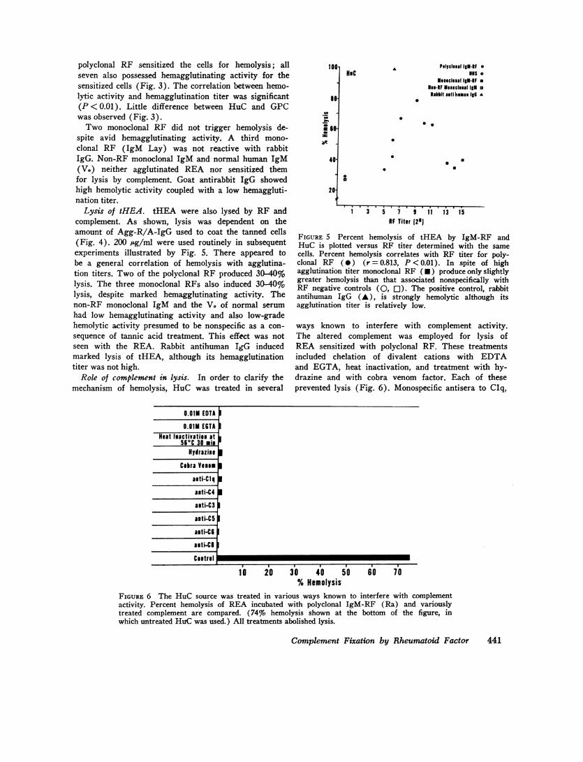
polyclonal RF sensitized the cells for hemolysis; allseven also possessed hemagglutinating activity for thesensitized cells (Fig. 3). The correlation between hemo-lytic activity and hemagglutination titer was significant(P <0.01). Little difference between HuC and GPCwas observed (Fig. 3).
Two monoclonal RF did not trigger hemolysis de-spite avid hemagglutinating activity. A third mono-clonal RF (IgM Lay) was not reactive with rabbitIgG. Non-RF monoclonal IgM and normal human IgM(V.) neither agglutinated REA nor sensitized themfor lysis by complement. Goat antirabbit IgG showedhigh hemolytic activity coupled with a low hemaggluti-nation titer.
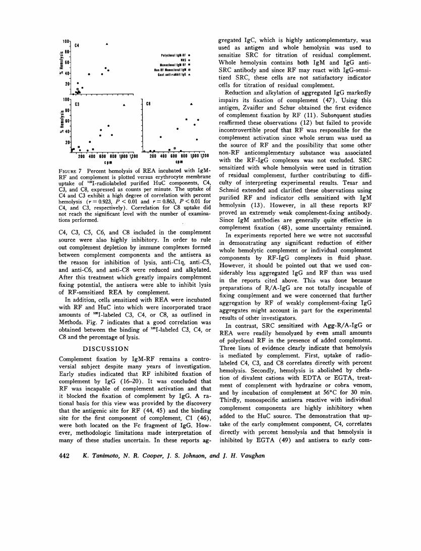
Lysis of tHEA. tHEA were also lysed by RF andcomplement. As shown, lysis was dependent on theamount of Agg-R/A-IgG used to coat the tanned cells(Fig. 4). 200 ,Ag/ml were used routinely in subsequentexperiments illustrated by Fig. 5. There appeared tobe a general correlation of hemolysis with agglutina-tion titers. Two of the polyclonal RF produced 30-40%lysis. The three monoclonal RFs also induced 30-40%lysis, despite marked hemagglutinating activity. Thenon-RF monoclonal IgM and the V. of normal serumhad low hemagglutinating activity and also low-gradehemolytic activity presumed to be nonspecific as a con-sequence of tannic acid treatment. This effect was notseen with the REA. Rabbit antihuman IgG inducedmarked lysis of tHEA, although its hemagglutinationtiter was not high.
Role of complement in lysis. In order to clarify themechanism of hemolysis, HuC was treated in several
100l
S10
E leOm
_f
40'
20-
HOC
.
Pelyclsal 1g-lFNlS o
MEseclemal 1gM-CF .Nei-IF Memeclemal 1gM o
Rabbit a.ti ho.al IgC A
.
.
0a
1 3 5 7 9 11RF Titer 12')
13 15
FIGURE 5 Percent hemolysis of tHEA by IgM-RF andHuC is plotted versus RF titer determined with the samecells. Percent hemolysis correlates with RF titer for poly-clonal RF (0) (r = 0.813, P < 0.01). In spite of highagglutination titer monoclonal RF (*) produce only slightlygreater hemolysis than that associated nonspecifically withRF negative controls (0, [1). The positive control, rabbitantihuman IgG (A), is strongly hemolytic although itsagglutination titer is relatively low.
ways known to interfere with complement activity.The altered complement was employed for lysis ofREA sensitized with polyclonal RF. These treatmentsincluded chelation of divalent cations with EDTAand EGTA, heat inactivation, and treatment with hy-drazine and with cobra venom factor. Each of theseprevented lysis (Fig. 6). Monospecific antisera to Clq,
0.01M EDTA
0.01M EGTAHeat Inactivatien at
560C 30 miHydrazine
Cobra Veome
alti-Clq
anti-C4
auti-C3
aiti-C5
anti-C6
anti-CS
Ceitral
I
I
I
I I I---=~~~~~*
10 20 30 40 50 60 70% Hemolysis
FIGURE 6 The HuC source was treated in various ways known to interfere with complementactivity. Percent hemolysis of REA incubated with polyclonal IgM-RF (Ra) and variouslytreated complement are compared. (74% hemolysis shown at the bottom of the figure, inwhich untreated HuCwas used.) All treatments abolished lysis.
Complement Fixation by Rheumatoid Factor 441
C4
Palyclelal IgM.RFNHS o
MoseclalaIgMRFNaR-RF Momeclanal 1gM o
Goat antirabbit IgG A
C3 A
. .
rn.I.
200 400 600 800 1,P000 1;0cpu
C8
a
200 400 600 800 100 1200Cpu
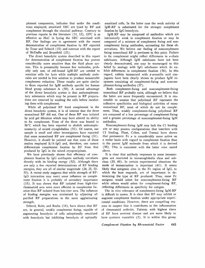
FIGURE 7 Percent hemolysis of REA incubated with IgM-RF and complement is plotted versus erythrocyte membraneuptake of '"I-radiolabeled purified HuC components, C4,C3, and C8, expressed as counts per minute. The uptake ofC4 and C3 exhibit a high degree of correlation with percenthemolysis (r = 0.923, P < 0.01 and r = 0.863, P <0.01 forC4, and C3, respectively). Correlation for C8 uptake didnot reach the significant level with the number of examina-tions performed.
C4, C3, C5, C6, and C8 included in the complementsource were also highly inhibitory. In order to ruleout complement depletion by immune complexes formedbetween complement components and the antisera as
the reason for inhibition of lysis, anti-Clq, anti-C5,and anti-C6, and anti-C8 were reduced and alkylated.After this treatment which greatly impairs complementfixing potential, the antisera were able to inhibit lysisof RF-sensitized REA by complement.
In addition, cells sensitized with REAwere incubatedwith RF and HuC into which were incorporated traceamounts of 'I-labeled C3, C4, or C8, as outlined inMethods. Fig. 7 indicates that a good correlation was
obtained between the binding of "SI-labeled C3, C4, or
C8 and the percentage of lysis.
DISCUSSION
Complement fixation by IgM-RF remains a contro-versial subject despite many years of investigation.Early studies indicated that RF inhibited fixation ofcomplement by IgG (16-20). It was concluded thatRF was incapable of complement activation and thatit blocked the fixation of complement by IgG. A ra-
tional basis for this view was provided by the discoverythat the antigenic site for RF (44, 45) and the bindingsite for the first component of complement, C1 (46),were both located on the Fc fragment of IgG. How-ever, methodologic limitations made interpretation ofmany of these studies uncertain. In these reports ag-
gregated IgC, which is highly anticomplementary, wasused as antigen and whole hemolysin was used tosensitize SRC for titration of residual complement.Whole hemolysin contains both IgM and IgG anti-SRC antibody and since RF may react with IgG-sensi-tized SRC, these cells are not satisfactory indicatorcells for titration of residual complement.
Reduction and alkylation of aggregated IgG markedlyimpairs its fixation of complement (47). Using thisantigen, Zvaifler and Schur obtained the first evidenceof complement fixation by RF (11). Subsequent studiesreaffirmed these observations (12) but failed to provideincontrovertible proof that RF was responsible for thecomplement activation since whole serum was used asthe source of RF and the possibility that some othernon-RF anticomplementary substance was associatedwith the RF-IgG complexes was not excluded. SRCsensitized with whole hemolysin were used in titrationof residual complement, further contributing to diffi-culty of interpreting experimental results. Tesar andSchmid extended and clarified these observations usingpurified RF and indicator cells sensitized with IgMhemolysin (13). However, in all these reports RFproved an extremely weak complement-fixing antibody.Since IgM antibodies are generally quite effective incomplement fixation (48), some uncertainty remained.
In experiments reported here we were not successfulin demonstrating any significant reduction of eitherwhole hemolytic complement or individual complementcomponents by RF-IgG complexes in fluid phase.However, it should be pointed out that we used con-siderably less aggregated IgG and RF than was usedin the reports cited above. This was done becausepreparations of R/A-IgG are not totally incapable offixing complement and we were concerned that furtheraggregation by RF of weakly complement-fixing IgGaggregates might account in part for the experimentalresults of other investigators.
In contrast, SRC sensitized with Agg-R/A-IgG orREA were readily hemolyzed by even small amountsof polyclonal RF in the presence of added complement.Three lines of evidence clearly indicate that hemolysisis mediated by complement. First, uptake of radio-labeled C4, C3, and C8 correlates directly with percenthemolysis. Secondly, hemolysis is abolished by chela-tion of divalent cations with EDTA or EGTA, treat-ment of complement with hydrazine or cobra venom,and by incubation of complement at 560C for 30 min.Thirdly, monospecific antisera reactive with individualcomplement components are highly inhibitory whenadded to the HuC source. The demonstration that up-take of the early complement component, C4, correlatesdirectly with percent hemolysis and that hemolysis isinhibited by EGTA (49) and antisera to early com-
442 K. Tanimoto, N. R. Cooper, J. S. Johnson, and J. H. Vaughan
100-
.__ 80-
; 60-w
s- 40-
20-
100-
. 80-M._'7 60-i
20
A
00
a a&--&-ML ------- =-
plement components, indicates that under the condi-tions employed, sensitized SRC are lysed by RF andcomplement through the classical pathway. Contrary toprevious reports in the literature (11, 12), GPC is aseffective as HuC in lysing the SRC sensitized withRF. Our findings are in agreement with the recentdemonstration of complement fixation by RF reportedby Tesar and Schmid (15) and contrast with the reportof McDuffie and Brumfield (21).
The direct hemolytic system described in this reportfor demonstration of complement fixation has provenconsiderably more sensitive than the fluid phase sys-tem. This is presumably because few molecules, prob-ably only one, of cell-bound IgM-RF are needed tosensitize cells for lysis while multiple antibody mole-cules are needed in free solution to produce measurablecomplement reduction. These results are quite similarto those reported for IgM antibody specific for humanblood group substance A (50). A second advantageof the direct hemolytic system is that anticomplemen-tary substances which may be present in RF prepara-tions are removed by washing the cells before incubat-ing them with complement.
While all polyclonal RF fixed complement in thedirect hemolytic system, it is of interest that the threemonoclonal RF did not. One of these RF was isolatedby acid gel filtration which may have altered its abilityto fix complement. None of the three was heated to560C which has been reported to abolish anticomple-mentarity of mixed cryoglobulins (51). Of course, oursample is small and other investigators have reportedthat some monoclonal RF are complement fixing (51).However, it should be pointed out that none of thosestudies employed R/A-IgG and, therefore, one cannotdifferentiate complement fixation by RF from thatproduced by IgG in the mixed cryoprecipitate.
We have previously shown that efficiency of com-plement fixation by IgG antihapten antibody correlatesdirectly with its binding energy (52). Although thereare only a few reported determinations of RF bindingenergies, they are all of similar magnitude (24, 25, 53-55). A recent study suggests that while strength of RF-IgG interaction may exert some influence on comple-ment fixation it is probably of secondary importance(15). It was shown that RF isolated from high-titerrheumatoid sera were more efficient in complement fix-ation than RF isolated from low-titer sera. The influenceof binding energies was minimized by adjusting allpurified RF preparations to the same agglutinatingstrength.
Schmid, Roitt, and Rocha (14), have shown that RFare, in general, weakly complement fixing, capable ofaugmenting hemolysis of cells suboptimally sensitizedwith hemolysin but inhibiting hemolysis of optimally
sensitized cells. In the latter case the weak activity ofIgM-RF is substituted for the stronger complementfixation by IgG hemolysin.
IgM-RF may be composed of antibodies which areintrinsically weak in complement fixation or may becomposed of a mixture of complement fixing and non-complement fixing antibodies, accounting for these ob-servations. We believe our finding of noncomplementfixing monoclonal RF is pertinent to this point. Failureto fix complement might reflect differences in u-chainsubclasses. Although IgM subclasses have not beenclearly demonstrated, one may be encouraged to thisbelief by analogy with IgG subclasses which do ex-hibit differences in complement fixation (56). In thisregard, rabbits immunized with p-arsanilic acid con-jugates have been clearly shown to produce IgM re-sponses consisting of complement-fixing and noncom-plement-fixing antibodies (57).
Both complement-fixing and noncomplement-fixingmonoclonal RF probably exist, although we believe thatthe latter are more frequently encountered. It is rea-sonable to assume that polyclonal RF represent thecollective specificities and biological activities of manymonoclonal RF, most of which do not fix comple-ment. Thus, weakly complement-fixing polyclonal RFare composed of a low percentage of complement-fixingand a greater percentage of noncomplement-fixing IgMantibodies.
Noncomplement-fixing IgM may lack the C1 bindingsite or may possess configurations that interfere withC1 binding. Plaut, Cohen, and Tomasi have shownthat pentameric Fc ,u is considerably more active ona molar basis with regard to complement fixation thanis the parent IgM molecule from which it is derived(58). This is consistent with the latter view statedabove.
It is clear that antibody responses to some immuno-gens are restricted in immunoglobulin class and sub-class (59, 60). In certain experimental situations themode of immunization is important (61). It seemslikely that antigenic sites in the Fc region of IgG, towhich the host responds, are of importance in de-termining the type of RF produced. Thus, some Fcantigens would select for noncomplement-fixing RFwhile others would select for complement-fixing RF,reflecting differences in specificity for antigen.
The in vivo relevance of complement-fixing IgM-RFis difficult to assess. It is clear that RF may inhibit oraugment complement fixation under appropriate experi-mental conditions. However, there are compelling rea-sons to suspect that it contributes to the inflammationof rheumatoid arthritis. Patients with highest titersof RF have severest disease and are more likely tohave systemic vasculitis (5). It is within this group
Complement Fixation by Rheumatoid Factor 443

that one finds patients who exhibit depressed serumcomplement titers (6) and hypercatabolism of C3 (62).Human RF have been shown to augment immune dam-age in experimental animals lending further evidencesupporting a pathogenetic role (7, 8).
The direct hemolytic assay of RF described in thisreport is readily adaptable to whole serum and can beused to assess rapidly the complement-fixing activityof RF from large numbers of patients. This should pro-vide important correlations with respect to the naturalcourse of disease, and response to therapy in patientswith rheumatoid arthritis.
ACKNOWLEDGMENTS
The authors gratefully acknowledge Ms. Charlotte Klemmeand Ms. Ruth Newman for valuable technical assistanceand Ms. Pamela Wentworth for preparation of the manu-script.
This work was supported in part by U. S. Public HealthService Grants AM 05693, AM 15118, and AI-07007 fromthe National Institutes of Health. Dr. Cooper is the re-cipient of Research Career Development Award 5-K4-AI-33630. Dr. Tanimoto was supported in part by a special re-search fellowship from the Arthritis Foundation, San DiegoChapter.
REFERENCES
1. Ruddy, S., I. Gigli, and K. F. Austen. 1972. The com-plement system of man. N. Engl. J. Med. 287: 489, 545,592, 642.
2. Mfiller-Eberhard, H. J. 1968. Chemistry and reactionmechanisms of complement. Adv. Immunol. 8: 1-80.
3. Schmid, F. R., N. S. Cooper, M. Ziff, and C. Mc-Ewen. 1961. Arteritis in rheumatoid arthritis. Am. J.Med. 30: 56-83.
4. Kniker, W. T., and C. G. Cochrane. 1965. Pathogenicfactors in vascular lesions of experimental serum sick-ness. J. Exp. Med. 122: 83-97.
5. Mongan, E. S., R. M. Cass, R. F. Jacox, and J. H.Vaughan. 1969. A study of the relation of the seronega-tive and seropositive rheumatoid arthritis to each otherand to necrotizing vasculitis. Am. J. Med. 47: 23-35.
6. Franco, A. F., and P. H. Schur. 1971. Hypocomple-mentemia in rheumatoid arthritis. Arthritis Rheum. 14:231-238.
7. DeHoratius, R. J., and R. C. Williams, Jr. 1972. Rheu-matoid factor accentuation of pulmonary lesions asso-ciated with experimental diffuse proliferative lung dis-ease. Arthritis Rheunn. 15: 293-301.
8. McCormick, J. N., J. Day, C. J. Morris, and A. G. S.Hill. 1969. The potentiating effect of rheumatoid ar-thritis serum in the immediate phase of nephrotoxicnephritis. Clin. Exp. Immunol. 4: 17-28.
9. Conn, D. L., F. C. McDuffie, and P. J. Dyck. 1972.Immunopathologic study of sural nerves in rheumatoidarthritis. Arthritis Rheum. 15: 135-143.
10. Pernis, B., C. B. Ballabio, and G. Chiappino. 1963.Presenza del fattore reumatoide nelle lesioni vascolariin casi di artrite reumatoide aggravata (maligna). Stu-dio con anti-corpi fluorescent. Reumatismo. 15: 187-199.
11. Zvaifler, N. J., and P. H. Schur. 1968. Reactions ofaggregated mercaptoethanol treated gamma globulin
with rheumatoid factor: precipitation and complementfixation studies. Arthritis Rheum. 9: 523-536.
12. Zvaifler, N. J. 1969. Rheumatoid factor and the fixa-tion of complement. Part IV. Possible biological func-tions of rheumatoid factor in synovial space. Ann. N. Y.Acad. Sci. 168: 146-160.
13. Tesar, J. T., and F. R. Schmid. 1970. Conversion ofsoluble immune complexes into complement-fixing ag-gregates by IgM-rheumatoid factor. J. Innmntnol. 105:1206-1214.
14. Schmid, F. R., I. M. Roitt, and M. J. Rocha. 1970.Complement fixation by a two-component antibody sys-tem: immunoglobulin G and immunoglobulin M anti-globulin (rheumatoid factor), paradoxical effect relatedto immunoglobulin G concentration. J. Exp. Med. 132:673-683.
15. Tesar, J. T., and F. R. Schmid. 1973. Heterogeneityamong rheumatoid factors for complement fixation. J.Iminnuol. 110: 993-1002.
16. Heimer, R. F., M. Levin, A. Primack, J. M. Corcos,and C. Nosenzo. 1962. Inhibition of complement fixationby human serum. J. Immunol. 89: 382-390.
17. Zvaifler, N. J., and K. J. Bloch. 1962. Rheumatoid fac-tor-an inhibitor of the complement fixation reaction.Arthritis Rheum. 5: 127. (Abstr.).
18. Heimer, R., F. M. Levin, and M. F. Kahn. 1963. In-hibition of complement fixation by human serum. II.The activity of a y-1M globulin and rheumatoid factorin complement fixation reactions. J. Immnunol. 91: 866-872.
19. Davis, J. S., IV, and A. J. Bollet. 1964. Protection ofa complement-sensitive enzyme system by rheumatoidfactor. J. Immunol. 92: 139-144.
20. Gough, W. W., and J. S. Davis, IV. 1966. Effects ofrheumatoid factor on complement levels in vivo. Ar-thritis Rheum. 9: 555-565.
21. McDuffie, F. C., and H. W. Brumfield. 1972. Effect ofrheumatoid factor on complement-mediated phagocyto-sis. J. Clin. Invest. 51: 3007-3014.
22. Metzger, H. 1967. Characterization of a human macro-globulin. V. A Waldenstrom macroglobulin with anti-body activity. Proc. Natl. Acad. Sci. U. S. A. 57: 1490-1497.
23. Hirose, S., and A. G. Osler. 1967. Interaction of rheu-matoid factors with urea-denatured human gammaglobulin and its subunits. J. Immunol. 98: 628-637.
24. Stone, M. J., and H. Metzger. 1968. Binding propertiesof a Waldenstrom macroglobulin antibody. J. Biol.Chemt. 243: 5977-5984.
25. Stone, M. J. 1973. Studies on monoclonal antibodies. I.The specificity and binding properties of a Waldenstrommacroglobulin with anti-^yG activity. J. Lab. Clin. Med.81: 393-409.
26. Nilsson, U. R., and H. J. Miuller-Eberhard. 1965. Iso-lation of 8,F-globulin from human serum and its char-acterization as the fifth component of complement. J.Exp. Med. 122: 277-298.
27. Muiller-Eberhard, H. J., and C. E. Biro. 1963. Isolationand description of the fourth component of complement.J. Exp. Med. 118: 447-466.
28. Manni, J. A., and H. J. Muller-Eberhard. 1969. Theeighth component of human complement (C8): isola-tion, characterization and hemolytic efficiency. J. Exp.Med. 130: 1145-1160.
29. Arroyave, C. M., and H. J. Miuller-Eberhard. 1971.Isolation of the sixth component of complement fromhuman serum. Immunochemistry. 8: 995-1006.
444 K. Tanimoto, N. R. Cooper, J. S. Johnson, and J. H. Vaughan
30. Kohler, P. F., and H. J. Mfiller-Eberhard. 1969. Com-plement-immunoglobulin relation: deficiency of Clq as-sociated with impaired immunoglobulin G synthesis.Science (Wash. D. C.). 163: 474-475.
31. Heller, G., A. S. Jacobson, and M. H. Kolodny. 1949.A modification of the hemagglutination test for rheu-matoid arthritis. Proc. Soc. Exp. Biol. Med. 72: 316-323.
32. Heller, G., A. S. Jacobson, M. H. Kolodny, and W. H.Kammerer. 1954. The hemagglutination test for rheu-matoid arthritis. II. The influence of human plasmafraction II (gamma globulin) on the reaction. J. Im-munol. 72: 66-78.
33. Mayer, M. 1951. Kabat and Mayer's Experimental Im-munochemistry. Charles C. Thomas, Publisher, Spring-field, Ill. 2nd edition. 133.
34. Borsos, T., and H. J. Rapp. 1963. Chromatographicseparation of the first component of complement and itsassay on a molecular basis. J. Immunol. 91: 851-858.
35. Cooper, N. R., M. J. Polley, and H. J. Miiller-Eber-hard. 1970. The second component of human comple-ment (C2) : quantitative molecular analysis of itsreactions in immune hemolysis. Immuino-chemistry. 7:341-356.
36. Cooper, N. R., and H. J. Mfiller-Eberhard. 1970. Thereaction mechanism of human C5 in immune hemolysis.J. Exp. Med. 132: 775-793.
37. Boyden, S. V. 1951. The adsorption of proteins onerythrocytes treated with tannic acid and subsequenthemagglutination by antiprotein sera. J. Exp. Med. 93:107-120.
38. McConahey, P. J., and F. J. Dixon. 1966. A method oftrace iodination of proteins for immunologic studies. Int.Arch. Allergy Appl. Immunol. 29: 185-189.
39. Ferrone, S., N. R. Cooper, M. A. Pellegrino, and R. A.Reisfeld. 1973. Interaction of histocompatibility (HL-A)antibodies and complement with synchronized humanlymphoid cells in continuous culture. J. Exp. Med.137: 55-68.
40. Cochrane, C. G., and H. J. Mfiller-Eberhard. 1968. Thederivation of two distinct anaphylatoxin activities fromthe third and fifth components of human complement.J. Exp. Med. 127: 371-386.
41. Pickering, R. J., M. R. Wolfson, R. A. Good, and H.Gewurz. 1969. Passive hemolysis by serum and cobravenom factor: a new mechanism in inducing membranedamage by complement. Proc. Natl. Acad. Sci. U. S. A.62: 521-527.
42. Muiller-Eberhard, H. J., and K. E. Fjellstrom. 1971.Isolation of the anticomplementary protein from cobravenom and its mode of action on C3. J. Immunol. 107:1666-1672.
43. Gotze, O., and H. J. Muiller-Eberhard. 1971. The C3-activator system: an alternate pathway of comple-ment activation. J. Exp. Med. 134: 90s-108s.
44. Goodman, J. W. 1961. Reaction of rheumatoid serawith fragments of papain-digested rabbit gamma glob-ulin. Proc. Soc. Exp. Biol. Med. 106: 822-825.
45. Franklin, E. C. 1961. The interaction of rheumatoidfactors and gamma globulins. Minerva Med. 2: 804.
46. Ishizaka, K., T. Ishizaka, and T. Sugahara. 1962. Bio-
logical activity of soluble antigen-antibody complexes.VII. Roles of an antibody fragment in the induction ofbiological activities. J. Immunol. 88: 690-701.
47. Wiedermann, G., P. Miescher, and E. C. Franklin. 1964.Effect of mercaptoethanol on complement binding abilityof human 7S gamma globulin. Proc. Soc. Exp. Biol.Med. 116: 448452.
48. Borsos, T., and H. J. Rapp. 1965. Complement fixationon cell surfaces by 19S and 7S antibodies. Science(Wash. D. C.). 150: 505-506.
49. Fine, D. P., S. R. Marney, Jr., D. G. Colley, J. S.Sergent, and R. M. Des Prez. 1972. C3 shunt activationin human serum chelated with EGTA. J. Immunol.109: 807-809.
50. Ishizaka, T., T. Tada, and K. Ishizaka. 1968. Fixationof C' and C'la by rabbit yG- and -yM-antibodies withparticulate and soluble antigens. J. Immunol. 100: 1145-1153.
51. Balizs, V., and M. M. Frohlich. 1966. Anticomplemen-tary effect of cryoglobulinemic sera and isolated cryo-globulins. Am. J. Med. Sci. 251: 89-94.
52. Fauci, A. S., M. M. Frank, and J. S. Johnson. 1970.The relationship between antibody affinity and theefficiency of complement fixation. J. Immnunol. 105: 215-220.
53. Normansell, D. E. 1970. Anti-v globulins in rheumatoidarthritis sera. I. Studies on the 22S complex. Immuno-chemistry. 7: 787-797.
54. Normansell, D. E. 1971. Anti-vy globulin in rheumatoidarthritis sera. II. The reactivity of anti-y globulinrheumatoid factors with altered y-globulin. Immuno-chemistry. 8: 593-602.
55. Normansell, D. E. 1972. Anti-y-globulins in rheumatoidarthritis sera. III. The reactivity of anti-vy-globulinrheumatoid factors with heterologous yG-globulin. Im-munochemistry. 9: 725-736.
56. Ishizaka, T.. K. Ishizaka, S. Salmon, and H. Fuden-berg. 1967. Biologic activities of aggregated y-globulin.VIII. Aggregated immunoglobulins of different classes.J. Immunol. 99: 82-91.
57. Hoyer, L. W., T. Borsos, H. J. Rapp, and W. E.Vannies. 1968. Heterogeneity of rabbit IgM antibodyas detected by C'la fixation. J. Exp. Med. 127: 589-603.
58. Plaut, A. G., S. Cohen, and T. B. Tomasi, Jr. 1972.Immunoglobulin M: fixation of human complement byFc fragment. Science (Wash. D. C.). 176: 55-56.
59. Yount, W. J., M. M. Dorner, H. G. Kunkel, and E. A.Kabat. 1968. Studies on human antibodies. VI. Selec-tive variations in subgroup composition and geneticmarkers. J. Exp. Med. 127: 633-646.
60. Nussenzweig, V., and B. Benacerraf. 1967. Antihaptenantibody specificity and L chain type. J. Exp. Med.126: 727-743.
61. Benacerraf, B., Z. Ovary, K. J. Bloch, and E. C.Franklin. 1963. Properties of guinea pig 7S antibodies.I. Electrophoretic separation of two types of guineapig 7S antibodies. J. Exp. Med. 117: 937-949.
62. Weinstein, A., K. Peters, D. Brown, and R. Bluestone.1972. Metabolism of the third component of complement(C3) in patients with rheumatoid arthritis. ArthritisRheum. 15: 49-56.
Complement Fixation by Rheumatoid Factor 445