complete blood count - stritch school of medicine · complete blood count andrea dean md assistant...
TRANSCRIPT
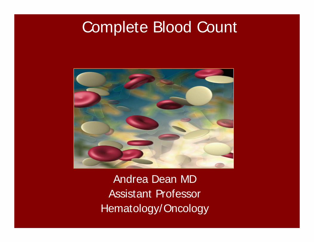
Complete Blood Count
Andrea Dean MDAssistant Professor
Hematology/Oncology
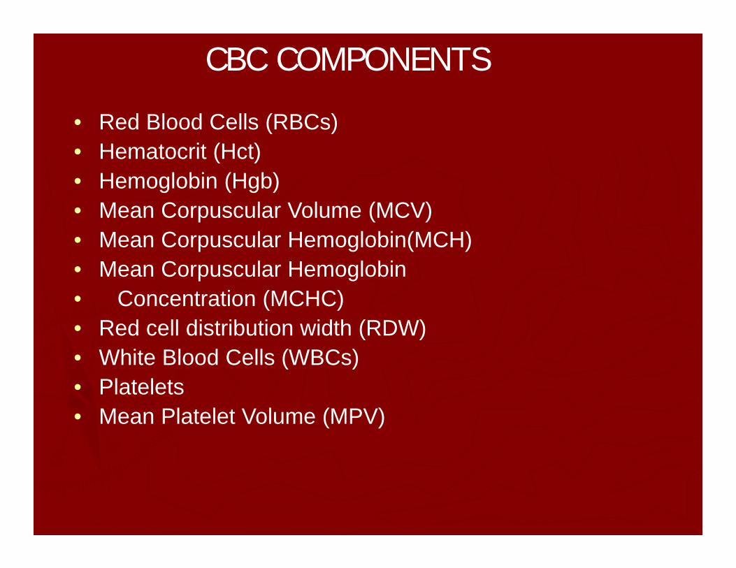
• Red Blood Cells (RBCs)• Hematocrit (Hct)• Hemoglobin (Hgb)• Mean Corpuscular Volume (MCV)• Mean Corpuscular Hemoglobin(MCH)• Mean Corpuscular Hemoglobin• Concentration (MCHC)• Red cell distribution width (RDW)• White Blood Cells (WBCs)• Platelets• Mean Platelet Volume (MPV)
CBC COMPONENTS
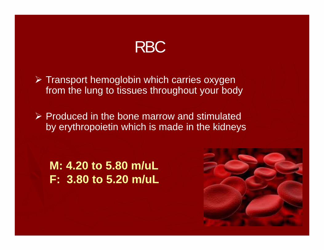
Transport hemoglobin which carries oxygen from the lung to tissues throughout your body
Produced in the bone marrow and stimulated by erythropoietin which is made in the kidneys
M: 4.20 to 5.80 m/uLF: 3.80 to 5.20 m/uL
RBC
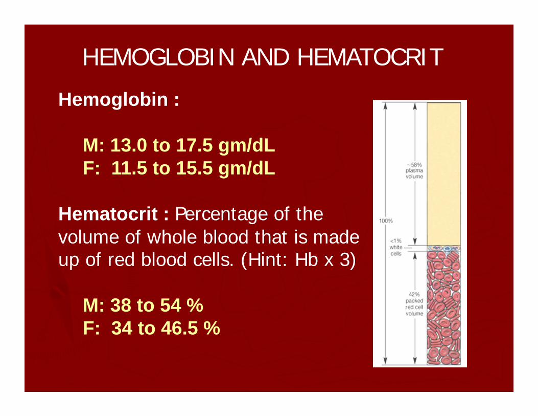
Hemoglobin :
M: 13.0 to 17.5 gm/dL F: 11.5 to 15.5 gm/dL
Hematocrit : Percentage of the volume of whole blood that is made up of red blood cells. (Hint: Hb x 3)
M: 38 to 54 % F: 34 to 46.5 %
HEMOGLOBIN AND HEMATOCRIT
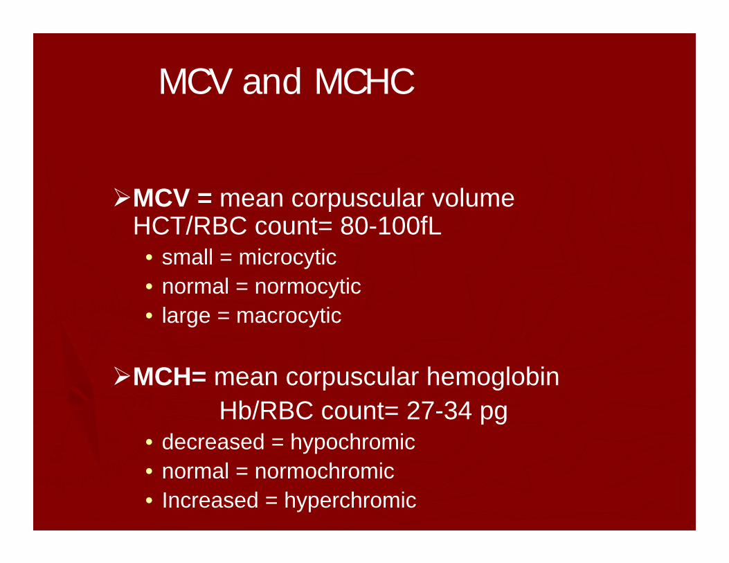
MCV = mean corpuscular volume HCT/RBC count= 80-100fL
• small = microcytic• normal = normocytic• large = macrocytic
MCH= mean corpuscular hemoglobin Hb/RBC count= 27-34 pg
• decreased = hypochromic• normal = normochromic• Increased = hyperchromic
MCV and MCHC
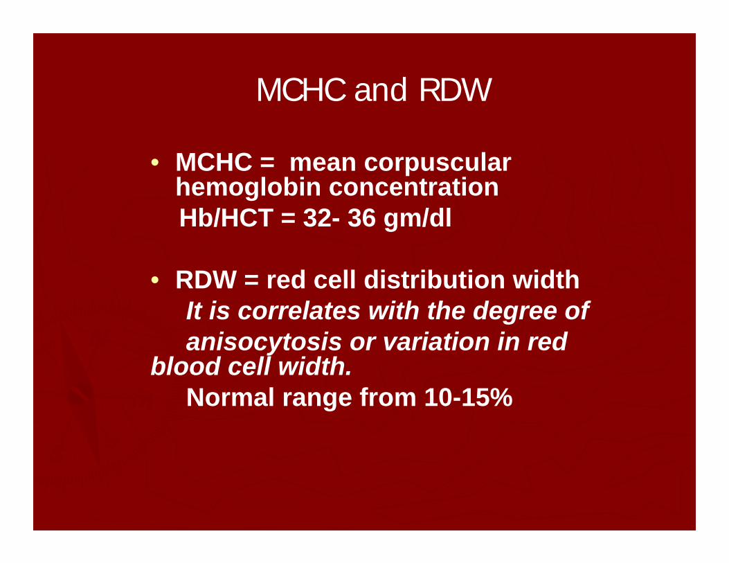
• MCHC = mean corpuscular hemoglobin concentrationHb/HCT = 32- 36 gm/dl
• RDW = red cell distribution widthIt is correlates with the degree of anisocytosis or variation in red
blood cell width.Normal range from 10-15%
MCHC and RDW
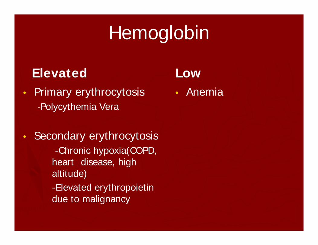
Hemoglobin
Elevated• Primary erythrocytosis
-Polycythemia Vera
• Secondary erythrocytosis-Chronic hypoxia(COPD, heart disease, high altitude)-Elevated erythropoietin due to malignancy
Low• Anemia
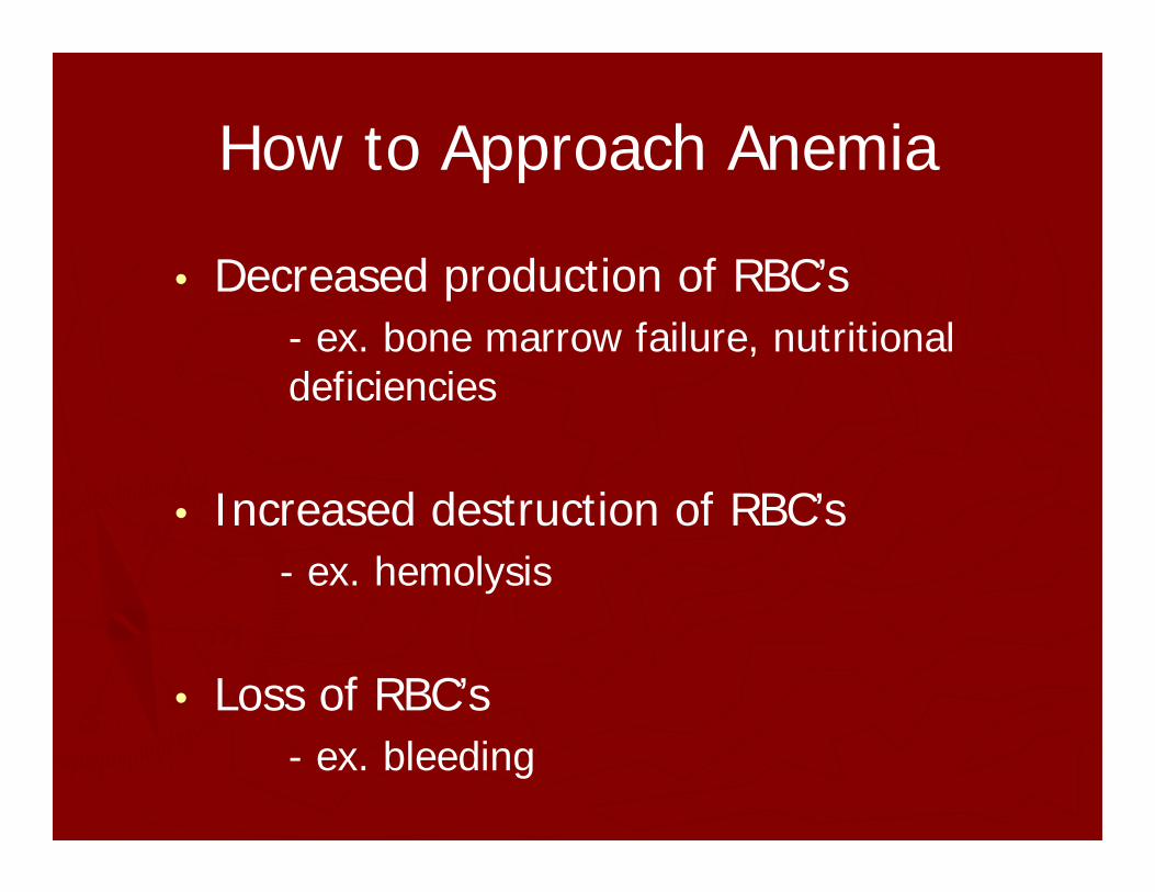
How to Approach Anemia
• Decreased production of RBC’s- ex. bone marrow failure, nutritional deficiencies
• Increased destruction of RBC’s- ex. hemolysis
• Loss of RBC’s- ex. bleeding
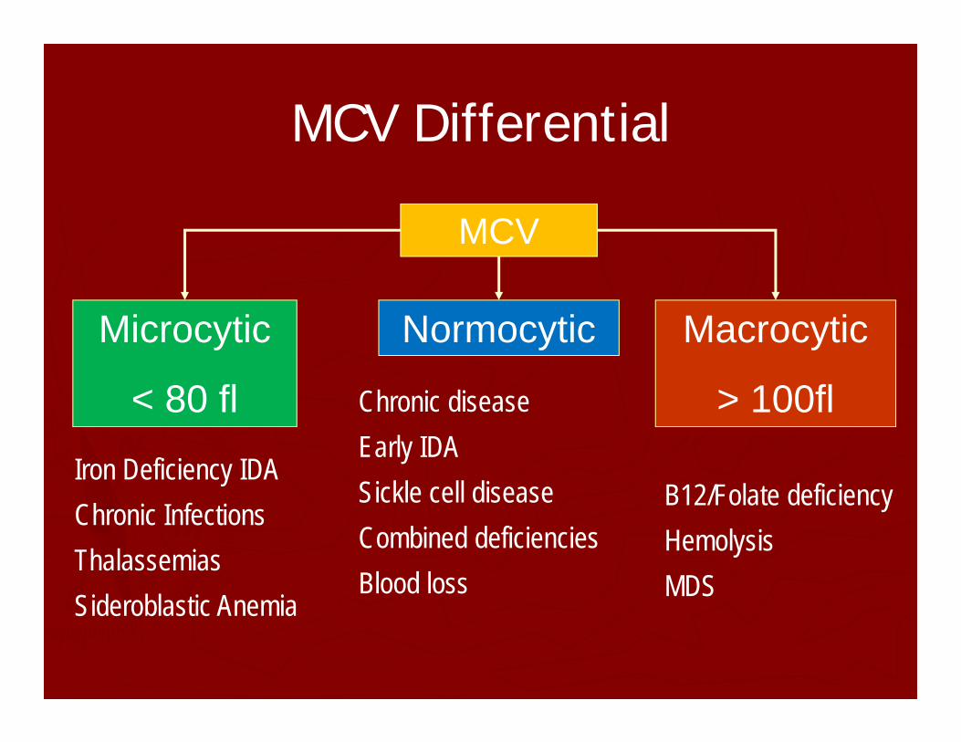
Microcytic
< 80 fl
MCV
Normocytic Macrocytic
> 100fl
Iron Deficiency IDAChronic InfectionsThalassemiasSideroblastic Anemia
Chronic diseaseEarly IDASickle cell diseaseCombined deficienciesBlood loss
B12/Folate deficiencyHemolysisMDS
MCV Differential
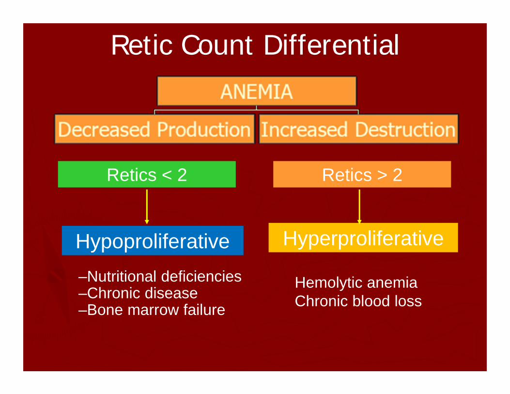
Hypoproliferative Hyperproliferative
Retics < 2 Retics > 2
–Nutritional deficiencies –Chronic disease–Bone marrow failure
Hemolytic anemiaChronic blood loss
Retic Count Differential
Microcytic Anemia
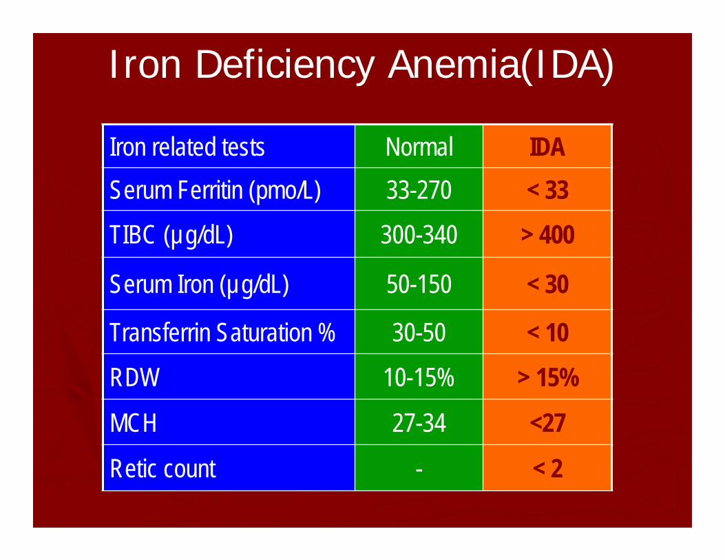
Iron Deficiency Anemia(IDA)
Iron related tests Normal IDASerum Ferritin (pmo/L) 33-270 < 33TIBC (µg/dL) 300-340 > 400
Serum Iron (µg/dL) 50-150 < 30
Transferrin Saturation % 30-50 < 10RDW 10-15% > 15%
MCH 27-34 <27
Retic count - < 2
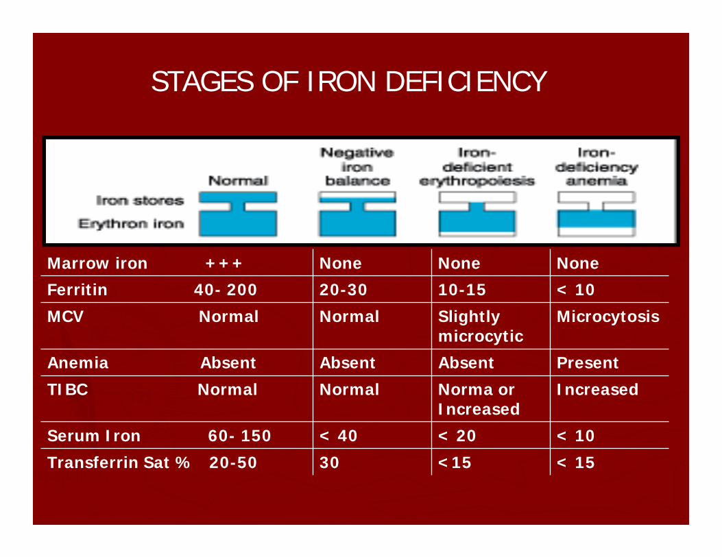
STAGES OF IRON DEFICIENCY
Marrow iron +++ None None None
Ferritin 40- 200 20-30 10-15 < 10
MCV Normal Normal Slightlymicrocytic
Microcytosis
Anemia Absent Absent Absent Present
TIBC Normal Normal Norma or Increased
Increased
Serum Iron 60- 150 < 40 < 20 < 10
Transferrin Sat % 20-50 30 <15 < 15
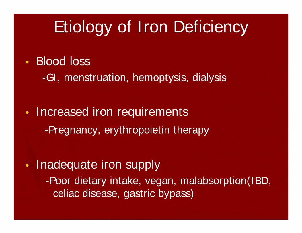
Etiology of Iron Deficiency
• Blood loss-GI, menstruation, hemoptysis, dialysis
• Increased iron requirements-Pregnancy, erythropoietin therapy
• Inadequate iron supply-Poor dietary intake, vegan, malabsorption(IBD,
celiac disease, gastric bypass)

Treatment for IDA
• Oral iron is first line treatment (ferrous sulfate/gluconate)
A. Ca-tums, Phosphate, antacids ↓absorptionB. Ascorbic acid (orange juice)↑absorption
• Reserve parenteral Rx. for oral intolerance• Packed cell transfusion in emergency• Continue Fe Rx at least 3 months after normal
Hb
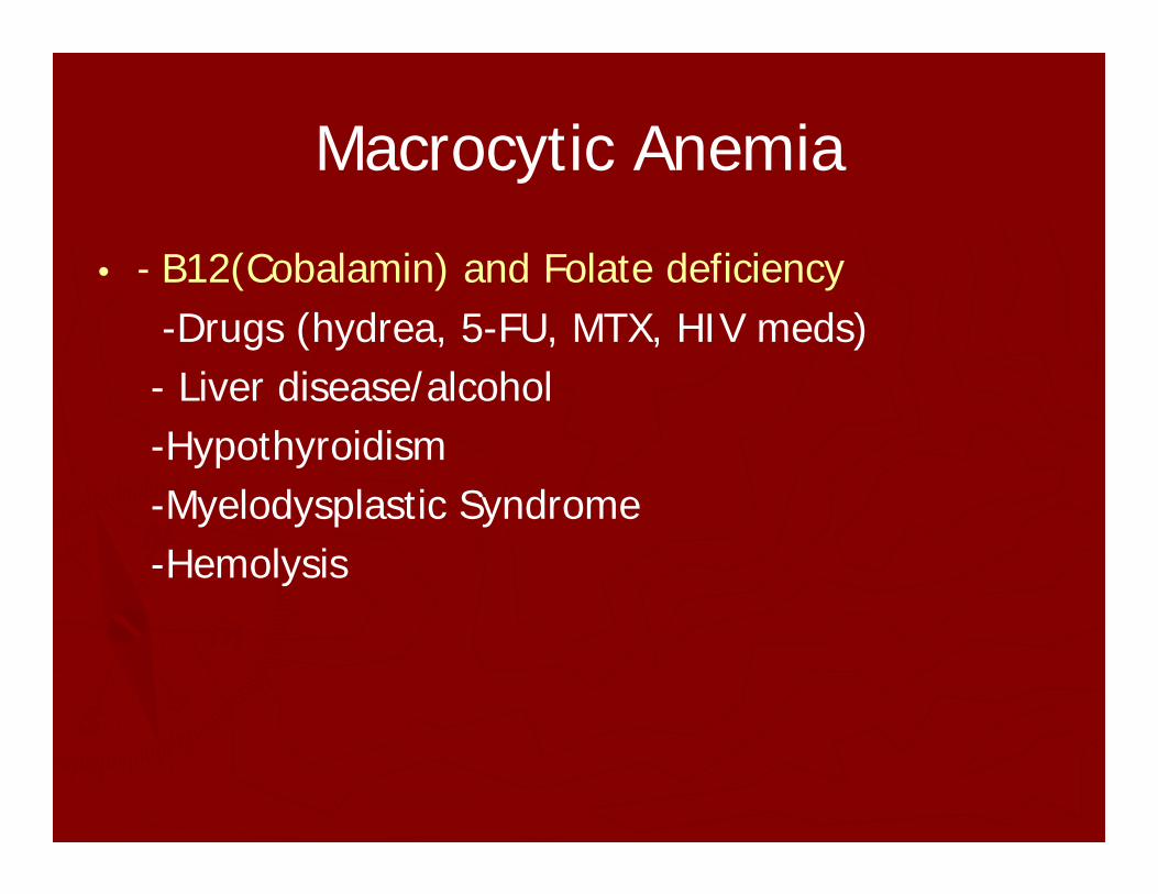
Macrocytic Anemia
• - B12(Cobalamin) and Folate deficiency-Drugs (hydrea, 5-FU, MTX, HIV meds)- Liver disease/alcohol-Hypothyroidism-Myelodysplastic Syndrome-Hemolysis
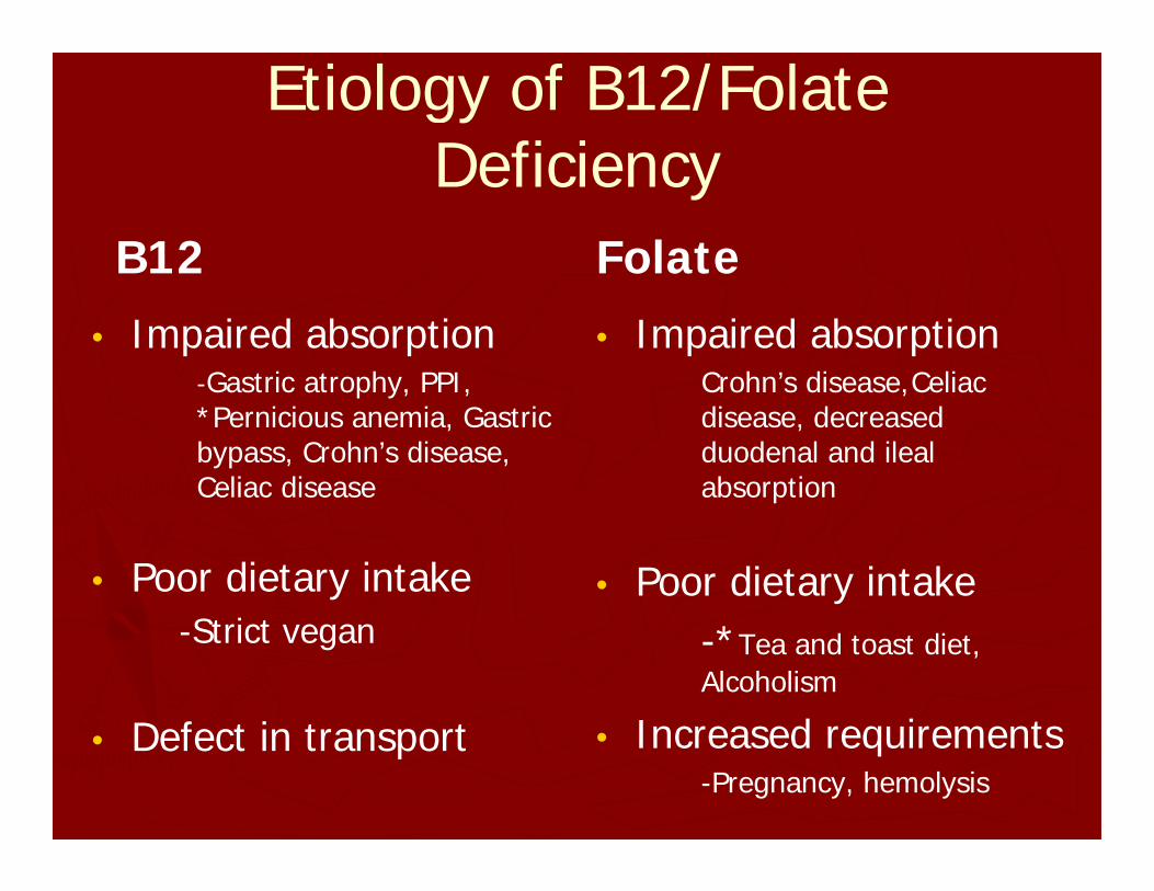
Etiology of B12/Folate Deficiency
B12• Impaired absorption
-Gastric atrophy, PPI, *Pernicious anemia, Gastric bypass, Crohn’s disease, Celiac disease
• Poor dietary intake-Strict vegan
• Defect in transport
Folate• Impaired absorption
Crohn’s disease,Celiac disease, decreased duodenal and ileal absorption
• Poor dietary intake-*Tea and toast diet, Alcoholism
• Increased requirements-Pregnancy, hemolysis

B12(Cobalamin) Deficiency
• Symptoms : weakness, depression, memory loss, unsteady gait and clumsiness (posterior and later columns degeneration)
• Diagnosed by B12 levels< 200 pg/ml
• Methylmalonic acid and homocysteine elevated in early deficiency
• Tx: oral B12 or B12 IM injections
Folate Deficiency
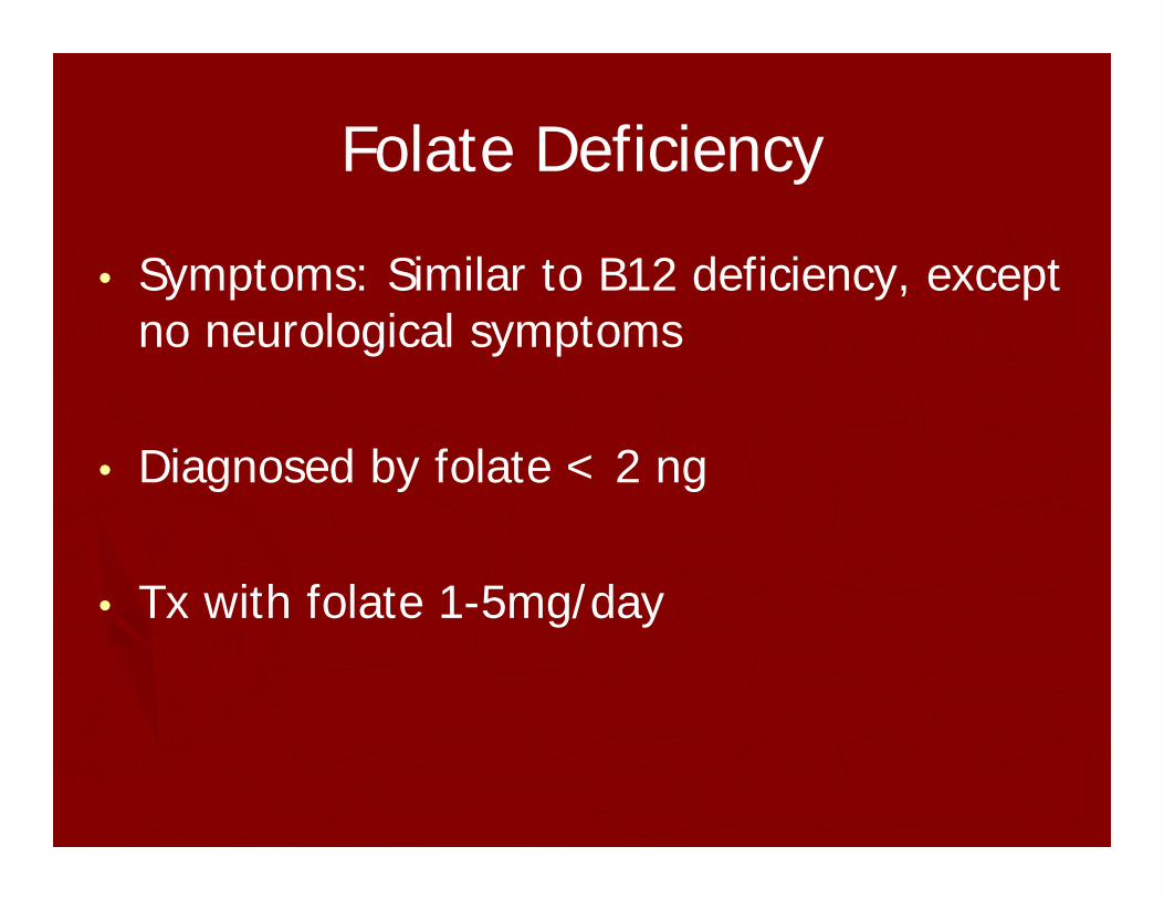
• Symptoms: Similar to B12 deficiency, except no neurological symptoms
• Diagnosed by folate < 2 ng
• Tx with folate 1-5mg/day
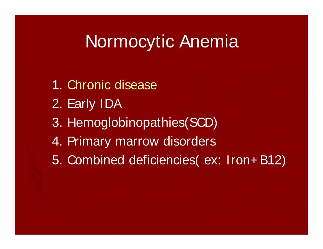
Normocytic Anemia
1. Chronic disease2. Early IDA3. Hemoglobinopathies(SCD)4. Primary marrow disorders5. Combined deficiencies( ex: Iron+B12)
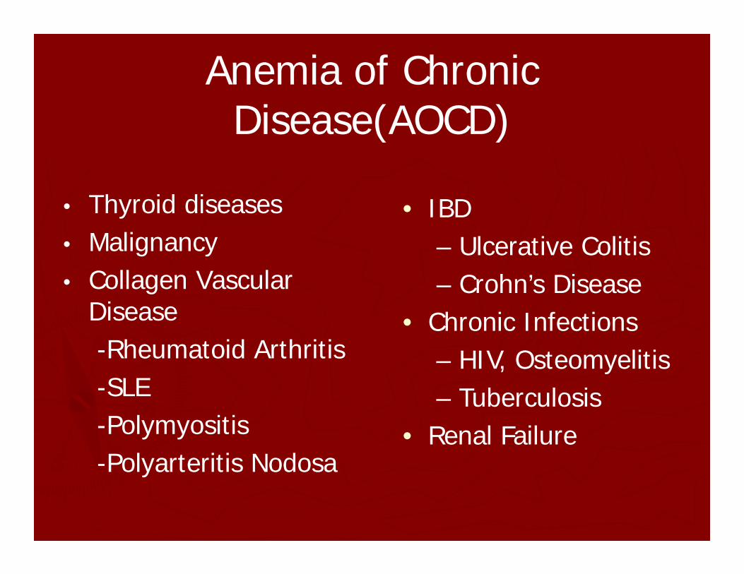
Anemia of Chronic Disease(AOCD)
• Thyroid diseases• Malignancy• Collagen Vascular
Disease-Rheumatoid Arthritis-SLE-Polymyositis-Polyarteritis Nodosa
• IBD– Ulcerative Colitis– Crohn’s Disease
• Chronic Infections– HIV, Osteomyelitis– Tuberculosis
• Renal Failure
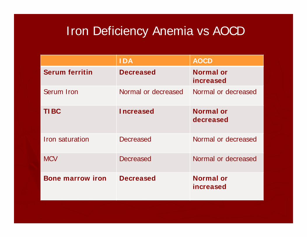
Iron Deficiency Anemia vs AOCD
IDA AOCD
Serum ferritin Decreased Normal or increased
Serum Iron Normal or decreased Normal or decreased
TIBC Increased Normal or decreased
Iron saturation Decreased Normal or decreased
MCV Decreased Normal or decreased
Bone marrow iron Decreased Normal or increased
• WBCs are involved in the immune response• The normal range: 3.5 – 10.5x10^9 K/L• Two types of WBC:
1) Granulocytes consist of:– Neutrophils: 50 - 70% – Eosinophils: 1 - 5% – Basophils: up to 1% 2) Agranulocytes consist of:- Lymphocytes: 20 - 40% – Monocytes: 1 - 6%
White Blood Cells (WBC)
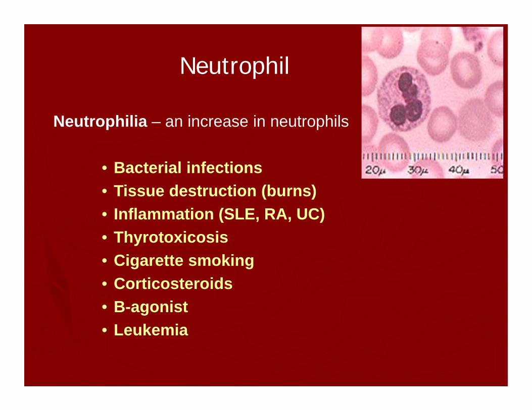
Neutrophilia – an increase in neutrophils
• Bacterial infections • Tissue destruction (burns)• Inflammation (SLE, RA, UC)• Thyrotoxicosis• Cigarette smoking• Corticosteroids• B-agonist• Leukemia
Neutrophil
Neutropenia – a decrease in neutrophils
• Decreased bone marrow production
• Medications ( ex. dapsone, cephalosporins)
• Immune related (ex. SLE, RA)
• Post acute infection (HSV, CMV, HIV, EBV)
Neutrophil
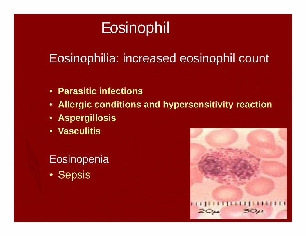
Eosinophilia: increased eosinophil count
• Parasitic infections• Allergic conditions and hypersensitivity reaction• Aspergillosis• Vasculitis
Eosinopenia• Sepsis
Eosinophil
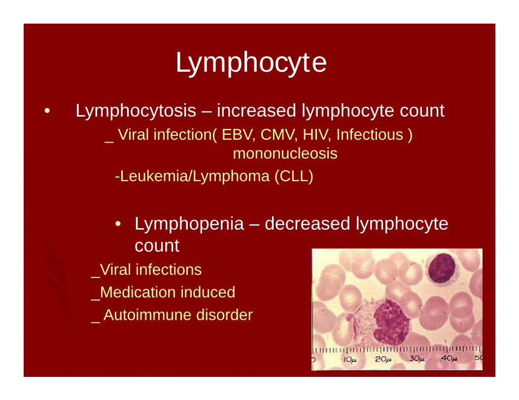
• Lymphocytosis – increased lymphocyte count_ Viral infection( EBV, CMV, HIV, Infectious )
mononucleosis-Leukemia/Lymphoma (CLL)
• Lymphopenia – decreased lymphocyte count
_Viral infections_Medication induced_ Autoimmune disorder
Lymphocyte
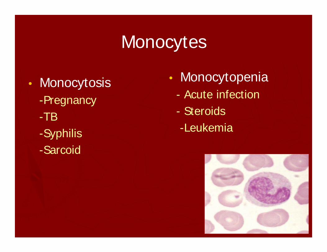
Monocytes
• Monocytosis-Pregnancy-TB-Syphilis-Sarcoid
• Monocytopenia- Acute infection- Steroids-Leukemia
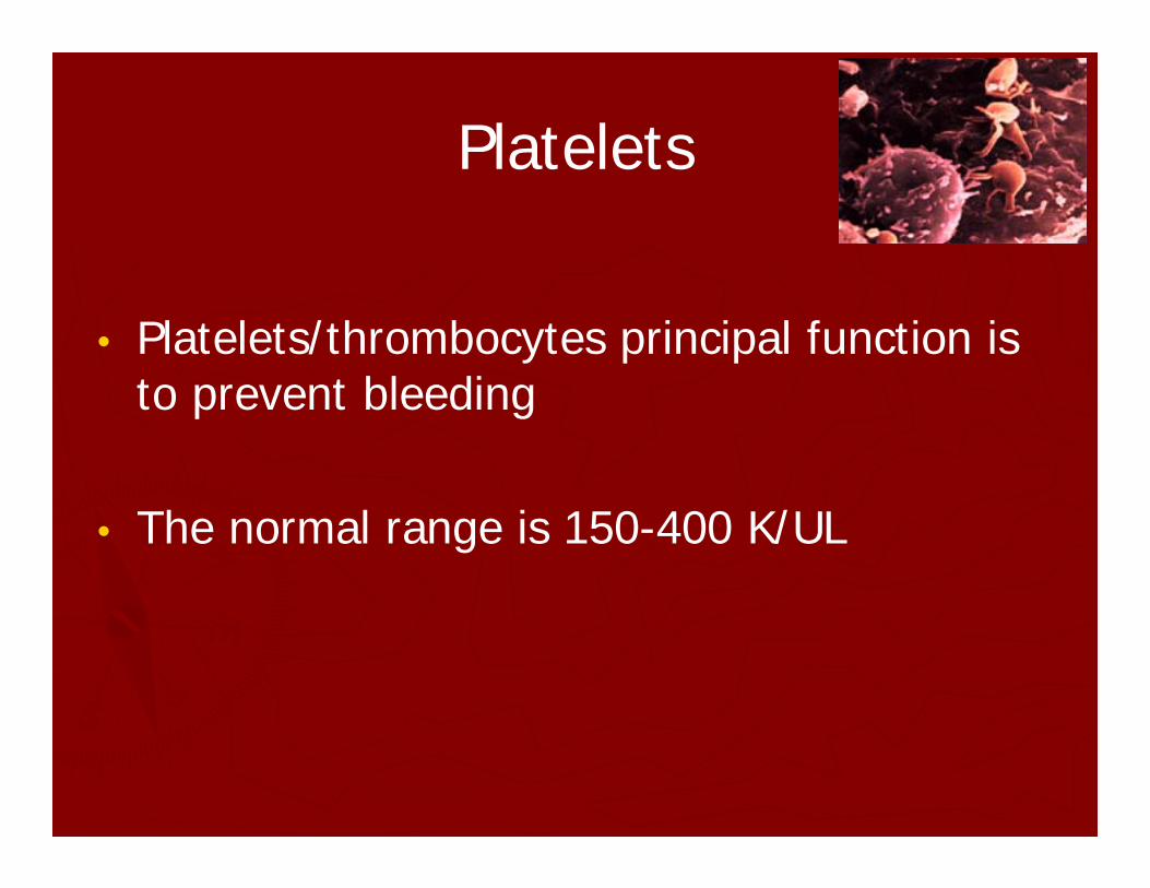
Platelets
• Platelets/thrombocytes principal function is to prevent bleeding
• The normal range is 150-400 K/UL
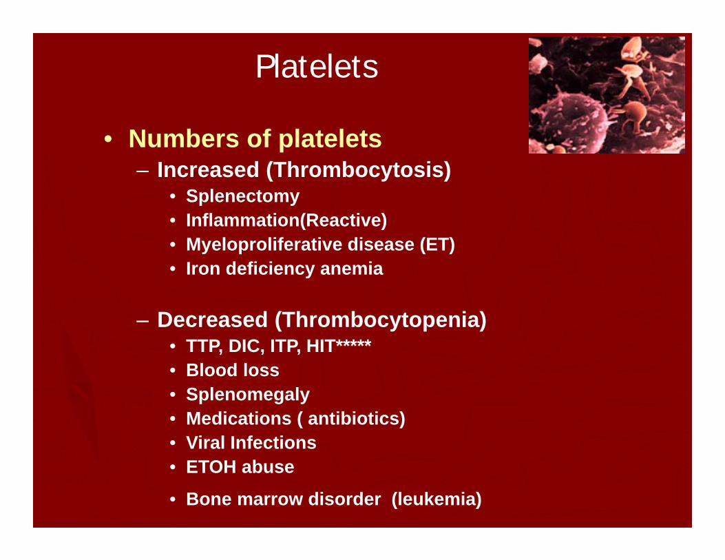
• Numbers of platelets – Increased (Thrombocytosis)
• Splenectomy• Inflammation(Reactive)• Myeloproliferative disease (ET)• Iron deficiency anemia
– Decreased (Thrombocytopenia)• TTP, DIC, ITP, HIT*****• Blood loss• Splenomegaly• Medications ( antibiotics)• Viral Infections• ETOH abuse
• Bone marrow disorder (leukemia)
Platelets