complete control retroviral inducible mammalian … control retroviral inducible mammalian...
TRANSCRIPT
Complete Control Retroviral Inducible Mammalian Expression System
Instruction Manual
Catalog #217564 (pFB-ERV), #240028 (pCFB-EGSH) Revision D.0
For Research Use Only. Not for use in diagnostic procedures. 217564-12
LIMITED PRODUCT WARRANTY This warranty limits our liability to replacement of this product. No other warranties of any kind, express or implied, including without limitation, implied warranties of merchantability or fitness for a particular purpose, are provided by Agilent. Agilent shall have no liability for any direct, indirect, consequential, or incidental damages arising out of the use, the results of use, or the inability to use this product.
ORDERING INFORMATION AND TECHNICAL SERVICES Email [email protected]
World Wide Web
www.genomics.agilent.com
Telephone Location Telephone
United States and Canada 800 227 9770
Austria 01 25125 6800
Benelux 02 404 92 22
Denmark 45 70 13 00 30
Finland 010 802 220
France 0810 446 446
Germany 0800 603 1000
Italy 800 012575
Netherlands 020 547 2600
Spain 901 11 68 90
Sweden 08 506 4 8960
Switzerland 0848 8035 60
UK/Ireland 0845 712 5292
All Other Countries Please visit www.agilent.com/genomics/contactus
Complete Control Retroviral Inducible Mammalian Expression System
Materials Provided .............................................................................................................................. 1
Storage Conditions .............................................................................................................................. 1
Additional Materials Required .......................................................................................................... 1
Notices to Purchaser ........................................................................................................................... 2
Safety Considerations ......................................................................................................................... 4
Introduction ......................................................................................................................................... 5
Overview of Ecdysone-Regulatable Gene Expression ..................................................................... 7
Overview of Replication-Defective Retroviral Gene Transfer Systems ......................................... 9
Description of the Vectors ................................................................................................... 11
The pFB-ERV Vector .......................................................................................................... 12
The pCFB-EGSH Vector ..................................................................................................... 13
Overview of the Cell Selection Procedure ....................................................................................... 14
Preparing the pCFB-EGSH Expression Vector Construct ........................................................... 15
Digestion and Ligation Protocol .......................................................................................... 16
Transforming Competent Cells with the pCFB-EGSH Construct ....................................... 17
Transfection Protocol........................................................................................................................ 18
Transient Cotransfection of Expression Construct and pFB-ERV to Determine
Plasmid/Host Cell Compatibility ................................................................................. 18
Production of Stable Receptor-Expressing Cell Lines ................................................................... 19
Day 1: Preparing for Production of Virus by Transfection ................................................. 20
Day 2: Transfecting Cells .................................................................................................... 21
Day 3: Preparing for the Transduction ................................................................................ 22
Day 4: Transducing the Target Cells ................................................................................... 23
Isolating G418-Resistant Stable Clones and Expanding the Colonies ................................ 24
Examining Receptor Expression ......................................................................................... 24
Preparing pCFB-EGSH Viral Stocks .............................................................................................. 25
Day 1: Preparing for Production of Virus by Transfection ................................................. 26
Day 2: Transfecting Cells .................................................................................................... 27
Day 3: Preparing for the Transduction ................................................................................ 28
Day 4: Transducing the Target Cells ................................................................................... 29
Monitoring Gene Expression by Anti-HA Immunodetection ............................................. 30
Troubleshooting ................................................................................................................................ 31

Preparation of Media and Reagents ................................................................................................ 33
References .......................................................................................................................................... 34
Endnotes ............................................................................................................................................. 34
MSDS Information ............................................................................................................................ 34
Complete Control Retroviral Inducible Mammalian Expression System 1
Complete Control Retroviral Inducible Mammalian Expression System
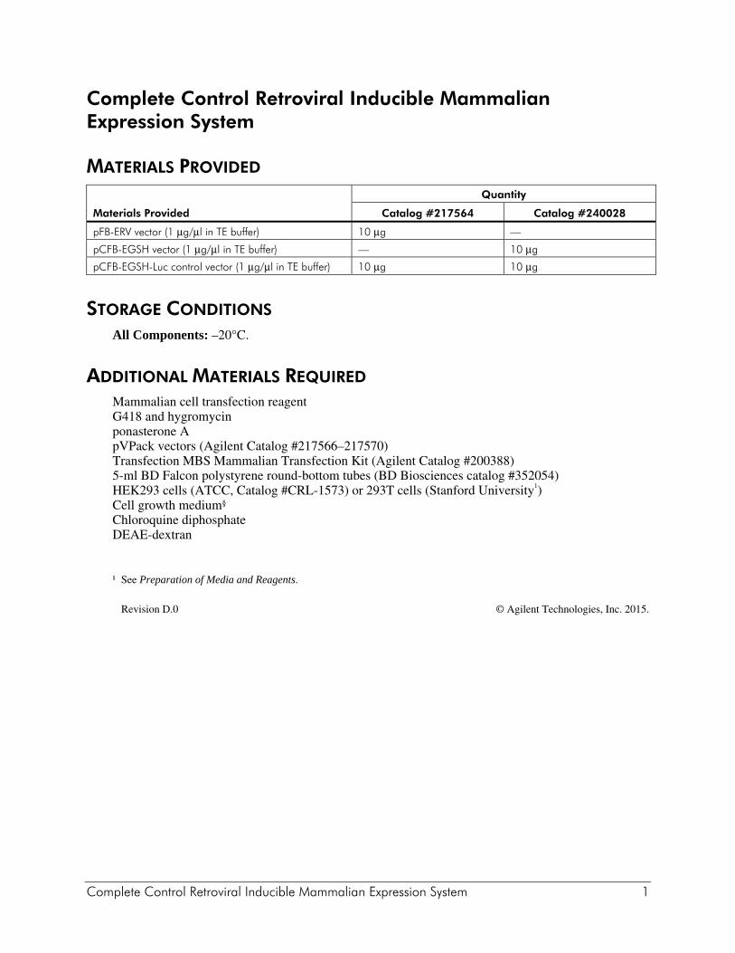
MATERIALS PROVIDED
Materials Provided
Quantity
Catalog #217564 Catalog #240028
pFB-ERV vector (1 μg/μl in TE buffer) 10 μg —
pCFB-EGSH vector (1 μg/μl in TE buffer) — 10 μg
pCFB-EGSH-Luc control vector (1 μg/μl in TE buffer) 10 μg 10 μg
STORAGE CONDITIONS All Components: –20°C.
ADDITIONAL MATERIALS REQUIRED Mammalian cell transfection reagent G418 and hygromycin ponasterone A pVPack vectors (Agilent Catalog #217566–217570) Transfection MBS Mammalian Transfection Kit (Agilent Catalog #200388) 5-ml BD Falcon polystyrene round-bottom tubes (BD Biosciences catalog #352054) HEK293 cells (ATCC, Catalog #CRL-1573) or 293T cells (Stanford University1) Cell growth medium§ Chloroquine diphosphate DEAE-dextran
§ See Preparation of Media and Reagents.
Revision D.0 © Agilent Technologies, Inc. 2015.
2 Complete Control Retroviral Inducible Mammalian Expression System
NOTICES TO PURCHASER Use of this product is covered by U.S. Patent No. 6,723,531 Complete Control Mammalian Expression System This product is the subject of one or more of U.S. Patent Nos. 5,514,578 and 6,245,531, and European Patent 0517805 licensed to Agilent Technologies Inc. The purchase of this product conveys to the buyer the non-transferable right to use the purchased amount of the product and components of the product in research conducted by the buyer outside of plants (whether the buyer is an academic or for-profit entity). The buyer cannot sell or otherwise transfer (a) this product (b) its components or (c) materials made using this product or its components to a third party or otherwise use this product or its components or materials made using this product or its components for commercial purposes. The buyer may transfer information or materials made through the use of this product to a scientific collaborator, provided that such transfer is not for the commercial purposes of the buyer, and that such collaborator agrees in writing (a) not to transfer such materials to any third party, and (b) to use such transferred materials and/or information solely for research and not for commercial purposes. Commercial purposes means any activity by a party for consideration and may include, but is not limited to: (1) use of the product or its components in manufacturing; (2) use of the product or its components to provide a service, information, or data; (3) use of the product or its components for therapeutic, diagnostic or prophylactic purposes; or (4) resale of the product or its components, whether or not such product or its components are resold for use in research. If the purchaser is not willing to accept the limitations of the limited use statement, Agilent is willing to accept return of the products with a full refund. For information on purchasing a license to this product for purposes other than research, contact Intrexon Corporation www.dna.com to obtain a commercial license. FOR LABORATORY USE ONLY A license (from Promega for research reagent products and from The Regents of the University of California for all other fields) is needed for any commercial sale of nucleic acid contained within or derived from this product.

Complete Control Retroviral Inducible Mammalian Expression System 3
NON-COMMERCIAL RESEARCH USE LICENSE FOR NONPROFIT ENTITIES
Agilent agrees to sell, and Licensee agrees to purchase, Agilent Vitality hrGFP products provided herewith (referred to as the “Products”) on the following terms and conditions. (For purposes of this License, “Licensee” shall include any person or entity which ordered the Products or at any time uses the Products.) LICENSEE’S ACCEPTANCE OF DELIVERY AND/OR USE OF THE PRODUCTS SHALL CONSTITUTE LICENSEE’S BINDING AGREEMENT TO THE FOLLOWING TERMS AND CONDITIONS. IF LICENSEE IS UNWILLING TO ACCEPT SUCH TERMS AND CONDITIONS, AGILENT IS WILLING TO ACCEPT RETURN OF THE PRODUCTS PRIOR TO ANY USE OF THE PRODUCTS, FOR A FULL REFUND.
1. The Products, containing DNA sequences encoding for mutant green fluorescent protein (GFP) from a novel marine organism, variants or proteins thereof, are proprietary to Agilent and licensed for non-commercial research purposes only. Licensee agrees that the Products will be used only at non-commercial entities and only in research that is not funded by or pledged to be licensed to commercial entities. Commercial entities and nonprofit entities conducting research funded by or in collaboration with commercial entities require a commercial license from Agilent Technologies, Inc. Licensee may modify only the non-coding region outside of the nucleic acid encoding the fluorescent protein of the Products to facilitate research. Licensee shall not have any rights to (i) modify the coding region of the nucleic acid encoding the fluorescent protein of the Products, (ii) offer the Products or any component, derivative or modification thereof for resale, or (iii) distribute, transfer, or otherwise provide access to, the Products or any component, derivative or modification thereof to any third party for any purpose or use.
2. Except as set forth above, no other rights, express or implied, are conveyed to Licensee. No rights are granted to Licensee to use the Products for (i) the provision of services to any for-profit third party (e.g., screening and profiling), (ii) diagnostic applications, (iii) methods employed in screens to evaluate compounds (e.g., high throughput screening (“HTS”), (iv) profiling chemicals for selectivity, bioavailability, drug metabolism or toxicity, (v) use in vivo in multicellular organisms (and methods therein) for gene therapy, (vi) quality control or quality assurance processes, including food and environmental testing, or (vii) use in manufacturing. 3. The Products shall be used solely on premises under the control of Licensee, and in compliance with all laws, regulations, rules and guidelines applicable to the Products and their use, testing, handling, or other disposition thereof, or otherwise applicable to Licensee’s activities hereunder.
4. Title to the Products shall remain at all times with Agilent and shall not transfer to Licensee.
5. Agilent warrants that, at the time of shipment, the Products will conform to the specifications which accompany the Products. This warranty limits Agilent’s liability to replacement of the Products. AGILENT MAKES NO OTHER WARRANTIES, EXPRESS OR IMPLIED, WITH RESPECT TO THE PRODUCTS, INCLUDING ANY WARRANTIES OF MERCHANTABILITY OR FITNESS FOR ANY PARTICULAR PURPOSE OR THAT THE PRODUCTS DO NOT INFRINGE ANY PROPRIETARY RIGHTS OF ANY THIRD PARTY. Licensee hereby waives, releases and renounces any and all warranties, guarantees, obligations, liabilities, rights and remedies, express or implied, arising by law or otherwise, with respect to the usefulness or freedom from defects of the Products, including, but not limited to, (a) any implied warranty or merchantability or fitness for a particular purpose, (b) any implied warranty arising from course of performance, course of dealing or usage in the trade, and (c) any obligation, right, liability, claim or remedy for (1) loss of use, revenue or profit, or any other damages, (2) infringement of third party intellectual property rights, and (3) incidental or consequential damages.
4 Complete Control Retroviral Inducible Mammalian Expression System
6. Licensee agrees to bear all risks associated with the Products and their use, testing, handling or other disposition thereof, and all risks associated with Licensee's use of Products purchased under this License. Licensee hereby assumes all risks of damage or injury to Licensee's facilities, employees or agents and to any third party arising from possession or use of the Products. Agilent shall have no liability to Licensee, its employees or agents or to any third party, regardless of the form or theory of action (whether contract, tort or otherwise, including, but not limited to, negligence and strict liability), for any direct, indirect, consequential, incidental or other damages arising out of or relating to the Products or this License.
7. Licensee shall indemnify, defend and hold Agilent, its affiliates, distributors, suppliers, directors, officers, employees and agents, harmless from and against any and all claims, actions, demands, liabilities, damages and expenses (including attorneys’ fees) relating to or arising out of any damage or injury, including, but not limited to, product liability and intellectual property infringement claims of any nature, alleged to have been caused by the Products or the use, testing, handling or other disposition thereof or Licensee’s activities hereunder.
8. Licensee may at any time properly dispose of the Products in a manner which ensures their prompt destruction and is consistent with all applicable laws, regulations, rules and guidelines.
9. No modification or waiver of any terms or conditions of this License shall be effective unless in a writing signed by Licensee and an authorized representative of Agilent Technologies, Inc. For information on purchasing a license to use the Products for commercial purposes, including commercial research purposes, please contact the Director of Business Development, 11011 North Torrey Pines Road, La Jolla, California 92037, telephone number (858) 535-5400, facsimile number (858) 535-0071.
SAFETY CONSIDERATIONS The host range of the virus is dictated not by the DNA but by the env gene used in the packaging system. Virus produced from VSV-G-based and other polytropic packaging systems are capable of infecting human cells. The National Institute of Health has designated retroviral vectors such as MMLV as Biosafety Level 2. Appropriate caution should be used in the production and use of recombinant retrovirus. For more information, see Biosafety in Microbiological and Biomedical Laboratories (4th edition). It is available on the Web sites of both the National Institutes of Health at http://bmbl.od.nih.gov and the Office of Health and Safety of the Centers for Disease Control and Prevention at www.cdc.gov/od/ohs (Click the link to Biosafety Information).
Complete Control Retroviral Inducible Mammalian Expression System 5
INTRODUCTION DNA vector-based systems that allow precise control of gene expression in vivo have become invaluable for the study of gene function in a variety of organisms, particularly when applied to the study of developmental and other biological processes for which the timing or dosage of gene expression is critical to gene function. Such systems have also been successfully used to overexpress toxic or disease-causing genes, to induce gene targeting, and to express antisense RNA. Inducible systems are currently being used by pharmaceutical companies to facilitate screening for inhibitors of clinically relevant biological pathways, and potential applications for gene therapy are being explored. The Agilent Complete Control ecdysone-inducible plasmid vectors are based on the insect molting hormone ecdysone, which can stimulate transcriptional activation in mammalian cells harboring the ecdysone receptor protein from Drosophila melanogaster.2 The system has a number of advantages over alternative systems. Firstly, the lipophilic nature and short in vivo half-life of the ecdysone analog ponasterone A (ponA) allows efficient penetrance into all tissues including brain, resulting in rapid and potent inductions and rapid clearance. Secondly, ecdysteroids are not known, nor are they expected, to affect mammalian physiology in any measurable way. Thirdly, the heterodimeric ponA responsive receptor and receptor DNA recognition element have been genetically altered such that trans-activation of endogenous genes by the ecdysone receptor, or of the ponA-responsive expression cassette by endogenous transcription factors, is extremely unlikely. In addition, it has been found that in the absence of inducer the heterodimer remains bound at the promoter in a complex with corepressors and histone deacetylase, and is thus tightly repressed until ligand binding, at which time high-level transcriptional activation occurs (i.e., the heterodimer is converted from a tight repressor to a trans-activator). In transient assays and stable cell lines harboring receptor expression plasmids in combination with a plasmid bearing an inducible luciferase expression cassette, induction ratios of 1,000-fold have been achieved.3 A limitation to the use of plasmid-based vectors for controlled gene expression is the fact that many cell types of academic, industrial or clinical interest are difficult or virtually impossible to transfect using current transfection methods. In particular, primary human cell lines, lymphocytes, neurons and other nondividing cells are best transduced using viral delivery systems. The most popular and user-friendly of these are the retroviral vectors. Infection with retroviruses often yields transduction efficiencies close to 100%, and the proviral copy number can be easily controlled by varying the multiplicity of infection (MOI). This latter feature is particularly important for inducible systems, for which low basal expression and high induction ratios are affected by copy number. Thus infection of the target cell with virus at an optimal MOI should yield a high frequency of clones capable of mediating desirable expression profiles without exhaustive colony screening.

6 Complete Control Retroviral Inducible Mammalian Expression System
With the vectors pFB-ERV and pCFB-EGSH, we have adapted the ecdysone inducible components of the Complete Control System for retroviral delivery. Used together, we have attained induction ratios of >1,000-fold with these vectors in tissue culture cells.
Complete Control Retroviral Inducible Mammalian Expression System 7
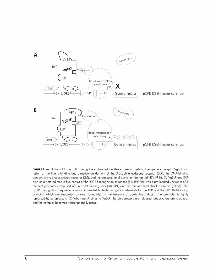
OVERVIEW OF ECDYSONE-REGULATABLE GENE EXPRESSION The ecdysone receptor (EcR) is a member of the retinoid-X-receptor (RXR) family of nuclear receptors and is composed of three domains: an N-terminal activation domain (AD), a central DNA-binding domain (DBD), and a C-terminal ligand-binding and dimerization domain (LBD). In insect cells, EcR and the nuclear receptor ultraspiracle (USP) form a promoter-bound heterodimer, which regulates transcription (see Figure 1). In the absence of ecdysone, the receptor heterodimer binds to corepressors and tightly represses transcription.4 When ecdysone binds to the EcR LBD, the corepressors are released, coactivators are recruited to the complex, and transcriptional activation is enabled. In mammalian cells harboring the EcR gene, EcR heterodimerizes with RXR, the mammalian homologue of USP. The EcR–RXR heterodimer binds to multiple copies of the ecdysone-responsive element (EcRE), and in the absence of ponA, represses transcription of an expression cassette. When ponA binds to the receptor, the receptor complex activates transcription of a reporter gene or a gene of interest. To avoid pleiotropic interactions with endogenous pathways in mammalian host cells, both the EcRE recognition sequence and the EcR protein were modified. The EcRE sequence was modified to create a synthetic recognition site that does not bind any endogenous transcription factors. The wild-type EcRE sequence consists of two inverted repeat sequences separated by a single nucleotide: AGTGCA N TGCACT. The EcRE sequence was changed to AGTGCA N1 TGTTCT (and renamed E/GRE). Recognition of the synthetic E/GRE recognition sequence by either a steroid receptor or a wild-type RXR heterodimer receptor is extremely unlikely, as these receptors recognized only the wild-type perfect inverted repeat. The E/GRE recognition sequence has imperfect inverted half sites separated by one nucleotide. A wild-type RXR heterodimer requires single nucleotide separation of the inverted repeats, and the majority bind to direct repeats rather than inverted repeats (EcRE is an exception). The EcR protein was modified to create a synthetic ecdysone-binding receptor that does not transactivate any host genes. Three amino acids in the EcR DBD were mutated to change its DNA-binding specificity to that of the glucocorticoid receptor (GR), which recognizes the half-site AGAACA.2 Like all steroid receptors and unlike RXR receptors, the GR protein homodimerizes and recognizes two inverted repeat sequences separated by three nucleotides. The GR–EcR fusion protein (GEcR) retains the ability to dimerize with RXR and activate, with ponA-dependence, reporter genes that contain the synthetic E/GRE recognition sequence. The GEcR receptor was further modified by replacing the EcR AD with the more potent VP16 AD. The result of all the modifications is the synthetic ecdysone-binding receptor VgEcR. VgEcR is a fusion of the ligand-binding and dimerization domain of the D. melanogaster ecdysone receptor, the DNA-binding domain of the glucocorticoid receptor, and the transcription activation domain of herpes simplex virus (HSV) VP16.
8 Complete Control Retroviral Inducible Mammalian Expression System
FIGURE 1 Regulation of transcription using the ecdysone-inducible expression system. The synthetic receptor VgEcR is a fusion of the ligand-binding and dimerization domain of the Drosophila ecdysone receptor (EcR), the DNA-binding domain of the glucocorticoid receptor (GR), and the transcriptional activation domain of HSV VP16. (A) VgEcR and RXR bind as a heterodimer to five copies of the E/GRE recognition sequence (5× E/GRE), which are located upstream of a minimal promoter composed of three SP1 binding sites (3× SP1) and the minimal heat shock promoter (mHSP). The E/GRE recognition sequence consists of inverted half-site recognition elements for the RXR and the GR DNA-binding domains (which are separated by one nucleotide). In the absence of ponA (the inducer), the promoter is tightly repressed by corepressors. (B) When ponA binds to VgEcR, the corepressors are released, coactivators are recruited, and the complex becomes transcriptionally active.
RXR
RXR
EcR
GR
Coactivator
Corepressor
PonA
B
pCFB-EGSH vector construct
VgEcR
RXR
RXR
EcR
GR
5× E/GRE 3× SP1
Gene of interest
Corepressor
X
CoactivatorA
pCFB-EGSH vector construct
VgEcR
VP16
Vp16
mHSP
5× E/GRE 3× SP1 mHSP
Gene of interest
Complete Control Retroviral Inducible Mammalian Expression System 9
OVERVIEW OF REPLICATION-DEFECTIVE RETROVIRAL GENE TRANSFER SYSTEMS
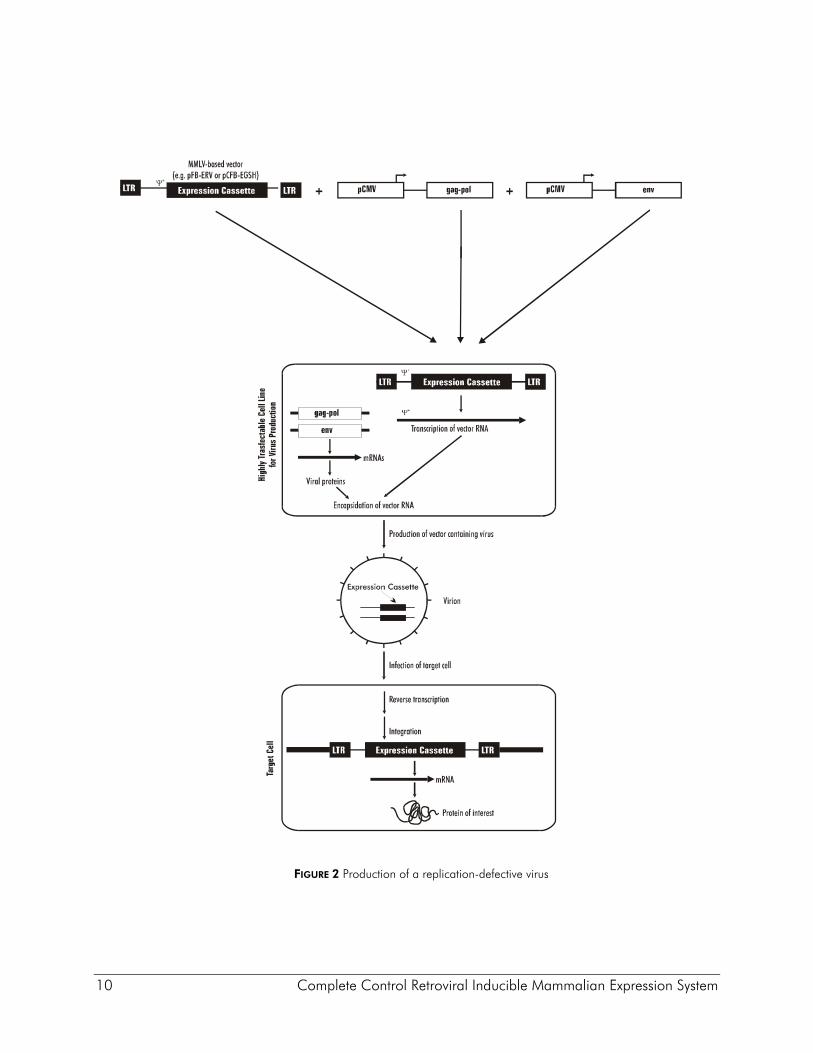
Non-replicating retroviral vectors contain all of the cis elements required for transcription of mRNA molecules encoding a gene of interest, and packaging of these transcripts into infectious virus particles (Figure 2). The vectors are typically comprised of an E. coli plasmid backbone containing a pair of 600 base pair viral long terminal repeats (LTRs) between which the gene of interest is inserted. The LTR is divided into 3 regions. The U3 region contains the retroviral promoter/enhancer. The U3 region is flanked in the 3´ direction by the R region, which contains the viral polyadenylation signal (pA), followed by the U5 region which, along with R, contains sequences that are critical for reverse transcription. Expression of the viral RNA is initiated within the U3 region of the 5´ LTR and is terminated in the R region of the 3´ LTR. Between the 5´ LTR and the coding sequence for the gene of interest resides an extended version of the viral packaging signal (ψ+), which is required in cis for the viral RNA to be packaged into virion particles. In order to generate infectious virus particles that carry the gene of interest, specialized packaging cell lines have been generated that contain chromosomally integrated expression cassettes for viral Gag, Pol and Env proteins, all of which are required in trans to make virus. The gag gene encodes internal structural proteins, pol encodes reverse transcriptase (RT) and integrase, and the env gene encodes the viral envelope protein, which resides on the viral surface and facilitates infection of the target cell by direct interaction with cell type-specific receptors; thus the host range of the virus is dictated not by the DNA vector but by the choice of the env gene used to construct the packaging cell. The packaging cell line is transfected with the vector DNA, and at this point either stable viral producer cell lines may be selected (providing the vector has an appropriate selectable marker), or mRNAs that are transiently transcribed from the vector are encapsidated and bud off into the cell supernatant. These supernatants are collected, and used to infect target cells. Upon infection of the target cell, the viral RNA molecule is reverse transcribed by RT (which is present in the virion particle), and the cDNA of the gene of interest, flanked by the LTRs, is integrated into the host DNA. Because the vector itself carries none of the viral proteins, once a target cell is infected the LTR expression cassette is incapable of proceeding through another round of virus production. Recent advances in transfection technology have allowed the production of high titer viral supernatants following transient cotransfection of the viral vector together with expression vectors encoding the gag, pol and env genes (Figure 2),5, 6 obviating the need for the production and maintenance of stable packaging cell lines. For example, Agilent pVPack gag-pol and env-expressing packaging vectors consistently give rise to titers of >107 infectious units (IU)/ml when cotransfected with the pFB-hrGFP control vector (Agilent Catalog #240027), using a 293-derived cell line for virus production.
10 Complete Control Retroviral Inducible Mammalian Expression System
FIGURE 2 Production of a replication-defective virus
Complete Control Retroviral Inducible Mammalian Expression System 11
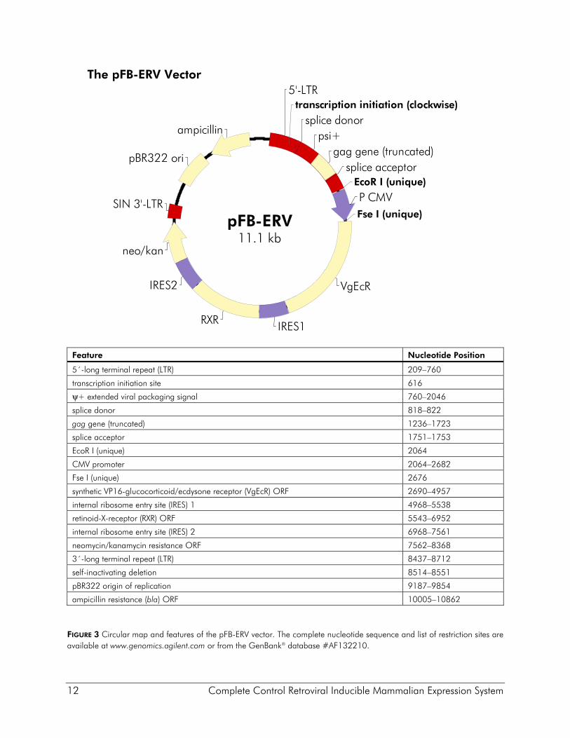
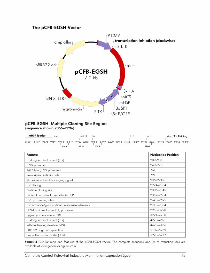
Description of the Vectors The pFB-ERV vector was derived from the high-titer MoMLV vector pFBNeo5 for efficient delivery of the ecdysone receptor proteins VgEcR and RXR (Figure 3). In the vector pFB-ERV the ecdysone receptor and the neomycin-resistance open reading frame (ORF) are expressed from a tricistronic message with the neomycin resistance ORF expressed at the end of the message. Thus, maintenance of infected cell lines in G418 ensures expression of the transcript encoding the receptor genes. The tricistronic transcript is expressed from the CMV promoter, which is flanked by unique EcoR I and Fse I sites so that a cell type-specific promoter of interest may be substituted. The viral promoter within the 3´ LTR has been deleted to make this a self-inactivating (SIN) vector. Upon infection and chromosomal integration into the target cell genome, the SIN deletion is transferred to the 5´ LTR, resulting in an integrated expression cassette in which only the CMV promoter is active. Cells containing an estimated single integrated viral expression cassette can be selected in as high as 1 mg/ml G418, although 600 μg/ml is routinely used. The vector pCFB-EGSH contains an ecdysone-inducible expression cassette inserted between the viral LTRs in the antisense orientation relative to that for the viral promoter (see Figure 4). The U3 promoter within the 5´ LTR of the vector has been replaced with the CMV promoter to increase production of viral RNA in packaging cells, thereby increasing the titer of the viral supernatants. Potential interference from the proviral 5´ LTR is obviated due to the SIN deletion. The inducible expression cassette contains a multiple cloning site that contains three contiguous copies of the HA epitope (3× HA) positioned for fusion at the C-terminus of the protein of interest. A second expression cassette in which the hygromycin-resistance gene is expressed from the TK promoter is located downstream (relative to transcription from the LTRs) of the inducible cassette. A pBR322 origin and ampicillin-resistance gene allow pCFB-EGSH to be propagated in prokaryotes. The pCFB-EGSH-Luc vector contains the luciferase reporter gene and is intended for use as a positive control vector to test the expression of the VgEcR and RXR receptors in pFB-ERV-containing cell lines. The pCFB-EGSH-Luc vector is derived from the pCFB-EGSH vector and has the luciferase gene inserted in the MCS. The pCFB-EGSH-Luc vector does not contain the HA epitope sequence.
12 Complete Control Retroviral Inducible Mammalian Expression System
The pFB-ERV Vector
Feature Nucleotide Position
5´-long terminal repeat (LTR) 209–760
transcription initiation site 616
ψ+ extended viral packaging signal 760–2046
splice donor 818–822
gag gene (truncated) 1236–1723
splice acceptor 1751–1753
EcoR I (unique) 2064
CMV promoter 2064–2682
Fse I (unique) 2676
synthetic VP16-glucocorticoid/ecdysone receptor (VgEcR) ORF 2690–4957
internal ribosome entry site (IRES) 1 4968–5538
retinoid-X-receptor (RXR) ORF 5543–6952
internal ribosome entry site (IRES) 2 6968–7561
neomycin/kanamycin resistance ORF 7562–8368
3´-long terminal repeat (LTR) 8437–8712
self-inactivating deletion 8514–8551
pBR322 origin of replication 9187–9854
ampicillin resistance (bla) ORF 10005–10862
FIGURE 3 Circular map and features of the pFB-ERV vector. The complete nucleotide sequence and list of restriction sites are available at www.genomics.agilent.com or from the GenBank® database #AF132210.
5'-LTR
psi+
transcription initiation (clockwise)
splice donor
gag gene (truncated)
P CMVEcoR I (unique)
splice acceptor
Fse I (unique)
VgEcR
IRES1RXR
IRES2
neo/kan
SIN 3'-LTR
pBR322 ori
ampicillin
pFB-ERV11.1 kb
Complete Control Retroviral Inducible Mammalian Expression System 13
The pCFB-EGSH Vector
Feature Nucleotide Position
5´-long terminal repeat (LTR) 209–935
CMV promoter 249–773
TATA box (CMV promoter) 761
transcription initiation site 791
ψ+ extended viral packaging signal 936–2212
3× HA tag 2224–2304
multiple cloning site 2306–2345
minimal heat shock promoter (mHSP) 2352–2634
3× Sp1 binding sites 2648–2695
5× ecdysone/glucocorticoid responsive elements 2710–2884
HSV-thymidine kinase (TK) promoter 2950–3220
hygromycin resistance ORF 3221–4258
3´-long terminal repeat (LTR) 4270–4621
self-inactivating deletion (SIN) 4423–4466
pBR322 origin of replication 5102–5769
ampicillin resistance (bla) ORF 5920–6777
FIGURE 4 Circular map and features of the pCFB-EGSH vector. The complete sequence and list of restriction sites are available at www.genomics.agilent.com
hygromycinP TK
P CMV
5'-LTR
3x HAMCS
psi+
SIN 3'-LTR
ampicillin
pBR322 ori
transcription initiation (clockwise)
5x E/GRE3x SP1
mHSP
pCFB-EGSH7.0 kb
STOP
mHSP leader
pCFB-EGSH Multiple Cloning Site Region(sequence shown 2355–2296)
Stu IPme I Hind III Sac IPac I
CAC AGC TAG CGT TTA AAC TTA AGC TTA ATT AAC GTA CGA GGC CTG AGC TCG TAC CCA TATSTOP STOP STOP
start 3× HA tag
14 Complete Control Retroviral Inducible Mammalian Expression System
OVERVIEW OF THE CELL SELECTION PROCEDURE Establishing the ponA-inducible expression system in cultured cells involves the following sequence of procedures:
♦ Establish that the vectors function adequately in the proposed target cell type by cotransfecting and/or co-infecting the pFB-ERV and pCFB-EGSH-Luc vectors, inducing with ponA, and assaying for luciferase activity.
♦ Produce stable receptor-expressing cell lines by infection with pFB-ERV virus, selection and expansion of G418-resistant colonies, and screening for the best receptor cell line by transient transfection of pCFB-EGSH-Luc followed by induction with ponA.
♦ While establishing the receptor-expressing cell line, insert gene of interest into the pCFB-EGSH expression cassette.
♦ Verify expression of the gene of interest by transfection/infection of the receptor cell line or cotransfection/infection of the target cell line with pFB-ERV, induction with ponA, and detection by function, phenotype or western blotting.
♦ Infect the receptor cell line with the pCFB-EGSH derivative harboring the gene of interest, select and expand hygromycin-resistant colonies, and screen for the best double-stable cell line by function, phenotype or western blotting following ponA induction.

Complete Control Retroviral Inducible Mammalian Expression System 15
PREPARING THE PCFB-EGSH EXPRESSION VECTOR CONSTRUCT
The pCFB-EGSH vector contains 5 unique restriction sites in the MCS for insertion of the gene of interest (see Figure 4). See reference 7 for protocols covering basic DNA manipulations. The gene of interest to be inserted into the pCFB-EGSH vector should contain a Kozak translation initiation sequence.8 If the gene does not contain a stop codon, insert the gene in frame with one of the four stop codons found in the MCS or in frame with the 3× HA epitope tag. We recommend dephosphorylating the digested vector with calf intestinal alkaline phosphatase (CIAP) prior to ligation with the insert DNA. If the vector is digested with more than one restriction enzyme, the small fragment between the two restriction sites (which appears as background) can be removed by electrophoresing the DNA on an agarose gel and recovering the desired vector by electroelution. After purification and ethanol precipitation of the DNA, resuspend the DNA in TE buffer (see Preparation of Media and Reagents). The concentration of the vector DNA should be the same as the concentration of the insert DNA (~0.1 μg/μl). For ligation, the ideal insert-to-vector ratio is variable; however, a reasonable starting ratio is 2:1 (measured in available picomole ends). The insert-to-vector ratio is calculated as follows:
picomole ends / microgram of DNA 2 × 10
number of base pairs × 660
6
=
16 Complete Control Retroviral Inducible Mammalian Expression System
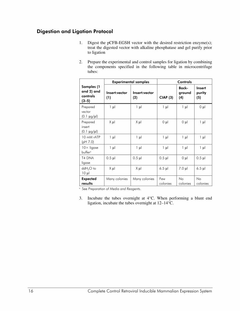
Digestion and Ligation Protocol
1. Digest the pCFB-EGSH vector with the desired restriction enzyme(s); treat the digested vector with alkaline phosphatase and gel purify prior to ligation
2. Prepare the experimental and control samples for ligation by combining the components specified in the following table in microcentrifuge tubes:
Samples (1 and 2) and controls (3–5)
Experimental samples Controls
Insert:vector (1)
Insert:vector (2)
CIAP (3)
Back-ground (4)
Insert purity (5)
Prepared vector (0.1 μg/μl)
1 μl 1 μl 1 μl 1 μl 0 μl
Prepared insert (0.1 μg/μl)
X μl X μl 0 μl 0 μl 1 μl
10 mM rATP (pH 7.0)
1 μl 1 μl 1 μl 1 μl 1 μl
10× ligase buffera
1 μl 1 μl 1 μl 1 μl 1 μl
T4 DNA ligase
0.5 μl 0.5 μl 0.5 μl 0 μl 0.5 μl
ddH2O to 10 μl
X μl X μl 6.5 μl 7.0 μl 6.5 μl
Expected results
Many colonies Many colonies Few colonies
No colonies
No colonies
a See Preparation of Media and Reagents. 3. Incubate the tubes overnight at 4°C. When performing a blunt end
ligation, incubate the tubes overnight at 12–14°C.
Complete Control Retroviral Inducible Mammalian Expression System 17
Transforming Competent Cells with the pCFB-EGSH Construct
1. Transform competent cells with 1–2 μl of the ligation mixture.
2. Plate the transformation on LB–ampicillin agar plates (see Preparation of Media and Reagents).
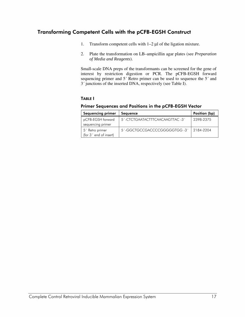
Small-scale DNA preps of the transformants can be screened for the gene of interest by restriction digestion or PCR. The pCFB-EGSH forward sequencing primer and 5´ Retro primer can be used to sequence the 5´ and 3´ junctions of the inserted DNA, respectively (see Table I). TABLE I
Primer Sequences and Positions in the pCFB-EGSH Vector
Sequencing primer Sequence Position (bp)
pCFB-EGSH forward sequencing primer
5´-CTCTGAATACTTTCAACAAGTTAC -3´ 2398-2375
5´ Retro primer (for 3´ end of insert)
5´-GGCTGCCGACCCCGGGGGTGG -3´ 2184-2204
18 Complete Control Retroviral Inducible Mammalian Expression System
TRANSFECTION PROTOCOL
Transient Cotransfection of Expression Construct and pFB-ERV to Determine Plasmid/Host Cell Compatibility
Note We recommend cesium chloride-banded DNA preps (or a comparable protocol) of positive transformants before transfection into cultured cells.
The cultured cell line chosen for expression of the gene of interest can be transiently cotransfected with the pFB-ERV receptor vector and pCFB-EGSH-Luc, or the pCFB-EGSH construct containing the gene of interest as a quick method to determine whether or not the expression construct can be induced in the cell line. The preferred method of transfection depends on the chosen cultured cell line. See the Agilent genomics web site at www.genomics.agilent.com for information on calcium phosphate- and liposome-mediated transfection kits. Transfect duplicate samples of the cells (at minimum) to enable a comparison of uninduced and induced expression levels.
Note Transfect cells with several different amounts of DNA in order to determine the optimal concentration. The optimal amount of DNA will vary between cell lines and transfection methods. Follow the manufacturer's transfection protocol. Expression of the inserted gene can be induced by adding 1–10 μM ponA (diluted from a 1 mM stock, see Preparation of Media and Reagents) to the medium 4–20 hours before harvesting the cells. Parameters will need to be optimized for each particular system.
Results from transient assays should be used to ascertain whether the gene of interest can be induced in the cell line of choice and to determine the maximal expression levels that can be expected in stable cells or transgenic animals. Uninduced pCFB-EGSH background expression will be considerably higher in transient transfections than stable transfections. This background is due to the high copy number of the plasmid and accessibility of the free nuclear DNA to core transcription factors. In stably transduced cells, however, the pCFB-EGSH vector is integrated into the host chromosome and is naturally repressed. In cases where the target cell line is not efficiently transfected with available transfection reagents, the vectors may be tested by preparing virus using the protocols described below, and co-infecting the target cell line using various ratios of pFB-ERV and pCFB-EGSH viral supernatants. As stated in the above note for transient cotransfection, the induction ratios for co-infections will likely not approach those attainable by two-step sequential clonal selection of double-stable cell lines, but may be useful for determining whether the system and the target cell line are compatible.
Complete Control Retroviral Inducible Mammalian Expression System 19
PRODUCTION OF STABLE RECEPTOR-EXPRESSING CELL LINES Although it is possible to select double-stable cell lines following simultaneous co-infection with both pFB-ERV and the pCFB-EGSH-derived viruses, the chances of obtaining a double-stable line with both negligible uninduced background and high-level induced expression is much greater using a two-step selection procedure. First, a pFB-ERV-transduced population is screened for optimal expression of the RXR and VgEcR receptor proteins, and then the best of the receptor-expressing clones is infected with the pEGSH-derivative, and screened for double-stable clones with low background and high-level induced expression.
Note The steps performed in this section, Production of Stable Receptor-Expressing Cell Lines, need to be carried out under sterile conditions in a laminar flow hood.
Prior to production of virus, users should be thoroughly familiar with the suggestions and Web sites described in Safety Considerations. All virus work should be performed in a designated virus work area.
The use of target cell lines harboring retrovirus capable of encapsidating vector proviral RNA can result in the undesirable spread of vector-derived virus. There are a number of published protocols describing assays for detection of replication-competent retrovirus based on mobilization of a provirus containing a detectable marker (“marker rescue”) or by direct detection of reverse transcriptase activity in tissue culture supernatants. It is strongly recommended that all cell lines to be used for viral production or infection be first tested for the presence of endogenous retrovirus using one of the assays described.9
Although a variety of transfection protocols and producer cell lines may be successfully used to produce virus using the pFB-ERV plasmid, the following protocol consistently results in the production of viral titers of >105 G418-resistant colony forming units (cfu)/ml using NIH3T3 cells as target. pFB-ERV virus is transiently produced following cotransfection of 293 cells with the gag-pol and env-expressing packaging vectors pVPack-GP and one of the pVPack env-expressing vectors. The protocol employs the Transfection MBS Mammalian transfection kit, modified according to Pear and colleagues.6 Although excellent results may be obtained using 293 cells, we recommend the use of the 293 cell derivative, 293T, which has been shown to transfect with a significantly greater efficiency.1
20 Complete Control Retroviral Inducible Mammalian Expression System
Day 1: Preparing for Production of Virus by Transfection
293T Host Cell Preparation Split 293T cells at 2.5–3.0 × 10⁶ cells per 60-mm tissue culture dish in growth medium (See Preparation of Media and Reagents) 24 hours before the transfection and incubate at 37°C until needed. For some target cells it may be necessary to concentrate the virus in order to achieve a high-level efficiency of infection. In this case multiple 100-mm dishes are seeded with 6.3–7.5 × 10⁶ cells per dish.
Note To achieve optimal titers, it is important that the 293T cells are healthy and growing exponentially. Cells should be passaged at high density, and ideally passaged no more than 20 times (no more than approximately 2 months); it is thus prudent to initially prepare a large number of frozen vials of the cells while they are at a low passage and healthy. Care should be taken to avoid clumping of the cells during passaging and plating for transfection.
Plasmid DNA Preparation DNA preparations of high purity should be used for the transfections.
1. Pipette 3 μg (60-mm dishes) or 6 μg (100-mm dishes) of each the following into a clean 1.5-ml microcentrifuge tube; prepare one tube for each transfection to be carried out. pFB-ERV pVPack-GP (gag-pol-expressing vector) One of the four env-expressing vectors (pVPack-Eco, pVPack-Ampho,
pVPack-VSV-G, pVPack-10A1)
2. To each of the tubes containing the mixed vector DNA, add 1 ml 100% (v/v) ethanol and 0.1 × volume of 3 M sodium acetate to the DNA mixture; mix by inverting the tube, incubate at –80°C for 30 minutes. Collect the DNA pellet by centrifugation at 12,000 × g for 10 minutes at 4°C. Aspirate and discard the supernatant. Add 1 ml 70% (v/v) ethanol to the tube, vortex briefly, and collect the DNA pellet by centrifugation at 12,000 × g for 5 minutes at 4°C. Remove and discard the supernatant; close the cap of the tube. Store wet pellets at 4°C overnight.
Complete Control Retroviral Inducible Mammalian Expression System 21
Day 2: Transfecting Cells
Note The procedure on Day 2 will take a minimum of 10 hours to complete.
Adding the MBS-Containing Medium to the Cells
1. Inspect the host cells that were split the day before; they should be approximately 80% confluent. [If cells are significantly less than 80% confluent, viral supernatants may be harvested 72 hours post-transfection rather than 48 hours.]
2. Prepare the MBS-containing medium. This must be done immediately prior to the transfection. 4 ml per 60-mm dish, or 10 ml for each 100-mm dish, should be prepared. See Preparation of Media and Reagents.
3. Add MBS-containing medium to each dish (4 ml per 60-mm dish, 10 ml for each 100-mm dish) and return the dishes to the 37°C incubator. This must be completed 20-30 minutes before the addition of the DNA suspension.
Adding the DNA Suspension to the Cells
1. Remove the microcentrifuge tubes containing the wet DNA pellets from storage at 4°C and transfer them to the laminar flow hood.
2. Resuspend each DNA pellet in sterile H2O (450 μl for 60-mm dishes, 900 μl for 100-mm dishes) and transfer the liquid to separate BD Falcon polystyrene tubes.
3. To each resuspended DNA pellet add Solution I (50 μl or 100 μl for 60-mm or 100-mm dishes, respectively) and Solution II (500 μl or 1.0 ml for 60-mm or 100-mm dishes, respectively) from the Transfection MBS Mammalian Transfection Kit.
4. Gently resuspend any precipitate in the DNA suspension by pipetting the suspension up and down with a pipettor set at 500 μl. The DNA suspension should appear clear to opaque. Allow the DNA suspension to sit at room temperature for 10 minutes.
22 Complete Control Retroviral Inducible Mammalian Expression System
5. Remove the dishes to be transfected from the incubator and add the DNA suspension onto the dishes in a dropwise fashion, swirling gently to prevent the cells from being lifted from the dish and to distribute the DNA suspension evenly.
Note From this point on, it should be assumed that infectious virus is present in the supernatant of the transfected cells. Gloves and disposable lab coats should be worn while working with the virus. We recommend that gloved hands be sprayed intermittently with ethanol. When pipetting cell culture supernatants and transferring dishes to and from the laminar flow hood, aerosols should be avoided. In case of spills, follow the procedures recommended in the UCSD Vector Development Lab Web site http://medicine.ucsd.edu/gt/MoMuLV.html. Guidelines for disposal of dirty pipets and plasticware are also given in the UCSD Vector Development Lab Web site.
6. Return the tissue culture dishes to the 37°C incubator.
7. After incubating for 3 hours, remove the medium from the dishes and replace it with growth medium (4 ml for 60-mm dishes, 10 ml for 100-mm dishes) supplemented with 25 μM chloroquine (see Preparation of Media and Reagents). Return the dishes to the 37°C incubator.
8. After incubating for an additional 6–7 hours, remove the growth medium containing 25 μM chloroquine and replace with growth medium—no chloroquine.
Day 3: Preparing for the Transduction
1. Remove growth medium from 293T dishes and replace with of fresh growth medium (3.0 ml for 60-mm dishes, 7.5 ml for 100-mm dishes). Return the dishes to the 37°C incubator.
Note If virus is to be harvested 72 hours post-transfection rather than 48 hours, steps 2 and 3 should be carried out on Day 4.
2. Split the target cells, seeding 5 × 10⁵ cells per 100-mm dish. This seeding density may vary with the cell line; 20–30% confluency is desirable.
3. Return the dishes to the 37°C incubator overnight.
Complete Control Retroviral Inducible Mammalian Expression System 23
Day 4: Transducing the Target Cells
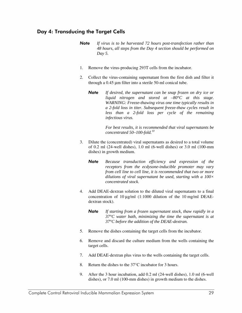
Note If virus is to be harvested 72 hours post-transfection rather than 48 hours, all steps from the Day 4 section should be performed on Day 5.
1. Remove the virus-producing 293T cells from the incubator.
2. Collect the virus-containing supernatant from the first dish and filter it through a 0.45 μm filter into a sterile 50-ml conical tube.
Note If desired, the supernatant can be snap frozen on dry ice or liquid nitrogen and stored at -80°C at this stage. WARNING: Freeze-thawing virus one time typically results in a 2-fold loss in titer. Subsequent freeze-thaw cycles result in less than a 2-fold loss per cycle of the remaining infectious virus.
3. Dilute viral supernatants as desired to a total volume of 3.0 ml in growth medium.
Note Because transduction efficiency and expression of the receptors from the CMV promoter may vary from cell line to cell line, it is recommended that two or more dilutions of viral supernatant be used (e.g., undiluted and 1:10)
4. Add DEAE-dextran solution to the diluted viral supernatants to a final concentration of 10 μg/ml (1:1000 dilution of the 10 mg/ml DEAE-dextran stock. See Preparation of Media and Reagents).
Note If starting from a frozen supernatant stock, thaw rapidly in a 37°C water bath, minimizing the time the supernatant is at 37°C before the addition of the DEAE-dextran.
5. Remove the dishes containing the target cells from the incubator.
6. Remove and discard the culture medium from the wells containing the target cells.
7. Add DEAE-dextran plus virus to the wells containing the target cells.
8. Return the dishes to the 37°C incubator for 3 hours.
9. After the 3 hour incubation, add 7.0 ml growth medium to the dishes.
10. One day following the infection, split the cells at least 1:20 and seed into 100-mm dishes.
24 Complete Control Retroviral Inducible Mammalian Expression System
Use of Antibiotics for Cell Selection Not all mammalian cell lines are equally sensitive to the antibiotic G418 and hygromycin. The minimal lethal concentration can range from 100 μg/ml to 1 mg/ml. The antibiotic concentration to be used for selection must be determined for each cell line before beginning the experiment. Consult the available literature on the sensitivity of cell lines to G418 and hygromycin. If no information about the sensitivity of a particular cell line is available, a simple way to determine sensitivity is to grow cultures in a multiwell dish with a range of antibiotic concentrations between the individual wells. The optimal concentration is the lowest one that kills all of the cells within 10–14 days. (Rapidly dividing cells may be killed more readily since the antibiotic appears to act mainly on dividing cells.) In some cases, it may be possible to reduce the concentration of the antibiotic after initial selection and still maintain selective pressure for the marker gene. For example, NIH3T3 cells are generally selected in 400 μg/ml G418, but selective pressure for the neomycin-resistance (Neor) gene can be maintained in 250 μg/ml.
Isolating G418-Resistant Stable Clones and Expanding the Colonies
1. Select for transfected cells by adding G418 (100 μg/ml–1 mg/ml, depending on the cell line) to the medium.
2. Isolate individual clones and expand the resulting colonies.
Examining Receptor Expression Examine the expanded clones for expression of the VgEcR and RXR receptors by transiently transfecting the G418-resistant clones with the pCFB-EGSH-Luc vector or infecting with packaged pCFB-EGSH-Luc virus particles and inducing luciferase transcription with ponA.
Note examine at least 30 individual clones, as the site and number of proviral integrations will affect how well the receptors are expressed. For most cell types, relatively small quantities of pCFB-EGSH-Luc (10–100 ng/105 cells plated) should give induction ratios of ≥ 20-fold and reach ≥105 RLUs when fully induced in these assays.
1. For each expanded clone to be tested, plate cells in duplicate at the recommended density for the transfection procedure to be used. If clones are to be screened by infection, plate 1–2 × 10⁴ cells per well in 24-well dishes.
2. Transfect clones with pCFB-EGSH-Luc using the transfection method preferred for the target cell line. Alternatively, pCFB-EGSH-Luc virus may be prepared using the procedures described below. If clones are to be tested by infection, it is recommended that the pCFB-EGSH-Luc viral supernatants be concentrated 50- to 100-fold prior to infection of the expanded pFB-ERV clones.
Complete Control Retroviral Inducible Mammalian Expression System 25
3. One day following transfection, add 1-10 μM ponA to one of each of the duplicate wells, and an equivalent volume of 100% ethanol to the other well. Induce for 18-24 hours. If clones are to be screened by infection, add ponA to the media 48 hours after infection, and induce for >24 hours.
4. Perform luciferase assays, and select the clone(s) showing the highest induction(s).
PREPARING PCFB-EGSH VIRAL STOCKS
Note Prior to production of virus, users should be thoroughly familiar with the suggestions and Web sites described in Safety Considerations. All virus work should be performed in a designated virus work area. All cell lines to be used for production of or infection by retrovirus should first be tested for the presence of endogenous retrovirus.
Although a variety of transfection protocols and producer cell lines may be successfully used to produce virus using the pCFB-EGSH plasmid, the following protocol consistently results in the production of viral titers >105 hygromycin-resistant colony forming units (cfu)/ml using NIH3T3 cells as target. pCFB-EGSH-derived virus is transiently produced following cotransfection of 293 cells with the gag-pol and env-expressing packaging vectors pVPack-GP and one of the pVPack env-expressing vectors. Because it will likely be necessary to concentrate the virus for best results, the methods described in reference 10 should be considered. The protocol employs the Transfection MBS Mammalian transfection kit, modified according to Pear and colleagues.6 Although excellent results may be obtained using 293 cells, we recommend the use of the 293 cell derivative 293T, which has been shown to transfect with a significantly greater efficiency.1
Note The steps performed in this section, Preparing pCFB-EGSH Viral Stocks, need to be carried out under sterile conditions in a laminar flow hood.
26 Complete Control Retroviral Inducible Mammalian Expression System
Day 1: Preparing for Production of Virus by Transfection
293T Host Cell Preparation Split 293T cells at 2.5–3.0 × 10⁶ cells per 60-mm tissue culture dish in growth medium 24 hours before the transfection and incubate at 37°C until needed. For some target cells it may be necessary to concentrate the virus in order to achieve a high-level efficiency of infection. In this case multiple 100-mm dishes are seeded with 6.3–7.5 × 10⁶ cells per dish.
Note To achieve optimal titers, it is important that the 293T cells are healthy and growing exponentially. Cells should be passaged at high density, and ideally passaged no more than 20 times (no more than approximately 2 months); it is thus prudent to initially prepare a large number of frozen vials of the cells while they are at a low passage and healthy. Care should be taken to avoid clumping of the cells during passaging and plating for transfection.
Plasmid DNA Preparation DNA preparations of high purity should be used for the transfections.
1. Pipette 3 μg (60-mm dishes) or 6 μg (100-mm dishes) of each the following into a clean 1.5-ml microcentrifuge tube; prepare one tube for each transfection to be carried out. pCFB-EGSH-derivative pVPack-GP (gag-pol-expressing vector) pVPack-VSV-G
2. To each of the tubes containing the mixed vector DNA, add 1 ml 100% (v/v) ethanol and 0.1 × volume of 3 M sodium acetate to the DNA mixture; mix by inverting the tube, incubate at –80°C for 30 minutes. Collect the DNA pellet by centrifugation at 12,000 × g for 10 minutes at 4°C. Aspirate and discard the supernatant. Add 1 ml 70% (v/v) ethanol to the tube, vortex briefly, and collect the DNA pellet by centrifugation at 12,000 × g for 5 minutes at 4°C. Remove and discard the supernatant; close the cap of the tube. Store wet pellets at 4°C overnight.
Complete Control Retroviral Inducible Mammalian Expression System 27
Day 2: Transfecting Cells
Note The procedure on Day 2 will take a minimum of 10 hours to complete.
Adding the MBS-Containing Medium to the Cells
1. Inspect the host cells that were split the day before; they should be approximately 80% confluent. [If cells are significantly less than 80% confluent, viral supernatants may be harvested 72 hours post-transfection rather than 48 hours.]
2. Prepare the MBS-containing medium. This must be done immediately prior to the transfection. 4 ml per 60-mm dish, or 10 ml for each 100-mm dish, should be prepared.
3. Add MBS-containing medium to each dish (4 ml per 60-mm dish, 10 ml for each 100-mm dish) and return the dishes to the 37°C incubator. This must be completed 20–30 minutes before the addition of the DNA suspension.
Adding the DNA Suspension to the Cells
1. Remove the microcentrifuge tubes containing the wet DNA pellets from storage at 4°C and transfer them to the laminar flow hood.
2. Resuspend each DNA pellet in sterile H2O (450 μl for 60-mm dishes, 900 μl for 100-mm dishes) and transfer the liquid to separate BD Falcon polystyrene tubes.
3. To each resuspended DNA pellet add Solution I (50 μl or 100 μl for 60-mm or 100-mm dishes, respectively) and Solution II (500 μl or 1.0 ml for 60-mm or 100-mm dishes, respectively) from the Transfection MBS Mammalian Transfection Kit.
4. Gently resuspend any precipitate in the DNA suspension by pipetting the suspension up and down with a pipettor set at 500 μl. The DNA suspension should appear clear to opaque. Allow the DNA suspension to sit at room temperature for 10 minutes.
28 Complete Control Retroviral Inducible Mammalian Expression System
5. Remove the dishes to be transfected from the incubator and add the DNA suspension onto the dishes in a dropwise fashion, swirling gently to prevent the cells from being lifted from the dish and to distribute the DNA suspension evenly.
Note From this point on, it should be assumed that infectious virus is present in the supernatant of the transfected cells. Gloves and disposable lab coats should be worn while working with the virus. We recommend that gloved hands be sprayed intermittently with ethanol. When pipetting cell culture supernatants and transferring dishes to and from the laminar flow hood, aerosols should be avoided. In case of spills, follow the procedures recommended in the UCSD Vector Development Lab Web site http://medicine.ucsd.edu/gt/MoMuLV.html. Guidelines for disposal of dirty pipets and plasticware are also given in the UCSD Vector Development Lab Web site
6. Return the tissue culture dishes to the 37°C incubator.
7. After incubating for 3 hours, remove the medium from the dishes and replace it with growth medium (4 ml for 60-mm dishes, 10 ml for 100-mm dishes) supplemented with 25 μM chloroquine. Return the dishes to the 37°C incubator.
8. After incubating for an additional 6–7 hours, remove the growth medium containing 25 μM chloroquine and replace with growth medium—no chloroquine.
Day 3: Preparing for the Transduction
1. Remove growth medium from 293T dishes and replace with of fresh growth medium (3.0 ml for 60-mm dishes, 7.5 ml for 100-mm dishes). Return the dishes to the 37°C incubator.
Note If virus is to be harvested 72 hours post-transfection rather than 48 hours, steps 2 and 3 should be carried out on Day 4.
2. Split the pFB-ERV-transduced receptor-expressing target cells. If cells are to be induced following mass infection of the receptor cell line by pCFB-EGSH virus, i.e., without selecting double-stable clones with hygromycin, seed 1–2 × 10⁴ cells in duplicate for each viral dilution for 24-well dishes, or 105 cells per well for 6-well dishes. If hygromycin-resistant double-stable cell lines are to be selected, seed 5 × 10⁵ cells per 100-mm dish. This seeding density may vary with the cell line; 20–30% confluency is desirable.
3. Return the dishes to the 37°C incubator overnight.
Complete Control Retroviral Inducible Mammalian Expression System 29
Day 4: Transducing the Target Cells
Note If virus is to be harvested 72 hours post-transfection rather than 48 hours, all steps from the Day 4 section should be performed on Day 5.
1. Remove the virus-producing 293T cells from the incubator.
2. Collect the virus-containing supernatant from the first dish and filter it through a 0.45 μm filter into a sterile 50-ml conical tube.
Note If desired, the supernatant can be snap frozen on dry ice or liquid nitrogen and stored at –80°C at this stage. WARNING: Freeze-thawing virus one time typically results in a 2-fold loss in titer. Subsequent freeze-thaw cycles result in less than a 2-fold loss per cycle of the remaining infectious virus.
For best results, it is recommended that viral supernatants be concentrated 50–100-fold.10
3. Dilute the (concentrated) viral supernatants as desired to a total volume of 0.2 ml (24-well dishes), 1.0 ml (6-well dishes) or 3.0 ml (100-mm dishes) in growth medium.
Note Because transduction efficiency and expression of the receptors from the ecdysone-inducible promoter may vary from cell line to cell line, it is recommended that two or more dilutions of viral supernatant be used, starting with a 100× concentrated stock.
4. Add DEAE-dextran solution to the diluted viral supernatants to a final concentration of 10 μg/ml (1:1000 dilution of the 10 mg/ml DEAE-dextran stock).
Note If starting from a frozen supernatant stock, thaw rapidly in a 37°C water bath, minimizing the time the supernatant is at 37°C before the addition of the DEAE-dextran.
5. Remove the dishes containing the target cells from the incubator.
6. Remove and discard the culture medium from the wells containing the target cells.
7. Add DEAE-dextran plus virus to the wells containing the target cells.
8. Return the dishes to the 37°C incubator for 3 hours.
9. After the 3 hour incubation, add 0.2 ml (24-well dishes), 1.0 ml (6-well dishes), or 7.0 ml (100-mm dishes) in growth medium to the dishes.
30 Complete Control Retroviral Inducible Mammalian Expression System
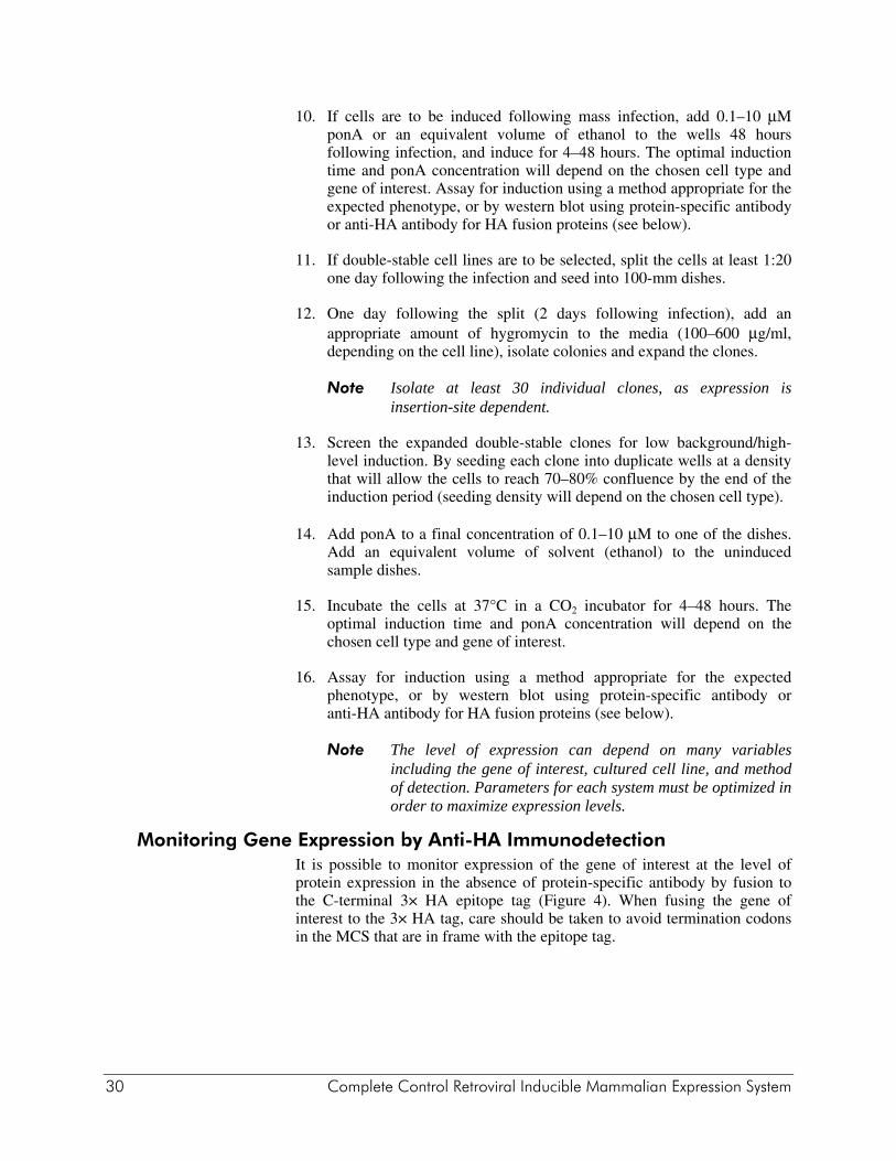
10. If cells are to be induced following mass infection, add 0.1–10 μM ponA or an equivalent volume of ethanol to the wells 48 hours following infection, and induce for 4–48 hours. The optimal induction time and ponA concentration will depend on the chosen cell type and gene of interest. Assay for induction using a method appropriate for the expected phenotype, or by western blot using protein-specific antibody or anti-HA antibody for HA fusion proteins (see below).
11. If double-stable cell lines are to be selected, split the cells at least 1:20 one day following the infection and seed into 100-mm dishes.
12. One day following the split (2 days following infection), add an appropriate amount of hygromycin to the media (100–600 μg/ml, depending on the cell line), isolate colonies and expand the clones.
Note Isolate at least 30 individual clones, as expression is insertion-site dependent.
13. Screen the expanded double-stable clones for low background/high-level induction. By seeding each clone into duplicate wells at a density that will allow the cells to reach 70–80% confluence by the end of the induction period (seeding density will depend on the chosen cell type).
14. Add ponA to a final concentration of 0.1–10 μM to one of the dishes. Add an equivalent volume of solvent (ethanol) to the uninduced sample dishes.
15. Incubate the cells at 37°C in a CO2 incubator for 4–48 hours. The optimal induction time and ponA concentration will depend on the chosen cell type and gene of interest.
16. Assay for induction using a method appropriate for the expected phenotype, or by western blot using protein-specific antibody or anti-HA antibody for HA fusion proteins (see below).
Note The level of expression can depend on many variables including the gene of interest, cultured cell line, and method of detection. Parameters for each system must be optimized in order to maximize expression levels.
Monitoring Gene Expression by Anti-HA Immunodetection It is possible to monitor expression of the gene of interest at the level of protein expression in the absence of protein-specific antibody by fusion to the C-terminal 3× HA epitope tag (Figure 4). When fusing the gene of interest to the 3× HA tag, care should be taken to avoid termination codons in the MCS that are in frame with the epitope tag.
Complete Control Retroviral Inducible Mammalian Expression System 31
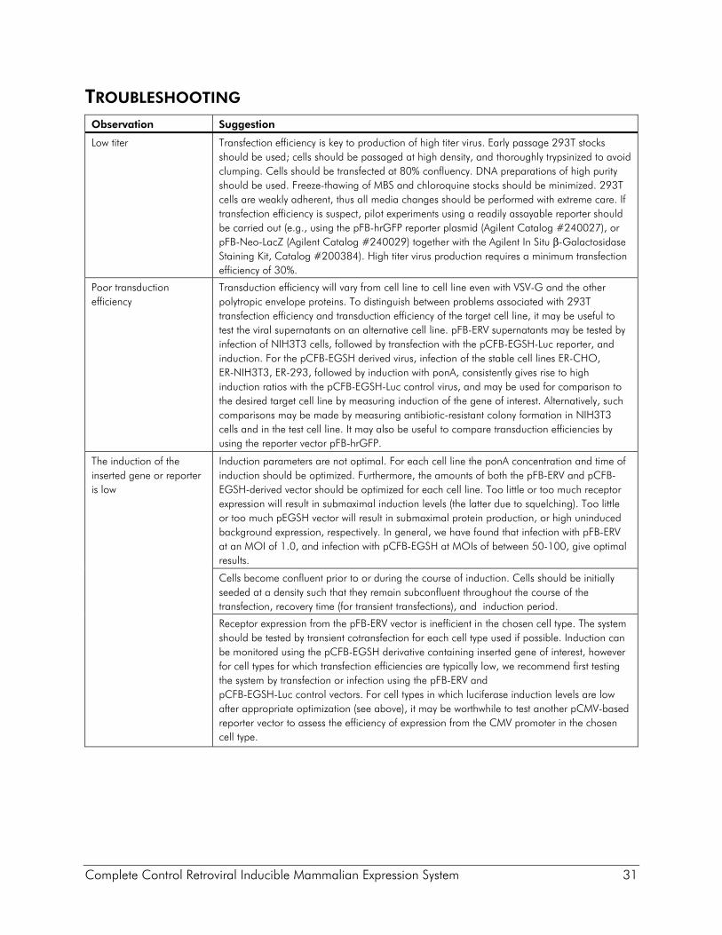
TROUBLESHOOTING Observation Suggestion
Low titer Transfection efficiency is key to production of high titer virus. Early passage 293T stocks should be used; cells should be passaged at high density, and thoroughly trypsinized to avoid clumping. Cells should be transfected at 80% confluency. DNA preparations of high purity should be used. Freeze-thawing of MBS and chloroquine stocks should be minimized. 293T cells are weakly adherent, thus all media changes should be performed with extreme care. If transfection efficiency is suspect, pilot experiments using a readily assayable reporter should be carried out (e.g., using the pFB-hrGFP reporter plasmid (Agilent Catalog #240027), or pFB-Neo-LacZ (Agilent Catalog #240029) together with the Agilent In Situ β-Galactosidase Staining Kit, Catalog #200384). High titer virus production requires a minimum transfection efficiency of 30%.
Poor transduction efficiency
Transduction efficiency will vary from cell line to cell line even with VSV-G and the other polytropic envelope proteins. To distinguish between problems associated with 293T transfection efficiency and transduction efficiency of the target cell line, it may be useful to test the viral supernatants on an alternative cell line. pFB-ERV supernatants may be tested by infection of NIH3T3 cells, followed by transfection with the pCFB-EGSH-Luc reporter, and induction. For the pCFB-EGSH derived virus, infection of the stable cell lines ER-CHO, ER-NIH3T3, ER-293, followed by induction with ponA, consistently gives rise to high induction ratios with the pCFB-EGSH-Luc control virus, and may be used for comparison to the desired target cell line by measuring induction of the gene of interest. Alternatively, such comparisons may be made by measuring antibiotic-resistant colony formation in NIH3T3 cells and in the test cell line. It may also be useful to compare transduction efficiencies by using the reporter vector pFB-hrGFP.
The induction of the inserted gene or reporter is low
Induction parameters are not optimal. For each cell line the ponA concentration and time of induction should be optimized. Furthermore, the amounts of both the pFB-ERV and pCFB-EGSH-derived vector should be optimized for each cell line. Too little or too much receptor expression will result in submaximal induction levels (the latter due to squelching). Too little or too much pEGSH vector will result in submaximal protein production, or high uninduced background expression, respectively. In general, we have found that infection with pFB-ERV at an MOI of 1.0, and infection with pCFB-EGSH at MOIs of between 50-100, give optimal results.
Cells become confluent prior to or during the course of induction. Cells should be initially seeded at a density such that they remain subconfluent throughout the course of the transfection, recovery time (for transient transfections), and induction period.
Receptor expression from the pFB-ERV vector is inefficient in the chosen cell type. The system should be tested by transient cotransfection for each cell type used if possible. Induction can be monitored using the pCFB-EGSH derivative containing inserted gene of interest, however for cell types for which transfection efficiencies are typically low, we recommend first testing the system by transfection or infection using the pFB-ERV and pCFB-EGSH-Luc control vectors. For cell types in which luciferase induction levels are low after appropriate optimization (see above), it may be worthwhile to test another pCMV-based reporter vector to assess the efficiency of expression from the CMV promoter in the chosen cell type.

32 Complete Control Retroviral Inducible Mammalian Expression System
Observation Suggestion
The induction of the inserted gene or reporter is low (CONTINUED)
Expression of the inserted gene is inefficient in the chosen cell type. Confirm that the pCFB-EGSH-Luc reporter can be induced by transient cotransfection or co-infection in the chosen cell type. If the control reporter vector can be efficiently induced, inefficient expression of the protein of interest may be due to poor translation (e.g., resulting from inefficient codon usage), or to stability problems due to inappropriate post-translational processing or targeting of the protein of interest. In such cases it is prudent to test transient induced expression in one or more alternative cell lines, and it may be useful to assess whether the gene of interest can be expressed constitutively from a more robust vector, e.g., directly from the pCMV promoter.
Total protein levels from uninduced and induced extracts are not normalized. Equivalent amounts of protein should be used when comparing expression levels.
In stable cells, pFB-ERV and/or pCFB-EGSH-derived proviruses are chromosomally integrated at sites that do not allow optimal expression. Optimal double-stable cell line production requires that both vectors are integrated at positions in the genome that a) allow expression of optimal levels of the receptors, and b) allow maximal induction of the gene of interest but minimal uninduced background expression. Although stable cell lines that are produced by simultaneous co-infection of pFB-ERV and the pCFB-EGSH do yield double-stable cell lines that may give adequate induction ratios, the frequency of getting both viruses integrated at sites for optimal performance is much lower than the two-step selection method. We recommend first producing stable pFB-ERV cell lines and screening at least 25 colonies by transient transfection of the pCFB-EGSH-Luc reporter. The pFB-ERV cell line that shows the highest level of induced expression by this assay can now be used to produce double-stable cell lines using the pCFB-EGSH-derived plasmid. It is again recommended that a minimum of twenty colonies be screened for one that gives low background and high induction levels.
Complete Control Retroviral Inducible Mammalian Expression System 33
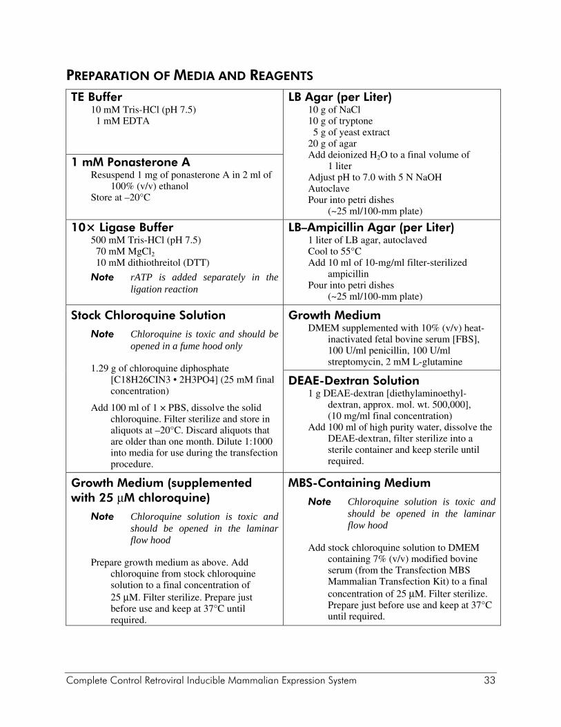
PREPARATION OF MEDIA AND REAGENTS TE Buffer
10 mM Tris-HCl (pH 7.5) 1 mM EDTA
LB Agar (per Liter)10 g of NaCl 10 g of tryptone 5 g of yeast extract 20 g of agar Add deionized H2O to a final volume of
1 liter Adjust pH to 7.0 with 5 N NaOH Autoclave Pour into petri dishes
(~25 ml/100-mm plate)
1 mM Ponasterone A Resuspend 1 mg of ponasterone A in 2 ml of
100% (v/v) ethanol Store at –20°C
10× Ligase Buffer 500 mM Tris-HCl (pH 7.5) 70 mM MgCl2 10 mM dithiothreitol (DTT)
Note rATP is added separately in the ligation reaction
LB–Ampicillin Agar (per Liter)1 liter of LB agar, autoclaved Cool to 55°C Add 10 ml of 10-mg/ml filter-sterilized
ampicillin Pour into petri dishes
(~25 ml/100-mm plate)
Stock Chloroquine Solution
Note Chloroquine is toxic and should be opened in a fume hood only
1.29 g of chloroquine diphosphate [C18H26CIN3 • 2H3PO4] (25 mM final concentration)
Add 100 ml of 1 × PBS, dissolve the solid chloroquine. Filter sterilize and store in aliquots at –20°C. Discard aliquots that are older than one month. Dilute 1:1000 into media for use during the transfection procedure.
Growth Medium DMEM supplemented with 10% (v/v) heat-
inactivated fetal bovine serum [FBS], 100 U/ml penicillin, 100 U/ml streptomycin, 2 mM L-glutamine
DEAE-Dextran Solution 1 g DEAE-dextran [diethylaminoethyl-
dextran, approx. mol. wt. 500,000], (10 mg/ml final concentration)
Add 100 ml of high purity water, dissolve the DEAE-dextran, filter sterilize into a sterile container and keep sterile until required.
Growth Medium (supplemented with 25 μM chloroquine)
Note Chloroquine solution is toxic and should be opened in the laminar flow hood
Prepare growth medium as above. Add chloroquine from stock chloroquine solution to a final concentration of 25 μM. Filter sterilize. Prepare just before use and keep at 37°C until required.
MBS-Containing Medium
Note Chloroquine solution is toxic and should be opened in the laminar flow hood
Add stock chloroquine solution to DMEM containing 7% (v/v) modified bovine serum (from the Transfection MBS Mammalian Transfection Kit) to a final concentration of 25 μM. Filter sterilize. Prepare just before use and keep at 37°C until required.

34 Complete Control Retroviral Inducible Mammalian Expression System
REFERENCES 1. DuBridge, R. B., Tang, P., Hsia, H. C., Leong, P. M., Miller, J. H. et al. (1987) Mol
Cell Biol 7(1):379-87. 2. No, D., Yao, T. P. and Evans, R. M. (1996) Proc Natl Acad Sci U S A 93(8):3346-51. 3. yborski, D. L. and Vaillancourt, P. (1999) Strategies 12(1):1-4. 4. Chen, J. D., Umesono, K. and Evans, R. M. (1996) Proc Natl Acad Sci U S A
93(15):7567-71. 5. Felts, K., Bauer, J. C. and Vaillancourt, P. (1999) Strategies 12(2):74-77. 6. Pear, W. S., Scott, M. L. and Nolan, G. P. (1997). Generation of High-Titer, Helper-
Free Retroviruses by Transient Transfection. In Methods in Molecular Medicine: Gene Therapy Protocols,P. D. Robbins (Ed.). Humana Press, Totawa, New Jersey.
7. Sambrook, J., Fritsch, E. F. and Maniatis, T. (1989). Molecular Cloning: A Laboratory Manual. Cold Spring Harbor Laboratory Press, Cold Spring Harbor, NY.
8. Kozak, M. (1999) Gene 234(2):187-208. 9. Cepko, C. and Pear, W. S. (1996). Detection of Helper Virus in Retrovirus Stocks (Unit
9.13). In Current Protocols in Molecular Biology,F. M. Ausubel, R. Brent, R. E. Kingston, D. D. Moore, J. G. Seidmanet al. (Eds.). John Wiley and Sons, New York.
10. Cepko, C. (1997). Large-scale Preparation and Concentration of Retrovirus Stocks (Unit 9.12). In Current Protocols in Molecular Biology,F. M. Ausubel, R. Brent, R. E. Kingston, D. D. Moore, J. G. Seidmanet al. (Eds.). John Wiley and Sons, New York.
ENDNOTES GenBank® is a registered trademark of the U.S. Department of Health and Human Services.
MSDS INFORMATION Material Safety Data Sheets (MSDSs) are provided online at http://www.genomics.agilent.com. MSDS documents are not included with product shipments.