complete tumor resection
TRANSCRIPT

7/23/2019 Complete Tumor Resection
http://slidepdf.com/reader/full/complete-tumor-resection 1/5
Korean J Gastroenterol Vol. 66 No. 2, 122-126
http://dx.doi.org/10.4166/kjg.2015.66.2.122
pISSN 1598-9992 eISSN 2233-6869
CASE REPORT
Korean J Gastroenterol, Vol. 66 No. 2, August 2015
www.kjg.or.kr
부갑상선 호르몬 관련 펩티드를 분비하는 간세포암종의 완전 절제
김은경, 김진수, 신기철, 이길태, 한철주, 김상범1, 구윤희
한국원자력의학원 원자력병원 내과, 외과1
Complete Tumor Resection for a Hepatocellular Carcinoma Secreting Parathyroid
Hormone-related Peptide
Eun Kyoung Kim, Jin Su Kim, Ki Chul Shin, Gil Tae Lee, Chul Ju Han, Sang Beom Kim1, and Yun Hyi Ku
Departments of Internal Medicine and Surgery1, Korea Cancer Center Hospital, Korea Institute of Radiological and Medical Sciences,
Seoul, Korea
Hepatocellular carcinoma (HCC) is the fifth most common cancer in Korea. Diverse paraneoplastic syndromes can occur in
patients with HCC, but parathyroid hormone-related peptide (PTH-rP)-induced hypercalcemia is uncommon. Hypercalcemia due
to PTH or particularly PTH-rP-secreting HCC is associated with poor outcomes. We report a 71-year-old man who presented
with symptoms of vague abdominal discomfort, somnolence, lethargy, nausea, vomiting, and weight loss. Imaging studies revealed
a large HCC without metastasis. The laboratory findings showed elevated serum calcium level, low intact parathyroid hormone
(iPTH) level and elevated PTH-rP level. These results led to a diagnosis of a PTH-rP-secreting HCC and paraneoplastic hypercal-
cemia. After emergency management of the hypercalcemia, the patient underwent an extended right hemihepatectomy with
cholecystectomy. One year after the surgery, he is alive with normal calcium, PTH-rP, and iPTH levels. This case demonstrates
that the rare phenomenon of life-threatening hypercalcemia caused by HCC should not be overlooked. These symptoms offer
a good opportunity to diagnose HCC early. Radical tumor resection makes it possible to cure patients with PTH-rP-secretingHCC. Korean J Gastroenterol 2015;66:122-126)
Key Words: Hepatocellular carcinoma; Parathyroid hormone-related protein; Paraneoplastic syndromes
Received January 17, 2015. Revised March 23, 2015. Accepted March 26, 2015.
CC This is an open access article distributed under the terms of the Creative Commons Attribution Non-Commercial License (http://creativecommons.org/licenses/
by-nc/4.0) which permits unrestricted non-commercial use, distribution, and reproduction in any medium, provided the original work is properly cited.
Copyright © 2015. Korean Society of Gastroenterology.
교신저자: 구윤희, 01812, 서울시 노원구 노원길 75, 원자력병원 내과
Correspondence to:
Yun Hyi Ku, Department of Internal Medicine, Korea Cancer Center Hospital, 75 Nowon-gil, Nowon-gu, Seoul 01812, Korea. Tel: +82-2-970- 1211,
Fax: +82-2-970-2438, E-mail: [email protected]
Financial support: None. Conflict of interest: None.
INTRODUCTION
Hypercalcemia is a feature of paraneoplastic syndromes
often associated with malignancies of the lungs, breasts,
ovaries, lymphatic system, or other organs. Although there
are various causes of hypercalcemia in malignancies, para-
thyroid hormone-related peptide (PTH-rP)-associated hyper-
calcemia is the most common cause.1,2
According to the National Cancer Information Center
(www.cancer.go.kr) in Korea, hepatocellular carcinoma (HCC)
was the sixth most common cancer diagnosed in 2012, after
thyroid cancer, stomach cancer, colon cancer, lung cancer
and breast cancer. Patients with HCC may display a variety of
paraneoplastic syndromes including hypercholesterolemia,
hypoglycemia, erythrocytosis, and hypercalcemia.3,4
A few cases of hypercalcemia associated with PTH-rP-se-
creting HCC have been reported. However, no case that was
cured has been described in Korea. Herein, we describe a
71-year-old man who was cured of a PTH-rP-secreting HCC by
undergoing complete tumor resection.
7/23/2019 Complete Tumor Resection
http://slidepdf.com/reader/full/complete-tumor-resection 2/5
Kim EK, et al. PTH-rP Secreting Hepatocellular Carcinoma 123
Vol. 66 No. 2, August 2015
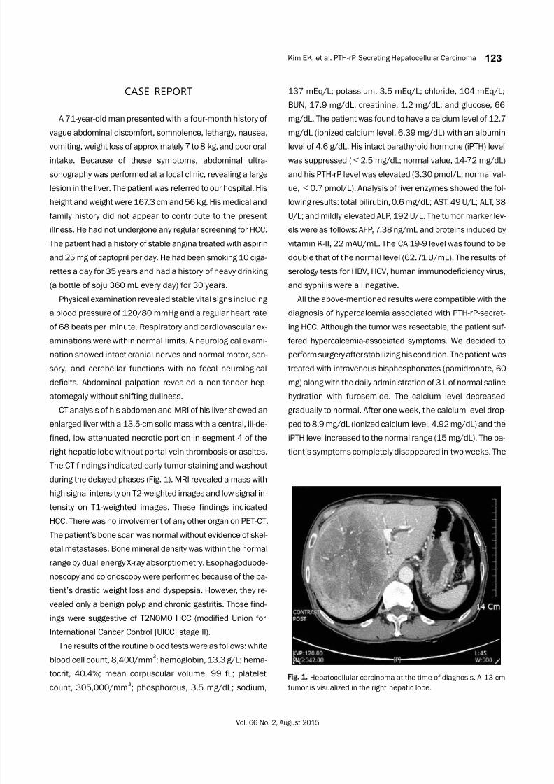
Fig. 1. Hepatocellular carcinoma at the time of diagnosis. A 13-cm
tumor is visualized in the right hepatic lobe.
CASE REPORT
A 71-year-old man presented with a four-month history of
vague abdominal discomfort, somnolence, lethargy, nausea,
vomiting, weight loss of approximately 7 to 8 kg, and poor oral
intake. Because of these symptoms, abdominal ultra-
sonography was performed at a local clinic, revealing a large
lesion in the liver. The patient was referred to our hospital. His
height and weight were 167.3 cm and 56 kg. His medical and
family history did not appear to contribute to the present
illness. He had not undergone any regular screening for HCC.
The patient had a history of stable angina treated with aspirin
and 25 mg of captopril per day. He had been smoking 10 ciga-
rettes a day for 35 years and had a history of heavy drinking(a bottle of soju 360 mL every day) for 30 years.
Physical examination revealed stable vital signs including
a blood pressure of 120/80 mmHg and a regular heart rate
of 68 beats per minute. Respiratory and cardiovascular ex-
aminations were within normal limits. A neurological exami-
nation showed intact cranial nerves and normal motor, sen-
sory, and cerebellar functions with no focal neurological
deficits. Abdominal palpation revealed a non-tender hep-
atomegaly without shifting dullness.
CT analysis of his abdomen and MRI of his liver showed anenlarged liver with a 13.5-cm solid mass with a central, ill-de-
fined, low attenuated necrotic portion in segment 4 of the
right hepatic lobe without portal vein thrombosis or ascites.
The CT findings indicated early tumor staining and washout
during the delayed phases (Fig. 1). MRI revealed a mass with
high signal intensity on T2-weighted images and low signal in-
tensity on T1-weighted images. These findings indicated
HCC. There was no involvement of any other organ on PET-CT.
The patient’s bone scan was normal without evidence of skel-
etal metastases. Bone mineral density was within the normal
range by dual energy X-ray absorptiometry. Esophagoduode-
noscopy and colonoscopy were performed because of the pa-
tient’s drastic weight loss and dyspepsia. However, they re-
vealed only a benign polyp and chronic gastritis. Those find-
ings were suggestive of T2N0M0 HCC (modified Union for
International Cancer Control [UICC] stage II).
The results of the routine blood tests were as follows: white
blood cell count, 8,400/mm3; hemoglobin, 13.3 g/L; hema-
tocrit, 40.4%; mean corpuscular volume, 99 fL; platelet
count, 305,000/mm3
; phosphorous, 3.5 mg/dL; sodium,
137 mEq/L; potassium, 3.5 mEq/L; chloride, 104 mEq/L;
BUN, 17.9 mg/dL; creatinine, 1.2 mg/dL; and glucose, 66
mg/dL. The patient was found to have a calcium level of 12.7
mg/dL (ionized calcium level, 6.39 mg/dL) with an albumin
level of 4.6 g/dL. His intact parathyroid hormone (iPTH) level
was suppressed (<2.5 mg/dL; normal value, 14-72 mg/dL)
and his PTH-rP level was elevated (3.30 pmol/L; normal val-
ue,<0.7 pmol/L). Analysis of liver enzymes showed the fol-
lowing results: total bilirubin, 0.6 mg/dL; AST, 49 U/L; ALT, 38
U/L; and mildly elevated ALP, 192 U/L. The tumor marker lev-
els were as follows: AFP, 7.38 ng/mL and proteins induced by
vitamin K-II, 22 mAU/mL. The CA 19-9 level was found to be
double that of the normal level (62.71 U/mL). The results of
serology tests for HBV, HCV, human immunodeficiency virus,and syphilis were all negative.
All the above-mentioned results were compatible with the
diagnosis of hypercalcemia associated with PTH-rP-secret-
ing HCC. Although the tumor was resectable, the patient suf-
fered hypercalcemia-associated symptoms. We decided to
perform surgery after stabilizing his condition. The patient was
treated with intravenous bisphosphonates (pamidronate, 60
mg) along with the daily administration of 3 L of normal saline
hydration with furosemide. The calcium level decreased
gradually to normal. After one week, the calcium level drop-ped to 8.9 mg/dL (ionized calcium level, 4.92 mg/dL) and the
iPTH level increased to the normal range (15 mg/dL). The pa-
tient’s symptoms completely disappeared in two weeks. The

7/23/2019 Complete Tumor Resection
http://slidepdf.com/reader/full/complete-tumor-resection 3/5
124 김은경 등. 부갑상선 호르몬 관련 펩티드 분비 간세포암종
The Korean Journal of Gastroenterology
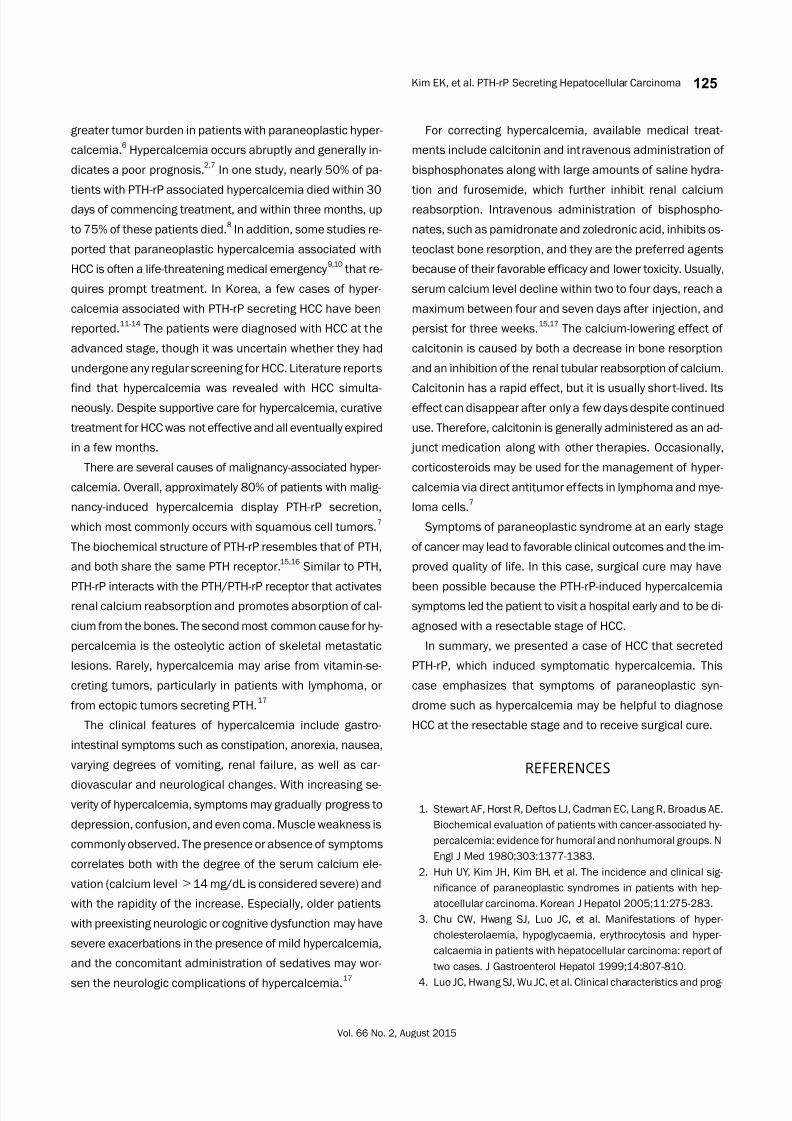
Fig. 2. Immunohistochemistry of the tumor specimen. (A) Microscopically tumor cells show trabecular growth patterns separated by sinusoids(H&E, ×400). (B) Positive staining for glypican-3 (×400). (C) Negative staining for CK19 (×400). (D) Negative staining for parathyroid hormone (×400).
patient then underwent a successful extended right hemi-
hepatectomy with cholecystectomy. There was no sign of
metastasis in any of the lymph nodes analyzed and no evi-
dence of cirrhosis in the liver tissue. Hematoxylin and eo-
sin-stained tissue revealed the presence of a typical HCC.
Immunohistochemical staining of the tissue showed positive
staining for glypican-3, negative staining for CA 19-9, and
negative staining for PTH (Fig. 2). All of the histopathological
findings were compatible with our pre-operative diagnosis.
Two weeks after the surgery, the patient was found to have
a calcium level of 8.9 mg/dL (ionized calcium level, 4.33
mg/dL) with an albumin level of 3.3 g/dL. His iPTH was 20
mg/mL (normal value, 14-72 mg/dL) and his PTH-rP was not
detectable (normal value,<
0.7 pmol/L). After 15 months,an abdominal CT showed no evidence of HCC recurrence and
results of subsequent serial blood tests were all within the
normal range. The patient is still alive with no evidence of
recurrence.
DISCUSSION
The risk of hypercalcemia induced by PTH-rP associated
with HCC was first assessed in 1982 in a retrospective study.
Of the 152 patients analyzed, 5.2% had hypercalcemia due
to HCC without bone involvement.5 A study in the Korean
Journal of Hepatology reported that the prevalence of para-
neoplastic syndromes associated with HCC in Korea is 35%
in Korea is 43.6%.2 The most common paraneoplastic syn-
drome was hypercholesterolemia (14.5%), followed by hypo-
glycemia (12.7%), and hypercalcemia (7.8%).2
Paraneoplastic hypercalcemia in patients with HCC is as-sociated with more advanced disease, as evidenced by sig-
nificantly higher AFP levels, bilobar disease, multiple lesions
and more advanced TNM stage at diagnosis, which reflect a
7/23/2019 Complete Tumor Resection
http://slidepdf.com/reader/full/complete-tumor-resection 4/5
Kim EK, et al. PTH-rP Secreting Hepatocellular Carcinoma 125
Vol. 66 No. 2, August 2015
greater tumor burden in patients with paraneoplastic hyper-
calcemia.6 Hypercalcemia occurs abruptly and generally in-
dicates a poor prognosis.2,7
In one study, nearly 50% of pa-
tients with PTH-rP associated hypercalcemia died within 30
days of commencing treatment, and within three months, up
to 75% of these patients died.8 In addition, some studies re-
ported that paraneoplastic hypercalcemia associated with
HCC is often a life-threatening medical emergency9,10
that re-
quires prompt treatment. In Korea, a few cases of hyper-
calcemia associated with PTH-rP secreting HCC have been
reported.11-14
The patients were diagnosed with HCC at the
advanced stage, though it was uncertain whether they had
undergone any regular screening for HCC. Literature reports
find that hypercalcemia was revealed with HCC simulta-neously. Despite supportive care for hypercalcemia, curative
treatment for HCC was not effective and all eventually expired
in a few months.
There are several causes of malignancy-associated hyper-
calcemia. Overall, approximately 80% of patients with malig-
nancy-induced hypercalcemia display PTH-rP secretion,
which most commonly occurs with squamous cell tumors.7
The biochemical structure of PTH-rP resembles that of PTH,
and both share the same PTH receptor.15,16
Similar to PTH,
PTH-rP interacts with the PTH/PTH-rP receptor that activatesrenal calcium reabsorption and promotes absorption of cal-
cium from the bones. The second most common cause for hy-
percalcemia is the osteolytic action of skeletal metastatic
lesions. Rarely, hypercalcemia may arise from vitamin-se-
creting tumors, particularly in patients with lymphoma, or
from ectopic tumors secreting PTH.17
The clinical features of hypercalcemia include gastro-
intestinal symptoms such as constipation, anorexia, nausea,
varying degrees of vomiting, renal failure, as well as car-
diovascular and neurological changes. With increasing se-
verity of hypercalcemia, symptoms may gradually progress to
depression, confusion, and even coma. Muscle weakness is
commonly observed. The presence or absence of symptoms
correlates both with the degree of the serum calcium ele-
vation (calcium level>14 mg/dL is considered severe) and
with the rapidity of the increase. Especially, older patients
with preexisting neurologic or cognitive dysfunction may have
severe exacerbations in the presence of mild hypercalcemia,
and the concomitant administration of sedatives may wor-
sen the neurologic complications of hypercalcemia.17
For correcting hypercalcemia, available medical treat-
ments include calcitonin and intravenous administration of
bisphosphonates along with large amounts of saline hydra-
tion and furosemide, which further inhibit renal calcium
reabsorption. Intravenous administration of bisphospho-
nates, such as pamidronate and zoledronic acid, inhibits os-
teoclast bone resorption, and they are the preferred agents
because of their favorable efficacy and lower toxicity. Usually,
serum calcium level decline within two to four days, reach a
maximum between four and seven days after injection, and
persist for three weeks.15,17
The calcium-lowering effect of
calcitonin is caused by both a decrease in bone resorption
and an inhibition of the renal tubular reabsorption of calcium.
Calcitonin has a rapid effect, but it is usually short-lived. Itseffect can disappear after only a few days despite continued
use. Therefore, calcitonin is generally administered as an ad-
junct medication along with other therapies. Occasionally,
corticosteroids may be used for the management of hyper-
calcemia via direct antitumor effects in lymphoma and mye-
loma cells.7
Symptoms of paraneoplastic syndrome at an early stage
of cancer may lead to favorable clinical outcomes and the im-
proved quality of life. In this case, surgical cure may have
been possible because the PTH-rP-induced hypercalcemiasymptoms led the patient to visit a hospital early and to be di-
agnosed with a resectable stage of HCC.
In summary, we presented a case of HCC that secreted
PTH-rP, which induced symptomatic hypercalcemia. This
case emphasizes that symptoms of paraneoplastic syn-
drome such as hypercalcemia may be helpful to diagnose
HCC at the resectable stage and to receive surgical cure.
REFERENCES
1. Stewart AF, Horst R, Deftos LJ, Cadman EC, Lang R, Broadus AE.
Biochemical evaluation of patients with cancer-associated hy-
percalcemia: evidence for humoral and nonhumoral groups. N
Engl J Med 1980;303:1377-1383.
2. Huh UY, Kim JH, Kim BH, et al. The incidence and clinical sig-
nificance of paraneoplastic syndromes in patients with hep-
atocellular carcinoma. Korean J Hepatol 2005;11:275-283.
3. Chu CW, Hwang SJ, Luo JC, et al. Manifestations of hyper-
cholesterolaemia, hypoglycaemia, erythrocytosis and hyper-
calcaemia in patients with hepatocellular carcinoma: report of
two cases. J Gastroenterol Hepatol 1999;14:807-810.
4. Luo JC, Hwang SJ, Wu JC, et al. Clinical characteristics and prog-

7/23/2019 Complete Tumor Resection
http://slidepdf.com/reader/full/complete-tumor-resection 5/5
126 김은경 등. 부갑상선 호르몬 관련 펩티드 분비 간세포암종
The Korean Journal of Gastroenterology
nosis of hepatocellular carcinoma patients with paraneoplastic
syndromes. Hepatogastroenterology 2002;49:1315-1319.
5. Oldenburg WA, van Heerden JA, Sizemore GW, Abboud CF,
Sheedy PF 2nd. Hypercalcemia and primary hepatic tumors.
Arch Surg 1982;117:1363-1366.6. Chang PE, Ong WC, Lui HF, Tan CK. Epidemiology and prognosis
of paraneoplastic syndromes in hepatocellular carcinoma. ISRN
Oncol 2013. doi: 10.1155/2013/684026.
7. Lumachi F, Brunello A, Roma A, Basso U. Medical treatment of
malignancy-associated hypercalcemia. Curr Med Chem 2008;
15:415-421.
8. Ralston SH, Gallacher SJ, Patel U, Campbell J, Boyle IT. Cancer-
associated hypercalcemia: morbidity and mortality. Clinical ex-
perience in 126 treated patients. Ann Intern Med 1990;112:
499-504.
9. Ghobrial MW, George J, Mannam S, Henien SR. Severe hyper-
calcemia as an initial presenting manifestation of hepato-
cellular carcinoma. Can J Gastroenterol 2002;16:607-609.
10. Mahoney EJ, Monchik JM, Donatini G, De Lellis R. Life-threat-
ening hypercalcemia from a hepatocellular carcinoma secreting
intact parathyroid hormone: localization by sestamibi sin-
gle-photon emission computed tomographic imaging. Endocr
Pract 2006;12:302-306.
11. Lee MH, Son JH, Jang WY, et al. A case of hepatocellular carcino-
ma with hypercholesterolemia and hypercalcemia as paraneo-
plastic syndrome. Korean J Gastroenterol 1999;34:271-276.
12. Kim JH, Kim BH, Lee DH, et al. A case of hepatocellular carcino-
ma presenting with four paraneoplastic syndromes such as hy-percholesterolemia, hypoglycemia, hypercalcemia, and erythr-
ocytosis. Korean J Gastroenterol 2003;41:316-320.
13. Lee D, Kim IH, Kim SH, et al. A case of sclerosing hepatocellular
carcinoma with hypercalcemia treated with transcatheter arte-
rial chemoembolization. Korean J Med 2000;59:104-108.
14. Yoon SY, Lee CR, Lee JH, Choi SJ, Son SP. A case of humoral hyper-
calcemia of malignancy associated with hepatoma: a case in
which both PTHrP and 1,25 (OH) 2D were elevated. J Korean Soc
Endocrinol 1999;14:197-202.
15. Berenson JR. Treatment of hypercalcemia of malignancy with
bisphosphonates. Semin Oncol 2002;29(6 Suppl 21):12-18.
16. Bilezikian JP, Singer FR. Acute management of hypercalcemia
due to parathyroid hormone and parathyroid hormone-related
protein. In: Bilezikian JP, Marcus R, Levine MA, eds. The Parathy-
roids: basic and clinical concepts. New York: Raven Press, 1994:
359-372.
17. Stewart AF. Clinical practice. Hypercalcemia associated with
cancer. N Engl J Med 2005;352:373-379.