comprehensivestructuralandbiochemical analysis...
TRANSCRIPT
Article
Comprehensive Structural and Biochemical Analysis
of the TerminalMyxalamid ReductaseDomain for theEngineered Production of Primary AlcoholsGraphical Abstract
Highlights
d Highest resolution and first cofactor-bound structure of a
terminal reductase domain
d Computational modeling advances hypotheses made from
the crystal structure
d Biochemical analysis defines residues critical for substrate
specificity and catalysis
d Result-based engineering enabled improved reduction of
highly reduced substrates
Barajas et al., 2015, Chemistry & Biology 22, 1018–1029August 20, 2015 ª2015 Elsevier Ltd All rights reservedhttp://dx.doi.org/10.1016/j.chembiol.2015.06.022
Authors
Jesus F. Barajas, Ryan M. Phelan,
Andrew J. Schaub, ..., Ray Luo, Jay D.
Keasling, Shiou-Chuan Tsai
[email protected] (J.D.K.),[email protected] (S.-C.T.)
In Brief
Barajas et al. report the structure of a
unique termination domain employed in
the reductive release of NRPS-generated
natural products. The crystal structure,
combined with computational and
biochemical investigations, provide a
comprehensive understanding of key
factors that govern catalysis in this class
of termination domains.
Accession Numbers
4U7W
4W4T

Chemistry & Biology
Article
Comprehensive Structural and Biochemical Analysisof the Terminal Myxalamid Reductase Domainfor the Engineered Production of Primary AlcoholsJesus F. Barajas,1,6 Ryan M. Phelan,2,3,6 Andrew J. Schaub,1 Jaclyn T. Kliewer,1 Peter J. Kelly,1 David R. Jackson,1
Ray Luo,1 Jay D. Keasling,2,3,4,5,* and Shiou-Chuan Tsai1,*1Department of Molecular Biology and Biochemistry, Chemistry, and Pharmaceutical Sciences, University of California, Irvine, Irvine,
CA 92697, USA2Joint Bioenergy Institute, 5885 Hollis Street, Emeryville, CA 94608, USA3QB3 Institute, University of California, Berkeley, Berkeley, CA 94270, USA4Department of Chemical and Biomolecular Engineering and Department of Bioengineering, University of California, Berkeley, Berkeley,
CA 94720, USA5Physical Biosciences Division, Lawrence Berkeley National Laboratory, Berkeley, CA 94720, USA6Co-first author
*Correspondence: [email protected] (J.D.K.), [email protected] (S.-C.T.)
http://dx.doi.org/10.1016/j.chembiol.2015.06.022
SUMMARY
The terminal reductase (R) domain from the non-ribo-somal peptide synthetase (NRPS) module MxaA inStigmatella aurantiaca Sga15 catalyzes a non-proc-essive four-electron reduction to produce the myxa-lamide family of secondary metabolites. Despitewidespread use in nature, a lack of structuraland mechanistic information concerning reductiverelease from polyketide synthase (PKS) and NRPSassembly lines principally limits our ability to rede-sign R domains with altered or improved activity.Here we report crystal structures for MxaA R, bothin the absence and, for the first time, in the presenceof the NADPH cofactor. Molecular dynamics simula-tions were employed to provide a deeper under-standing of this domain and further identify residuescritical for structural integrity, substrate binding, andcatalysis. Aggregate computational and structuralfindings provided a basis for mechanistic investiga-tions and, in the process, delivered a rationallyaltered variant with improved activity toward highlyreduced substrates.
INTRODUCTION
Polyketide synthases (PKSs) and non-ribosomal peptide synthe-
tases (NRPSs) are large, multi-modular protein assemblies
capable of generating chemically diverse and complex mole-
cules. To produce these compounds, PKS and NRPS pro-
grammed assembly occurs through the use of either acyl-
coenzyme A or amino acid building blocks, respectively, to
provide natural products that have applications in numerous
sectors in the world economy (e.g., pharmaceutical and agro-
chemical) (Hertweck, 2009). The myxobacterium Stigmatella
aurantiaca Sga15 contains a modular PKS/NRPS hybrid respon-
1018 Chemistry & Biology 22, 1018–1029, August 20, 2015 ª2015 El
sible for the biosynthesis of the myxalamids (Figure 1), potent in-
hibitors of the respiratory electron transport chain (Gerth et al.,
1983; Silakowski et al., 2001). Interestingly, this hybrid system
uses a rare termination mechanism to release the final com-
pound as a primary alcohol (Gaitatzis et al., 2001; Silakowski
et al., 2001) as opposed to more common chain release mecha-
nisms that produce, for instance, macrolactones or macrolac-
tams (Du and Lou, 2010).
The biosynthesis of myxalamid is amulti-step process initiated
by a type I modular PKS consisting of six modules to bio-
synthesize a polyene intermediate 3 (Figure 1A) that is translo-
cated to the NRPS module for final processing (Silakowski
et al., 2001). In the terminal NRPS module, MxaA, the adenyla-
tion (A) domain activates alanine as a building block while the
condensation (C) domain catalyzes peptide bond formation be-
tween alanine and the PKS-generated intermediate 3 to yield 4
(Konz and Marahiel, 1999). The last step in biosynthesis requires
the reductive release of myxalamid (mxa) from the phosphopan-
tetheine (pPant) prosthetic group covalently attached to the pep-
tidyl carrier protein (PCP). This action is catalyzed by a recently
described class of NADPH-dependent terminal reductase (R)
domains that execute chain termination by a 4e� non-processive
reduction to generate primary alcohols (Figure 1) (Chhabra et al.,
2012; Du and Lou, 2010; Silakowski et al., 2001). To accomplish
this, the PCP-bound thioester is first reduced to the aldehyde in-
termediate 5, which, following reduction by a second NADPH
equivalent, affords the final 2-aminopropanol-containing prod-
uct 6 (Figure 1A).
Given the ability to rationally program PKS and NRPS biosyn-
thesis (Hahn and Stachelhaus, 2006; Menzella et al., 2005; Poust
et al., 2014; Sherman, 2005; Weissman and Leadlay, 2005), cur-
rent research is focused on the study of R domains in order to
expand the range of available characterized termination mecha-
nisms. In light of the immense scope of possible products
that could be generated by PKSs, terminating R domains can
provide a unique route to numerous alcohol-containing com-
pounds to replace existing petroleum-derived fuels or commod-
ity chemicals (Atsumi et al., 2008; Chu and Majumdar, 2012;
Peralta-Yahya et al., 2012). However, to reliably engineer new
sevier Ltd All rights reserved
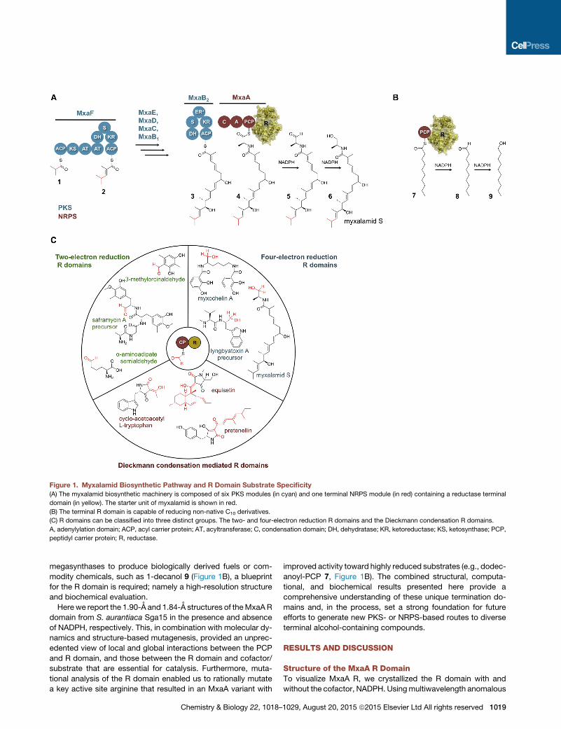
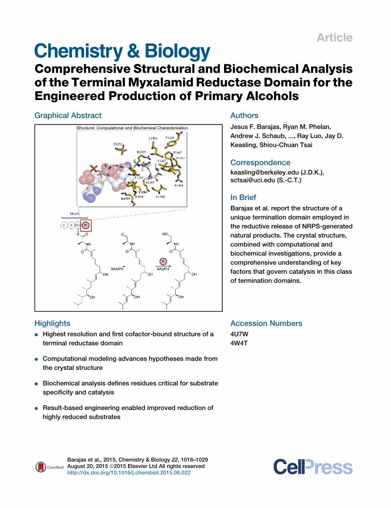
Figure 1. Myxalamid Biosynthetic Pathway and R Domain Substrate Specificity
(A) The myxalamid biosynthetic machinery is composed of six PKS modules (in cyan) and one terminal NRPS module (in red) containing a reductase terminal
domain (in yellow). The starter unit of myxalamid is shown in red.
(B) The terminal R domain is capable of reducing non-native C10 derivatives.
(C) R domains can be classified into three distinct groups. The two- and four-electron reduction R domains and the Dieckmann condensation R domains.
A, adenylylation domain; ACP, acyl carrier protein; AT, acyltransferase; C, condensation domain; DH, dehydratase; KR, ketoreductase; KS, ketosynthase; PCP,
peptidyl carrier protein; R, reductase.
megasynthases to produce biologically derived fuels or com-
modity chemicals, such as 1-decanol 9 (Figure 1B), a blueprint
for the R domain is required; namely a high-resolution structure
and biochemical evaluation.
Here we report the 1.90-A and 1.84-A structures of theMxaAR
domain from S. aurantiaca Sga15 in the presence and absence
of NADPH, respectively. This, in combination with molecular dy-
namics and structure-based mutagenesis, provided an unprec-
edented view of local and global interactions between the PCP
and R domain, and those between the R domain and cofactor/
substrate that are essential for catalysis. Furthermore, muta-
tional analysis of the R domain enabled us to rationally mutate
a key active site arginine that resulted in an MxaA variant with
Chemistry & Biology 22, 1018–
improved activity toward highly reduced substrates (e.g., dodec-
anoyl-PCP 7, Figure 1B). The combined structural, computa-
tional, and biochemical results presented here provide a
comprehensive understanding of these unique termination do-
mains and, in the process, set a strong foundation for future
efforts to generate new PKS- or NRPS-based routes to diverse
terminal alcohol-containing compounds.
RESULTS AND DISCUSSION
Structure of the MxaA R DomainTo visualize MxaA R, we crystallized the R domain with and
without the cofactor, NADPH. Usingmultiwavelength anomalous
1029, August 20, 2015 ª2015 Elsevier Ltd All rights reserved 1019
Figure 2. Structure of the MxaA R Domain
(A) The MxaA R domain monomer is composed of
an N-terminal subdomain that contains an NADPH
Rossmann fold (in blue) and a C-terminal sub-
domain that contains a helix-turn-helix motif
(shown in green). The NADPH cofactor is dis-
played as gray sticks.
(B) The MxaA R domain crystallizes as a dimer;
monomer A is shown in yellow and monomer B is
shown in gray.
(C) The cofactor NADPH binds to the TGxxGxxG
motif close to the T, Y, and K catalytic site. An SA-
omit map of the NADPH cofactor contoured at
1.0s is shown in the gray isomesh map.
See also Figure S3 and Table S1.
diffraction (MAD) with a selenomethionine-substituted protein,
the structure of MxaA R was determined to a resolution of
1.95 A. The apo MxaA R structure was further refined to 1.84 A
(Table S1). MxaA R was solved as a dimer with a root-mean-
square deviation (RMSD) of 0.45 A between monomers A
and B. The overall structure contains strong architectural similar-
ities to type E short-chain dehydrogenases/reductases (SDRs)
that contain an N-terminal NADPH-binding region and a
C-terminal substrate-binding subdomain (Figure 2) (Jornvall
et al., 1995). Structural alignment with the type E SDR from
Agrobacterium tumefaciens (PDB: 4ID9) displays an RMSD of
3.88 A through 119 residues of the alpha carbon backbone.
Hidden Markov models show that MxaA R has structural and
sequence similarities with the extended type E SDRs based on
the Kallberg et al. classification (Kallberg et al., 2010). The N-ter-
minal subdomain contains an extended NADPH-binding a/b
Rossmann fold with seven parallel beta sheets (b3-b2-b1-b4-
b5-b6-b10) flanked by five alpha helices (a2-a3-a4-a6-a8-a11)
(Figure 2A; Figures S2 and S3). These structural features corre-
late well with the previously solved Nrp R domain structure
(PDB: 4DQV) with an RMSD of 2.19 A through 279 residues of
the alpha carbon backbone (Chhabra et al., 2012). Similar fea-
tures include a canonical tyrosine-dependent catalytic triad
(T1283, K1315, and Y1311) and a distinctive helix-turn-helix
(HTH) motif (a16-a17) found in all structurally known terminating
reductase domains.
Substrate recognition in the SDR family occurs in the C-termi-
nal subdomain (Kavanagh et al., 2008). Consequently, while the
N-terminal subdomains in SDRs are highly conserved, C-termi-
nal domains often differ in sequence. The C-terminal subdomain
of MxaA R consists of five helices (a12-a15-a16-a17-a20) and
two parallel beta sheets (b9-b11), which are substantially larger
(�130 residues) than those found in typical SDRs (Figures
S3A–S3C) (Jornvall et al., 1995; Kallberg et al., 2010; Kavanagh
et al., 2008). A notable inserted HTH motif (a16-a17) between
residues Y1431 and Q1456 contains several conserved hydro-
phobic residues (W1433, L1437, L1450, L1451) frequently pre-
sent in R domains that conduct PKS or NRPS chain termination
with 2- or 4-electron reductions (Figure 1C) (Bergmann et al.,
1020 Chemistry & Biology 22, 1018–1029, August 20, 2015 ª2015 Elsevier Ltd All rights rese
2007; Gaitatzis et al., 2001; Gomez-Escri-
bano et al., 2012; Li et al., 2008; Mas-
schelein et al., 2015; Silakowski et al.,
2001).
To further distinguish true biological interfaces from lattice
contacts in the crystal structure, we further analyzed the MxaA
R domain utilizing the Evolutionary Protein-Protein Interface
Classifier (EPPIC) server (Duarte et al., 2012), which relies on
evolutionary data to detect biological interfaces and PDBePISA
(Krissinel and Henrick, 2007). The EPPIC server was unable to
reliably determine biologically relevant surface interfaces due
to the lack of homolog sequences for comparison. PDBePISA
generated a Complex Formation Significance Score of 0.00,
suggesting that the surface interface displayed by the MxaA ho-
modimer is a result of crystal packing. The average interface area
between bothmonomers was calculated to be 656.9 A2, which is
3.85% of the total solvent accessible area. This constituted a
total of 22 and 20 buried surface residues for monomers A and
B, respectively. It is also well known that biological interfaces
tend to exhibit large areas, with the majority of cases exceeding
1,000 A2 (Jones and Thornton, 1996). Furthermore, evidence for
its biological monomeric state was gathered from analytical size
exclusion chromatography experiments comparing the MxaA
PCP-R didomain to known protein standards. Overall, these re-
sults suggest that MxaA R exists in a biologically monomeric
form rather than the crystallographically observed homodimeric
state.
Structure Analysis of NADPH-Bound R DomainCurrently the Nrp terminal R domain (PDB: 4DQV) from Myco-
bacteria smegmatis involved in glycopeptide biosynthesis and
the AusA R domain (PDB: 4F6C, 4F6L) from Staphyloccoccus
aureus involved in pyrazinone biosynthesis are the sole PKS-
or NRPS-associated R domains to have a structure reported.
While these monodomain structures have been solved with
moderate resolution (2.30 A for NRP and 2.81 A for AusA), the
lack of bound NADPH leaves key structural and mechanistic de-
tails rather unclear (Chhabra et al., 2012; Wyatt et al., 2012). In
order to define residues required for cofactor binding in MxaA
R, co-crystals of MxaA R complexed with NADPH were solved
by molecular replacement of the apo structure to 1.90 A (Table
S1). NADPH binds to the well-known Rossmann fold, which
has a conserved nucleotide-binding motif TGxxGxxG, with the
rved

central diphosphate moiety hydrogen bonding to the peptide
backbone of G1155, T1157, G1158, L1160, and G1161 (Fig-
ure 2C; Figure S3D). Furthermore, the G1155 carbonyl forms a
hydrogen bond with the adenosine 30-hydroxyl group while the
adenosine 20-phosphate oxygen interacts with highly conserved
T1157, R1181, and R1191. Both the 20- and 30-hydroxyl groupsof the nicotinamide-containing ribose ring hydrogen bond with
K1315 and Y1311. The nicotinamide amine hydrogen bonds
with the G1338 carbonyl. Together, these interactions serve to
tightly bind NADPH (Kd = 45 ± 3.7 mM) and properly orient it in
the active site for reduction of the pPant-bound intermediate to
the terminal alcohol.
Several coordinated water molecules are present between the
catalytic residues Y1311, T1283 and the non-catalytic S1285.
One water molecule is positioned 2.7 A from the hydroxyl of
Y1311 and 2.8 A from T1283, possibly occupying the oxyanion
hole that these two residues create to assist in thioester and
aldehyde reduction. T1283 and S1285 bind a second water
molecule in the active site, although its positioning does not pro-
vide a clear role in catalysis. With respect to these observations,
several SDR studies suggest that ordered water molecules in the
active sitemight participate in a proton relay system involving the
hydroxyl of Y1311, 20-hydroxyl of the nicotinamide ribose and
K1315 (Eklund et al., 1982; Oppermann et al., 2003). Structural
comparison of the apo and NADPH-bound MxaA R domain
show slight conformational changes with an overall RMSD of
0.63 A through the entire backbone. From these small differ-
ences, the C-terminal subdomain experiences a slightly higher
conformational change upon NADPH binding compared with
the complete monomer, with an RMSD difference of 0.73 A.
Molecular Dynamics AnalysisTo elucidate the structural dynamics of NADPH and substrate
binding in MxaA R, we conductedmolecular dynamics (MD) sim-
ulations by analyzing conformational changes in 100-ns MD
runs. Atomic coordinates of the MxaA R domain were obtained
from the NADPH-bound MxaA R domain (chain B) crystal struc-
ture. The ff14SB forcefield in Amber14 was used for the protein
and the general AMBER force field was used for the NADPH
cofactor (Case et al., 2014a, 2014b; Gotz et al., 2012; Hornak
et al., 2006; Wang et al., 2004, 2006; Wickstrom et al., 2009).
NADPH was parameterized using Gaussian 09 to obtain the
initial electrostatic potential using the HF/6-31G(d,p) basis set,
followed by the use of antechamber to obtain the HF/6-
31G(d,p) restricted electrostatic potential fit with final overall
net charge of �4. The system was explicitly solvated with a
buffer of 10 A TIP3P waters in a truncated octahedron box after
neutralizing with counter ions. A two-system minimization was
performed using SANDER and PMEMDwas used for production
runs (Verdonk et al., 2003).
TheNADPH-boundMxaAR domainwas allowed to equilibrate
after heating the system to 300 K and subsequently allowed to
run over 100 ns. 2D RMSDmaps were generated using Chimera
and an in-house MATLAB script by comparing RMSD fluctua-
tions of the protein backbone. The maps revealed an RMSD
range of 0.61–2.41 A for the MxaA R domain (Figure 3A). Further
dissection of the N- versus C-terminal subdomains revealed
RMSD ranges of 0.54–1.49 A and 0.52–2.25 A, respectively (Fig-
ures 3B and 3C). These results, combined with RMSD values
Chemistry & Biology 22, 1018–
found in our crystal structures, indicate higher flexibility and
movement of the C-terminal subdomain.
The most noticeable region of flexibility was observed in the
C-terminalHTHmotif, specifically theconservedhydrophobic res-
idues between Y1430 and Q1455 of a16-a17, which display an
average RMSD of 0.82 A in the NADPH-bound model (Figure 2C;
Figures S4G–S4I). Numerous salt bridges are critical in stabilizing
the a16-a17 HTH motif, such as R1426 and E1436 (Figures S4A–
S4C). During the 100-ns NADPH-bound run, the zC of R1426
maintains a distance of %6.0 A with either εO of E1436. D1444,
the turn residue between helix 16 and helix 17, also maintains a
tight salt bridge interaction with a distance %3.5 A between the
zC of R1364 through stochastic sampling of either helix 13
D1444 dO during 73.2% of the simulation (Figure S4A). Moderate
electrostatic interactions were observed between helix 12 R1357
andhelix 17E1446,with the zCofR1357maintaining a distance%
6.0 A for 68.7% of the simulation. The catalytic triad (T1283,
Y1311, and K1315) exhibits little movement with an average of
0.07 A per residue throughout the entire 100-ns run. The phos-
phate attached to the nicotinamide ribose 50 carbon remains sta-
ble within 1.99 and 2.09 A of the Rossmann TGxxGxxG motif.
A representative cluster ensemble was generated fromMDus-
ing RMSD scoring as implemented in Chimera (Pettersen et al.,
2004). RMSD scoring reduced the initial set of 1,000 frames
generated to the 46 most unique frames. In silico docking of
the pPant-bound substrate using all of the 46 unique frames
from the previous MD run by the programGOLD revealed a large
binding cavity under the a16-a17 HTHmotif (Figure 2A) (Verdonk
et al., 2003). In order to identify substrate-binding residues for
MD analysis, we docked the myxalamid substrate (see the
next section on docking analysis) using the 46 unique clusters
from the NADPH-bound MxaA R MD analysis. We ranked the
docking solutions using the ChemPLP scoring function and iden-
tified the most consistent binding orientation of the myxalamid
substrate by tallying residues involved in substrate binding (Fig-
ure 4B). The top ChemPLP docking solution that was pre-
screened by binding orientation was used for MD analysis of
MxaA R with the myxalamid substrate. Using the same MD sys-
tem parameters as before, we allowed the pPant-bound sub-
strate and NADPH-bound R domain chain B simulation to run
for 100 ns. RMSD 2D map analysis of the MxaA R domain in
complex with the pPant substrate revealed an RMSD range of
0.60–1.99 A (Figure 3D). This value is lower than the RMSD range
for the R domain with NADPH bound but lacking substrate (Fig-
ure 3A) and indicates a decrease in protein motion upon sub-
strate binding. The NADPH-binding N-terminal subdomain
demonstrates a similar RMSD range of 0.55–1.49 A, whether
substrate is bound or not, while the C terminus reduces its flex-
ibility upon substrate binding (Figures 3C and 3F).
In light of results that indicated that substrate binding stabi-
lized the C terminus, we opted to focus additional attention on
the C-terminal HTH motif, specifically those residues that are
steadied through interactions with the substrate (Figures 4A–
4C). The terminal HTH motif displays a slightly lower average
RMSD of 0.76 A, while the pPant moiety exhibits larger move-
ments than the sequestered myxalamid segment. D1353 shows
strong hydrogen bonding with the amide moiety of the pPant
group, averaging a 3.0-A distance for more than 70 ns of the
MD run (Figure S4E). The amide carbonyl group of the terminal
1029, August 20, 2015 ª2015 Elsevier Ltd All rights reserved 1021

100
1000
200
300
400
600
700
800
900
500
0.5
1.0
1.5
2.0
0.5
1.0
1.5
2.0
0.5
1.0
1.5
2.0
0.5
1.0
1.5
2.0
0.5
1.0
1.5
2.0
100 200 300 400 500 600 700 800 900 1000 0.5
1.0
1.5
2.0
Time (ns)
Tim
e (n
s)
F E MxaA R + NADPH
+ Mxa-pPant MxaA R N-term + NADPH
+ Mxa-pPant
Å Å Å
Å Å Å
MxaA R C-term + NADPH + Mxa-pPant
100
1000
200
300
400
600
700
800
900
500
100 200 300 400 500 600 700 800 900 1000 Time (ns)
Tim
e (n
s)
100
1000
200
300
400
600
700
800
900
500
100 200 400 500 600 700 800 900 1000 Time (ns)
Tim
e (n
s)
100
1000
200
300
400
600
700
800
900
500
100 200 400 500 600 700 800 900 1000 Time (ns)
Tim
e (n
s)
100
1000
200
300
400
600
700
800
900
500
100 200 400 500 600 700 800 900 1000 Time (ns)
Tim
e (n
s)
100
1000
200
300
400
600
700
800
900
500
100 200 400 500 600 700 800 900 1000 Time (ns)
Tim
e (n
s)
D
C B A MxaA R + NADPH MxaA R N-term + NADPH MxaA R C-term + NADPH
300
300
300
300 300
Figure 3. Molecular Dynamic Analysis
(A) 2D RMSD map analysis of the MxaA R domain backbone with NADPH over the entire 100-ns molecular dynamics simulation. Low RMSD is observed in blue
and high RMSD is observed in red.
(B and C) Dissecting the N- versus C-terminal subdomain of MxaA R bound to NADPH reveals higher RMSD deviations in the C-terminal subdomain.
(D) 2D RMSD map was generated with the MxaA R domain bound to NADPH and docked with mxa-pPant.
(E and F) Dissection of the N- versus C-terminal subdomain of the bound NADPHmxa-pPant R domain demonstrates a decrease in movement of the C-terminal
subdomain.
See also Figure S4.
alanine in the mxa intermediate generates a tight 2.0-A interac-
tionwith R1339, highlighting the likely importance of electrostatic
interactions between myxalamid and the R domain (Figure S4D).
The methyl-branched diene moiety of the mxa substrate forms
intramolecular hydrophobic interactions, kinking the aliphatic
substrate back toward the pPant thioester to minimize its hydro-
phobic surface area. F1248 in MxaA R aids in stabilizing these
hydrophobic interactions. The C9 and C15 hydroxyl groups in
myxalamid intermediate 4 hydrogen bond with S1285 and
D1461, respectively (Figures 4A and 4C). S1285 forms an
average 3.0-A hydrogen bond with the terminal hydroxyl group
in the myxalamid intermediate for more than 95% of the MD
simulation. Similarly, D1461 forms an average 3.2-A hydrogen
bond with the first hydroxyl group for more than 80% of the MD
run. Both S1285 and D1461 along with R1339 and F1248 appear
to play a key role in myxalamid substrate recognition and orient-
ing the substrate for reduction byNADPH. In summary,MD simu-
lation results suggest that the C-terminal domain is highly mobile
until myxalamid substrate binding quenches movement, more
precisely at the HTH motif, for the first round of reduction.
Docking Analysis of pPant-Mxa Substrate and PCPDomainInitial structural analysis inparallelwith theNADPH-boundMxaAR
MD analysis revealed a large substrate cavity with various poten-
1022 Chemistry & Biology 22, 1018–1029, August 20, 2015 ª2015 El
tial substrate-binding residues. Using in silico docking, we further
probed for residues important in substrate binding by docking the
pPant-tethered mxa intermediate in the R domain active site. The
100-ns MD simulation of NADPH-bound MxaA R identified 46
unique clusters, indicative of 46 distinct MxaA R domain confor-
mations. One frame from each cluster was obtained and was
used as the receptor to dock against the pPant-mxa ligand using
the program GOLD (Liebeschuetz et al., 2012). Each frame
generated 100 solutions that were scored and ranked using the
ChemPLP scoring function (Hildebrand et al., 2009). The program
LIGPLOT, in parallel with visual inspection, was used to analyze
and identify ligand-protein residue interactions (Wallace et al.,
1995). The residue-ligand interactions between 2.5 and 4.0 A for
each frame were tallied and a heatmap was generated, indicative
of ligand-residue proximity in different R domain conformations
(Figure 4B). Not surprisingly, the catalytic triad was revealed to
frequently associate with the pPant-bound substrate. T1283
showed interactions close to the thioester linkage in 28 out of
the 46 frames. In addition, Y1311 interacted with the thioester in
18 of the 46 frames. The water-coordinating S1285 associated
with the thioester in 35 of the 46 frames. The majority of residues
that interacted with the mxa portion of the ligand were localized
on the C-terminal subdomain. Of the 46 frames, 37 showed that
R1339 engages in electrostatic interactions with one of the two
carbonyl groups in the substrate near the thioester linkage.
sevier Ltd All rights reserved
Residue Interaction Probability
O
O
HN
O
SNN
H
O
O
O
OH
OP
HO
O-O
-O O
H
H2N NH
NH
O
H
H
O-
O
H
NH
OH H2N
NH
NH
OH
High Frame Count
Low Frame Count
K1315 Y1311
T1283
V1308
R1468
P1467
D1461
Y1430
L1454
V1457
R1339 D1353
S1285
R1339
T1283
F1248
L1454
V1457
P1467
R1468 Y1430
S1285
V1308
D1461
D1353
Val 1121 1Gly 1158 1Leu 1189 1Tyr 1199 1Pro 1288 1Ser 1306 1Thr 1341 1Trp 1349 1Val 1380 1Met 1414 1Gln 1449 1Leu 1249 2Tyr 1250 2Pro 1251 2Ser 1282 2Leu 1287 2Leu 1359 2Val 1281 3Trp 1433 3Leu 1245 4Arg 4
Pro 1369 4Leu 1377 5Trp 1415 5Gln 1456 6Leu 1160 7Asn 1247 7Leu 1289 8Gly 1310 8Val 1470 8Val 1371 10Pro 1458 10Phe 1159 11Gly 1309 12Ala 1312 13Leu 1375 13Thr 1358 16Val 1284 17Val 1340 17Asn 1350 18Tyr 1311 19Val 20
Pro 1337 20Arg 1468 20Tyr 1430 21Phe 1453 22Asp 1352 23Asp 1376 24Val 1355 25Leu 1454 25Val 1457 26Gly 1338 27Asp 1353 27Met 1469 27Thr 1283 28Asp 1461 28Val 1308 30Leu 1354 32Ser 1285 34Pro 1467 34Phe 1248 36Arg 1339 37
A B
C
K1315 Y1311
T1283
V1308
R1468
P1467 D1461
Y1430
L1454
R1339 D1353
F1248 S1285
D
V1457
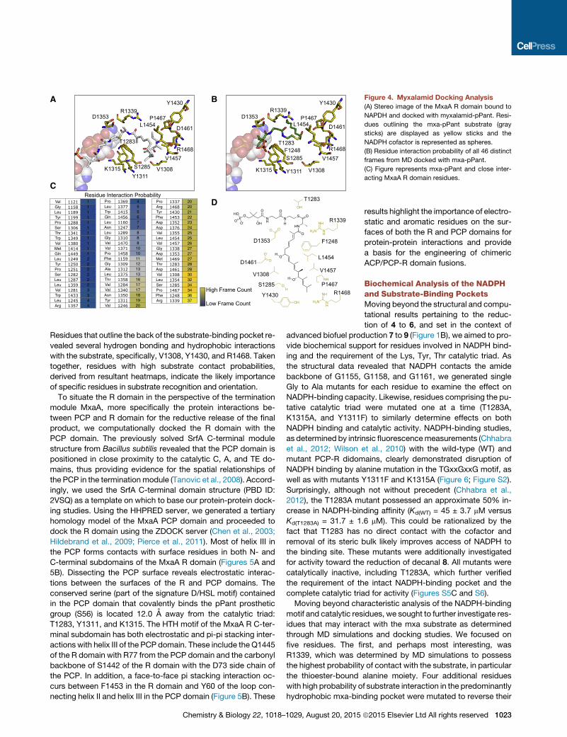
Figure 4. Myxalamid Docking Analysis
(A) Stereo image of the MxaA R domain bound to
NAPDH and docked with myxalamid-pPant. Resi-
dues outlining the mxa-pPant substrate (gray
sticks) are displayed as yellow sticks and the
NADPH cofactor is represented as spheres.
(B) Residue interaction probability of all 46 distinct
frames from MD docked with mxa-pPant.
(C) Figure represents mxa-pPant and close inter-
acting MxaA R domain residues.
Residues that outline the back of the substrate-binding pocket re-
vealed several hydrogen bonding and hydrophobic interactions
with the substrate, specifically, V1308, Y1430, and R1468. Taken
together, residues with high substrate contact probabilities,
derived from resultant heatmaps, indicate the likely importance
of specific residues in substrate recognition and orientation.
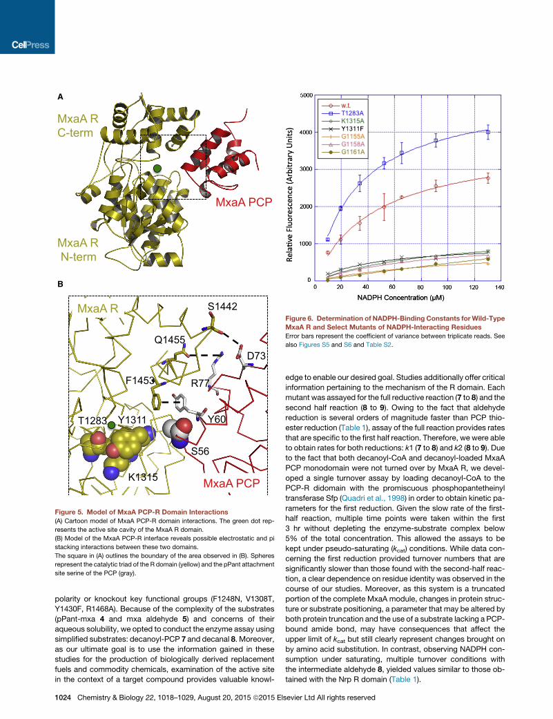
To situate the R domain in the perspective of the termination
module MxaA, more specifically the protein interactions be-
tween PCP and R domain for the reductive release of the final
product, we computationally docked the R domain with the
PCP domain. The previously solved SrfA C-terminal module
structure from Bacillus subtilis revealed that the PCP domain is
positioned in close proximity to the catalytic C, A, and TE do-
mains, thus providing evidence for the spatial relationships of
the PCP in the termination module (Tanovic et al., 2008). Accord-
ingly, we used the SrfA C-terminal domain structure (PBD ID:
2VSQ) as a template on which to base our protein-protein dock-
ing studies. Using the HHPRED server, we generated a tertiary
homology model of the MxaA PCP domain and proceeded to
dock the R domain using the ZDOCK server (Chen et al., 2003;
Hildebrand et al., 2009; Pierce et al., 2011). Most of helix III in
the PCP forms contacts with surface residues in both N- and
C-terminal subdomains of the MxaA R domain (Figures 5A and
5B). Dissecting the PCP surface reveals electrostatic interac-
tions between the surfaces of the R and PCP domains. The
conserved serine (part of the signature D/HSL motif) contained
in the PCP domain that covalently binds the pPant prosthetic
group (S56) is located 12.0 A away from the catalytic triad:
T1283, Y1311, and K1315. The HTH motif of the MxaA R C-ter-
minal subdomain has both electrostatic and pi-pi stacking inter-
actions with helix III of the PCP domain. These include the Q1445
of the R domain with R77 from the PCP domain and the carbonyl
backbone of S1442 of the R domain with the D73 side chain of
the PCP. In addition, a face-to-face pi stacking interaction oc-
curs between F1453 in the R domain and Y60 of the loop con-
necting helix II and helix III in the PCP domain (Figure 5B). These
Chemistry & Biology 22, 1018–1029, August 20, 2015
results highlight the importance of electro-
static and aromatic residues on the sur-
faces of both the R and PCP domains for
protein-protein interactions and provide
a basis for the engineering of chimeric
ACP/PCP-R domain fusions.
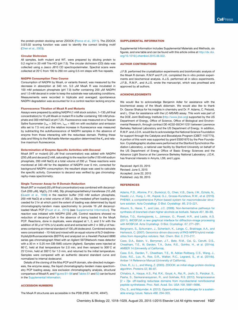
Biochemical Analysis of the NADPHand Substrate-Binding PocketsMoving beyond the structural and compu-
tational results pertaining to the reduc-
tion of 4 to 6, and set in the context of
advanced biofuel production 7 to 9 (Figure 1B), we aimed to pro-
vide biochemical support for residues involved in NADPH bind-
ing and the requirement of the Lys, Tyr, Thr catalytic triad. As
the structural data revealed that NADPH contacts the amide
backbone of G1155, G1158, and G1161, we generated single
Gly to Ala mutants for each residue to examine the effect on
NADPH-binding capacity. Likewise, residues comprising the pu-
tative catalytic triad were mutated one at a time (T1283A,
K1315A, and Y1311F) to similarly determine effects on both
NADPH binding and catalytic activity. NADPH-binding studies,
as determined by intrinsic fluorescencemeasurements (Chhabra
et al., 2012; Wilson et al., 2010) with the wild-type (WT) and
mutant PCP-R didomains, clearly demonstrated disruption of
NADPH binding by alanine mutation in the TGxxGxxG motif, as
well as with mutants Y1311F and K1315A (Figure 6; Figure S2).
Surprisingly, although not without precedent (Chhabra et al.,
2012), the T1283A mutant possessed an approximate 50% in-
crease in NADPH-binding affinity (Kd(WT) = 45 ± 3.7 mM versus
Kd(T1283A) = 31.7 ± 1.6 mM). This could be rationalized by the
fact that T1283 has no direct contact with the cofactor and
removal of its steric bulk likely improves access of NADPH to
the binding site. These mutants were additionally investigated
for activity toward the reduction of decanal 8. All mutants were
catalytically inactive, including T1283A, which further verified
the requirement of the intact NADPH-binding pocket and the
complete catalytic triad for activity (Figures S5C and S6).
Moving beyond characteristic analysis of the NADPH-binding
motif and catalytic residues, we sought to further investigate res-
idues that may interact with the mxa substrate as determined
through MD simulations and docking studies. We focused on
five residues. The first, and perhaps most interesting, was
R1339, which was determined by MD simulations to possess
the highest probability of contact with the substrate, in particular
the thioester-bound alanine moiety. Four additional residues
with high probability of substrate interaction in the predominantly
hydrophobic mxa-binding pocket were mutated to reverse their
ª2015 Elsevier Ltd All rights reserved 1023
MxaA R C-term
MxaA
MxaA R N-term
A
MxaA PCP
MxaA R S1442
F1453
Q1455
S56
Y60
R77
D73
Y1311
K1315
T1283
B
Figure 5. Model of MxaA PCP-R Domain Interactions
(A) Cartoon model of MxaA PCP-R domain interactions. The green dot rep-
resents the active site cavity of the MxaA R domain.
(B) Model of the MxaA PCP-R interface reveals possible electrostatic and pi
stacking interactions between these two domains.
The square in (A) outlines the boundary of the area observed in (B). Spheres
represent the catalytic triad of the R domain (yellow) and the pPant attachment
site serine of the PCP (gray).
Figure 6. Determination of NADPH-Binding Constants forWild-Type
MxaA R and Select Mutants of NADPH-Interacting Residues
Error bars represent the coefficient of variance between triplicate reads. See
also Figures S5 and S6 and Table S2.
polarity or knockout key functional groups (F1248N, V1308T,
Y1430F, R1468A). Because of the complexity of the substrates
(pPant-mxa 4 and mxa aldehyde 5) and concerns of their
aqueous solubility, we opted to conduct the enzyme assay using
simplified substrates: decanoyl-PCP 7 and decanal 8. Moreover,
as our ultimate goal is to use the information gained in these
studies for the production of biologically derived replacement
fuels and commodity chemicals, examination of the active site
in the context of a target compound provides valuable knowl-
1024 Chemistry & Biology 22, 1018–1029, August 20, 2015 ª2015 El
edge to enable our desired goal. Studies additionally offer critical
information pertaining to the mechanism of the R domain. Each
mutant was assayed for the full reductive reaction (7 to 8) and the
second half reaction (8 to 9). Owing to the fact that aldehyde
reduction is several orders of magnitude faster than PCP thio-
ester reduction (Table 1), assay of the full reaction provides rates
that are specific to the first half reaction. Therefore, we were able
to obtain rates for both reductions: k1 (7 to 8) and k2 (8 to 9). Due
to the fact that both decanoyl-CoA and decanoyl-loaded MxaA
PCP monodomain were not turned over by MxaA R, we devel-
oped a single turnover assay by loading decanoyl-CoA to the
PCP-R didomain with the promiscuous phosphopantetheinyl
transferase Sfp (Quadri et al., 1998) in order to obtain kinetic pa-
rameters for the first reduction. Given the slow rate of the first-
half reaction, multiple time points were taken within the first
3 hr without depleting the enzyme-substrate complex below
5% of the total concentration. This allowed the assays to be
kept under pseudo-saturating (kcat) conditions. While data con-
cerning the first reduction provided turnover numbers that are
significantly slower than those found with the second-half reac-
tion, a clear dependence on residue identity was observed in the
course of our studies. Moreover, as this system is a truncated
portion of the complete MxaA module, changes in protein struc-
ture or substrate positioning, a parameter that may be altered by
both protein truncation and the use of a substrate lacking a PCP-
bound amide bond, may have consequences that affect the
upper limit of kcat but still clearly represent changes brought on
by amino acid substitution. In contrast, observing NADPH con-
sumption under saturating, multiple turnover conditions with
the intermediate aldehyde 8, yielded values similar to those ob-
tained with the Nrp R domain (Table 1).
sevier Ltd All rights reserved

Table 1. Specific and Relative Activities for Wild-Type and Select
Mutants with Respect to the First- and Second-Half Reactions
Full Reaction (7 to 9)
Second-Half Reaction
(8 to 9)
Enzyme
Activity (pmol/
min/mg MxaA)
Activity
Relative to
Wild-Type
Enzyme
Activity (mmol/
min/mg MxaA)
Activity
Relative to
Wild-Type
WT 3.69 ± 0.19 1.00 21.5 ± 1.7 1.00
F1248N 1.24 ± 0.03 0.34 27.2 ± 4.9 1.26
V1380T 1.86 ± 0.10 0.50 34.1 ± 0.7 1.58
R1339A 15.19 ± 0.46 4.11 134.41 ± 12.5 6.22
Y1430F 2.45 ± 1.56 0.66 37.4 ± 4.1 1.73
R1468A 1.10 ± 0.10 0.30 26.8 ± 3.1 1.24
With respect to the first reduction, we found mutations of the
four residues that define the mxa-binding pocket to cause signif-
icant reductions in activity (Table 1). Mutation of residues closer
to the NADPH-binding site (F1248N, approximately 65% reduc-
tion in activity) caused a greater reduction in activity than those
buried deeper in the pocket Y1430F and V1380T, (approximately
45% reduction in activity). This is likely due to reduced substrate-
residue interactions, as indicated by docking simulations with
the non-native substrates 7 and 8. R1468, while buried deep in
the binding pocket, still appeared to have an important role in
the first-half reaction as demonstrated by the sharp reduction
in activity with the R1468A mutant. Interestingly, for the second
reduction, the same mutations moderately increased activity
compared with theWT. Aggregate results reveal a high probabil-
ity that the first-half reaction is the rate-limiting step of this overall
process and acutely sensitive to binding pocket mutations, while
aldehyde reduction appears to be more robust. In our investiga-
tion, the second-half reaction turnover rate was actually
improved by disruption of the binding pocket and active site
entrance, suggesting that, for the second-half reduction, prod-
uct release might be rate limiting.
In addition to exploring the mutational tolerance of the binding
pocket, we were interested to determine if R1339, as indicated
by MD and docking simulations, interacted with the substrate.
Computational data hinted at an electrostatic interaction be-
tween the R1339 guanidino group and the terminal alanine moi-
ety contained within mxa. Therefore, we aimed to determine if
R1339 in fact has an impact on catalysis. While kinetic analysis
of substrates lacking a terminal alanyl thioester or alanal moiety,
as found in mxa substrates 4 and 5, cannot definitively demon-
strate the role that R1339 plays during catalysis with the native
substrate, comparison of the C10 substrate used in our studies
with both WT and R1339A MxaA R provides a general under-
standing as to the nature of the residue-substrate interaction.
Of significant importance, particularly in light of our goals to
apply this enzyme in the production of fully reduced alcohols,
we found that R1339A dramatically improved the ability of
MxaA R to reduce C10 substrates with a 4.1- and 6.2-fold in-
crease in activity for the first and second reduction, respectively.
The large increases in activity can be rationalized by the fact that
reduction of the thioester or aldehyde is guided by interactions
between the PCP, R domain, and pPant arm and R1339 appears
to be poised to interact with incoming substrates (Figure 4). Both
Chemistry & Biology 22, 1018–
the first and second reductions with alternate substrates are
improved by removal of the mismatched residue-substrate po-
larity (i.e., hydrocarbon-guanidino interaction) and, accordingly,
are facilitated by an increase in the hydrophobicity of the active
site tunnel (Figure S6). These biochemical findings support the
combined crystal structure and computational data and set the
stage for future endeavors to further tune the active site to in-
crease the turnover of aliphatic substrates.
ConclusionsProducts generated by PKSs and NRPSs require release from
pPant-tethered carrier proteins contained in megasynthases.
Both thioesterase and R domains mediate chain release to pro-
vide distinct terminal functional groups to enrich the chemical
diversity of polyketide and non-ribosomal peptide natural prod-
ucts (Du and Lou, 2010). R domains are an NADPH-dependent
class of SDR-like enzymes capable of reductively releasing
acyl and peptide intermediates from the pPant-tethered carrier
protein. Prior to this study, no cofactor bound structure was
available for modular enzyme-associated terminal R domains.
Here, we report the crystal structure, with significantly increased
resolution, of the myxalamid PKS-NRPS terminal R domain that
catalyzes the non-processive four-electron reduction of 4 to 6
and decanoyl-PCP (7) to 1-decanol (9). Computational MD and
biochemical analysis support assertions that the C-terminal sub-
domain of the R domain is the most flexible region, responsible
for substrate binding and selectivity. With respect to kinetic pa-
rameters, the first reduction of decanoyl-PCP (7) to yield decanal
(8) is significantly slower than the second reduction of decanal (8)
to 1-decanol (9), thus providing insight into the rate-limiting step
during R domain-mediated product release. Structure-based
mutations helped to determine residues important for substrate
binding and reduction. Furthermore, mutational analysis of the
putative gatekeeping residue (R1339) improved reduction of
both 7 and 8. Combined, the mechanistic insights gained by
our comprehensive investigation of MxaA R provide not only a
deeper understanding of the structural and catalytic features
required for activity but set a foundation for future engineering ef-
forts using modular catalyst-associated R domains. Efforts in
combining R domainswith novel PKS- or NRPS-based assembly
lines could produce alternate substrates that, for example, could
be screened for new bioactivity or used in the production of bio-
logically derived commodity chemicals.
SIGNIFICANCE
Termination domains found inmodular catalysts (i.e. polyke-
tides synthases and non-ribosomal peptide synthetases) are
responsible for the release of covalently attached intermedi-
ates and, in the process, generate functional group diversity
contingent on the mechanism employed. We focus on a
member of the 4-electron reducing domain (R) class, MxaA
R from myxalamid biosynthesis, and report the highest res-
olution structure to date of the apo- and, for the first time,
cofactor-bound enzyme. Molecular dynamics simulations
delivered an improved picture, beyond traditional structural
studies, of key protein-protein and protein-substrate inter-
actions, which, combined with structural data, provided
the basis for biochemical investigations. Mutational analysis
1029, August 20, 2015 ª2015 Elsevier Ltd All rights reserved 1025

focused both on the putative catalytic residues and sub-
strate-binding pocket to define the necessity for the
catalytic triad and reveal select residues that are highly influ-
ential in catalysis. The combined data provide an unparal-
leled view of this unique termination mechanism that spans
from macromolecular movements essential for catalysis to
the identification of key substrate-residue interactions. In
aggregate, the studies presented here will aid efforts to
improve these domains for the production of diverse primary
alcohols. This possibility was highlighted by the enhance-
ment of activity toward fully saturated compounds, specif-
ically C10 derivatives, through mutation guided by our
structural and biochemical results.
EXPERIMENTAL PROCEDURES
Molecular Dynamics
MDwas carried out using AMBER 14 (Case et al., 2014a, 2014b). Both protein
and ligand were prepared for docking using the program Chimera (Pettersen
et al., 2004). Charges were calculated using the AMBER ff14SB force field. Se-
lenomethionine residues were converted to methionine residues, solvent was
deleted, and hydrogens were added. LEaP was used to neutralize the system
by adding eight Na+ ions and solvating the apoenzyme in a 10-A water buffer
TIP3P truncated octahedron box. The fully solvated system contained 42,865
atoms. Minimization using SANDER was performed in two stages to remove
any steric clashes present in the initial crystal structure. The initial stage was
carried out over 2,500 steps for the solvent and ions with the protein and
cofactor restrained by a force constant of 500 kcal/mol/A2, followed by a sec-
ond stage carried out over 5,000 steps of the entire system. A short 20-ps
simulation with weak restraints (force constant of 10 kcal/mol/A2 on the protein
and cofactor) was used to heat up the system to a temperature of 300 K using a
Langevin temperature equilibration scheme. Periodic boundary conditions
were used, along with a non-bonded interaction cutoff of 10 A. For the simu-
lation, hydrogen atoms were constrained using the SHAKE algorithm, allowing
for a 2-fs time step. The simulation was run over 100 ns (50,000,000 time
steps). Simulation speeds of 4.0 ns/day were observed. A representative clus-
ter ensemble was generated from MD using RMSD scoring as implemented in
Chimera 1.9 (Pettersen et al., 2004). RMSD scoring reduced the initial set of
1,000 frames generated to the 46 most unique frames. Molecular graphics
and analysis were performed with the UCSF Chimera package. RMSD scoring
was also used to calculate changes in the C-terminal and N-terminal domains.
Highly mobile residues were identified in a similar approach.
Protein Expression and Purification
The recombinant WT and mutant MxaA R monodomains with an N-terminal
His6x tag were expressed in BL21 (DE3) E. coli cells (Novagen). Cells contain-
ing the MxaA R domain plasmid were grown to OD600 = 0.6 at 37�C in LB me-
dium containing 50 mg/ml kanamycin. The cell cultures were cooled to 18�Cand expression was induced using 0.5 mM isopropyl b-D-1-thiogalactopyra-
noside (IPTG). The cell cultures were incubated for an additional 16 hr at
18�C and harvested by centrifugation at 5,525 relative centrifugal force
(RCF) for 15 min. The cell pellets were resuspended in 50 mM Tris-HCl
(pH 7.5), 10% glycerol, 10 mM imidazole, 300 mM NaCl, and 1 mg/ml lyso-
zyme. Resuspended cells were cooled on ice for 30min and the cells were dis-
rupted using sonication. The cell debris was cleared by centrifugation at
21,036 RCF for 1 hr. The supernatant was collected and batch bound to HisPur
Cobalt Resin (Thermo Scientific) for 1 hr at 4�C.MxaARwas purified according
to the manufacturer’s instructions using an imidazole step gradient. Fractions
containing pure protein were determined by SDS-PAGE and fractions contain-
ing MxaA R were combined and dialyzed against 50 mM Tris-HCl (pH 7.5),
10% glycerol, 300 mM NaCl at 4�C for 12 hr. Removal of the N-terminal His6tag was conducted by incubating the dialyzed MxaA R at 18�C for 24 hr with
thrombin from bovine plasma (Sigma-Aldrich) at a concentration of 2 U/mg
of MxaA R protein and 3.5 mM CaCl2. Removal of thrombin and further purifi-
cation of MxaA R was conducted by anion exchange chromatography using
HiTrap Q FF (GE Healthcare) according to the manufacturer’s instructions. Pu-
1026 Chemistry & Biology 22, 1018–1029, August 20, 2015 ª2015 El
rified MxaA R was dialyzed against crystallization buffer, which consisted of
25 mM Tris-HCl (pH 7.5), 5% glycerol, and 1 mM DTT.
Selenomethionine-substituted (SeMet) MxaA R protein was produced in
Bl21 (DE3) E. coli strain in M9 minimal medium using metabolic inhibition of
the methionine biosynthetic pathway (Van Duyne et al., 1993). Five milliliters
of an LB culture grown overnight was used to inoculate 2 3 1 l of LB, which
was allowed to grow at 37�C in the presence of 50 mg/mL kanamycin until
OD600 = 0.6 was reached. The resulting cells were pelleted at 5,525 RCF for
15 min and washed three times by suspension in 40 ml of M9 medium
and then transferred to 2 3 1 l of M9 medium containing 50 mg/ml kanamycin
and the following amino acids: lysine, phenylalanine, and threonine (100mg/L);
isoleucine, leucine, and valine (50 mg/L); and L-selenomethionine (40 mg/L)
(Sigma). The temperature was reduced to 18�C and the mixture was induced
with 0.5 mM IPTG and allowed to grow overnight for 16 hr. The cells were
harvested and purified following the WT procedure. The incorporation of sele-
nomethionine (10 residues in total) was confirmed by MALDI-TOF mass
spectrometry.
Crystallization, Data Processing, Refinement and Analysis
Both native and SeMet crystals of the WT MxaA R domain (9 mg/ml) grew in
0.22 M ammonium acetate, 28% PEG 3350, and 0.1 M HEPES (pH 7.7) over-
night at 25�C using the hanging drop vapor diffusion method. NADPH-bound
MxaA R crystals formed similarly with the exception of incubating NADPH and
MxaA R at a 5:1 M ratio for 1 hr at 4�C prior to crystal tray setup. Crystals were
cryoprotected in well solution and flash frozen in liquid nitrogen prior to data
collection. Data was collected at beamline 12-2 at the Stanford Synchrotron
Radiation Lightsource (SSRL) for SeMet crystals. Prior to data collection, initial
frames were assessed for quality and redundancy using Mosflm and Web-ice
(Gonzalez et al., 2008; Leslie and Powell, 2007). MAD data was collected to
1.70 A for SeMet MxaA R at l = 0.9792 A (selenium peak), l = 0.9611 A (inflec-
tion), l = 0.9794 A (remote). For MAD data collection, the exposure time was
set to 0.2 s; 0.15� oscillation width for 1,920 frames. All data were processed
using Mosflm to the P21 space group (Battye et al., 2011). Native NADPH-
bound MxaA R data were collected at the Advance Light Source beamline
822 at the Lawrence Berkeley National Laboratory. Single monochromatic
X-ray diffraction data (l = 0.9775 A, 700 frames at 0.5� oscillation width for
1-s exposure) were collected to 1.84 A and processed with Mosflm using
the P21 space group. Resolution cutoff was based on a combination of data
completeness, R values, and CC values. Initial phases for the MAD dataset
were obtained using PHENIX Autosol and 9 of the 10 heavy-atom derivatives
were located (Terwilliger et al., 2009). The initial model was constructed
using PHENIX Autobuild. Refinement was done using PHENIX.REFINE and
COOT (Emsley and Debreczeni, 2012; Adams et al., 2010). Improved phases
were used in COOT to model missing side residues manually and waters
were added during the last refinement cycles. For the NADPH co-crystal struc-
ture, PHENIX LigandFit was used to model the NADPH upon obtaining the
initial model and phases from PHENIX Autosol (Adams et al., 2010). Both
apo and NADPH-bound structures were validated using PROCHECK and
PDB_REDO (Joosten et al., 2012; Laskowski et al., 1993). Structural analyses
such as structural superimposition, electrostatic potentials, and figure gener-
ation used in the manuscript were done using PyMOL (Schrodinger LLC,
2013).
In Silico Docking
The docking program GOLD was used for docking between the MxaA R
domain and the phosphopantetheine-tethered myxalamid intermediate (Ver-
donk et al., 2003). Both protein and ligand were prepared for docking by
removing waters, adding hydrogens, and converting the PDB files to Mol2 files
using the program Chimera (Huang et al., 1996). The MxaA R ligand-binding
pocket was defined as residues within 20 A of the hydrogen atom on the hy-
droxyl group of T1283. Docking was performed using the default settings
with 100 docking trials performed. The docking solutions were ranked using
the ChemPLP scoring functions. MD simulations generated 46 clusters with
significant RMSD differences. A frame from each cluster was used to dock
the phosphopantetheine-tethered myxalamid using the same docking param-
eters. Prior to MxaA PCP-R domain docking, a PCP homology model was
generated using the structure prediction HHpred (Hildebrand et al., 2009).
The R domain monomer was docked with the PCP homology model using
sevier Ltd All rights reserved
the protein-protein docking server ZDOCK (Pierce et al., 2011). The ZDOCK
3.0/3.02 scoring function was used to identify the correct binding motif
(Chen et al., 2003).
Circular Dichroism
All samples, both mutant and WT, were prepared by diluting protein to
0.2 mg/ml in 20 mM Tris-HCl (pH 7.5). The circular dichroism (CD) data was
collected using a Jasco J810 CD spectropolarimeter. Spectral scans were
collected at 20�C from 190 to 260 nm using 0.5-nm steps with five repeats.
NADPH Consumption Time Course
Consumption of NADPH by MxaA, or variants thereof, was measured by the
decrease in absorption at 340 nm. 5.0 mM MxaA R was incubated in
100 mM potassium phosphate (pH 7.0) buffer containing 200 mM NADPH
and 1.0 mM decanal in order to keep the substrate near saturating conditions.
Measurements were recorded in triplicate and averaged; spontaneous
NADPH degradation was accounted for in a control reaction lacking enzyme.
Fluorescence Titration of MxaA R and Mutants
Assays were prepared by adding NADPH (1mM stock solution, 1–130 mMfinal
concentration) to 10 mMMxaA or mutant R in buffer containing 100 mM phos-
phate and 300 mMNaCl at pH 7.25. Fluorescence was measured on a Teacan
Safire fluorometer (lex = 340 nm, lem = 460 nm with excitation and emission
slits set to 7.5 nm) and the relative increase in fluorescence was measured
by subtracting the autofluorescence of NADPH samples in the absence of
enzyme from those interacting with the reductase domain. Plotting these
data and fitting to the Michaelis-Menten equation determined the Kd and rela-
tive maximum fluorescence.
Determination of Enzyme-Specific Activities with Decanal
MxaA (WT or mutant) (20 mM final concentration) was added with NADPH
(250 mM) and decanal (2mM, saturating) to the reaction buffer (150mMsodium
phosphate, 200 mM NaCl) at a total volume of 200 ml. These reactions were
monitored at 340 nM for the depletion of NADPH over 6 min, corrected for
background NADPH consumption; the resultant slope was used to calculate
the specific activity. Conversion to decanol was verified by gas chromatog-
raphy-mass spectrometry.
Single Turnover Assay for R Domain Reduction
MxaA (WT ormutant) (50 mMfinal concentration) was combinedwith decanoyl-
CoA (200 mM), MgCl2 (10 mM), Sfp phosphopantetheinyl transferase (10 mM)
(Quadri et al., 1998) in the reaction buffer (150 mM sodium phosphate,
200 mM NaCl) at a total volume of 300 ml. Sfp-mediated pPant loading pro-
ceeded for 2 hr at which point the extent of loading was determined by liquid
chromatography-tandem mass spectrometry to provide 19 mM decanoyl-
loaded MxaA PCP (Poust et al., 2015) (see Supplemental Information). The
reaction was initiated with NADPH (250 mM). Control reactions showed no
reduction of decanoyl-CoA in the absence of being loaded to the MxaA
PCP. Reactions, done in duplicate, were stopped at 1, 2, and 3 hr with the
addition of 30 ml of 10% (v/v) acetic acid and extracted with 23 300 ml of hex-
anes containing an internal standard of 100 mMdodecanol. Combined extracts
were concentrated�10-fold andmixedwith an equal volume ofN,O-bis(trime-
thylsilyl)trifluoroacetamide (BSTFA) and analyzed on a Hewlett Packard 6890
series gas chromatograph fitted with an Agilent 5973Network mass detector
with a 30 m 3 0.25 mm DB-5MS column (Agilent). Samples were injected at
80�C, held at that temperature for 2.0 min, and then ramped to 300�C at
25�C/min, held at 300�C for 1.0 min, and returned to the initial temperature.
Samples were compared with an authentic decanol standard curve and
normalized to internal dodecanol.
Details of the cloning of the Mxa PCP and R domain, site-directed mutagen-
esis, the enzyme assay, the liquid chromatography-tandem mass spectrom-
etry PCP loading assay, size exclusion chromatography analysis, structural
comparison ofMxaAR, and Figures S1–S6 and Tables S1 and S2 can be found
in the Supplemental Information.
ACCESSION NUMBERS
The MxaA R structures are accessible in the PDB (PDB: 4U7W, 4W4T).
Chemistry & Biology 22, 1018–
SUPPLEMENTAL INFORMATION
Supplemental Information includes Supplemental Materials and Methods, six
figures, and one table and can be found with this article online at http://dx.doi.
org/10.1016/j.chembiol.2015.06.022.
AUTHOR CONTRIBUTIONS
J.F.B. performed the crystallization experiments and bioinformatic analysis of
the MxaA R domain. R.M.P and P.J.K. completed the in vitro protein experi-
ments and biochemical analysis. A.J.S. performed all in silico experiments.
J.F.B., R.M.P., and A.J.S. wrote the manuscript, which was proofread and
approved by all authors.
ACKNOWLEDGMENTS
We would like to acknowledge Benjamin Adler for assistance with the
biochemical assay of the MxaA didomain. We would also like to thank
Dr. Gaurav Shakya for his insights in chemistry and Dr. P. Adams, C. Petzold,
and L. Chan for assistance with the LC-MS/MS assay. This work was part of
the DOE Joint BioEnergy Institute (http://www.jbei.org) supported by the US
Department of Energy, Office of Science, Office of Biological and Environ-
mental Research, through contract DE-AC02-05CH11231 between Lawrence
Berkeley National Laboratory and the US Department of Energy. In addition,
R.M.P. and J.D.K. would like to acknowledge the National Science Foundation
for support through the Catalysis and Biocatalysis Program (CBET-1437775).
A portion of this work was supported by grant ES001670 by the Pew Founda-
tion. Crystallographic studies were performed at the Stanford Synchrotron Ra-
diation Laboratory, a national user facility by Stanford University on behalf of
the US Department of Energy Office of Basic Energy Sciences and the
Advance Light Source at the Lawrence Berkeley National Laboratory. J.D.K.
has financial interests in Amyris, LS9, and Lygos.
Received: April 23, 2015
Revised: June 5, 2015
Accepted: June 22, 2015
Published: July 30, 2015
REFERENCES
Adams, P.D., Afonine, P.V., Bunkoczi, G., Chen, V.B., Davis, I.W., Echols, N.,
Headd, J.J., Hung, L.-W., Kapral, G.J., Grosse-Kunstleve, R.W., et al. (2010).
PHENIX: a comprehensive Python-based system for macromolecular struc-
ture solution. Acta Crystallogr. D Biol. Crystallogr. 66, 213–221.
Atsumi, S., Hanai, T., and Liao, J.C. (2008). Non-fermentative pathways for
synthesis of branched-chain higher alcohols as biofuels. Nature 451, 86–89.
Battye, T.G., Kontogiannis, L., Johnson, O., Powell, H.R., and Leslie, A.G.
(2011). iMOSFLM: a new graphical interface for diffraction-image processing
with MOSFLM. Acta Crystallogr. D Biol. Crystallogr. 67, 271–281.
Bergmann, S., Schumann, J., Scherlach, K., Lange, C., Brakhage, A.A., and
Hertweck, C. (2007). Genomics-driven discovery of PKS-NRPS hybrid metab-
olites from Aspergillus nidulans. Nat. Chem. Biol. 3, 213–217.
Case, D.A., Babin, V., Berryman, J.T., Betz, R.M., Cai, Q., Cerutti, D.S.,
Cheatham, T.E., III, Darden, T.A., Duke, R.E., Gohlke, H., et al. (2014a).
AMBER 14 (University of California).
Case, D.A., Darden, T., Cheatham, T.E., III, Adrian Roitberg, C.S., Wang, J.,
Duke, R.E., Luo, R., Roe, D.R., Walker, R.C., Legrand, S., et al. (2014b).
Amber 14 Reference Manual (University of California).
Chen, R., Li, L., and Weng, Z. (2003). ZDOCK: an initial-stage protein-docking
algorithm. Proteins 52, 80–87.
Chhabra, A., Haque, A.S., Pal, R.K., Goyal, A., Rai, R., Joshi, S., Panjikar, S.,
Pasha, S., Sankaranarayanan, R., and Gokhale, R.S. (2012). Nonprocessive
[2 + 2]e- off-loading reductase domains from mycobacterial nonribosomal
peptide synthetases. Proc. Natl. Acad. Sci. USA 109, 5681–5686.
Chu, S., and Majumdar, A. (2012). Opportunities and challenges for a sustain-
able energy future. Nature 488, 294–303.
1029, August 20, 2015 ª2015 Elsevier Ltd All rights reserved 1027

Du, L., and Lou, L. (2010). PKS and NRPS release mechanisms. Nat. Prod.
Rep. 27, 255–278.
Duarte, J.M., Srebniak, A., Scharer, M.A., and Capitani, G. (2012). Protein
interface classification by evolutionary analysis. BMC Bioinformatics 13, 334.
Eklund, H., Plapp, B.V., Samama, J.P., and Branden, C.I. (1982). Binding of
substrate in a ternary complex of horse liver alcohol dehydrogenase. J. Biol.
Chem. 257, 14349–14358.
Emsley, P., and Debreczeni, J.E. (2012). The use of molecular graphics in
structure-based drug design. Methods Mol. Biol. 841, 143–159.
Gaitatzis, N., Kunze, B., andMuller, R. (2001). In vitro reconstitution of themyx-
ochelin biosynthetic machinery of Stigmatella aurantiaca Sg a15: biochemical
characterization of a reductive release mechanism from nonribosomal peptide
synthetases. Proc. Natl. Acad. Sci. USA 98, 11136–11141.
Gerth, K., Jansen, R., Reifenstahl, G., Hofle, G., Irschik, H., Kunze, B.,
Reichenbach, H., and Thierbach, G. (1983). The myxalamids, new antibiotics
from Myxococcus xanthus (Myxobacterales). I. Production, physico-chemical
and biological properties, and mechanism of action. J. Antibiot. 36, 1150–
1156.
Gomez-Escribano, J.P., Song, L.J., Fox, D.J., Yeo, V., Bibb, M.J., and Challis,
G.L. (2012). Structure and biosynthesis of the unusual polyketide alkaloid coe-
limycin P1, a metabolic product of the cpk gene cluster of Streptomyces coeli-
color M145. Chem. Sci. 3, 2716–2720.
Gonzalez, A., Moorhead, P., McPhillips, S.E., Song, J., Sharp, K., Taylor, J.R.,
Adams, P.D., Sauter, N.K., and Soltis, S.M. (2008). Web-Ice: integrated
data collection and analysis for macromolecular crystallography. J. Appl.
Crystallogr. 41, 176–184.
Gotz, A.W., Williamson, M.J., Xu, D., Poole, D., Le Grand, S., and Walker,
R.C. (2012). Routine microsecond molecular dynamics simulations with
AMBER on GPUs. 1. Generalized born. J. Chem. Theory Comput. 8, 1542–
1555.
Hahn, M., and Stachelhaus, T. (2006). Harnessing the potential of communica-
tion-mediating domains for the biocombinatorial synthesis of nonribosomal
peptides. Proc. Natl. Acad. Sci. USA 103, 275–280.
Hertweck, C. (2009). The biosynthetic logic of polyketide diversity. Angew.
Chem. Int. Ed. Engl. 48, 4688–4716.
Hildebrand, A., Remmert, M., Biegert, A., and Soding, J. (2009). Fast and
accurate automatic structure prediction with HHpred. Proteins 77 (Suppl 9 ),
128–132.
Hornak, V., Abel, R., Okur, A., Strockbine, B., Roitberg, A., and Simmerling, C.
(2006). Comparison of multiple Amber force fields and development of
improved protein backbone parameters. Proteins 65, 712–725.
Huang, C.C., Couch, G.S., Pettersen, E.F., and Ferrin, T.E. (1996). Chimera: an
extensible molecular modeling application constructed using standard com-
ponents. Pac. Symp. Biocomput. 1, 724.
Jones, S., and Thornton, J.M. (1996). Principles of protein-protein interactions.
Proc. Natl. Acad. Sci. USA 93, 13–20.
Joosten, R.P., Joosten, K., Murshudov, G.N., and Perrakis, A. (2012).
PDB_REDO: constructive validation, more than just looking for errors. Acta
Crystallogr. D Biol. Crystallogr. 68, 484–496.
Jornvall, H., Persson, B., Krook, M., Atrian, S., Gonzalez-Duarte, R., Jeffery, J.,
and Ghosh, D. (1995). Short-chain dehydrogenases/reductases (SDR).
Biochemistry 34, 6003–6013.
Kallberg, Y., Oppermann, U., and Persson, B. (2010). Classification of the
short-chain dehydrogenase/reductase superfamily using hidden Markov
models. FEBS J. 277, 2375–2386.
Kavanagh, K.L., Jornvall, H., Persson, B., and Oppermann, U. (2008). Medium-
and short-chain dehydrogenase/reductase gene and protein families: the SDR
superfamily: functional and structural diversity within a family of metabolic and
regulatory enzymes. Cell. Mol. Life Sci. 65, 3895–3906.
Konz, D., and Marahiel, M.A. (1999). How do peptide synthetases generate
structural diversity? Chem. Biol. 6, R39–R48.
Krissinel, E., and Henrick, K. (2007). Inference of macromolecular assemblies
from crystalline state. J. Mol. Biol. 372, 774–797.
1028 Chemistry & Biology 22, 1018–1029, August 20, 2015 ª2015 El
Laskowski, R.A., MacArthur, M.W., Moss, D.S., and Thornton, J.M. (1993).
PROCHECK: a program to check the stereochemical quality of protein struc-
tures. J. Appl. Crystallogr. 26, 283–291.
Leslie, A.G.W., and Powell, H.R. (2007). Processing diffraction data with
MOSFLM. In Evolving Methods for Macromolecular Crystallography, J.L.S.
Randy and J. Read, eds. (Springer), pp. 41–51.
Li, Y., Weissman, K.J., and Muller, R. (2008). Myxochelin biosynthesis: direct
evidence for two- and four-electron reduction of a carrier protein-bound thio-
ester. J. Am. Chem. Soc. 130, 7554–7555.
Liebeschuetz, J.W., Cole, J.C., and Korb, O. (2012). Pose prediction and virtual
screening performance of GOLD scoring functions in a standardized test.
J. Comput. Aided Mol. Des. 26, 737–748.
Masschelein, J., Clauwers, C., Awodi, U.R., Stalmans, K., Vermaelen, W.,
Lescrinier, E., Aertsen, A., Michiels, C., Challis, G.L., and Lavigne, R. (2015).
A combination of polyunsaturated fatty acid, nonribosomal peptide and poly-
ketide biosynthetic machinery is used to assemble the zeamine antibiotics.
Chem. Sci. 6, 923–929.
Menzella, H.G., Reid, R., Carney, J.R., Chandran, S.S., Reisinger, S.J., Patel,
K.G., Hopwood, D.A., and Santi, D.V. (2005). Combinatorial polyketide biosyn-
thesis by de novo design and rearrangement of modular polyketide synthase
genes. Nat. Biotechnol. 23, 1171–1176.
Oppermann, U., Filling, C., Hult, M., Shafqat, N., Wu, X., Lindh, M., Shafqat, J.,
Nordling, E., Kallberg, Y., Persson, B., et al. (2003). Short-chain dehydroge-
nases/reductases (SDR): the 2002 update. Chem. Biol. Interact. 143–144,
247–253.
Peralta-Yahya, P.P., Zhang, F., del Cardayre, S.B., and Keasling, J.D. (2012).
Microbial engineering for the production of advanced biofuels. Nature 488,
320–328.
Pettersen, E.F., Goddard, T.D., Huang, C.C., Couch, G.S., Greenblatt, D.M.,
Meng, E.C., and Ferrin, T.E. (2004). UCSF Chimera–a visualization system
for exploratory research and analysis. J. Comput. Chem. 25, 1605–1612.
Pierce, B.G., Hourai, Y., and Weng, Z. (2011). Accelerating protein docking in
ZDOCK using an advanced 3D convolution library. PLoS One 6, e24657.
Poust, S., Hagen, A., Katz, L., and Keasling, J.D. (2014). Narrowing the gap be-
tween the promise and reality of polyketide synthases as a synthetic biology
platform. Curr. Opin. Biotechnol. 30, 32–39.
Poust, S., Phelan, R.M., Deng, K., Katz, L., Petzold, C.J., and Keasling, J.D.
(2015). Divergent mechanistic routes for the formation of gem-dimethyl groups
in the biosynthesis of complex polyketides. Angew. Chem. Int. Ed. Engl. 54,
2370–2373.
Quadri, L.E., Weinreb, P.H., Lei, M., Nakano, M.M., Zuber, P., and Walsh, C.T.
(1998). Characterization of Sfp, a Bacillus subtilis phosphopantetheinyl
transferase for peptidyl carrier protein domains in peptide synthetases.
Biochemistry 37, 1585–1595.
Schrodinger LLC. (2013). The PyMOL Molecular Graphics System
(Schrodinger LLC).
Sherman, D.H. (2005). The Lego-ization of polyketide biosynthesis. Nat.
Biotechnol. 23, 1083–1084.
Silakowski, B., Nordsiek, G., Kunze, B., Blocker, H., and Muller, R. (2001).
Novel features in a combined polyketide synthase/non-ribosomal peptide syn-
thetase: the myxalamid biosynthetic gene cluster of the myxobacterium
Stigmatella aurantiaca Sga15. Chem. Biol. 8, 59–69.
Tanovic, A., Samel, S.A., Essen, L.O., andMarahiel, M.A. (2008). Crystal struc-
ture of the termination module of a nonribosomal peptide synthetase. Science
321, 659–663.
Terwilliger, T.C., Adams, P.D., Read, R.J., McCoy, A.J., Moriarty, N.W.,
Grosse-Kunstleve, R.W., Afonine, P.V., Zwart, P.H., and Hung, L.W. (2009).
Decision-making in structure solution using Bayesian estimates of map qual-
ity: the PHENIX AutoSol wizard. Acta Crystallogr. D Biol. Crystallogr. 65,
582–601.
Van Duyne, G.D., Standaert, R.F., Karplus, P.A., Schreiber, S.L., and Clardy, J.
(1993). Atomic structures of the human immunophilin FKBP-12 complexes
with FK506 and rapamycin. J. Mol. Biol. 229, 105–124.
sevier Ltd All rights reserved

Verdonk, M.L., Cole, J.C., Hartshorn, M.J., Murray, C.W., and Taylor, R.D.
(2003). Improved protein-ligand docking using GOLD. Proteins 52,
609–623.
Wallace, A.C., Laskowski, R.A., and Thornton, J.M. (1995). LIGPLOT: a pro-
gram to generate schematic diagrams of protein-ligand interactions. Protein
Eng. 8, 127–134.
Wang, J., Wolf, R.M., Caldwell, J.W., Kollman, P.A., and Case, D.A. (2004).
Development and testing of a general amber force field. J. Comput. Chem.
25, 1157–1174.
Wang, J., Wang, W., Kollman, P.A., and Case, D.A. (2006). Automatic atom
type and bond type perception in molecular mechanical calculations. J. Mol.
Graph. Model. 25, 247–260.
Chemistry & Biology 22, 1018–
Weissman, K.J., and Leadlay, P.F. (2005). Combinatorial biosynthesis of
reduced polyketides. Nat. Rev. Microbiol. 3, 925–936.
Wickstrom, L., Okur, A., andSimmerling, C. (2009). Evaluating the performance
of the ff99SB force field based on NMR scalar coupling data. Biophys. J. 97,
853–856.
Wilson, R.A., Gibson, R.P., Quispe, C.F., Littlechild, J.A., and Talbot,
N.J. (2010). An NADPH-dependent genetic switch regulates plant infec-
tion by the rice blast fungus. Proc. Natl. Acad. Sci. USA 107, 21902–
21907.
Wyatt, M.A., Mok, M.C., Junop, M., and Magarvey, N.A. (2012). Heterologous
expression and structural characterisation of a pyrazinone natural product as-
sembly line. Chembiochem 13, 2408–2415.
1029, August 20, 2015 ª2015 Elsevier Ltd All rights reserved 1029