conception and development of the fetal tissue bank
TRANSCRIPT
J Clin Pathol 1981 ;34:240-248
Conception and development of the FetalTissue BankSYLVIA D LAWLER*
From the Department of Cytogenetics and Immunogenetics, Institute of Cancer Research and The RoyalMarsden Hospital, Fulham Road, London SW3 6JJ, UK
The Fetal Tissue Bank is financed by a SpecialProject Grant from the Medical Research Council.It is located at the Royal Marsden Hospital, FulhamRoad, in the Department of Cytogenetics andImmunology, and its purpose is to collect deadfetuses for dissection and to distribute the tissues.The Bank is located in a cancer hospital because itwas founded by Humphrey Kay in 1957 with theidea of providing supplies of human fetal tissue fortransplantation to patients with leukaemia. Thedemands for fetal tissue have changed and expanded;thus the Bank now supplies material for diverseactivities. It is used by virologists, oncologists ofvarious disciplines, and geneticists as well as byclinicians.
Organisation of the Bank
The successful operation of the Bank has beenmaintained because of the collaboration of manyobstetricians. Fetuses are collected within the Lon-don area and brought back to the hospital as rapidlyas possible for immediate dissection by surgicaltheatre techniques in a 'sterile' room. Certainclinical data are obtained, in particular the date ofthe last menstrual period before the termination ofpregnancy. Each fetus is given a number, and thetissues are distributed, the names of the recipientsbeing recorded. There is an answerphone service.
Source of fetuses
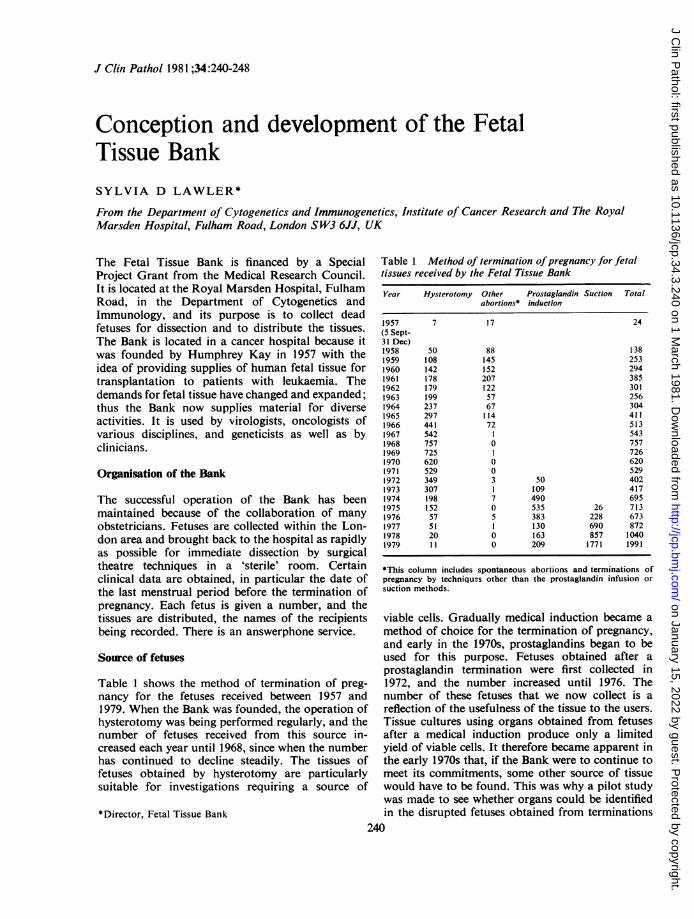
Table 1 shows the method of termination of preg-nancy for the fetuses received between 1957 and1979. When the Bank was founded, the operation ofhysterotomy was being performed regularly, and thenumber of fetuses received from this source in-creased each year until 1968, since when the numberhas continued to decline steadily. The tissues offetuses obtained by hysterotomy are particularlysuitable for investigations requiring a source of
*Director, Fetal Tissue Bank
Table 1 Method of termination ofpregnancy for fetaltissues received by the Fetal Tissue Bank
Year Hysterotomy Other Prostaglandin Suction Totalabortions* induction
1957 7 17 24(5 Sept-31 Dec)1958 50 88 1381959 108 145 2531960 142 152 2941961 178 207 3851962 179 122 3011963 199 57 2561964 237 67 3041965 297 114 4111966 441 72 5131967 542 1 5431968 757 0 7571969 725 1 7261970 620 0 6201971 529 0 5291972 349 3 50 4021973 307 1 109 4171974 198 7 490 6951975 152 0 535 26 7131976 57 5 383 228 6731977 51 1 130 690 8721978 20 0 163 857 10401979 11 0 209 1771 1991
*This column includes spontaneous abortions and terminations ofpregnancy by techniques other than the prostaglandin infusion orsuction methods.
viable cells. Gradually medical induction became amethod of choice for the termination of pregnancy,and early in the 1970s, prostaglandins began to beused for this purpose. Fetuses obtained after aprostaglandin termination were first collected in1972, and the number increased until 1976. Thenumber of these fetuses that we now collect is areflection of the usefulness of the tissue to the users.Tissue cultures using organs obtained from fetusesafter a medical induction produce only a limitedyield of viable cells. It therefore became apparent inthe early 1970s that, if the Bank were to continue tomeet its commitments, some other source of tissuewould have to be found. This was why a pilot studywas made to see whether organs could be identifiedin the disrupted fetuses obtained from terminations
240
on January 15, 2022 by guest. Protected by copyright.
http://jcp.bmj.com
/J C
lin Pathol: first published as 10.1136/jcp.34.3.240 on 1 M
arch 1981. Dow
nloaded from

Conception and development of the Fetal Tissue Bank
of pregnancy by the suction method.'We started to dissect disrupted fetuses in 1975,
since when the number collected has steadily in-creased. The development of this technique hasenabled virologists to continue to use human fetaltissues for the isolation of viruses and the prepara-tion of vaccines.
Ethical considerations
In 1970 an advisory group, with Sir John Peel aschairman, was appointed by the Secretary of Statefor Social Services and the Secretaries of State forScotland and Wales to consider the ethical, medical,social, and legal implications of using fetuses andfetal material for research. This group took extensiveevidence from the Medical Research Council andother organisations. A report was published in 19722giving a recommended code of practice for the use offetuses and fetal material in research. This reportrecommended that all research using the fetus, fetaltissue, or fetal material should be approved by anappropriate committee in the institution in whichthe research was undertaken, and that this committeeshould accept responsibility for ensuring that suchinvestigations were ethical.
In the Fetal Tissue Bank, although it is not legallybinding, we have followed the tenets of the 'PeelReport'. All users of the bank have to sign anethical clearance form:
'I. certify that theresearch on human fetal material for which I amresponsible has been approved by my localEthical Committee or equivalent body.
Signed ..........................Date ...
and the function of the Fetal Tissue Bank in theRoyal Marsden Hospital has been sanctioned by theEthical Committee of the Royal Marsden Hospital.The tenets of the 'Peel Report' that are relevant to
the operation of the Fetal Tissue Bank, and to whichwe adhere, are as follows:
'The minimal limit of viability for human fetusesshould be regarded as 20 weeks' gestational age.This corresponds to a weight of approximately 400-500 grammes.The use of the whole dead fetus or tissues from
dead fetuses for medical research is permissiblesubject to the following conditions:(i) The provisions of the Human Tissue Act are
observed;(ii) Where the provisions of the Human Tissue Act
do not apply there is no known objection on thepart of the parent who has had an opportunity
to declare any wishes about the disposal of thefetus;
(iii) Dissection of the dead fetus or experiments onthe fetus or fetal material do not occur in theoperating theatre or place of delivery;
(iv) There is no monetary exchange for fetuses orfetal material;
(v) Full records are kept by the relevant institution.'
Estimation of fetal age
It is always important to know the age of the fetaltissue, whether it is being used for clinical purposesor for research. The age of the fetus may be ex-pressed as menstrual age or gestational age. Thestarting point for menstrual age is the first day of thelast menstrual period. Others prefer to use gesta-tional age, which begins 14 days after the start of thelast menstrual period. When a menstrual cycle is not28 days in length, the estimated gestational age isfrom 14 days before the next anticipated menstrua-tion.3 Whichever measurement is used, there isinherent risk of inaccuracy because errors of recallcan occur.Data on fetal measurements correlated with the
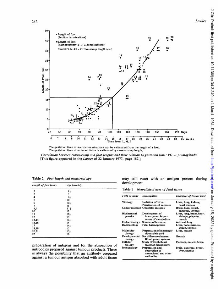
reported menstrual dates have been used in theFetal Tissue Bank in order to give an assessment ofthe age of the fetus. When whole fetuses are avail-able, the crown-rump length is the measurement ofchoice, and we use the simple formula that thecrown-rump length in centimetres +4 is equivalentto menstrual age in weeks.When we started using disrupted fetuses we
decided to use the length of foot as the fetal measure-ment. Foot length was compared with crown-rumplength by using whole fetuses, as shown in theFigure. The estimates of menstrual age based on footlength now in use in the Fetal Tissue Bank are shownin Table 2.
Non-clinical uses of fetal tissue
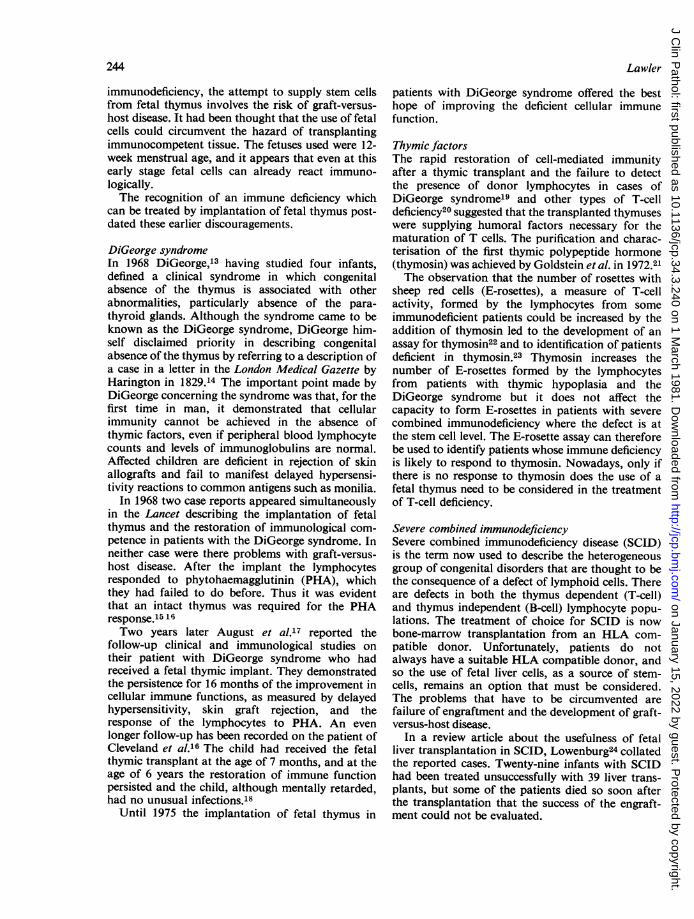
In a typical month, for example, July 1980, wereceived 173 disrupted fetuses, of which 107 werediscarded because no organs were identified. Fromthe remaining 66 fetuses with identifiable organs,327 specimens of tissue were distributed, and fromthe 28 prostaglandin-induced abortions of wholefetuses, 206 specimens of tissue were distributed. Theservice that the Fetal Tissue Bank provides can besummarised in tabular form (Table 3).The tissues from the disrupted fetuses are suitable
for cell investigations and are essential for the tissuecultures needed in virological studies. Tissues fromprostaglandin induction are suitable for the work ononcofetal antigens, since they can be used in the
241
on January 15, 2022 by guest. Protected by copyright.
http://jcp.bmj.com
/J C
lin Pathol: first published as 10.1136/jcp.34.3.240 on 1 M
arch 1981. Dow
nloaded from
A Length of foot(Suction terminations)
*Length of foot(Hysterectomy & P. G. terminations)
Numbers 5-20 = Crown-rump length (cm)
1314 13.
10
9 J
<|.136 *~~~~*^12m AAAA A 7 A
AA A AA A
AAA AA A
A AA AAAA"A AAI
19 19 20a U %
18 17 18
19 17* 1616
1714 14 a 8
*16 5 16
1413 12/14U*U
12 / A
13as
50 60. I 1 150 6'0 70 80 90 100 1 10 120 130 140
6 7 8 9 10 11 12 13 14 15 16 17 18 19 20 21 22 23Time from L. M. P
The gestation time of suction terminations can be estimated from the length of a foot.The gestation time of an intact fetus is estimated by crown-rump length.
Correlation between crown-rump andfoot lengths and their relation to gestation time: PG[This figure appeared in the Lancet of 22 January 1977, page 187.]
Table 2 Foot length and menstrual age
Length offoot (mm) Age (weeks)
2 813 94 9j5 106 lo+7 118,9 11i
10 1211 12i12 1313,14 13j15,16 1417 14j18,19 1520,21 15S22 16
preparation of antigens and for the absorption ofantibodies prepared against tumour products. Thereis always the possibility that an antibody preparedagainst a tumour antigen absorbed with adult tissue
may still react with an antigen present duringdevelopment.
Table 3 Non-clinical uses offetal tissue
Field ofstudy Investigation Examples of tissues used
Virology Isolation of virus. Liver, lung, kidney,Preparation of vaccines nasal mucosa
Cancer research Oncofetal antigens Brain, liver, breast,pancreas, thymus
Biochemical Development of Liver, lung, brain, heart,genetics isoenzymes. Inborn kidneys, placenta,
errors ofmetabolism muscleEndocrinology Sources ofhormone Adrenal, lungHaematology Fetal haemopoiesis Liver, bone-marrow,
spleen, thymusMolecular Preparation ofmessenger Liver, muscle
biology ribonucleic acidDevelopmental Sex differences in man. Gonads
biology Blood group systemsCellular Study oftrophoblast Placenta, muscle, brain
biology receptor mechanismsImmunology Preparation and Brain, pancreas, breast,
absorption of liver, thymusmonoclonal and otherantibodies
242 Lawyer
50-
45-
40-
35-
e 30
xa 25-0
*20-
15-
10-
5-
40 150 160 170 Days
24 25 Weeks
prostaglandin.
a, . IUT~
on January 15, 2022 by guest. Protected by copyright.
http://jcp.bmj.com
/J C
lin Pathol: first published as 10.1136/jcp.34.3.240 on 1 M
arch 1981. Dow
nloaded from

Conception and development of the Fetal Tissue Bank
Clinical uses of fetal tissue
BIOLOGY OF FETAL THYMUSIn a preliminary communication in 1961,4 Millerdescribed the effect of neonatal thymectomy in miceand proposed that the thymus at an early stage inlife plays an important role in the immunologicalresponse. He also produced evidence that engraftinga thymus restored the capacity to reject allogeneicskin grafts, which was one of the functions lost bythe neonatally thymectomised mice. In 19625 Millergave more details of these experiments when heconcluded that during very early life the thymus"produces the progenitors of immunologically com-petent cells which subsequently mature and migrateto other sites'. His experiments, however, did notexclude the possibility that the young thymus pro-duces a humoral factor that is necessary for thematuration of lymphocytes elsewhere in the body.Also in 1962, Kay et al.6 described the develop-
ment of the thymus in the human fetus and discussedits relation to immunological potential. They record-ed the weights of human fetal thymuses in relationto crown-rump length and body weight. They in-ferred that the thymus grows regularly during thefirst two trimesters but that the growth rate levels offat the beginning of the third trimester. After that,growth rate declines until birth and continues to doso thereafter. The initial up-gradient from 12 to 24weeks was deduced from information collected inthe Fetal Tissue Bank, but the data on older fetuseswere obtained from the literature. The data on theolder fetuses may have been less accurate because ofthe way in which they were collected, and also be-cause the thymus could have undergone involutionduring the delay between death and delivery of thefetus. The authors compared the growth of thespleen with that of the thymus and found that thespleen lagged behind the thymus in growth, but thatthe increase in relative splenic weight continueduntil birth.
In comparing the three aspects of lymphoidmaturation that can be subjected to quantitativeanalysis, namely, the development of spleen, thymus,and blood lymphocytes, Kay et al.6 found that thesedata from the human source were compatible withthe hypothesis that cells from the thymus colonisethe other lymphoid tissues of the body. Thus, bydeduction in man, the same conclusion was reachedas that from the experimental approach in mice.
IMMUNODEFICIENCY IN INFANCYSyndromes diagnosed in infancy, attributable to afailure of the development or function of the immunesystem, have been recognised since 1950. At first theimmunological deficiencies were described as humoral
immunity, cellular immunity, or both. The trans-plantation of fetal tissue came to play a role in thetreatment of some of these immunological de-ficiencies.The immune deficiency that is associated with the
lack of gammaglobulins was described by Bruton in1952.7 Once the diagnosis was made, treatment withgammaglobulin was used to control the infectionsthat are the salient features of the disease, which isinherited as a sex-linked recessive.
'Swiss type' agammaglobulinaemiaIn the next few years variants of this humoraldisorder were described, and one of these, whichcame to be known as the 'Swiss' form of agamma-globulinaemia, had been described by Glanzmannand Riniker in 1950.8 This syndrome was defined bysevere lymphopenia, absence of all immunoglobu-lins, hypoplasia of the lymphoreticular system, andan autosomal recessive inheritance pattern. Nowa-days the term combined immunodeficiency is used todescribe immune disorders in which there is adeficiency of both antibody responses and cell-mediated immunity.
In 1965 Hitzig et al.9 described an attempt to treattwo patients with the 'Swiss form of agammaglobu-linaemia' by implanting human fetal thymuses whichhad been collected in the Fetal Tissue Bank. In bothchildren a transient improvement in lymphocytecount was seen, but both died later of pulmonaryinfection.Two earlier attempts at immunological reconsti-
tution with thymus grafts in lymphopenic patientshad also been clinically unsuccessful. In a case ofalymphocytosis and agammaglobulinaemia, Rosenet al.10 used the thymic tissue from a 5-month-oldinfant, but this did not populate the lymphoid tissueof the recipient. Harboe"l made the importantobservation that in a 5-month-old boy with hypo-gammaglobulinaemia and severe lymphocytopenia,thymus implantation induced the formation of amonoclonal gamma-G and a monoclonal gamma-Mantibody, but the patient died of pneumonia twomonths later.
In 1968 Hong et al.12 reported the successfultreatment of an 8-month-old boy with (Swiss type)immunological deficiency complicated by oralmoniliasis. The presence of cells with a femalekaryotype in the bone-marrow established theengraftment. Unfortunately, the cells of the graftreacted against those of the host, producing acharacteristic skin eruption, one of the signs ofgraft-versus-host disease. The patient died ofpneumonia. The lessons learned from this case werethat although thymus graft alone is not sufficient torestore immune function in patients with combined
243
on January 15, 2022 by guest. Protected by copyright.
http://jcp.bmj.com
/J C
lin Pathol: first published as 10.1136/jcp.34.3.240 on 1 M
arch 1981. Dow
nloaded from
Lawler
immunodeficiency, the attempt to supply stem cellsfrom fetal thymus involves the risk of graft-versus-host disease. It had been thought that the use of fetalcells could circumvent the hazard of transplantingimmunocompetent tissue. The fetuses used were 12-week menstrual age, and it appears that even at thisearly stage fetal cells can already react immuno-logically.The recognition of an immune deficiency which
can be treated by implantation of fetal thymus post-dated these earlier discouragements.
DiGeorge syndromeIn 1968 DiGeorge,'13 having studied four infants,defined a clinical syndrome in which congenitalabsence of the thymus is associated with otherabnormalities, particularly absence of the para-thyroid glands. Although the syndrome came to beknown as the DiGeorge syndrome, DiGeorge him-self disclaimed priority in describing congenitalabsence of the thymus by referring to a description ofa case in a letter in the London Medical Gazette byHarington in 1829.14 The important point made byDiGeorge concerning the syndrome was that, for thefirst time in man, it demonstrated that cellularimmunity cannot be achieved in the absence ofthymic factors, even if peripheral blood lymphocytecounts and levels of immunoglobulins are normal.Affected children are deficient in rejection of skinallografts and fail to manifest delayed hypersensi-tivity reactions to common antigens such as monilia.
In 1968 two case reports appeared simultaneouslyin the Lancet describing the implantation of fetalthymus and the restoration of immunological com-petence in patients with the DiGeorge syndrome. Inneither case were there problems with graft-versus-host disease. After the implant the lymphocytesresponded to phytohaemagglutinin (PHA), whichthey had failed to do before. Thus it was evidentthat an intact thymus was required for the PHAresponse.'5 16Two years later August et al.17 reported the
follow-up clinical and immunological studies ontheir patient with DiGeorge syndrome who hadreceived a fetal thymic implant. They demonstratedthe persistence for 16 months of the improvement incellular immune functions, as measured by delayedhypersensitivity, skin graft rejection, and theresponse of the lymphocytes to PHA. An evenlonger follow-up has been recorded on the patient ofCleveland et al.'6 The child had received the fetalthymic transplant at the age of 7 months, and at theage of 6 years the restoration of immune functionpersisted and the child, although mentally retarded,had no unusual infections.18
Until 1975 the implantation of fetal thymus in
patients with DiGeorge syndrome offered the besthope of improving the deficient cellular immunefunction.
Thymic factorsThe rapid restoration of cell-mediated immunityafter a thymic transplant and the failure to detectthe presence of donor lymphocytes in cases ofDiGeorge syndrome'9 and other types of T-celldeficiency20 suggested that the transplanted thymuseswere supplying humoral factors necessary for thematuration of T cells. The purification and charac-terisation of the first thymic polypeptide hormone(thymosin) was achieved by Goldstein et al. in 1972.21The observation that the number of rosettes with
sheep red cells (E-rosettes), a measure of T-cellactivity, formed by the lymphocytes from someimmunodeficient patients could be increased by theaddition of thymosin led to the development of anassay for thymosin22 and to identification of patientsdeficient in thymosin.23 Thymosin increases thenumber of E-rosettes formed by the lymphocytesfrom patients with thymic hypoplasia and theDiGeorge syndrome but it does not affect thecapacity to form E-rosettes in patients with severecombined immunodeficiency where the defect is atthe stem cell level. The E-rosette assay can thereforebe used to identify patients whose immune deficiencyis likely to respond to thymosin. Nowadays, only ifthere is no response to thymosin does the use of afetal thymus need to be considered in the treatmentof T-cell deficiency.
Severe combined immunodeficiencySevere combined immunodeficiency disease (SCID)is the term now used to describe the heterogeneousgroup of congenital disorders that are thought to bethe consequence of a defect of lymphoid cells. Thereare defects in both the thymus dependent (T-cell)and thymus independent (B-cell) lymphocyte popu-lations. The treatment of choice for SCID is nowbone-marrow transplantation from an HLA com-patible donor. Unfortunately, patients do notalways have a suitable HLA compatible donor, andso the use of fetal liver cells, as a source of stem-cells, remains an option that must be considered.The problems that have to be circumvented arefailure of engraftment and the development of graft-versus-host disease.
In a review article about the usefulness of fetalliver transplantation in SCID, Lowenburg24 collatedthe reported cases. Twenty-nine infants with SCIDhad been treated unsuccessfully with 39 liver trans-plants, but some of the patients died so soon afterthe transplantation that the success of the engraft-ment could not be evaluated.
'2AA
on January 15, 2022 by guest. Protected by copyright.
http://jcp.bmj.com
/J C
lin Pathol: first published as 10.1136/jcp.34.3.240 on 1 M
arch 1981. Dow
nloaded from
Conception and development of the Fetal Tissue Bank
One factor that has to be considered is that cases
of SCID form such a heterogeneous clinical group.Within this group of diseases one entity has beendefined on the basis of a biochemical defect. Theimmunological deficiency has been associated withthe absence of the enzyme adenosine deaminase(ADA), which is essential for the successful forma-tion of lymphocytes.25 Cases ofADA deficiency havebeen corrected by repeated blood transfusion26whereby the enzyme is passively transferred.
Lowenburg24 tabulated the clinical and immuno-logical data on six successful fetal liver transplants.Three of these cases had been published elsewhere27-29but the other three were unpublished cases, describedat the Third Workshop of the International Co-operative Group for Bone-marrow Transplantationin Man (combined immunodeficiency) held atTarrytown, USA, in 1976. The proceedings of thismeeting have not been published. These six patientswere aged between 3 and 22 months; two were ADAnegative, the others positive. Fresh fetal liver cellswere used in all cases, which is an important point,because among the cases where engraftment failed,cells recovered after cryopreservation had been usedin some cases. This demonstrates the need forspecial care in the preservation and recovery of cells,including careful monitoring of the viability of thecells administered. Consideration has been given tothe optimal age of fetal tissue administered and theconsensus seems to be that the younger the fetus, thebetter. The donor fetuses in the successful cases
were almost all less than 12 weeks old. Buckley29suggested that 'younger fetal livers may be more
effective because of their relative enrichment forhaematological stem cells as compared to liver ofolder gestational age'. In successful cases, the num-
ber of cells administered per kg body weight was
often lower than in the unsuccessful cases, so cellnumbers may not be a critical factor. Intraperitonealadministration of the cells was used in the successfulcases. Some of the cases had repeated transplanta-tions before reconstitution was achieved, which is inaccord with what happens with bone-marrow trans-plantation where more than one transplant may alsobe required.30
Recently, a case has been made for the uise of fetalliver and thymus for immunological reconstitutionof patients, with variants of SCID, who do not havea suitable bone-marrow donor.31 These variants ofSCID require both a source of stem cells and a
source of thymic factors for successful immuno-logical reconstitution.
APLASTIC ANAEMIA
Since fetal liver provides a source of bone-marrowstem cells, a relevant question is whether it can be
used for transplantation in cases of haemopoieticdisorders. As early as 1961 Bodley Scott et al.32reported 14 cases with different types of bone-marrow hypoplasia who had been treated by theinjection of fetal haemopoietic cells. Out of fourcases of chronic pancytopenia, two had remissions,and the timing of these suggested that the fetal cellsmay have played a role, even though chimaerismwas not detected.The use of fetal liver for transplantation in cases
of aplastic anaemia has now been superseded by theuse of allogeneic bone-marrow from HLA-matcheddonors, child or adult. However, there still remainsthe problem of the case where no suitable livingdonor is available. Recently, interest has revived inthis application of human fetal liver, and the FetalTissue Bank has supplied fetal liver to Dr EGluckman and her colleagues for this purpose.33Implants of human fetal liver were attempted in fourchildren aged 4 to 13 years who had severe bone-marrow aplasia, in two cases following infectivehepatitis. The fetal liver graft was done after stoppingother treatments and after conditioning with cyclo-phosphamide. The fetuses used were aged between10 and 18 weeks, and the fetal cells were injectedintravenously. Two of the patients died of infections,one on the 44th day and the second on the 62nd day.The third patient was alive 26 months after thegraft. There was no evidence of bone-marrow con-version to donor type, although the patient is nowin a stable haematological state. The fourth patientshows a partial haematological reconstitution,which is autologous, but this child also receivedsteroids and a graft of stem cells from the mother.The interpretation of the results on these cases is
very difficult, and so it is impossible to know whatpart was played by the fetal liver graft in the twocases where there was a partial reconstitution.Nevertheless it appears that these two cases justifythe continuation of such attempts at reconstitutionin cases where suitable donors are not available.
Research in the Fetal Tissue Bank
Throughout the years the biology of fetal cells hasbeen studied in the Tissue Bank particularly, but notexclusively, in tissues that are relevant to trans-plantation.
RED CELL ANTIGENSWhen a fetus provides a potential source of haemo-poietic cells it is important to be able to determinethe blood groups of the fetus so that the red cells ofthe recipient can be examined for the presence ofdonor cells.
In 1962 blood from 39 fetuses was tested for the
245
on January 15, 2022 by guest. Protected by copyright.
http://jcp.bmj.com
/J C
lin Pathol: first published as 10.1136/jcp.34.3.240 on 1 M
arch 1981. Dow
nloaded from
246
ABO blood groups by Constandoulakis and Kay.34They found that the A and B antigens are readilydetectable but that no distinction between the Aland A2 antigens could be made. The A and B anti-gens of fetal red cells were found to be weaker thanthose of adults in absorptive power, and there wasno increase in antigenic strength during fetal life.The next year Constantoulakis et al.35 reported thatin fetal life the A2 gene is expressed as a phenotypesimilar to A. in adults, the red cells being agglutina-ted weakly by anti-A obtained from group B donorsbut strongly by anti-A from group 0 donors. Theyalso showed that the H antigen is expressed on fetalcells of all ABO groups and, although weaker thanin adults, the relationship to the A and B antigens isthe same. Concerning the antigenic strength of fetalantigens an interesting observation had been madeon the P blood group system by Ikin et al.,36 whofound that younger fetuses were more frequently andmore strongly Pi than were older fetuses.
Others have shown that the MN, Rhesus, Lua,Kell, Duffy, and Kidd antigens can be detected onfetal red cells.37 Therefore when blood is available,fetal red cells can be typed without difficulty. Whendisrupted fetuses are being used, a blood samplecannot be obtained but a suspension of haemo-poietic cells can be prepared from fetal liver fromwhich at least the ABO group can be determined.38
HLA ANTIGENSThe developments in the understanding of the HLAsystem, which have led to such advances in the choiceof suitable donors for bone-marrow transplantation,raise the possibility of selecting donors when usingfetal tissues. In order to do this it is essential to beable to determine the HLA type of the donor fetus.
If blood is available, fetal lymphocytes can betyped in the same way as those of adults.We have shown that fetal fibroblasts can also be
used as a reliable source of information on theHLA-A and B antigens of the fetus.39 The study wasdone by determining the antigens of lymphocytesand fibroblasts from the same fetus. Correlationswere found to be sufficiently good to warrant theuse of fibroblasts in the determination of the HLA-antigens of disrupted fetuses, particularly if culturesfrom more than one tissue were tested, for example,lung and skin.
FETAL CHROMOSOMESAfter transplantation the presence of donor cells inthe recipient can be identified by using the chromo-somes of dividing lymphocytes as a marker for theorigin of the cell. The easiest way to exploit thetechnique is when donor and recipient are ofdifferent sex. Applying the technique to another
Lawyer
problem, Kay and Margoles40 looked for trans-placental passage of maternal lymphocytes in malefetuses. They did not find any female cells among895 examined from 33 male fetuses.The sex of a disrupted fetus can be determined by
looking for sex chromatin or examining the chromo-somes in fibroblasts grown in tissue culture.
ALPHA-FETOPROTEIN (AFP)Many workers use fetal tissue from the Bank intheir studies of oncofetal antigens. Lawler andNash4' showed that fetal skin and muscle was a richsource of AFP, the highest yields being given byfetuses up to 16 weeks menstrual age. Antibodiessuitable for radioimmunoassay were prepared byimmunising rabbits with concentrates from thissource.
HYDATIDIFORM MOLESHydatidiform moles can be classified into twoentities, complete and partial mole, each of whichhas distinct pathological and genetic features. Thecomplete mole is characterised by cystic swelling andgross trophoblastic proliferation in the absence ofany evidence of fetal development.42 43 By chromo-some analysis these have been shown to be almostalways XX and to be entirely androgenetic in origin,that is, the chromosome complement comes entirelyfrom the male parent with no genetic representationof the female genome.44-46 On the other hand, apartial mole shows focal trophoblastic hyperplasia,a range of normal to swollen villi, and, most impor-tant, it is associated with the presence of a fetus atsome stage. Genetically, partial moles are triploid,and there is a chromosomal contribution from thefemale parent.47Between August 1978 and the end of July 1980,
3426 disrupted fetuses obtained from pregnanciesterminated by the suction method were collected bythe Fetal Tissue Bank. Among these were threehydatidiform moles, of which two were diagnosedas complete and one as partial mole. This incidenceof molar pregnancy of 1 in 1142 is somewhat higherthan the frequency usually quoted for Caucasians.48These early therapeutic abortions were all examinedmacroscopically for the presence of molar changes.Therefore it is possible that the higher estimate ofthe frequency of hydatidiform mole is correct.Molar pregnancies among spontaneous abortionsthat are not examined by medical personnel mayescape diagnosis.The Fetal Tissue Bank staff are also participating
in an elective study of hydatidiform moles.49Chromosomal polymorphisms and biochemicalgenetic markers are being used to study the geneticorigin of complete and partial moles. So far we have
on January 15, 2022 by guest. Protected by copyright.
http://jcp.bmj.com
/J C
lin Pathol: first published as 10.1136/jcp.34.3.240 on 1 M
arch 1981. Dow
nloaded from
Conception and development of the Fetal Tissue Bank 247
found that complete moles are XX and geneticallyhomozygous for the male contribution. Partialmoles, on the other hand, have been found to betriploid, to show evidence of maternal contribution,and to have arisen by dispermy or failure at the firstpaternal meiotic division.The study of moles is being made because of our
interest in premalignant conditions. Under a regis-tration scheme devised by the Department of Healthand Social Security and the Royal College ofObstetricians and Gynaecologists, patients are fol-lowed up after evacuation of a molar pregnancy, andsubsequent trophoblastic activity is monitored bymeasuring levels of human chorionic gonado-trophin.50 Thus, in the elective study, we have theopportunity to relate the data on the hydatidiformmole to the clinical outcome. In the United King-dom, up to 10% of patients require treatment withchemotherapy because of persistent trophoblasticactivity, and about one-third of them are likely tohave choriocarcinoma.51 Although most patientswho require treatment after a molar pregnancy havehad a complete mole, there is evidence that treatmentmay be required after a partial mole.52 Among 58hydatidiform moles studied at the Royal MarsdenHospital between July 1978 and June 1980, 44 werecomplete and 14 partial. At the time of writing(August 1980), six patients have required treatmentand all of these had complete moles.
The future of the Tissue Bank
There is no doubt that fetal tissue will have applica-tions in service and research work for many years tocome. If obstetric practice does not change radically,then the Fetal Tissue Bank should be able to supplythe tissues that are required. The needs of virologistsare likely to continue at least at the present level forthe foreseeable future because human fetal cellcultures are the tissues of choice for the isolation ofviruses and the preparation of vaccines. Oncologistscontinue to require fetal tissue to help sort outneoantigens from development ones. The requestsfor tissue from molecular biologists are continuallyincreasing. Apart from the present sources in thehydatidiform moles we have the possibility ofsupplying characterised homozygous human cells.Finally, in supplying organs for transplantation, weare considering the possibility of creating a store ofcryopreserved, tissue-typed cells to prepare for theera of matched fetal tissue transplants.
At present the Fetal Tissue Bank is operated by twopart-time medical personnel, Dr MJ Allan and Dr LWong; a scientist, Miss RA Fisher; a junior tech-nician, a porter, and a clerk. Mr AB Hockley, chief
technician in the Department of Cytogenetics andImmunology, has contributed to the operation of theBank for many years. Among the staff who workedboth with me and with Dr HEM Kay are Dr BMarkowski, Dr C Margoles, and Mrs DL Boyd. Iam deeply indebted to all the past and present staff.The work of the Bank would not be possible
without the cooperation of many obstetricians andtheir staff, and of the director and staff of the LondonPrivate Nursing Homes. Those who are working, orhave worked, in the Bank are most grateful to themall.
I thank Dr L Wong for suggestions and criticismsof the manuscript, and Miss P Leach for helping meto prepare it.
References
Markowski B, Lawler SD. Use of early fetal tissuesobtained from suction termination of pregnancy. Lancet1977;1 :186-8.
sThe use offetuses and fetal material for research. Reportof the Advisory Group, Department of Health andSocial Security, Scottish Home and Health Department,Welsh Office. London: Her Majesty's Stationery Office,1972.
Shepard TH. Growth and development of the humanembryo and fetus. In: Endocrine and genetic diseases ofchildhood and adolescence, 2nd edn. Ed LI Gardner.Philadelphia, London, Toronto: WB Saunders Co, 1975.
4Miller JFAP. Immunological function of the thymus.Lancet 1961 ;2:748-9.
mMiller JFAP. Effect of neonatal thymectomy on the im-munological responsiveness of the mouse. Proc RoyalSoc 1962;156:415-28.
Kay HEM, Playfair JHL, Wolfendale M, Hopper PK.Nature 1962;196:238-40.
7 Bruton OC. Agammaglobulinemia. Pediatrics 1952;9 :722-8.8 Glanzmann E, Riniker P. Essentielle Lymphocytophthise.
Ein neues Krankenheitsbild aus der Sauglingspathologie.Ann Paediat (Basel) 1950;175:1-32.
* Hitzig WH, Kay HEM, Cottier H. Familial lymphopeniawith agammaglobulinaemia. An attempt at treatment byimplantation of foetal thymus. Lancet 1965;2:151-4.
Rosen FS, Gitlin D, Janeway CA. Alymphocytosis,agammaglobulinaemia, homografts, and delayed hyper-sensitivity: study of a case. Lancet 1962;2:380-1.
Harboe M, Pande H, Brandtzaeg P, Tveter KJ, Hjort PF.Synthesis of donor type yG-globulin following thymustransplantation in hypo-y-globulinaemia with severelymphocytopenia. Scand J Haematol 1966 ;3:351-74.
Hong R, Cooper MD, Allan MJG, Kay HEM, MeuwissenH, Good RA. Immunological restitution in lymphopenicimmunological deficiency syndrome. Lancet 1968 ;1:503-6.
13 DiGeorge AM. Congenital absence of the thymusand its immunologic consequences: Concurrence withcongenital hypoparathyroidism. In: Immunologic de-ficiency diseases in man. Ed D Bergsma. Birth DefectsOriginal Article Series, IV, 116-23. The NationalFoundation-March of Dimes. 1968.
14 Harington H. Absence of the thymus gland. LondonMedical Gazette 1829;3:314-5.
15 August CS, Rosen FS, Filler RM, Janeway CA, MarkowskiB, Kay HEM. Implantation of a foetal thymus, restoringimmunological competence in a patient with thymic
on January 15, 2022 by guest. Protected by copyright.
http://jcp.bmj.com
/J C
lin Pathol: first published as 10.1136/jcp.34.3.240 on 1 M
arch 1981. Dow
nloaded from
248 Lawler
aplasia (DiGeorge's syndrome). Lancet 1968 ;2 :1210-1.16 Cleveland WW, Fogel BJ, Brown WT, Kay HEM. Foetal
thymic transplant in a case of DiGeorge's syndrome.Lancet 1968;2:1211-4.
17 August CS, Berkel Al, Levey RH, Rosen FS, Kay HEM.Establishment of immunological competence in a childwith congenital thymic aplasia by a graft of fetal thymus.Lancet 1970;1:1080-3.
18 Cleveland WW. Immunologic reconstitution in theDiGeorge syndrome by fetal thymic transplant. In:Immunodeficiency in man and animals. Ed D Bergsma.Birth Defects Original Article Series, XI, 352-6. TheNational Foundation-March of Dimes. 1975.
19 Kay HEM. Foetal thymus transplants in man. In:Ontogeny of acquired immnunity. A Ciba FoundationSymposium (23-25 November 1971), Amsterdam:Elsevier-Excerpta Medica-North-Holland, 249-60.
20 Foroozanfar N, Yamamura M, Watson G, Weaver P.Belton EM, Lawler S, Hobbs JR. Successful thymusgraft for T-cell deficiency in a 6-year-old boy. Br MedJ 1975;1:314-5.
Goldstein AL, Guha A, Zatz MM, Hardy MA, White A.Purification and biological activity of thymosin, ahormone of the thymus gland. Proc Natl Ac.ad Sci USA1972;69:1800-3.
22 Goldstein AL, Thurman GB, Cohen GH, Hooper JA.Thymosin: chemistry, biology and clinical application.In: Biological activity of thymic hormones. Ed DW VanBekkum. Rotterdam: Kookyer Scientific Publications,1975; 173-97.
2:1 Wara DW, Ammann AJ. Activation of T-cell rosettes inimmunodeficient patients by thymosin. Ann N Y AcadSci 1975;249:308-15.
21 Lowenberg B, Vossen JMJJ, Dooren LJ. Transplantationof fetal liver cells in the treatment of severe combinedimmunodeficiency disease. Blut 1977;34:181-95.
25 Giblett ER, Anderson JE, Cohen F, Pollara B, MeuwissenHJ. Adenosine-deaminase deficiency in two patientswith severely impaired celullar immunity. Lancet 1972;2:1067-9.
26 Polmar SH, Wetzler EM, Stern RC. Hirschhorn R.Restoration of in-vitro lymphocyte responses withexogenous adenosine deaminase in a patient with severecombined immunodeficiency. Lancet 1975 ;2 :743-6.
27 Keightley RG, Lawton AR, Cooper MD. Successful fetalliver transplantation in a child with severe combinedimmunodeficiency. Lancet 1975;2:850-3.
28 Ackeret C, Pluss HJ, Hitzig WH. Hereditary severe com-bined immunodeficiency and adenosine deaminasedeficiency. Pediatr Res 1976;10:67-70.
2!1 Buckley RH, Whisnant JK, Schiff RI, Gilbertsen RB,Huang AT, Platt MS. N Engl J Med 1976;294:1076-81.
Biggar WD, Park B, Niosi P, Yunis EJ, Good RA. Bonemarrow transplantation in combined immunodeficiencydisease. In: Tissue tYping and organ transplantation. EdEJ Yunis, RR Gatti, BD Amos. New York and London:Academic Press, 1973.
3' Pahwa R, Pahwa S, Good RA, Iincefy GS, O'Reilly RJ.Rationale for combined use of fetal liver and thymus forimmunological reconstitution in patients with variants ofsevere combined immunodeficiency. Proc Natl Acad SciUSA 1977;74:3002-5.
32 Bodley Scott R, Matthias JQ, Constandoulakis M, Kay
HEM, Lucas PF, Whiteside JD. Hypoplastic anaemiatreated by transfusion of foetal haemopoietic cells.Lancet 1961 ;2:1385-8.
33 Harousseau JL, Devergie A, Lawler S, Gluckman E.Schaison G. Implant of fetal liver in severe bone marrowaplasia. No,(lo Rev Fr Hematol. Societe FranQaised'Hematologie-Resumes du Congres de Poitiers, May1980.
3' Constandoulakis M, Kay HEM. A and B antigens of thehuman foetal erythrocyte. Br J Haematol 1962;8:57-63.
30 Constantoulakis M, Kay HEM, Giles CM, Parkin DM.Observations on the A, gene and H antigen in foetal life.Br J Haematol 1963;9:63-7.
361kin EW, Kay HEM, Playfair JHL, Mourant AE. Pantigen in the human foetus. Nature 1961;192:883.
37 Race RR, Sanger E. Blood groups in man. 6th edn. Oxford.London, Edinburgh, Melbourne: Blackwell ScientificPublications, 1975.
38 Wong L. Unpublished observations. 1980.39Singh SPN, Vyramuthu N, Margoles C, Lawler SD. HLA
typing of human fetal fibroblasts. Transplantation 1979;28 :262-3.
40 Kay HEM, Margoles C. Chromosomes of human fetallymphocytes: frequency of abnormalities and absence ofmaternal cells. Lancet 1971 ;2:733-5.
'1 Lawler SD, Nash SG. Alpha-fetoprotein in fetal skin andmuscle. Br J Obstet Gynaecol 1977;84:51-4.
42 Vassilakos P, Riotton G, Kajii T. Hydatidiform mole: twoentities. A morphologic and cytogenetic study with someclinical considerations. Am J Obstet Gvnecol 1977;127:167-70.
43 Szulman AE, Surti U. The syndromes of hydatidifornimole. 11. Morphologic evolution of the complete andpartial mole. Am J Obstet Gynecol 1978;132:20-7.
4' Kajii T, Ohama K. Androgenetic origin of hydatidifornmole. Nature 1977;268:633-4.
4; Jacobs PA, Hassold TJ, Matsuyama AM, Newlands IM.Chromosome constitution of gestational trophoblasticdisease. Lancet 1978;2:49.
46 Wake N, Takagi N, Sasaki N. Androgenesis as a cause ofhydatidiform mole. J Natl Cancer Inst 1978 ;60:51-7.
4 Szulman AE, Surti U. The syndromes of hydatidiformmole. 1. Cytogenetic and morphological correlations.Am J Obstet Gynecol 1978;131:665-71.
48 Bagshawe KD, Lawler SD. Choriocarcinoma. In:Cancer epidemiology and prevention. Ed D Schottenfeld,J Fraumeni Jr. Philadelphia, London, Toronto: WBSaunders Co, in press.
49 Lawler SD, Pickthall VJ, Fisher RA, Povey S, Wyn EvansM, Szulman AE. Genetic studies of complete andpartial hydatidiform moles. Lancet 1979;2:580.
5U Anonymous. New follow-up of hydatidiform mole. BrMed J 1972 ;iv:685-6.
51 Stone M, Dent J, Kardana A, Bagshawe KD. Relationshipof oral contraception to development of trophoblastictumour after evacuation of hydatidiform mole. Br JObstet Gynaecol 1976;83:913-6.
52 Stone M, Bagshawe KD. Hydatidiform mole: two entities.Lancet 1976;1:535-6.
Requests for reprints to: Dr Sylvia Lawler, Departmentof Cytogenetics and Immunogenetics, The Royal MarsdenHospital, Fulham Road, London SW3 6JJ.
on January 15, 2022 by guest. Protected by copyright.
http://jcp.bmj.com
/J C
lin Pathol: first published as 10.1136/jcp.34.3.240 on 1 M
arch 1981. Dow
nloaded from