conceptual and experimental tools to understand...
TRANSCRIPT

BI86CH14-Ismagilov ARI 18 May 2017 17:11
Conceptual and ExperimentalTools to Understand SpatialEffects and TransportPhenomena in NonlinearBiochemical Networks Illustratedwith Patchy Switching
Rebecca R. Pompano,1 Andrew H. Chiang,2
Christian J. Kastrup,3 and Rustem F. Ismagilov4
1Department of Chemistry, University of Virginia, Charlottesville, Virginia 22904;email: [email protected] of Chemistry and Institute for Biophysical Dynamics, The University of Chicago,Chicago, Illinois 60637; email: [email protected] Smith Laboratories and Department of Biochemistry and Molecular Biology,University of British Columbia, Vancouver, British Columbia V6T 1Z4, Canada;email: [email protected] of Chemistry and Chemical Engineering, California Institute of Technology,Pasadena, California 91125; email: [email protected]
Annu. Rev. Biochem. 2017. 86:333–56
The Annual Review of Biochemistry is online atbiochem.annualreviews.org
https://doi.org/10.1146/annurev-biochem-060815-014207
Copyright c© 2017 by Annual Reviews.All rights reserved
Keywords
state switching, signaling, flow, mass transfer, Damkohler number,microfluidics
Abstract
Many biochemical systems are spatially heterogeneous and exhibit nonlinearbehaviors, such as state switching in response to small changes in the localconcentration of diffusible molecules. Systems as varied as blood clotting,intracellular calcium signaling, and tissue inflammation are all heavily in-fluenced by the balance of rates of reaction and mass transport phenomenaincluding flow and diffusion. Transport of signaling molecules is also affectedby geometry and chemoselective confinement via matrix binding. In this re-view, we use a phenomenon referred to as patchy switching to illustrate theinterplay of nonlinearities, transport phenomena, and spatial effects. Patchyswitching describes a change in the state of a network when the local concen-tration of a diffusible molecule surpasses a critical threshold. Using patchyswitching as an example, we describe conceptual tools from nonlinear dy-namics and chemical engineering that make testable predictions and providea unifying description of the myriad possible experimental observations. Wedescribe experimental microfluidic and biochemical tools emerging to testconceptual predictions by controlling transport phenomena and spatial dis-tribution of diffusible signals, and we highlight the unmet need for in vivotools.
333
Click here to view this article'sonline features:
• Download figures as PPT slides• Navigate linked references• Download citations• Explore related articles• Search keywords
ANNUAL REVIEWS Further
Ann
u. R
ev. B
ioch
em. 2
017.
86:3
33-3
56. D
ownl
oade
d fr
om w
ww
.ann
ualr
evie
ws.
org
Acc
ess
prov
ided
by
Cal
ifor
nia
Inst
itute
of
Tec
hnol
ogy
on 0
6/28
/17.
For
per
sona
l use
onl
y.

BI86CH14-Ismagilov ARI 18 May 2017 17:11
Contents
INTRODUCTION . . . . . . . . . . . . . . . . . . . . . . . . . . . . . . . . . . . . . . . . . . . . . . . . . . . . . . . . . . . . . . . 334TRANSPORT AND THRESHOLD KINETICS DRIVE
PATCHY SWITCHING. . . . . . . . . . . . . . . . . . . . . . . . . . . . . . . . . . . . . . . . . . . . . . . . . . . . . . . 334Definition of Patchy Switching . . . . . . . . . . . . . . . . . . . . . . . . . . . . . . . . . . . . . . . . . . . . . . . . . . 334Dynamics of Patchy Switching Predicted by the Damkohler Number . . . . . . . . . . . . . 336Predicting Patchy Switching in Response to Patch Size . . . . . . . . . . . . . . . . . . . . . . . . . . . 337
MICROSCALE CONTROL OVER TRANSPORT PHENOMENA. . . . . . . . . . . . . . . 339Controlling Patch Size and Clustering . . . . . . . . . . . . . . . . . . . . . . . . . . . . . . . . . . . . . . . . . . . 339Chemically Controlling Diffusivity . . . . . . . . . . . . . . . . . . . . . . . . . . . . . . . . . . . . . . . . . . . . . . 342Utilizing Confinement . . . . . . . . . . . . . . . . . . . . . . . . . . . . . . . . . . . . . . . . . . . . . . . . . . . . . . . . . . 343Manipulating Flow and Shear . . . . . . . . . . . . . . . . . . . . . . . . . . . . . . . . . . . . . . . . . . . . . . . . . . . 345
CONCLUSIONS AND OUTLOOK. . . . . . . . . . . . . . . . . . . . . . . . . . . . . . . . . . . . . . . . . . . . . . 346To Which Biological Systems Might Patchy Switching Apply? . . . . . . . . . . . . . . . . . . . . 346Case Study: Possible Patchy Switching in the Lesions of Multiple Sclerosis . . . . . . . . 346Outlook on Tools for Testing the Role of Patchy Switching
In Vitro and In Vivo . . . . . . . . . . . . . . . . . . . . . . . . . . . . . . . . . . . . . . . . . . . . . . . . . . . . . . . . . 348
INTRODUCTION
Biochemical reactions rarely occur in a perfectly homogeneous environment. With the exceptionof a well-mixed test tube, molecules and cells are dispersed unevenly through complex matrices,whether inside a cell, culture dish, or tissue. Furthermore, the reactions themselves are moving inspace—molecules and cells are transported by diffusion, convection, and cellular motion, exceptwhen bound to relatively stationary support structures such as the extracellular matrix (ECM).
Historically, scientists have had better tools for controlling rates of reactions than for ma-nipulating transport phenomena. Quantification of the rates of reactions affecting signalingmolecules—for example, rate of enzymatic activity, rate of cellular secretion, and levels ofgene expression—is standard in biological experiments, and modulating reaction rates (via smallmolecules, siRNA, inactivating antibodies, etc.) is common in basic science and medicine. In con-trast, characterization and especially control of transport phenomena (1)—for example, diffusion,flow, effects of spatial arrangement, and effects of binding to the ECM (2–4)—have been morechallenging. Yet, when these experiments are performed, they suggest that changes in transportrates can be significant enough to perturb the local concentration of a signal. For example, bind-ing of basic fibroblast growth factor (bFGF) to heparan sulfate (Kd ∼ 24 nM) slows its diffusion(5) through the basement membrane by >125-fold. Such a large change in local concentrationcan strongly affect the state of a biochemical network when the kinetics of the network dependnonlinearly on concentration (6). If a system shows threshold dependence on the concentrationof the signal [e.g., in growth factor signaling (7)], this change may be sufficient to drive switchingto a new state.
TRANSPORT AND THRESHOLD KINETICS DRIVEPATCHY SWITCHING
Definition of Patchy Switching
To illustrate the interplay of nonlinear reaction kinetics, changes in local concentrations, and masstransport phenomena, in this review we focus our attention on biological systems that switch their
334 Pompano et al.
Ann
u. R
ev. B
ioch
em. 2
017.
86:3
33-3
56. D
ownl
oade
d fr
om w
ww
.ann
ualr
evie
ws.
org
Acc
ess
prov
ided
by
Cal
ifor
nia
Inst
itute
of
Tec
hnol
ogy
on 0
6/28
/17.
For
per
sona
l use
onl
y.

BI86CH14-Ismagilov ARI 18 May 2017 17:11
DiffusivityFlow
Geometry
+ Production– Degradation
– InhibitionDa =
Rate of reaction
Rate of transport
Da << 1 Da >> 1
Thresholdconcentration
Patchy switching
Low [trigger] on patch
System is OFF System is ON
High [trigger] on patch
Figure 1In patchy switching, local accumulation of diffusible triggers causes transitions between biological states,shown here for OFF and ON (red starburst) states. Triggers accumulate to high concentration (dark blue)only when their removal by transport ( green arrows) is slower than their net production (red arrows). Thecompetition between reaction and transport is described by a unitless quantity termed the Damkohlernumber (Da).
state in response to a change in local concentrations of diffusible molecules, which we call triggers(Figure 1). Although this phenomenon may sound specialized, it is actually quite common. Intu-itive examples of state switching occur in blood coagulation (clotted/not clotted), cellular signalingnetworks (signaling on/off ), quorum sensing (swarming/resting), neural dynamics (firing/not fir-ing), and stem cell differentiation (pluripotent/differentiated). For a more rigorous description ofstate switching, we refer the reader to reviews of dynamical systems (8) and transitions betweenmacro states in statistical thermodynamics (9). Triggers that control the state of a system as a re-sult of a change in concentration include small molecules, proteins, and enzymes. For simplicity,here we focus on molecular triggers, although such state switching may also occur in response tochanges in the local concentration of larger structures, such as vesicles or cells. All of these molec-ular triggers are subject to mass transport, and therefore their local concentrations are controlledby the balance of production, degradation, and mass transport phenomena.
A dramatic effect arises if two conditions are met: (a) triggers are localized or produced in acluster we call a patch and (b) the system can switch from OFF to ON when triggers are presentabove a critical threshold concentration. When the local concentration of triggers near a patchpasses through its critical threshold value, we call the state change patchy switching (Figure 1).For a concrete example, consider a small injury to a blood vessel that exposes blood to a patchof subendothelial tissue. This patch of damaged vasculature contains proteins, including tissuefactor (TF) and collagen, which begin adhering platelets and activating an enzymatic cascade thatcan lead to a blood clot (10). Clotting occurs only when activated enzymes accumulate abovea threshold concentration (11, 12), so this scenario meets the two conditions described above.Whether or not a clot forms and grows from the patch of damage depends on the balance betweenthe net rate of enzyme activation (activation minus consumption and inhibition) and the rate ofremoval of those enzymes by flow or diffusion (13).
The concept of patchy switching is not new, though appreciation for it has remained mostlyconfined to specific fields. Switching driven by mass transport effects is predicted by classicaltheory in chemistry, including work by Turing on the chemical basis of morphogenesis and work
www.annualreviews.org • Tools for Patchy Switching in Biology 335
Ann
u. R
ev. B
ioch
em. 2
017.
86:3
33-3
56. D
ownl
oade
d fr
om w
ww
.ann
ualr
evie
ws.
org
Acc
ess
prov
ided
by
Cal
ifor
nia
Inst
itute
of
Tec
hnol
ogy
on 0
6/28
/17.
For
per
sona
l use
onl
y.

BI86CH14-Ismagilov ARI 18 May 2017 17:11
on the chemical dynamics of nonequilibrium systems (14, 15), and related approaches in chemicalengineering (16). Patchy switching phenomena are a core concept in mathematical biology andecology, especially in the context of population dynamics (17, 18). The critical balance amongproduction, rapid degradation, and limited transport of growth factors through the extracellularmatrices is a factor in tissue engineering design (3, 19); this balance has also been described forcancer niches and microenvironments (20, 21) and stem cell signaling (22). In this review, weuse these ideas to illustrate the underlying principles that can describe many seemingly disparateexamples of switching from biochemistry and cell and tissue biology.
Although any nonlinear system will respond to any change in spatial distribution (23), we havechosen to focus on the specific example of patchy switching because conceptually it is relativelysimple, yet it can show dynamics that appear counterintuitive unless transport phenomena areconsidered. Now is an exciting time to study the effects of spatial distribution and mass transportof diffusible signals because newly emerging microfluidic and biochemical tools are revealing theinterplay of reactions, transport, and spatial effects on the cellular scale. Throughout this review,we speculate on biochemical systems in which phenomena such as patchy switching are likely tooccur, and we provide suggestions on how emerging tools might be used to probe these systems.
Dynamics of Patchy Switching Predicted by the Damkohler Number
The relationship between transport and concentration in dynamical systems can quickly becomecomplex, making it difficult to model and develop the intuition to make predictions. For example, inblood coagulation, the transport of coagulation enzymes is influenced by diffusion, flow, binding tocell surfaces and protein scaffolds, and secretion from platelets; importantly, the reaction networkdisplays nonlinear responses due to multiple positive and negative feedback reactions. How doesone begin to interpret and probe the interplay of reactions and transport phenomena in complexsystems like this? Fortunately, concepts from chemical engineering can be used to unify theseemingly disparate features of a system, such as reaction rate, clustering of molecules, and bindingto the ECM, into a single coherent description. For example, for patchy switching, all of thesefeatures are integrated into a simple dimensionless number that predicts the state of the system,termed the Damkohler number (Da). Da is the ratio of production and transport, and convenientlyit can be defined in terms of either rates (R, 1/s or mol/s) or timescales (τ , s):
Da = Rreaction
Rremoval= τremoval
τreaction
When Da� 1, triggers exceed a threshold concentration and the system will turn ON, whereaswhen Da � 1 the system will turn OFF (Figure 1). It is important to note that these are netrates. Thus, Rreaction is the net rate of the production of triggers—in other words, it is equal tothe difference between the total rate of all reactions that produce triggers and the total rate of allreactions that degrade or inhibit them. Alternatively, using timescales enables the estimation ofDa even when rates are unknown. Thus, τ reaction can be defined empirically as the time neededfor a system to reach the threshold concentration of the trigger (in the absence of transport).Similarly, Rremoval and τ removal refer to the net removal of triggers from the region of the patchby mass transport phenomena: diffusion, flow, transport by motile cells, etc. For example, in asystem in which triggers are produced by the patch, τ removal would be decreased by faster flow pastthe patch, whereas it would be increased by the trigger binding to matrix structures. Changes inspatial distribution drive patchy switching by changing τ removal.
336 Pompano et al.
Ann
u. R
ev. B
ioch
em. 2
017.
86:3
33-3
56. D
ownl
oade
d fr
om w
ww
.ann
ualr
evie
ws.
org
Acc
ess
prov
ided
by
Cal
ifor
nia
Inst
itute
of
Tec
hnol
ogy
on 0
6/28
/17.
For
per
sona
l use
onl
y.

BI86CH14-Ismagilov ARI 18 May 2017 17:11
C C C C
a b c d
withproduction
withdiffusion
withthreshold
C*
Nopatch
Smallpatch
Largepatch Activators produce
triggers Triggers are removedby diffusion and flow
OFF OFF
Da << 1Da < 1
ONDa > 1
Figure 2Balance of production and removal of triggers controls patchy switching and is described by the Damkohler number (Da). In eachpanel, the plot above depicts the concentration of triggers (C) for each scenario. (a) Three scenarios for the distribution of activators( gray spheres). “No patch” refers to molecules or cells being dispersed. (b) Activators produce triggers (blue) at a fixed rate per activator(red arrows). (c) Diffusion removes triggers ( green arrows) with greater effect on smaller patches. (d ) For a certain thresholdconcentration of triggers (C∗, gray dotted line), only the large patch accumulates triggers above the threshold and has Da > 1 (ON, redstarburst). The no patch and small patch groups are both below the threshold and have Da < 1 (OFF).
Predicting Patchy Switching in Response to Patch Size
As one example of the effect of spatial distribution on the state of a nonlinear biological system,consider the effect of patch size. Suppose that a patch consists of a cluster of activators (e.g., TFmolecules). Each activator produces triggers (e.g., activated clotting enzymes) at a constant rate.Therefore, τ reaction is independent of the size of the patch in the absence of mass transport. Thus,isolated activators, small patches, and large patches all would increase the local concentration ofthe trigger on the same timescale (Figure 2a,b). In contrast, τ removal depends on the size of thepatch. For diffusion, the timescale is τD ∼ x2/D (seconds), where x is the radius of the patch(meters) and D is the diffusion constant (m2/s) of the trigger. A freely diffusing trigger is removedrapidly from isolated molecules or cells, more slowly from a small patch, and slowest from a largepatch (Figure 2c). Removal of a trigger by flow would have a pattern similar to diffusion butis not depicted here. In summary, the larger the patch, the more the trigger accumulates. Fora sufficiently large patch (larger than a so-called critical patch size), Da is �1, and the triggeraccumulates above the threshold concentration to turn the system ON (e.g., clotted). In contrast,systems with smaller or no patches remain OFF (Da � 1) (Figure 2d). Thus, changes in thesize of a patch can also initiate patchy switching. Once state switching is initiated, it can evenpropagate outward from the patch (e.g., Figure 3a) in the presence of positive feedback loops,such as autocatalytic production of thrombin during blood clotting (24).
We emphasize that this example of state switching is not meant to be all-encompassing. We usethis type of patchy switching (switching ON when the local concentration of diffusible moleculescrosses a threshold) throughout the review for clarity, but the tools and methods described applyto a broad range of state switching systems in which mass transport phenomena play a role. Forexample, if the patch in Figure 2 produced inhibitors instead of triggers, then larger patches wouldbe OFF and smaller patches would be ON. Here we have focused on binary switches betweentwo possible states, but the same reasoning also applies to biological systems that have more thantwo potential states, such as stem cells, which can remain pluripotent or differentiate into any ofseveral different cell types.
State switching in response to patch size has been observed in several biochemical and cellularsystems (Figure 3). For example, the enzymatic network of blood coagulation could be switched
www.annualreviews.org • Tools for Patchy Switching in Biology 337
Ann
u. R
ev. B
ioch
em. 2
017.
86:3
33-3
56. D
ownl
oade
d fr
om w
ww
.ann
ualr
evie
ws.
org
Acc
ess
prov
ided
by
Cal
ifor
nia
Inst
itute
of
Tec
hnol
ogy
on 0
6/28
/17.
For
per
sona
l use
onl
y.

BI86CH14-Ismagilov ARI 18 May 2017 17:11
a i
b i
c i
d i
300 μm 300 μm 300 μm300 μm
Pancreatic
Parotid
500 μm500 μm500 μm500 μm500 μm500 μm500 μm500 μm
200 μm microcolony 500 μm microcolony
8 mm
4 mm
6 mm
2.4 mm
No regeneration No regeneration
Regeneration Regeneration
Very low density Low density
Threshold density High density
aa bb dd ffcc ee
aa bb dd ffcc ee
ii
Notclotted
ClottedClot
tim
e (m
in)
Patch size (μM)10
10
100
100 1,000
[Ca2+
] (μM
)
iiPancreaticParotid
[caged-InsP3] (μM)
5
4
3
2
1
1 10 1000
ii
ii
200
160
020406080
100120140
500 1,000
Num
ber o
f neu
rite
s
Size of microwell forcolony formation (μm)
** **
1,000
500
8.0 7.0 6.0 5.0 4.0 3.0 2.4
Mea
sure
d re
gene
rati
ng h
air n
umbe
r
Plucked area diameter (mm)
Saturated density:All follicles inthe region are
plucked
Zone of quorumsensing behavior
200 hairs are plucked fromdifferent sized regions
Regeneratingunplucked hairs
Regeneratingplucked hairs
Density too low:Plucked hairs do not regenerate
338 Pompano et al.
Ann
u. R
ev. B
ioch
em. 2
017.
86:3
33-3
56. D
ownl
oade
d fr
om w
ww
.ann
ualr
evie
ws.
org
Acc
ess
prov
ided
by
Cal
ifor
nia
Inst
itute
of
Tec
hnol
ogy
on 0
6/28
/17.
For
per
sona
l use
onl
y.

BI86CH14-Ismagilov ARI 18 May 2017 17:11
from a quiescent to a clotted state by increasing the size of micropatterned patches of TF, eventhough the total quantity of TF remained the same (Figure 3a) (25). As discussed above, oneexpects to observe patchy switching in this network because there are threshold responses to sol-uble triggers involved in coagulation (12), such as the proteases thrombin (26) and factor Xa (11).Eukaryotic calcium signaling is another threshold-mediated system in which autocatalysis plays arole, and patchy switching has been observed here as well. Specifically, the ubiquitous intracellularinositol (1,4,5)-triphosphate (IP3) receptor releases soluble Ca2+ from the endoplasmic reticulumin response to above-threshold concentrations of IP3 (27). This release drives a positive feedbackloop in which the released Ca2+ ions activate nearby IP3 receptor clusters to release more calcium(28, 29). Interestingly, when soluble IP3 was loaded at varying concentrations into murine pan-creatic and parotid cells, Ca2+ signals transitioned from small local spikes to global waves, a statechange that depended on the density of IP3 receptors in the cells (Figure 3b) (30).
In another example, at larger scales, the direction of stem cell differentiation was shown to bepotentiated by the size of stem cell embryoid bodies generated in microfluidic wells (3, 27). Studiesby two independent groups have shown that small embryoid bodies remained inert to neurodiffer-entiation, whereas larger clusters (>250–350 μm in diameter) adopted a neural morphology withextensive neurite formation (Figure 3c) (27, 31). The mechanism of this is still unclear and mayor may not be due to diffusible signals, although the role of diffusible stem cell signals is beingactively investigated (22, 32).
In the sections that follow, still using patchy switching as the example, we discuss recentdevelopments in microfluidics and chemistry that provide experimental control over factors thataffect the local balance (Da) of reactions (Figure 4a) and transport (Figure 4b), such as patch size,clustering, confinement, and the effects of flow and changing diffusivity.
MICROSCALE CONTROL OVER TRANSPORT PHENOMENA
Controlling Patch Size and Clustering
Changing the patch size, x, by patterning cells or molecules (while keeping their numbers constant)affects the Da number by changing the diffusion time of soluble triggers away from the patch,τD ∼ x2/D (Figures 2c and 4b,i). To help determine whether patchy switching plays a role in asystem of interest, a wide range of tools, many based on photochemistry (33) and soft lithography(a range of biocompatible micropatterning techniques) (29), can be used to pattern moleculesand cells onto in vitro surfaces (Figure 5a) (34). Multiple molecular or cellular entities can bepatterned separately or in close proximity to one another (Figure 5b,c) (35–39), for example, to
←−−−−−−−−−−−−−−−−−−−−−−−−−−−−−−−−−−−−−−−−−−−−−−−−−−−−−−−−−−−−−−−−−−−−−−−−−−−−−−−−−−−−−−−−−−Figure 3Switching in response to changes in spatial distribution. (a, i ) Blood coagulation (blue) is initiated on micropatterned patches of tissuefactor (red ) only if they are larger than a critical size, quantified in subpanel ii. Panel a adapted with permission fromReference 25, c© 2006 National Academy of Sciences, USA. (b, i, top) Only localized Ca2+ puffs were produced in response to localrelease of photocaged inositol (1,4,5)-triphosphate (IP3) in murine pancreatic cells expressing low density of inositol trisphosphatereceptors (InsP3R); (b, i, bottom) global Ca2+ (blue) waves were switched on in response to local release of photocaged IP3 in murineparotid cells expressing high density of InsP3R (30). (b, ii ) A plot showing that parotid cells have a lower threshold IP3 concentrationfor maximum Ca2+ release than the pancreatic cells. Panel b adapted from Reference 30 with permission. (c, i ) Embryoid bodiespreferentially differentiated into neural precursor cells ( green) with long neurites (red, quantified in subpanel ii ) only when they were atleast 500 μm in size. Panel c adapted from Reference 27 with permission. (d, i ) Plucking hairs on the skin of mice induced amplifiedhair regeneration only when hairs were plucked above a threshold density, quantified in subpanel ii. Red circles in images show theregions from which 200 hairs were plucked 30 days prior to the photo. Panel d adapted from Reference 63 with permission.
www.annualreviews.org • Tools for Patchy Switching in Biology 339
Ann
u. R
ev. B
ioch
em. 2
017.
86:3
33-3
56. D
ownl
oade
d fr
om w
ww
.ann
ualr
evie
ws.
org
Acc
ess
prov
ided
by
Cal
ifor
nia
Inst
itute
of
Tec
hnol
ogy
on 0
6/28
/17.
For
per
sona
l use
onl
y.

BI86CH14-Ismagilov ARI 18 May 2017 17:11
a Alter kinetics b Alter transport
Increaseproductionrate
Increaseproductionrate
Increaseproductionrate
Increasedegradation
rate
Increasedegradation
rate
Increasedegradation
rate
i Patch size or clustering
Degradeinhibitory ECM Inhibit
on ECM
ii Diffusivity iii Confinement iv Convection or active transport
Enlarge patch Assemble patcheson scaffold Confine patch Remove by
flow or shear
Remove by active transport
Partially confinepatchCluster patches Accumulate
triggers on ECM
Figure 4Various mechanisms for inducing patchy switching by changing the Damkohler number. (a) Altering the local concentration of atrigger (blue) by increasing its rate of production by the activators in the patch ( gray spheres) can turn a system with threshold kineticsON (red starburst). Inhibitory binding of a trigger on the extracellular matrix (ECM) also can turn the system OFF, whereas degradingthe ECM can restore the ON state. (b) Altering transport (i–iii ) by limiting diffusional flux increases the local concentration and canturn the system ON, whereas increasing removal (iv) by increasing the rate of convection or active transport ( green arrows) decreasesthe local concentration and can turn the system OFF. Each of these processes can also occur in reverse.
a
100 μm
b
50 μm
c
250 μm 200 μm
dd e
10 mm
Figure 5Controlling patch size and clustering using chemistry, microfluidics, and local drug delivery systems. (a) Two-dimensional patches ofhuman oral squamous carcinoma cells micropatterned using a peel-off perylene stencil. Panel a reproduced from Reference 34 withpermission. (b) Three-dimensional (3D) tumor spheroids formed and stained (inset, green) in microfluidic traps. Panel b adapted fromReference 37 with permission. (c) Two-dimensional chemical patterning of 3D microcultures containing rat fibroblasts ( green) orhuman lung adenocarcinoma cells (blue) using DNA templates. Panel c reproduced from Reference 35 with permission. (d ) Localizedchemical stimulation (red ) of a murine lymph node slice cultured in a microfluidic chamber. The slice was immunostained for B cells( green) and counterstained with Hoechst (blue), allowing specific regions to be targeted for stimulation. Panel d reproduced fromReference 53 with permission from The Royal Society of Chemistry. (e) Microchip implanted in a human to locally deliver discretedoses of a hormone fragment, which increased bone formation in women with osteoporosis. Panel e adapted from Reference 160 withpermission.
340 Pompano et al.
Ann
u. R
ev. B
ioch
em. 2
017.
86:3
33-3
56. D
ownl
oade
d fr
om w
ww
.ann
ualr
evie
ws.
org
Acc
ess
prov
ided
by
Cal
ifor
nia
Inst
itute
of
Tec
hnol
ogy
on 0
6/28
/17.
For
per
sona
l use
onl
y.
BI86CH14-Ismagilov ARI 18 May 2017 17:11
study heterotypic interactions among patches (39). In addition to providing the ability to culturematerials on two-dimensional (2D) surfaces, these micropatterning and microfluidic tools arewell suited to the generation of size-controlled three-dimensional (3D) cultures of microtissues,tumor spheroids, and stem cell organoids (40). Microfluidic laminar flow is another simple wayto control the spatial distribution of cells in 2D or 3D, often by patterning parallel lanes ofcells, gels, and stimuli. Finally, true 3D patterning is now possible as a result of advances inbiomaterials, chemistry, and 3D bioprinting, so that molecules and cells can be patterned on orwithin functionalized polymeric scaffolds or matrices for more realistic, tissue-like geometries (41–45). These patterning tools have been invaluable for evaluating the effect of patch size on bloodcoagulation (Figure 3a) (25) and stem cell differentiation (Figure 3c) (27, 31, 46–48). Patterningcan also distinguish the role of cell–cell contacts from that of soluble signaling (28) to help assessthe extent to which transport-mediated signals drive a particular system. For example, in a studyof tumor invasiveness, a patterned coculture of mammary epithelial cells and human mammaryfibroblasts was generated with and without a cell-free lane between them (49), revealing thatthe epithelial transition to an invasive phenotype was initiated by diffusible signals but requiredcell–cell contact to be completed.
In contrast to building patterned cell cultures, patchy distributions also can be created whenpreexisting in vitro cell cultures (50–52) or intact ex vivo tissue slices (Figure 5d) (53–58) arelocally stimulated using spatially resolved microfluidic delivery. Modern microfluidic designs thatstimulate local regions of neuronal cells (59), essentially creating a patch on a subcellular level,have built on early work by pioneers such as Campenot (60). Delivery of soluble molecules orproteins to spatially structured cells and tissues has the potential to enable investigation of patchyswitching and related dynamics, as described in the next paragraph.
We note that not all clusters are patches and not all spatial effects are due to the patchy switch-ing mechanism described here. For example, even if large patches turn the system ON and smallpatches do not, a critical question is whether the effect is driven by greater accumulation of dif-fusible triggers (patchy switching as in Figure 2d) or simply by having a larger quantity of theactivator (not patchy switching). Microfluidic and micropatterning experiments provide the con-trol needed to tease apart these differences. For example, a microfluidic device was used to delivera potassium chloride solution to local regions of murine brain slices, causing the cortical spreadingdepressions that simulate a migraine (58). By varying the radius of the delivery region and theconcentration of potassium, the researchers showed that it was the total quantity of potassium, notthe area over which it was spread, that determined whether spreading depressions occurred (61). Aresponse to a larger quantity of activator is not the same as a response to its spatial distribution anddoes not require nonlinear kinetics. The result of this potassium stimulation experiment contrastswith the patchy switching observed for blood clotting when TF was micropatterned into patchesof varying sizes while holding the total quantity constant (Figure 3a).
Clustering of patches and variations in patch density are other mechanisms that can change theDa number and induce switching (Figure 3b, i). As patches are brought closer together, diffusionof triggers from one patch overlaps with diffusion of triggers from nearby patches, and ultimatelyclosely spaced small patches functionally merge together. On the molecular scale, self-assemblyof signaling molecules on scaffolds (62) is a remarkable example of clustering. Tethering diffusingenzymes and substrates to a scaffold can raise the effective local concentration substantially; suchclustering can alter propagation of information through signaling pathways (62) and could leadto switching. Clustering can play a role at the tissue scale as well and can have marked effects onorgan-level function. For example, a link was made recently between clustering-induced patchyswitching and stimulated hair regrowth. Specifically, plucking a few hairs from the back of amouse caused robust hair regrowth but only if the hairs were plucked above a threshold density
www.annualreviews.org • Tools for Patchy Switching in Biology 341
Ann
u. R
ev. B
ioch
em. 2
017.
86:3
33-3
56. D
ownl
oade
d fr
om w
ww
.ann
ualr
evie
ws.
org
Acc
ess
prov
ided
by
Cal
ifor
nia
Inst
itute
of
Tec
hnol
ogy
on 0
6/28
/17.
For
per
sona
l use
onl
y.

BI86CH14-Ismagilov ARI 18 May 2017 17:11
(Figure 3d) (63). The effect was mediated by local transport and accumulation of inflammatoryproteins.
The micropatterning techniques described above make it easy to test the effect of clustering invitro. For example, in blood coagulation, micropatterning was used to generate clusters of sub-threshold patches of Bacillus bacteria on an otherwise inert surface. When human blood plasmawas exposed to these surfaces, unclustered patches of Bacillus did not initiate clotting, but clus-tered patches induced a robust coagulation response (64). These experiments demonstrated thata bacterial species could initiate the full coagulation cascade via a quorum acting mechanism, andthe work identified a bacterial zinc metalloprotease InhA1 as the activator of human thrombin andfactor Xa. Controlled clustering or localization of cells has also been used to study the complexeffects of autocrine and paracrine signaling, for example, to separate the effects of cell–cell contactand soluble signaling on the liver-specific function of hepatocyte–stromal cocultures (65) or oncolony formation by murine embryonic stem cells (66).
Tools are also emerging to test the role of spatial distribution and patches in vivo, in the contextof immune responses, the growth of tumors, and the local effects of xenografts or subcutaneousvaccinations. Microfluidic tools are potentially more invasive than optical probes and need to berefined to be used deep in tissues in vivo (67, 68). Genetic manipulation can be used to createpatchy distributions by utilizing temporally and spatially specific promoters or by creating chimericor mosaic animals—for example, in a chimeric mouse model of amyotrophic lateral sclerosis(69), although it is not known whether patch size plays a role in this system. In vivo patches ofcontrolled sizes can be created by microneedles, grafting, ultrasound, insertion of artificial tissuescaffolds and hydrogels, and local drug delivery systems (3, 70–74). In mouse models of ischemicinjury, for example, local hydrogel-mediated delivery of vascular endothelial growth factor (VEGF)and insulin-like growth factor-1 (IGF1) in tandem resulted in increased muscle weight. Suchexperiments suggest that spatial and temporal control play a key role in revascularization andregeneration in vivo (71, 75). Numerous other local drug delivery systems are being developedthat could be used to create large patches. The goal of most drug delivery systems is to maintain asufficiently high local concentration of a therapeutic drug in the target tissue while using low andsafe total amounts of the drugs; however, these systems could be extended to test the role of patchesin vivo. Light can also be used to create patches in vivo. Laser-induced injury of blood vessels(76) is used extensively to create patches of damaged vasculature and initiate blood coagulation;however, this technique has not yet been used to test if patchy switching occurs during initiationof coagulation in vivo. Dynamically, patches of cells can be created and switching can be inducedin vivo using optogenetics (77), dynamic patterning (36), and possibly the microfluidic tools forlocal chemical stimulation described above.
Chemically Controlling Diffusivity
Another method to induce patchy switching is to chemically reduce the diffusivity, D, of triggerscausing them to accumulate locally (Figure 4b, ii). Diffusion of triggers can be restricted by bind-ing them to or incorporating them into large, low-mobility objects such as proteins, vesicles, andexosomes (78) or by oligomerization and fibril formation. For example, intracellular binding ofCa2+ ions to soluble calcium-binding proteins enables local accumulation of Ca2+ and promotesinitiation of global calcium waves (79). Furthermore, binding of trigger molecules to nondiffusingobjects, such as cell surfaces, intracellular scaffolds, or the ECM, further reduces the effectivediffusion coefficient. For example, during coagulation in a damaged blood vessel, activated en-zymes bind to the area of exposed endothelium and aggregated platelets, which raises their localconcentration and amplifies the cascade (12, 80). In the context of tissue growth and repair as well
342 Pompano et al.
Ann
u. R
ev. B
ioch
em. 2
017.
86:3
33-3
56. D
ownl
oade
d fr
om w
ww
.ann
ualr
evie
ws.
org
Acc
ess
prov
ided
by
Cal
ifor
nia
Inst
itute
of
Tec
hnol
ogy
on 0
6/28
/17.
For
per
sona
l use
onl
y.

BI86CH14-Ismagilov ARI 18 May 2017 17:11
as inflammation, many cytokines, chemokines, and growth factors bind to proteoglycans on cellsurfaces or to components of the ECM, which limits diffusive loss and enables local chemoselec-tive accumulation despite constant interstitial, vascular, or lymphatic flow (3, 5, 73, 81–83). TheECM and closely packed cell bodies also reduce diffusivity by decreasing the free volume of thetissue, increasing tortuosity, and introducing sieving (84). Reduced diffusion through the ECMis well established for a variety of systems including ischemic injuries (85), brain tissue (86, 87),tumor microenvironment (84, 88, 89), mucosal linings in the gut and other organs (90), bacterialbiofilms (91), fibrosis on implanted materials (92), and stem cell embryoid bodies (93). In additionto reducing the diffusion of triggers, binding to the ECM may change their chemical or functionalproperties, for example, by protecting chemokines and cytokines from enzymatic degradation orpreventing binding to cellular receptors (1, 94). Modifying the trafficking of molecules into andout of cells is another biological mechanism for controlling transport and switches and occurs, forexample, by regulating endocytosis (95). Controlled uptake of chemokines, for example, has beenshown to allow cells to create their own chemotactic gradient for self-directed migration (96).
Tools are emerging to manipulate diffusion chemoselectively by altering binding events. Boththe signaling proteins [e.g., chemokines (82, 97, 98)] and the ECM (2, 4, 73, 97, 99) have beenmodified in vitro and in vivo, yielding insights into the roles of restricted diffusion. For exam-ple, mutations in the glycosaminoglycan (GAG)-binding sites of three chemokines, monocytechemoattractant protein-1/CCL2, macrophage inflammatory protein-1β/CCL4, and RANTES(regulated on activation normal T cell expressed and secreted)/CCL5, were designed to preventbinding of the chemokines to heparan sulfate (82, 98). Although these mutants were chemoat-tractive in static in vitro conditions, they failed to recruit cells after injection into the peritonealcavity in mice, thus confirming that GAG binding is essential for proper spatial localization andfunction of these chemokines in vivo. Decreased diffusion can be engineered as well; for example,TG-VEGF, a VEGF variant with an amino-terminus factor XIIIa substrate sequence, was designedto bind directly to fibrin in blood clots and fibrin glue tissue sealants, raising the local VEGF con-centration long enough to enhance angiogenesis (2). To engineer the ECM, proteins or hydrogelshave been designed to present specific binding motifs for soluble factors (73, 100). For example,a fibrin matrix was engineered to express a chemokine-binding motif, the twelfth–fourteenthtype III repeats of fibronectin (FN III12–14); this matrix slowed diffusive loss of platelet-derivedgrowth factor-BB and increased tissue growth responses (4). Accumulation of soluble factors isdistinct from presentation of cell-binding motifs such as RGD (arginylglycylaspartic acid); how-ever, chemokine- and integrin-binding motifs can certainly function synergistically as well (101,102). The ability of microfluidics to pattern and deliver reagents brings spatial resolution to dif-fusion experiments. Microfluidics and micropatterning can also be used to pattern ECM proteins(Figure 6a) (103, 104), inhibitors of binding, or other molecules (e.g., affinity reagents and recep-tors, and size-selective or chemo-selective matrices) that can help control diffusivity. As describedabove, microfluidic tools also enable local delivery of reagents, which can be used to measure diffu-sion through the ECM or measure transport in even more complex systems such as gap junctions(105), to provide insights into mechanisms of transport and switching.
Utilizing Confinement
A complementary approach to changing Da to induce patchy switching is restricting diffusion usinggeometric constraints, such as physical confinement (Figure 4b, iii). Confinement promotes theaccumulation of secreted molecules and can raise the local concentration of triggers above thethreshold value, inducing switching. Confinement of diffusible triggers is common in nature andis the basis for vesicles, organelles, cells, and other biological compartments. Experimentally,
www.annualreviews.org • Tools for Patchy Switching in Biology 343
Ann
u. R
ev. B
ioch
em. 2
017.
86:3
33-3
56. D
ownl
oade
d fr
om w
ww
.ann
ualr
evie
ws.
org
Acc
ess
prov
ided
by
Cal
ifor
nia
Inst
itute
of
Tec
hnol
ogy
on 0
6/28
/17.
For
per
sona
l use
onl
y.
BI86CH14-Ismagilov ARI 18 May 2017 17:11
a Diffusion in ECM b Confinement c Partial confinement d Shear
Shear rate = 100 s–1
Shear rate = 1,000 s–1
TF 2.5 6.3 12.5300 μm 5 μm
50-n
m re
d be
ads
50-n
m re
d be
ads
Gel
Gel
50-n
m g
reen
bea
ds
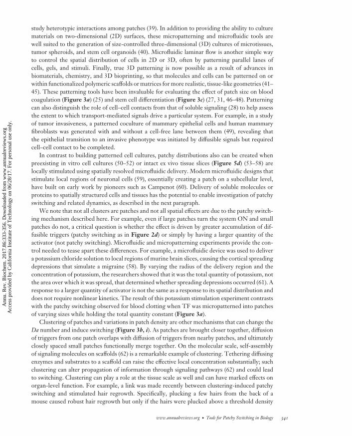
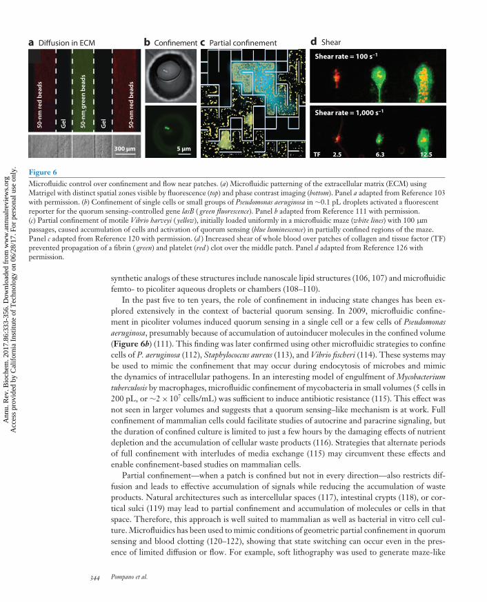
Figure 6Microfluidic control over confinement and flow near patches. (a) Microfluidic patterning of the extracellular matrix (ECM) usingMatrigel with distinct spatial zones visible by fluorescence (top) and phase contrast imaging (bottom). Panel a adapted from Reference 103with permission. (b) Confinement of single cells or small groups of Pseudomonas aeruginosa in ∼0.1 pL droplets activated a fluorescentreporter for the quorum sensing–controlled gene lasB ( green fluorescence). Panel b adapted from Reference 111 with permission.(c) Partial confinement of motile Vibrio harveyi ( yellow), initially loaded uniformly in a microfluidic maze (white lines) with 100 μmpassages, caused accumulation of cells and activation of quorum sensing (blue luminescence) in partially confined regions of the maze.Panel c adapted from Reference 120 with permission. (d ) Increased shear of whole blood over patches of collagen and tissue factor (TF)prevented propagation of a fibrin ( green) and platelet (red ) clot over the middle patch. Panel d adapted from Reference 126 withpermission.
synthetic analogs of these structures include nanoscale lipid structures (106, 107) and microfluidicfemto- to picoliter aqueous droplets or chambers (108–110).
In the past five to ten years, the role of confinement in inducing state changes has been ex-plored extensively in the context of bacterial quorum sensing. In 2009, microfluidic confine-ment in picoliter volumes induced quorum sensing in a single cell or a few cells of Pseudomonasaeruginosa, presumably because of accumulation of autoinducer molecules in the confined volume(Figure 6b) (111). This finding was later confirmed using other microfluidic strategies to confinecells of P. aeruginosa (112), Staphylococcus aureus (113), and Vibrio fischeri (114). These systems maybe used to mimic the confinement that may occur during endocytosis of microbes and mimicthe dynamics of intracellular pathogens. In an interesting model of engulfment of Mycobacteriumtuberculosis by macrophages, microfluidic confinement of mycobacteria in small volumes (5 cells in200 pL, or ∼2× 10⁷ cells/mL) was sufficient to induce antibiotic resistance (115). This effect wasnot seen in larger volumes and suggests that a quorum sensing–like mechanism is at work. Fullconfinement of mammalian cells could facilitate studies of autocrine and paracrine signaling, butthe duration of confined culture is limited to just a few hours by the damaging effects of nutrientdepletion and the accumulation of cellular waste products (116). Strategies that alternate periodsof full confinement with interludes of media exchange (115) may circumvent these effects andenable confinement-based studies on mammalian cells.
Partial confinement—when a patch is confined but not in every direction—also restricts dif-fusion and leads to effective accumulation of signals while reducing the accumulation of wasteproducts. Natural architectures such as intercellular spaces (117), intestinal crypts (118), or cor-tical sulci (119) may lead to partial confinement and accumulation of molecules or cells in thatspace. Therefore, this approach is well suited to mammalian as well as bacterial in vitro cell cul-ture. Microfluidics has been used to mimic conditions of geometric partial confinement in quorumsensing and blood clotting (120–122), showing that state switching can occur even in the pres-ence of limited diffusion or flow. For example, soft lithography was used to generate maze-like
344 Pompano et al.
Ann
u. R
ev. B
ioch
em. 2
017.
86:3
33-3
56. D
ownl
oade
d fr
om w
ww
.ann
ualr
evie
ws.
org
Acc
ess
prov
ided
by
Cal
ifor
nia
Inst
itute
of
Tec
hnol
ogy
on 0
6/28
/17.
For
per
sona
l use
onl
y.

BI86CH14-Ismagilov ARI 18 May 2017 17:11
microfluidic geometries that were then filled with liquid cultures of motile Vibrio harveyi. Themicrobes were initially distributed uniformly, but after 1–3 h they accumulated in the dead endsof the maze and activated quorum-dependent responses, presumably responding to accumulatingautocrine signals in partially confined regions (Figure 6c) (120). Furthermore, when compressivestress was used to decrease intercellular space in cultures of endothelial cells, it enhanced autocrineepidermal growth factor (EGF) signaling and increased EGF receptor activation equivalent to a10-fold increase in soluble EGF (117). These results show that confinement is especially effec-tive for activating signaling that incorporates positive feedback loops, including autocatalysis inblood coagulation, or autocrine EGF signaling and quorum sensing. Partial confinement wouldbe particularly effective in combination with chemoselective confinement by the ECM, whichwould capture triggers such as cytokines or growth factors while letting nutrients and waste passthrough unhindered. Existing patterning tools can be combined with chemistry to decorate theECM with binding functionalities (44) or to use the sieving (84) effects of ECM to study the effectsof chemoselective confinement on patchy switching.
Manipulating Flow and Shear
Fluid flow provides another way to induce switching by changing the local concentration of solubletriggers, by either delivering them to a patch or washing them out (Figure 4b, iv). In solutionor in bulk tissue, flow is best described by the linear flow velocity, V (meters per second). Neara surface, such as the endothelium, the effectiveness of transport is best described by the shearrate, γ = V/L (seconds−1), where L is a characteristic length (meters) over which the flow velocityis changing, such as the radius of a microfluidic channel or blood vessel. Shear rate controls thedynamics in systems such as blood coagulation (123), and its effects on transport are distinct fromshear-induced mechanical stress that can also induce signaling processes.
How does one know whether diffusion or flow is more important to transporting triggers ina particular system? The relative contributions of flow and diffusion to transport of a triggerare quantified by a dimensionless quantity termed the Peclet number: Pe = LV/D. At high Pe(Pe � 1), flow dominates, whereas at low Pe (Pe � 1), diffusion dominates. However, often thetwo work in concert: The combination of flow and diffusion generates gradients, so that the ef-fect of flow extends beyond where the flow actually is (83). For example, in solid tumors, flowthrough vessels delivers oxygen and nutrients that then diffuse to generate regions of proliferatingtissue surrounded by hypoxic and then necrotic tissue (124). A similar mode of transport occurswithin biofilms (91). Hydrostatic pressure from blood flow drives slow interstitial flow (0.1–2 μm/s)through soft tissues, which redistributes soluble molecules into gradients (83). Incorporating theseflows into tissue-engineered models is essential to recreating biological functions such as chemo-taxis (81).
Microfluidic techniques are well suited to controlling fluid flow in vitro at the size and volumescales most relevant to patchy switching and are already being exploited for this purpose (125). Forexample, microfluidic tools have been used to test the effects of flow and shear on the initiationof blood coagulation and platelet activation (Figure 6d). When human whole blood or plasmaflowed through microfluidic channels over surface-patterned patches of TF, the coagulation cas-cade switched ON only when low shear rates were used (126, 127). Bacterial quorum sensing hasalso been tested for its response to flow. In one experiment, 3D picoliter lobster traps were fabri-cated inside a microchannel by cross-linking the protein bovine serum albumin (BSA) (112), andP. aeruginosa bearing a fluorescent reporter for quorum sensing was cultured inside the trap. Quo-rum sensing was then induced simply by lowering the flow rate of media over the trap, indicatingthe sensitivity of this behavior to the transport conditions around the colony.
www.annualreviews.org • Tools for Patchy Switching in Biology 345
Ann
u. R
ev. B
ioch
em. 2
017.
86:3
33-3
56. D
ownl
oade
d fr
om w
ww
.ann
ualr
evie
ws.
org
Acc
ess
prov
ided
by
Cal
ifor
nia
Inst
itute
of
Tec
hnol
ogy
on 0
6/28
/17.
For
per
sona
l use
onl
y.

BI86CH14-Ismagilov ARI 18 May 2017 17:11
Microfluidic flow has also been used to control state switching in mammalian cell cultures,often by using controlled perfusion to wash out autocrine or paracrine cell-secreted factors. Sucha system was used to probe the mechanism of autocrine signaling by mouse embryonic stem cellsin maintaining a self-renewing state (22). Gentle perfusion over the cells induced a shift away fromself-renewal into an epiblast state, apparently not by simply removing autocrine growth factorsbut by removing secreted matrix-remodeling factors such as matrix metalloproteases (22). Thisexperiment illustrates the impact of both flow and ECM binding in cell fate decisions. Flow has alsobeen used to control directional heterotypic cell–cell interactions, as has been reviewed previously(39). Recently, an elegant microfluidic device design combined control over interstitial flow withcontrol over flow-mediated paracrine signaling; endothelial cells seeded into the device switchedinto vessel formation only when paracrine signals were present (128). Combining microfluidicswith biomaterials to control diffusion and flow simultaneously provides an opportunity to mimicin vitro complex biological gradients (129) and test the effects of slow interstitial flow (130). Toolsto modify the transport of molecules and induce flow are also being developed. For example,micromotor and self-propelling particles are being developed to actively transport their cargoin vitro and in vivo. Self-propelling particles have been used to transport triggers of coagulationagainst flow and deep into wounds to treat severe hemorrhage in large animals (131). Micromotorshave been used to transport drugs throughout gastric acid in the stomach (132) and to transporttheir cargo into target cells (133).
CONCLUSIONS AND OUTLOOK
To Which Biological Systems Might Patchy Switching Apply?
The tools discussed here may provide the greatest insights for biological systems with the hallmarkcharacteristics of patchy switching—in other words, those that display spatial heterogeneity, aredriven by diffusible signals, show highly nonlinear (e.g., threshold) responses to those signals, andare influenced by factors that affect mass transport, such as flow, partial confinement, changes inclustering or patch size, and binding to the ECM. Many systems meet most or all of these criteria.For example, inflammation is a highly nonlinear process that is driven by the local concentra-tion of cell-secreted cytokines (134); both the cells and the cytokines are mobile and respond todiffusion, interstitial flow, clustering and confinement in tight spaces, and binding to the ECM(135). Cellular decisions in stem cell niches, cancer niches, developmental niches, sites of woundhealing, sites of chronic inflammation, and even bacterial biofilms are similarly dependent on thespatial distribution of secreted factors that interact with the microenvironment in multiple ways.Furthermore, many of these systems include positive feedbacks, which could lead to spatial propa-gation of the switched state (Figure 3a) and also to stabilization of one state against perturbations.We call this stabilization state imprinting. In niches (e.g., stem cell niches, cancer niches, devel-opmental niches) in particular, such stabilization could occur from extensive interactions betweenthe cells and the ECM (20, 21), during which a patch of cells that has undergone a state switchcan modify the surrounding ECM to change its affinity for soluble signals and further stabilize thestate against perturbations, effectively converting a transient switching event to a chronic one.
Case Study: Possible Patchy Switching in the Lesions of Multiple Sclerosis
At first glance, it may seem that real biological systems are much too intricate to map onto thepatchy switching paradigm. Therefore, it is worth examining a case study. To discuss one exam-ple, we turn to localized inflammation in the demyelinating lesions of multiple sclerosis (MS),
346 Pompano et al.
Ann
u. R
ev. B
ioch
em. 2
017.
86:3
33-3
56. D
ownl
oade
d fr
om w
ww
.ann
ualr
evie
ws.
org
Acc
ess
prov
ided
by
Cal
ifor
nia
Inst
itute
of
Tec
hnol
ogy
on 0
6/28
/17.
For
per
sona
l use
onl
y.
BI86CH14-Ismagilov ARI 18 May 2017 17:11
a
200 µm
b
Dura
Arachnoid
Subarachnoidspace
Pia WM
GM Cerebral sulcus
CD20
CD20
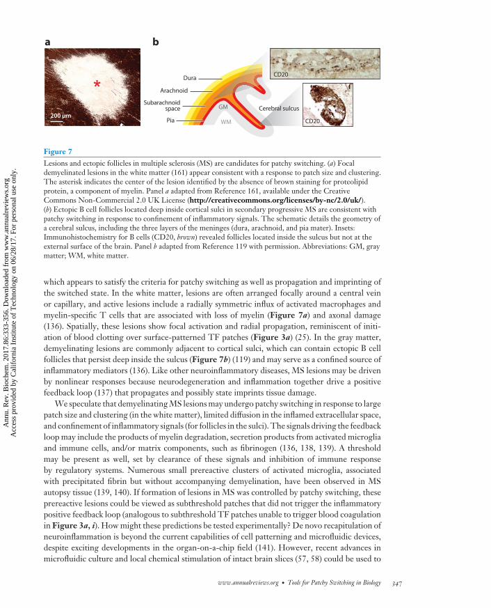
Figure 7Lesions and ectopic follicles in multiple sclerosis (MS) are candidates for patchy switching. (a) Focaldemyelinated lesions in the white matter (161) appear consistent with a response to patch size and clustering.The asterisk indicates the center of the lesion identified by the absence of brown staining for proteolipidprotein, a component of myelin. Panel a adapted from Reference 161, available under the CreativeCommons Non-Commercial 2.0 UK License (http://creativecommons.org/licenses/by-nc/2.0/uk/).(b) Ectopic B cell follicles located deep inside cortical sulci in secondary progressive MS are consistent withpatchy switching in response to confinement of inflammatory signals. The schematic details the geometry ofa cerebral sulcus, including the three layers of the meninges (dura, arachnoid, and pia mater). Insets:Immunohistochemistry for B cells (CD20, brown) revealed follicles located inside the sulcus but not at theexternal surface of the brain. Panel b adapted from Reference 119 with permission. Abbreviations: GM, graymatter; WM, white matter.
which appears to satisfy the criteria for patchy switching as well as propagation and imprinting ofthe switched state. In the white matter, lesions are often arranged focally around a central veinor capillary, and active lesions include a radially symmetric influx of activated macrophages andmyelin-specific T cells that are associated with loss of myelin (Figure 7a) and axonal damage(136). Spatially, these lesions show focal activation and radial propagation, reminiscent of initi-ation of blood clotting over surface-patterned TF patches (Figure 3a) (25). In the gray matter,demyelinating lesions are commonly adjacent to cortical sulci, which can contain ectopic B cellfollicles that persist deep inside the sulcus (Figure 7b) (119) and may serve as a confined source ofinflammatory mediators (136). Like other neuroinflammatory diseases, MS lesions may be drivenby nonlinear responses because neurodegeneration and inflammation together drive a positivefeedback loop (137) that propagates and possibly state imprints tissue damage.
We speculate that demyelinating MS lesions may undergo patchy switching in response to largepatch size and clustering (in the white matter), limited diffusion in the inflamed extracellular space,and confinement of inflammatory signals (for follicles in the sulci). The signals driving the feedbackloop may include the products of myelin degradation, secretion products from activated microgliaand immune cells, and/or matrix components, such as fibrinogen (136, 138, 139). A thresholdmay be present as well, set by clearance of these signals and inhibition of immune responseby regulatory systems. Numerous small prereactive clusters of activated microglia, associatedwith precipitated fibrin but without accompanying demyelination, have been observed in MSautopsy tissue (139, 140). If formation of lesions in MS was controlled by patchy switching, theseprereactive lesions could be viewed as subthreshold patches that did not trigger the inflammatorypositive feedback loop (analogous to subthreshold TF patches unable to trigger blood coagulationin Figure 3a, i). How might these predictions be tested experimentally? De novo recapitulation ofneuroinflammation is beyond the current capabilities of cell patterning and microfluidic devices,despite exciting developments in the organ-on-a-chip field (141). However, recent advances inmicrofluidic culture and local chemical stimulation of intact brain slices (57, 58) could be used to
www.annualreviews.org • Tools for Patchy Switching in Biology 347
Ann
u. R
ev. B
ioch
em. 2
017.
86:3
33-3
56. D
ownl
oade
d fr
om w
ww
.ann
ualr
evie
ws.
org
Acc
ess
prov
ided
by
Cal
ifor
nia
Inst
itute
of
Tec
hnol
ogy
on 0
6/28
/17.
For
per
sona
l use
onl
y.

BI86CH14-Ismagilov ARI 18 May 2017 17:11
test whether patchy switching can drive the onset of local inflammation ex vivo. For example, alocalized pulse of cytokines or myelin degradation products could be delivered to a rodent brainslice in arrays of focal regions of varying size. The first prediction is that for a given concentration oftrigger, inflammatory monocytes or lymphocytes would be recruited only when the local deliveryregions exceeded a critical size. The second prediction is that recruitment takes place, and theswitch to the inflammatory state would persist and propagate outward.
Outlook on Tools for Testing the Role of Patchy SwitchingIn Vitro and In Vivo
Knowing that a system of interest is undergoing patchy switching is invaluable because it allowsone to use Da as a unifying framework to predict how switching dynamics may be affected bychanges in the many factors that affect transport rates (Figure 4). Obviously, not all systems thatappear patchy or satisfy the criteria for patchy switching would actually show patchy switching.For example, if relative transport and reaction rates differ drastically or the threshold is too farfrom the resting value, small changes in reaction or transport rates may be insufficient to cross thecritical value of Da. Furthermore, alternative mechanisms (81) such as mechanotransduction (142)and cell–cell (49, 141) and cell–matrix contacts (142) would in most cases influence the range ofparameters in which patchy switching could occur and, in some cases, would dominate over effectsof diffusible signaling and Da number. Thus, to confirm or disprove patchy switching in eachsystem of interest, one needs to measure and modulate transport rates and the spatial distributionof potential trigger molecules using a chemically selective approach. Microfluidic and chemicaltools are essential to accomplish this.
In vitro, some of the microfluidic tools we have discussed in this review are still at the proof-of-concept stage, and few are sufficiently robust for use by nonexperts, although they are potentiallyavailable through collaboration with microfluidics laboratories. But some microfluidic tools arenow commercially available and are producing biological insights, and other tools are likely tomature rapidly toward widespread use. We expect the use of these tools to increase as advances aremade in tissue engineering and in other techniques that enable complex processes to be validatedex vivo and in vitro.
In vivo and ex vivo, additional spatially resolved analytical tools are urgently needed and havebegun to appear recently. For measurements with high spatial, temporal, and chemical resolu-tion, microsampling techniques combined with off-line analysis (50–52, 143) could be utilizedbut are invasive (144). In vivo electrochemistry with probes a few micrometers in diameter canprovide information on electroactive molecules; this technique has been particularly useful inbrain research (145) and may also become useful in immunity research (146). Imaging approachesare naturally spatially resolved, and confocal and two-photon imaging techniques in live tissue(147) are becoming increasingly accessible to nonspecialist researchers. Optical imaging, as wellas positron emission tomography (PET), functional magnetic resonance imaging (fMRI), and dif-fusion MRI techniques, can be done noninvasively but require that chemical wash-in, wash-out,or caged probes be developed for each molecule of interest (148, 149). Genetically encoded probesfor MRI and ultrasound techniques (150, 151) are particularly intriguing for noninvasively mon-itoring biological processes in vivo. Spatially resolved analysis of RNA and protein expression isparticularly exciting and has been called for recently (152).
Once developed, methods for spatially resolved analysis in vivo will be useful for identifyingniches that chemoselectively accumulate or lose trigger molecules and also for visualizing the ac-tivity of the cells and remodeling enzymes responsible for creating such niches. Furthermore, suchmethods could identify regions of confinement and chemoselective confinement as well as changes
348 Pompano et al.
Ann
u. R
ev. B
ioch
em. 2
017.
86:3
33-3
56. D
ownl
oade
d fr
om w
ww
.ann
ualr
evie
ws.
org
Acc
ess
prov
ided
by
Cal
ifor
nia
Inst
itute
of
Tec
hnol
ogy
on 0
6/28
/17.
For
per
sona
l use
onl
y.

BI86CH14-Ismagilov ARI 18 May 2017 17:11
in local flow, and these methods could be used to measure the size of patches, identify clusters ofpatches, and monitor their switching. Such tools could also improve basic understanding of in vivotransport rates; how they are affected by matrix composition; how they change with inflammation,stress, disease, aging, and physical activity; and how they affect patchy switching in vivo. Diag-nostically, such tools could support early detection and monitoring of treatment by identifyingsubthreshold, near-threshold, and above-threshold patches and corresponding niches—for exam-ple, by identifying early fibrinogen deposits in MS or precancerous lesions in sites predisposed tocancer (20)—or by monitoring sites of chronic inflammation.
Creating patches and controlling rates of transport to control switching in vivo are even morechallenging than analyzing them, but progress has been made in developing tools that can doso. Creating patches of known sizes could potentially be accomplished by using optogenetic tech-niques based on model organisms (153) or light-activated probes (154, 155), either of which providea resolution of ∼0.2–2 μm depending on tissue thickness and optical conditions. Alternatively,patches could be created by using focused ultrasound (resolution of a few millimeters) (156), orthe more invasive microfluidic tools (resolution of tens of micrometers to millimeters) (143, 157).In terms of controlling transport, flow is a powerful factor to control switching, and methods tocontrol local flow rates of blood, as well as interstitial, cerebrospinal, lymph, and synovial flu-ids, are still needed. Approaches are also particularly needed for chemoselective local synthesis,degradation, or modification of ECM, which would provide control over the binding of triggersto ECM in patches, and would allow one to modify, create, or erase patches and niches. To meetthis need, synthetic biomaterials, delivered to specific spatial areas, could act as niches and providecontrol of diffusion in a chemoselective fashion. Their effects could be enhanced further usingpartial confinement approaches (e.g., mimicking the ectopic follicles in Figure 7b) to promoteswitching. Such approaches could be used to create synthetic preimprinted niches with geometric,cytokine-binding, and transport properties suitable for initiation and propagation of a desired re-sponse and have been applied in a few promising models of adaptive immunity (158, 159). Similarniches are needed to allow controlled initiation of other processes such as neuroinflammationand wound healing. We speculate that methods for chemoselective control of transport in vivoalso could be applied therapeutically to erase pathological microenvironments (e.g., prereactivelesions in MS or premetastatic niches in cancer) or create supportive microenvironments (e.g., inwound healing, tissue regeneration, or vaccination). Overall, the emerging spatially resolved tools,together with advances in understanding of biological complexity, make this an exciting time tostudy and manipulate patchy switching and related biological phenomena in nonlinear networksthat are controlled by the balance of transport and reaction rates.
DISCLOSURE STATEMENT
The authors are not aware of any affiliations, memberships, funding, or financial holdings thatmight be perceived as affecting the objectivity of this review.
ACKNOWLEDGMENTS
This work was supported in part by a National Institutes of Health Director’s Pioneer Award(DP10D003584, R.F.I.), a Jacobs Institute for Molecular Engineering for Medicine Award (R.F.I.),a National Science Foundation Emerging Frontiers in Research and Innovation Award (1137089,R.F.I.), an Individual Biomedical Research Award from The Hartwell Foundation (R.R.P.), and aCanadian Institutes of Health Research Award (MSH-130166, C.J.K.). We thank Natasha Shelbyfor contributions to writing and editing this manuscript.
www.annualreviews.org • Tools for Patchy Switching in Biology 349
Ann
u. R
ev. B
ioch
em. 2
017.
86:3
33-3
56. D
ownl
oade
d fr
om w
ww
.ann
ualr
evie
ws.
org
Acc
ess
prov
ided
by
Cal
ifor
nia
Inst
itute
of
Tec
hnol
ogy
on 0
6/28
/17.
For
per
sona
l use
onl
y.

BI86CH14-Ismagilov ARI 18 May 2017 17:11
LITERATURE CITED
1. Tayalia P, Mooney DJ. 2009. Controlled growth factor delivery for tissue engineering. Adv. Mater.21:3269–85
2. Ehrbar M, Zeisberger SM, Raeber GP, Hubbell JA, Schnell C, Zisch AH. 2008. The role of activelyreleased fibrin-conjugated VEGF for VEGF receptor 2 gene activation and the enhancement of angio-genesis. Biomaterials 29:1720–29
3. Lee K, Silva EA, Mooney DJ. 2011. Growth factor delivery-based tissue engineering: general approachesand a review of recent developments. J. R. Soc. Interface 8:153–70
4. Martino MM, Hubbell JA. 2010. The 12th-14th type III repeats of fibronectin function as a highlypromiscuous growth factor-binding domain. FASEB J. 24:4711–21
5. Dowd CJ, Cooney CL, Nugent MA. 1999. Heparan sulfate mediates bFGF transport through basementmembrane by diffusion with rapid reversible binding. J. Biol. Chem. 274:5236–44
6. Tyson JJ, Chen KC, Novak B. 2003. Sniffers, buzzers, toggles and blinkers: dynamics of regulatory andsignaling pathways in the cell. Curr. Opin. Cell Biol. 15:221–31
7. Pickup M, Novitskiy S, Moses HL. 2013. The roles of TGFβ in the tumour microenvironment. Nat.Rev. Cancer 13:788–99
8. Astrom KJ, Murray R. 2003. Feedback Systems: An Introduction for Scientists and Engineers. Princeton, NJ:Princeton Univ. Press
9. Phillips R, Kondev J, Theriot J. 2008. Physical Biology of the Cell. New York: Garland Science10. Fogelson AL, Neeves KB. 2015. Fluid mechanics of blood clot formation. Annu. Rev. Fluid Mech. 47:377–
40311. Jesty J, Beltrami E. 2005. Positive feedbacks of coagulation: their role in threshold regulation. Arterioscler.
Thromb. Vasc. Biol. 25:2463–6912. Mann KG, Brummel K, Butenas S. 2003. What is all that thrombin for? J. Thromb. Haemost. 1:1504–1413. Rana K, Neeves KB. 2016. Blood flow and mass transfer regulation of coagulation. Blood Rev. 30:357–6814. Epstein IR, Showalter K. 1996. Nonlinear chemical dynamics: oscillations, patterns, and chaos. J. Phys.
Chem. 100:13132–4715. Turing AM. 1952. The chemical basis of morphogenesis. Philos. Trans. R. Soc. B 237:37–7216. Luss D, Sheintuch M. 2005. Spatiotemporal patterns in catalytic systems. Catal. Today 105:254–7417. Holmes EE, Lewis MA, Banks JE, Veit RR. 1994. Partial differential equations in ecology: spatial
interactions and population dynamics. Ecology 75:17–2918. Okubo A, Levin SA. 2001. Diffusion and Ecological Problems: Modern Perspectives. New York: Springer19. Saltzman WM, Olbricht WL. 2002. Building drug delivery into tissue engineering design. Nat. Rev.
Drug Discov. 1:177–8620. Bissell MJ, Hines WC. 2011. Why don’t we get more cancer? A proposed role of the microenvironment
in restraining cancer progression. Nat. Med. 17:320–2921. Lu P, Weaver VM, Werb Z. 2012. The extracellular matrix: a dynamic niche in cancer progression.
J. Cell Biol. 196:395–40622. Przybyla LM, Voldman J. 2012. Attenuation of extrinsic signaling reveals the importance of matrix
remodeling on maintenance of embryonic stem cell self-renewal. PNAS 109:835–4023. Epstein IR. 1995. The consequences of imperfect mixing in autocatalytic chemical and biological systems.
Nature 374:321–2724. Runyon MK, Kastrup CJ, Johnson-Kerner BL, Ha TG, Ismagilov RF. 2008. Effects of shear rate on
propagation of blood clotting determined using microfluidics and numerical simulations. J. Am. Chem.Soc. 130:3458–64
25. Kastrup CJ, Runyon MK, Shen F, Ismagilov RF. 2006. Modular chemical mechanism predicts spa-tiotemporal dynamics of initiation in the complex network of hemostasis. PNAS 103:15747–52
26. Pompano RR, Li HW, Ismagilov RF. 2008. Rate of mixing controls rate and outcome of autocatalyticprocesses: theory and microfluidic experiments with chemical reactions and blood coagulation. Biophys.J. 95:1531–43
27. Choi YY, Chung BG, Lee DH, Khademhosseini A, Kim JH, Lee SH. 2010. Controlled-size embryoidbody formation in concave microwell arrays. Biomaterials 31:4296–303
350 Pompano et al.
Ann
u. R
ev. B
ioch
em. 2
017.
86:3
33-3
56. D
ownl
oade
d fr
om w
ww
.ann
ualr
evie
ws.
org
Acc
ess
prov
ided
by
Cal
ifor
nia
Inst
itute
of
Tec
hnol
ogy
on 0
6/28
/17.
For
per
sona
l use
onl
y.

BI86CH14-Ismagilov ARI 18 May 2017 17:11
28. Bhatia SN, Balis UJ, Yarmush ML, Toner M. 1999. Effect of cell–cell interactions in preservation ofcellular phenotype: cocultivation of hepatocytes and nonparenchymal cells. FASEB J. 13:1883–900
29. Xia Y, Whitesides GM. 1998. Soft lithography. Annu. Rev. Mater. Sci. 28:153–8430. Giovannucci DR, Bruce JIE, Straub SV, Arreola J, Sneyd J, et al. 2002. Cytosolic Ca2+ and Ca2+-
activated Cl− current dynamics: insights from two functionally distinct mouse exocrine cells. J. Physiol.540:469–84
31. Chen Y-H, Peng C-C, Tung Y-C. 2015. Flip channel: a microfluidic device for uniform-sized embryoidbody formation and differentiation. Biomicrofluidics 9:054111
32. Przybyla LM, Theunissen TW, Jaenisch R, Voldman J. 2013. Matrix remodeling maintains ESC self-renewal by activating Stat3. Stem Cells 31:1097–106
33. Kloxin AM, Kasko AM, Salinas CN, Anseth KS. 2009. Photodegradable hydrogels for dynamic tuningof physical and chemical properties. Science 324:59–63
34. Tan CP, Seo BR, Brooks DJ, Chandler EM, Craighead HG, Fischbach C. 2009. Parylene peel-off arraysto probe the role of cell–cell interactions in tumour angiogenesis. Integr. Biol. 1:587–94
35. Li CY, Wood DK, Hsu CM, Bhatia SN. 2011. DNA-templated assembly of droplet-derived PEGmicrotissues. Lab Chip 11:2967–75
36. Robertus J, Browne WR, Feringa BL. 2010. Dynamic control over cell adhesive properties usingmolecular-based surface engineering strategies. Chem. Soc. Rev. 39:354–78
37. Wu LY, Di Carlo D, Lee LP. 2008. Microfluidic self-assembly of tumor spheroids for anticancer drugdiscovery. Biomed. Microdevices 10:197–202
38. Yarmush ML, King KR. 2009. Living-cell microarrays. Annu. Rev. Biomed. Eng. 11:235–5739. Zervantonakis IK, Kothapalli CR, Chung S, Sudo R, Kamm RD. 2011. Microfluidic devices for studying
heterotypic cell-cell interactions and tissue specimen cultures under controlled microenvironments.Biomicrofluidics 5:14
40. Lee GH, Lee JS, Wang X, Hoon Lee S. 2016. Bottom-up engineering of well-defined 3D microtissuesusing microplatforms and biomedical applications. Adv. Healthc. Mater. 5:56–74
41. Albrecht DR, Underhill GH, Wassermann TB, Sah RL, Bhatia SN. 2006. Probing the role of multicel-lular organization in three-dimensional microenvironments. Nat. Methods 3:369–75
42. Gu L, Mooney DJ. 2016. Biomaterials and emerging anticancer therapeutics: engineering the microen-vironment. Nat. Rev. Cancer 16:56–66
43. Murphy SV, Atala A. 2014. 3D bioprinting of tissues and organs. Nat. Biotech. 32:773–8544. Woolfson DN, Mahmoud ZN. 2010. More than just bare scaffolds: towards multi-component and
decorated fibrous biomaterials. Chem. Soc. Rev. 39:3464–7945. Yin X, Mead BE, Safaee H, Langer R, Karp JM, Levy O. 2016. Engineering stem cell organoids. Cell
Stem Cell 18:25–3846. Hwang YS, Chung BG, Ortmann D, Hattori N, Moeller HC, Khademhosseini A. 2009. Microwell-
mediated control of embryoid body size regulates embryonic stem cell fate via differential expression ofWNT5a and WNT11. PNAS 106:16978–83
47. Lee LH, Peerani R, Ungrin M, Joshi C, Kumacheva E, Zandstra PW. 2009. Micropatterning of humanembryonic stem cells dissects the mesoderm and endoderm lineages. Stem Cell Res. 2:155–62
48. Peerani R, Zandstra PW. 2010. Enabling stem cell therapies through synthetic stem cell-niche engi-neering. J. Clin. Invest. 120:60–70
49. Sung KE, Yang N, Pehlke C, Keely PJ, Eliceiri KW, et al. 2011. Transition to invasion in breast cancer:A microfluidic in vitro model enables examination of spatial and temporal effects. Integr. Biol. 3:439–50
50. Chen D, Du WB, Liu Y, Liu WS, Kuznetsov A, et al. 2008. The chemistrode: a droplet-based microflu-idic device for stimulation and recording with high temporal, spatial, and chemical resolution. PNAS105:16843–48
51. Kaigala GV, Lovchik RD, Drechsler U, Delamarche E. 2011. A vertical microfluidic probe. Langmuir27:5686–93
52. Saha-Shah A, Green CM, Abraham DH, Baker LA. 2016. Segmented flow sampling with push–pulltheta pipettes. Analyst 141:1958–65
53. Ross AE, Belanger MC, Woodroof JF, Pompano RR. 2017. Spatially resolved microfluidic stimulationof lymphoid tissue ex vivo. Analyst 142:649–59
www.annualreviews.org • Tools for Patchy Switching in Biology 351
Ann
u. R
ev. B
ioch
em. 2
017.
86:3
33-3
56. D
ownl
oade
d fr
om w
ww
.ann
ualr
evie
ws.
org
Acc
ess
prov
ided
by
Cal
ifor
nia
Inst
itute
of
Tec
hnol
ogy
on 0
6/28
/17.
For
per
sona
l use
onl
y.
BI86CH14-Ismagilov ARI 18 May 2017 17:11
54. Chang TC, Mikheev AM, Huynh W, Monnat RJ, Rostomily RC, Folch A. 2014. Parallel microfluidicchemosensitivity testing on individual slice cultures. Lab Chip 14:4540–51
55. Huang Y, Williams JC, Johnson SM. 2012. Brain slice on a chip: opportunities and challenges of applyingmicrofluidic technology to intact tissues. Lab Chip 12:2103–17
56. Mohammed JS, Caicedo HH, Fall CP, Eddington DT. 2008. Microfluidic add-on for standard electro-physiology chambers. Lab Chip 8:1048–55
57. Queval A, Ghattamaneni NR, Perrault CM, Gill R, Mirzaei M, et al. 2010. Chamber and microfluidicprobe for microperfusion of organotypic brain slices. Lab Chip 10:326–34
58. Tang YT, Kim J, Lopez-Valdes HE, Brennan KC, Ju YS. 2011. Development and characterization of amicrofluidic chamber incorporating fluid ports with active suction for localized chemical stimulation ofbrain slices. Lab Chip 11:2247–54
59. Yi Y, Park J, Lim J, Lee CJ, Lee S-H. 2015. Central nervous system and its disease models on a chip.Trends Biotechnol. 33:762–76
60. Campenot RB. 1977. Local control of neurite development by nerve growth factor. PNAS 74:4516–1961. Tang YT, Mendez JM, Theriot JJ, Sawant PM, Lopez-Valdes HE, et al. 2014. Minimum conditions for
the induction of cortical spreading depression in brain slices. J. Neurophysiol. 112:2572–7962. Good MC, Zalatan JG, Lim WA. 2011. Scaffold proteins: hubs for controlling the flow of cellular
information. Science 332:680–8663. Chen C-C, Wang L, Plikus MV, Jiang TX, Murray PJ, et al. 2015. Organ-level quorum sensing directs
regeneration in hair stem cell populations. Cell 161:277–9064. Kastrup CJ, Boedicker JQ, Pomerantsev AP, Moayeri M, Bian Y, et al. 2008. Spatial localization of
bacteria controls coagulation of human blood by ‘quorum acting.’ Nat. Chem. Biol. 4:742–5065. Hui EE, Bhatia SN. 2007. Micromechanical control of cell–cell interactions. PNAS 104:5722–2666. Rosenthal A, Macdonald A, Voldman J. 2007. Cell patterning chip for controlling the stem cell microen-
vironment. Biomaterials 28:3208–1667. Lee WH, Ngernsutivorakul T, Mabrouk OS, Wong J-MT, Dugan CE, et al. 2016. Microfabrication
and in vivo performance of a microdialysis probe with embedded membrane. Anal. Chem. 88:1230–3768. Slaney TR, Nie J, Hershey ND, Thwar PK, Linderman J, et al. 2011. Push-pull perfusion sampling
with segmented flow for high temporal and spatial resolution in vivo chemical monitoring. Anal. Chem.83:5207–13
69. Clement AM, Nguyen MD, Roberts EA, Garcia ML, Boillee S, et al. 2003. Wild-type nonneuronal cellsextend survival of SOD1 mutant motor neurons in ALS mice. Science 302:113–17
70. Kastrup CJ, Nahrendorf M, Figueiredo JL, Lee H, Kambhampati S, et al. 2012. Painting blood vesselsand atherosclerotic plaques with an adhesive drug depot. PNAS 109:21444–49
71. Kolambkar YM, Dupont KM, Boerckel JD, Huebsch N, Mooney DJ, et al. 2011. An alginate-basedhybrid system for growth factor delivery in the functional repair of large bone defects. Biomaterials32:65–74
72. Mitragotri S, Burke PA, Langer R. 2014. Overcoming the challenges in administering biopharmaceuti-cals: formulation and delivery strategies. Nat. Rev. Drug Discov. 13:655–72
73. Patterson J, Martino MM, Hubbell JA. 2010. Biomimetic materials in tissue engineering. Mater. Today13:14–22
74. Schoellhammer CM, Schroeder A, Maa R, Lauwers GY, Swiston A, et al. 2015. Ultrasound-mediatedgastrointestinal drug delivery. Sci. Transl. Med. 7:310ra168
75. Borselli C, Storrie H, Benesch-Lee F, Shvartsman D, Cezar C, et al. 2010. Functional muscle regener-ation with combined delivery of angiogenesis and myogenesis factors. PNAS 107:3287–92
76. Furie B, Furie BC. 2012. Formation of the clot. Thromb. Res. 130(Suppl. 1):S44–4677. Tischer D, Weiner OD. 2014. Illuminating cell signalling with optogenetic tools. Nat. Rev. Mol. Cell
Biol. 15:551–5878. Peinado H, Lavotshkin S, Lyden D. 2011. The secreted factors responsible for pre-metastatic niche
formation: old sayings and new thoughts. Semin. Cancer Biol. 21:139–4679. Dargan SL, Schwaller B, Parker I. 2004. Spatiotemporal patterning of IP3-mediated Ca2+ signals in
Xenopus oocytes by Ca2+-binding proteins. J. Physiol. 556:447–61
352 Pompano et al.
Ann
u. R
ev. B
ioch
em. 2
017.
86:3
33-3
56. D
ownl
oade
d fr
om w
ww
.ann
ualr
evie
ws.
org
Acc
ess
prov
ided
by
Cal
ifor
nia
Inst
itute
of
Tec
hnol
ogy
on 0
6/28
/17.
For
per
sona
l use
onl
y.

BI86CH14-Ismagilov ARI 18 May 2017 17:11
80. Ivanciu L, Stalker TJ. 2015. Spatiotemporal regulation of coagulation and platelet activation during thehemostatic response in vivo. J. Thromb. Haemost. 13:1949–59
81. Griffith LG, Swartz MA. 2006. Capturing complex 3D tissue physiology in vitro. Nat. Rev. Mol. Cell Biol.7:211–24
82. Proudfoot AEI. 2006. The biological relevance of chemokine–proteoglycan interactions. Biochem. Soc.Trans. 34:422–26
83. Swartz MA, Fleury ME. 2007. Interstitial flow and its effects in soft tissues. Annu. Rev. Biomed. Eng.9:229–56
84. Magzoub M, Jin S, Verkman AS. 2008. Enhanced macromolecule diffusion deep in tumors after en-zymatic digestion of extracellular matrix collagen and its associated proteoglycan decorin. FASEB J.22:276–84
85. Kim J, Cao L, Shvartsman D, Silva EA, Mooney DJ. 2011. Targeted delivery of nanoparticles to ischemicmuscle for imaging and therapeutic angiogenesis. Nano Lett. 11:694–700
86. Dityatev A, Seidenbecher CI, Schachner M. 2010. Compartmentalization from the outside: the extra-cellular matrix and functional microdomains in the brain. Trends Neurosci. 33:503–12
87. Sykova E, Nicholson C. 2008. Diffusion in brain extracellular space. Physiol. Rev. 88:1277–34088. Chauhan VP, Lanning RM, Diop-Frimpong B, Mok W, Brown EB, et al. 2009. Multiscale measurements
distinguish cellular and interstitial hindrances to diffusion in vivo. Biophys. J. 97:330–3689. Netti PA, Berk DA, Swartz MA, Grodzinsky AJ, Jain RK. 2000. Role of extracellular matrix assembly in
interstitial transport in solid tumors. Cancer Res. 60:2497–50390. Khanvilkar K, Donovan MD, Flanagan DR. 2001. Drug transfer through mucus. Adv. Drug Deliv. Rev.
48:173–9391. Stewart PS. 2003. Diffusion in biofilms. J. Bacteriol. 185:1485–9192. Ward KW. 2008. A review of the foreign-body response to subcutaneously-implanted devices: The role
of macrophages and cytokines in biofouling and fibrosis. J. Diabetes Sci. Technol. 2:768–7793. Sachlos E, Auguste DT. 2008. Embryoid body morphology influences diffusive transport of inductive
biochemicals: a strategy for stem cell differentiation. Biomaterials 29:4471–8094. Hynes RO. 2009. The extracellular matrix: not just pretty fibrils. Science 326:1216–1995. Francavilla C, Papetti M, Rigbolt KT, Pedersen AK, Sigurdsson JO, et al. 2016. Multilayered proteomics
reveals molecular switches dictating ligand-dependent EGFR trafficking. Nat. Struct. Mol. Biol. 23:608–18
96. Dona E, Barry JD, Valentin G, Quirin C, Khmelinskii A, et al. 2013. Directional tissue migration througha self-generated chemokine gradient. Nature 503:285–89
97. Dai E, Liu LY, Wang H, McIvor D, Sun YM, et al. 2010. Inhibition of chemokine-glycosaminoglycaninteractions in donor tissue reduces mouse allograft vasculopathy and transplant rejection. PLOS ONE5:e10510
98. Proudfoot AEI, Handel TM, Johnson Z, Lau EK, LiWang P, et al. 2003. Glycosaminoglycan bindingand oligomerization are essential for the in vivo activity of certain chemokines. PNAS 100:1885–90
99. Brown JM, Xia J, Zhuang B, Cho KS, Rogers CJ, et al. 2012. A sulfated carbohydrate epitope inhibitsaxon regeneration after injury. PNAS 109:4768–73
100. Custodio CA, Reis RL, Mano JF. 2014. Engineering biomolecular microenvironments for cell instructivebiomaterials. Adv. Healthc. Mater. 3:797–810
101. Martino MM, Tortelli F, Mochizuki M, Traub S, Ben-David D, et al. 2011. Engineering the growthfactor microenvironment with fibronectin domains to promote wound and bone tissue healing. Sci.Transl. Med. 3:100ra89
102. Das RK, Zouani OF. 2014. A review of the effects of the cell environment physicochemical nanoarchi-tecture on stem cell commitment. Biomaterials 35:5278–93
103. Wong AP, Perez-Castillejos R, Christopher Love J, Whitesides GM. 2008. Partitioning microfluidicchannels with hydrogel to construct tunable 3-D cellular microenvironments. Biomaterials 29:1853–61
104. Tien J, Nelson CM. 2014. Microstructured extracellular matrices in tissue engineering and development:an update. Ann. Biomed. Eng. 42:1413–23
105. Chen S, Lee LP. 2010. Non-invasive microfluidic gap junction assay. Integr. Biol. 2:130–38
www.annualreviews.org • Tools for Patchy Switching in Biology 353
Ann
u. R
ev. B
ioch
em. 2
017.
86:3
33-3
56. D
ownl
oade
d fr
om w
ww
.ann
ualr
evie
ws.
org
Acc
ess
prov
ided
by
Cal
ifor
nia
Inst
itute
of
Tec
hnol
ogy
on 0
6/28
/17.
For
per
sona
l use
onl
y.
BI86CH14-Ismagilov ARI 18 May 2017 17:11
106. Chan V, Novakowski SK, Law S, Klein-Bosgoed C, Kastrup CJ. 2015. Controlled transcription ofexogenous mRNA in platelets using protocells. Angew. Chem. Int. Ed. Engl. 54:13590–93
107. Lizana L, Bauer B, Orwar O. 2008. Controlling the rates of biochemical reactions and signaling networksby shape and volume changes. PNAS 105:4099–104
108. Gorris HH, Walt DR. 2010. Analytical chemistry on the femtoliter scale. Angew. Chem. Int. Ed. Engl.49:3880–95
109. Vincent ME, Liu WS, Haney EB, Ismagilov RF. 2010. Microfluidic stochastic confinement enhancesanalysis of rare cells by isolating cells and creating high density environments for control of diffusiblesignals. Chem. Soc. Rev. 39:974–84
110. Weitz M, Muckl A, Kapsner K, Berg R, Meyer A, Simmel FC. 2014. Communication and computationby bacteria compartmentalized within microemulsion droplets. J. Am. Chem. Soc. 136:72–75
111. Boedicker JQ, Vincent ME, Ismagilov RF. 2009. Microfluidic confinement of single cells of bacteria insmall volumes initiates high-density behavior of quorum sensing and growth and reveals its variability.Angew. Chem. Int. Ed. Engl. 48:5908–11
112. Connell JL, Wessel AK, Parsek MR, Ellington AD, Whiteley M, Shear JB. 2010. Probing prokaryoticsocial behaviors with bacterial “lobster traps.” mBio 1:e00202-10
113. Carnes EC, Lopez DM, Donegan NP, Cheung A, Gresham H, et al. 2010. Confinement-induced quorumsensing of individual Staphylococcus aureus bacteria. Nat. Chem. Biol. 6:41–45
114. Hagen SJ, Son M, Weiss JT, Young JH. 2010. Bacterium in a box: sensing of quorum and environmentby the LuxI/LuxR gene regulatory circuit. J. Biol. Phys. 36:317–27
115. Luthuli BB, Purdy GE, Balagadde FK. 2015. Confinement-induced drug-tolerance in mycobacteriamediated by an efflux mechanism. PLOS ONE 10:e0136231
116. Yu HM, Meyvantsson I, Shkel IA, Beebe DJ. 2005. Diffusion dependent cell behavior in microenviron-ments. Lab Chip 5:1089–95
117. Tschumperlin DJ, Dai G, Maly IV, Kikuchi T, Laiho LH, et al. 2004. Mechanotransduction throughgrowth-factor shedding into the extracellular space. Nature 429:83–86
118. Sato T, van Es JH, Snippert HJ, Stange DE, Vries RG, et al. 2011. Paneth cells constitute the niche forLgr5 stem cells in intestinal crypts. Nature 469:415–18
119. Magliozzi R, Howell O, Vora A, Serafini B, Nicholas R, et al. 2007. Meningeal B-cell follicles in secondaryprogressive multiple sclerosis associate with early onset of disease and severe cortical pathology. Brain130:1089–104
120. Park S, Wolanin PM, Yuzbashyan EA, Silberzan P, Stock JB, Austin RH. 2003. Motion to form a quorum.Science 301:188
121. Ribbe J, Maier B. 2016. Density-dependent differentiation of bacteria in spatially structured open systems.Biophys. J. 110:1648–60
122. Shen F, Pompano RR, Kastrup CJ, Ismagilov RF. 2009. Confinement regulates complex biochemicalnetworks: initiation of blood clotting by “diffusion acting.” Biophys. J. 97:2137–45
123. Hathcock JJ. 2006. Flow effects on coagulation and thrombosis. Arterioscler. Thromb. Vasc. Biol. 26:1729–37
124. Wyffels K, Kaanders J, Rijken P, Bussink J, van den Hoogen FJA, et al. 2000. Vascular architecture andhypoxic profiles in human head and neck squamous cell carcinomas. Br. J. Cancer 83:674–83
125. Kuo JS, Chiu DT. 2011. Controlling mass transport in microfluidic devices. Annu. Rev. Anal. Chem.4:275–96
126. Okorie UM, Denney WS, Chatterjee MS, Neeves KB, Diamond SL. 2008. Determination of sur-face tissue factor thresholds that trigger coagulation at venous and arterial shear rates: amplification of100 fM circulating tissue factor requires flow. Blood 111:3507–13
127. Shen F, Kastrup CJ, Liu Y, Ismagilov RF. 2008. Threshold response of initiation of blood coagulationby tissue factor in patterned microfluidic capillaries is controlled by shear rate. Arterioscler. Thromb. Vasc.Biol. 28:2035–41
128. Alonzo LF, Moya ML, Shirure VS, George SC. 2015. Microfluidic device to control interstitial flow-mediated homotypic and heterotypic cellular communication. Lab Chip 15:3521–29
129. Choi NW, Cabodi M, Held B, Gleghorn JP, Bonassar LJ, Stroock AD. 2007. Microfluidic scaffolds fortissue engineering. Nat. Mater. 6:908–15
354 Pompano et al.
Ann
u. R
ev. B
ioch
em. 2
017.
86:3
33-3
56. D
ownl
oade
d fr
om w
ww
.ann
ualr
evie
ws.
org
Acc
ess
prov
ided
by
Cal
ifor
nia
Inst
itute
of
Tec
hnol
ogy
on 0
6/28
/17.
For
per
sona
l use
onl
y.

BI86CH14-Ismagilov ARI 18 May 2017 17:11
130. Bonvin C, Overney J, Shieh AC, Dixon JB, Swartz MA. 2010. A multichamber fluidic device for 3D cul-tures under interstitial flow with live imaging: development, characterization, and applications. Biotechnol.Bioeng. 105:982–91
131. Baylis JR, Yeon JH, Thomson MH, Kazerooni A, Wang X, et al. 2015. Self-propelled particles thattransport cargo through flowing blood and halt hemorrhage. Sci. Adv. 1:e1500379
132. Gao W, Dong R, Thamphiwatana S, Li J, Gao W, et al. 2015. Artificial micromotors in the mouse’sstomach: a step toward in vivo use of synthetic motors. ACS Nano 9:117–23
133. Srivastava SK, Medina-Sanchez M, Koch B, Schmidt OG. 2016. Medibots: dual-action biogenic mi-crodaggers for single-cell surgery and drug release. Adv. Mater. 28:832–37
134. Schmitz ML, Weber A, Roxlau T, Gaestel M, Kracht M. 2011. Signal integration, crosstalk mechanismsand networks in the function of inflammatory cytokines. Biochim. Biophys. Acta Mol. Cell Res. 1813:2165–75
135. Vaday GG, Lider O. 2000. Extracellular matrix moieties, cytokines, and enzymes: dynamic effects onimmune cell behavior and inflammation. J. Leukoc. Biol. 67:149–59
136. Lassmann H. 2011. Review: the architecture of inflammatory demyelinating lesions: implications forstudies on pathogenesis. Neuropathol. Appl. Neurobiol. 37:698–710
137. Gao HM, Hong JS. 2008. Why neurodegenerative diseases are progressive: Uncontrolled inflammationdrives disease progression. Trends Immunol. 29:357–65
138. Akassoglou K, Adams RA, Bauer J, Mercado P, Tseveleki V, et al. 2004. Fibrin depletion decreasesinflammation and delays the onset of demyelination in a tumor necrosis factor transgenic mouse modelfor multiple sclerosis. PNAS 101:6698–703
139. Marik C, Felts PA, Bauer J, Lassmann H, Smith KJ. 2007. Lesion genesis in a subset of patients withmultiple sclerosis: a role for innate immunity? Brain 130:2800–15
140. van der Valk P, Amor S. 2009. Preactive lesions in multiple sclerosis. Curr. Opin. Neurol. 22:207–13141. Bhatia SN, Ingber DE. 2014. Microfluidic organs-on-chips. Nat. Biotech. 32:760–72142. Choi CK, Breckenridge MT, Chen CS. 2010. Engineered materials and the cellular microenvironment:
a strengthening interface between cell biology and bioengineering. Trends Cell Biol. 20:705–14143. Wang M, Hershey ND, Mabrouk OS, Kennedy RT. 2011. Collection, storage, and electrophoretic
analysis of nanoliter microdialysis samples collected from awake animals in vivo. Anal. Bioanal. Chem.400:2013–23
144. Cepeda DE, Hains L, Li D, Bull J, Lentz SI, Kennedy RT. 2015. Experimental evaluation and com-putational modeling of tissue damage from low-flow push–pull perfusion sampling in vivo. J. Neurosci.Methods 242:97–105
145. Bucher ES, Wightman RM. 2015. Electrochemical analysis of neurotransmitters. Annu. Rev. Anal. Chem.8:239–61
146. Andersson U, Tracey KJ. 2012. Neural reflexes in inflammation and immunity. J. Exp. Med. 209:1057–68147. Germain RN, Robey EA, Cahalan MD. 2012. A decade of imaging cellular motility and interaction
dynamics in the immune system. Science 336:1676–81148. Reynolds F, Kelly KA. 2011. Techniques for molecular imaging probe design. Mol. Imaging 10:407–19149. Porterfield WB, Prescher JA. 2015. Tools for visualizing cell–cell ‘interactomes.’ Curr. Opin. Chem. Biol.
24:121–30150. Shapiro MG, Goodwill PW, Neogy A, Yin M, Foster FS, et al. 2014. Biogenic gas nanostructures as
ultrasonic molecular reporters. Nat. Nanotechnol. 9:311–16151. Shapiro MG, Ramirez RM, Sperling LJ, Sun G, Sun J, et al. 2014. Genetically encoded reporters for
hyperpolarized xenon magnetic resonance imaging. Nat. Chem. 6:629–34152. Crosetto N, Bienko M, van Oudenaarden A. 2015. Spatially resolved transcriptomics and beyond. Nat.
Rev. Genet. 16:57–66153. Miesenbock G. 2009. The optogenetic catechism. Science 326:395–99154. Ryu KA, Stutts L, Tom JK, Mancini RJ, Esser-Kahn AP. 2014. Stimulation of innate immune cells by
light-activated TLR7/8 agonists. J. Am. Chem. Soc. 136:10823–25155. Bansal A, Zhang Y. 2014. Photocontrolled nanoparticle delivery systems for biomedical applications.
Acc. Chem. Res. 47:3052–60
www.annualreviews.org • Tools for Patchy Switching in Biology 355
Ann
u. R
ev. B
ioch
em. 2
017.
86:3
33-3
56. D
ownl
oade
d fr
om w
ww
.ann
ualr
evie
ws.
org
Acc
ess
prov
ided
by
Cal
ifor
nia
Inst
itute
of
Tec
hnol
ogy
on 0
6/28
/17.
For
per
sona
l use
onl
y.

BI86CH14-Ismagilov ARI 18 May 2017 17:11
156. Aryal M, Arvanitis CD, Alexander PM, McDannold N. 2014. Ultrasound-mediated blood–brain barrierdisruption for targeted drug delivery in the central nervous system. Adv. Drug Deliv. Rev. 72:94–109
157. Jeong J-W, McCall JG, Shin G, Zhang Y, Al-Hasani R, et al. 2015. Wireless optofluidic systems forprogrammable in vivo pharmacology and optogenetics. Cell 162:662–74
158. Purwada A, Jaiswal MK, Ahn H, Nojima T, Kitamura D, et al. 2015. Ex vivo engineered immuneorganoids for controlled germinal center reactions. Biomaterials 63:24–34
159. Singh A, Peppas NA. 2014. Hydrogels and scaffolds for immunomodulation. Adv. Mater. 26:6530–41160. Farra R, Sheppard NF Jr., McCabe L, Neer RM, Anderson JM, et al. 2012. First-in-human testing of a
wirelessly controlled drug delivery microchip. Sci. Transl. Med. 4:122ra21161. Chang A, Smith MC, Yin XH, Fox RJ, Staugaitis SM, Trapp BD. 2008. Neurogenesis in the chronic
lesions of multiple sclerosis. Brain 131:2366–75
356 Pompano et al.
Ann
u. R
ev. B
ioch
em. 2
017.
86:3
33-3
56. D
ownl
oade
d fr
om w
ww
.ann
ualr
evie
ws.
org
Acc
ess
prov
ided
by
Cal
ifor
nia
Inst
itute
of
Tec
hnol
ogy
on 0
6/28
/17.
For
per
sona
l use
onl
y.
ANNUAL REVIEWSConnect With Our Experts
New From Annual Reviews:Annual Review of Cancer Biologycancerbio.annualreviews.org • Volume 1 • March 2017
Co-Editors: Tyler Jacks, Massachusetts Institute of Technology Charles L. Sawyers, Memorial Sloan Kettering Cancer Center
The Annual Review of Cancer Biology reviews a range of subjects representing important and emerging areas in the field of cancer research. The Annual Review of Cancer Biology includes three broad themes: Cancer Cell Biology, Tumorigenesis and Cancer Progression, and Translational Cancer Science.
TABLE OF CONTENTS FOR VOLUME 1:• How Tumor Virology Evolved into Cancer Biology and
Transformed Oncology, Harold Varmus• The Role of Autophagy in Cancer, Naiara Santana-Codina,
Joseph D. Mancias, Alec C. Kimmelman• Cell Cycle–Targeted Cancer Therapies, Charles J. Sherr,
Jiri Bartek• Ubiquitin in Cell-Cycle Regulation and Dysregulation
in Cancer, Natalie A. Borg, Vishva M. Dixit• The Two Faces of Reactive Oxygen Species in Cancer,
Colleen R. Reczek, Navdeep S. Chandel• Analyzing Tumor Metabolism In Vivo, Brandon Faubert,
Ralph J. DeBerardinis• Stress-Induced Mutagenesis: Implications in Cancer
and Drug Resistance, Devon M. Fitzgerald, P.J. Hastings, Susan M. Rosenberg
• Synthetic Lethality in Cancer Therapeutics, Roderick L. Beijersbergen, Lodewyk F.A. Wessels, René Bernards
• Noncoding RNAs in Cancer Development, Chao-Po Lin, Lin He
• p53: Multiple Facets of a Rubik’s Cube, Yun Zhang, Guillermina Lozano
• Resisting Resistance, Ivana Bozic, Martin A. Nowak• Deciphering Genetic Intratumor Heterogeneity
and Its Impact on Cancer Evolution, Rachel Rosenthal, Nicholas McGranahan, Javier Herrero, Charles Swanton
• Immune-Suppressing Cellular Elements of the Tumor Microenvironment, Douglas T. Fearon
• Overcoming On-Target Resistance to Tyrosine Kinase Inhibitors in Lung Cancer, Ibiayi Dagogo-Jack, Jeffrey A. Engelman, Alice T. Shaw
• Apoptosis and Cancer, Anthony Letai• Chemical Carcinogenesis Models of Cancer: Back
to the Future, Melissa Q. McCreery, Allan Balmain• Extracellular Matrix Remodeling and Stiffening Modulate
Tumor Phenotype and Treatment Response, Jennifer L. Leight, Allison P. Drain, Valerie M. Weaver
• Aneuploidy in Cancer: Seq-ing Answers to Old Questions, Kristin A. Knouse, Teresa Davoli, Stephen J. Elledge, Angelika Amon
• The Role of Chromatin-Associated Proteins in Cancer, Kristian Helin, Saverio Minucci
• Targeted Differentiation Therapy with Mutant IDH Inhibitors: Early Experiences and Parallels with Other Differentiation Agents, Eytan Stein, Katharine Yen
• Determinants of Organotropic Metastasis, Heath A. Smith, Yibin Kang
• Multiple Roles for the MLL/COMPASS Family in the Epigenetic Regulation of Gene Expression and in Cancer, Joshua J. Meeks, Ali Shilatifard
• Chimeric Antigen Receptors: A Paradigm Shift in Immunotherapy, Michel Sadelain
ANNUAL REVIEWS | CONNECT WITH OUR EXPERTS
650.493.4400/800.523.8635 (us/can)www.annualreviews.org | [email protected]
ONLINE NOW!
Ann
u. R
ev. B
ioch
em. 2
017.
86:3
33-3
56. D
ownl
oade
d fr
om w
ww
.ann
ualr
evie
ws.
org
Acc
ess
prov
ided
by
Cal
ifor
nia
Inst
itute
of
Tec
hnol
ogy
on 0
6/28
/17.
For
per
sona
l use
onl
y.

BI86-FrontMatter ARI 1 June 2017 10:4
Annual Review ofBiochemistry
Volume 86, 2017ContentsAt the Intersection of Chemistry, Biology, and Medicine
Christopher T. Walsh � � � � � � � � � � � � � � � � � � � � � � � � � � � � � � � � � � � � � � � � � � � � � � � � � � � � � � � � � � � � � � � � � � � � � � � � � � 1
Protein Misfolding DiseasesF. Ulrich Hartl � � � � � � � � � � � � � � � � � � � � � � � � � � � � � � � � � � � � � � � � � � � � � � � � � � � � � � � � � � � � � � � � � � � � � � � � � � � � � � �21
Protein Misfolding, Amyloid Formation, and Human Disease:A Summary of Progress Over the Last DecadeFabrizio Chiti and Christopher M. Dobson � � � � � � � � � � � � � � � � � � � � � � � � � � � � � � � � � � � � � � � � � � � � � � � � �27
Structural Studies of Amyloid Proteins at the Molecular LevelDavid S. Eisenberg and Michael R. Sawaya � � � � � � � � � � � � � � � � � � � � � � � � � � � � � � � � � � � � � � � � � � � � � � � �69
Mechanisms and Functions of Spatial Protein Quality ControlEmily Mitchell Sontag, Rahul S. Samant, and Judith Frydman � � � � � � � � � � � � � � � � � � � � � � � � � �97
The Ubiquitin System, Autophagy, and Regulated Protein DegradationAlexander Varshavsky � � � � � � � � � � � � � � � � � � � � � � � � � � � � � � � � � � � � � � � � � � � � � � � � � � � � � � � � � � � � � � � � � � � � � � 123
Ubiquitin Ligases: Structure, Function, and RegulationNing Zheng and Nitzan Shabek � � � � � � � � � � � � � � � � � � � � � � � � � � � � � � � � � � � � � � � � � � � � � � � � � � � � � � � � � � � 129
Mechanisms of Deubiquitinase Specificity and RegulationTycho E.T. Mevissen and David Komander � � � � � � � � � � � � � � � � � � � � � � � � � � � � � � � � � � � � � � � � � � � � � � � 159
Proteasomal and Autophagic Degradation SystemsIvan Dikic � � � � � � � � � � � � � � � � � � � � � � � � � � � � � � � � � � � � � � � � � � � � � � � � � � � � � � � � � � � � � � � � � � � � � � � � � � � � � � � � � � � 193
Mechanisms of Autophagy InitiationJames H. Hurley and Lindsey N. Young � � � � � � � � � � � � � � � � � � � � � � � � � � � � � � � � � � � � � � � � � � � � � � � � � � 225
Systems Biology of MetabolismJens Nielsen � � � � � � � � � � � � � � � � � � � � � � � � � � � � � � � � � � � � � � � � � � � � � � � � � � � � � � � � � � � � � � � � � � � � � � � � � � � � � � � � � 245
Metabolite Measurement: Pitfalls to Avoid and Practices to FollowWenyun Lu, Xiaoyang Su, Matthias S. Klein, Ian A. Lewis, Oliver Fiehn,
and Joshua D. Rabinowitz � � � � � � � � � � � � � � � � � � � � � � � � � � � � � � � � � � � � � � � � � � � � � � � � � � � � � � � � � � � � � � 277
Isocitrate Dehydrogenase Mutation and (R)-2-Hydroxyglutarate:From Basic Discovery to Therapeutics DevelopmentLenny Dang and Shin-San Michael Su � � � � � � � � � � � � � � � � � � � � � � � � � � � � � � � � � � � � � � � � � � � � � � � � � � � 305
v
Ann
u. R
ev. B
ioch
em. 2
017.
86:3
33-3
56. D
ownl
oade
d fr
om w
ww
.ann
ualr
evie
ws.
org
Acc
ess
prov
ided
by
Cal
ifor
nia
Inst
itute
of
Tec
hnol
ogy
on 0
6/28
/17.
For
per
sona
l use
onl
y.

BI86-FrontMatter ARI 1 June 2017 10:4
Conceptual and Experimental Tools to Understand Spatial Effects andTransport Phenomena in Nonlinear Biochemical NetworksIllustrated with Patchy SwitchingRebecca R. Pompano, Andrew H. Chiang, Christian J. Kastrup,
and Rustem F. Ismagilov � � � � � � � � � � � � � � � � � � � � � � � � � � � � � � � � � � � � � � � � � � � � � � � � � � � � � � � � � � � � � � � 333
Biochemistry of Catabolic Reductive DehalogenationMaeva Fincker and Alfred M. Spormann � � � � � � � � � � � � � � � � � � � � � � � � � � � � � � � � � � � � � � � � � � � � � � � � � 357
Electric Fields and Enzyme CatalysisStephen D. Fried and Steven G. Boxer � � � � � � � � � � � � � � � � � � � � � � � � � � � � � � � � � � � � � � � � � � � � � � � � � � � � 387
Eukaryotic DNA Replication ForkPeter M.J. Burgers and Thomas A. Kunkel � � � � � � � � � � � � � � � � � � � � � � � � � � � � � � � � � � � � � � � � � � � � � � � 417
Telomerase Mechanism of Telomere SynthesisR. Alex Wu, Heather E. Upton, Jacob M. Vogan, and Kathleen Collins � � � � � � � � � � � � � � � � 439
Site-Specific Self-Catalyzed DNA Depurination: A BiologicalMechanism That Leads to Mutations and Creates SequenceDiversityJacques R. Fresco and Olga Amosova � � � � � � � � � � � � � � � � � � � � � � � � � � � � � � � � � � � � � � � � � � � � � � � � � � � � � � 461
A New Facet of Vitamin B12: Gene Regulation by Cobalamin-BasedPhotoreceptorsS. Padmanabhan, Marco Jost, Catherine L. Drennan,
and Montserrat Elıas-Arnanz � � � � � � � � � � � � � � � � � � � � � � � � � � � � � � � � � � � � � � � � � � � � � � � � � � � � � � � � � � 485
Engineering and In Vivo Applications of RiboswitchesZachary F. Hallberg, Yichi Su, Rebekah Z. Kitto, and Ming C. Hammond � � � � � � � � � � � � 515
Cyclic GMP–AMP as an Endogenous Second Messenger in InnateImmune Signaling by Cytosolic DNAKazuki Kato, Hiroki Omura, Ryuichiro Ishitani, and Osamu Nureki � � � � � � � � � � � � � � � � � � 541
A Bright Future for Antibiotics?Donna Matzov, Anat Bashan, and Ada Yonath � � � � � � � � � � � � � � � � � � � � � � � � � � � � � � � � � � � � � � � � � � 567
Molecular Characteristics and Biological Functions of Surface-Activeand Surfactant ProteinsMargaret Sunde, Chi L.L. Pham, and Ann H. Kwan � � � � � � � � � � � � � � � � � � � � � � � � � � � � � � � � � � � 585
How α-Helical Motifs Form Functionally Diverse Lipid-BindingCompartmentsLucy Malinina, Dinshaw J. Patel, and Rhoderick E. Brown � � � � � � � � � � � � � � � � � � � � � � � � � � � � � 609
The Evolution of Organellar Coat Complexes and Organization of theEukaryotic CellMichael P. Rout and Mark C. Field � � � � � � � � � � � � � � � � � � � � � � � � � � � � � � � � � � � � � � � � � � � � � � � � � � � � � � � 637
vi Contents
Ann
u. R
ev. B
ioch
em. 2
017.
86:3
33-3
56. D
ownl
oade
d fr
om w
ww
.ann
ualr
evie
ws.
org
Acc
ess
prov
ided
by
Cal
ifor
nia
Inst
itute
of
Tec
hnol
ogy
on 0
6/28
/17.
For
per
sona
l use
onl
y.
BI86-FrontMatter ARI 1 June 2017 10:4
Endoplasmic Reticulum–Plasma Membrane Contact SitesYasunori Saheki and Pietro De Camilli � � � � � � � � � � � � � � � � � � � � � � � � � � � � � � � � � � � � � � � � � � � � � � � � � � � 659
Mitochondrial Machineries for Protein Import and AssemblyNils Wiedemann and Nikolaus Pfanner � � � � � � � � � � � � � � � � � � � � � � � � � � � � � � � � � � � � � � � � � � � � � � � � � � � 685
Oxidative StressHelmut Sies, Carsten Berndt, and Dean P. Jones � � � � � � � � � � � � � � � � � � � � � � � � � � � � � � � � � � � � � � � � 715
Multiple Functions and Regulation of Mammalian PeroxiredoxinsSue Goo Rhee and In Sup Kil � � � � � � � � � � � � � � � � � � � � � � � � � � � � � � � � � � � � � � � � � � � � � � � � � � � � � � � � � � � � � � 749
Redox-Based Regulation of Bacterial Development and BehaviorAbigail J. Sporer, Lisa J. Kahl, Alexa Price-Whelan, and Lars E.P. Dietrich � � � � � � � � � 777
Extracellular Heme Uptake and the Challenge of Bacterial CellMembranesWeiliang Huang and Angela Wilks � � � � � � � � � � � � � � � � � � � � � � � � � � � � � � � � � � � � � � � � � � � � � � � � � � � � � � � � 799
Teaching Old Dyes New Tricks: Biological Probes Built fromFluoresceins and RhodaminesLuke D. Lavis � � � � � � � � � � � � � � � � � � � � � � � � � � � � � � � � � � � � � � � � � � � � � � � � � � � � � � � � � � � � � � � � � � � � � � � � � � � � � � � 825
Microbial Rhodopsins: Diversity, Mechanisms, and OptogeneticApplicationsElena G. Govorunova, Oleg A. Sineshchekov, Hai Li, and John L. Spudich � � � � � � � � � � � 845
Cellular Electron Cryotomography: Toward Structural Biology In SituCatherine M. Oikonomou and Grant J. Jensen � � � � � � � � � � � � � � � � � � � � � � � � � � � � � � � � � � � � � � � � � � � 873
Errata
An online log of corrections to Annual Review of Biochemistry articles may be found athttp://www.annualreviews.org/errata/biochem
Contents vii
Ann
u. R
ev. B
ioch
em. 2
017.
86:3
33-3
56. D
ownl
oade
d fr
om w
ww
.ann
ualr
evie
ws.
org
Acc
ess
prov
ided
by
Cal
ifor
nia
Inst
itute
of
Tec
hnol
ogy
on 0
6/28
/17.
For
per
sona
l use
onl
y.