conformal radiotherapy - department of healthfile/1038-conformal-radiotherapy-assessment... · this...
TRANSCRIPT
Conformal radiotherapy
November 2001
MSAC application 1038
Assessment report

© Commonwealth of Australia 2001
ISSN (Print) 1443-7120 ISSN (Online) 1443-7139 ISBN
First printed: March 2002
This work is copyright. Apart from any use as permitted under the Copyright Act 1968 no part may be reproduced by any process without written permission from AusInfo. Requests and inquiries concerning reproduction and rights should be directed to the Manager, Legislative Services, AusInfo, GPO Box 1920, Canberra, ACT, 2601.
Electronic copies of the report can be obtained from the Medical Service Advisory Committee’s Internet site at:
http://www.msac.gov.au
Hard copies of the report can be obtained from:
The Secretary Medical Services Advisory Committee Department of Health and Ageing Mail Drop 107 GPO Box 9848 Canberra ACT 2601
Enquiries about the content of the report should be directed to the above address.
The Medical Services Advisory Committee is an independent committee which has been established to provide advice to the Commonwealth Minister for Health and Ageing on the strength of evidence available on new and existing medical technologies and procedures in terms of their safety, effectiveness and cost-effectiveness. This advice will help to inform Government decisions about which medical services should attract funding under Medicare.
This report was prepared for the Medical Services Advisory Committee (MSAC) by Davina Ghersi, Sally Wortley and Glenn Salkeld from The NHMRC Clinical Trials Centre, University of Sydney and the MSAC Supporting Committee for Conformal Radiotherapy. The report was endorsed by the Commonwealth Minister for Health and Ageing on 5 February 2002
Publication approval number: 3012
Conformal Radiotherapy iii
Contents
Executive summary ............................................................................................................ vi Introduction..........................................................................................................................1 Background ......................................................................................................................... 2
Conformal radiotherapy ............................................................................................................... 2 Clinical need/burden of disease.................................................................................................. 5 Existing procedures / Comparator............................................................................................. 7 Marketing status of the technology ............................................................................................ 7 Current reimbursement arrangement ......................................................................................... 7
Approach to assessment ...................................................................................................... 8 Review of literature ....................................................................................................................... 8 Expert advice ............................................................................................................................... 13
Results of assessment .........................................................................................................14 Is it safe? ....................................................................................................................................... 14 Is it effective? ............................................................................................................................... 14 Conformal radiotherapy for prostate cancer........................................................................... 14 Conformal radiotherapy for other cancers .............................................................................. 33 What are the economic considerations? .................................................................................. 45 Quality Assurance and Occupational Health and Safety....................................................... 49
Conclusions ....................................................................................................................... 54 Safety ............................................................................................................................................. 54 Effectiveness ................................................................................................................................ 54 Cost-effectiveness ....................................................................................................................... 54 Other considerations .................................................................................................................. 54
Recommendation .............................................................................................................. 55 Appendix A MSAC terms of reference and membership............................................. 56 Appendix B Supporting committee ............................................................................. 58 Appendix C Studies included in the review .................................................................. 59 Appendix D Radiation toxicity grading ............................................................................ 92 Appendix E Toxicity for case series.................................................................................. 93 Abbreviations..................................................................................................................... 95 References.......................................................................................................................... 97
iv
Tables
Table 1 Measures of disease burden for selected conditions ........................................................ 5
Table 2 Number of linear accelerators per state and territory in Australia ................................. 6
Table 3 Search strategy......................................................................................................................... 9
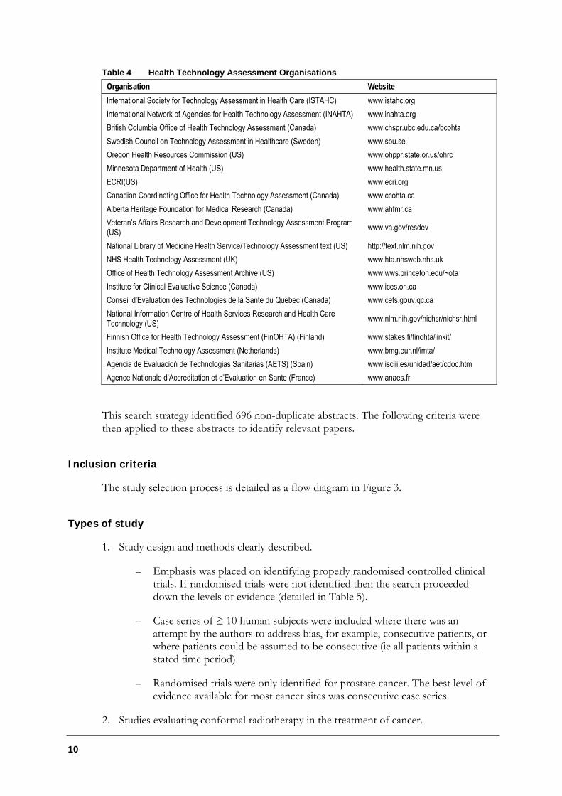
Table 4 Health Technology Assessment Organisations................................................................ 10
Table 5 Designation of levels of evidence ...................................................................................... 12
Table 6 Outcomes to be addressed in the literature ...................................................................... 12
Table 7 Biochemical control ............................................................................................................. 17
Table 8 Overall survival ..................................................................................................................... 17
Table 9 Acute morbidity (prostate cancer)...................................................................................... 18
Table 10 Late morbidity (prostate cancer) ........................................................................................ 18
Table 11 Effect of dose on local control in 105 patients................................................................ 26
Table 12 Log rank test of overall survival for T1-T4 ........................................................................ 27
Table 13 Acute toxicity for IMRT vs CRT ....................................................................................... 28
Table 14 Late toxicity for IMRT vs CRT.......................................................................................... 28
Table 15 Incidence of acute toxicity in patients with high vs low dose CRT.............................. 29
Table 16 Incidence of late toxicity in patients treated with high vs low dose CRT.................... 30
Table 17 Efficacy outcomes for patients with uterine cancer ........................................................ 42
Table 18 Treatment related morbidity for patients with uterine cancer ....................................... 43
Table 19 Mean cost per treatment field with and without MLC ................................................... 46
Table 20 Linear Accelerator - Advanced Features........................................................................... 47
Table 21 Linear Accelerator – Costs of Advanced Features .......................................................... 47
Table 22 Linear Accelerator – National Annual Equivalent Costs of MLC, EPI....................... 47
Table 23 Randomised trials of conformal radiotherapy for prostate cancer ............................... 59
Table 24 Dose response studies for prostate cancer ....................................................................... 60
Table 25 The role CRT in patients with brain carcinoma............................................................... 62
Table 26 The role of conformal radiotherapy in patients with liver carcinoma .......................... 65
Table 27 The role of conformal radiotherapy in patients with sarcoma ...................................... 70
Table 28 The role of CRT in patients with carcinoma of the uterine cervix ............................... 72
Table 29 The role of conformal radiotherapy in patients with breast cancer .............................. 73
Table 30 The role of conformal radiotherapy in patients with head and neck carcinoma ........ 74
Table 31 The role of conformal radiotherapy in patients with NSCLC....................................... 82
Table 32 Studies included in the economic analysis of CRT.......................................................... 89
Table 33 Acute radiation toxicity grading using modified RTOG criteria ................................... 92
Conformal Radiotherapy v
Table 34 Delayed radiation toxicity grading using RTOG and LENT criteria............................ 92
Table 35 Toxicity for other cancers ................................................................................................... 93
Figures
Figure 1 Defining the target volumes.................................................................................................. 3
Figure 3 Flow diagram of study selection process .......................................................................... 11
Figure 4 Taken from MD Anderson Study: Kaplan-Meier FFF curves for all patients by dose randomisation (70 Gy vs 78 Gy).. ....................................................................... 16
Figure 5 Bladder toxicity ..................................................................................................................... 20
Figure 6 Gastrointestinal toxicity....................................................................................................... 21
vi
Executive summary
The procedure
Conformal radiotherapy (also known as three-dimensional conformal radiotherapy or 3DCRT) is a method of delivering radiotherapy that uses computer planning and treatment systems to tailor the size and shape of the dose area to the ideal target volume, with maximal exclusion of the surrounding normal tissue.
Medical Services Advisory Committee – role and approach
The Medical Services Advisory Committee (MSAC) is a key element of a measure taken by the Commonwealth Government to strengthen the role of evidence in health financing decisions in Australia. MSAC advises the Commonwealth Minister for Health and Ageing on the evidence relating to the safety, effectiveness and cost-effectiveness of new and existing medical technologies and procedures, and under what circumstances public funding should be supported.
A rigorous assessment of the available evidence is thus the basis of decision making when funding is sought under Medicare. A team from the NHMRC Clinical Trials Centre, University of Sydney was engaged to conduct a systematic review of literature and an economic analysis on conformal radiotherapy. A supporting committee with expertise in this area then evaluated the evidence and provided advice to MSAC.
MSAC’s assessment of Conformal Radiotherapy
Conformal radiotherapy (CRT) is a method of delivering radiotherapy that has two main aims:
1. To improve dose distribution by tailoring a high-dose radiation volume to an accurately defined target volume; and
2. To reduce the volume of the surrounding normal tissues receiving radiation.
In turn, it is expected that this will decrease the incidence of late effects and allow for escalation of the radiation dose to the tumour.
In recent years there have been significant improvements in the field of radiotherapy. Advances in computer hardware and software, and medical imaging have led to the development of new technology for improving external beam treatment planning, dose delivery and verification of radiotherapy. Three-dimensional treatment planning systems (3D RTP), multileaf collimators and on-line electronic portal imaging are examples of this technology. Within this review of conformal radiotherapy these technological developments will be discussed.
The evidence for the efficacy and safety of conformal radiotherapy is based on three completed randomised studies that compare conformal with standard or conventional radiotherapy for prostate cancer (level II evidence), several prospective non-randomised
Conformal Radiotherapy vii
studies, and a number of uncontrolled case series reports. The issues of quality assurance and occupational health and safety in relation to conformal radiotherapy are also discussed in this review.
Clinical need
Radiotherapy is one of the main treatment modalities for cancer. In 1999/2000 there were over 585,000 instances of patients claiming radiotherapy or therapeutic nuclear medicine under Medicare as definitive therapy for cancer (Commonwealth Department of Health and Aged Care 2000). This figure also includes re-treatments and second courses so it is possible that this total may be an overestimate. A perhaps more helpful figure is reported in a recent document by the Faculty of Radiation Oncology which states that currently around 40% of patients diagnosed with cancer in Australia received radiation therapy, with a proposed national benchmark of 50-55% of patients per year receiving radiation therapy (Faculty of Radiation Oncology, Australian Institute of Radiography, & Australasian College of Physical Scientists and Engineers in Medicine 2001).
Like surgery, radiotherapy is a loco-regional treatment modality. Failure to achieve loco-regional control of a cancer can increase the risk of distant metastases and decrease survival. It is postulated that improvements in radiotherapy techniques and delivery can result in improvements in loco-regional control of disease.
Safety
Irradiation is a well-established method of treating cancer. Its side effects (both acute and long-term) are well known. Generally, tolerance of normal tissues is the limiting factor for the dose of radiation that can be delivered to a tumour. The presence and severity of acute and long-term side effects are related to the ability to spare normal tissues from exposure to radiation, the total dose of radiation administered, and the dose schedule; for example, higher or lower total dose delivered using more or less fractions. The aim of conformal radiotherapy is to limit exposure of normal tissues to radiation and increase the dose to the tumour.
A review of the literature indicates that in the treatment of prostate cancer, delivery of similar total doses of radiotherapy using a conformal approach results in reduced toxicity to that experienced using conventional radiotherapy, with the greatest benefit appearing to be in terms of both acute and late gastrointestinal toxicity.
There is also randomised evidence to suggest that delivery of higher total doses of radiotherapy in the treatment of prostate cancer using a conformal approach results in similar toxicity to that experienced using conventional radiotherapy.
Limited indicative data from comparative non-randomised studies also suggests that the incidence of toxicity for some indications may be lower using conformal radiotherapy than for standard radiotherapy. However, the data for these other indications is relatively small and of poor quality.
viii
Effectiveness
The body of evidence on which the efficacy outcomes is based is relatively small. From the three randomised trials included, only two trials had any information on efficacy.
Based on this limited data it would appear that, in the treatment of prostate cancer, conformal radiotherapy results in similar efficacy to that experienced using conventional radiotherapy when delivering similar doses.
There is also some randomised and non-randomised evidence to suggest that higher doses of radiotherapy, delivered by conformal radiotherapy, may result in increased efficacy for patients with carcinoma of the prostate.
Cost effectiveness
In terms of the economic analysis, components of conformal radiotherapy (CRT) were evaluated. The most information provided dealt with the costs of multileaf collimators (MLC) in comparison to shielding blocks, with seven papers purporting to measure the costs and/or benefits of MLC.
The main cost implications for MLC are:
• Its ability to decrease the average duration of radiation treatment and hence increase the productivity of the linear accelerator (by increasing patient throughput); and
• The reduction, if not elimination, of the need to manufacture blocks. Cost savings arising from reduced mould room labour and supplies.
There is some data indicating that, based on the additional costs of MLC alone, CRT appears to be both more effective and less costly than standard radiotherapy (RT) in some patients groups. However, this data is not comprehensive enough to draw definitive conclusions regarding the cost-effectiveness of conformal radiotherapy.
Quality Assurance and Occupational Health and Safety
A primarily narrative review of quality assurance and occupational health and safety issues was conducted in relation to equipment and technology used in the delivery of conformal radiotherapy. In recent years there has been an increase in the sophistication and complexity of radiotherapy treatment and significant advances in computer hardware, software and medical imaging devices for improving external beam treatment planning, dose delivery and verification of radiotherapy.
The use of these devices, specifically the application of multileaf collimators and electronic portal imaging in radiotherapy treatment, also have occupational health and safety implications for patients and radiotherapy staff.
It would appear from the literature available that there are some occupational health and safety benefits in using multileaf collimators in comparison to shielding blocks when treating patients with conformal radiotherapy.

Conformal Radiotherapy ix
Recommendation
MSAC recommended that on the strength of evidence pertaining to the safety, efficacy and cost of conformal radiotherapy that public funding should be supported for this procedure and that intensity modulated radiation therapy should be reviewed again at a later date when substantial additional data are available relating to safety, effectiveness and cost effectiveness.
The Minister for Health and Ageing accepted this recommendation on 5 February 2002.

x Conformal Radiotherapy
Conformal Radiotherapy 1
Introduction
The Medical Services Advisory Committee (MSAC) has reviewed the use of conformal radiotherapy, which is a therapeutic intervention for cancer. MSAC evaluates new and existing health technologies and procedures for which funding is sought under the Medicare Benefits Scheme in terms of their safety, effectiveness and cost-effectiveness, while taking into account other issues such as access and equity. MSAC adopts an evidence-based approach to its assessments, based on reviews of the scientific literature and other information sources, including clinical expertise.
MSAC’s terms of reference and membership are at Appendix A. MSAC is a multidisciplinary expert body, comprising members drawn from such disciplines as diagnostic imaging, pathology, surgery, internal medicine and general practice, clinical epidemiology, health economics, consumer affairs and health administration.
This report summarises the assessment of current evidence for conformal radiotherapy for cancer.
2 Conformal Radiotherapy
Background
Conformal radiotherapy
The procedure
Conformal radiotherapy (CRT) is a method of delivering radiotherapy that uses three dimensional computer planning and treatment systems to tailor the size and shape of the dose area to conform tightly to the shape of the tumour. As such, conformal radiotherapy is also often referred to as three-dimensional conformal radiotherapy or 3DCRT.
There seems to be general agreement in the literature that conformal radiotherapy has two main aims:
• To improve dose distribution by tailoring a high-dose radiation volume to an accurately defined target volume; and
• To reduce the volume of the surrounding normal tissues receiving radiation.
In turn, it is expected that this will decrease the incidence of late effects and allow for escalation of the radiation dose to the tumour.
The delivery of 3DCRT is a multi-step process (Horwitz & Hanks 2000). While different names exist for these steps there are a number of processes that constitute what we commonly understand as three-dimensional conformal radiotherapy (Kolitsi et al 1997; Purdy 1997; Cardinale & Kavanagh 2000). These steps can be broadly broken down into:
1. Patient data acquisition 2. Three-dimensional treatment planning 3. Three-dimensional dose delivery and optimisation 4. Treatment verification and treatment execution
1. Patient data acquisition
As the goal of conformal radiotherapy is to tightly shape the high dose of radiation to the tumour, accurate and detailed information regarding the patient and the tumour is essential. Conformal radiotherapy begins with the immobilisation of the patient in the treatment position with the use of individualised casts. From here, a three-dimensional image is attained either in a computed tomography (CT) simulation suite or by conventional radiation therapy simulation. Radiopaque markers are placed on the patient’s skin to aid in repositioning and multiple cross-sectional slices of the region of interest are taken, with the number of CT slices dependent upon factors such as location and size of the tumour. While CT is the most commonly used imaging modality for data acquisition, magnetic resonance imaging (MRI) and nuclear imaging such as single-photon emission computed tomography (SPECT) or positron emission tomography (PET) may also be employed.
Conformal Radiotherapy 3
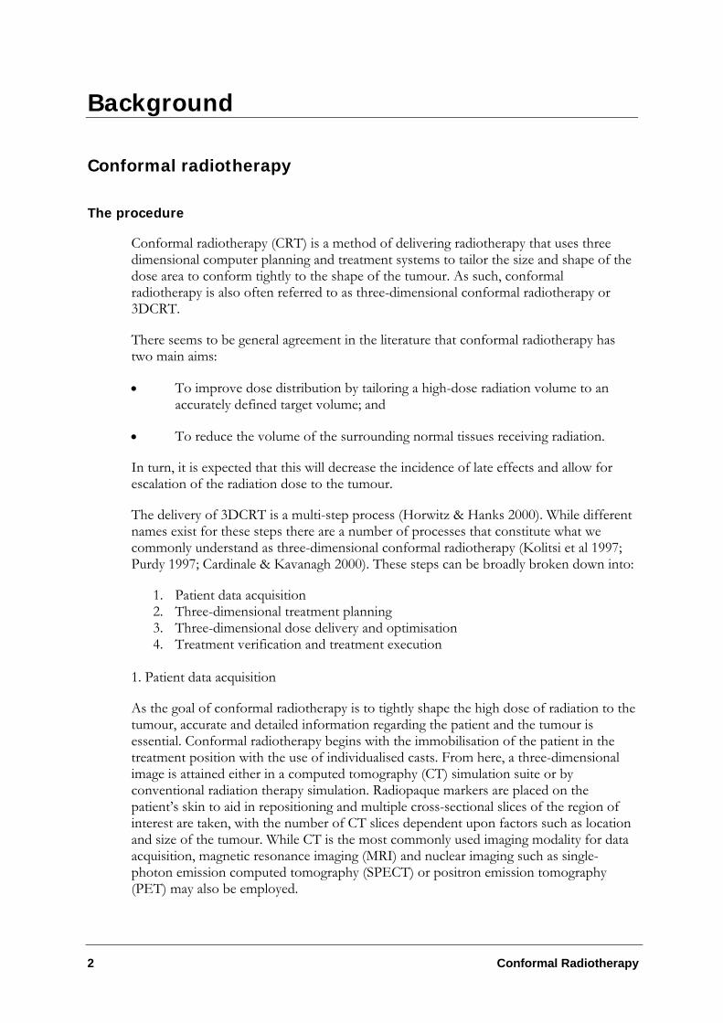
Once the 3D information has been acquired the next step is to delineate the target volume and normal tissue structures (Figure 1). This is done by defining the gross tumour volume (GTV), which is dependent upon the anatomy of the patient, and the clinical target volume (CTV), which incorporates the GTV and/or subclinical disease (Emami et al 1997). These two volumes are used in the subsequent process to define the planning target volume (PTV), which is defined by specifying the margins that must be added around the CTV to compensate for the effects of the organ, tumour and patient movements, as well as inaccuracies in beam and patient set-up (Purdy 1997).
Figure 1 Defining the target volumes Source: (Image Guided Therapy QA Center at Washington University 2001)
2. Three-dimensional treatment planning
The data acquired in step 1 are used to generate a three-dimensional representation of each structure using appropriate computer software. The geometry of the radiation fields are then defined to optimise the dose distribution using a beam’s eye view (BEV) display (Purdy 1997). The BEV is part of the three-dimensional planning system and is a tool that allows the practitioner to view the dose distribution within the body in three dimensions and directly visualise the organs and structures the beam will traverse. The use of non-coplanar beams in conformal radiotherapy also assists with this process. Non-coplanar beams can enter and exit the patient from any arbitrary angle unlike conventional radiotherapy, which uses coplanar beams. Using the BEV and non-coplanar beams it is possible to view, manipulate and calculate the angle of irradiation and number of fields to conform precisely to the dimension of the target, sparing as much as possible the adjacent normal tissue (Lichter & Ten Haken 1995). This information is then used to determine the shape of the radiation fields. The shaping is achieved using customised blocks or multileaf collimators.
a) Customised blocks
These are created using low melting point alloys such as cerrobend. Once cast, they are positioned and attached to a tray. This tray is then manually lifted and inserted into the treatment machine.
b) Multileaf collimators
The multileaf collimator is an automated device that is built into the head of the radiotherapy treatment machine (linear accelerator). This device is
4 Conformal Radiotherapy
computer controlled and moves a variable number of metal leaves that shape the radiation field.
3. Three-dimensional dose delivery and optimisation
During this stage treatment plans are measured using tools such as dose volume histograms (DVH), which allows the practitioner to evaluate the dose distribution throughout the volume of normal tissues and tumour, and alter the treatment plan if needed (Cardinale & Kavanagh 2000). Once a treatment plan has been checked and approved, the documentation is generated for the beam and shaping devices. These parameters are then transferred to the treatment machine and the treatment is delivered (Purdy 1997).
4. Treatment verification and treatment execution
Immediately following the delivery of treatment, the accuracy and validity of the treatment is confirmed through radiographic verification, portal imaging being the most common technique (Boyle & McPadden 2000). A standard portal image is prepared much like a x-ray. The use of electronic portal imaging (EPI) is becoming increasingly common. EPI digitally captures the field size, shape and position on a computer that can then be viewed and compared to the original image. This ability to verify on-line patient position, field alignment, block shaping and movement throughout the entire radiotherapy treatment is advantageous in terms of quality assurance (Lavertu, Girouard, & Pouliot 2000). The EPI process is also significantly less time consuming than using the standard portal imaging method.
The above discussion has primarily centred on three-dimensional conformal radiotherapy. However, it should be noted that there exists a more advanced form of 3DCRT called intensity modulated radiation therapy (IMRT). In IMRT the intensity of the radiotherapy beam can be varied during the treatment, usually by computer-controlled movement of the MLC leaves. IMRT can be dynamic (where the machine, in particular the MLC leaves, and/or couch move while the radiation beam is on), or static (where the radiation beam is turned off while machine and couch movements take place). The main advantage of IMRT over conventional 3DCRT is that it allows even greater conformity of dose to the target volume. Depending on the treatment priorities, the target dose can be even more homogeneous, and/or the dose to critical structures can be reduced (Tubiana & Eschwege 2000).
Intended purpose
Conformal radiotherapy is used in the treatment of a wide variety of cancers. It is considered particularly suited to malignant tumours in sites of complex anatomy, irregularly shaped tumours, tumours adjacent to radiation-sensitive structures such as the spinal cord, bowel and intra-abdominal organs, and malignancies that have a documented high local failure with current radiotherapy doses (Vijayakumar & Chen 1995). Published randomised controlled trial evidence on the efficacy and safety of conformal radiotherapy primarily relates to prostate cancer. Other cancer sites such as lung, head and neck, brain and the hepatobiliary tract may also be treated with CRT. However, there is limited controlled evidence in these indications.
Conformal Radiotherapy 5
This review will focus primarily on the use of conformal radiotherapy in patients with prostate cancer, but will also provide information on the evidence available for conformal radiotherapy in other cancer indications.
Clinical need/burden of disease
Cancer contributes considerably to morbidity and mortality in the Australian population. Although cancer ranks eighth in direct health system costs, it is the most common cause of premature death and the second most common cause of death overall in Australia, and is recognised by the government as a National Health Priority Area (AIHW 1997).
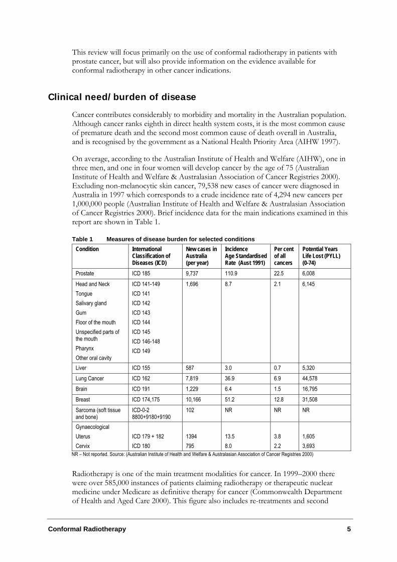
On average, according to the Australian Institute of Health and Welfare (AIHW), one in three men, and one in four women will develop cancer by the age of 75 (Australian Institute of Health and Welfare & Australasian Association of Cancer Registries 2000). Excluding non-melanocytic skin cancer, 79,538 new cases of cancer were diagnosed in Australia in 1997 which corresponds to a crude incidence rate of 4,294 new cancers per 1,000,000 people (Australian Institute of Health and Welfare & Australasian Association of Cancer Registries 2000). Brief incidence data for the main indications examined in this report are shown in Table 1.
Table 1 Measures of disease burden for selected conditions Condition International
Classification of Diseases (ICD)
New cases in Australia (per year)
Incidence Age Standardised Rate (Aust 1991)
Per cent of all cancers
Potential Years Life Lost (PYLL) (0-74)
Prostate ICD 185 9,737 110.9 22.5 6,008
Head and Neck Tongue Salivary gland Gum Floor of the mouth Unspecified parts of the mouth Pharynx Other oral cavity
ICD 141-149 ICD 141 ICD 142 ICD 143 ICD 144 ICD 145 ICD 146-148 ICD 149
1,696 8.7 2.1 6,145
Liver ICD 155 587 3.0 0.7 5,320
Lung Cancer ICD 162 7,819 36.9 6.9 44,578
Brain ICD 191 1,229 6.4 1.5 16,795
Breast ICD 174,175 10,166 51.2 12.8 31,508
Sarcoma (soft tissue and bone)
ICD-0-2 8800+9180+9190
102 NR NR NR
Gynaecological Uterus Cervix
ICD 179 + 182 ICD 180
1394 795
13.5 8.0
3.8 2.2
1,605 3,693
NR – Not reported. Source: (Australian Institute of Health and Welfare & Australasian Association of Cancer Registries 2000)
Radiotherapy is one of the main treatment modalities for cancer. In 1999–2000 there were over 585,000 instances of patients claiming radiotherapy or therapeutic nuclear medicine under Medicare as definitive therapy for cancer (Commonwealth Department of Health and Aged Care 2000). This figure also includes re-treatments and second
6 Conformal Radiotherapy
courses so it is possible that this total may be an overestimate. A perhaps more helpful figure is reported in a recent document by the Faculty of Radiation Oncology which states that currently around 40% of patients diagnosed with cancer in Australia received radiation therapy, with a proposed national benchmark of 50–55% of patients per year receiving radiation therapy (Faculty of Radiation Oncology, Australian Institute of Radiography, & Australasian College of Physical Scientists and Engineers in Medicine 2001).
Like surgery, radiotherapy is a loco-regional treatment modality. Failure to achieve loco-regional control of a cancer can increase the risk of distant metastases and decreases survival. It is postulated that improvements in radiotherapy techniques and delivery can result in improvements in loco-regional control of disease (Vijayakumar & Chen 1995).
Conformal radiotherapy is based on three premises: 1) that a higher rate of local control can improve the survival rate; 2) that dose escalation can increase tumour control; and 3) that higher doses can be delivered due to the decreased occurrence of radiation complications, as a result of the conforming of the dose and sparing of normal tissue from radiation (Tubiana & Eschwege 2000).
CRT use in Australia
In Australia, radiation oncology services are provided through both public and private sectors with all states and territories, except the Northern Territory, having radiation oncology treatment facilities (Faculty of Radiation Oncology, Australian Institute of Radiography, & Australasian College of Physical Scientists and Engineers in Medicine 2001). Radiotherapy can be delivered as either external beam radiotherapy or brachytherapy. The vast majority of patients in Australia receiving radiotherapy are treated with external beam therapy using a linear accelerator. Conformal radiotherapy is considered a form of external beam therapy and one that has been increasingly used as a treatment of choice in Australia.
As stated in the 2001 National Strategic Plan for Radiation Oncology (Australia), there were 99 linear accelerators in radiation oncology centres throughout Australia (Faculty of Radiation Oncology, Australian Institute of Radiography, & Australasian College of Physical Scientists and Engineers in Medicine 2001). Table 2 shows the number in each state and territory.
Table 2 Number of linear accelerators per state and territory in Australia State Number of linear accelerators New South Wales (including Australian Capital Territory) 37 Victoria 24 Queensland 17 South Australia 9 Western Australia 8 Tasmania 4
Source: (Faculty of Radiation Oncology, Australian Institute of Radiography, & Australasian College of Physical Scientists and Engineers in Medicine 2001)
Over the past two decades linear accelerators have become more sophisticated, with EPI devices (EPIDs) and MLCs becoming an integral component of the liner accelerator
Conformal Radiotherapy 7
hardware. There are 25 machines using EPI and 32 linear accelerators with MLCs in Australia. These advances in technology and growth in equipment have helped lead to the increased use of conformal radiotherapy in this country. However, as technology has changed, so has the practice of conformal radiotherapy. For instance, fields that may have been originally shaped with cerrobend blocks are now shaped using MLC. It would also appear that conformal radiotherapy is now commonly regarded by many practitioners as ‘standard’ treatment and IMRT as the new practice (Zelefsky et al 2000). Both these issues have led to dilemmas when evaluating the evidence.
Existing procedures / Comparator
In this review, conformal radiotherapy is compared to standard or conventional radiotherapy (RT). For the majority of cases, standard or conventional radiotherapy is considered to be two-dimensional radiotherapy. Two-dimensional (2D) radiation therapy came about through the use of CT scans in treatment planning. Treatment planning and dose calculations are performed from a single two-dimensional slice (contour) through a given treatment volume. Practitioners use bony landmarks on plain simulation radiographs to identify the tumour and important normal structures to draw blocks and align treatment beams (Cardinale & Kavanagh 2000).
Marketing status of the technology
The multileaf collimator device used for the delivery of CRT is listed with the Australian Therapeutic Goods Administration, with the listing number of:
Elekta: L31967 Varian: L14534 Siemens: L37972
Current reimbursement arrangement
Conformal radiotherapy using multileaf collimators, electronic portal imaging and integrated systems networking is not currently covered under the Medicare Benefits Scheme (MBS) or the radiotherapy Capital Equipment List. There are MBS items that relate to radiotherapy and to the placement of lead alloy blocks for shielding specific tissues from cross radiation and shaping the volume of the tissue to be irradiated. However, the capital cost of the multileaf collimator, electronic portal imaging equipment and integrated systems networking are not included.
8 Conformal Radiotherapy
Approach to assessment
Review of literature
The medical literature was searched to identify relevant studies and reviews. Searches were conducted in the following databases from their commencement until the end of March 2001.
• Medline/Pre-Medline
• EBM Reviews – Best Evidence
• EMBASE
• The Cochrane Library
• Current Contents
• ISTAHC Online database (International Society for Technology Assessment in Health Care)
• NHS Centre for Reviews and Dissemination databases
− DARE (Database of Abstracts and Reviews of Effectiveness) − EED (Economic Evaluation Database) − HTA (Health Technology Assessment Database)
• Oshrom: Occupational Health and Safety
- NIOSHTIC (National Institute for Occupational Safety and Health)
- CISDOC (International Occupational Safety and Health Information Centre)
- HSELINE (Health and Safety Executive Library Information)
- MHIDAS (Major Hazard Incident Data Service)
Search strategy
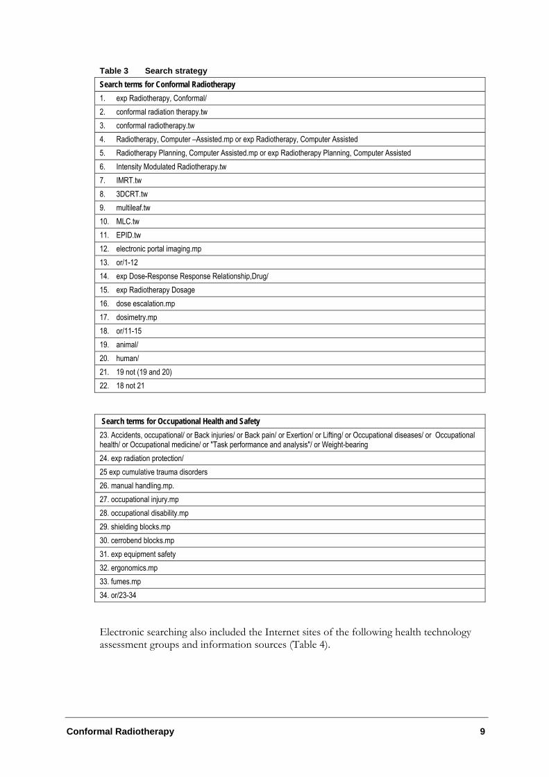
The search strategy shown in Table 3 was used to identify papers on conformal radiotherapy in Medline, CINAHL and Best Evidence. The same search strategy was used for EMBASE, replacing MeSH terms with EMTREE terms.
Conformal Radiotherapy 9
Table 3 Search strategy Search terms for Conformal Radiotherapy 1. exp Radiotherapy, Conformal/ 2. conformal radiation therapy.tw 3. conformal radiotherapy.tw 4. Radiotherapy, Computer –Assisted.mp or exp Radiotherapy, Computer Assisted 5. Radiotherapy Planning, Computer Assisted.mp or exp Radiotherapy Planning, Computer Assisted 6. Intensity Modulated Radiotherapy.tw 7. IMRT.tw 8. 3DCRT.tw 9. multileaf.tw 10. MLC.tw 11. EPID.tw 12. electronic portal imaging.mp 13. or/1-12 14. exp Dose-Response Response Relationship,Drug/ 15. exp Radiotherapy Dosage 16. dose escalation.mp 17. dosimetry.mp 18. or/11-15 19. animal/ 20. human/ 21. 19 not (19 and 20) 22. 18 not 21
Search terms for Occupational Health and Safety 23. Accidents, occupational/ or Back injuries/ or Back pain/ or Exertion/ or Lifting/ or Occupational diseases/ or Occupational health/ or Occupational medicine/ or "Task performance and analysis"/ or Weight-bearing 24. exp radiation protection/ 25 exp cumulative trauma disorders 26. manual handling.mp. 27. occupational injury.mp 28. occupational disability.mp 29. shielding blocks.mp 30. cerrobend blocks.mp 31. exp equipment safety 32. ergonomics.mp 33. fumes.mp 34. or/23-34
Electronic searching also included the Internet sites of the following health technology assessment groups and information sources (Table 4).
10
Table 4 Health Technology Assessment Organisations Organisation Website International Society for Technology Assessment in Health Care (ISTAHC) www.istahc.org International Network of Agencies for Health Technology Assessment (INAHTA) www.inahta.org British Columbia Office of Health Technology Assessment (Canada) www.chspr.ubc.edu.ca/bcohta Swedish Council on Technology Assessment in Healthcare (Sweden) www.sbu.se Oregon Health Resources Commission (US) www.ohppr.state.or.us/ohrc Minnesota Department of Health (US) www.health.state.mn.us ECRI(US) www.ecri.org Canadian Coordinating Office for Health Technology Assessment (Canada) www.ccohta.ca Alberta Heritage Foundation for Medical Research (Canada) www.ahfmr.ca Veteran’s Affairs Research and Development Technology Assessment Program (US) www.va.gov/resdev
National Library of Medicine Health Service/Technology Assessment text (US) http://text.nlm.nih.gov NHS Health Technology Assessment (UK) www.hta.nhsweb.nhs.uk Office of Health Technology Assessment Archive (US) www.wws.princeton.edu/~ota Institute for Clinical Evaluative Science (Canada) www.ices.on.ca Conseil d’Evaluation des Technologies de la Sante du Quebec (Canada) www.cets.gouv.qc.ca National Information Centre of Health Services Research and Health Care Technology (US) www.nlm.nih.gov/nichsr/nichsr.html
Finnish Office for Health Technology Assessment (FinOHTA) (Finland) www.stakes.fi/finohta/linkit/ Institute Medical Technology Assessment (Netherlands) www.bmg.eur.nl/imta/ Agencia de Evaluacioń de Technologias Sanitarias (AETS) (Spain) www.isciii.es/unidad/aet/cdoc.htm Agence Nationale d’Accreditation et d’Evaluation en Sante (France) www.anaes.fr
This search strategy identified 696 non-duplicate abstracts. The following criteria were then applied to these abstracts to identify relevant papers.
Inclusion criteria
The study selection process is detailed as a flow diagram in Figure 3.
Types of study
1. Study design and methods clearly described.
– Emphasis was placed on identifying properly randomised controlled clinical trials. If randomised trials were not identified then the search proceeded down the levels of evidence (detailed in Table 5).
– Case series of ≥ 10 human subjects were included where there was an attempt by the authors to address bias, for example, consecutive patients, or where patients could be assumed to be consecutive (ie all patients within a stated time period).
– Randomised trials were only identified for prostate cancer. The best level of evidence available for most cancer sites was consecutive case series.
2. Studies evaluating conformal radiotherapy in the treatment of cancer.
Conformal Radiotherapy 11
3. English language articles reporting primary data and published in a peer reviewed journal (not abstracts).
4. Studies not duplicated or superseded by a subsequent study with the same purpose from the same institution.
5. The study must report information on at least one of the outcomes of interest.
6. Excluded were:
– Dose calculation, planning and immobilisation studies.
– Studies where the intervention was confounded by the presence of another treatment (eg chemotherapy).
– Studies in brachytherapy.
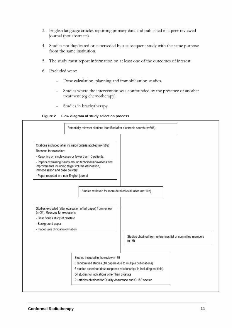
Figure 2 Flow diagram of study selection process
Potentially relevant citations identified after electronic search (n=696)
Citations excluded after inclusion criteria applied (n= 589) Reasons for exclusion: - Reporting on single cases or fewer than 10 patients; - Papers examining issues around technical innovations and improvements including target volume delineation, immobilisation and dose delivery. - Paper reported in a non-English journal
Studies retrieved for more detailed evaluation (n= 107)
Studies excluded (after evaluation of full paper) from review (n=34). Reasons for exclusions - Case series study of prostate - Background paper - Inadequate clinical information
Studies included in the review n=79 3 randomised studies (10 papers due to multiple publications) 6 studies examined dose response relationship (14 including multiple) 34 studies for indications other than prostate 21 articles obtained for Quality Assurance and OH&S section
Studies obtained from references list or committee members (n= 6)
12
Seventy-nine papers thus form the basis of this review, including three randomised studies evaluating patients with prostate cancer and thirty-four papers examining other cancer indications.
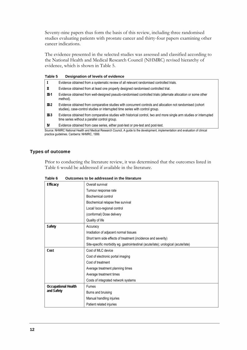
The evidence presented in the selected studies was assessed and classified according to the National Health and Medical Research Council (NHMRC) revised hierarchy of evidence, which is shown in Table 5.
Table 5 Designation of levels of evidence I Evidence obtained from a systematic review of all relevant randomised controlled trials. II Evidence obtained from at least one properly designed randomised controlled trial. III-1 Evidence obtained from well-designed pseudo-randomised controlled trials (alternate allocation or some other
method). III-2 Evidence obtained from comparative studies with concurrent controls and allocation not randomised (cohort
studies), case-control studies or interrupted time series with control group. III-3 Evidence obtained from comparative studies with historical control, two and more single arm studies or interrupted
time series without a parallel control group. IV Evidence obtained from case series, either post-test or pre-test and post-test.
Source: NHMRC National Health and Medical Research Council, A guide to the development, implementation and evaluation of clinical practice guidelines. Canberra: NHMRC, 1999.
Types of outcome
Prior to conducting the literature review, it was determined that the outcomes listed in Table 6 would be addressed if available in the literature.
Table 6 Outcomes to be addressed in the literature Efficacy Overall survival
Tumour response rate Biochemical control Biochemical relapse free survival Local/ loco-regional control (conformal) Dose delivery Quality of life
Safety Accuracy Irradiation of adjacent normal tissues Short term side effects of treatment (incidence and severity) Site-specific morbidity eg. gastrointestinal (acute/late); urological (acute/late)
Cost Cost of MLC device Cost of electronic portal imaging Cost of treatment Average treatment planning times Average treatment times Costs of integrated network systems
Occupational Health and Safety
Fumes Burns and bruising Manual handling injuries Patient related injuries
Conformal Radiotherapy 13
Existing reviews
The Alberta Heritage Foundation for Medical Research listed a Techscan report on Intensity Modulated Radiation Therapy. This is not a health technology assessment (HTA) but a brief horizon scanning document on the purpose and potential implications of the emerging technology.
Expert advice
A supporting committee with expertise in radiation oncology was established to evaluate the evidence and provide advice to MSAC from a clinical perspective. In selecting members for supporting committees, MSAC’s practice is to approach the appropriate medical colleges, specialist societies and associations and consumer bodies for nominees. Membership of the supporting committee is provided at Appendix B.
14
Results of assessment
Is it safe?
Irradiation is a well-established method of treating cancer. Its side effects (both acute and long-term) have been established and are well known. A number of systems exist that can be used to score toxicity including the recently updated Common Toxicity Criteria (CTC) Radiation Therapy Oncology Group (RTOG) criteria for acute toxicity, and the Late Effects Normal Tissues (LENT) for late effects (Appendix D).
Tolerance of normal tissues is generally the limiting factor for the dose of radiation that can be delivered to a tumour (Overgaard & Bartelink, 1995). The presence and severity of acute and long-term side effects is related to the ability to spare normal tissues from exposure to radiation, the total dose of radiation administered, and the dose schedule; for example, higher or lower total dose delivered using more or less fractions. The aim of conformal radiotherapy is to limit exposure of normal tissues to radiation and increase the dose to the tumour. The hope is that this will improve local control and reduce the severity of side effects. Alternatively, as the total volume of irradiated tissue is reduced with conformal radiotherapy, there is concern that this could have an adverse effect on local tumour control (Dearnaley et al 1999).
Is it effective?
The ability to evaluate effectiveness was influenced by the availability of good quality evidence. The sections of this report relating to effectiveness have therefore been separated into:
• A comparison of standard radiation therapy with conformal radiation therapy for prostate cancer; and
• A description of the evidence available for other cancer indications.
Conformal radiotherapy for prostate cancer
A total of three completed randomised studies have been identified that compare conformal with conventional radiotherapy for prostate cancer. The features of these trials are summarised below and in Table 23 (Appendix C).
Allocation concealment in the randomisation process is regarded as particularly important in protecting against bias and will be graded using the Cochrane approach as follows:
Grade A - Clearly adequate concealment Grade B - Possibly adequate Grade C - Clearly inadequate concealment
Conformal Radiotherapy 15
About the trials
1. Royal Marsden Hospital (RMH) (Carrie & Ginestet 1997; Dearnaley et al 1999; Huddart et al 1996; Tait et al 1993; Tait et al 1997)
This trial began in 1988 as the Royal Marsden Hospital Pelvic Radiotherapy Trial with all patients (both men and women) undergoing CT planning in preparation for pelvic radiotherapy being eligible. The quality of the randomisation process was clearly adequate and hence graded as A. Of the 274 patients randomised to the original trial, 144 had prostate cancer and this was extended to recruit a total of 225 patients with T1-4 (TNM classification, UICC, 1997) prostate cancer (111 to conventional and 114 to conformal) by the time the trial was closed to accrual in 1995. Patients were randomised by an independent randomisation service using a randomised permuted block design. All patients received a standard dose of 60–64 Gy in daily 2 Gy fractions. Treatment was delivered to patients randomised to conventional radiotherapy (111) with a three-field technique, and to patients in the conformal radiotherapy arm (114) with customised cerrobend blocks. Information on acute and late side effects, biochemical control (Prostate Specific Antigen (PSA) failure), local recurrence and overall survival have been reported. The minimum follow-up period at the time of the most recent publication was two years (median 3.6 years).
2. MD Anderson (MDA) (Nguyen, Pollack, & Zagars 1998; Pollack et al 1996; Pollack et al 2000; Storey et al 2000)
This single-centre randomised trial opened to accrual in 1993 and recruited 305 patients with T2-4 prostate cancer, of which 301 were considered assessable (150 in the conventional arm and 151 in the conformal radiotherapy arm). As the method of randomisation has not been reported, the quality of the randomisation process could only be determined as being possibly adequate and hence graded as B. All patients received an initial dose of 46 Gy using a conventional four-field box. Those randomised to the conventional therapy arm received a conventional four-field boost of 2 Gy per fraction to a total dose of 70 Gy to the isocentre. Patients randomised to the conformal arm received a six field 3DCRT boost to a total dose of 78 Gy to the isocentre. Conformal radiotherapy fields were shaped using blocks. The main endpoint for this trial was freedom from biochemical and/or disease failure (FFF) defined as ‘time from completion of treatment to an increasing PSA level and/or clinical-radiographic relapse’ (Pollack et al 2000). Information on disease failure (clinical-radiographic relapse), biochemical control (PSA failure), freedom from distant metastases, overall survival, and early and late side effects have also been reported. Median follow-up time was 40 months.
3. Daniel Den Hoed Cancer Centre (DDH) (Brada & Baumert 1999)
This trial commenced in June 1994 and recruited 266 patients with T1-4 prostate cancer. As the method of randomisation has not been reported the quality of the randomisation process could only be determined as being possibly adequate and hence graded as B. All patients were planned in the same way and received a dose of 66 Gy. Patients receiving conventional treatment were treated with rectangular, open fields and patients in the conformal radiotherapy arm were treated with conformally shaped fields using a multileaf collimator. The main aim of this trial was to evaluate acute toxicity. Hence, only this information is reported with some related details on dose received.
16
Results
Local control
The RMH trial was the only study to report on local control, defined as ‘clinically judged maintenance of local control’. After a median follow-up of 3.6 years there was no statistically significant difference in the actuarial rates of local control two years (96% in the conventional group vs 97% in the conformal group) or five years after treatment (83% vs 78%, p=0.40).
Freedom from biochemical and/or disease failure (FFF)
The primary outcome of the MDA trial was survival analysis of freedom from biochemical and/or disease failure. This was defined as ‘time from completion of treatment to an increasing PSA level and/or clinical-radiographic relapse’ (Pollack et al 2000). Biochemical failure was defined as three or more increases in the PSA as per the American Society of Therapeutic Radiation Oncology (ASTRO) consensus guidelines. Information has only been reported on the combined outcome and it is not possible to separate biochemical failure from disease failure. Clinical-radiographic relapse was not defined.
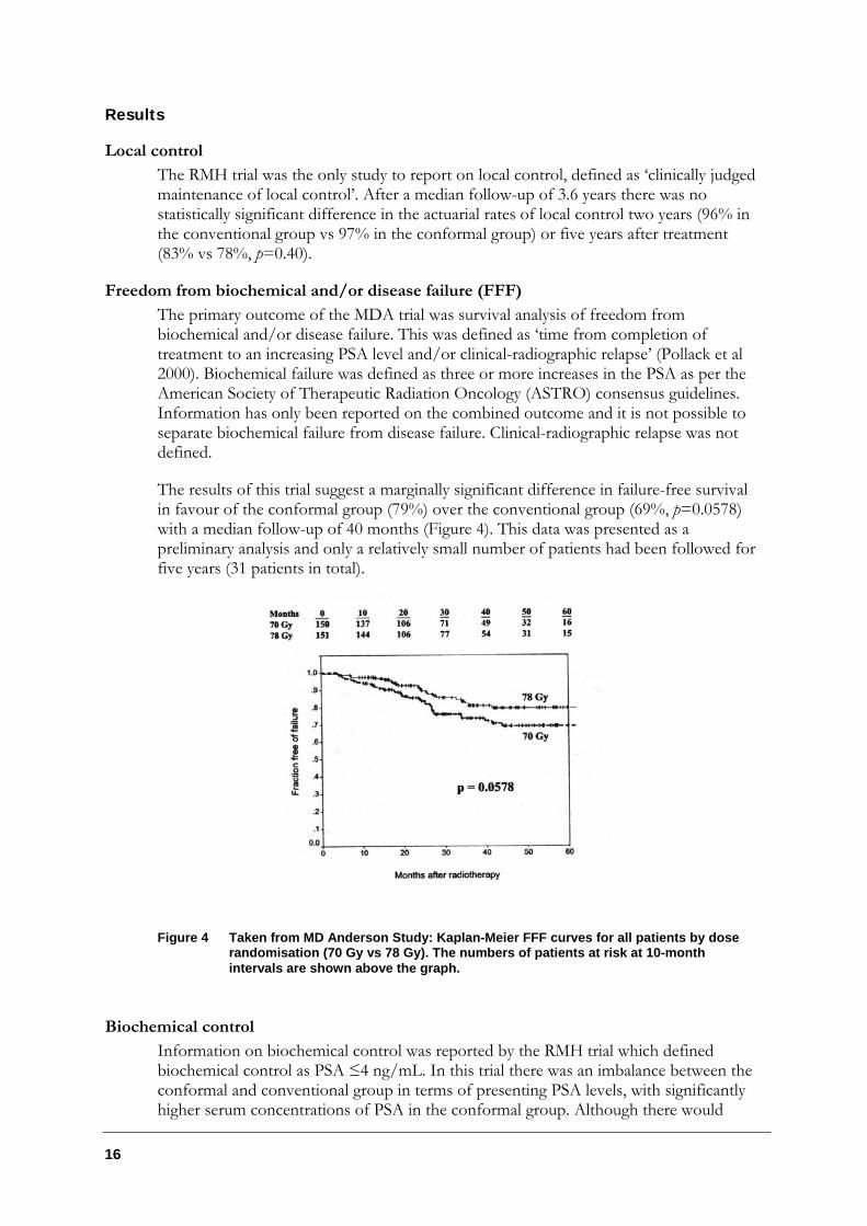
The results of this trial suggest a marginally significant difference in failure-free survival in favour of the conformal group (79%) over the conventional group (69%, p=0.0578) with a median follow-up of 40 months (Figure 4). This data was presented as a preliminary analysis and only a relatively small number of patients had been followed for five years (31 patients in total).
Biochemical control
Information on biochemical control was reported by the RMH trial which defined biochemical control as PSA ≤4 ng/mL. In this trial there was an imbalance between the conformal and conventional group in terms of presenting PSA levels, with significantly higher serum concentrations of PSA in the conformal group. Although there would
Figure 4 Taken from MD Anderson Study: Kaplan-Meier FFF curves for all patients by dose randomisation (70 Gy vs 78 Gy). The numbers of patients at risk at 10-month intervals are shown above the graph.
Conformal Radiotherapy 17
appear to be a significant difference in the PSA failure rates in favour of the conformal group, this difference was no longer significant (p = 0.28) when patients were stratified for their PSA concentration at presentation (Table 7).
Table 7 Biochemical control Study Patients with biochemical control at 2 years Patients with biochemical control at 5 years Conventional (95%CI) Conformal (95%CI) Conventional (95%CI) Conformal (95%CI) RMH 54% (44–63) 71% (62–79) 31% (21–42) 39% (27–50)
Note: p = 0.02
Overall survival
Overall survival was not the main outcome in any of the trials identified and survival curves have not been published. Both the RMH and MDA trials reported in the text of their reports that overall survival at five years was similar in the two treatment groups (Table 8). The MDA study also reported no difference in five-year survival when the pre-treatment PSA level was taken into account (Pollack et al 2000).
Table 8 Overall Survival
Study Overall survival at 2 years Overall survival at 5 years Conventional Conformal Conventional Conformal RMH 90% 91% 64% 66% (see note) MDA Not reported Not reported 90% 91%
Note: p = 0.57
Freedom from distant metastases
Only the MDA trial has reported information on metastases. The overall five-year freedom-from-distant-metastases rates were 95% in the conventional group and 98% in the conformal group (p=0.054).
Side effects / Treatment related morbidity
The most common side effects associated with radiotherapy (both short and long-term) in the treatment of prostate cancer involve the gastrointestinal tract (including rectal pain and bleeding) and bladder (including cystitis and incontinence). Radiation toxicity grades are described in Appendix D.
On the following pages information from the MDA trial has been combined with the RMH and DDH trials, although it is recognised that the side effects experienced on this trial will be different owing to the greater dose received in the conformal arm, and the fact that all patients received on average 46 Gy of conventional radiotherapy. Therefore, plots are presented that both include and exclude information from the MDA trial.
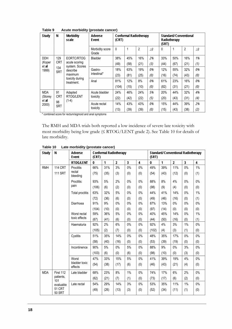
The DDH and MDA trials both reported a low incidence of severe acute toxicity with most morbidity being low grade (≤ RTOG grade 2). Table 9 shows the details of acute morbidity.
18
Table 9 Acute morbidity (prostate cancer) Study N Morbidity
scale Adverse Event
Conformal Radiotherapy (CRT)
Standard/ Conventional Radiotherapy (SRT)
Morbidity score Grade
0 1 2 ≥3 0 1 2 ≥3
Bladder 38% (49)
45% (58)
16% (21)
2% (3)
33% (44)
50% (67)
16% (21)
1% (1)
Gastro-intestinal*
18% (23)
63% (81)
19% (25)
0% (0)
12% (16)
55% (74)
32% (43)
0% (0)
DDH (Koper et al 1999)
129 CRT 134 SRT
EORTC/RTOG acute scoring system. Scores describe maximum toxicity during treatment. Anal 81%
(104) 12% (15)
8% (10)
0% (0)
61% (82)
23% (31)
16% (21)
0% (0)
Acute bladder toxicity
24% (22)
46% (42)
24% (22)
5% (5)
20% (20)
44% (43)
32% (31)
4% (4)
MDA (Storey et al 2000)
91 CRT 98 SRT
Adapted RTOG/LENT (1-4)
Acute rectal toxicity
14% (13)
43% (39)
43% (39)
0% (0)
15% (15)
44% (43)
39% (38)
2% (2)
* combined score for rectum/sigmoid and anal symptoms
The RMH and MDA trials both reported a low incidence of severe late toxicity with most morbidity being low grade (≤ RTOG/LENT grade 2). See Table 10 for details of late morbidity.
Table 10 Late morbidity (prostate cancer) Study N Adverse
Event Conformal Radiotherapy (CRT)
Standard/ Conventional Radiotherapy (SRT)
RTOG/LENT 0 1 2 3 4 0 1 2 3 4 Proctitis rectal bleeding
66% (75)
31% (35)
3% (3)
0% (0)
0% (0)
49% (54)
39% (43)
11% (12)
0% (0)
1% (1)
Proctitis: pain
93% (106)
5% (6)
2% (2)
0% (0)
0% (0)
88% (98)
8% (9)
4% (4)
0% (0)
0% (0)
Total proctitis 63% (72)
32% (36)
5% (6)
0% (0)
0% (0)
44% (49)
41% (46)
14% (16)
0% (0)
1% (1)
Diarrhoea 91% (104)
9% (10)
0% (0)
0% (0)
0% (0)
87% (97)
13% (14)
0% (0)
0% (0)
0% (0)
Worst rectal toxic effects
59% (67)
36% (41)
5% (6)
0% (0)
0% (0)
40% (44)
45% (50)
14% (16)
0% (0)
1% (1)
Haematuria 92% (105)
2% (2)
6% (7)
0% (0)
0% (0)
92% (102)
4% (4)
3% (3)
1% (1)
0% (0)
Cystitis 51% (58)
35% (40)
14% (16)
0% (0)
0% (0)
48% (53)
35% (39)
17% (19)
0% (0)
0% (0)
Incontinence 90% (103)
5% (6)
0% (0)
5% (6)
0% (0)
88% (98)
9% (10)
0% (0)
3% (3)
0% (0)
RMH 114 CRT 111 SRT
Worst bladder toxic effects
47% (54)
33% (38)
15% (17)
5% (6)
0% (0)
41% (46)
39% (43)
19% (21)
4% (4)
0% (0)
Late bladder 68% (62)
23% (21)
8% (7)
1% (1)
0% (0)
74% (73)
17% (17)
6% (6)
2% (2)
0% (0)
MDA First 112 patients, 101 evaluable 51 CRT 50 SRT
Late rectal 54% (49)
29% (26)
14% (13)
3% (3)
0% (0)
53% (52)
35% (34)
11% (11)
1% (1)
0% (0)
Conformal Radiotherapy 19
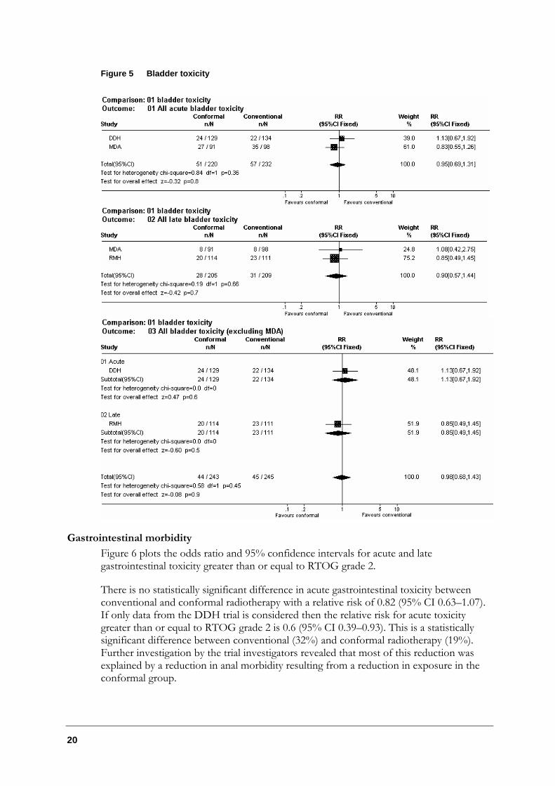
Bladder morbidity
Figure 5 plots the odds ratio and 95% confidence intervals (CI) for acute and late bladder toxicity greater than or equal to RTOG grade 2.
There is no statistically significant difference in acute bladder toxicity between conventional and conformal radiotherapy with a relative risk of 0.95 (95% CI 0.69–1.31). If only the DDH trial data is considered (ie information from the MDA trial is excluded) then the relative risk for acute toxicity greater than or equal to RTOG grade 2 is 1.13 (95% CI 0.67–1.92).
There is no statistically significant difference in late bladder toxicity between conventional and conformal radiotherapy with a relative risk of 0.90 (95% CI 0.57–1.44). If only data from the RMH trial is considered (ie information from the MDA trial is excluded) then the relative risk for late toxicity greater than or equal to RTOG grade 2 is 0.85 (95% CI 0.49–1.45).
20
Figure 5 Bladder toxicity
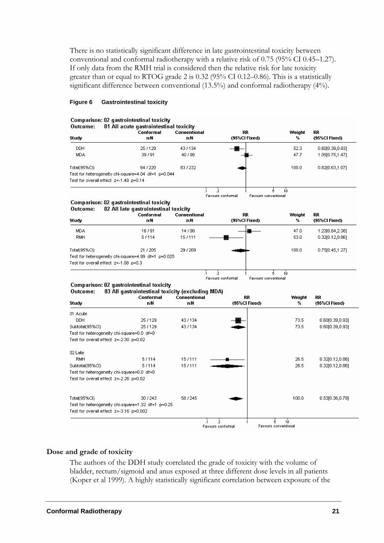
Gastrointestinal morbidity
Figure 6 plots the odds ratio and 95% confidence intervals for acute and late gastrointestinal toxicity greater than or equal to RTOG grade 2.
There is no statistically significant difference in acute gastrointestinal toxicity between conventional and conformal radiotherapy with a relative risk of 0.82 (95% CI 0.63–1.07). If only data from the DDH trial is considered then the relative risk for acute toxicity greater than or equal to RTOG grade 2 is 0.6 (95% CI 0.39–0.93). This is a statistically significant difference between conventional (32%) and conformal radiotherapy (19%). Further investigation by the trial investigators revealed that most of this reduction was explained by a reduction in anal morbidity resulting from a reduction in exposure in the conformal group.
Conformal Radiotherapy 21
There is no statistically significant difference in late gastrointestinal toxicity between conventional and conformal radiotherapy with a relative risk of 0.75 (95% CI 0.45–1.27). If only data from the RMH trial is considered then the relative risk for late toxicity greater than or equal to RTOG grade 2 is 0.32 (95% CI 0.12–0.86). This is a statistically significant difference between conventional (13.5%) and conformal radiotherapy (4%).
Figure 6 Gastrointestinal toxicity
Dose and grade of toxicity
The authors of the DDH study correlated the grade of toxicity with the volume of bladder, rectum/sigmoid and anus exposed at three different dose levels in all patients (Koper et al 1999). A highly statistically significant correlation between exposure of the
22
anus and anal toxicity was found, but no such correlation was found between exposed volume and bladder or rectum/sigmoid toxicity.
The MDA study also found no relationship between acute bladder or rectal toxicity and dose (volume of bladder or rectum receiving more than 60 Gy) for patients receiving conformal radiotherapy.
Quality of Life
The RMH trial used a patient morbidity (self-assessment) questionnaire to monitor patient well-being. The questionnaire was completed by patients on the first day of treatment, weekly during and for four weeks following radiotherapy, then monthly for two months. Information on medications was poorly completed and it has been suggested that this is problematic as morbidity scored by the patient was influenced by the medication prescribed (Koper et al 1999; Tait et al 1997). Information on well-being was reported for 261 of the 266 patients randomised to the pelvic radiotherapy trial and therefore includes information on patients who do not have prostate cancer. The authors concluded that pelvic radiotherapy affects bowel motions, micturition, tiredness and weakness, with no measurable effect on nausea or abdominal pain. No significant difference was found between conventional and conformal radiotherapy in terms of proportion of patients experiencing symptoms, severity of symptoms, time to worst symptoms or time to return to baseline symptoms.
The MDA trial sent questionnaires to the first 112 patients randomised with more than two years of follow-up (Nguyen, Pollack, & Zagars 1998). Not all of the 101 patients who responded completed every question. The focus of the questionnaire was bladder, bowel and sexual function. Data on bladder and bowel symptoms were graded using modified RTOG and LENT scales. They are included in this report in the section ‘Side effects / Treatment-related morbidity’ and hence, are not reported further here. In relation to sexual function, it was reported that potency decreased from 80% before radiotherapy to 51%. Of those who were potent before radiotherapy, 64% retained potency. Potency was defined as erections adequate for intercourse at least a few times since the completion of radiotherapy. Differences between conventional and conformal radiotherapy were not compared statistically.
Conformal Radiotherapy 23
Conclusions
There is some randomised evidence to suggest that:
• In the treatment of prostate cancer, delivery of similar total doses of radiotherapy using a conformal approach results in similar efficacy and reduced toxicity to that experienced using conventional radiotherapy.
• In the treatment of prostate cancer, delivery of higher total doses of radiotherapy using a conformal approach results in increased efficacy and similar toxicity to that experienced using conventional radiotherapy.
• The greatest benefit would appear to be in terms of gastrointestinal toxicity, both acute and late.
• A reasonable proportion of the reduction in acute gastrointestinal toxicity may be explained by a reduction in anal morbidity resulting from a reduction in exposure using conformal radiotherapy.
Dose response relationship in prostate cancer
In the previous section the MDA trial (Pollack et al 2000) examined dose escalation using CRT boost radiotherapy compared to RT. This section examines the efficacy and safety of dose escalation in patients with prostate cancer receiving high dose CRT versus low dose CRT as well as the use of different conformal techniques such as IMRT.
Conformal radiotherapy is built on the premise that in reducing the overall exposure of normal tissues receiving radiation, complications are reduced, thereby permitting dose escalation to the target volume (Tubiana & Eschwege 2000). Further, it is thought that in escalating the dose to target volume, tumour control is increased and that this higher rate of local control can improve survival rates.
Like the evidence for assessing the differences between standard and conformal radiotherapy using conventional dose, large dose-escalation studies of reasonable quality with long-term follow-up have primarily been undertaken in patients with prostate cancer. These studies are reported on below; however, it should be noted that this section is based on non-randomised (level III-2/III-3) evidence and as such, bias is likely to exist. Differences in prognostic factors and dose ranges, as well as reporting on selected sub-groups for some outcomes, also makes evaluating the role of dose escalation difficult. Consequently, caution should be exercised when interpreting or generalising these results.
Intensity-modulated radiation therapy (IMRT)
As mentioned in the background section of this report, IMRT is a more advanced form of 3DCRT. IMRT uses 3D treatment planning techniques to optimise delivery of radiation to an irregularly shaped volume (Verhey 1999). In IMRT, the beam intensity is varied across the treatment field. The intensity of the radiation exposure in one portion of the field is modified depending on whether tumour or critical normal structures are
24
present in the beam pathway. Hence, the beam is divided into multiple beamlets, and each can have a different intensity (Grant & Woo 1999). As a result of varying the beam intensity across those shaped fields, IMRT can yield dose distributions that conform closely to the three-dimensional shape of the target volume while reducing the dose to normal structures even further than is possible with CRT. This differs from traditional 3DCRT where a ‘beams eye view’ of the tumour is used to shape beams from chosen directions. With IMRT, the shaping and intensity variations are determined by optimisation software which seeks the best solution to a set of dose constraints which the user specifies (so-called ‘inverse planning’). The role of the MLC is also increased in IMRT due to the more dynamic nature of the treatment.
About the trials
Memorial Sloan Kettering Cancer Centre (MSKCC) - IMRT vs CRT (Zelefsky et al 2000)
This trial commenced in September 1992 and recruited 232 patients with T1-3 prostate cancer. The aim of the study was to compare acute and late toxicities of high dose radiation for prostate cancer delivered by either CRT or IMRT. Sixty-one (61) patients were treated with conventional 3DCRT to 72 Gy, followed by a 9 Gy boost. One hundred and seventy-one patients received the entire 81 Gy with IMRT. Twenty patients were also randomly selected and planned concomitantly by both techniques to ascertain further differences in the two approaches. Acute and late urinary and rectal toxicities were scored according to the RTOG morbidity grading scale. Follow-up evaluations were performed at three and six months.
Memorial Sloan Kettering Cancer Center (MSKCC) (Skwarchuk et al 2000; Zelefsky et al 1998a; Zelefsky et al 1998c; Zelefsky et al 1998b; Zelefsky et al 1999)
An earlier dose escalation study evaluating high dose 3DCRT was also undertaken at the Memorial Sloan-Kettering Center (MSKCC). Accrual of patients began in October 1988 and 743 patients were recruited with stages T1c-T3 tumours. All patients were treated with 3DCRT that targeted the prostate and seminal vesicles. The prescription dose was 64.8 Gy for 96 patients, 70.2 Gy for 266 patients, 75.6 Gy for 320 patients and 81.0 Gy for 61 patients (Skwarchuk et al 2000). Follow-up evaluations were performed at three and six month intervals after treatment, with a minimum follow-up of 12 months for 136 patients, followed by 4–8 years. Acute and late toxicity was scored according to the RTOG scale. Multiple papers have been published on this study. The way in which clinically significant (grade III or IV) acute and late toxicity information is reported varies between the papers analysing this patient population. As a result this information is not included in the tables below but is included in the text. It would also appear that the group of 61 patients treated to 81 Gy are part of the later dose study evaluating IMRT and CRT also undertaken at MSKCC (Zelefsky et al 2000).
Centre Antoine Lacassagne (Bey et al 2000)
In another multi-institutional dose escalation study, Bey et al (2000) evaluated the feasibility of increasing the dose from 66 Gy to 80 Gy in 164 patients with stage T1b-T3 prostate cancer. Patients were treated at one of five French institutions equipped for 3DCRT and received a dose of 66, 70, 74, 78 or 80 Gy. For analysis, patients were divided into two groups; group 1 receiving the standard dose of 66–70 Gy (46 patients,
Conformal Radiotherapy 25
mean follow-up 32 months) and the second group receiving 74–80 Gy (118 patients, mean follow-up 17.5 months). The dose delivered to the rectal wall was limited to 75 Gy, while the limit for the seminal vesicles was set at 72 Gy. Ninety patients had their seminal vesicles irradiated. Late effects were graded using a 0–4 scale, adapted from a glossary used for complications in gynaecological cancers and were evaluated six months after treatment. A quality of life questionnaire (QLQ-C30) was also employed prior and post treatment, however, only 77 patients were evaluable.
National Cancer Institute (NCI) 3DOG/RTOG 9406 Study (Michalski et al 2000)
The National Cancer Institute funded nine institutions with 3DCRT planning capabilities to develop a multi-institutional trial to determine the maximum tolerated dose of radiation that can be delivered to the prostate gland in men with prostate cancer (<T3). Michalski et al (2000) report on preliminary toxicity of 288 men with low-risk prostate cancer in this study treated on the first two dose levels (68.4 Gy and 73.8 Gy respectively) of the phase I/II 3DOG/RTOG 9406 dose escalation protocol. Patients were stratified into three treatment groups, as determined by local disease and risk factors, with each of these groups having three dose levels. The paper by Michalski et al (2000) reports on the first two groups and dose levels. Comparisons are also made between the RTOG 9406 study and the RTOG 7506 and 7706 treatment controls to take into account the different length of follow-up between the historical experiences and the current study (Lawton et al 1991). This is not the ideal method and could result in bias. Acute toxicity was defined as occurring within 120 days from the start of treatment and was graded according to the RTOG acute radiation morbidity criteria. Late complications were considered to be those appearing 120 days post treatment and were scored in accordance with the RTOG late radiation morbidity scoring scale. Median follow-up ranged from 2.2 years (Group 2, level II) to 3.4 years (Group 1, level I). However, the authors note that as a result of the inclusion of patients receiving neoadjuvant and or adjuvant hormone therapy, there is the potential for confounding of results (Michalski et al 2000).
Fox Chase Cancer Centre (FCCC) (Hanks et al 1998; Hanks et al 2000; Hanks et al 1996; Hanks et al 1997)
This study reports on 232 consecutive patients treated with 3DCRT between June 1989 and October 1992. Patients with various stages, grades and PSA levels are included. Dose was escalated from 63 Gy to 79 Gy. Biochemical freedom from disease (no evidence of disease (bNED)) rates were reported. Each dose group was subdivided by pre-treatment PSA level (<10, 10–19 and ≥20 ng/mL). A later paper is also published by the FCCC group, however, this paper is primarily interested in sub-group analysis of the higher dose groups.
University of Michigan, University of California and FCCC (Hanks et al 2000)
This study is a retrospective analysis of 180 patients treated with 3DCRT at three institutions. Patients who had a Gleason score 8–10 adenocarcinoma of the prostate and a known pre-treatment PSA were included in the study and were divided into two groups for analysis; Group 1, T 1-2 and Group 2, T 3-4. The main aim of this study was to evaluate the effect of high dose 3DCRT. Outcomes that were reported included freedom from PSA failure (bNED control) and survival median follow-up was 36 months.
26
RT-01
This ongoing randomised trial is being conducted by the UK Medical Research Council Radiotherapy Working Party. Recruitment commenced in January 1998. The aim of this trial is to compare 74 Gy delivered by high dose conformal radiotherapy with standard 64 Gy conformal radiotherapy in patients with stage T1b-T3a prostate cancer. The protocol was developed from a randomised pilot study that commenced at the Royal Marsden NHS Trust and Institute of Cancer Research in 1995. The primary endpoints of the trial are local tumour control, biochemical prostate specific antigen (PSA) failure, development of metastases, survival, and incidence of acute and late radiation induced side-effects.
Results
Local control
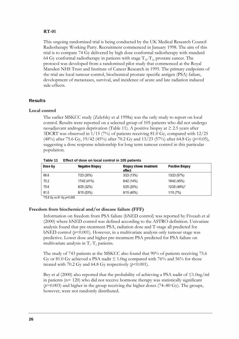
The earlier MSKCC study (Zelefsky et al 1998a) was the only study to report on local control. Results were reported on a selected group of 105 patients who did not undergo neoadjuvant androgen deprivation (Table 11). A positive biopsy at ≥ 2.5 years after 3DCRT was observed in 1/15 (7%) of patients receiving 81.0 Gy, compared with 12/25 (48%) after 75.6 Gy, 19/42 (45%) after 70.2 Gy and 13/23 (57%) after 64.8 Gy (p<0.05), suggesting a dose response relationship for long term tumour control in this particular population.
Table 11 Effect of dose on local control in 105 patients Dose Gy Negative Biopsy Biopsy shows treatment
effect Positive Biopsy
66.6 7/23 (30%) 3/23 (13%) 13/23 (57%) 70.2 17/42 (41%) 6/42 (14%) 19/42 (45%) 75.6 8/25 (32%) 5/25 (20%) 12/25 (48%)* 81.0 8/15 (53%) 6/15 (40%) 1/15 (7%) *75.6 Gy vs 81 Gy p=0.005
Freedom from biochemical and/or disease failure (FFF)
Information on freedom from PSA failure (bNED control) was reported by Fiveash et al (2000) where bNED control was defined according to the ASTRO definition. Univariate analysis found that pre-treatment PSA, radiation dose and T-stage all predicted for bNED control (p<0.001). However, in a multivariate analysis only tumour stage was predictive. Lower dose and higher pre-treatment PSA predicted for PSA failure on multivariate analysis in T1-T2 patients.
The study of 743 patients at the MSKCC also found that 90% of patients receiving 75.6 Gy or 81.0 Gy achieved a PSA nadir ≤ 1.0ng compared with 76% and 56% for those treated with 70.2 Gy and 64.8 Gy respectively (p<0.001).
Bey et al (2000) also reported that the probability of achieving a PSA nadir of ≤1.0ng/ml in patients (n= 120) who did not receive hormone therapy was statistically significant (p=0.003) and higher in the group receiving the higher doses (74–80 Gy). The groups, however, were not randomly distributed.
Conformal Radiotherapy 27
In the FCCC study (1998) a dose response for patients with pre-treatment PSA >10ng/ml was observed based on five-year bNED results. No dose response was observed for patients with pre-treatment PSA <10ng/ml.
Overall survival
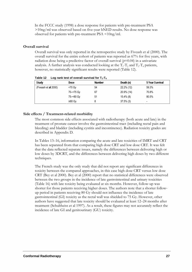
Overall survival was only reported in the retrospective study by Fiveash et al (2000). The overall survival for the entire cohort of patients was reported as 67% for five years, with radiation dose being a predictive factor of overall survival (p=0.04) in a univariate analysis. A further analysis was conducted looking at the T1-T2 and T3-T4 patients, however, no statistically significant results were reported (Table 12).
Table 12 Log rank test of overall survival for T1-T4 Study Dose Number Death (n) 5 Year Survival
<70 Gy 54 22.2% (12) 59.3% 70–<75 Gy 67 20.9% (14) 70.8% 75–<80 Gy 51 15.4% (8) 80.5%
(Fiveash et al 2000)
≥80 Gy 8 37.5% (3) -
Side effects / Treatment-related morbidity
The most common side effects associated with radiotherapy (both acute and late) in the treatment of prostate cancer involve the gastrointestinal tract (including rectal pain and bleeding) and bladder (including cystitis and incontinence). Radiation toxicity grades are described in Appendix D.
In Tables 13–16, information comparing the acute and late toxicities of IMRT and CRT has been separated from that comparing high dose CRT and low dose CRT. It was felt that the data reflected separate issues, namely the differences between delivering high or low doses by 3DCRT, and the differences between delivering high doses by two different techniques.
The French study was the only study that did not report any significant differences in toxicity between the compared approaches, in this case high dose CRT versus low dose CRT (Bey et al 2000). Bey et al (2000) report that no statistical differences were observed between the two groups in the incidence of late gastrointestinal and urinary toxicities (Table 16) with late toxicity being evaluated at six months. However, follow-up was shorter for those patients receiving higher doses. The authors note that a shorter follow-up period in patients receiving 80 Gy should not influence the incidence of late gastrointestinal (GI) toxicity as the rectal wall was shielded to 75 Gy. However, other authors have suggested that late toxicity should be evaluated at least 12–24 months after treatment (Schultheiss et al 1997). As a result, these figures may not accurately reflect the incidence of late GI and genitourinary (GU) toxicity.
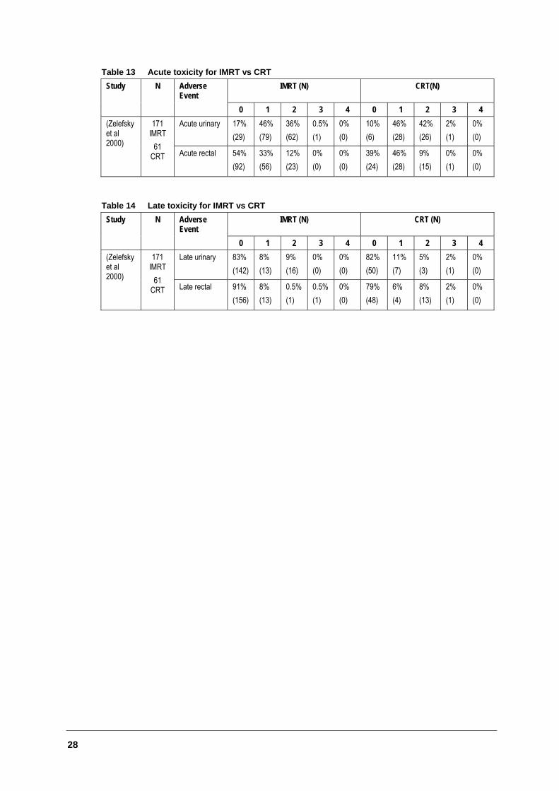
28
Table 13 Acute toxicity for IMRT vs CRT Study N Adverse
Event IMRT (N) CRT(N)
0 1 2 3 4 0 1 2 3 4 Acute urinary 17%
(29) 46% (79)
36% (62)
0.5% (1)
0% (0)
10% (6)
46% (28)
42% (26)
2% (1)
0% (0)
(Zelefsky et al 2000)
171 IMRT
61 CRT Acute rectal 54%
(92) 33% (56)
12% (23)
0% (0)
0% (0)
39% (24)
46% (28)
9% (15)
0% (1)
0% (0)
Table 14 Late toxicity for IMRT vs CRT Study N Adverse
Event IMRT (N) CRT (N)
0 1 2 3 4 0 1 2 3 4 Late urinary 83%
(142) 8% (13)
9% (16)
0% (0)
0% (0)
82% (50)
11% (7)
5% (3)
2% (1)
0% (0)
(Zelefsky et al 2000)
171 IMRT
61 CRT Late rectal 91%
(156) 8% (13)
0.5% (1)
0.5% (1)
0% (0)
79% (48)
6% (4)
8% (13)
2% (1)
0% (0)
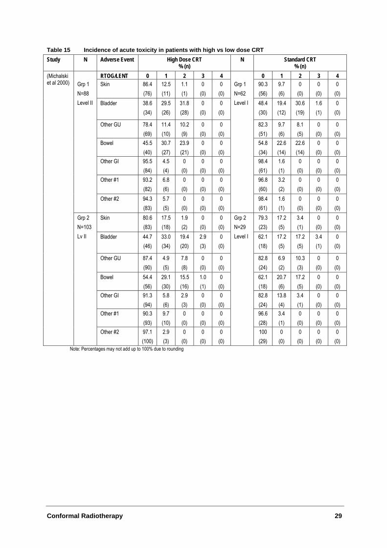
Conformal Radiotherapy 29
Table 15 Incidence of acute toxicity in patients with high vs low dose CRT Study N Adverse Event High Dose CRT
% (n) N Standard CRT
% (n) RTOG/LENT 0 1 2 3 4 0 1 2 3 4
Skin 86.4 (76)
12.5 (11)
1.1 (1)
0 (0)
0 (0)
90.3 (56)
9.7 (6)
0 (0)
0 (0)
0 (0)
Bladder 38.6 (34)
29.5 (26)
31.8 (28)
0 (0)
0 (0)
48.4 (30)
19.4 (12)
30.6 (19)
1.6 (1)
0 (0)
Other GU
78.4 (69)
11.4 (10)
10.2 (9)
0 (0)
0 (0)
82.3 (51)
9.7 (6)
8.1 (5)
0 (0)
0 (0)
Bowel 45.5 (40)
30.7 (27)
23.9 (21)
0 (0)
0 (0)
54.8 (34)
22.6 (14)
22.6 (14)
0 (0)
0 (0)
Other GI 95.5 (84)
4.5 (4)
0 (0)
0 (0)
0 (0)
98.4 (61)
1.6 (1)
0 (0)
0 (0)
0 (0)
Other #1 93.2 (82)
6.8 (6)
0 (0)
0 (0)
0 (0)
96.8 (60)
3.2 (2)
0 (0)
0 (0)
0 (0)
Grp 1 N=88 Level II
Other #2 94.3 (83)
5.7 (5)
0 (0)
0 (0)
0 (0)
Grp 1 N=62 Level I
98.4 (61)
1.6 (1)
0 (0)
0 (0)
0 (0)
Skin 80.6 (83)
17.5 (18)
1.9 (2)
0 (0)
0 (0)
79.3 (23)
17.2 (5)
3.4 (1)
0 (0)
0 (0)
Bladder 44.7 (46)
33.0 (34)
19.4 (20)
2.9 (3)
0 (0)
62.1 (18)
17.2 (5)
17.2 (5)
3.4 (1)
0 (0)
Other GU 87.4 (90)
4.9 (5)
7.8 (8)
0 (0)
0 (0)
82.8 (24)
6.9 (2)
10.3 (3)
0 (0)
0 (0)
Bowel 54.4 (56)
29.1 (30)
15.5 (16)
1.0 (1)
0 (0)
62.1 (18)
20.7 (6)
17.2 (5)
0 (0)
0 (0)
Other GI 91.3 (94)
5.8 (6)
2.9 (3)
0 (0)
0 (0)
82.8 (24)
13.8 (4)
3.4 (1)
0 (0)
0 (0)
Other #1 90.3 (93)
9.7 (10)
0 (0)
0 (0)
0 (0)
96.6 (28)
3.4 (1)
0 (0)
0 (0)
0 (0)
(Michalski et al 2000)
Grp 2 N=103 Lv II
Other #2 97.1 (100)
2.9 (3)
0 (0)
0 (0)
0 (0)
Grp 2 N=29 Level I
100 (29)
0 (0)
0 (0)
0 (0)
0 (0)
Note: Percentages may not add up to 100% due to rounding
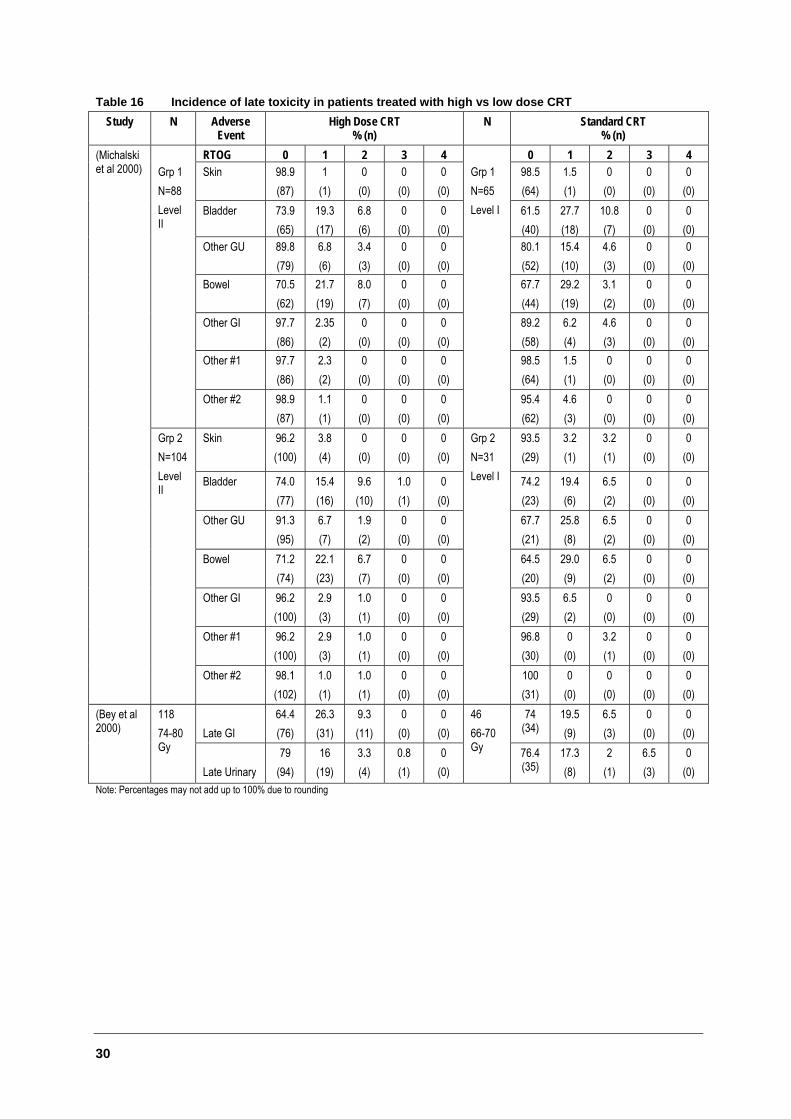
30
Table 16 Incidence of late toxicity in patients treated with high vs low dose CRT Study N Adverse
Event High Dose CRT
% (n) N Standard CRT
% (n) RTOG 0 1 2 3 4 0 1 2 3 4
Skin 98.9 (87)
1 (1)
0 (0)
0 (0)
0 (0)
98.5 (64)
1.5 (1)
0 (0)
0 (0)
0 (0)
Bladder 73.9 (65)
19.3 (17)
6.8 (6)
0 (0)
0 (0)
61.5 (40)
27.7 (18)
10.8 (7)
0 (0)
0 (0)
Other GU
89.8 (79)
6.8 (6)
3.4 (3)
0 (0)
0 (0)
80.1 (52)
15.4 (10)
4.6 (3)
0 (0)
0 (0)
Bowel 70.5 (62)
21.7 (19)
8.0 (7)
0 (0)
0 (0)
67.7 (44)
29.2 (19)
3.1 (2)
0 (0)
0 (0)
Other GI 97.7 (86)
2.35 (2)
0 (0)
0 (0)
0 (0)
89.2 (58)
6.2 (4)
4.6 (3)
0 (0)
0 (0)
Other #1 97.7 (86)
2.3 (2)
0 (0)
0 (0)
0 (0)
98.5 (64)
1.5 (1)
0 (0)
0 (0)
0 (0)
Grp 1 N=88 Level II
Other #2 98.9 (87)
1.1 (1)
0 (0)
0 (0)
0 (0)
Grp 1 N=65 Level I
95.4 (62)
4.6 (3)
0 (0)
0 (0)
0 (0)
Skin 96.2 (100)
3.8 (4)
0 (0)
0 (0)
0 (0)
93.5 (29)
3.2 (1)
3.2 (1)
0 (0)
0 (0)
Bladder 74.0 (77)
15.4 (16)
9.6 (10)
1.0 (1)
0 (0)
74.2 (23)
19.4 (6)
6.5 (2)
0 (0)
0 (0)
Other GU
91.3 (95)
6.7 (7)
1.9 (2)
0 (0)
0 (0)
67.7 (21)
25.8 (8)
6.5 (2)
0 (0)
0 (0)
Bowel 71.2 (74)
22.1 (23)
6.7 (7)
0 (0)
0 (0)
64.5 (20)
29.0 (9)
6.5 (2)
0 (0)
0 (0)
Other GI 96.2 (100)
2.9 (3)
1.0 (1)
0 (0)
0 (0)
93.5 (29)
6.5 (2)
0 (0)
0 (0)
0 (0)
Other #1 96.2 (100)
2.9 (3)
1.0 (1)
0 (0)
0 (0)
96.8 (30)
0 (0)
3.2 (1)
0 (0)
0 (0)
(Michalski et al 2000)
Grp 2 N=104 Level II
Other #2 98.1 (102)
1.0 (1)
1.0 (1)
0 (0)
0 (0)
Grp 2 N=31 Level I
100 (31)
0 (0)
0 (0)
0 (0)
0 (0)
Late GI
64.4 (76)
26.3 (31)
9.3 (11)
0 (0)
0 (0)
74 (34)
19.5 (9)
6.5 (3)
0 (0)
0 (0)
(Bey et al 2000)
118 74-80 Gy
Late Urinary 79
(94) 16
(19) 3.3 (4)
0.8 (1)
0 (0)
46 66-70 Gy 76.4
(35) 17.3 (8)
2 (1)
6.5 (3)
0 (0)
Note: Percentages may not add up to 100% due to rounding
Conformal Radiotherapy 31
Bladder
In the MSKCC study comparing the toxicity resultant from IMRT and CRT, it was found that although there was a decrease in acute grade II and III GU toxicity with IMRT, the differences were not statistically significant. There were also no differences between the two treatment techniques in terms of late GU toxicity.
The earlier MSKCC study found that the overall rate of clinically significant acute urinary toxicity (grade III or IV) was 0.7% with five patients presenting with urinary retention requiring catheter placement. In terms of clinically significant late urinary toxicity (grade III or IV) eight (1%) patients developed grade III urethral strictures requiring dilation. The authors also note that the rates of late grade II GU toxicity were 13% and 8% for patients receiving ≥75.6 Gy versus ≤70.2 Gy, respectively (p=0.002) (Zelefsky et al 1999).
Preliminary analysis of the NCI study found that within the two groups and two dose levels, relatively few patients experienced grade III bladder toxicities (0–3%), while there were no cases of acute grade IV or V toxicities and only one patient in the Group 2-Level II arm experienced bladder toxicity ≥ grade III.
Gastrointestinal
In the MSKCC IMRT trial it was reported that the combined rates of acute grade I and II rectal toxicity for IMRT were significantly decreased (p=0.05) compared to those treated with 3DCRT, with a concomitant increase in the number of patients having no toxicity (p=0.05) (Table 13). A significant decrease in late grade II rectal bleeding with IMRT was also found (p=0.0001).
Results from the MSKCC 3DCRT study indicate that one patient (0.1%) experienced acute grade III GI symptoms and five patients (0.7%) developed late grade III proctitis with associated rectal bleeding. One patient (0.1%) treated to 64.8 Gy with a history of inflammatory bowel disease also developed late grade IV toxicity.
The NCI study reported a single acute grade II bowel toxicity in a Group 1 patient treated to dose level II. There were no grade IV or V bowel or other GI toxicities reported.
Dose and grade of toxicity (DVH analysis)
Boersma et al (1993a) report on the relationship between the percentage of the rectal wall exposed to radiotherapy and the incidence of severe rectal bleeding. It was found that toxicity was greater in patients with greater than 40-50% of the rectal wall receiving more than 65 Gy (p=0.02).
In the trial undertaken by the MSKCC on CRT there was an increase in grade II rectal bleeding from 6% in patients treated with ≤70.2 Gy to 17% in patients receiving ≥75.6 Gy (p<0.001). The authors cite this information, along with an analysis of DVH in patients receiving higher doses, as reasons why the latter trial using IMRT was undertaken (Zelefsky et al 2000). It was conjectured that conforming more tightly to the PTV using improved techniques would decrease the incidence of rectal toxicity. Zelefsky et al (2000) report that in the 20 patients planned by both techniques, IMRT did appear to reduce the volume of the rectal wall compared to 3DCRT.
32
The authors of the NCI study also undertook a dose-volume analysis and found that in the case of acute bladder toxicity, when the percent of bladder receiving greater than the reference dose (65 Gy) exceeded 30%, there was an increased risk of developing acute bladder toxicity. A similar experience was also described for late bladder toxicity. Michalski et al ( 2000) also note there was a greater incidence of acute bladder toxicity in patients receiving prior hormone therapy.
Quality of life
Quality of life was examined in the French study (Bey et al 2000). Two or more questionnaires were completed by 77 of 164 patients. No statistically significant differences were found between the two groups in terms of the global quality of life analysis. This may be a result of the poor response rate or the fact that the patients were not randomly distributed and that clinically and statistically significant differences were reported between the two groups. It is also unclear how many individuals in each of the two groups were included in the global quality of life analysis.
One of the papers from the first MSKCC study also looked at erectile function in 544 patients with normal erectile function before CRT (Zelefsky et al 1999). It was found that the five-year actuarial likelihood of impotence among patients who received ≥75.6 Gy was 68% compared with 52% for those treated with less than 70.2 Gy (p<0.001).
Conclusions
There is some limited indicative evidence to suggest that:
• In the treatment of prostate cancer, delivery of higher total doses of radiotherapy using a conformal approach may result in improved local control.
• A considerable proportion of toxicity experienced by prostate cancer patients may be explained by the percentage of the rectal and bladder wall receiving high dose radiotherapy.
• In the treatment of prostate cancer, preliminary results indicate that IMRT appears to have the potential to lower the percentage of rectal wall receiving high dose radiotherapy thereby achieving a lower incidence of rectal toxicity.
• Randomised evidence is needed to more clearly determine the benefit of high dose CRT and IMRT for patients with prostate cancer.
Conformal Radiotherapy 33
Conformal radiotherapy for other cancers
Thirty-four (34) papers were identified for indications other than prostate cancer in relation to conformal radiotherapy.
The main indications and tumour sites studied in terms of conformal radiotherapy were the brain, hepatobiliary tract, bronchogenic and head and neck carcinomas. Conformal radiotherapy is considered particularly suited to such sites due to the close proximity of other normal tissues and the poor local control rate with conventional treatment for many of these cancers.
As conformal radiotherapy represents a relatively new change in radiotherapy practice there is a lack of long-term data on the efficacy and safety of CRT. Follow-up data within the papers is usually reported at one and two years, with a number of papers calculating actuarial estimates for progression-free survival or overall survival. More publications reported on toxicity than efficacy information and a summary of the toxicity information is presented in Appendix E. The issue of dose escalation was also raised in a number of papers.
The majority of the papers report on a relatively small number of patients and as a result extreme percentages should be viewed with caution. Due to the lack of data in respect to conformal radiotherapy in indications other than prostate cancer, papers that included patients who were receiving concurrent chemotherapy were also included.
In general, the quality and amount of information reported in these papers varied quite widely, making it difficult to draw definitive conclusions regarding the efficacy and safety of CRT in these indications. The following information is therefore a description of the evidence available for other cancer indications.
Limitations of data/Methodological issues
Consistency in relation to technology
• Within the papers there did not appear to be a general definition of standard or conformal radiotherapy.
• There was a lack of consistency in the way conformal radiotherapy was delivered. The delivery of conformal radiotherapy to patients also differed throughout the papers, and within individual papers. Technology changes were reflected in papers; for example, in one study some patients had fields shaped with cerrobend blocks and others had fields shaped with a multileaf collimator (Dawson et al 2000; Dawson et al 2000; Robertson et al 1993; Alheit et al 1999).
• Also, these changes in technology were not taken into account within the papers. Often, authors did not delineate between the different techniques and analyse the group receiving an earlier technique as historical controls.
34
Issues within the patient population
• Within many papers there was a vast range of patients, with some undergoing primary radiation, and others undergoing booster radiation, post-operative irradiation or re-irradiation (eg for recurrence) (Dawson et al 2000).
• The method of recruiting patients was often unclear ie whether patients were consecutive or selected.
• Comparisons were also made difficult in the papers that evaluated multiple related indications such as sarcoma and head and neck cancer, due to the heterogeneity of these cancers.
Manner in which information is reported in studies
• Authors did not report information in a consistent manner. For example, acute and late toxicity were often defined differently by different authors.
• Also, it was often not possible to determine at what time point after treatment complications developed, or efficacy information was reported.
• There was a general lack of consistency in dose schedules among the papers. Many papers reported a wide range of doses, however the differences between patients who received higher or lower doses were not necessarily reported (Alheit et al 1999; Robertson et al 1997a; Armstrong et al 1997).
• The clinical target volumes were also defined differently within the papers making comparisons regarding dose difficult.
• Many papers included patients who were receiving other treatments, such as chemotherapy (Alheit et al 1999; Cheng et al 1999; Graham et al 1999; Pommier et al 2000).
• There appeared to be some overlap of patient groups between publications, particularly relating to those patients with liver cancer. It is difficult to tell from these papers whether they contain the same patients, or subsets of the same patients.
• Many papers had short follow-up periods.
• Censoring of data occurred within many of the papers.
• Many patients were considered ‘unevaluable’ and it was often unclear how many patients were included in the analysis.
Brain tumours
Efficacy and dose escalation
There were three case series that examined conformal radiotherapy in patients with brain tumours (See Appendix C). The first is a consecutive case series of 38 patients treated with post-operative irradiation for glioblastoma multiforme between 1985 and 1995 (Nakagawa et al 1998). Patients seen prior to 1991 received whole brain radiotherapy in
Conformal Radiotherapy 35
addition to conformal therapy, and patients seen from 1991 received only conformal radiotherapy. Twenty-one (21) patients received a dose of 60–80 Gy, while seventeen (17) patients, who were recruited after 1990, were given a dose of 90 Gy (Nakagawa et al 1998). Disease recurred in 32 of 38 patients (84%) with a median follow-up of 79.3 months (22.3–154.7 months). The authors make comparisons between the high and low dose groups, however, this is inappropriate for a number of reasons including the longer follow-up available for the low dose group, the small number of patients, and the study design. The median regrowth-free survival period for patients in this study was five months, and median survival was approximately 20 months. The authors themselves acknowledge that, ‘this is a highly selected and favourable patient population’ (Nakagawa et al 1998).
In the paper by Lee et al (1999) patients with high-grade astrocytomaswere treated with high dose conformal radiotherapy. Fifteen (15) patients were treated with 70 Gy and twenty-one (21) with 80 Gy. The paper reported on recurrence, with 32 of 36 patients presenting with recurrence inside the region given the high-dose volume. Three patients were considered marginal while one fell outside the high-dose region. From this, the authors concluded that even higher doses might be necessary to eradicate high-grade astrocytomas. Toxicity is alluded to in the paper and acknowledged as a possible issue; however, it is not reported on.
The third study, by Alheit et al (1999), reported on 24 cases (method of selection unclear) of meningioma (benign and malignant). The patients had little in common other than meningioma. Little clinically relevant information is reported. It is difficult to draw any definitive conclusions from this information.
Treatment related morbidity
Nakagawa et al (1998) and Alheit et al (1999) also reported on toxicity in relation to patients with brain tumours (Appendix E). Little information was reported in the papers about those patients who developed complications post-treatment. Nakagawa et al (1998) report that the two patients who developed radiation necrosis both received 90 Gy, however, no dose volume analysis was completed. In the paper by Alheit et al (1999), the short follow-up of cases with meningiomas prevents any assessment of late toxicity.
Pituitary adenomas
Jalali et al (2000) describe 22 patients with residual or recurrent pituitary adenomas treated with fractionated stereotactic conformal radiotherapy (SCRT). The outcomes of interest were vision, endocrine function and toxicity. The study included patients who had 45 Gy in 25 fractions or 50 Gy in 30 fractions, as well as either three or four field treatments. While a number of outcomes were reported in the paper, the lack of long-term follow-up data makes assessing tumour control and toxicity difficult.
Liver
Efficacy
The role of conformal radiotherapy in patients with liver cancer was detailed in six papers. It would appear that in four of these papers, patient groups overlap or are very similar (Robertson et al 1993; Robertson et al 1995; Robertson et al 1997a; Robertson et al 1997b), though the time period for patient selection was not always stated. The type of liver cancer also varied within the six papers with patients having hepatocellular carcinoma, bile duct cancer (cholangiocarcinoma) or hepatic metastases. The papers also
36
assessed the role of chemotherapy agents, such as intra-arterial hepatic fluorodeoxyuridine in combination with conformal radiotherapy, and different doses were given depending upon patient selection. Follow-up data of patients within some of the studies was also a little unclear. Given these limitations, it is difficult to ascertain the true effect of conformal radiotherapy on patients with liver cancer.
In looking at the outcome of efficacy, the majority of papers reported on tumour response, tumour failure and survival. Not all papers reported on all patients in terms of tumour response as a number were considered unevaluable. In terms of failure and survival, the most common site of failure was the liver, while median survival in the four papers by Robertson et al, which may be reporting on the same patients, ranged from 16 to 19 months.
Treatment related morbidity
Toxicity is outlined in Appendix E. There were several reports of toxicity greater than grade III and a number of cases of gastrointestinal bleeding were reported (Cheng et al 1999; Robertson et al 1995; Robertson et al 1997a). The numbers, however, are still quite small.
Lung cancer
Efficacy
There were five papers that reported on conformal radiotherapy and the efficacy outcomes of survival, tumour response and local control in patients with lung cancer. Varying dose schedules were also used within and between papers, which is not surprising given the interest in dose escalation for patients with lung cancer. A number of studies also included patients receiving concurrent chemotherapy. It has been suggested that local control figures presented in the papers may be an underestimate of the extent of disease control as bronchoscopies were not performed to confirm disease (Armstrong et al 1997).
Armstrong et al (1997) report on anatomic failure patterns and survival in 45 patients. Tumour stage was stage I/II in 13%, IIIa in 42% and IIIb in 44% of patients. The majority of patients had symptomatic locally advanced disease and 67% were treated with an ongoing dose escalation protocol, while the remaining 33% received normal doses. It was found that of 38 patients (seven did not complete 3DCRT due to disease progression), thoracic progression occurred in 18 patients (46%). However bronchoscopies were not performed after CRT. Distant metastases occurred in 31% of patients. Median survival (all patients) was 15.7 months and survival was 32% at two years and 12% at 59 months.
Bahri et al (1999) also report on survival for 35 evaluable patients (seven were excluded from series) with stage II-III unresectable non-small cell lung cancer (NSCLC). Of these patients, 20 received concurrent chemotherapy, while the median dose to the GTV was 63 Gy (range 50–68.4 Gy). It is reported that the overall one-year and two-year survival for all patients was 70.2% and 51.2% respectively, while local control was 23.3% at two years. These results are confounded by the use of concurrent chemotherapy, which reportedly significantly increased survival for patients with stage III disease. Bahri et al (1999) report a one-year survival of 78.6% for those with concurrent chemotherapy compared to 53.3% for the RT or sequential chemotherapy group.
Conformal Radiotherapy 37
Sibley et al (1995) retrospectively evaluated 37 patients with stage IIIa or IIIb NSCLC treated with high dose CRT. The median local progression-free survival period for patients in this study was 15.6 months, and median survival was approximately 19.5 months. Overall one and two-year actuarial survival was 75% and 37% respectively, with a local progression-free two-year survival rate of 23% (only based on 28 patients). Local progression was also documented in 18 of 28 evaluable patients.
The modified phase I trial by Socinski et al (2000) evaluated the role of 3DCRT in patients with stage III unresectable NSCLC. Disease progression was noted in 11% of patients and early death occurred in 7% of patients. The median survival was approximately 21 months. The one and two-year estimated survival rates were 69% and 45% respectively with one-year progression-free survival being 41%. Again these results were confounded by chemotherapy.
Nakagawa et al (2000) report on the use of stereotactic radiosurgery and conformal radiotherapy in fifteen patients with 22 thoracic tumours from various primary malignancies (21 tumours evaluable). These patients are evaluated in terms of tumour control and palliation of symptoms. In looking at tumour response rates, from a total of 21, 12 tumours had complete responses, seven had partial responses, and two had no changes. One patient was observed to have local recurrence, and at the end of 82 months, five of the 15 patients had died. The authors indicate that some caution should be exercised in interpreting these statistics due to the heterogenous nature of the group. However, it should also be noted that the paper has other limitations, such as the fact that in relation to clinical information, the number of tumours is frequently reported rather than the number of patients. One patient was also considered unevaluable due to death one month after treatment.
Dose escalation
Excluding prostate cancer, there is more information available on dose escalation for patients with non-small cell lung cancer (NSCLC) than is available for other indications (Emami 1996). The complex anatomy of the thorax and the close proximity of critical normal structures to target volumes have prevented the use of high doses using standard radiotherapy practices. As a result, NSCLC appears to be a good candidate for conformal radiotherapy, particularly if local control rates can be increased.
The MSKCC trial reports on 52 patients with inoperable NSCLC enrolled on a phase I study between 1991 and 1996 treated with 3DCRT (Rosenzweig et al 2000). The primary aim of the study was to estimate the maximum tolerated dose of external beam radiotherapy. The initial dose level was 70.2 Gy and each subsequent level increased the total dose by 5.4 Gy. A radiation dose level was considered complete when 10 patients received the dose without unacceptable acute morbidity. Of those initially assigned to the 70.2 Gy level (20), only 14 patients received the dose with two of the patients developing acute grade III radiation pneumonitis and one grade V (fatal) radiation pneumonitis. A further 18 patients were then entered on a modified study with three patients presenting with grade III toxicity at 70.2 Gy. Ten patients were treated at 75.6 Gy, with one patient developing pulmonary toxicity and another oesophageal toxicity. In total, there were six cases of acute grade III pulmonary complications and two cases of acute grade III oesophageal toxicity, while three patients experienced late grade III pulmonary complications. The authors noted that in patients with a normal tissue complication probability (NTCP) of >10%, there was a 27% rate of acute grade II or greater
38
pulmonary toxicity. In contrast, there were no cases of radiation pneumonitis in patients with a NTCP of <10% (p=0.05). The median survival time was reported at 11 months.
MD Anderson Cancer Centre also report on a randomised phase I/II dose trial (Cox et al 1990) of 884 patients with lung carcinoma. The dose given to patients is delivered in a hyperfractionated manner. Patients were randomised to receive minimum total doses of 60.0, 64.8, 69.6, 74.4 and 79.2 Gy. While no significant differences in the risks of acute or late effects in the five treatment arms were reported, there were six cases of late grade ≥III toxicity in the 60.0 Gy group and 24 cases in the 79.2 Gy group. Survival rates were also reported but on a selected group of patients. In this group of patients it was found that while there no differences in survival between the three higher dose arms, two-year survival rates for those receiving 69.6 Gy were significantly improved than those on the lower two groups (p=0.02).
Similarly, McGibney et al (1999) report on the potential of continuous hyperfractionated accelerated radiotherapy (CHART) for NSCLC with and without the application of 3DCRT. This is primarily a planning study and the results are not reported here as this was considered outside the scope of the review.
Treatment related morbidity
Toxicity was reported by all the papers and was mainly assessed in terms of weight loss, pneumonitis and oesophageal and pulmonary toxicity. Several of the papers also made observations in relation to factors predicting acute and late toxicity. Most of the papers reported cases of RTOG toxicity grade III or above in relation to oesophageal toxicity (Armstrong et al 1997; Maguire et al 1999; Socinski et al 2000) while one paper also reported on ≥ grade III pulmonary toxicity (Armstrong et al 1995). Cases of pneumonitis were also noted in a number of papers. In terms of factors that may have some impact on toxicity, Armstrong et al (1997) note that a significant correlation would seem to exist between the occurrence of severe radiation pneumonitis and volume of lung receiving ≥25 Gy. Graham et al (1999) and Maguire et al (1999) also identified factors in multivariate analyses that significantly predicted pneumonitis and oesophageal toxicity, these being the percentage volume of the total lung exceeding 20 Gy, percentage of oesophageal volume of >50 Gy (p=0.02) and the maximum percent of circumference treated >80 Gy (p=0.02).
Head and neck cancer
Efficacy
Seven papers form the basis of the efficacy comment in relation to conformal radiotherapy and head and neck cancer. This includes one study on patients with paranasal sinus and nasal cavity tumour malignancies and two studies on patients with nasopharyngeal carcinoma. As stated earlier, one of the difficulties in relation to this indication is the heterogenous nature of head and neck cancer and the subsequent variation in patient selection for studies. Within the papers there were patients with both primary or recurrent head and neck cancer, as well as patients that were undergoing adjunct chemotherapy (Dawson et al 2000). Both three dimensional conformal radiotherapy and intensity-modulated radiotherapy were also used in the head and neck studies.
The main outcomes of interest were tumour response rate, loco-regional control and toxicity. In terms of tumour response and loco-regional control, Dawson et al (2000)
Conformal Radiotherapy 39
report two and five-year actuarial loco-regional recurrence rates for all patients receiving irradiation for primary or recurrent head and neck cancers of 21% and 25%, respectively. In another study undertaken at the University of Michigan (Dawson et al 2001), one and two-year actuarial loco-regional recurrence-free survival was found to be 38.5% and 19.5%, respectively, for patients receiving irradiation for primary or recurrent head and neck cancers. Actuarial relapse and survival rates were also reported in this paper, with one and two-year relapse free survival rates of 28.6% and 15.9% and one and two-year survival rates of 51.1% and 32.6%. It should be noted that is was not always clear what denominator was used in respect to the above figures.
Butler et al (1999) assessed tumour response, with 19 of 20 patients defined as having complete response (CR) after simultaneous modulated accelerated treatment (SMART). After follow-up of two and five months, two patients were found to have lung metastases and an additional two patients were discovered to have local recurrence. Chao et al (2000) also report on tumour response in 17 patients receiving IMRT with squamous cell carcinoma (SCC) of the head and neck. Case selection was not stated and nine patients also received concurrent cisplatin chemotherapy. It was found that nine of 11 patients with gross tumour irradiation or re-irradiation, and five of six patients with post-operative irradiation, achieved CR, with two patients in the re-irradiation group presenting with local recurrence.
Three studies were identified that examined patients with specific head and neck tumours (Nishioka et al 2000; Pommier et al 2000; Chang et al 2000). The study by Pommier et al (2000) describes a relatively small study of forty (40) consecutive patients with paranasal sinus and nasal cavity tumours. Chemotherapy was performed in eight cases and immunotherapy in two cases. Efficacy outcomes of survival and local control rate are recorded. In the study, the total dose delivered was ≤60 Gy for 37 patients, with a median dose of 64 Gy. One and two-year survival rates were reported as 75.6% and 65.9%, respectively. Local relapse occurred in eight patients and post-operative relapse with progression in one case. Six cases of isolated metastases and one of lung metastases were also reported. Pommier et al (2000) note that macroscopic residual disease was reported in 11 out of 30 patients who underwent surgery.
In the paper by Nishioka et al (2000) 18 patients with both residual or recurrent nasopharyngeal carcinoma (NPC) were included in the study. Patients with NPC were given CRT as a booster after conventional radiotherapy (patients with local residual disease n=12) or as re-irradiation (patients with local recurrence n=6). Survival and local control were the reported outcomes. Those patients with booster therapy (n=12) had three-year survival and local control rates of 67% and 70% respectively, while those with re-irradiation (n=6) had a two-year control rate of 25%. Of the 12 patients who received booster therapy, three patients developed local recurrence; outside the booster fields in two cases, and inside in one case. Six patients died of the disease. Three failures were also noted in the recurrence group with persistent residual disease in two patients and relapse outside the field in another case. Five patients died of disease. The authors note that there was no clear relationship between dose and local control. It should be noted that the patient population these results are based on is relatively small and CRT was delivered as a booster after RT.
In the study by Chang et al (2000), 151 patients from a study population of 186 patients with locally recurrent nasopharyngeal carcinoma underwent ‘standard’ radiotherapy. The remaining patients (35) received conformal radiotherapy, due to a change in the institution’s delivery of radiotherapy. While survival rates are reported, they are not
40
analysed in terms of technique (RT vs CRT). As it is more likely that the results will reflect the efficacy of standard radiotherapy, they are not reported here.
Dose escalation
Papers with a primary aim of investigating dose escalation in head and neck cancer patients were not identified. Some discussion was undertaken in the studies in respect to the dose limit that should be assigned to normal tissues such as the salivary glands or optic chiasm. Head and neck cancer, however, is identified as an area where dose escalation by the use of 3DCRT and IMRT deserves further evaluation (Tubiana & Eschwege 2000).
Treatment related morbidity
More papers reported on toxicity than efficacy in respect to head and neck cancer (Appendix E). In particular, the issue of xerostomia was highlighted due to its impact on the quality of life of patients after radiotherapy. Occurrences of grade I skin toxicities (Butler et al 1999) to grade III xerostomia (Chao et al 2000) were reported in the papers. Ascertaining when toxicity was measured was a problem in some of the papers. At times it was unclear if grading was undertaken during, or post, treatment. This has implications for the rates of complications reported in the papers and should be taken into consideration. Debate was also undertaken within two papers (Chao et al 2000; Eisbruch et al 1999b) as to the optimal dose that should be given to the parotid gland in order to reduce some of the complications associated with radiotherapy. Chao et al (2000) note that IMRT appears to have the potential to reduce the incidence of xerostomia.
In respect to paranasal and nasal malignancies, Pommier et al (2000) state that CRT appears to result in a low rate of toxicity, with three grade III mucositis and one superficial keratitis being reported. One patient experienced blindness three years after radiotherapy, while another died of meningitis three months after the completion of CRT. Nishioka et al (2000) also report a low rate of toxicity in their population of patients with nasopharyngeal carcinoma (Appendix E).
Brizel et al (1999) report on dose distributions of conventional (two-dimensional) versus conformal (three-dimensional) treatment plans in relation to patients with paranasal sinus malignancies. Twenty patients were planned using a 2D approach and then compared with treatment received from a 3D plan using the same dose on normal tissue complication probabilities (NTCP). In this paper study subjects acted as their own controls. It was found that 3D plans resulted in fewer complications in all organs except the contralateral eye. As this study involves some theoretical assumptions it would seem practical to exercise some caution in interpreting the results.
Chang et al (2000) report that all patients with nasopharyngeal carcinoma included in their study had a history of prior radiotherapy treatment, while a significant proportion of the patients were also being treated with chemotherapy. The doses delivered to the tumour areas also differed within the study population. Only brief information on complications is reported in this paper. The authors note that a significant difference was observed between the standard radiotherapy treatment (SRT) group and the CRT group in terms of severe complications (p=0.004), with 9.3% in the CRT group experiencing complications in comparison to 22.9% of the SRT group. This may be explained in part by the shorter follow-up period in the CRT group. It may also be influenced by other factors such as dose or chemotherapy. It is unclear, however, whether these factors have
Conformal Radiotherapy 41
been taken into account in this paper as the primary aim of the papers was not to describe differences between treatment techniques.
Sarcoma Efficacy
Three papers looked at the use of conformal radiotherapy in sarcoma cases. Again, it is difficult to formulate conclusions from these two papers about the role of conformal radiotherapy in this indication, especially given the heterogenous nature of sarcoma. In the paper by Greiner et al (1992) 21 patients with soft tissue sarcoma were treated with high dose conformal radiotherapy. It was found that three (3) patients developed local tumour progression, one (1) inside the treatment volume, and two (2) outside, while five (5) patients had metastases. Survival rates (3 year actuarial 67%; 5 year actuarial 33%) and complication rates are also given in the paper, and it is perhaps important to note that some patients were also undergoing chemotherapy at the time of treatment.
There were two papers reporting on skull base tumours (Debus et al 1997; Rosenberg et al 1999). The paper by Rosenberg et al (1999) is of little clinical relevance as the primary focus of the paper is on the differentiation of chordoma from chondrosarcoma, rather than the role of conformal radiotherapy in sarcoma (Rosenberg et al 1999). The study by Debus et al (1997) also contains little efficacy information as the primary focus of the paper is on the long-term incidence of brainstem toxicity in patients treated for skull base tumours with high dose conformal radiotherapy. Survival rates of the 367 patients are reported as 94% at five years and 86% at 10 years.
Treatment related morbidity and dose escalation
Debus et al (1997) report on the long-term incidence of brainstem toxicity. It was found that 17 of 348 patients had radiation-induced toxicity, ranging from RTOG grade I to grade IV. In a multivariate analysis the volume of the brainstem receiving >60 cobalt gray equivalent (CGE), diabetes and the number of surgical procedures at the base of the skull, were associated with increased risk of brainstem toxicity. Actuarial rates of five and 10-year toxicity free survival are reported as 89% and 83% respectively. While this paper does have limitations in terms of censoring of data and lack of histological confirmation of patients, it is suggestive of issues around volume effect and toxicity. The authors conclude that tolerance of the brainstem to radiotherapy appears to be significantly related to the volume of the brainstem receiving a high dose, rather than the maximum dose delivered to the brainstem.
Breast cancer
Efficacy
No information on efficacy was identified.
Treatment related morbidity
Kiricuta et al (2000) report on 62 patients selected to undergo conformal radiotherapy, with a homogeneous dose of 46–56 Gy to the target volume. The target volume was considered to be the breast or chest wall and the loco-regional lymphatics, and was treated as a single unit. Data is reported on acute and late lung reactions with five out of 62 patients presenting with grade I and grade II Southwest Oncology Group (SWOG) acute side effects, ie radiation pneumonitis. grade I late toxicities occurred in four out of

42
62 patients in areas that received around or greater than 70% of the prescribed dose. While the authors note a follow-up of one, three and six months, it is not explicitly stated in the papers at what time points these toxicities have been measured. In addition, no information is recorded on patient characteristics.
In a recent paper by Kestin et al (2000) a new technique is reported that improves dose uniformity and potentially reduces acute toxicity with tangential whole breast radiotherapy using IMRT. No patients experienced RTOG grade III or greater acute skin toxicity at least one month after treatment. The results, however, are still preliminary.
Uterine and cervical cancer
Efficacy
Yamazaki et al (2000) report on 74 patients who underwent surgery for stage I, II or III SCC or adenosquamous cell carcinoma of the cervix. Thirty-four (34) patients treated with irregularly shaped four-field irradiation were compared to a historical control group of 40 patients who had received conventional two-field radiotherapy. Fields were constructed using conventional lead blocks. It is unclear whether patients were selected or consecutive. The authors report that no statistical differences were found in the survival, relapse-free survival and pelvic control rate between the two-field and irregularly shaped four field groups (Table 17).
Table 17 Efficacy outcomes for patients with uterine cancer Study Relapse free survival rates
5yrs Pelvic control rates 5yrs Overall survival at 5 years
Conformal Standard Conformal Standard Conformal Conventional (Yamazaki et al 2000) 92% 100% 100% 94% 85% 93%
Treatment related morbidity
The two safety outcomes examined in the Yamazaki et al (2000) paper were oedema of the leg and bowel obstruction. It is unclear what grading scale the authors used, however, it is noted that grade II requires medical treatment and grade III requires surgical treatment. It was found that the actual five-year complication-free rates of grade II oedema were 71.4% and 96.9% for SRT and CRT respectively, with the difference being statistically significant (p=0.0123) (Table 18). Bowel complications were compared six months after treatment between the two groups. The incidence of grade II-III bowel complications in the irregularly shaped four-field technique group was significantly lower than that in the two-field technique group (p<0.05). However, this does not take into consideration the time of onset of late complications. Thus, the figure reported may be an underestimate as the follow-up period is also shorter for patients with the four-field technique.
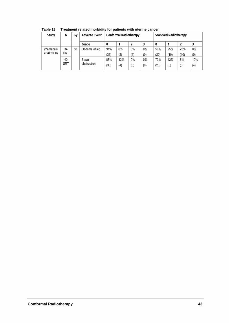
Conformal Radiotherapy 43
Table 18 Treatment related morbidity for patients with uterine cancer Study N Gy Adverse Event Conformal Radiotherapy Standard Radiotherapy
Grade 0 1 2 3 0 1 2 3 34
CRT Oedema of leg
91% (31)
6% (2)
3% (1)
0% (0)
50% (20)
25% (10)
25% (10)
0% (0)
(Yamazaki et al 2000)
40 SRT
50
Bowel obstruction
88% (30)
12% (4)
0% (0)
0% (0)
70% (28)
13% (5)
8% (3)
10% (4)
44
Conclusions
• Substantial methodological limitations exist in the papers evaluating CRT in indications other than prostate cancer. These limitations compromise the ability to draw substantive conclusions based on this data.
• CRT still results in acute and late toxicity. For some indications (such as carcinoma of the cervix and nasopharyngeal carcinoma) the complication rate with CRT may be lower than for conventional radiotherapy. However, poor quality data limits our ability to draw robust conclusions.
• Some low level evidence exists to suggest that CRT, as well as IMRT, may be of benefit in the sparing of normal tissue structures and may contribute to the decrease in toxicity.
• Dose escalation using CRT may be of benefit to some subgroups of patients, however, further evidence is needed in this area.
• There is some evidence to suggest that a reasonable proportion of the toxicity experienced by some patients may be explained by the percentage of the structure receiving high dose radiotherapy.
Conformal Radiotherapy 45
What are the economic considerations?
Multileaf collimators, like all new technologies, represent an additional cost to current therapy. Foroudi et al (2000) estimate that MLC, whether fitted as original equipment or retrofitted, represents a significant additional cost. Depending upon the type of MLC and the supplier, costs can range from $325,000 for a standard MLC, up to $615,000 for a 120 leaf MLC (Foroudi et al 2000). The economic impact of MLC can be viewed in terms of its impact on patient outcomes and on the overall operating cost for radiation therapy.
Cost-effectiveness studies
Seven papers have purported to measure costs and/or benefits of MLC. All of these studies are based on the premise that the outcomes for patients undergoing MLC treatment, compared to non-MLC treatment, are at least equivalent. Only Horwitz et al (1999), in a cost-effectiveness analysis of MLC radiation therapy for patients with clinically localised prostate cancer, reported improved rates of biochemical (bNED) control as an outcome.
Cost implications
Given that clinical equivalence is a reasonable assumption (and clinical superiority can be established in some patient groups with localised cancer) the focus on the cost implications of MLC is reasonable.
The main cost implications for MLC are:
• Its ability to decrease the average duration of radiation treatment and hence increase the productivity of the linear accelerator (by increasing patient throughput); and
• The reduction, if not elimination, of the need to manufacture blocks. Cost savings arising from reduced mould room labour and supplies.
The extent to which these cost savings offset the additional annuitised cost of purchasing and maintaining MLC depends on the assumptions made in the economic study and the cost structure of the radiation therapy department. All but one of the papers summarised in Table 32 are based on studies conducted in the US or Canada. Health service costs in these countries are not generalisable to Australia. Furthermore, all the US studies use reimbursement fees or charges (not costs), making cost comparisons difficult, if not impossible.
The only local economic evaluation was conducted by Foroudi et al (2000) and compared the costs of MLC to non-MLC radiation treatment over a three-month period at the joint radiation oncology departments of Westmead Hospital and Nepean Hospital. The study included all of the capital costs associated with radiation treatment (with and without MLC) and annual recurrent costs (staff, consumables and overheads) of a linear accelerator. Output was measured by the number of fields treated per machine over three months. Data were also collected (for one month and one week) using the equivalent
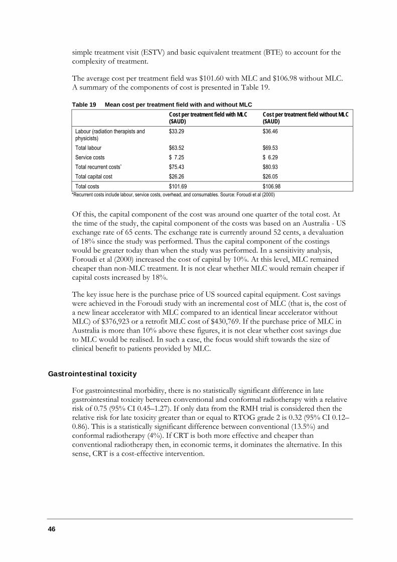
46
simple treatment visit (ESTV) and basic equivalent treatment (BTE) to account for the complexity of treatment.
The average cost per treatment field was $101.60 with MLC and $106.98 without MLC. A summary of the components of cost is presented in Table 19.
Table 19 Mean cost per treatment field with and without MLC Cost per treatment field with MLC
($AUD) Cost per treatment field without MLC ($AUD)
Labour (radiation therapists and physicists)
$33.29 $36.46
Total labour $63.52 $69.53 Service costs $ 7.25 $ 6.29 Total recurrent costs* $75.43 $80.93 Total capital cost $26.26 $26.05 Total costs $101.69 $106.98
*Recurrent costs include labour, service costs, overhead, and consumables. Source: Foroudi et al (2000)
Of this, the capital component of the cost was around one quarter of the total cost. At the time of the study, the capital component of the costs was based on an Australia - US exchange rate of 65 cents. The exchange rate is currently around 52 cents, a devaluation of 18% since the study was performed. Thus the capital component of the costings would be greater today than when the study was performed. In a sensitivity analysis, Foroudi et al (2000) increased the cost of capital by 10%. At this level, MLC remained cheaper than non-MLC treatment. It is not clear whether MLC would remain cheaper if capital costs increased by 18%.
The key issue here is the purchase price of US sourced capital equipment. Cost savings were achieved in the Foroudi study with an incremental cost of MLC (that is, the cost of a new linear accelerator with MLC compared to an identical linear accelerator without MLC) of $376,923 or a retrofit MLC cost of $430,769. If the purchase price of MLC in Australia is more than 10% above these figures, it is not clear whether cost savings due to MLC would be realised. In such a case, the focus would shift towards the size of clinical benefit to patients provided by MLC.
Gastrointestinal toxicity
For gastrointestinal morbidity, there is no statistically significant difference in late gastrointestinal toxicity between conventional and conformal radiotherapy with a relative risk of 0.75 (95% CI 0.45–1.27). If only data from the RMH trial is considered then the relative risk for late toxicity greater than or equal to RTOG grade 2 is 0.32 (95% CI 0.12–0.86). This is a statistically significant difference between conventional (13.5%) and conformal radiotherapy (4%). If CRT is both more effective and cheaper than conventional radiotherapy then, in economic terms, it dominates the alternative. In this sense, CRT is a cost-effective intervention.
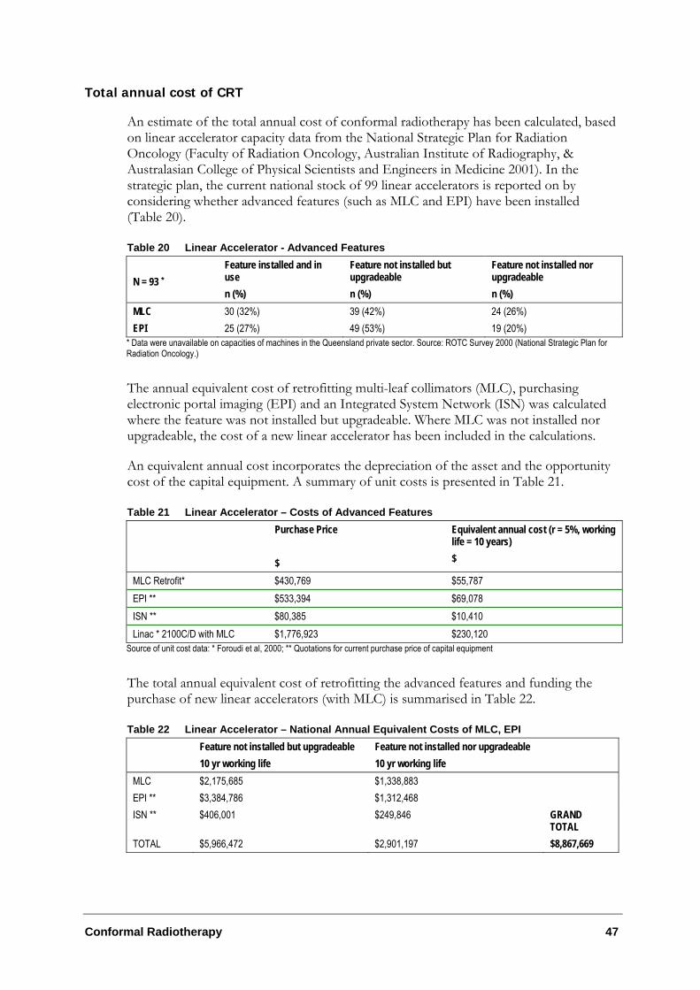
Conformal Radiotherapy 47
Total annual cost of CRT
An estimate of the total annual cost of conformal radiotherapy has been calculated, based on linear accelerator capacity data from the National Strategic Plan for Radiation Oncology (Faculty of Radiation Oncology, Australian Institute of Radiography, & Australasian College of Physical Scientists and Engineers in Medicine 2001). In the strategic plan, the current national stock of 99 linear accelerators is reported on by considering whether advanced features (such as MLC and EPI) have been installed (Table 20).
Table 20 Linear Accelerator - Advanced Features N = 93 *
Feature installed and in use n (%)
Feature not installed but upgradeable n (%)
Feature not installed nor upgradeable n (%)
MLC 30 (32%) 39 (42%) 24 (26%) EPI 25 (27%) 49 (53%) 19 (20%)
* Data were unavailable on capacities of machines in the Queensland private sector. Source: ROTC Survey 2000 (National Strategic Plan for Radiation Oncology.)
The annual equivalent cost of retrofitting multi-leaf collimators (MLC), purchasing electronic portal imaging (EPI) and an Integrated System Network (ISN) was calculated where the feature was not installed but upgradeable. Where MLC was not installed nor upgradeable, the cost of a new linear accelerator has been included in the calculations.
An equivalent annual cost incorporates the depreciation of the asset and the opportunity cost of the capital equipment. A summary of unit costs is presented in Table 21.
Table 21 Linear Accelerator – Costs of Advanced Features
Purchase Price $
Equivalent annual cost (r = 5%, working life = 10 years) $
MLC Retrofit* $430,769 $55,787 EPI ** $533,394 $69,078 ISN ** $80,385 $10,410 Linac * 2100C/D with MLC $1,776,923 $230,120
Source of unit cost data: * Foroudi et al, 2000; ** Quotations for current purchase price of capital equipment
The total annual equivalent cost of retrofitting the advanced features and funding the purchase of new linear accelerators (with MLC) is summarised in Table 22.
Table 22 Linear Accelerator – National Annual Equivalent Costs of MLC, EPI
Feature not installed but upgradeable 10 yr working life
Feature not installed nor upgradeable 10 yr working life
MLC $2,175,685 $1,338,883 EPI ** $3,384,786 $1,312,468 ISN ** $406,001 $249,846 GRAND
TOTAL TOTAL $5,966,472 $2,901,197 $8,867,669
48
Summary
There is some data that indicates that based on the additional costs of MLC alone, CRT appears to be both more effective and less costly than standard RT in some patients groups. However, this data is not comprehensive enough to draw definitive conclusions regarding the cost-effectiveness of conformal radiotherapy.
In addition to MLC, EPI and ISN are components that aid in the delivery of CRT. It is unclear how these additional components will affect the incremental cost-effectiveness ratio.
The estimated total annual equivalent cost of retrofitting MLC, EPI and ISN to existing upgradeable linear accelerators and funding the purchases of new linear accelerators with these features is approximately $9 million.
Conformal Radiotherapy 49
Quality Assurance and Occupational Health and Safety
Quality Assurance (QA)
In recent years there has been a significant increase in the sophistication and complexity of radiotherapy treatment (Fraass et al 1998). Advances in computer hardware and software, and medical imaging have led to the development of new technologies for improving external beam treatment planning, dose delivery and verification of radiotherapy. Three-dimensional treatment planning systems (3D RTP), multileaf collimators and on-line electronic portal imaging are examples of this technology (Michalski & Purdy 1998). The very nature of conformal radiotherapy has meant that these devices have been incorporated into practice. This in turn has implications for quality assurance and quality improvement.
The processes of treatment planning, delivery and verification have in part been discussed in the background section of this report. The focus of this section will be on the application of new technologies, such as integrated oncology management systems and multileaf collimators, to quality assurance in conformal radiotherapy.
Multileaf collimator
Multileaf collimators (MLCs) are a good example of the technological advances made in the field of radiotherapy. Developed for field shaping, and primarily as a replacement for custom shaped blocks, MLCs have had a great impact on the adoption of conformal techniques (Carlson 2001). According to Dunscombe and Roberts (2000), the application of an MLC presents many potential advantages to patients, staff and the radiotherapy institution, including quality assurance.
In comparing the efficacy of MLCs to customised shielding blocks, Powlis et al (1993b) found that equivalent dose distributions were obtained from the two field shaping systems. LoSasso et al (1995) report similar results and state that in some cases MLCs are superior to custom blocks in regards to accuracy. Purdy and Klein (1997) note that in terms of shielding blocks, errors can occur due to the incorrect specification of the magnification factor, block misalignment caused by tray movement in the slot, inaccuracies in the styrofoam cutting process and errors in the mounting or incorrect positioning of the block on the tray (for example, right versus left). It is also possible to accidentally omit the tray, or to place the tray in the wrong orientation. These errors can result in problems such as the shielding of the tumour site or non-shielding of vital structures. A study of the clinical implementation of a commercial MLC as reported by Klein et al (1995) shows that, in addition to a significant decrease in in-field positioning errors, there is also a 25% reduction in in-treatment room time.
MLC also permits rapid field editing by eliminating the need to recast and remount blocks for field adjustments. It permits on-the-spot modification of the field aperture if the portal image reveals that anatomical landmarks are not at expected locations relative to the boundary.
50
Integrated systems networking
Today’s radiotherapy departments utilise computer networking to enable data and images to be transferred and accessed as required (these may be local area networks, or even wide area networks for institutes with multiple sites.) Patient management systems incorporating record and verify (R and V) software utilise a central file server/data base and are interfaced to computer controlled linear accelerators and treatment planning computers.
Conformal radiotherapy using MLCs fits within this concept of an integrated department. The leaf position coordinates of each conformal field are generated as part of the treatment planning process, then saved to file and exported. The field shapes can then be reviewed at any MLC workstation or elsewhere, and imported to the R and V system. During treatment delivery, the MLC controller on the linear accelerator reads the leaf position coordinates. Treatment proceeds when the controller has driven the leaves to the correct positions and the R and V system has confirmed their position.
In addition to the benefits relating to quality control, this process is extremely efficient. The initial export and import of MLC files is very straightforward and daily treatment is fully automated. Departments with more than one MLC machine can readily effect patient transfers in the event of a machine breakdown, even to another site.
Electronic portal imaging (EPI)
Like multileaf collimators, EPI is a new device and is still undergoing development and improvement (Shalev 1996). The impetus for on-line EPI came about as a result of a need for an improved portal imaging system to enhance verification of conformal radiotherapy (Herman et al 2001).
A wide range of factors contribute to set-up uncertainty and therefore the potential for error in field placement. These include erratic patient movement, patient repositioning uncertainties, inter-treatment organ motion uncertainties and intra-treatment organ motion uncertainties due to rhythmic effects such as breathing during treatment. Adherence to quality assurance principles means that it is the responsibility of the department to assess and correct as far as possible for these errors.
Van Dyk (1998) states that port films have been in use for many years as a means of confirmation of the accuracy of beam placement. As port films require developing, there is a delay between the time they are taken and the time they are reviewed. Mostly, the radiation oncologist views the port film after the patient leaves. Therefore, generally these films provide only a retrospective analysis of the field position. Any adjustments necessary are performed before the next treatment. Poor image quality due to a lack of contrast and a large amount of scatter radiation can also be a problem (Hatherly et al 2001).
If individual patient positioning problems indicate the need for field verification prior to treatment a port film is taken with the patient on the treatment couch. The film is processed and reviewed, and any adjustments made before the treatment is initiated. This is an inefficient system, not only because of the time involved but also because the patient may move after the film is taken and before the field is treated.
EPI allows an instant on-line image to be created and reviewed before the treatment, which can achieve savings in time. If the patient is receiving palliative treatment the
Conformal Radiotherapy 51
number of treatment fractions may be as low as one, but is usually between five and ten. Therefore, one geometric miss may constitute 10–100% of the prescribed treatment, impacting significantly on the final treatment outcome.
Thomason (1998) reports that when using EPI, a digital electronic portal image can be obtained within a few seconds and be available for immediate review. Effective use of an EPI will minimise both random and systematic errors, which, by virtue of a geometric miss, can result in the reduction of the dose to the target volume and/or overdose to normal critical tissue. Gross error, including incorrect or misplaced shielding (if using blocks), incorrect collimator rotation, incorrect field size or patient positioning can be detected before a large fraction of the dose has been delivered.
As outlined in the American Association of Physicists in Medicine (AAPM) radiation therapy committee taskforce report on EPI (2001), electronic portal imaging has also been used for quality assurance of treatment machines and treatment techniques such as radiosurgery and dynamic treatment delivery. Investigators have used the EPI device for the design and verification of compensating filters. Electronic portal imaging allows more precise, quantitative results to be obtained with much less effort than would have been achievable using conventional QA tools.
All port films are stored with the patient’s simulator films for the legally required time. The result is a growing number of x-rays requiring storage space. EPI storage is usually onto compact disc (CD). The high cost of radiographic film is eliminated and the fast process of image capture and display improves patient throughput, reducing the cost of staff time per patient.
Occupational Health and Safety Issues
Occupational health and safety issues in relation to conformal radiotherapy largely relate to the application of multileaf collimators and electronic portal imaging in radiotherapy treatment.
Studies specifically pertaining to occupational health and safety in the radiotherapy industry are scant. There is, however, a considerable amount of anecdotal evidence in relation to occupational health and safety concerning beam shaping devices that highlights some of the main issues. It would seem from the literature that there are three main areas of concern for staff as well as some concerns regarding patient-related safety, discussed below.
Fumes inhalation
The main potential for staff to become exposed to toxic fumes is in the casting of shielding blocks and the cutting of the polystyrene moulds for these blocks. While there has been a decrease in the use of pure lead to construct shielding blocks due to the concern over health effects, the alternative, alloy shielding, still contains heavy metals such as cadmium, bismuth, tin and lead. Although not as hazardous as pure lead, the potential health effects of these heavy metals are still of concern to staff. Most studies, while acknowledging the potential for staff to be exposed to toxic vapours, and the hazards of inhaling metallic dust particles and skin absorption of metal alloy as a result of shield block fabrication (DeMeyer et al 1988; Karzmark & Huisman 1972), have reported that the casting process does not seem to present an inhalation hazard to employees
52
(McCullough & Senjem 1981). This is primarily due to the implementation of safety procedures, such as the installation of ventilated rooms and fume extraction equipment to prevent exposure of staff to possible toxic fumes. Multileaf collimators decrease the workload of shielding block fabrication and hence reduce the amount of possible exposure to fumes.
Radiographic portal imaging also produce fumes. Hazardous substances such as glutaraldehyde are used in the developing process. Exposure to these fumes is considered an occupational health and safety risk (Vyas et al 2000). According to Worthington (2001), glutaraldehyde can cause skin and eye irritation and has been linked to occupational asthma among hospital staff. Spillage is also a problem and any effluent from the process must be disposed of thoughtfully (Coulter K, personal communication).
Burns and bruising injuries
The second issue of staff injuries is also of import in the Australian context. It is noted in the paper by DeMeyer et al (1986) that potential hazards to personnel using shielding blocks include burns from handling molten alloy as well as bruises to hands or feet from dropped blocks (Purdy 1983). This is supported by incident records of radiotherapy departments in Australia, which report cuts and bruises to fingers of staff working with shielding trays and blocks. Again, there is relatively little information concerning this issue and what is reported is largely anecdotal. However, it can be assumed that these types of injuries would disappear with the use of multileaf collimators.
Manual handling injuries
In Australia, incorrect manual handling practices account for a significant number of work related injuries, with up to one third of all work injuries in Australia occurring during manual handling (National Occupational Health and Safety Commission 1990). The Australian National Code of Practice on manual handling recommends lifting no more than 16kg in a standard position (National Occupational Health and Safety Commission 1990). Conventional shielding trays with several shielding blocks mounted on them can weigh several kilograms and while most shielding blocks are less than 16kg, it is necessary for these blocks to be lifted repetitively up on to the support trays for each beam, often above shoulder, or even head, height (Lee 1995). In conformal radiotherapy, as the number of fields given to patients is greater than in conventional radiotherapy, radiotherapy staff in a department using CRT and shielding blocks will consequently be required to undertake more manual handling. In a letter to the editor of an occupational health journal, a clinician noted that the lifting of blocks onto the linear accelerator above shoulder height was resulting in radiotherapy staff having back pain and accidents (Beresford 1994). Aribisal (1993) cites a similar situation where to make a customised shielding block a radiation therapist was required to pour a full cup of cerrobend alloy weighing several kilograms into a mould, thus placing substantial strain on the therapist’s ligaments and causing nerve compression and wrist flexion.
Incident statistics obtained from Australian radiotherapy departments seem to be in accordance with these descriptions. The data indicates that back and shoulder pain from lifting shielding blocks is a major contributor to staff injuries and may in fact be underestimated due to masking of work injuries by sick leave. Figures from the National Workers Compensation statistics database (2001) indicate that in 1998–1999 there were
Conformal Radiotherapy 53
21 cases of sprains and strained joints in radiographers in Australia. Again this is likely to be an underestimate of cases due to the method of reporting.
Miller and O’Brien (2001) report on the prevalence of lower back pain among radiation therapists and note that activities such as lifting heavy blocks would seem to put radiation therapists at risk for lower back pain. While the study was relatively small (n=19) and radiation therapists were not randomly selected, 89% of female respondents and 55% of male respondents reported having experienced lower back pain as a result of a task undertaken as part of their work. Miller and O’Brien (2001) propose a number of reasons for such a high percentage including current staff shortages and the lack of ergonomic storage and placement of blocks. The American Association of Physicists in Medicine (AAPM) surveyed US radiotherapy departments and found that not only was ergonomic improvement one of the main reasons for purchase of a multileaf collimator, but that this purchase led to a decrease in workload for therapists. Similarly, in a study concerned with timing and cost effectiveness of beam shaping devices, conventional shielding blocks were compared to multileaf collimation. It was reported that using a multileaf collimator eliminates the need to lift blocks up on to the support trays for each beam and thus reduces the physical workload for staff (Helyer & Heisig 1995). The use of a multileaf collimator in conformal radiotherapy therefore appears to reduce work practices that result in occupational injuries and health hazards for radiotherapy staff.
Patient-related injuries
In addition to staff injuries, patient-related injuries have also been identified as an area of concern in relation to the use of shielding blocks. Experts in the field of radiotherapy have reported instances where cerrobend blocks used for field shaping have fallen on patients, resulting in significant injury (Coulter K, personal communication).
54
Conclusions
Safety
• Limited randomised evidence suggests that acute and late gastrointestinal toxicity is reduced for patients with carcinoma of the prostate treated with conformal radiotherapy compared to standard radiotherapy.
• There is some indicative data from comparative non-randomised studies to suggest that incidence of toxicity for some indications may be lower using conformal radiotherapy than for standard radiotherapy. However, the data for these other indications is relatively small and of poor quality.
Effectiveness
• In the treatment of prostate cancer, conformal radiotherapy results in similar efficacy to that experienced using conventional radiotherapy using similar doses.
• There is some limited randomised and non-randomised evidence to suggest that higher doses of radiotherapy, delivered by conformal radiotherapy, may result in increased efficacy for patients with carcinoma of the prostate.
Cost-effectiveness
• There is some data indicating that based on the additional costs of MLC alone, CRT appears to be both more effective and less costly than standard RT in some patients groups. However, this data is not comprehensive enough to draw definitive conclusions regarding the cost-effectiveness of conformal radiotherapy.
• It is unclear how additional components (EPI and ISN) will affect the incremental cost-effectiveness ratio.
• At this stage there is little data to determine the cost-effectiveness of IMRT.
Other considerations
• There is some descriptive data that would seem to indicate that there are occupational health and safety benefits in using multileaf collimators in comparison to shielding blocks when treating patients with conformal radiotherapy.
Conformal Radiotherapy 55
Recommendation
MSAC recommended that on the strength of evidence pertaining to the safety, efficacy and cost of conformal radiotherapy that public funding should be supported for this procedure and that intensity modulated radiation therapy should be reviewed again at a later date when substantial additional data are available relating to safety, effectiveness and cost effectiveness.
- The Minister for Health and Ageing accepted this recommendation on 5 February 2002 -
56 Conformal Radiotherapy
Appendix A MSAC terms of reference and membership
MSAC's terms of reference are to:
• advise the Minister for Health and Ageing on the strength of evidence pertaining to new and emerging medical technologies and procedures in relation to their safety, effectiveness and cost-effectiveness and under what circumstances public funding should be supported;
• advise the Minister for Health and Ageing on which new medical technologies and procedures should be funded on an interim basis to allow data to be assembled to determine their safety, effectiveness and cost-effectiveness;
• advise the Minister for Health and Ageing on references related either to new and/or existing medical technologies and procedures; and
• undertake health technology assessment work referred by the Australian Health Ministers’ Advisory Council (AHMAC), and report its findings to AHMAC.
The membership of MSAC comprises a mix of clinical expertise covering pathology, nuclear medicine, surgery, specialist medicine and general practice, plus clinical epidemiology and clinical trials, health economics, consumers, and health administration and planning:
Member Expertise or Affiliation
Mr Stephen Blamey (Chair) general surgery
Professor Bruce Barraclough general surgery
Professor Syd Bell pathology
Dr Paul Craft clinical epidemiology and oncology
Professor Ian Fraser reproductive medicine
Associate Professor Jane Hall
Dr Terri Jackson
health economics
health economics
Ms Rebecca James
Professor Brendon Kearney
consumer health issues
health administration and planning
Mr Alan Keith Assistant Secretary, Diagnostics and Technology Branch, Commonwealth Department of Health and Ageing
Associate Professor Richard King internal medicine
Dr Ray Kirk
Dr Michael Kitchener
health research
nuclear medicine
Mr Lou McCallum
Emeritus Professor Peter Phelan
consumer health issues
paediatrics
Dr Ewa Piejko
Dr David Robinson
Professor John Simes
general practice
plastic surgery
clinical epidemiology and clinical trials
Conformal Radiotherapy 57
Professor Richard Smallwood Chief Medical Officer, Commonwealth Department of Health and Ageing
Professor Bryant Stokes neurological surgery, representing the Australian Health Ministers’ Advisory Council
Associate Professor Ken Thomson radiology
Dr Douglas Travis urology
Professor David Weedon pathology (Chair until 24/08/01)
Ms Hilda Bastian consumer health issues (Member until 24/08/01)
Dr Ross Blair vascular surgery (New Zealand)(Member until 24/08/01)
Dr Paul Hemming general practice (Member until 24/08/01)
58 Conformal Radiotherapy
Appendix B Supporting committee
Supporting committee for MSAC application 1038 for conformal radiotherapy
Dr John Primrose (Chair) MBBS (Hons), FRACR Senior Medical Adviser Health Access and Financing Division Commonwealth Department of Health and Ageing Canberra
Adviser to MSAC
Mr James Cramb MSc, MACPSEM Director of the Physical Science Department Peter MacCallum Cancer Institute Melbourne
nominated by the Australasian College of Physical Scientists and Engineers in Medicine
Ms Kristine Coulter Certificate of Competence, Australian Institute of Radiography (Radiation Therapy), Member-AIR Director of Radiation Therapy Services North Queensland Oncology Service
nominated by the Australian Institute of Radiography
Dr Geoffrey Delaney MBBS (Hons), FRANZCR Staff Specialist Cancer Therapy Centre Liverpool Hospital
nominated by the Royal Australian and New Zealand College of Radiologists
Professor Martin Lavin MBBS (Hons), PhD Professor of Molecular Oncology University of Queensland; Assistant Director Queensland Institute of Medical Research
nominated by the
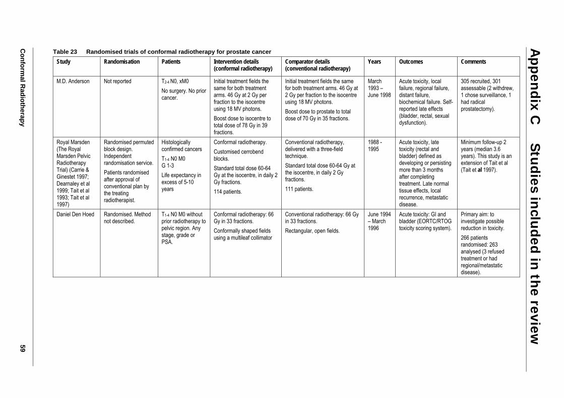
Conform
al Radiotherapy
59
Table 23 Randomised trials of conformal radiotherapy for prostate cancer Study
Randomisation Patients Intervention details (conformal radiotherapy)
Comparator details (conventional radiotherapy)
Years Outcomes Comments
M.D. Anderson Not reported T2-4 N0, xM0 No surgery. No prior cancer.
Initial treatment fields the same for both treatment arms. 46 Gy at 2 Gy per fraction to the isocentre using 18 MV photons. Boost dose to isocentre to total dose of 78 Gy in 39 fractions.
Initial treatment fields the same for both treatment arms. 46 Gy at 2 Gy per fraction to the isocentre using 18 MV photons. Boost dose to prostate to total dose of 70 Gy in 35 fractions.
March 1993 – June 1998
Acute toxicity, local failure, regional failure, distant failure, biochemical failure. Self-reported late effects (bladder, rectal, sexual dysfunction).
305 recruited, 301 assessable (2 withdrew, 1 chose surveillance, 1 had radical prostatectomy).
Royal Marsden (The Royal Marsden Pelvic Radiotherapy Trial) (Carrie & Ginestet 1997; Dearnaley et al 1999; Tait et al 1993; Tait et al 1997)
Randomised permuted block design. Independent randomisation service. Patients randomised after approval of conventional plan by the treating radiotherapist.
Histologically confirmed cancers T1-4 N0 M0 G 1-3 Life expectancy in excess of 5-10 years
Conformal radiotherapy. Customised cerrobend blocks. Standard total dose 60-64 Gy at the isocentre, in daily 2 Gy fractions. 114 patients.
Conventional radiotherapy, delivered with a three-field technique. Standard total dose 60-64 Gy at the isocentre, in daily 2 Gy fractions. 111 patients.
1988 - 1995
Acute toxicity, late toxicity (rectal and bladder) defined as developing or persisting more than 3 months after completing treatment. Late normal tissue effects, local recurrence, metastatic disease.
Minimum follow-up 2 years (median 3.6 years). This study is an extension of Tait et al (Tait et al 1997).
Daniel Den Hoed Randomised. Method not described.
T1-4 N0 M0 without prior radiotherapy to pelvic region. Any stage, grade or PSA.
Conformal radiotherapy: 66 Gy in 33 fractions. Conformally shaped fields using a multileaf collimator
Conventional radiotherapy: 66 Gy in 33 fractions. Rectangular, open fields.
June 1994 – March 1996
Acute toxicity: GI and bladder (EORTC/RTOG toxicity scoring system).
Primary aim: to investigate possible reduction in toxicity. 266 patients randomised: 263 analysed (3 refused treatment or had regional/metastatic disease).
Appen
dix C
Studies in
cluded in
the review
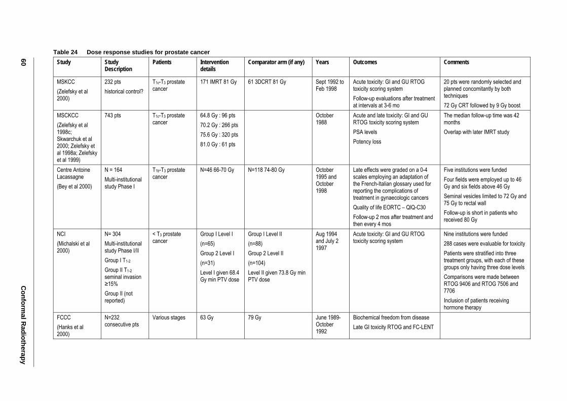
60 C
onformal R
adiotherapy
Table 24 Dose response studies for prostate cancer Study
Study Description
Patients Intervention details
Comparator arm (if any) Years Outcomes Comments
MSKCC (Zelefsky et al 2000)
232 pts historical control?
T1c-T3 prostate cancer
171 IMRT 81 Gy 61 3DCRT 81 Gy Sept 1992 to Feb 1998
Acute toxicity: GI and GU RTOG toxicity scoring system Follow-up evaluations after treatment at intervals at 3-6 mo
20 pts were randomly selected and planned concomitantly by both techniques 72 Gy CRT followed by 9 Gy boost
MSCKCC (Zelefsky et al 1998c; Skwarchuk et al 2000; Zelefsky et al 1998a; Zelefsky et al 1999)
743 pts T1c-T3 prostate cancer
64.8 Gy : 96 pts 70.2 Gy : 266 pts 75.6 Gy : 320 pts 81.0 Gy : 61 pts
October 1988
Acute and late toxicity: GI and GU RTOG toxicity scoring system PSA levels Potency loss
The median follow-up time was 42 months Overlap with later IMRT study
Centre Antoine Lacassagne (Bey et al 2000)
N = 164 Multi-institutional study Phase I
T1b-T3 prostate cancer
N=46 66-70 Gy N=118 74-80 Gy October 1995 and October 1998
Late effects were graded on a 0-4 scales employing an adaptation of the French-Italian glossary used for reporting the complications of treatment in gynaecologic cancers Quality of life EORTC – QlQ-C30 Follow-up 2 mos after treatment and then every 4 mos
Five institutions were funded Four fields were employed up to 46 Gy and six fields above 46 Gy Seminal vesicles limited to 72 Gy and 75 Gy to rectal wall Follow-up is short in patients who received 80 Gy
NCI (Michalski et al 2000)
N= 304 Multi-institutional study Phase I/II Group I T1-2 Group II T1-2 seminal invasion ≥15% Group II (not reported)
< T3 prostate cancer
Group I Level I (n=65) Group 2 Level I (n=31) Level I given 68.4 Gy min PTV dose
Group I Level II (n=88) Group 2 Level II (n=104) Level II given 73.8 Gy min PTV dose
Aug 1994 and July 2 1997
Acute toxicity: GI and GU RTOG toxicity scoring system
Nine institutions were funded 288 cases were evaluable for toxicity Patients were stratified into three treatment groups, with each of these groups only having three dose levels Comparisons were made between RTOG 9406 and RTOG 7506 and 7706 Inclusion of patients receiving hormone therapy
FCCC (Hanks et al 2000)
N=232 consecutive pts
Various stages 63 Gy 79 Gy June 1989-October 1992
Biochemical freedom from disease Late GI toxicity RTOG and FC-LENT
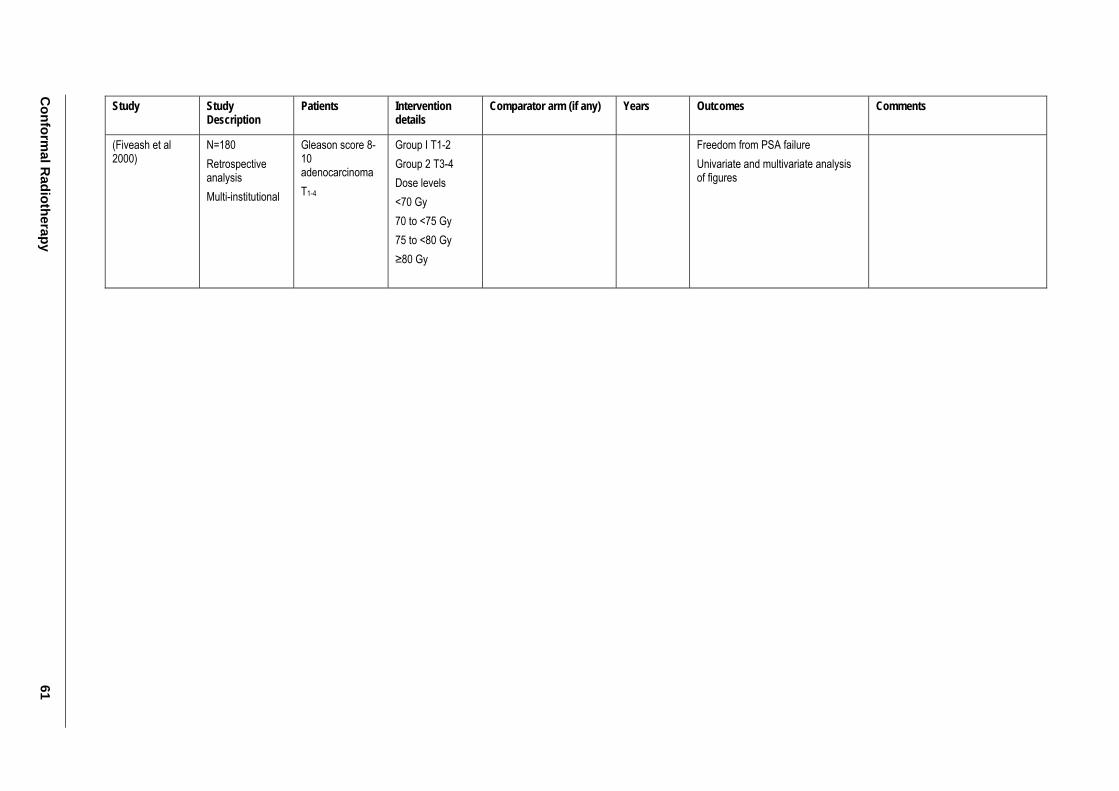
Conform
al Radiotherapy
61
Study
Study Description
Patients Intervention details
Comparator arm (if any) Years Outcomes Comments
(Fiveash et al 2000)
N=180 Retrospective analysis Multi-institutional
Gleason score 8-10 adenocarcinoma T1-4
Group I T1-2 Group 2 T3-4 Dose levels <70 Gy 70 to <75 Gy 75 to <80 Gy ≥80 Gy
Freedom from PSA failure Univariate and multivariate analysis of figures
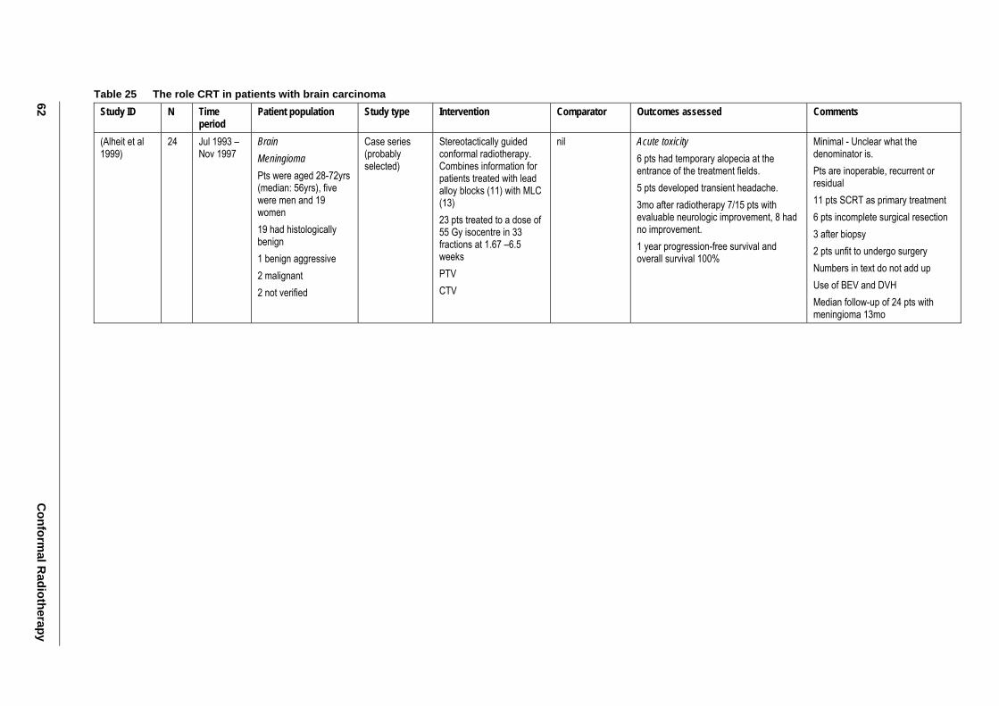
62 C
onformal R
adiotherapy
Table 25 The role CRT in patients with brain carcinoma Study ID N Time
period Patient population Study type Intervention Comparator Outcomes assessed Comments
(Alheit et al 1999)
24 Jul 1993 – Nov 1997
Brain Meningioma Pts were aged 28-72yrs (median: 56yrs), five were men and 19 women 19 had histologically benign 1 benign aggressive 2 malignant 2 not verified
Case series (probably selected)
Stereotactically guided conformal radiotherapy. Combines information for patients treated with lead alloy blocks (11) with MLC (13) 23 pts treated to a dose of 55 Gy isocentre in 33 fractions at 1.67 –6.5 weeks PTV CTV
nil Acute toxicity 6 pts had temporary alopecia at the entrance of the treatment fields. 5 pts developed transient headache. 3mo after radiotherapy 7/15 pts with evaluable neurologic improvement, 8 had no improvement. 1 year progression-free survival and overall survival 100%
Minimal - Unclear what the denominator is. Pts are inoperable, recurrent or residual 11 pts SCRT as primary treatment 6 pts incomplete surgical resection 3 after biopsy 2 pts unfit to undergo surgery Numbers in text do not add up Use of BEV and DVH Median follow-up of 24 pts with meningioma 13mo
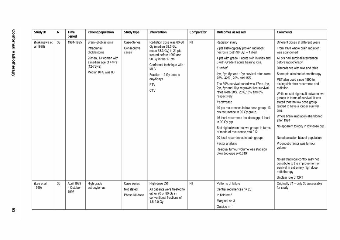
Conform
al Radiotherapy
63
Study ID N Time period
Patient population Study type Intervention Comparator Outcomes assessed Comments
(Nakagawa et al 1998)
38 1984-1995 Brain- glioblastoma Intracranial glioblastoma 25men, 13 women with a median age of 47yrs (12-73yrs) Median KPS was 80
Case-Series Consecutive cases
Radiation dose was 60-80 Gy (median 68.5 Gy, mean 68.3 Gy) in 21 pts treated before 1990 and 90 Gy in the 17 pts Conformal technique with MLC Fraction – 2 Gy once a day/5days PTV CTV
Nil Radiation injury 2 pts Histologically proven radiation necrosis (both 90 Gy) – 1 died 4 pts with grade II acute skin injuries and 3 with Grade II acute hearing loss. Survival 1yr, 2yr, 5yr and 10yr survival rates were 75%, 42% , 20% and 15%. The 50% survival period was 17mo. 1yr, 2yr, 5yr and 10yr regrowth-free survival rates were 28%, 25%,13% and 8% respectively. Recurrence 19 pts recurrences in low dose group; 13 pts recurrence in 90 Gy group. 16 local recurrence low dose grp; 4 local in 90 Gy grp Stat sig between the two groups in terms of mode of recurrence p=0.012 20 local recurrences in both groups Factor analysis Residual tumour volume was stat sign btwn two grps p=0.019
Different doses at different years From 1991 whole brain radiation was abandoned All pts had surgical intervention before radiotherapy Discordance with text and table Some pts also had chemotherapy PET also used since 1990 to distinguish btwn recurrence and radiation. While no stat sig result between two groups in terms of survival, it was stated that the low dose group tended to have a longer survival time. Whole brain irradiation abandoned after 1991 No apparent toxicity in low dose grp Noted selection bias of population Prognostic factor was tumour volume Noted that local control may not contribute to the improvement of survival in extremely high dose radiotherapy Unclear role of CRT
(Lee et al 1999)
36
April 1989 – October 1995
High grade astrocytomas
Case series Not stated Phase I/II dose
High dose CRT All patients were treated to either 70 or 80 Gy in conventional fractions of 1.8-2.0 Gy
Nil Patterns of failure Central recurrences n= 26 In field n= 6 Marginal n= 3 Outside n= 1
Originally 71 – only 36 assessable for study
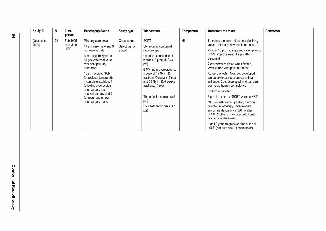
64 C
onformal R
adiotherapy
Study ID N Time period
Patient population Study type Intervention Comparator Outcomes assessed Comments
(Jalali et al 2000)
22 Feb 1995 and March 1999
Pituitary adenomas 14 pts were male and 8 pts were female Mean age 45.3yrs: 20-67 yrs with residual or recurrent pituitary adenomas 15 pts received SCRT for residual tumour after incomplete excision, 4 following progression after surgery and medical therapy and 3 for recurrent tumour after surgery alone.
Case-series Selection not stated
SCRT Stereotactic conformal radiotherapy Use of customised lead blocks (19 pts); MLC (3 pts) 6-MV linear accelerator to a dose of 45 Gy in 25 fractions /5weeks (18 pts) and 50 Gy in 30/6 weeks fractions. (4 pts) Three-field techniques (5 pts) Four field techniques (17 pts)
Nil Secretory tumours – 6 pts had declining values of initially elevated hormones. Vision - 15 pts had impaired vision prior to SCRT, improvement of 9 pts after treatment 2 cases where vision was affected, 2weeks and 7mo post treatment Adverse effects - Most pts developed temporary localised alopecia at beam entrance, 5 pts developed mild transient post radiotherapy somnolence Endocrine function 6 pts at the time of SCRT were on HRT. Of 6 pts with normal pituitary function prior to radiotherapy, 2 developed endocrine deficiency at 5/6mo after SCRT. 2 other pts required additional hormone replacement 1 and 2 year progression-free survival 100% (not sure about denominator)
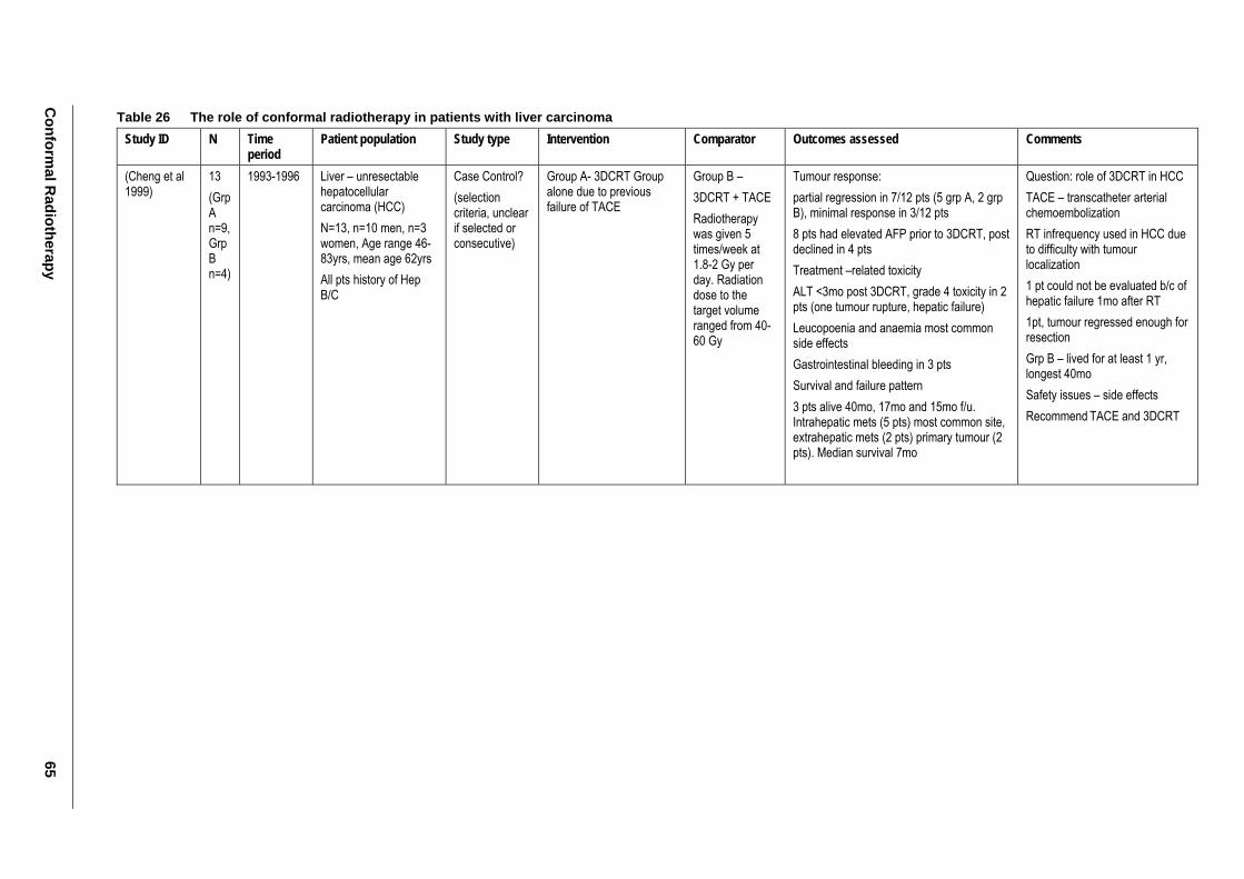
Conform
al Radiotherapy
65
Table 26 The role of conformal radiotherapy in patients with liver carcinoma Study ID N Time
period Patient population Study type Intervention Comparator Outcomes assessed Comments
(Cheng et al 1999)
13 (Grp A n=9, Grp B n=4)
1993-1996 Liver – unresectable hepatocellular carcinoma (HCC) N=13, n=10 men, n=3 women, Age range 46-83yrs, mean age 62yrs All pts history of Hep B/C
Case Control? (selection criteria, unclear if selected or consecutive)
Group A- 3DCRT Group alone due to previous failure of TACE
Group B – 3DCRT + TACE Radiotherapy was given 5 times/week at 1.8-2 Gy per day. Radiation dose to the target volume ranged from 40-60 Gy
Tumour response: partial regression in 7/12 pts (5 grp A, 2 grp B), minimal response in 3/12 pts 8 pts had elevated AFP prior to 3DCRT, post declined in 4 pts Treatment –related toxicity ALT <3mo post 3DCRT, grade 4 toxicity in 2 pts (one tumour rupture, hepatic failure) Leucopoenia and anaemia most common side effects Gastrointestinal bleeding in 3 pts Survival and failure pattern 3 pts alive 40mo, 17mo and 15mo f/u. Intrahepatic mets (5 pts) most common site, extrahepatic mets (2 pts) primary tumour (2 pts). Median survival 7mo
Question: role of 3DCRT in HCC TACE – transcatheter arterial chemoembolization RT infrequency used in HCC due to difficulty with tumour localization 1 pt could not be evaluated b/c of hepatic failure 1mo after RT 1pt, tumour regressed enough for resection Grp B – lived for at least 1 yr, longest 40mo Safety issues – side effects Recommend TACE and 3DCRT
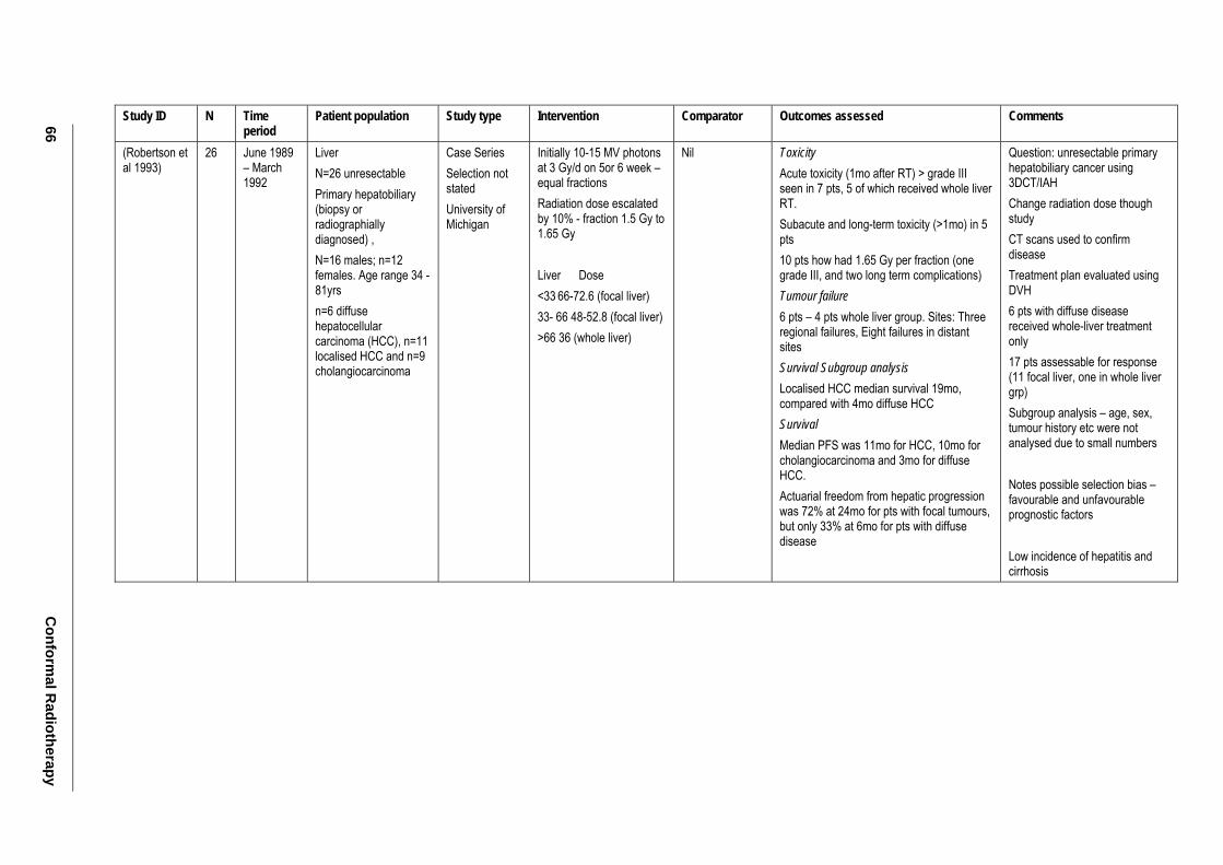
66 C
onformal R
adiotherapy
Study ID N Time period
Patient population Study type Intervention Comparator Outcomes assessed Comments
(Robertson et al 1993)
26 June 1989 – March 1992
Liver N=26 unresectable Primary hepatobiliary (biopsy or radiographially diagnosed) , N=16 males; n=12 females. Age range 34 - 81yrs n=6 diffuse hepatocellular carcinoma (HCC), n=11 localised HCC and n=9 cholangiocarcinoma
Case Series Selection not stated University of Michigan
Initially 10-15 MV photons at 3 Gy/d on 5or 6 week – equal fractions Radiation dose escalated by 10% - fraction 1.5 Gy to 1.65 Gy Liver Dose <33 66-72.6 (focal liver) 33- 66 48-52.8 (focal liver) >66 36 (whole liver)
Nil Toxicity Acute toxicity (1mo after RT) > grade III seen in 7 pts, 5 of which received whole liver RT. Subacute and long-term toxicity (>1mo) in 5 pts 10 pts how had 1.65 Gy per fraction (one grade III, and two long term complications) Tumour failure 6 pts – 4 pts whole liver group. Sites: Three regional failures, Eight failures in distant sites Survival Subgroup analysis Localised HCC median survival 19mo, compared with 4mo diffuse HCC Survival Median PFS was 11mo for HCC, 10mo for cholangiocarcinoma and 3mo for diffuse HCC. Actuarial freedom from hepatic progression was 72% at 24mo for pts with focal tumours, but only 33% at 6mo for pts with diffuse disease
Question: unresectable primary hepatobiliary cancer using 3DCT/IAH Change radiation dose though study CT scans used to confirm disease Treatment plan evaluated using DVH 6 pts with diffuse disease received whole-liver treatment only 17 pts assessable for response (11 focal liver, one in whole liver grp) Subgroup analysis – age, sex, tumour history etc were not analysed due to small numbers Notes possible selection bias – favourable and unfavourable prognostic factors Low incidence of hepatitis and cirrhosis
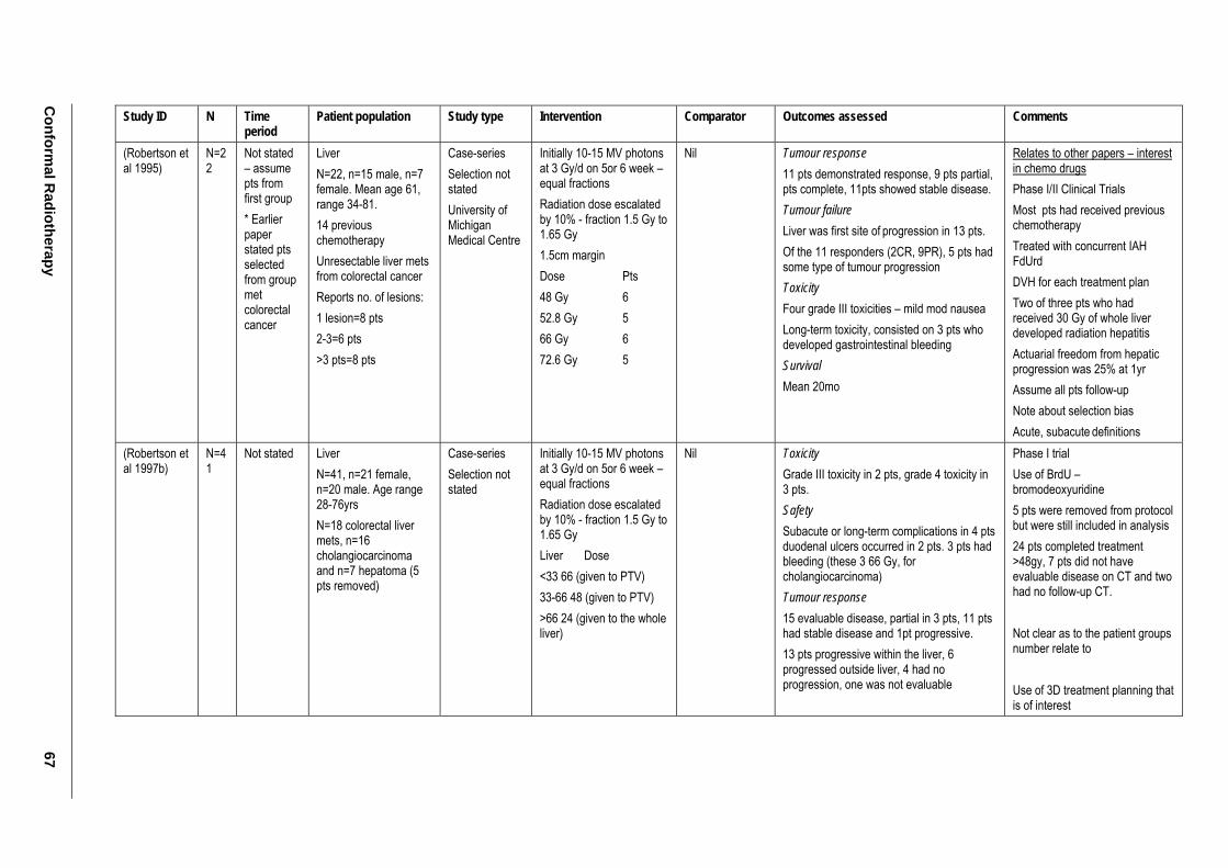
Conform
al Radiotherapy
67
Study ID N Time period
Patient population Study type Intervention Comparator Outcomes assessed Comments
(Robertson et al 1995)
N=22
Not stated – assume pts from first group * Earlier paper stated pts selected from group met colorectal cancer
Liver N=22, n=15 male, n=7 female. Mean age 61, range 34-81. 14 previous chemotherapy Unresectable liver mets from colorectal cancer Reports no. of lesions: 1 lesion=8 pts 2-3=6 pts >3 pts=8 pts
Case-series Selection not stated University of Michigan Medical Centre
Initially 10-15 MV photons at 3 Gy/d on 5or 6 week – equal fractions Radiation dose escalated by 10% - fraction 1.5 Gy to 1.65 Gy 1.5cm margin Dose Pts 48 Gy 6 52.8 Gy 5 66 Gy 6 72.6 Gy 5
Nil Tumour response 11 pts demonstrated response, 9 pts partial, pts complete, 11pts showed stable disease. Tumour failure Liver was first site of progression in 13 pts. Of the 11 responders (2CR, 9PR), 5 pts had some type of tumour progression Toxicity Four grade III toxicities – mild mod nausea Long-term toxicity, consisted on 3 pts who developed gastrointestinal bleeding Survival Mean 20mo
Relates to other papers – interest in chemo drugs Phase I/II Clinical Trials Most pts had received previous chemotherapy Treated with concurrent IAH FdUrd DVH for each treatment plan Two of three pts who had received 30 Gy of whole liver developed radiation hepatitis Actuarial freedom from hepatic progression was 25% at 1yr Assume all pts follow-up Note about selection bias Acute, subacute definitions
(Robertson et al 1997b)
N=41
Not stated Liver N=41, n=21 female, n=20 male. Age range 28-76yrs N=18 colorectal liver mets, n=16 cholangiocarcinoma and n=7 hepatoma (5 pts removed)
Case-series Selection not stated
Initially 10-15 MV photons at 3 Gy/d on 5or 6 week – equal fractions Radiation dose escalated by 10% - fraction 1.5 Gy to 1.65 Gy Liver Dose <33 66 (given to PTV) 33-66 48 (given to PTV) >66 24 (given to the whole liver)
Nil Toxicity Grade III toxicity in 2 pts, grade 4 toxicity in 3 pts. Safety Subacute or long-term complications in 4 pts duodenal ulcers occurred in 2 pts. 3 pts had bleeding (these 3 66 Gy, for cholangiocarcinoma) Tumour response 15 evaluable disease, partial in 3 pts, 11 pts had stable disease and 1pt progressive. 13 pts progressive within the liver, 6 progressed outside liver, 4 had no progression, one was not evaluable
Phase I trial Use of BrdU – bromodeoxyuridine 5 pts were removed from protocol but were still included in analysis 24 pts completed treatment >48gy, 7 pts did not have evaluable disease on CT and two had no follow-up CT. Not clear as to the patient groups number relate to Use of 3D treatment planning that is of interest
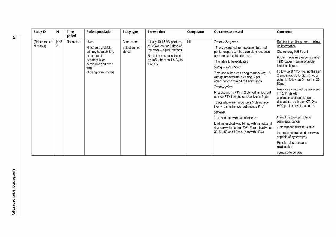
68 C
onformal R
adiotherapy
Study ID N Time period
Patient population Study type Intervention Comparator Outcomes assessed Comments
(Robertson et al 1997a)
N=22
Not stated Liver N=22 unresectable primary hepatobiliary cancer (n=11 hepatocellular carcinoma and n=11 with cholangiocarcinoma)
Case-series Selection not stated
Initially 10-15 MV photons at 3 Gy/d on 5or 6 days of the week – equal fractions Radiation dose escalated by 10% - fraction 1.5 Gy to 1.65 Gy
Nil Tumour Response 11 pts evaluated for response, 9pts had partial response, 1 had complete response and one had stable disease. 11 unable to be evaluated Safety – side effects 7 pts had subacute or long-term toxicity – 5 with gastrointestinal bleeding. 2 pts complications related to biliary tubes. Tumour failure First site within PTV in 2 pts, within liver but outside PTV in 6 pts, outside liver in 9 pts 10 pts who were responders 5 pts outside liver, 4 pts in the liver but outside PTV Survival 7 pts without evidence of disease Median survival was 16mo, with an actuarial 4-yr survival of about 20%. Four pts alive at 39, 51, 52 and 59 mo. (one with HCC)
Relates to earlier papers – follow-up information Chemo drug IAH FdUrd Paper makes reference to earlier 1993 paper in terms of acute toxicities figures Follow-up at 1mo, 1-2 mo then an 2-3mo intervals for 2yrs (median potential follow-up 54months, 27-69mo) Response could not be assessed in 10/11 pts with cholangiocarcinomas their disease not visible on CT. One HCC pt also developed mets One pt discovered to have pancreatic cancer 7 pts without disease, 3 alive liver outside irradiated area was capable of hypertrophy Possible dose-response relationship compare to surgery
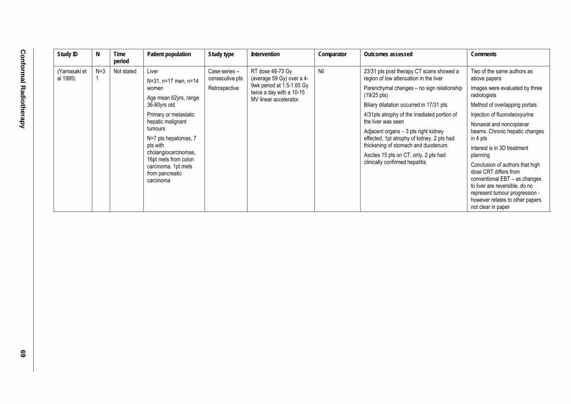
Conform
al Radiotherapy
69
Study ID N Time period
Patient population Study type Intervention Comparator Outcomes assessed Comments
(Yamasaki et al 1995)
N=31
Not stated Liver N=31, n=17 men, n=14 women Age mean 62yrs, range 36-80yrs old Primary or metastatic hepatic malignant tumours N=7 pts hepatomas, 7 pts with cholangiocarcinomas, 16pt mets from colon carcinoma, 1pt mets from pancreatic carcinoma
Case-series – consecutive pts Retrospective
RT dose 48-73 Gy (average 59 Gy) over a 4-9wk period at 1.5-1.65 Gy twice a day with a 10-15 MV linear accelerator.
Nil 23/31 pts post therapy CT scans showed a region of low attenuation in the liver Parenchymal changes – no sign relationship (19/25 pts) Biliary dilatation occurred in 17/31 pts 4/31pts atrophy of the irradiated portion of the liver was seen Adjacent organs – 3 pts right kidney effected, 1pt atrophy of kidney. 2 pts had thickening of stomach and duodenum. Ascites 15 pts on CT, only, 2 pts had clinically confirmed hepatitis
Two of the same authors as above papers Images were evaluated by three radiologists Method of overlapping portals Injection of fluorodeoxyurine Nonaxial and noncoplanar beams. Chronic hepatic changes in 4 pts Interest is in 3D treatment planning Conclusion of authors that high dose CRT differs from conventional EBT – as changes to liver are reversible, do no represent tumour progression - however relates to other papers not clear in paper
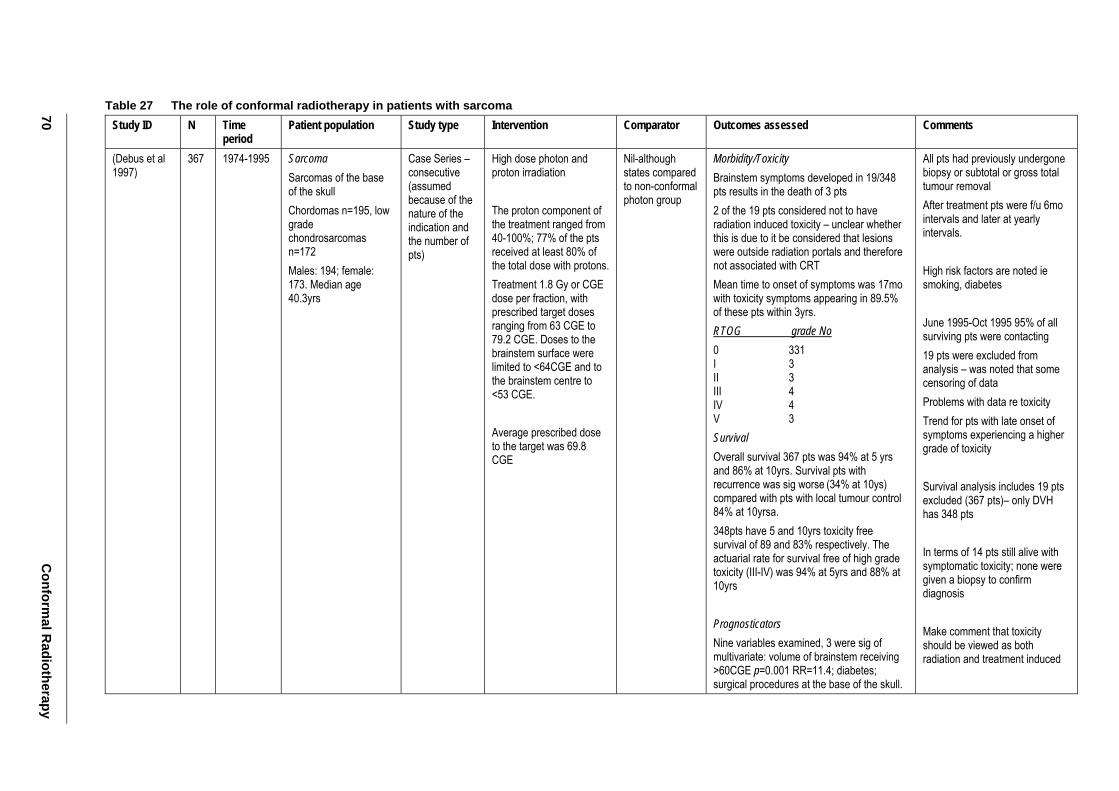
70 C
onformal R
adiotherapy
Table 27 The role of conformal radiotherapy in patients with sarcoma Study ID N Time
period Patient population Study type Intervention Comparator Outcomes assessed Comments
(Debus et al 1997)
367 1974-1995 Sarcoma Sarcomas of the base of the skull Chordomas n=195, low grade chondrosarcomas n=172 Males: 194; female: 173. Median age 40.3yrs
Case Series – consecutive (assumed because of the nature of the indication and the number of pts)
High dose photon and proton irradiation The proton component of the treatment ranged from 40-100%; 77% of the pts received at least 80% of the total dose with protons. Treatment 1.8 Gy or CGE dose per fraction, with prescribed target doses ranging from 63 CGE to 79.2 CGE. Doses to the brainstem surface were limited to <64CGE and to the brainstem centre to <53 CGE. Average prescribed dose to the target was 69.8 CGE
Nil-although states compared to non-conformal photon group
Morbidity/Toxicity Brainstem symptoms developed in 19/348 pts results in the death of 3 pts 2 of the 19 pts considered not to have radiation induced toxicity – unclear whether this is due to it be considered that lesions were outside radiation portals and therefore not associated with CRT Mean time to onset of symptoms was 17mo with toxicity symptoms appearing in 89.5% of these pts within 3yrs. RTOG grade No 0 331 I 3 II 3 III 4 IV 4 V 3 Survival Overall survival 367 pts was 94% at 5 yrs and 86% at 10yrs. Survival pts with recurrence was sig worse (34% at 10ys) compared with pts with local tumour control 84% at 10yrsa. 348pts have 5 and 10yrs toxicity free survival of 89 and 83% respectively. The actuarial rate for survival free of high grade toxicity (III-IV) was 94% at 5yrs and 88% at 10yrs Prognosticators Nine variables examined, 3 were sig of multivariate: volume of brainstem receiving >60CGE p=0.001 RR=11.4; diabetes; surgical procedures at the base of the skull.
All pts had previously undergone biopsy or subtotal or gross total tumour removal After treatment pts were f/u 6mo intervals and later at yearly intervals. High risk factors are noted ie smoking, diabetes June 1995-Oct 1995 95% of all surviving pts were contacting 19 pts were excluded from analysis – was noted that some censoring of data Problems with data re toxicity Trend for pts with late onset of symptoms experiencing a higher grade of toxicity Survival analysis includes 19 pts excluded (367 pts)– only DVH has 348 pts In terms of 14 pts still alive with symptomatic toxicity; none were given a biopsy to confirm diagnosis Make comment that toxicity should be viewed as both radiation and treatment induced
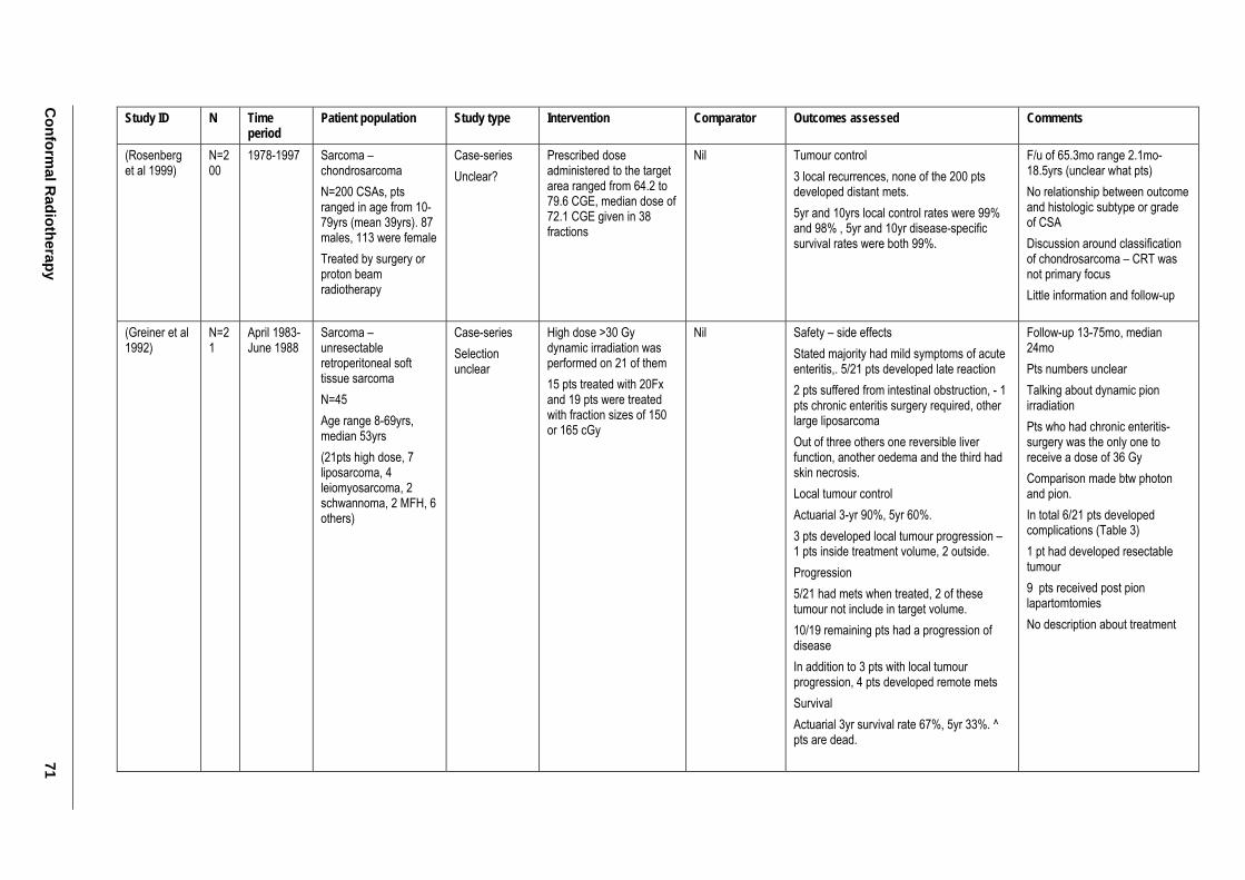
Conform
al Radiotherapy
71
Study ID N Time period
Patient population Study type Intervention Comparator Outcomes assessed Comments
(Rosenberg et al 1999)
N=200
1978-1997 Sarcoma – chondrosarcoma N=200 CSAs, pts ranged in age from 10-79yrs (mean 39yrs). 87 males, 113 were female Treated by surgery or proton beam radiotherapy
Case-series Unclear?
Prescribed dose administered to the target area ranged from 64.2 to 79.6 CGE, median dose of 72.1 CGE given in 38 fractions
Nil Tumour control 3 local recurrences, none of the 200 pts developed distant mets. 5yr and 10yrs local control rates were 99% and 98% , 5yr and 10yr disease-specific survival rates were both 99%.
F/u of 65.3mo range 2.1mo-18.5yrs (unclear what pts) No relationship between outcome and histologic subtype or grade of CSA Discussion around classification of chondrosarcoma – CRT was not primary focus Little information and follow-up
(Greiner et al 1992)
N=21
April 1983- June 1988
Sarcoma – unresectable retroperitoneal soft tissue sarcoma N=45 Age range 8-69yrs, median 53yrs (21pts high dose, 7 liposarcoma, 4 leiomyosarcoma, 2 schwannoma, 2 MFH, 6 others)
Case-series Selection unclear
High dose >30 Gy dynamic irradiation was performed on 21 of them 15 pts treated with 20Fx and 19 pts were treated with fraction sizes of 150 or 165 cGy
Nil Safety – side effects Stated majority had mild symptoms of acute enteritis,. 5/21 pts developed late reaction 2 pts suffered from intestinal obstruction, - 1 pts chronic enteritis surgery required, other large liposarcoma Out of three others one reversible liver function, another oedema and the third had skin necrosis. Local tumour control Actuarial 3-yr 90%, 5yr 60%. 3 pts developed local tumour progression – 1 pts inside treatment volume, 2 outside. Progression 5/21 had mets when treated, 2 of these tumour not include in target volume. 10/19 remaining pts had a progression of disease In addition to 3 pts with local tumour progression, 4 pts developed remote mets Survival Actuarial 3yr survival rate 67%, 5yr 33%. ^ pts are dead.
Follow-up 13-75mo, median 24mo Pts numbers unclear Talking about dynamic pion irradiation Pts who had chronic enteritis-surgery was the only one to receive a dose of 36 Gy Comparison made btw photon and pion. In total 6/21 pts developed complications (Table 3) 1 pt had developed resectable tumour 9 pts received post pion lapartomtomies No description about treatment
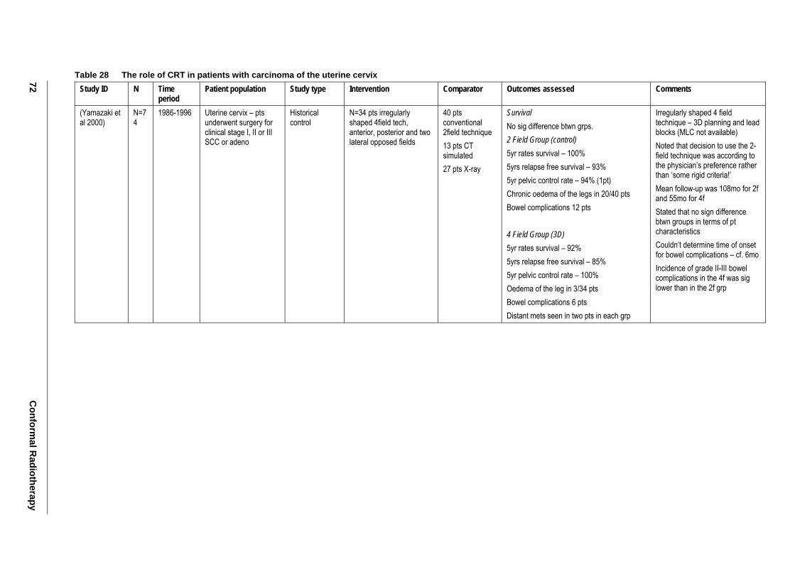
72 C
onformal R
adiotherapy
Table 28 The role of CRT in patients with carcinoma of the uterine cervix Study ID N Time
period Patient population Study type Intervention Comparator Outcomes assessed Comments
(Yamazaki et al 2000)
N=74
1986-1996 Uterine cervix – pts underwent surgery for clinical stage I, II or III SCC or adeno
Historical control
N=34 pts irregularly shaped 4field tech, anterior, posterior and two lateral opposed fields
40 pts conventional 2field technique 13 pts CT simulated 27 pts X-ray
Survival No sig difference btwn grps. 2 Field Group (control) 5yr rates survival – 100% 5yrs relapse free survival – 93% 5yr pelvic control rate – 94% (1pt) Chronic oedema of the legs in 20/40 pts Bowel complications 12 pts 4 Field Group (3D) 5yr rates survival – 92% 5yrs relapse free survival – 85% 5yr pelvic control rate – 100% Oedema of the leg in 3/34 pts Bowel complications 6 pts Distant mets seen in two pts in each grp
Irregularly shaped 4 field technique – 3D planning and lead blocks (MLC not available) Noted that decision to use the 2-field technique was according to the physician’s preference rather than ‘some rigid criteria!’ Mean follow-up was 108mo for 2f and 55mo for 4f Stated that no sign difference btwn groups in terms of pt characteristics Couldn’t determine time of onset for bowel complications – cf. 6mo Incidence of grade II-III bowel complications in the 4f was sig lower than in the 2f grp
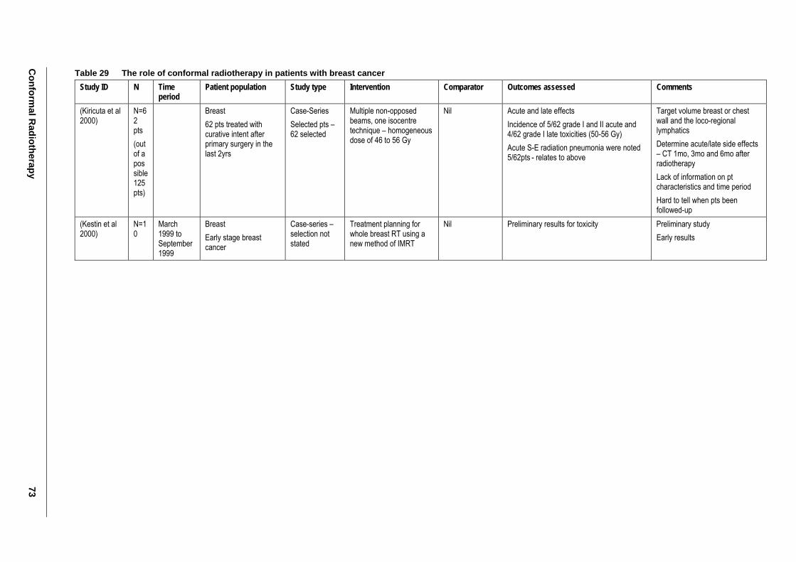
Conform
al Radiotherapy
73
Table 29 The role of conformal radiotherapy in patients with breast cancer Study ID N Time
period Patient population Study type Intervention Comparator Outcomes assessed Comments
(Kiricuta et al 2000)
N=62 pts (out of a possible 125pts)
Breast 62 pts treated with curative intent after primary surgery in the last 2yrs
Case-Series Selected pts – 62 selected
Multiple non-opposed beams, one isocentre technique – homogeneous dose of 46 to 56 Gy
Nil Acute and late effects Incidence of 5/62 grade I and II acute and 4/62 grade I late toxicities (50-56 Gy) Acute S-E radiation pneumonia were noted 5/62pts - relates to above
Target volume breast or chest wall and the loco-regional lymphatics Determine acute/late side effects – CT 1mo, 3mo and 6mo after radiotherapy Lack of information on pt characteristics and time period Hard to tell when pts been followed-up
(Kestin et al 2000)
N=10
March 1999 to September 1999
Breast Early stage breast cancer
Case-series –selection not stated
Treatment planning for whole breast RT using a new method of IMRT
Nil Preliminary results for toxicity Preliminary study Early results
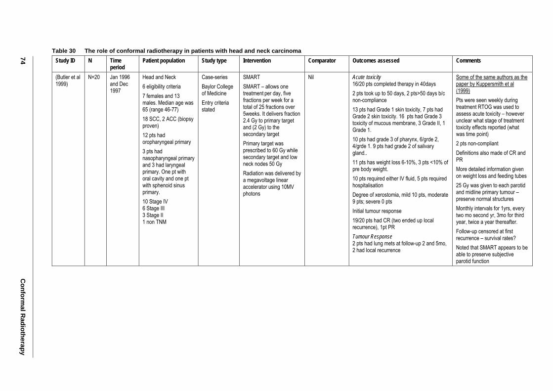
74 C
onformal R
adiotherapy
Table 30 The role of conformal radiotherapy in patients with head and neck carcinoma Study ID N Time
period Patient population Study type Intervention Comparator Outcomes assessed Comments
(Butler et al 1999)
N=20 Jan 1996 and Dec 1997
Head and Neck 6 eligibility criteria 7 females and 13 males. Median age was 65 (range 46-77) 18 SCC, 2 ACC (biopsy proven) 12 pts had oropharyngeal primary 3 pts had nasopharyngeal primary and 3 had laryngeal primary. One pt with oral cavity and one pt with sphenoid sinus primary. 10 Stage IV 6 Stage III 3 Stage II 1 non TNM
Case-series Baylor College of Medicine Entry criteria stated
SMART SMART – allows one treatment per day, five fractions per week for a total of 25 fractions over 5weeks. It delivers fraction 2.4 Gy to primary target and (2 Gy) to the secondary target Primary target was prescribed to 60 Gy while secondary target and low neck nodes 50 Gy Radiation was delivered by a megavoltage linear accelerator using 10MV photons
Nil Acute toxicity 16/20 pts completed therapy in 40days 2 pts took up to 50 days, 2 pts>50 days b/c non-compliance 13 pts had Grade 1 skin toxicity, 7 pts had Grade 2 skin toxicity. 16 pts had Grade 3 toxicity of mucous membrane, 3 Grade II, 1 Grade 1. 10 pts had grade 3 of pharynx, 6/grde 2, 4/grde 1. 9 pts had grade 2 of salivary gland.. 11 pts has weight loss 6-10%, 3 pts <10% of pre body weight. 10 pts required either IV fluid, 5 pts required hospitalisation Degree of xerostomia, mild 10 pts, moderate 9 pts; severe 0 pts Initial tumour response 19/20 pts had CR (two ended up local recurrence), 1pt PR Tumour Response 2 pts had lung mets at follow-up 2 and 5mo, 2 had local recurrence
Some of the same authors as the paper by Kuppersmith et al (1999) Pts were seen weekly during treatment RTOG was used to assess acute toxicity – however unclear what stage of treatment toxicity effects reported (what was time point) 2 pts non-compliant Definitions also made of CR and PR More detailed information given on weight loss and feeding tubes 25 Gy was given to each parotid and midline primary tumour – preserve normal structures Monthly intervals for 1yrs, every two mo second yr, 3mo for third year, twice a year thereafter. Follow-up censored at first recurrence – survival rates? Noted that SMART appears to be able to preserve subjective parotid function
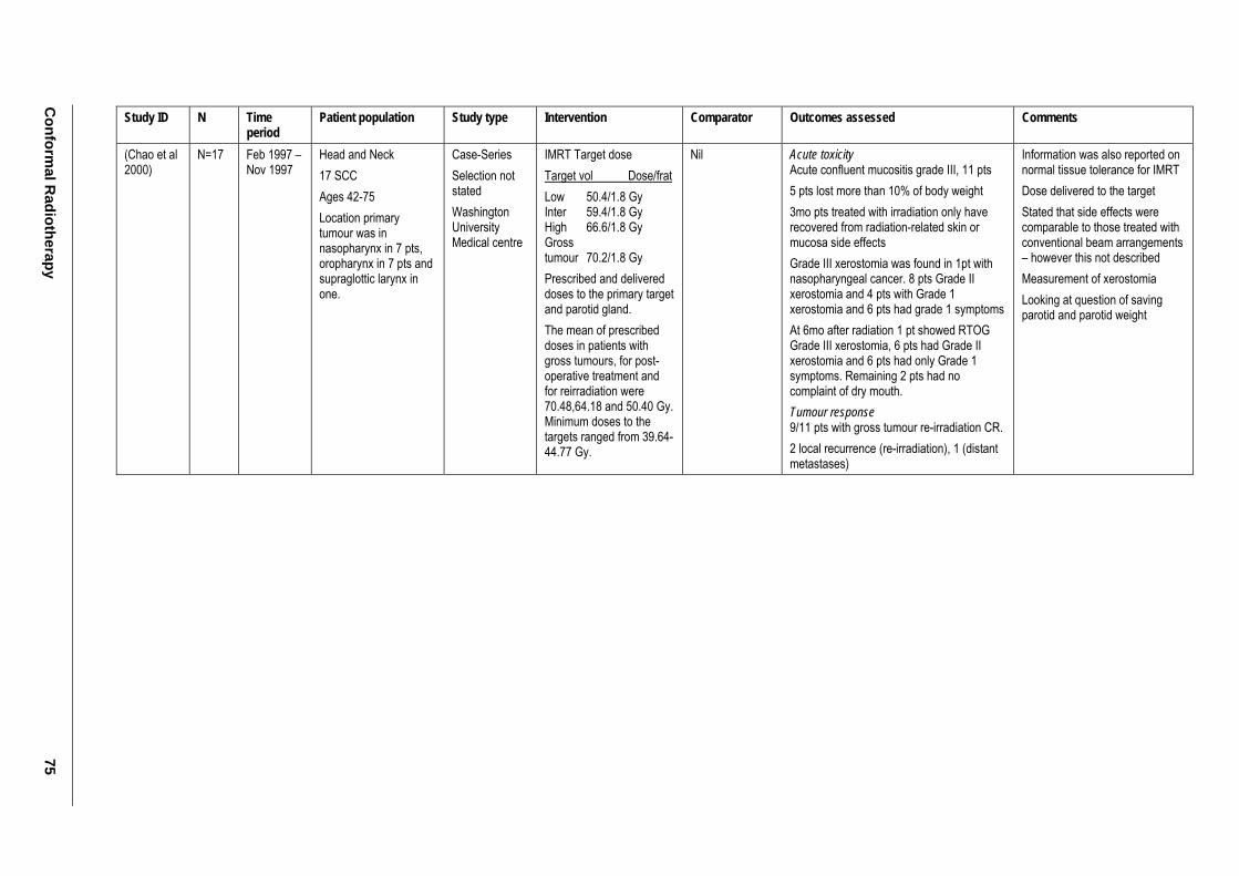
Conform
al Radiotherapy
75
Study ID N Time period
Patient population Study type Intervention Comparator Outcomes assessed Comments
(Chao et al 2000)
N=17 Feb 1997 – Nov 1997
Head and Neck 17 SCC Ages 42-75 Location primary tumour was in nasopharynx in 7 pts, oropharynx in 7 pts and supraglottic larynx in one.
Case-Series Selection not stated Washington University Medical centre
IMRT Target dose Target vol Dose/frat Low 50.4/1.8 Gy Inter 59.4/1.8 Gy High 66.6/1.8 Gy Gross tumour 70.2/1.8 Gy Prescribed and delivered doses to the primary target and parotid gland. The mean of prescribed doses in patients with gross tumours, for post-operative treatment and for reirradiation were 70.48,64.18 and 50.40 Gy. Minimum doses to the targets ranged from 39.64-44.77 Gy.
Nil Acute toxicity Acute confluent mucositis grade III, 11 pts 5 pts lost more than 10% of body weight 3mo pts treated with irradiation only have recovered from radiation-related skin or mucosa side effects Grade III xerostomia was found in 1pt with nasopharyngeal cancer. 8 pts Grade II xerostomia and 4 pts with Grade 1 xerostomia and 6 pts had grade 1 symptoms At 6mo after radiation 1 pt showed RTOG Grade III xerostomia, 6 pts had Grade II xerostomia and 6 pts had only Grade 1 symptoms. Remaining 2 pts had no complaint of dry mouth. Tumour response 9/11 pts with gross tumour re-irradiation CR. 2 local recurrence (re-irradiation), 1 (distant metastases)
Information was also reported on normal tissue tolerance for IMRT Dose delivered to the target Stated that side effects were comparable to those treated with conventional beam arrangements – however this not described Measurement of xerostomia Looking at question of saving parotid and parotid weight
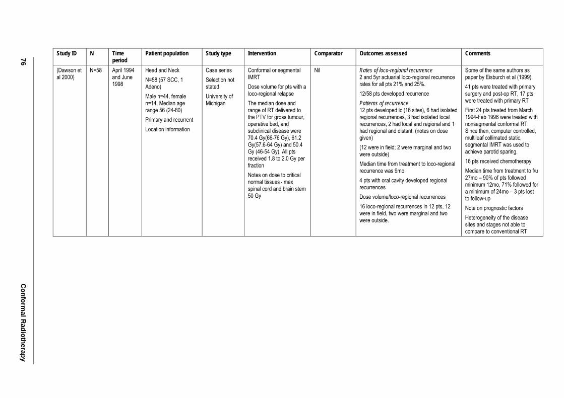
76 C
onformal R
adiotherapy
Study ID N Time period
Patient population Study type Intervention Comparator Outcomes assessed Comments
(Dawson et al 2000)
N=58 April 1994 and June 1998
Head and Neck N=58 (57 SCC, 1 Adeno) Male n=44, female n=14. Median age range 56 (24-80) Primary and recurrent Location information
Case series Selection not stated University of Michigan
Conformal or segmental IMRT Dose volume for pts with a loco-regional relapse The median dose and range of RT delivered to the PTV for gross tumour, operative bed, and subclinical disease were 70.4 Gy(66-76 Gy), 61.2 Gy(57.6-64 Gy) and 50.4 Gy (46-54 Gy). All pts received 1.8 to 2.0 Gy per fraction Notes on dose to critical normal tissues - max spinal cord and brain stem 50 Gy
Nil Rates of loco-regional recurrence 2 and 5yr actuarial loco-regional recurrence rates for all pts 21% and 25%. 12/58 pts developed recurrence Patterns of recurrence 12 pts developed lc (16 sites), 6 had isolated regional recurrences, 3 had isolated local recurrences, 2 had local and regional and 1 had regional and distant. (notes on dose given) (12 were in field; 2 were marginal and two were outside) Median time from treatment to loco-regional recurrence was 9mo 4 pts with oral cavity developed regional recurrences Dose volume/loco-regional recurrences 16 loco-regional recurrences in 12 pts, 12 were in field, two were marginal and two were outside.
Some of the same authors as paper by Eisburch et al (1999). 41 pts were treated with primary surgery and post-op RT, 17 pts were treated with primary RT First 24 pts treated from March 1994-Feb 1996 were treated with nonsegmental conformal RT. Since then, computer controlled, multileaf collimated static, segmental IMRT was used to achieve parotid sparing. 16 pts received chemotherapy Median time from treatment to f/u 27mo – 90% of pts followed minimum 12mo, 71% followed for a minimum of 24mo – 3 pts lost to follow-up Note on prognostic factors Heterogeneity of the disease sites and stages not able to compare to conventional RT
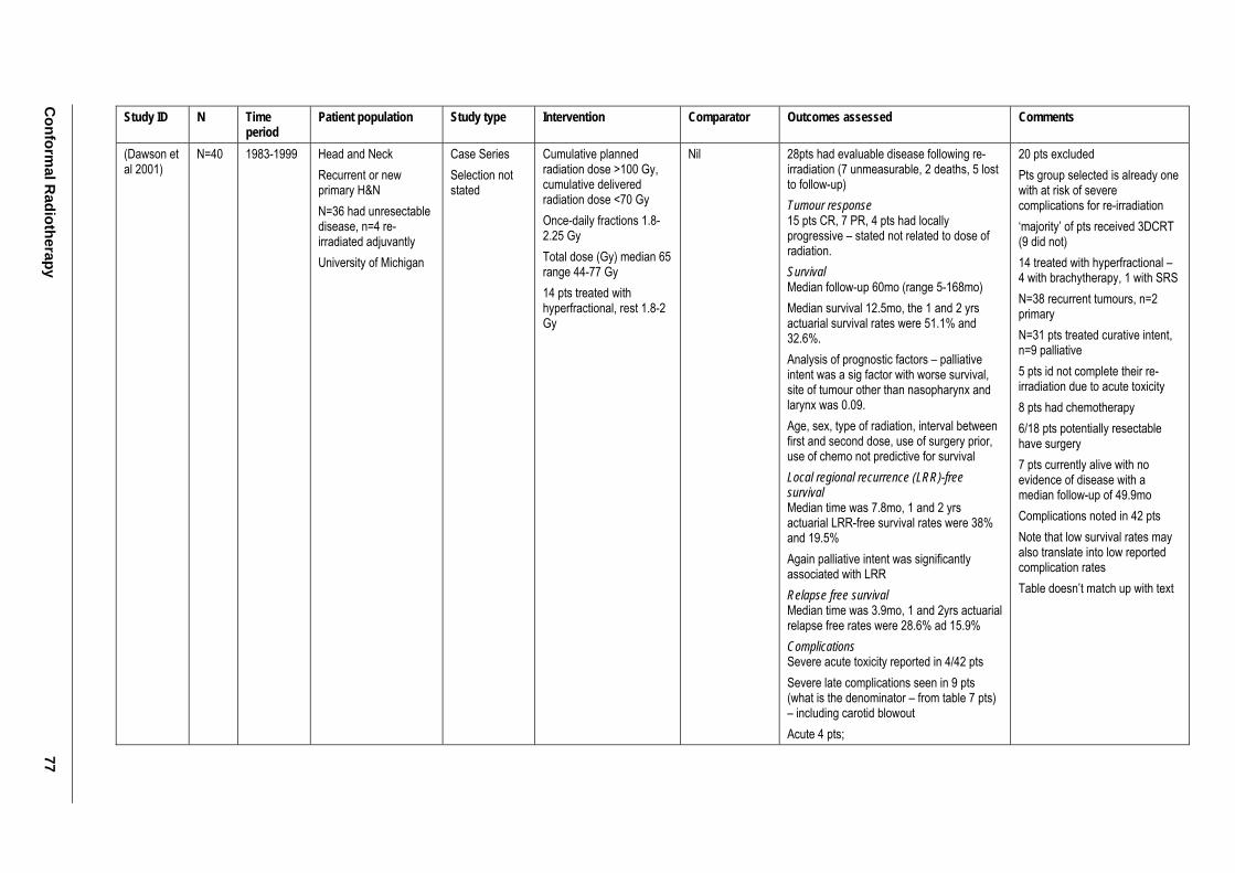
Conform
al Radiotherapy
77
Study ID N Time period
Patient population Study type Intervention Comparator Outcomes assessed Comments
(Dawson et al 2001)
N=40 1983-1999 Head and Neck Recurrent or new primary H&N N=36 had unresectable disease, n=4 re-irradiated adjuvantly University of Michigan
Case Series Selection not stated
Cumulative planned radiation dose >100 Gy, cumulative delivered radiation dose <70 Gy Once-daily fractions 1.8-2.25 Gy Total dose (Gy) median 65 range 44-77 Gy 14 pts treated with hyperfractional, rest 1.8-2 Gy
Nil 28pts had evaluable disease following re-irradiation (7 unmeasurable, 2 deaths, 5 lost to follow-up) Tumour response 15 pts CR, 7 PR, 4 pts had locally progressive – stated not related to dose of radiation. Survival Median follow-up 60mo (range 5-168mo) Median survival 12.5mo, the 1 and 2 yrs actuarial survival rates were 51.1% and 32.6%. Analysis of prognostic factors – palliative intent was a sig factor with worse survival, site of tumour other than nasopharynx and larynx was 0.09. Age, sex, type of radiation, interval between first and second dose, use of surgery prior, use of chemo not predictive for survival Local regional recurrence (LRR)-free survival Median time was 7.8mo, 1 and 2 yrs actuarial LRR-free survival rates were 38% and 19.5% Again palliative intent was significantly associated with LRR Relapse free survival Median time was 3.9mo, 1 and 2yrs actuarial relapse free rates were 28.6% ad 15.9% Complications Severe acute toxicity reported in 4/42 pts Severe late complications seen in 9 pts (what is the denominator – from table 7 pts) – including carotid blowout Acute 4 pts;
20 pts excluded Pts group selected is already one with at risk of severe complications for re-irradiation ‘majority’ of pts received 3DCRT (9 did not) 14 treated with hyperfractional – 4 with brachytherapy, 1 with SRS N=38 recurrent tumours, n=2 primary N=31 pts treated curative intent, n=9 palliative 5 pts id not complete their re-irradiation due to acute toxicity 8 pts had chemotherapy 6/18 pts potentially resectable have surgery 7 pts currently alive with no evidence of disease with a median follow-up of 49.9mo Complications noted in 42 pts Note that low survival rates may also translate into low reported complication rates Table doesn’t match up with text
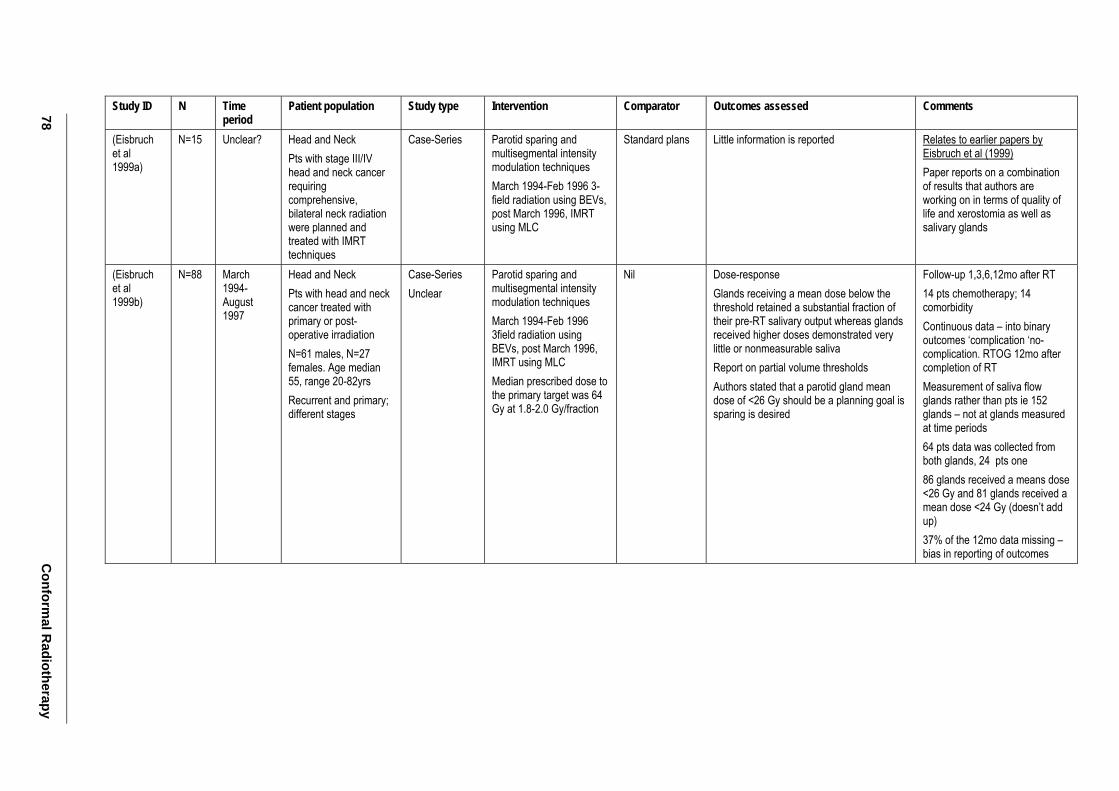
78 C
onformal R
adiotherapy
Study ID N Time period
Patient population Study type Intervention Comparator Outcomes assessed Comments
(Eisbruch et al 1999a)
N=15 Unclear? Head and Neck Pts with stage III/IV head and neck cancer requiring comprehensive, bilateral neck radiation were planned and treated with IMRT techniques
Case-Series Parotid sparing and multisegmental intensity modulation techniques March 1994-Feb 1996 3-field radiation using BEVs, post March 1996, IMRT using MLC
Standard plans Little information is reported Relates to earlier papers by Eisbruch et al (1999) Paper reports on a combination of results that authors are working on in terms of quality of life and xerostomia as well as salivary glands
(Eisbruch et al 1999b)
N=88 March 1994- August 1997
Head and Neck Pts with head and neck cancer treated with primary or post-operative irradiation N=61 males, N=27 females. Age median 55, range 20-82yrs Recurrent and primary; different stages
Case-Series Unclear
Parotid sparing and multisegmental intensity modulation techniques March 1994-Feb 1996 3field radiation using BEVs, post March 1996, IMRT using MLC Median prescribed dose to the primary target was 64 Gy at 1.8-2.0 Gy/fraction
Nil Dose-response Glands receiving a mean dose below the threshold retained a substantial fraction of their pre-RT salivary output whereas glands received higher doses demonstrated very little or nonmeasurable saliva Report on partial volume thresholds Authors stated that a parotid gland mean dose of <26 Gy should be a planning goal is sparing is desired
Follow-up 1,3,6,12mo after RT 14 pts chemotherapy; 14 comorbidity Continuous data – into binary outcomes ‘complication ‘no-complication. RTOG 12mo after completion of RT Measurement of saliva flow glands rather than pts ie 152 glands – not at glands measured at time periods 64 pts data was collected from both glands, 24 pts one 86 glands received a means dose <26 Gy and 81 glands received a mean dose <24 Gy (doesn’t add up) 37% of the 12mo data missing – bias in reporting of outcomes
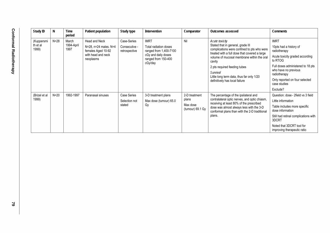
Conform
al Radiotherapy
79
Study ID N Time period
Patient population Study type Intervention Comparator Outcomes assessed Comments
(Kuppersmith et al 1999)
N=28 March 1994-April 1997
Head and Neck N=28, n=24 males. N=4 females Aged 10-92 with head and neck neoplasms
Case-Series Consecutive - retrospective
IMRT Total radiation doses ranged from 1,400-7100 cGy and daily doses ranged from 150-400 cGy/day
Nil Acute toxicity Stated that in general, grade III complications were confined to pts who were treated with a full dose that covered a large volume of mucosal membrane within the oral cavity 2 pts required feeding tubes Survival Little long term data, thus far only 1/20 definitively has local failure
IMRT 10pts had a history of radiotherapy Acute toxicity graded according to RTOG Full doses administered to 18 pts who have no previous radiotherapy Only reported on four selected case studies Exclude?
(Brizel et al 1999)
N=20 1992-1997 Paranasal sinuses Case Series Selection not stated
3-D treatment plans Max dose (tumour) 65.0 Gy
2-D treatment plans Max dose (tumour) 69.1 Gy
The percentage of the ipsilateral and contralateral optic nerves, and optic chiasm, receiving at least 80% of the prescribed dose was almost always less with the 3-D conformal plans than with the 2-D traditional plans.
Question: dose - 2field vs 3 field Little information Table includes more specific dose information Still had retinal complications with 3DCRT Noted that 3DCRT tool for improving therapeutic ratio
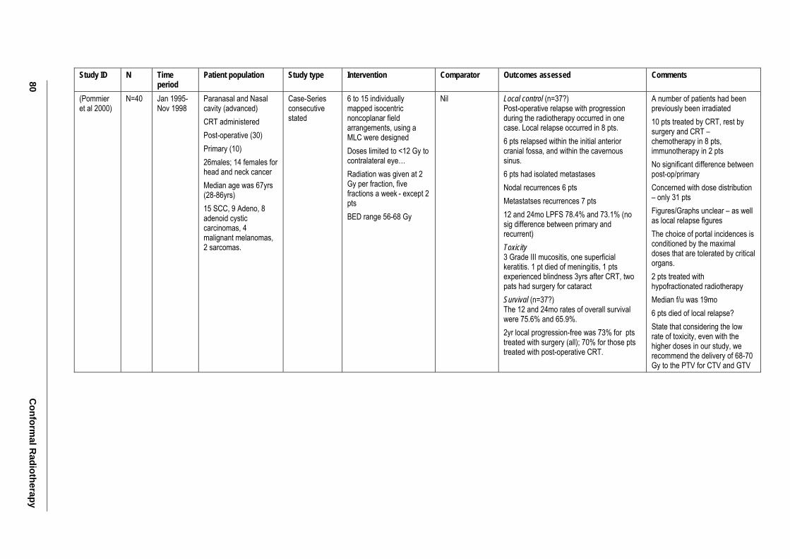
80 C
onformal R
adiotherapy
Study ID N Time period
Patient population Study type Intervention Comparator Outcomes assessed Comments
(Pommier et al 2000)
N=40 Jan 1995- Nov 1998
Paranasal and Nasal cavity (advanced) CRT administered Post-operative (30) Primary (10) 26males; 14 females for head and neck cancer Median age was 67yrs (28-86yrs) 15 SCC, 9 Adeno, 8 adenoid cystic carcinomas, 4 malignant melanomas, 2 sarcomas.
Case-Series consecutive stated
6 to 15 individually mapped isocentric noncoplanar field arrangements, using a MLC were designed Doses limited to <12 Gy to contralateral eye… Radiation was given at 2 Gy per fraction, five fractions a week - except 2 pts BED range 56-68 Gy
Nil Local control (n=37?) Post-operative relapse with progression during the radiotherapy occurred in one case. Local relapse occurred in 8 pts. 6 pts relapsed within the initial anterior cranial fossa, and within the cavernous sinus. 6 pts had isolated metastases Nodal recurrences 6 pts Metastatses recurrences 7 pts 12 and 24mo LPFS 78.4% and 73.1% (no sig difference between primary and recurrent) Toxicity 3 Grade III mucositis, one superficial keratitis. 1 pt died of meningitis, 1 pts experienced blindness 3yrs after CRT, two pats had surgery for cataract Survival (n=37?) The 12 and 24mo rates of overall survival were 75.6% and 65.9%. 2yr local progression-free was 73% for pts treated with surgery (all); 70% for those pts treated with post-operative CRT.
A number of patients had been previously been irradiated 10 pts treated by CRT, rest by surgery and CRT – chemotherapy in 8 pts, immunotherapy in 2 pts No significant difference between post-op/primary Concerned with dose distribution – only 31 pts Figures/Graphs unclear – as well as local relapse figures The choice of portal incidences is conditioned by the maximal doses that are tolerated by critical organs. 2 pts treated with hypofractionated radiotherapy Median f/u was 19mo 6 pts died of local relapse? State that considering the low rate of toxicity, even with the higher doses in our study, we recommend the delivery of 68-70 Gy to the PTV for CTV and GTV
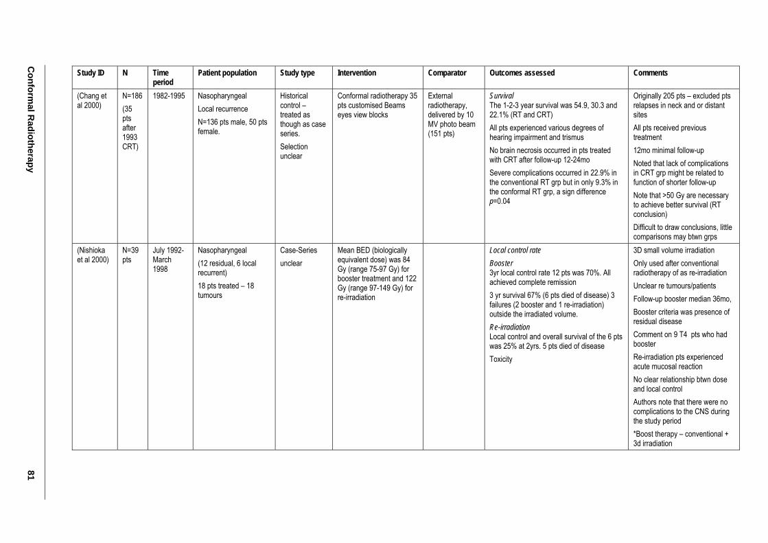
Conform
al Radiotherapy
81
Study ID N Time period
Patient population Study type Intervention Comparator Outcomes assessed Comments
(Chang et al 2000)
N=186 (35 pts after 1993 CRT)
1982-1995 Nasopharyngeal Local recurrence N=136 pts male, 50 pts female.
Historical control – treated as though as case series. Selection unclear
Conformal radiotherapy 35 pts customised Beams eyes view blocks
External radiotherapy, delivered by 10 MV photo beam (151 pts)
Survival The 1-2-3 year survival was 54.9, 30.3 and 22.1% (RT and CRT) All pts experienced various degrees of hearing impairment and trismus No brain necrosis occurred in pts treated with CRT after follow-up 12-24mo Severe complications occurred in 22.9% in the conventional RT grp but in only 9.3% in the conformal RT grp, a sign difference p=0.04
Originally 205 pts – excluded pts relapses in neck and or distant sites All pts received previous treatment 12mo minimal follow-up Noted that lack of complications in CRT grp might be related to function of shorter follow-up Note that >50 Gy are necessary to achieve better survival (RT conclusion) Difficult to draw conclusions, little comparisons may btwn grps
(Nishioka et al 2000)
N=39 pts
July 1992- March 1998
Nasopharyngeal (12 residual, 6 local recurrent) 18 pts treated – 18 tumours
Case-Series unclear
Mean BED (biologically equivalent dose) was 84 Gy (range 75-97 Gy) for booster treatment and 122 Gy (range 97-149 Gy) for re-irradiation
Local control rate Booster 3yr local control rate 12 pts was 70%. All achieved complete remission 3 yr survival 67% (6 pts died of disease) 3 failures (2 booster and 1 re-irradiation) outside the irradiated volume. Re-irradiation Local control and overall survival of the 6 pts was 25% at 2yrs. 5 pts died of disease Toxicity
3D small volume irradiation Only used after conventional radiotherapy of as re-irradiation Unclear re tumours/patients Follow-up booster median 36mo, Booster criteria was presence of residual disease Comment on 9 T4 pts who had booster Re-irradiation pts experienced acute mucosal reaction No clear relationship btwn dose and local control Authors note that there were no complications to the CNS during the study period *Boost therapy – conventional + 3d irradiation

82 C
onformal R
adiotherapy
Table 31 The role of conformal radiotherapy in patients with NSCLC Study ID N Time
period Patient population Study type Intervention Comparator Outcomes assessed Comments
(Armstrong et al 1997)
N=45 1990-1993 NSCLC N=45 (28 males; 17 females) Median age 65 (range 38-82) Tumour stage was I/II in 13%, IIIa in 42% and IIIb in 44%. SCC in 44% Adeno in 36%
Case-series Selection not stated
Anterior and posterior fields custom cerrobend blocking – 18 (10 fractions) 3DCRT 32.4 Gy (18 fractions) to both the elective and nodal areas – subsequently 50.4 Gy elective nodes and 70.2 Gy to gross disease
Nil Toxicity KPS unchanged in 64%, decreased in 22% and increased in 13%. Weight increased 20%, decreased 38% and unchanged in 42%. Oesophageal toxicity RTOG None in 24%, Grade 1 56%, Grade II 18%; and 2% in Grade III%. Pulmonary toxicity Radiation pneumonitis occurred in 4/45. NTCP/DVH (31 pts) Grade III or higher 4/14 with NTCP Looked at relationship between % lung volume and the risk of pneumonitis. Severe pulmonary toxicity occurred in 38% (3/8) pts >30% lung >25 Gy, versus 4% pts <30% receiving >25 Gy Anatomic failure Radiologically and or clinically evident thoracic progression occurred in 46%. Actuarial freedom falls to 27% at 59mo, distant mets occurred in 31%. Median survival is 15.7mo and survival is 32% at 2yrs and 12% at 59mo.
Previous articles have more detail Unclear at times whether reporting on 46 pts or 31 pts (refer earlier paper) 31 pts have DVH – these pts reported on in terms of NTCP Anatomic failure 7 pts did not complete 3DCRT – seen as an underestimation of disease Toxicity RTOG/EORTC Possible problem with NTCP model in oesophageal model Most significant toxicity was radiation pneumonitis *sig correlation between the occurrence of severe radiation pneumonitis and volume of lung receiving 25 Gy. Again unclear which patients were followed up and what T/N stage Median follow-up of 6 survivors is 43.5mo
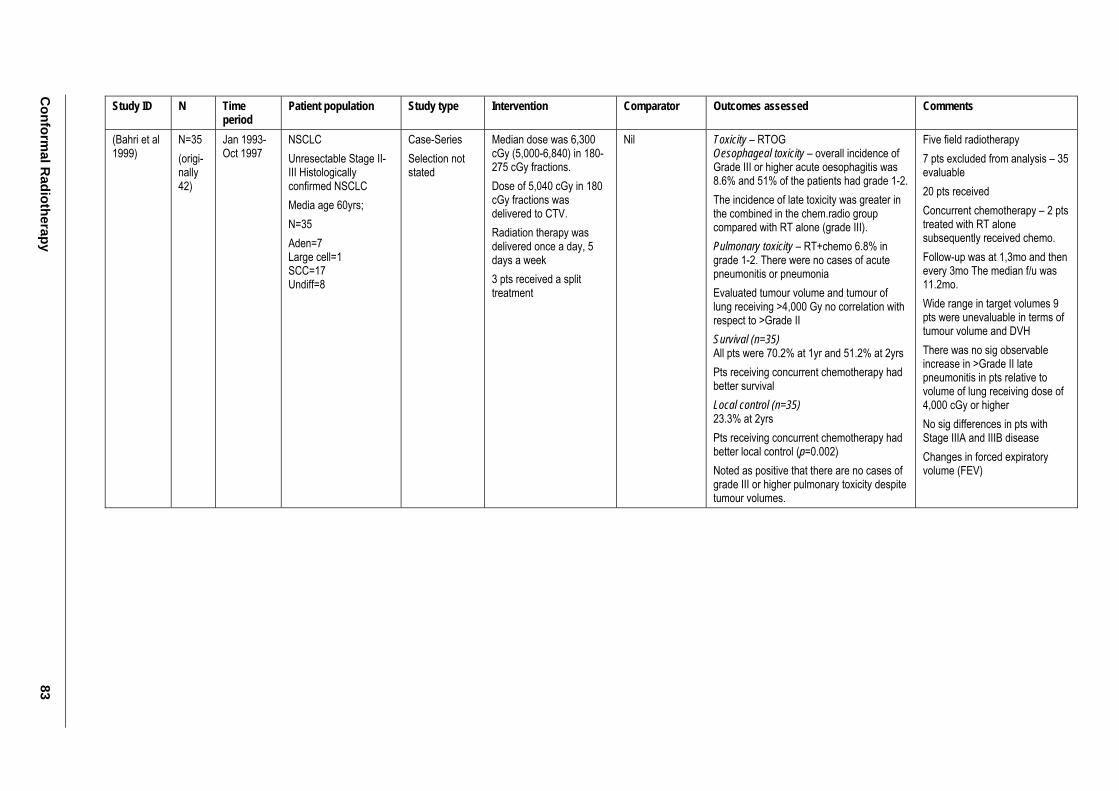
Conform
al Radiotherapy
83
Study ID N Time period
Patient population Study type Intervention Comparator Outcomes assessed Comments
(Bahri et al 1999)
N=35 (origi-nally 42)
Jan 1993-Oct 1997
NSCLC Unresectable Stage II-III Histologically confirmed NSCLC Media age 60yrs; N=35 Aden=7 Large cell=1 SCC=17 Undiff=8
Case-Series Selection not stated
Median dose was 6,300 cGy (5,000-6,840) in 180-275 cGy fractions. Dose of 5,040 cGy in 180 cGy fractions was delivered to CTV. Radiation therapy was delivered once a day, 5 days a week 3 pts received a split treatment
Nil Toxicity – RTOG Oesophageal toxicity – overall incidence of Grade III or higher acute oesophagitis was 8.6% and 51% of the patients had grade 1-2. The incidence of late toxicity was greater in the combined in the chem.radio group compared with RT alone (grade III). Pulmonary toxicity – RT+chemo 6.8% in grade 1-2. There were no cases of acute pneumonitis or pneumonia Evaluated tumour volume and tumour of lung receiving >4,000 Gy no correlation with respect to >Grade II Survival (n=35) All pts were 70.2% at 1yr and 51.2% at 2yrs Pts receiving concurrent chemotherapy had better survival Local control (n=35) 23.3% at 2yrs Pts receiving concurrent chemotherapy had better local control (p=0.002) Noted as positive that there are no cases of grade III or higher pulmonary toxicity despite tumour volumes.
Five field radiotherapy 7 pts excluded from analysis – 35 evaluable 20 pts received Concurrent chemotherapy – 2 pts treated with RT alone subsequently received chemo. Follow-up was at 1,3mo and then every 3mo The median f/u was 11.2mo. Wide range in target volumes 9 pts were unevaluable in terms of tumour volume and DVH There was no sig observable increase in >Grade II late pneumonitis in pts relative to volume of lung receiving dose of 4,000 cGy or higher No sig differences in pts with Stage IIIA and IIIB disease Changes in forced expiratory volume (FEV)
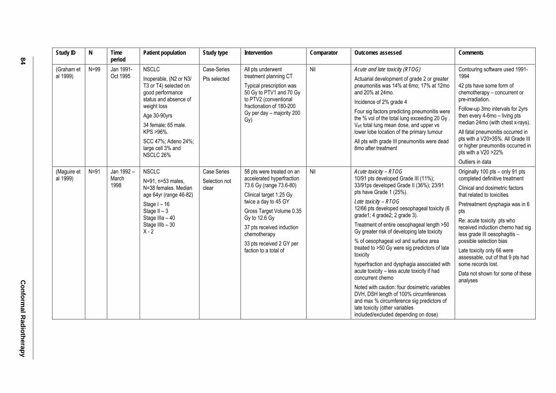
84 C
onformal R
adiotherapy
Study ID N Time period
Patient population Study type Intervention Comparator Outcomes assessed Comments
(Graham et al 1999)
N=99 Jan 1991-Oct 1995
NSCLC Inoperable, (N2 or N3/ T3 or T4) selected on good performance status and absence of weight loss Age 30-90yrs 34 female; 65 male. KPS >96%. SCC 47%; Adeno 24%; large cell 3% and NSCLC 26%
Case-Series Pts selected
All pts underwent treatment planning CT Typical prescription was 50 Gy to PTV1 and 70 Gy to PTV2 (conventional fractionation of 180-200 Gy per day – majority 200 Gy)
Nil Acute and late toxicity (RTOG) Actuarial development of grade 2 or greater pneumonitis was 14% at 6mo; 17% at 12mo and 20% at 24mo. Incidence of 2% grade 4 Four sig factors predicting pneumonitis were the % vol of the total lung exceeding 20 Gy . Veff, total lung mean dose, and upper vs lower lobe location of the primary tumour All pts with grade III pneumonitis were dead 8mo after treatment
Contouring software used 1991-1994 42 pts have some form of chemotherapy – concurrent or pre-irradiation. Follow-up 3mo intervals for 2yrs then every 4-6mo – living pts median 24mo (with chest x-rays). All fatal pneumonitis occurred in pts with a V20>35%. All Grade III or higher pneumonitis occurred in pts with a V20 >22% Outliers in data
(Maguire et al 1999)
N=91 Jan 1992 – March 1998
NSCLC N=91, n=53 males, N=38 females. Median age 64yr (range 46-82) Stage I – 16 Stage II – 3 Stage IIIa – 40 Stage IIIb – 30 X - 2
Case Series Selection not clear
58 pts were treated on an accelerated hyperfraction 73.6 Gy (range 73.6-80) Clinical target 1.25 Gy twice a day to 45 GY Gross Target Volume 0.35 Gy to 12.6 Gy 37 pts received induction chemotherapy 33 pts received 2 GY per faction to a total of
Nil Acute toxicity – RTOG 10/91 pts developed Grade III (11%); 33/91ps developed Grade II (36%); 23/91 pts have Grade 1 (25%). Late toxicity – RTOG 12/66 pts developed oesophageal toxicity (6 grade1; 4 grade2; 2 grade 3). Treatment of entire oesophageal length >50 Gy greater risk of developing late toxicity % of oesophageal vol and surface area treated to >50 Gy were sig predictors of late toxicity hyperfraction and dysphagia associated with acute toxicity – less acute toxicity if had concurrent chemo Noted with caution: four dosimetric variables DVH, DSH length of 100% circumferences and max % circumference sig predictors of late toxicity (other variables included/excluded depending on dose)
Originally 100 pts – only 91 pts completed definitive treatment Clinical and dosimetric factors that related to toxicities Pretreatment dysphagia was in 6 pts Re: acute toxicity pts who received induction chemo had sig less grade III oesophagitis – possible selection bias Late toxicity only 66 were assessable, out of that 9 pts had some records lost. Data not shown for some of these analyses
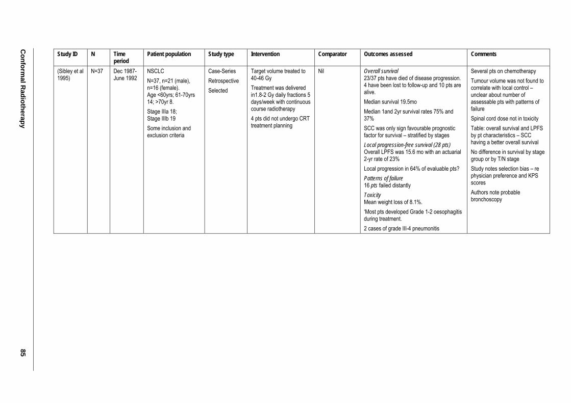
Conform
al Radiotherapy
85
Study ID N Time period
Patient population Study type Intervention Comparator Outcomes assessed Comments
(Sibley et al 1995)
N=37 Dec 1987-June 1992
NSCLC N=37, n=21 (male), n=16 (female). Age <60yrs; 61-70yrs 14; >70yr 8. Stage IIIa 18; Stage IIIb 19 Some inclusion and exclusion criteria
Case-Series Retrospective Selected
Target volume treated to 40-46 Gy Treatment was delivered in1.8-2 Gy daily fractions 5 days/week with continuous course radiotherapy 4 pts did not undergo CRT treatment planning
Nil Overall survival 23/37 pts have died of disease progression. 4 have been lost to follow-up and 10 pts are alive. Median survival 19.5mo Median 1and 2yr survival rates 75% and 37% SCC was only sign favourable prognostic factor for survival – stratified by stages Local progression-free survival (28 pts) Overall LPFS was 15.6 mo with an actuarial 2-yr rate of 23% Local progression in 64% of evaluable pts? Patterns of failure 16 pts failed distantly Toxicity Mean weight loss of 8.1%. ‘Most pts developed Grade 1-2 oesophagitis during treatment. 2 cases of grade III-4 pneumonitis
Several pts on chemotherapy Tumour volume was not found to correlate with local control – unclear about number of assessable pts with patterns of failure Spinal cord dose not in toxicity Table: overall survival and LPFS by pt characteristics – SCC having a better overall survival No difference in survival by stage group or by T/N stage Study notes selection bias – re physician preference and KPS scores Authors note probable bronchoscopy
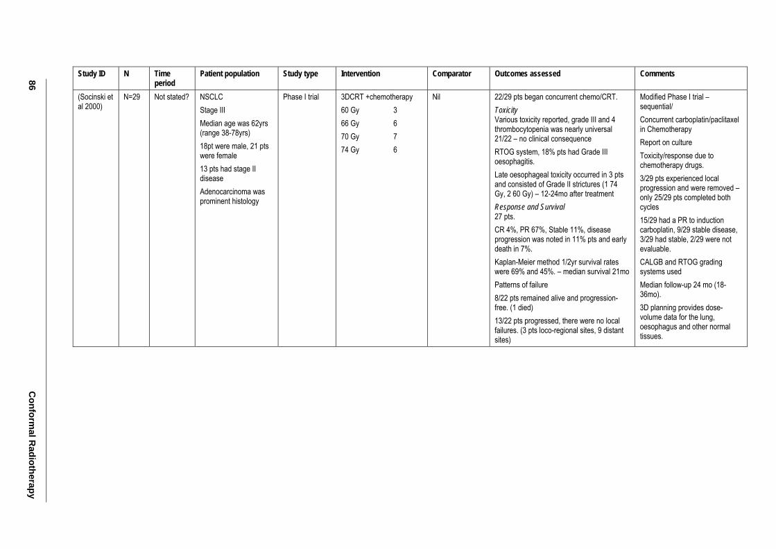
86 C
onformal R
adiotherapy
Study ID N Time period
Patient population Study type Intervention Comparator Outcomes assessed Comments
(Socinski et al 2000)
N=29 Not stated? NSCLC Stage III Median age was 62yrs (range 38-78yrs) 18pt were male, 21 pts were female 13 pts had stage II disease Adenocarcinoma was prominent histology
Phase I trial 3DCRT +chemotherapy 60 Gy 3 66 Gy 6 70 Gy 7 74 Gy 6
Nil 22/29 pts began concurrent chemo/CRT. Toxicity Various toxicity reported, grade III and 4 thrombocytopenia was nearly universal 21/22 – no clinical consequence RTOG system, 18% pts had Grade III oesophagitis. Late oesophageal toxicity occurred in 3 pts and consisted of Grade II strictures (1 74 Gy, 2 60 Gy) – 12-24mo after treatment Response and Survival 27 pts. CR 4%, PR 67%, Stable 11%, disease progression was noted in 11% pts and early death in 7%. Kaplan-Meier method 1/2yr survival rates were 69% and 45%. – median survival 21mo Patterns of failure 8/22 pts remained alive and progression-free. (1 died) 13/22 pts progressed, there were no local failures. (3 pts loco-regional sites, 9 distant sites)
Modified Phase I trial – sequential/ Concurrent carboplatin/paclitaxel in Chemotherapy Report on culture Toxicity/response due to chemotherapy drugs. 3/29 pts experienced local progression and were removed – only 25/29 pts completed both cycles 15/29 had a PR to induction carboplatin, 9/29 stable disease, 3/29 had stable, 2/29 were not evaluable. CALGB and RTOG grading systems used Median follow-up 24 mo (18-36mo). 3D planning provides dose-volume data for the lung, oesophagus and other normal tissues.
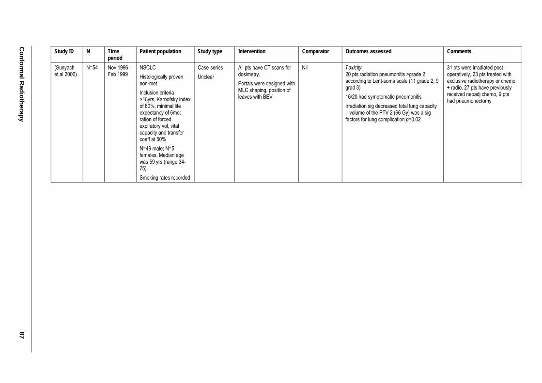
Conform
al Radiotherapy
87
Study ID N Time period
Patient population Study type Intervention Comparator Outcomes assessed Comments
(Sunyach et al 2000)
N=54 Nov 1996-Feb 1999
NSCLC Histologically proven non-met Inclusion criteria >18yrs, Karnofsky index of 80%, minimal life expectancy of 6mo; ration of forced expiratory vol, vital capacity and transfer coeff at 50% N=49 male; N=5 females. Median age was 59 yrs (range 34-75). Smoking rates recorded
Case-series Unclear
All pts have CT scans for dosimetry. Portals were designed with MLC shaping, position of leaves with BEV
Nil Toxicity 20 pts radiation pneumonitis >grade 2 according to Lent-soma scale (11 grade 2; 9 grad 3) 16/20 had symptomatic pneumonitis Irradiation sig decreased total lung capacity – volume of the PTV 2 (66 Gy) was a sig factors for lung complication p=0.02
31 pts were irradiated post-operatively, 23 pts treated with exclusive radiotherapy or chemo + radio. 27 pts have previously received neoadj chemo, 9 pts had pneumonectomy
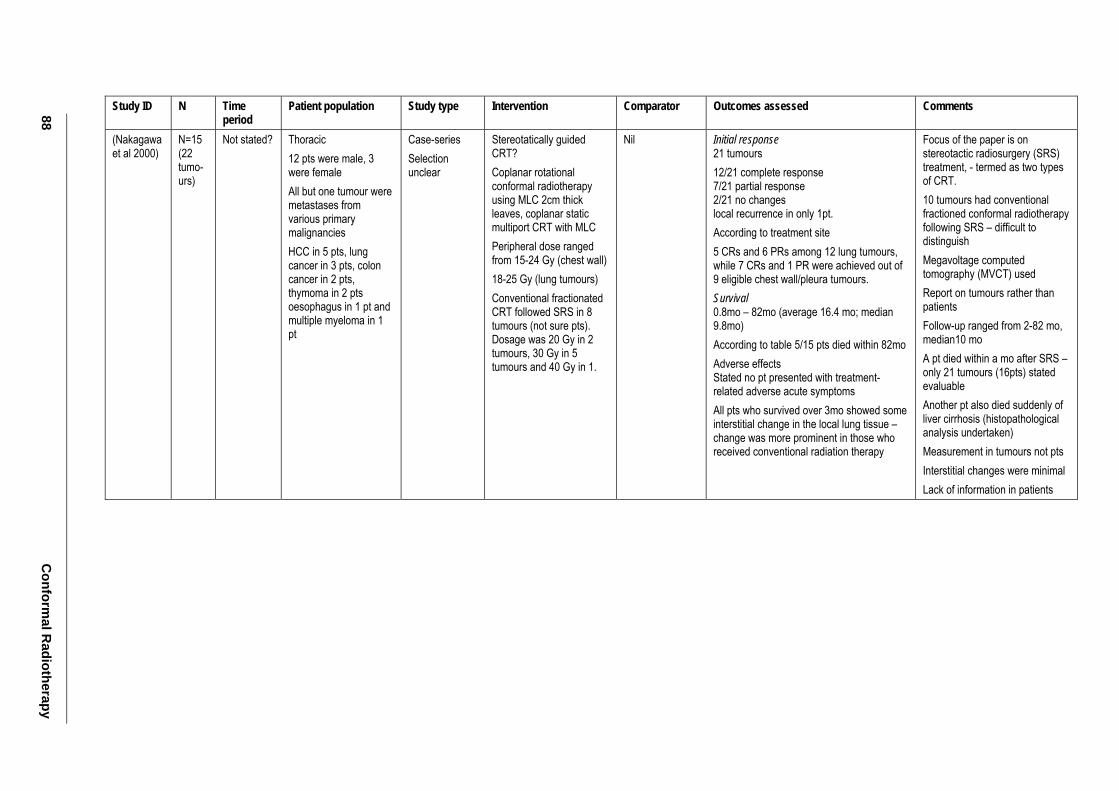
88 C
onformal R
adiotherapy
Study ID N Time period
Patient population Study type Intervention Comparator Outcomes assessed Comments
(Nakagawa et al 2000)
N=15 (22 tumo-urs)
Not stated? Thoracic 12 pts were male, 3 were female All but one tumour were metastases from various primary malignancies HCC in 5 pts, lung cancer in 3 pts, colon cancer in 2 pts, thymoma in 2 pts oesophagus in 1 pt and multiple myeloma in 1 pt
Case-series Selection unclear
Stereotatically guided CRT? Coplanar rotational conformal radiotherapy using MLC 2cm thick leaves, coplanar static multiport CRT with MLC Peripheral dose ranged from 15-24 Gy (chest wall) 18-25 Gy (lung tumours) Conventional fractionated CRT followed SRS in 8 tumours (not sure pts). Dosage was 20 Gy in 2 tumours, 30 Gy in 5 tumours and 40 Gy in 1.
Nil Initial response 21 tumours 12/21 complete response 7/21 partial response 2/21 no changes local recurrence in only 1pt. According to treatment site 5 CRs and 6 PRs among 12 lung tumours, while 7 CRs and 1 PR were achieved out of 9 eligible chest wall/pleura tumours. Survival 0.8mo – 82mo (average 16.4 mo; median 9.8mo) According to table 5/15 pts died within 82mo Adverse effects Stated no pt presented with treatment-related adverse acute symptoms All pts who survived over 3mo showed some interstitial change in the local lung tissue – change was more prominent in those who received conventional radiation therapy
Focus of the paper is on stereotactic radiosurgery (SRS) treatment, - termed as two types of CRT. 10 tumours had conventional fractioned conformal radiotherapy following SRS – difficult to distinguish Megavoltage computed tomography (MVCT) used Report on tumours rather than patients Follow-up ranged from 2-82 mo, median10 mo A pt died within a mo after SRS – only 21 tumours (16pts) stated evaluable Another pt also died suddenly of liver cirrhosis (histopathological analysis undertaken) Measurement in tumours not pts Interstitial changes were minimal Lack of information in patients
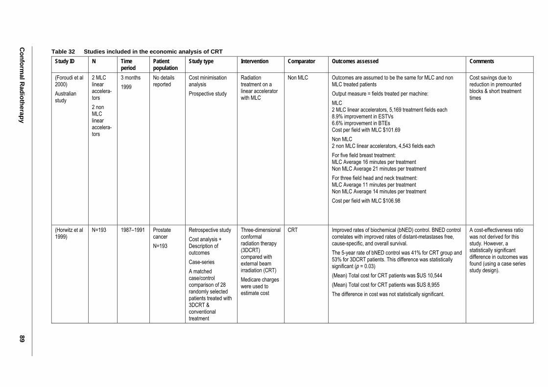
Conform
al Radiotherapy
89
Table 32 Studies included in the economic analysis of CRT Study ID N Time
period Patient population
Study type Intervention Comparator Outcomes assessed Comments
(Foroudi et al 2000) Australian study
2 MLC linear accelera-tors 2 non MLC linear accelera-tors
3 months 1999
No details reported
Cost minimisation analysis Prospective study
Radiation treatment on a linear accelerator with MLC
Non MLC Outcomes are assumed to be the same for MLC and non MLC treated patients Output measure = fields treated per machine: MLC 2 MLC linear accelerators, 5,169 treatment fields each 8.9% improvement in ESTVs 6.6% improvement in BTEs Cost per field with MLC $101.69 Non MLC 2 non MLC linear accelerators, 4,543 fields each For five field breast treatment: MLC Average 16 minutes per treatment Non MLC Average 21 minutes per treatment For three field head and neck treatment: MLC Average 11 minutes per treatment Non MLC Average 14 minutes per treatment Cost per field with MLC $106.98
Cost savings due to reduction in premounted blocks & short treatment times
(Horwitz et al 1999)
N=193 1987–1991 Prostate cancer N=193
Retrospective study Cost analysis + Description of outcomes Case-series A matched case/control comparison of 28 randomly selected patients treated with 3DCRT & conventional treatment
Three-dimensional conformal radiation therapy (3DCRT) compared with external beam irradiation (CRT) Medicare charges were used to estimate cost
CRT Improved rates of biochemical (bNED) control. BNED control correlates with improved rates of distant-metastases free, cause-specific, and overall survival. The 5-year rate of bNED control was 41% for CRT group and 53% for 3DCRT patients. This difference was statistically significant (p = 0.03) (Mean) Total cost for CRT patients was $US 10,544 (Mean) Total cost for CRT patients was $US 8,955 The difference in cost was not statistically significant.
A cost-effectiveness ratio was not derived for this study. However, a statistically significant difference in outcomes was found (using a case series study design).
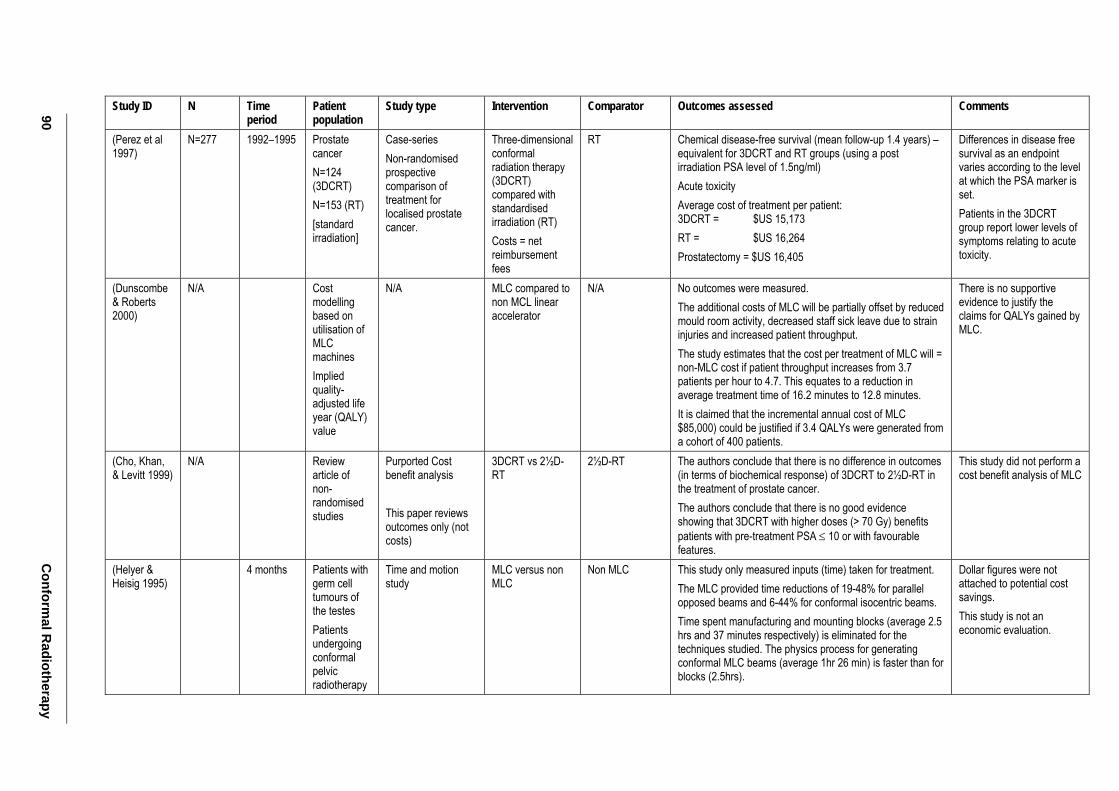
90 C
onformal R
adiotherapy
Study ID N Time period
Patient population
Study type Intervention Comparator Outcomes assessed Comments
(Perez et al 1997)
N=277 1992–1995 Prostate cancer N=124 (3DCRT) N=153 (RT) [standard irradiation]
Case-series Non-randomised prospective comparison of treatment for localised prostate cancer.
Three-dimensional conformal radiation therapy (3DCRT) compared with standardised irradiation (RT) Costs = net reimbursement fees
RT Chemical disease-free survival (mean follow-up 1.4 years) – equivalent for 3DCRT and RT groups (using a post irradiation PSA level of 1.5ng/ml) Acute toxicity Average cost of treatment per patient: 3DCRT = $US 15,173 RT = $US 16,264 Prostatectomy = $US 16,405
Differences in disease free survival as an endpoint varies according to the level at which the PSA marker is set. Patients in the 3DCRT group report lower levels of symptoms relating to acute toxicity.
(Dunscombe & Roberts 2000)
N/A Cost modelling based on utilisation of MLC machines Implied quality-adjusted life year (QALY) value
N/A MLC compared to non MCL linear accelerator
N/A No outcomes were measured. The additional costs of MLC will be partially offset by reduced mould room activity, decreased staff sick leave due to strain injuries and increased patient throughput. The study estimates that the cost per treatment of MLC will = non-MLC cost if patient throughput increases from 3.7 patients per hour to 4.7. This equates to a reduction in average treatment time of 16.2 minutes to 12.8 minutes. It is claimed that the incremental annual cost of MLC $85,000) could be justified if 3.4 QALYs were generated from a cohort of 400 patients.
There is no supportive evidence to justify the claims for QALYs gained by MLC.
(Cho, Khan, & Levitt 1999)
N/A Review article of non-randomised studies
Purported Cost benefit analysis This paper reviews outcomes only (not costs)
3DCRT vs 2½D-RT
2½D-RT The authors conclude that there is no difference in outcomes (in terms of biochemical response) of 3DCRT to 2½D-RT in the treatment of prostate cancer. The authors conclude that there is no good evidence showing that 3DCRT with higher doses (> 70 Gy) benefits patients with pre-treatment PSA ≤ 10 or with favourable features.
This study did not perform a cost benefit analysis of MLC
(Helyer & Heisig 1995)
4 months Patients with germ cell tumours of the testes Patients undergoing conformal pelvic radiotherapy
Time and motion study
MLC versus non MLC
Non MLC This study only measured inputs (time) taken for treatment. The MLC provided time reductions of 19-48% for parallel opposed beams and 6-44% for conformal isocentric beams. Time spent manufacturing and mounting blocks (average 2.5 hrs and 37 minutes respectively) is eliminated for the techniques studied. The physics process for generating conformal MLC beams (average 1hr 26 min) is faster than for blocks (2.5hrs).
Dollar figures were not attached to potential cost savings. This study is not an economic evaluation.
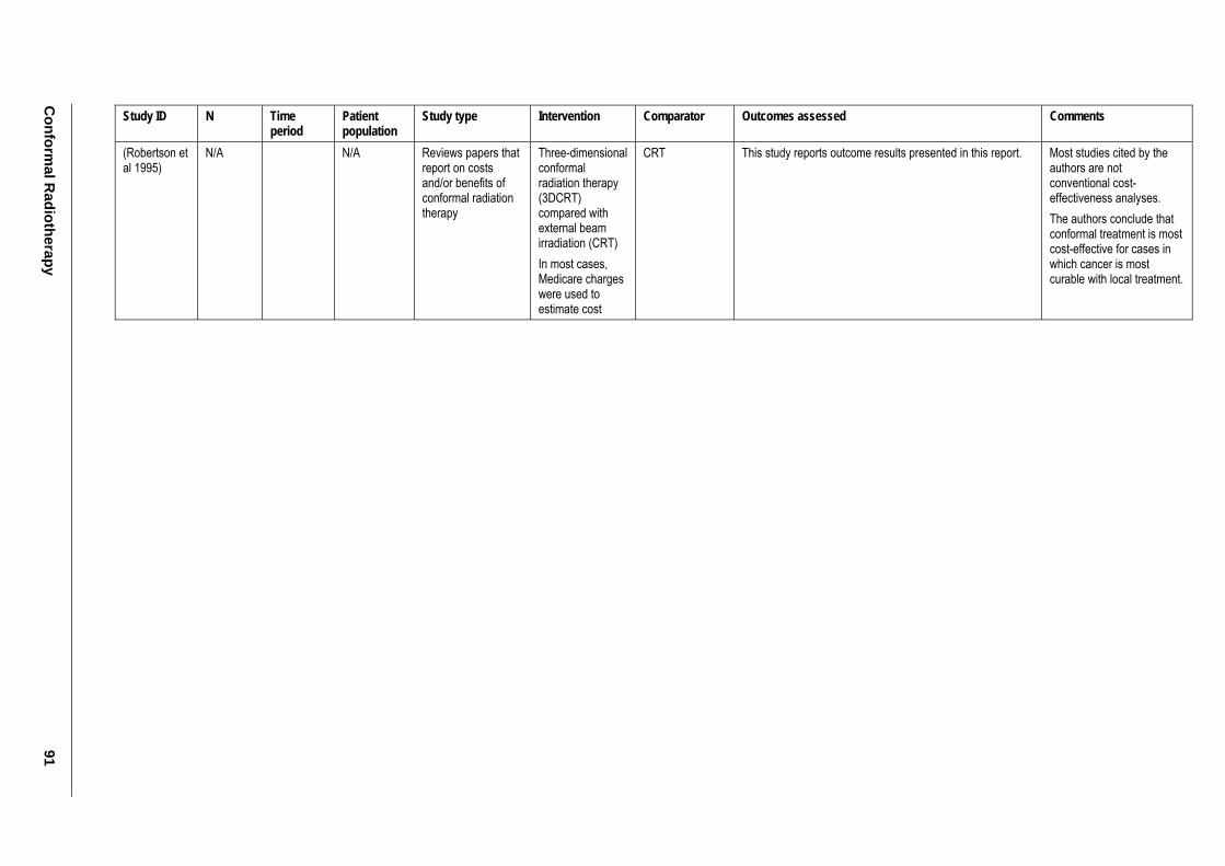
Conform
al Radiotherapy
91
Study ID N Time period
Patient population
Study type Intervention Comparator Outcomes assessed Comments
(Robertson et al 1995)
N/A N/A Reviews papers that report on costs and/or benefits of conformal radiation therapy
Three-dimensional conformal radiation therapy (3DCRT) compared with external beam irradiation (CRT) In most cases, Medicare charges were used to estimate cost
CRT This study reports outcome results presented in this report. Most studies cited by the authors are not conventional cost-effectiveness analyses. The authors conclude that conformal treatment is most cost-effective for cases in which cancer is most curable with local treatment.
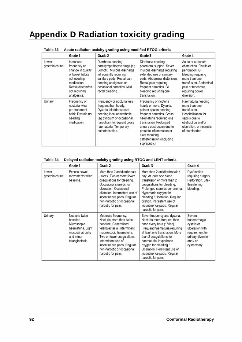
92 Conformal Radiotherapy
Appendix D Radiation toxicity grading
Table 33 Acute radiation toxicity grading using modified RTOG criteria Grade 1 Grade 2 Grade 3 Grade 4 Lower gastrointestinal
Increased frequency or change in quality of bowel habits not needing medication. Rectal discomfort not requiring analgesics.
Diarrhoea needing parasympatholytic drugs (eg Lomotil). Mucous discharge infrequently requiring sanitary pads. Rectal pain needing analgesics or occasional narcotics. Mild rectal bleeding.
Diarrhoea needing parenteral support. Sever mucous discharge requiring extended use of sanitary pads. Abdominal distension. Rectal pain requiring frequent narcotics. GI bleeding requiring one transfusion.
Acute or subacute obstruction. Fistula or perforation. GI bleeding requiring more than one transfusion. Abdominal pain or tenesmus requiring bowel diversion.
Urinary Frequency or nocturia twice pre-treatment habit. Dysuria not needing medication.
Frequency or nocturia less frequent than hourly. Dysuria, bladder spasm needing local anaesthetic (eg pyridium or occasional narcotics). Infrequent gross haematuria. Temporary catheterisation.
Frequency or nocturia hourly or more. Dysuria, pain or spasm needing frequent narcotics. Gross haematuria requiring one transfusion. Prolonged urinary obstruction due to prostate inflammation or clots requiring catheterisation (including suprapubic).
Haematuria needing more than one transfusion. Hospitalisation for sepsis due to obstruction and/or ulceration, or necrosis of the bladder.
Table 34 Delayed radiation toxicity grading using RTOG and LENT criteria Grade 1 Grade 2 Grade 3 Grade 4 Lower gastrointestinal
Excess bowel movements twice baseline.
More than 2 antidiarrhoeals / week. Two or more fewer coagulations for bleeding. Occasional steroids for ulceration. Occasional dilatation. Intermittent use of incontinence pads. Regular non-narcotic or occasional narcotic for pain.
More than 2 antidiarrhoeals / day. At least one blood transfusion or more than 2 coagulations for bleeding. Prolonged steroids per enema. Hyperbaric oxygen for bleeding / ulceration. Regular dilation. Persistent use of incontinence pads. Regular narcotic for pain.
Dysfunction requiring surgery. Perforation. Life-threatening bleeding.
Urinary Nocturia twice baseline. Microscopic haematuria. Light mucosal atrophy and minor telangiectasia.
Moderate frequency. Nocturia more than twice baseline. Generalised telangiectasia. Intermittent macroscopic haematuria. Two or fewer coagulations. Intermittent use of incontinence pads. Regular non-narcotic or occasional narcotic for pain.
Sever frequency and dysuria. Nocturia more frequent than once every hour (150cc). Frequent haematuria requiring at least one transfusion. More than 2 coagulations for haematuria. Hyperbaric oxygen for bleeding / ulceration. Persistent use of incontinence pads. Regular narcotic for pain.
Severe haemorrhagic cystitis or ulceration with requirement for urinary diversion and / or cystectomy.
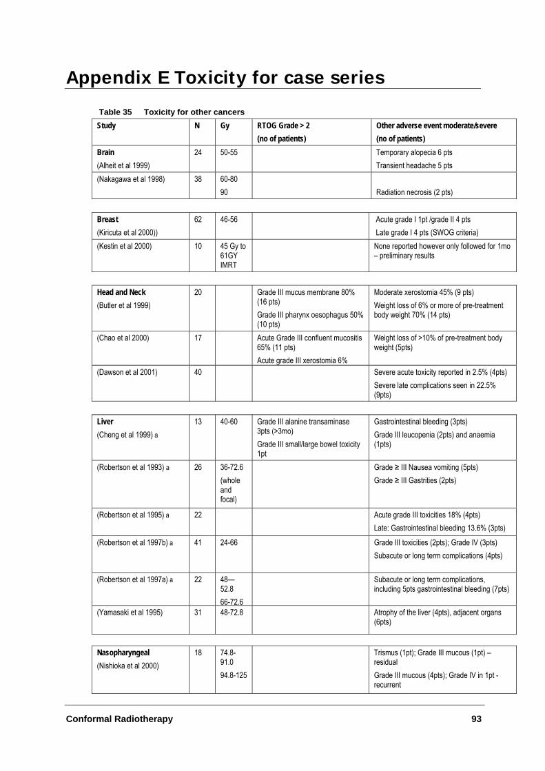
Conformal Radiotherapy 93
Appendix E Toxicity for case series
Table 35 Toxicity for other cancers Study N Gy RTOG Grade > 2
(no of patients) Other adverse event moderate/severe (no of patients)
Brain (Alheit et al 1999)
24 50-55 Temporary alopecia 6 pts Transient headache 5 pts
(Nakagawa et al 1998) 38 60-80 90
Radiation necrosis (2 pts)
Breast (Kiricuta et al 2000))
62 46-56 Acute grade I 1pt /grade II 4 pts Late grade I 4 pts (SWOG criteria)
(Kestin et al 2000) 10 45 Gy to 61GY IMRT
None reported however only followed for 1mo – preliminary results
Head and Neck (Butler et al 1999)
20 Grade III mucus membrane 80% (16 pts) Grade III pharynx oesophagus 50% (10 pts)
Moderate xerostomia 45% (9 pts) Weight loss of 6% or more of pre-treatment body weight 70% (14 pts)
(Chao et al 2000) 17 Acute Grade III confluent mucositis 65% (11 pts) Acute grade III xerostomia 6%
Weight loss of >10% of pre-treatment body weight (5pts)
(Dawson et al 2001) 40 Severe acute toxicity reported in 2.5% (4pts) Severe late complications seen in 22.5% (9pts)
Liver (Cheng et al 1999) a
13 40-60 Grade III alanine transaminase 3pts (>3mo) Grade III small/large bowel toxicity 1pt
Gastrointestinal bleeding (3pts) Grade III leucopenia (2pts) and anaemia (1pts)
(Robertson et al 1993) a 26 36-72.6 (whole and focal)
Grade ≥ III Nausea vomiting (5pts) Grade ≥ III Gastrities (2pts)
(Robertson et al 1995) a 22 Acute grade III toxicities 18% (4pts) Late: Gastrointestinal bleeding 13.6% (3pts)
(Robertson et al 1997b) a 41 24-66 Grade III toxicities (2pts); Grade IV (3pts) Subacute or long term complications (4pts)
(Robertson et al 1997a) a 22 48—52.8 66-72.6
Subacute or long term complications, including 5pts gastrointestinal bleeding (7pts)
(Yamasaki et al 1995) 31 48-72.8 Atrophy of the liver (4pts), adjacent organs (6pts)
Nasopharyngeal (Nishioka et al 2000)
18 74.8-91.0 94.8-125
Trismus (1pt); Grade III mucous (1pt) – residual Grade III mucous (4pts); Grade IV in 1pt -recurrent
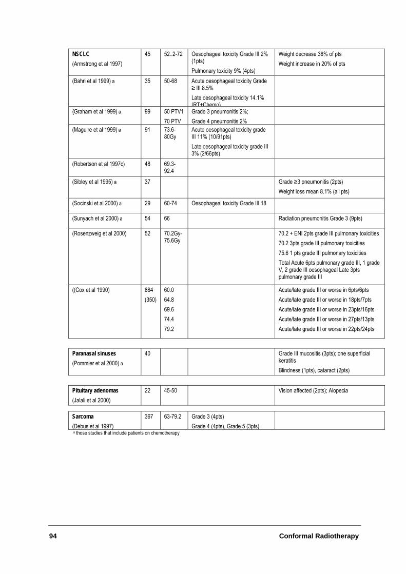
94 Conformal Radiotherapy
NSCLC (Armstrong et al 1997)
45 52..2-72 Oesophageal toxicity Grade III 2% (1pts) Pulmonary toxicity 9% (4pts)
Weight decrease 38% of pts Weight increase in 20% of pts
(Bahri et al 1999) a 35 50-68 Acute oesophageal toxicity Grade ≥ III 8.5% Late oesophageal toxicity 14.1% (RT+Chemo)
{Graham et al 1999) a 99 50 PTV1 70 PTV
Grade 3 pneumonitis 2%; Grade 4 pneumonitis 2%
(Maguire et al 1999) a 91 73.6-80Gy
Acute oesophageal toxicity grade III 11% (10/91pts) Late oesophageal toxicity grade III 3% (2/66pts)
(Robertson et al 1997c) 48 69.3-92.4
(Sibley et al 1995) a 37 Grade ≥3 pneumonitis (2pts) Weight loss mean 8.1% (all pts)
(Socinski et al 2000) a 29 60-74 Oesophageal toxicity Grade III 18
(Sunyach et al 2000) a 54 66 Radiation pneumonitis Grade 3 (9pts)
(Rosenzweig et al 2000) 52 70.2Gy-75.6Gy
70.2 + ENI 2pts grade III pulmonary toxicities 70.2 3pts grade III pulmonary toxicities 75.6 1 pts grade III pulmonary toxicities Total Acute 6pts pulmonary grade III, 1 grade V, 2 grade III oesophageal Late 3pts pulmonary grade III
((Cox et al 1990) 884 (350)
60.0 64.8 69.6 74.4 79.2
Acute/late grade III or worse in 6pts/6pts Acute/late grade III or worse in 18pts/7pts Acute/late grade III or worse in 23pts/16pts Acute/late grade III or worse in 27pts/13pts Acute/late grade III or worse in 22pts/24pts
Paranasal sinuses (Pommier et al 2000) a
40 Grade III mucositis (3pts); one superficial keratitis Blindness (1pts), cataract (2pts)
Pituitary adenomas (Jalali et al 2000)
22 45-50 Vision affected (2pts); Alopecia
Sarcoma (Debus et al 1997)
367 63-79.2 Grade 3 (4pts) Grade 4 (4pts), Grade 5 (3pts)
a those studies that include patients on chemotherapy
Conformal Radiotherapy 95
Abbreviations
3DCRT Three-dimensional conformal radiotherapy AAPM American Association of Physicists in Medicine ACC Adenoid cystic carcinoma AFP Alpha fetoprotein AIHW Australian Institute of Health and Welfare ASTRO American Society of Therapeutic Radiation Oncology BED Biologically equivalent dose BEV Beam’s eye view bNED Biochemical control no evidence of disease BTE Basic treatment equivalent CGE Cobalt Gray Equivalent CHART Continuous hyperfractionated accelerated radiotherapy CI Confidence interval CR Complete response CRT Conformal radiotherapy CT Computed tomography CTC Common Toxicity Criteria CTV Clinical target volume DDH Daniel Den Hoed DSH Dose surface histogram DVH Dose volume histogram EPI Electronic portal imaging EPID Electronic portal imaging device ESTV Equivalent simple treatment visit FCCC Fox Chase Cancer Centre FEV Forced expiratory volume FFF Freedom from failure GI Gastrointestinal GTV Gross tumour volume GU Genitourinary Gy Gray HCC Hepatocellular carcinoma HTA Health Technology Assessment IAH Intra-arterial hepatic chemotherapy IMRT Intensity Modulated Radiation Therapy ISN Integrated system network KPS Karnofsky Performance Score

96 Conformal Radiotherapy
LENT Late Effects Normal Tissues LPFS Local progression-free survival LRR Local regional recurrence MBS Medicare Benefits Scheme MDA MD Anderson MeSH Medical Subject Headings MLC Multileaf collimator MRI Magnetic resonance imaging MSAC Medical Services Advisory Committee MSKCC Memorial Sloan-Kettering Cancer Centre MVCT Megavoltage computed tomography NCI National Cancer Institute NHMRC National Health and Medical Research Council NPC Nasopharyngeal carcinoma NR Not reported NSCLC Non-small cell lung cancer NTCP Normal Tissue Complication Probability PET Positron emission tomography PR Partial response PSA Prostate Specific Antigen PTV Planning target volume PYLL Potential years life lost QA Quality assurance QALY Quality-adjusted life year QLQ Quality of life questionnaire R and V Record and verify RMH Royal Marsden Hospital RT Radiotherapy RTOG Radiation Therapy Oncology Group RTP Treatment planning system SCC Squamous cell carcinoma SCRT Stereotactic conformal radiotherapy SMART Simultaneous modulated accelerated treatment SPECT Single-photon emission computed tomography SRS Stereotactic radiosurgery SRT Standard radiotherapy SWOG Southwest Oncology Group TACE Transcatheter arterial chemoembolisation
Conformal Radiotherapy 97
References
Alheit, H., Saran, F. H., Warrington, A. P., Rosenberg, I., Perks, J., Jalali, R., Shepherd, S., Beardmore, C., Baumert, B., & Brada, M. 1999, "Stereotactically guided conformal radiotherapy for meningiomas", Radiotherapy & Oncology, vol. 50, no. 2, pp. 145-150.
Aribisala, E. 1993, "Cumulative Trauma Disorder: occupational hazard of the ' 90s", The Radiation Therapist pp. 27-29.
Armstrong, J., Raben, A., Zelefsky, M., Burt, M., Leibel, S., Burman, C., Kutcher, G., Harrison, L., Hahn, C., Ginsberg, R., Rusch, V., Kris, M., & Fuks, Z. 1997, "Promising survival with three-dimensional conformal radiation therapy for non-small cell lung cancer", Radiotherapy & Oncology, vol. 44, no. 1, pp. 17-22.
Armstrong, J. G., Zelefsky, M. J., Leibel, S. A., Burman, C., Han, C., Harrison, L. B., Kutcher, G. J., & Fuks, Z. Y. 1995, "Strategy for dose escalation using 3-dimensional conformal radiation therapy for lung cancer", Annals of Oncology, vol. 6, no. 7, pp. 693-697.
Australian Institute of Health and Welfare 1997, National Health Priority Areas: Cancer Control, Australian Institute of Health and Welfare, Canberra.
Australian Institute of Health and Welfare & Australasian Association of Cancer Registries 2000, Cancer in Australia 1997: Incidence and Mortality Data for 1997 and selected data for 1998 and 1999, Australian Institute of Health and Welfare, Canberra.
Bahri, S., Flickinger, J. C., Kalend, A. M., Deutsch, M., Belani, C. P., Sciurba, F. C., Luketich, J. D., & Greenberger, J. S. 1999, "Results of multifield conformal radiation therapy of nonsmall-cell lung carcinoma using multileaf collimation beams", Radiation Oncology Investigations, vol. 7, no. 5, pp. 297-308.
Beresford, J. 1994, "Manual handling in radiotherapy units", Occupational Health, vol. 46, no. 5, p. 170.
Bey, P., Carrie, C., Beckendorf, V., Ginestet, C., Aletti, P., Madelis, G., Luporsi, E., Pommier, P., Cowen, D., Gonzague-Casabianca, L., Simonian-Sauve, M., Maingon, P., Naudy, S., Lagrange, J., & Marcie, S. 2000, "Dose escalation with 3D-CRT in prostate cancer: French study of dose escalation with conformal 3D radiotherapy in prostate cancer-preliminary results", International Journal of Radiation Oncology, Biology, Physics, vol. 48, no. 2, pp. 513-517.
Boyle, T. & McPadden, E. 2000, "The contemporary use of radiation therapy in the management of lymphoma", Surgical Oncology Clinics of North America, vol. 9, no. 3, pp. 621-637.
Brada, M. & Baumert, B. 1999, "Focal fractionated conformal stereotactic boost following conventional radiotherapy of high-grade gliomas: a randomized phase III study. A joint study of the EORTC (22972) and the MRC (BR10)", Frontiers of Radiation Therapy & Oncology, vol. 33, pp. 241-243.
Brizel, D. M., Light, K., Zhou, S. M., & Marks, L. B. 1999, "Conformal radiation therapy
98 Conformal Radiotherapy
treatment planning reduces the dose to the optic structures for patients with tumors of the paranasal sinuses [see comments]", Radiotherapy & Oncology, vol. 51, no. 3, pp. 215-218.
Butler, E. B., Teh, B. S., Grant, W. H., Uhl, B. M., Kuppersmith, R. B., Chiu, J. K., Donovan, D. T., & Woo, S. Y. 1999, "Smart (simultaneous modulated accelerated radiation therapy) boost: a new accelerated fractionation schedule for the treatment of head and neck cancer with intensity modulated radiotherapy", International Journal of Radiation Oncology, Biology, Physics, vol. 45, no. 1, pp. 21-32.
Cardinale, R. M. & Kavanagh, B. D. 2000, "Conformal radiotherapy: what is it and why does it matter?", Surgical Oncology Clinics of North America, vol. 9, no. 3, pp. 415-434.
Carlson, D. 2001, "Intensity modulation using multileaf collimators: current status", Medical Dosimetry, vol. 26, no. 2, pp. 151-156.
Carrie, C. & Ginestet, C. 1997, "Acute toxicity in pelvic radiotherapy; a randomized trial of conformal versus conventional treatment", Radiotherapy & Oncology, vol. 44, no. 3, pp. 295-296.
Chang, J. T., See, L. C., Liao, C. T., Ng, S. H., Wang, C. H., Chen, I. H., Tsang, N. M., Tseng, C. K., Tang, S. G., & Hong, J. H. 2000, "Locally recurrent nasopharyngeal carcinoma", Radiotherapy & Oncology, vol. 54, no. 2, pp. 135-142.
Chao, K. S., Low, D. A., Perez, C. A., & Purdy, J. A. 2000, "Intensity-modulated radiation therapy in head and neck cancers: The Mallinckrodt experience", International Journal of Cancer, vol. 90, no. 2, pp. 92-103.
Cheng, S. H., Lin, Y. M., Chuang, V. P., Yang, P. S., Cheng, J. C., Huang, A. T., & Sung, J. L. 1999, "A pilot study of three-dimensional conformal radiotherapy in unresectable hepatocellular carcinoma", Journal of Gastroenterology & Hepatology, vol. 14, no. 10, pp. 1025-1033.
Cho, K. H., Khan, F. M., & Levitt, S. H. 1999, "Cost-benefit analysis of 3D conformal radiation therapy--treatment of prostate cancer as a model", Acta Oncologica, vol. 38, no. 5, pp. 603-611.
Commonwealth Department of Health and Aged Care. Medicare Benefits Schedule, November 1, 2000. DHAC Available from: http://www.health.gov.au/pubs/mbs/mbs6/default.htm.
Cox, J. D., Azarnia, N., Byhardt, R. W., Shin, K. H., Emami, B., & Pajak, T. F. 1990, "A randomized phase I/II trial of hyperfractionated radiation therapy with total doses of 60.0 Gy to 79.2 Gy: possible survival benefit with greater than or equal to 69.6 Gy in favorable patients with Radiation Therapy Oncology Group stage III non-small-cell lung carcinoma: report of Radiation Therapy Oncology Group 83-11", Journal of Clinical Oncology, vol. 8, no. 9, pp. 1543-1555.
Dawson, L. A., Anzai, Y., Marsh, L., Martel, M. K., Paulino, A., Ship, J. A., & Eisbruch, A. 2000, "Patterns of local-regional recurrence following parotid-sparing conformal and segmental intensity-modulated radiotherapy for head and neck cancer", International Journal of Radiation Oncology, Biology, Physics, vol. 46, no. 5, pp. 1117-1126.
Conformal Radiotherapy 99
Dawson, L. A., Myers, L. L., Bradford, C. R., Chepeha, D. B., Hogikyan, N., Teknos, T. N., Terrell, J. E., Wolf, G. T., & Eisbruch, A. 2001, "Conformal re-irradiation of recurrent and new primary head and neck cancer", International Journal of Radiation Oncology, Biology, Physics, vol. 50, no. 2, pp. 377-385.
Dearnaley, D. P., Khoo, V. S., Norman, A. R., Meyer, L., Nahum, A., Tait, D., Yarnold, J., & Horwich, A. 1999, "Comparison of radiation side-effects of conformal and conventional radiotherapy in prostate cancer: a randomised trial", Lancet, vol. 353, no. 9149, pp. 267-272.
Debus, J., Hug, E. B., Liebsch, N. J., O'Farrel, D., Finkelstein, D., Efird, J., & Munzenrider, J. E. 1997, "Brainstem tolerance to conformal radiotherapy of skull base tumors", International Journal of Radiation Oncology, Biology, Physics, vol. 39, no. 5, pp. 967-975.
DeMeyer, C. L., Whitehead, L. W., Jacobson, A. P., & Brown, D. G. 1986, "Potential exposure to metal fumes, particulates, and organic vapors during radiotherapy shielding block fabrication", Medical Physics, vol. 13, no. 5, pp. 748-750.
Dunscombe, P. B. & Roberts, G. 2000, "An economic framework for evaluating a multileaf collimator", International Journal of Technology Assessment in Health Care, vol. 16, no. 1, pp. 242-250.
Eisbruch, A., Dawson, L. A., Kim, H. M., Bradford, C. R., Terrell, J. E., Chepeha, D. B., Teknos, T. N., Anzai, Y., Marsh, L. H., Martel, M. K., Ten Haken, R. K., Wolf, G. T., & Ship, J. A. 1999a, "Conformal and intensity modulated irradiation of head and neck cancer: the potential for improved target irradiation, salivary gland function, and quality of life", Acta Oto-Rhino-Laryngologica Belgica, vol. 53, no. 3, pp. 271-275.
Eisbruch, A., Ten Haken, R. K., Kim, H. M., Marsh, L. H., & Ship, J. A. 1999b, "Dose, volume, and function relationships in parotid salivary glands following conformal and intensity-modulated irradiation of head and neck cancer [see comments]", International Journal of Radiation Oncology, Biology, Physics, vol. 45, no. 3, pp. 577-587.
Emami, B. 1996, "Three-dimensional conformal radiation therapy in bronchogenic carcinoma", Seminars in Radiation Oncology, vol. 6, no. 2, pp. 92-97.
Emami, B., Graham, M. V., Michalski, J. M., & Perez, C. A. 1997, "Three-dimensional conformal radiation therapy: clinical aspects," in Principles and Practice of Radiation Oncology, C. A. Perez & L. W. Brady, eds., Lippincott-Raven Publishers, Philadelphia, pp. 371-386.
Faculty of Radiation Oncology, R., Australian Institute of Radiography, & Australasian College of Physical Scientists and Engineers in Medicine 2001, National Strategic Plan for Radiation Oncology (Australia).
Fiveash, J. B., Hanks, G., Roach, M., Wang, S., Vigneault, E., McLaughlin, P. W., & Sandler, H. M. 2000, "3D conformal radiation therapy (3DCRT) for high grade prostate cancer: a multi-institutional review", International Journal of Radiation Oncology, Biology, Physics, vol. 47, no. 2, pp. 335-342.
Foroudi, F., Lapsley, H., Manderson, C., & Yeghiaian-Alvandi, R. 2000, "Cost-Minimization Analysis: radiation treatment with and without a multi-leaf collimator",
100 Conformal Radiotherapy
International Journal of Radiation Oncology, Biology., vol. 47, no. 5, pp. 1443-1448.
Fraass, B., Doppke, K., Hunt, M., Kutcher, G., Starkschall, G., Stern, R., & Van Dyk, J. 1998, "American Association of Physicists in Medicine Radiation Therapy Committee Task Group 53: quality assurance for clinical radiotherapy treatment planning", Medical Physics, vol. 25, no. 10, pp. 1773-1829.
Graham, M. V., Purdy, J. A., Emami, B., Harms, W., Bosch, W., Lockett, M. A., & Perez, C. A. 1999, "Clinical dose-volume histogram analysis for pneumonitis after 3D treatment for non-small cell lung cancer (NSCLC)", International Journal of Radiation Oncology, Biology, Physics, vol. 45, no. 2, pp. 323-329.
Grant, W. & Woo, S. Y. 1999, "Clinical and financial issues for intensity-modulated radiation therapy delivery", Seminars in Radiation Oncology, vol. 9, no. 1, pp. 99-107.
Greiner, R. H., Munkel, G., Blattmann, H., Coray, A., Kann, R., Pedroni, E., & Thum, P. 1992, "Conformal radiotherapy for unresectable retroperitoneal soft tissue sarcoma", International Journal of Radiation Oncology, Biology, Physics, vol. 22, no. 2, pp. 333-341.
Hanks, G. E., Hanlon, A. L., Pinover, W. H., Horwitz, E. M., Price, R. A., & Schultheiss, T. 2000, "Dose selection for prostate cancer patients based on dose comparison and dose response studies", International Journal of Radiation Oncology, Biology, Physics, vol. 46, no. 4, pp. 823-832.
Hanks, G. E., Hanlon, A. L., Schultheiss, T. E., Pinover, W. H., Movsas, B., Epstein, B. E., & Hunt, M. A. 1998, "Dose escalation with 3D conformal treatment: five year outcomes, treatment optimization, and future directions", International Journal of Radiation Oncology, Biology, Physics, vol. 41, no. 3, pp. 501-510.
Hanks, G. E., Lee, W. R., Hanlon, A. L., Hunt, M., Kaplan, E., Epstein, B. E., Movsas, B., & Schultheiss, T. E. 1996, "Conformal technique dose escalation for prostate cancer: biochemical evidence of improved cancer control with higher doses in patients with pretreatment prostate-specific antigen ≥10", International Journal of Radiation Oncology*Biology*Physics, vol. 35, no. 5, pp. 861-868.
Hanks, G. E., Schultheiss, T. E., Hanlon, A. L., Hunt, M., Lee, W. R., Epstein, B. E., & Coia, L. R. 1997, "Optimization of conformal radiation treatment of prostate cancer: report of a dose escalation study", International Journal of Radiation Oncology, Biology, vol. 37, no. 3, pp. 543-550.
Hatherly, K. E., Smylie, J. C., Rodger, A., Dally, M. J., Davis, S. R., & Millar, J. L. 2001, "A double exposed portal image comparison between electronic portal imaging hard copies and port films in radiation therapy treatment setup confirmation to determine its clinical application in a radiotherapy center", International Journal of Radiation Oncology, Biology, Physics, vol. 49, no. 1, pp. 191-198.
Helyer, S. J. & Heisig, S. 1995, "Multileaf collimation versus conventional shielding blocks: a time and motion study of beam shaping in radiotherapy", Radiotherapy & Oncology, vol. 37, no. 1, pp. 61-64.
Herman, M. G., Balter, J. M., Jaffray, D. A., McGee, K. P., Munro, P., Shalev, S., van Herk, M., & Wong, J. W. 2001, "Clinical use of electronic portal imaging: Report of
Conformal Radiotherapy 101
AAPM Radiation Therapy Committee Task Group 58", Medical Physics, vol. 28, no. 5, pp. 713-734.
Horwitz, E. M. & Hanks, G. E. 2000, "Three-dimensional conformal radiation therapy: what are the costs and benefits?", Surgical Oncology Clinics of North America, vol. 9, no. 3, pp. 455-467.
Horwitz, E. M., Hanlon, A. L., Pinover, W. H., & Hanks, G. E. 1999, "The cost effectiveness of 3D conformal radiation therapy compared with conventional techniques for patients with clinically localized prostate cancer :”International Journal of Radiation Oncology, Biology, Physics, vol. 45, no. 5, pp. 1219-1225.
Huddart, R. A., Nahum, A., Neal, A., McLean, M., Dearnaley, D. P., Law, M., Dyer, J., & Tait, D. 1996, "Accuracy of pelvic radiotherapy: prospective analysis of 90 patients in a randomised trial of blocked versus standard radiotherapy", Radiotherapy & Oncology, vol. 39, no. 1, pp. 19-29.
Image Guided Therapy QA Center at Washington University. 3D Conformal Radiation Therapy. 2001. Available at http://rtog3dqa.wustl.edu
Jalali, R., Brada, M., Perks, J. R., Warrington, A. P., Traish, D., Burchell, L., McNair, H., Thomas, D. G., Robinson, S., & Johnston, D. G. 2000, "Stereotactic conformal radiotherapy for pituitary adenomas: technique and preliminary experience", Clinical Endocrinology, vol. 52, no. 6, pp. 695-702.
Kestin, L. L., Sharpe, M. B., Frazier, R. C., Vicini, F. A., Yan, D., Matter, R. C., Martinez, A. A., & Wong, J. W. 2000, "Intensity modulated to improve dose uniformity wiuth tangential breast radiotherapy: initial clinical experience", International Journal of Radiation Oncology, Biology, Physics, vol. 48, no. 5, pp. 1559-1568.
Kiricuta, I. C., Gotz, U., Schwab, F., Fehn, M., & Neumann, H. H. 2000, "Target volume definition and target conformal irradiation technique for breast cancer patients", Acta Oncologica, vol. 39, no. 3, pp. 429-436.
Klein, E. E., Harms, W. B., Low, D. A., Willcut, V., & Purdy, J. A. 1995, "Clinical implementation of a commercial multileaf collimator: dosimetry, networking, simulation, and quality assurance", International Journal of Radiation Oncology, Biology, Physics, vol. 33, no. 5, pp. 1195-1208.
Kolitsi, Z., Dahl, O., Van Loon, R., Drouard, J., Van Dijk, J., Ruden, B. I., Chierego, G., & Rosenwald, J. C. 1997, "Quality assurance in conformal radiotherapy: DYNARAD consensus report on practice guidelines", Radiotherapy & Oncology, vol. 45, no. 3, pp. 217-223.
Koper, P. C., Stroom, J. C., van Putten, W. L., Korevaar, G. A., Heijmen, B. J., Wijnmaalen, A., Jansen, P. P., Hanssens, P. E., Griep, C., Krol, A. D., Samson, M. J., & Levendag, P. C. 1999, "Acute morbidity reduction using 3DCRT for prostate carcinoma: a randomized study", International Journal of Radiation Oncology, Biology, Physics, vol. 43, no. 4, pp. 727-734.
Kuppersmith, R. B., Greco, S. C., Teh, B. S., Donovan, D. T., Grant, W., Chiu, J. K., Cain, R. B., & Butler, E. B. 1999, "Intensity-modulated radiotherapy: first results with
102 Conformal Radiotherapy
this new technology on neoplasms of the head and neck", Ear, Nose, & Throat Journal, vol. 78, no. 4, pp. 238-6.
Lavertu, S., Girouard, L. M., & Pouliot, J. 2000, "Observation study of electronic portal images for off-line verification", Radiotherapy & Oncology, vol. 54, no. 1, pp. 47-55.
Lawton, C. A., Won, M., Pilepich, M. V., Asbell, S., Shipley, W. U., Hanks, G. E., Cox, J. D., Perez, C. A., Sause, W. T., Doggett, S. R. L., & Rubin, P. 1991, "Long-term treatment sequelae following external beam irradiation for adenocarconoma of the prostate: analysis of RTOG studies 7506 and 7706", International Journal of Radiation Oncology, Biology, Physics, vol. 21, pp. 935-939.
Lee, S. W., Fraass, B. A., Marsh, L. H., Herbort, K., Gebarski, S. S., Martel, M. K., Radany, E. H., Lichter, A. S., & Sandler, H. M. 1999, "Patterns of failure following high-dose 3-D conformal radiotherapy for high-grade astrocytomas: a quantitative dosimetric study", International Journal of Radiation Oncology, Biology, Physics, vol. 43, no. 1, pp. 79-88.
Lee, T. 1995. Multileaf Collimators in Radiotherapy. Project undertaken as part of a requirement for the Cumberland College of Health Sciences, University of Sydney. (Unpublished Work)
Lichter, A. S. & Ten Haken, R. K. 1995, "Three-dimensional treatment and conformal radiation dose delivery," in Important Advances in Oncology, V. T. DeVita, S. Hellman, & S. A. Rosenthal, eds., Lippincott Company, Philadelphia, pp. 95-109.
LoSasso, T. & Kutcher, G. J. 1995, "Multileaf collimation versus alloy blocks: analysis of geometric accuracy", International Journal of Radiation Oncology, Biology, Physics, vol. 32, no. 2, pp. 499-506.
Maguire, P. D., Sibley, G. S., Zhou, S. M., Jamieson, T. A., Light, K. L., Antoine, P. A., Herndon, J. E., Anscher, M. S., & Marks, L. B. 1999, "Clinical and dosimetric predictors of radiation-induced esophageal toxicity", International Journal of Radiation Oncology, Biology, Physics, vol. 45, no. 1, pp. 97-103.
Mc Gibney, C., Holmberg, O., McClean, B., Williams, C., McCrea, P., Sutton, P., & Armstrong, J. 1999, "Dose escalation of chart in non small cell lung cancer: is three dimensional conformal radiation therapy really necessary?", International Journal of Radiation Oncology Biology Physics , vol. 45, no. 2, pp. 339-350.
McCullough, E. C. & Senjem, D. H. 1981, "Airborne concentrations of toxic metals resulting from the use of low melting point lead alloys to construct radiotherapy shielding", Medical Physics, vol. 8, no. 1, pp. 111-114.
Michalski, J. M. & Purdy, J. A. 1998, "Innovations in three-dimensional treatment planning and quality assurance", Tumori, vol. 84, no. 2, pp. 127-139.
Michalski, J. M., Purdy, J. A., Winter, K., Roach, M., Vijayakumar, S., Sandler, H. M., Markoe, A. M., Ritter, M. A., Russell, K. J., Sailer, S., Harms, W. B., Perez, C. A., Wilder, R. B., Hanks, G. E., & Cox, J. D. 2000, "Preliminary report of toxicity following 3D radiation therapy for prostate cancer on 3DOG/RTOG 9406", International Journal of Radiation Oncology, Biology, Physics, vol. 46, no. 2, pp. 391-402.
Conformal Radiotherapy 103
Miller, S. D. & O'Brien, J. E. 2001, "Prevalence of Back Pain Among Radiation Therapists", Radiologic Technology, vol. 62, no. 6, pp. 460-466.
Nakagawa, K., Aoki, Y., Fujimaki, T., Tago, M., Terahara, A., Karasawa, K., Sakata, K., Sasaki, Y., Matsutani, M., & Akanuma, A. 1998, "High-dose conformal radiotherapy influenced the pattern of failure but did not improve survival in glioblastoma multiforme", International Journal of Radiation Oncology, Biology, Physics, vol. 40, no. 5, pp. 1141-1149.
Nakagawa, K., Aoki, Y., Tago, M., Terahara, A., & Ohtomo, K. 2000, "Megavoltage CT-assisted stereotactic radiosurgery for thoracic tumors: original research in the treatment of thoracic neoplasms", International Journal of Radiation Oncology, Biology, Physics, vol. 48, no. 2, pp. 449-457.
National Occupational Health and Safety Commission. National Code of Practice for Manual Handling. Available at http://www.nohsc.gov.au/OHSLegalObligations/NationalStandards/manhan.htm
Nguyen, L. N., Pollack, A., & Zagars, G. K. 1998, "Late effects after radiotherapy for prostate cancer in a randomized dose-response study: results of a self-assessment questionnaire", Urology, vol. 51, no. 6, pp. 991-997.
Nishioka, T., Shirato, H., Kagei, K., Fukuda, S., Hashimoto, S., & Ohmori, K. 2000, "Three-dimensional small-volume irradiation for residual or recurrent nasopharyngeal carcinoma", International Journal of Radiation Oncology, Biology, Physics, vol. 48, no. 2, pp. 495-500.
Perez, C. A., Michalski, J., Ballard, S., Drzymala, R., Kobeissi, B. J., Lockett, M. A., & Wasserman, T. H. 1997, "Cost benefit of emerging technology in localized carcinoma of the prostate", International Journal of Radiation Oncology, Biology, Physics, vol. 39, no. 4, pp. 875-883.
Pollack, A., Zagars, G. K., Smith, L. G., Lee, J., von Eschenbach, A. C., Antolak, J. A., Starkschall, G., & Rosen, I. 2000, "Preliminary results of a randomised radiotherapy dose escalation study comparing 70 Gy with 78 Gy for prostate cancer", Journal of Clinical Oncology, vol. 18, no. 23, pp. 3904-3911.
Pollack, A., Zagars, G. K., Starkschall, G., Childress, C. H., Kopplin, S., Boyer, A. L., & Rosen, I. I. 1996, "Conventional vs. conformal radiotherapy for prostate cancer: preliminary results of dosimetry and acute toxicity", International Journal of Radiation Oncology, Biology, Physics, vol. 34, no. 3, pp. 555-564.
Pommier, P., Ginestet, C., Sunyach, M., Zrounba, P., Poupart, M., Ceruse, P., Ciupea, C., Carrie, C., & Montbarbon, X. 2000, "Conformal radiotherapy for paranasal sinus and nasal cavity tumors: three-dimensional treatment planning and preliminary results in 40 patients", International Journal of Radiation Oncology, Biology, Physics, vol. 48, no. 2, pp. 485-493.
Powlis, W. D., Smith, A. R., Cheng, E., Galvin, J. M., Villari, F., Bloch, P., & Kligerman, M. M. 1993a, "Initiation of multi-leaf collimator conformal radiation therapy [see comments]", International Journal of Radiation Oncology, Biology, Physics, vol. 25, no. 2, pp.
104 Conformal Radiotherapy
171-179.
Powlis, W. D., Smith, A. R., Cheng, E., Galvin, J. M., Villari, F., Bloch, P., & Kligerman, M. M. 1993b, "Initiation of multileaf collimator conformal radiation therapy", International Journal of Radiation Oncology Biology Physics, vol. 25, no. 2, pp. 171-179.
Purdy, J. A. 1983, "Secondary Field Shaping," in Advances in Radiation Therapy Treatment Planning, A. Wright & A. Boyer, eds., American Institute of Physics Inc, New York, pp. 456-476.
Purdy, J. A. 1997, "Three-dimensional physics and treatment planning," in Principles and Practice of Radiation Oncology, C. A. Perez & L. W. Brady, eds., Lippincott-Raven Publishers, Philadelphia, pp. 343-369.
Purdy, J. A. & Klein, E. E. 1997, "External photn beam dosimety and treatment planning," in Principles and Practice of Radiation Oncology, C. A. Perez & L. W. Brady, eds., Lippincott-Raven Publishers, Philadelphia, pp. 281-320.
Robertson, J. M., Lawrence, T. S., Andrews, J. C., Walker, S., Kessler, M. L., & Ensminger, W. D. 1997a, "Long-term results of hepatic artery fluorodeoxyuridine and conformal radiation therapy for primary hepatobiliary cancers", International Journal of Radiation Oncology, Biology, Physics, vol. 37, no. 2, pp. 325-330.
Robertson, J. M., Lawrence, T. S., Dworzanin, L. M., Andrews, J. C., Walker, S., Kessler, M. L., DuRoss, D. J., & Ensminger, W. D. 1993, "Treatment of primary hepatobiliary cancers with conformal radiation therapy and regional chemotherapy", Journal of Clinical Oncology, vol. 11, no. 7, pp. 1286-1293.
Robertson, J. M., Lawrence, T. S., Walker, S., Kessler, M. L., Andrews, J. C., & Ensminger, W. D. 1995, "The treatment of colorectal liver metastases with conformal radiation therapy and regional chemotherapy", International Journal of Radiation Oncology, Biology, Physics, vol. 32, no. 2, pp. 445-450.
Robertson, J. M., McGinn, C. J., Walker, S., Marx, M. V., Kessler, M. L., Ensminger, W. D., & Lawrence, T. S. 1997b, "A phase I trial of hepatic arterial bromodeoxyuridine and conformal radiation therapy for patients with primary hepatobiliary cancers or colorectal liver metastases", International Journal of Radiation Oncology, Biology, Physics, vol. 39, no. 5, pp. 1087-1092.
Robertson, J. M., Ten Haken, R. K., Hazuka, M. B., Turrisi, A. T., Martel, M. K., Pu, A. T., Littles, J. F., Martinez, F. J., Francis, I. R., Quint, L. E., & Lichter, A. S. 1997c, "Dose escalation for non-small cell lung cancer using conformal radiation therapy", International Journal of Radiation Oncology, Biology, Physics, vol. 37, no. 5, pp. 1079-1085.
Rosenberg, A. E., Nielsen, G. P., Keel, S. B., Renard, L. G., Fitzek, M. M., Munzenrider, J. E., & Liebsch, N. J. 1999, "Chondrosarcoma of the base of the skull: a clinicopathologic study of 200 cases with emphasis on its distinction from chordoma", American Journal of Surgical Pathology, vol. 23, no. 11, pp. 1370-1378.
Rosenzweig, K. E., Mychalczak, B., Fuks, Z., Hanley, J., Burman, C., Ling, C. C., Armstrong, J., Ginsberg, R., Kris, M. G., Raben, A., & Leibel, S. 2000, "Final report of the 70.2-Gy and 75.6-Gy dose levels of a phase I dose escalation study using three-
Conformal Radiotherapy 105
dimensional conformal radiotherapy in the treatment of inoperable non-small cell lung cancer. [see comments]", Cancer Journal, vol. 6, no. 2, pp. 82-87.
Schultheiss, T. E., Lee, W. R., Hunt, M. A., Hanlon, A. L., Peter, R. S., & Hanks, G. E. 1997, "Late GI and GU complications in the treatment of prostate cancer", International Journal of Radiation Oncology, Biology, Physics, vol. 37, no. 1, pp. 3-11.
Shalev, S. 1996, "On-line portal imaging: contributions and limitations in clincial practice", Frontiers of radiation therapy and oncology pp. 156-167.
Sibley, G. S., Mundt, A. J., Shapiro, C., Jacobs, R., Chen, G., Weichselbaum, R., & Vijayakumar, S. 1995, "The treatment of stage III nonsmall cell lung cancer using high dose conformal radiotherapy", International Journal of Radiation Oncology, Biology, Physics, vol. 33, no. 5, pp. 1001-1007.
Skwarchuk, M. W., Jackson, A., Zelefsky, M. J., Venkatraman, E. S., Cowen, D. M., Levegrun, S., Burman, C. M., Fuks, Z., Leibel, S. A., & Ling, C. C. 2000, "Late rectal toxicity after conformal radiotherapy of prostate cancer (I): multivariate analysis and dose-response", International Journal of Radiation Oncology, Biology, Physics, vol. 47, no. 1, pp. 103-113.
Socinski, M. A., Rosenman, J. G., Schell, M. J., Halle, J., Russo, S., Rivera, M. P., Clark, J., Limentani, S., Fraser, R., Mitchell, W., & Detterbeck, F. C. 2000, "Induction carboplatin/paclitaxel followed by concurrent carboplatin/paclitaxel and dose-escalating conformal thoracic radiation therapy in unresectable stage IIIA/B nonsmall cell lung carcinoma: a modified Phase I trial", Cancer, vol. 89, no. 3, pp. 534-542.
Storey, M. R., Pollack, A., Zagars, G., Smith, L., Antolak, J., & Rosen, I. I. 2000, "Complications from radiotherapy dose escalation in prostate cancer: preliminary results of a randomised trial", International Journal of Radiation Oncology, Biology, Physics, vol. 48, no. 3, pp. 635-642.
Sunyach, M. P., Falchero, L., Pommier, P., Perol, M., Arpin, D., Vincent, M., Boutry, D., Rebatu, P., Ginestet, C., Martel-Lafay, I., Perol, D., & Carrie, C. 2000, "Prospective evaluation of early lung toxicity following three-dimensional conformal radiation therapy in non-small-cell lung cancer: preliminary results", International Journal of Radiation Oncology, Biology, Physics, vol. 48, no. 2, pp. 459-463.
Tait, D. M., Nahum, A. E., Meyer, L. C., Law, M., Dearnaley, D. P., Horwich, A., Mayles, W. P., & Yarnold, J. R. 1997, "Acute toxicity in pelvic radiotherapy; a randomised trial of conformal versus conventional treatment [see comments]", Radiotherapy & Oncology, vol. 42, no. 2, pp. 121-136.
Tait, D. M., Nahum, A. E., Rigby, L., Chow, M., Mayles, W. P., Dearnaley, D. P., & Horwich, A. 1993, "Conformal radiotherapy of the pelvis: assessment of acute toxicity", Radiotherapy & Oncology, vol. 29, no. 2, pp. 117-126.
Thomason, C. L. 1998, "Implementation and clinical use of portal imaging", Cancer Treatment & Research, vol. 93, pp. 66-69.
Tubiana, M. & Eschwege, F. 2000, "Conformal radiotherapy and intensity-modulated radiotherapy", Acta Oncologica, vol. 39, pp. 555-567.
106 Conformal Radiotherapy
UICC 1997, TNM classification of malignant tumours , Fifth edn, Wiley-Liss, New York.
Verhey, L. J. 1999, "Comparison of three-dimensional conformal radiation therapy and intensity-modulated radiation therapy systems", Seminars in Radiation Oncology, vol. 9, no. 1, pp. 78-98.
Vijayakumar, S. & Chen, G. T. Y. 1995, "Implementation of three dimensional conformal radiation therapy: prospects, opportunities, and challenges", International Journal of Radiation Oncology, Biology, Physics, vol. 33, no. 5, pp. 979-983.
Vyas, A., Pickering, C. A., Oldham, L. A., Francis, H. C., Fletcher, A. M., Merrett, T., & Niven, R. M. 2000, "Survey of symptoms, respiratory function, and immunology and their relation to glutaraldehyde and other occupational exposures among endoscopy nursing staff", Occupational & Environmental Medicine, vol. 57, no. 11, pp. 752-759.
Worthington, K. 2001, "Working with glutaraldehyde:take precautions and consider long-term alternatives", American Journal of Nursing, vol. 101, no. 6, p. 88.
Yamasaki, S. A., Marn, C. S., Francis, I. R., Robertson, J. M., & Lawrence, T. S. 1995, "High-dose localized radiation therapy for treatment of hepatic malignant tumors: CT findings and their relation to radiation hepatitis", American Journal of Roentgenology, vol.165, no. 1, pp. 79-84.
Yamazaki, A., Shirato, H., Nishioka, T., Hashimoto, S., Kitahara, T., Kagei, K., & Miyasaka, K. 2000, "Reduction of late complications after irregularly shaped four-field whole pelvic radiotherapy using computed tomographic simulation compared with parallel-opposed whole pelvic radiotherapy", Japanese Journal of Clinical Oncology, vol. 30, no. 4, pp. 180-184.
Zelefsky, M., Leibel, S. A., Gaudin, P. B., Kutcher, G. J., Fleshner, N. E., Venkatraman, E. S., Reuter, V. E., Fair, W. R., Ling, C. C., & Fuks, Z. 1998a, "Dose escalation with three-dimensional conformal radiation therapy affects the outcome in prostate cancer", International Journal of Radiation Oncology, Biology, Physics, vol. 41, no. 3, pp. 491-500.
Zelefsky, M. J., Cowen, D., Fuks, Z., Shike, M., Burman, C., Jackson, A., Venkatramen, E. S., & Leibel, S. A. 1999, "Long term tolerance of high dose three-dimensional conformal radiotherapy in patients with localized prostate carcinoma", Cancer, vol. 85, no. 11, pp. 2460-2468.
Zelefsky, M. J., Fuks, Z., Happersett, L., Lee, H. J., Ling, C. C., Burman, C. M., Hunt, M., Wolfe, T., Venkatraman, E. S., Jackson, A., Skwarchuk, M., & Leibel, S. A. 2000, "Clinical experience with intensity modulated radiation therapy (IMRT) in prostate cancer", Radiotherapy & Oncology, vol. 55, no. 3, pp. 241-249.
Zelefsky, M. J., Fuks, Z., Wolfe, T., Kutcher, G. J., Burman, C., Ling, C. C., Venkatraman, E. S., & Leibel, S. A. 1998b, "Locally advanced prostatic cancer: long-term toxicity outcome after three-dimensional conformal radiation therapy--a dose-escalation study", Radiology, vol. 209, no. 1, pp. 169-174.
Zelefsky, M. J., Leibel, S. A., Kutcher, G. J., & Fuks, Z. 1998c, "Three-dimensional conformal radiotherapy and dose escalation: where do we stand?", Seminars in Radiation Oncology, vol. 8, no. 2, pp. 107-114.