conformational analysis and automated receptor docking of selective arylacetamide-based κ-opioid...
TRANSCRIPT

Conformational Analysis and Automated Receptor Docking of SelectiveArylacetamide-Based K-Opioid Agonists
Govindan Subramanian, M. Germana Paterlini, Dennis L. Larson, Philip S. Portoghese, andDavid M. Ferguson*
Department of Medicinal Chemistry and Minnesota Supercomputer Institute, University of Minnesota,Minneapolis, Minnesota 55455
Received May 20, 1998
The three-dimensional structure, dynamics, and binding modes of representative κ-opioidagonists of the arylacetamide class (U50,488; U69,593; U62,066; CI-977; ICI199,441; ICI197,067;BRL52,537; and BRL52,656) have been investigated using molecular modeling techniques.Systematic exploration of the conformational space of the ligand combined with moleculardynamics (MD) simulations in water revealed consistent conformational preferences for allthe κ-agonists in this series. The results were further compared with available X-ray and 1D-and 2D-NMR data to identify potential “lead” conformers for molecular docking. Ligand bindingmodes were initially determined using automated docking of two of the ligands (U50,488 andBRL52,537) to the κ-opioid receptor. Extrapolation of the predicted binding mode to othermembers in this ligand series revealed similar docking preferences, with each ligand dockedalong the receptor helical axis. The binding modes were further refined using MD simulationsof the receptor-ligand complexes. The results show a that salt bridge is formed between theamino proton of the ligands and the carboxylate group of Asp138 in TM3. This interactionmost likely serves as a key anchoring point for the agonist association. Additional ligandcontacts were noted with κ-specific residues Ile294, Leu295, and Ala298, which may, in part,explain the κ-selectivity in this series. In comparing the arylacetamides with opiate-basedligands, no evidence was found to link these classes through a common binding motif (exceptfor the ion pair). The binding site model was also applied to explain the enantiomeric preferenceof U50,488 and to provide insight to the µ/κ-selectivity of representative ligands in this series.Overall, the results provide a structure-based rationale for ligand recognition that is consistentboth with site-directed mutagenesis experiments and structure-function relationship data.
Introduction
The past decade has witnessed an upsurge in thedevelopment of highly selective, non-peptide ligands forthe µ-, δ-, and κ-opioid receptors. While such com-pounds were developed as potential analgesics, theyhave applications as pharmacological tools as well.1Although a wide variety of opiate and opioid ligandshave been described,2 evidence has emerged for a uniqueclass of highly selective κ-agonists based on the U50,-488 (1; Figure 1) structural prototype.3 Representativesof this series (1-10) have been characterized as havingseveral key features, including the arylacetamide moietyand a basic amino group as part of a pyrrolidine ring.4While some similarities can be drawn between 1-10and opiate-based ligands, little evidence has surfacedto support comparative modeling of these ligand fami-lies. It is therefore not feasible to compare the bindingmodes or conformational preferences of 1-10 withopiates as molecular templates.5,6
Fortunately, some insights on the formation of 1-10can be obtained from the structure-activity relationship(SAR) data for this ligand series. By varying the aryl(1 vs 2)7,8 and amide (1 vs 9, 10)9-12 substituents,altering the chain length (spacer) between the aryl andamide group,13 modifying the cyclohexyl moiety (1 vs3-8),14-19 and changing the ring size that contains thetertiary nitrogen, SAR studies have identified various
means of optimizing the receptor-ligand interaction toimprove the κ-opioid receptor binding and selectivity.20,21
Although a simple spirotetrahydrofuranyl substituenton the cyclohexane ring in U50,488 [Ki(1) ) 0.9 nM]22
reveals an increased ligand binding [Ki(4) ) 0.3 nM],15b
dramatic effects in the µ/κ-selectivity are observed whenthe aryl group is modified in concert.7 Thus, CI-977 (5)displays greater selectivity (1575-fold)7 for the κ-receptoras compared to 1 and 4 (280-fold).15b Likewise, SARstudies have demonstrated enantioselective binding for5,8 with similar trends exhibited by other κ-agonists aswell. Other interesting chemical modifications display-ing selective κ-agonist activity are exemplified furtherby the ICI series (6-8)17-19,23 and the piperidine deriva-tives (9, 10).9,10
Despite these key developments in designing potentκ-agonists, the factors influencing the ligand bindingand selectivity to the opioid receptors are not yetunderstood. Elucidation of the three-dimensional struc-ture of the free ligand (1-10) and the receptor-ligandcomplex would afford a better understanding of themolecular recognition process at the κ-opioid receptor.In earlier work, we docked naltrexone-based compoundsto a model-built κ-opioid receptor and illustrated themessage-address concept of ligand recognition.6 Ourproposed receptor-ligand binding mode rationalized theobserved point mutations and provided insights to theselectivity of naltrexone-based derivatives.6 This recep-
4777J. Med. Chem. 1998, 41, 4777-4789
10.1021/jm9803166 CCC: $15.00 © 1998 American Chemical SocietyPublished on Web 10/28/1998

tor model has more recently been applied to explain thestructural basis for dynorphin A binding and selectiv-ity.24 Herein, we extend a similar, but much morerefined, computational approach in an effort to under-stand the three-dimensional structure, dynamics, andpharmacological features exhibited by the arylacetamideclass of compounds (1-10), for which experimental site-directed mutagenesis results are rather limited (Table1).25,26
Computational MethodsThe crystal structures of 1, 3, and 727 were used as starting
ligand geometries, while the three-dimensional structures of
2, 4-6, and 8-10 (for which X-ray data are unavailable) weremodel-built by appropriate modification of closely related X-raystructures. The partial atomic charges for 1-10 were obtainedfrom the multimolecular restrained electrostatic potential(RESP) charge fitting formalism28 discussed in detail later. Theinitial geometry and charges thus obtained were subsequentlyused for in vacuo geometry optimization and energy minimiza-tion employing the steepest descent and conjugate gradientmethods in the SANDER module of the AMBER 4.1 suite ofprograms.29 All computations adopted the all-atom Cornellet al. force field30 (parm94.dat of AMBER) which was modifiedto include the additional parameters listed in Table 2. Anonbonded cutoff of 8 Å was applied throughout, and thestructures were considered energy-minimized when a conver-gence tolerance of 0.001 kcal/mol was met.
The conformational space of 1-10 was then explored usingthe SPASMS dihedral driver option with a distance-dependentdielectric of 4r. In addition, molecular dynamics (MD) simula-tions were employed to obtain detailed information on the mostprominent local conformations of the free ligand. Accordingly,the in vacuo SANDER-optimized geometries (1-10) wereplaced in a periodic box of TIP3P water molecules.31 After thegeometry of the ligand was frozen, the solvent molecules alonewere energy-minimized (20 000 cycles or 0.1 kcal/mol rmsdeviation in energy) and equilibrated for 5 ps in a constanttemperature (300 K) bath. SANDER energy minimization(<0.01 kcal/mol rms deviation) and 2-ns MD simulation of theentire system followed this. SHAKE was used to constrainbonds involving hydrogen. A 1-fs time step was used alongwith a nonbonded cutoff of 8 Å at 1 atm of constant pressure.
Figure 1. Structures of representative κ-agonists. All ligands carry a unit positive charge.
Table 1. Experimental Change in the κ-Binding Affinity(Ki(mut)/Ki(wild)) of 1, 3, 5, and 6 for Specific Point Mutations
mutation 1a 3a 5b 6b
D105N 28.0 no binding 0.4T109Ac 1.2Y119Ac 1.9S123Ac 0.6D138A no bindingd no bindingd 110.0 45.0H291A 6.6 2.4I294A 9.8 1.5I294K 160.0 18.0E297A 0.7 0.3A298H 0.2 0.1G319V 6.3 28.0
a Reference 25. b Reference 46. c Reference 26. d D138N muta-tion.
4778 Journal of Medicinal Chemistry, 1998, Vol. 41, No. 24 Subramanian et al.

The temperature was maintained at 300 K using Berendsenalgorithm32 with a coupling constant of 0.2 ps.
The receptor-ligand complex with a probable ligand bindingorientation was built using various protocols and standardinput options (as described in the DOCK manual) in theautomated DOCK 3.5 suite of programs.33 The receptorcoordinates were taken from a model previously developed inour group to rationalize the binding of naltrexone-basedderivatives6 and dynorphin A(1-10).24 Additional detailsregarding the receptor structure, site-directed mutagenesisdata, and methods applied in developing the κ-model areavailable at our internet site (http://www.opioid.umn.edu). Themodel has also been compared with the recently publishedrhodopsin template of Baldwin.34 Although slight differencesin the orientation of the helices in the transmembrane (TM)domain are apparent, the juxtaposition of the residues withinthe putative binding pocket is well-conserved between the twomodels. The results reported here should therefore be trans-ferable to other GPCR receptor structures with similar tem-plates.
Since experimental mutagenesis (Table 1) and µ/κ-chimericdata26,35 strongly suggest this ligand series is bound withinthe TM domain of the receptor, the extracellular loop struc-tures were not considered here. The model-built TM helices6
(excluding the hydrogen atoms) were used to create thesolvent-accessible molecular surface (MS) employing the Con-nolly algorithm.36 The program SPHGEN in DOCK 3.5utilizes these points on the MS to generate spheres that fillthe “putative binding pocket” in the receptor. However, thetotal number of spheres thus obtained was quite large (>225)and branched out to areas away from the putative “bindingsite”. By visualizing all the spheres in the receptor usingMIDAS 2.0 graphics utility program,37 spheres that were awayfrom the “binding pocket” were identified and removed manu-ally from the sphere-cluster file. By this procedure, thenumber of spheres that act as a negative image and shapedescriptor for the κ-agonists in the “binding pocket” wasreduced to 84. Each sphere was assigned a close contactlimiting distance of 1.3 and 1.8 Å for the polar and nonpolar
atoms, respectively. In addition, a cutoff distance of 4.5 Å for“good contacts” with the receptor was used.33 The contact andforce-field grids generated using the DISTMAP and CHEM-GRID modules in DOCK 3.5 were used to score the differentorientations of the ligand bound to the receptor. The force-field scoring (ffscore) that evaluates both steric and electro-static contributions for each ligand binding orientation in thereceptor was applied. The energy-minimized crystal structureof 127a was docked into the receptor “binding pocket” using theSINGLE mode in DOCK 3.5, and all possible binding orienta-tions were searched. After identifying the most feasiblereceptor-ligand binding orientation (discussed later), thehydrogens were included to generate the tertiary structure ofthe complex.
Refinement of the receptor-ligand bound complex wasachieved by in vacuo energy minimization using a distance-dependent dielectric function of ε ) 4r and constraining theposition of the backbone atoms using a force constant of 5 kcal/mol. The energy-minimized structure was then used as thestarting point for subsequent 1-ns MD simulations, with thesame backbone constraints as in the minimization step. A 1-fstime step was used, and the nonbonded pair list was updatedevery 25 fs. The temperature of the system was maintainedat 300 K using the Berendsen algorithm with a 0.2-ps couplingconstant.
Parametrization. RESP Charge Derivation: Computa-tion of the electrostatic potential (ESP)-derived atomic chargesfor 1-10 using quantum mechanical approaches with reason-ably large basis sets is quite expensive. Consequently, theκ-agonists were divided into smaller fragments that aresuitable for ab initio calculations. The RESP charge-fittingformalism which derives the partial atomic charges of largemolecules from the combined ESP of the fragments38 wasemployed for 1-10. For instance, U50,488 (1) was divided intothree smaller fragments (a-d, Chart 1) without partitioningthe amide bond. Since the pyrrolidine ring nitrogen is proto-nated in 1 and the fragmentation pattern involves the (sp3)C-N(sp3) bond cleavage, both the protonated (b; eq 1) and neutral(d; eq 2) amino substituents on the cyclohexyl fragment wereconsidered. The HF/6-31G*-optimized geometries39 and ESPsof the fragments obtained using the Gaussian94 programpackage40 were utilized in the RESP charge-fitting procedure.The terminal blocking groups (i-iii, shown in dashed boxesin Chart 1) were then deleted to reassemble 1 from thefragments. The net charges of the blocking groups weredistributed over all the fragments, while retaining the totalmolecular charge. As both fragments (b and c) are positivelycharged in eq 1, the dissociating group carries a unit positivecharge (qi + qii ) 1.0), while the same is constrained to beneutral (qi + qiii ) 0) in eq 2.
Table 2. Additional AMBER Force-Field Parameters Used forκ-Agonists 1-10a
bonds Kb R0 (Å)
CA-CLb 244.6 1.719CA-CW 546.0 1.352CW-OA 427.0 1.351CN-OA 428.0 1.350
angles Kθ (kcal/mol/rad2) θ0 (deg)
HC-CT-Nb 30.2 109.0CA-CT-Cb 33.8 110.2CA-CT-Fb 46.8 109.0CA-CA-CLb 39.6 118.8CW-CA-HA 35.0 120.0H4-CW-OA 35.0 120.0H4-CW-CA 35.0 120.0CA-CA-CW 63.0 120.0CA-CA-CA 63.0 134.9CB-CA-CW 63.0 106.4CA-CW-OA 70.0 108.7CA-CN-OA 70.0 132.8CB-CN-OA 70.0 104.4CN-OA-CW 70.0 111.6CB-CA-CT 70.0 128.6CA-CT-N 80.0 112.0
dihedrals IDIVFcKφ
(kcal/mol/rad)phase(deg) periodicity
CL-CA-CA-HAb 4 15.0 180.0 2X -CW-OA-X 4 6.0 180.0 2X -CW-OA-X 4 6.0 180.0 2X -CA-CW-X 4 26.1 180.0 2
a Refer to text and ref 30 for atom definitions. b Derived fromMM2-87 force fields. c Factor by which Kφ is divided.
Chart 1
Docking of Arylacetamide-Based κ-Opioid Agonists Journal of Medicinal Chemistry, 1998, Vol. 41, No. 24 4779
To assess the quality of the partial charges obtained fromsuch fragmentation and recombination schemes, the RESPcharges were also computed for 1 (crystal structureorientation)27a as a whole from the HF/6-31G*-optimizedgeometries and ESPs. A plot of the RESP atomic charges ofU50,488 obtained from eqs 1 and 2 versus that for the wholemolecule gives an excellent straight line fit with a correlationcoefficient of 0.966 (eq 1) and 0.967 (eq 2). This clearlysuggests that the RESP charges derived here do not dependon the choice of fragment (b or d) or on the fragmentationscheme (eq 1 or 2). Hence, the partial charges obtained fromeq 2 were utilized for the MD and docking studies. In regardto the charges on the individual atoms, significant chargepolarization is observed only for the carbonyl group (qC )0.588; qO ) -0.549), the proton on the basic nitrogen (qH )0.333), and the amide nitrogen (qN ) -0.274). This suggeststhe feasibility of direct electrostatic or ion-pair interaction ofthe proton on the sp3 nitrogen with the electronegative ornegatively charged substituents on the receptor. Notably, thesp3 nitrogen (qN ) -0.078) and the chlorines (qCl ) -0.102)do not bear appreciable charges. A similar fragmentation andreassociation strategy was extended to obtain the partialatomic charges of the other κ-agonists in this series (seeSupporting Information). For example, gem-dimethyltetrahy-drofuran and protonated N-ethylpyrrolidine fragments wereused to compute the RESP charges for U62,066 relatives (3-5) and ICI derivatives (6-8). The pattern of the RESP atomiccharges in 2-10 did not show any appreciable deviations whencompared to 1. It should however be mentioned that the sp3
oxygen in the spiroether ring of 3-5 has a negative charge(qO ∼ -0.465) much greater than the sp2 oxygen in thebenzofuran ring (qO ∼ -0.270) of 2 and 5.
Having built the electrostatic environment around theligand, we incorporated the additional force-field parametersspecific for 1-10 into the AMBER parameter file (parm94.dat).29
Most of these parameters were either derived by analogy toexisting AMBER potentials or derived from the MM2 forcefield41 and are listed in Table 2. The stretching, bending, andtorsional force constants along with the geometric parametersfor the benzofuran ring in 2 and 5 were taken from the indolering of tryptophan. However, the C-O distances were short-ened by 0.03 Å from the corresponding C-N distances toaccount for the different electronegativities of the heteroatomsinvolved. The valence force constants for chlorine wereadapted from the MM2 force field since no analogous param-eter exist in AMBER. The nonbonded parameter for the sp2
oxygen (OA; R* ) 1.74 Å and ε ) 0.05 kcal/mol) in 2 and 5was taken from MM2 (parametrized for furan). The non-bonded chlorine parameters (CL; R* ) 2.25 Å and ε ) 0.2 kcal/mol) were reported by Veenstra et al.42 The extension of thegeneralized AMBER torsional potentials in parm94.dat to suchnonstandard molecules (1-10) was verified by comparing thecomputed in vacuo SPASMS29 and ab initio relative energiesof closely related U50,488 substructures, and the results aregiven in the Supporting Information.
ResultsConformational Space of κ-Agonists. Although
the key structural features of U50,488 derivatives havebeen identified by SAR (Figure 1),7-21 the conforma-tional preferences of the free ligand or that involved inthe receptor-ligand complex have not been investigatedin detail.23,43 In addition, the dynamic nature of thestructure is of additional interest since there are manyfreely rotatable bonds in each molecule.
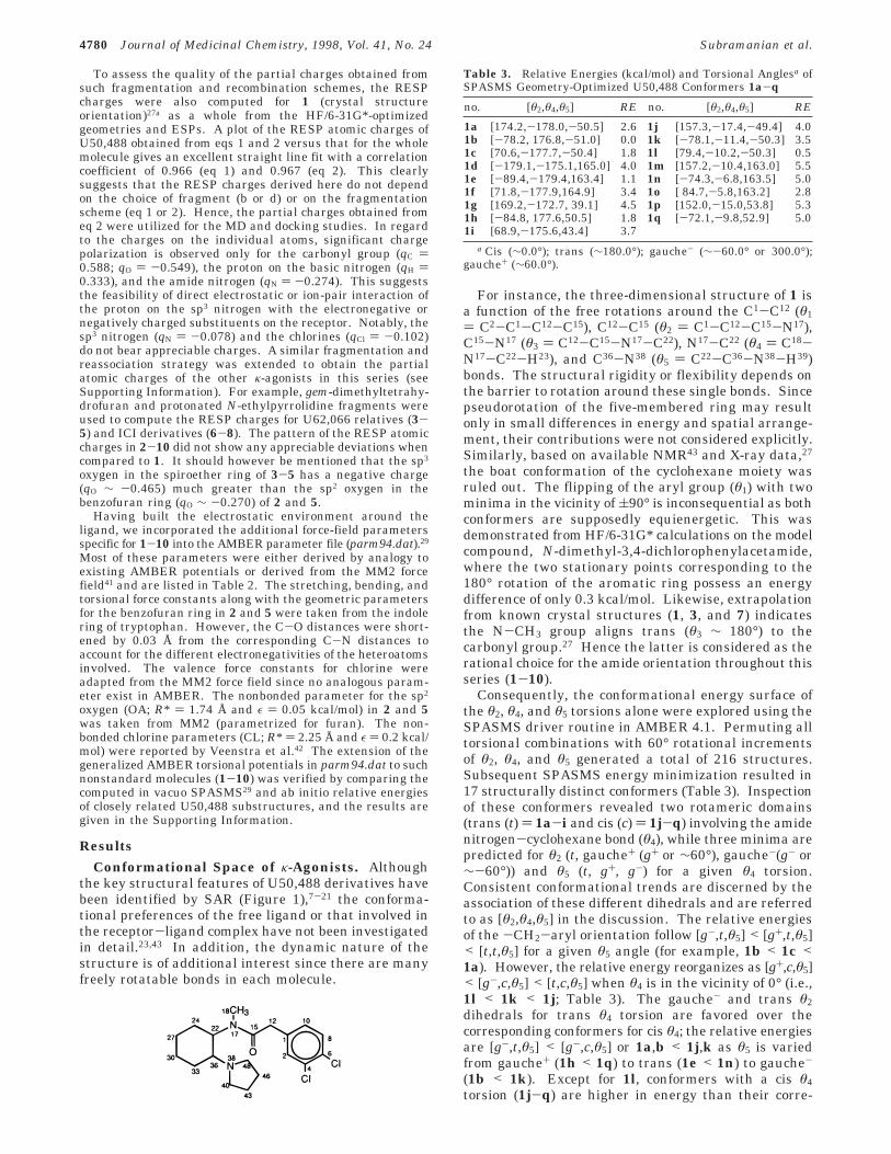
For instance, the three-dimensional structure of 1 isa function of the free rotations around the C1-C12 (θ1) C2-C1-C12-C15), C12-C15 (θ2 ) C1-C12-C15-N17),C15-N17 (θ3 ) C12-C15-N17-C22), N17-C22 (θ4 ) C18-N17-C22-H23), and C36-N38 (θ5 ) C22-C36-N38-H39)bonds. The structural rigidity or flexibility depends onthe barrier to rotation around these single bonds. Sincepseudorotation of the five-membered ring may resultonly in small differences in energy and spatial arrange-ment, their contributions were not considered explicitly.Similarly, based on available NMR43 and X-ray data,27
the boat conformation of the cyclohexane moiety wasruled out. The flipping of the aryl group (θ1) with twominima in the vicinity of (90° is inconsequential as bothconformers are supposedly equienergetic. This wasdemonstrated from HF/6-31G* calculations on the modelcompound, N-dimethyl-3,4-dichlorophenylacetamide,where the two stationary points corresponding to the180° rotation of the aromatic ring possess an energydifference of only 0.3 kcal/mol. Likewise, extrapolationfrom known crystal structures (1, 3, and 7) indicatesthe N-CH3 group aligns trans (θ3 ∼ 180°) to thecarbonyl group.27 Hence the latter is considered as therational choice for the amide orientation throughout thisseries (1-10).
Consequently, the conformational energy surface ofthe θ2, θ4, and θ5 torsions alone were explored using theSPASMS driver routine in AMBER 4.1. Permuting alltorsional combinations with 60° rotational incrementsof θ2, θ4, and θ5 generated a total of 216 structures.Subsequent SPASMS energy minimization resulted in17 structurally distinct conformers (Table 3). Inspectionof these conformers revealed two rotameric domains(trans (t) ) 1a-i and cis (c) ) 1j-q) involving the amidenitrogen-cyclohexane bond (θ4), while three minima arepredicted for θ2 (t, gauche+ (g+ or ∼60°), gauche-(g- or∼-60°)) and θ5 (t, g+, g-) for a given θ4 torsion.Consistent conformational trends are discerned by theassociation of these different dihedrals and are referredto as [θ2,θ4,θ5] in the discussion. The relative energiesof the -CH2-aryl orientation follow [g-,t,θ5] < [g+,t,θ5]< [t,t,θ5] for a given θ5 angle (for example, 1b < 1c <1a). However, the relative energy reorganizes as [g+,c,θ5]< [g-,c,θ5] < [t,c,θ5] when θ4 is in the vicinity of 0° (i.e.,1l < 1k < 1j; Table 3). The gauche- and trans θ2dihedrals for trans θ4 torsion are favored over thecorresponding conformers for cis θ4; the relative energiesare [g-,t,θ5] < [g-,c,θ5] or 1a,b < 1j,k as θ5 is variedfrom gauche+ (1h < 1q) to trans (1e < 1n) to gauche-
(1b < 1k). Except for 1l, conformers with a cis θ4torsion (1j-q) are higher in energy than their corre-
Table 3. Relative Energies (kcal/mol) and Torsional Anglesa ofSPASMS Geometry-Optimized U50,488 Conformers 1a-q
no. [θ2,θ4,θ5] RE no. [θ2,θ4,θ5] RE
1a [174.2,-178.0,-50.5] 2.6 1j [157.3,-17.4,-49.4] 4.01b [-78.2, 176.8,-51.0] 0.0 1k [-78.1,-11.4,-50.3] 3.51c [70.6,-177.7,-50.4] 1.8 1l [79.4,-10.2,-50.3] 0.51d [-179.1,-175.1,165.0] 4.0 1m [157.2,-10.4,163.0] 5.51e [-89.4,-179.4,163.4] 1.1 1n [-74.3,-6.8,163.5] 5.01f [71.8,-177.9,164.9] 3.4 1o [ 84.7,-5.8,163.2] 2.81g [169.2,-172.7, 39.1] 4.5 1p [152.0,-15.0,53.8] 5.31h [-84.8, 177.6,50.5] 1.8 1q [-72.1,-9.8,52.9] 5.01i [68.9,-175.6,43.4] 3.7
a Cis (∼0.0°); trans (∼180.0°); gauche- (∼-60.0° or 300.0°);gauche+ (∼60.0°).
4780 Journal of Medicinal Chemistry, 1998, Vol. 41, No. 24 Subramanian et al.

sponding trans θ4 forms (1a-i) and were thereforeexcluded from further consideration. Moreover, theX-ray structures of 1 and its relatives are illustrativeof the trans θ4 torsion (∼175°).27a Thus, among theremaining nine conformers (1a-i), the major differencerelates to the orientation of the pyrrolidine ring withrespect to cyclohexane (θ5). Among these, conformerswith a gauche- arrangement for θ5 are relatively lowerin energy than those with a trans θ5 angle, for a similarθ2 orientation (1a > 1d, 1b > 1e, 1c > 1f). The mostfavorable conformer of U50,488 was indeed 1b with a[g-,t,g-] arrangement and is 2.6 kcal/mol lower inenergy compared to the X-ray structure orientation([t,t,g-], 1a).27a
Although the relative energies of all nine isomers(1a-i) were within a few kcal/mol, the barrier torotation around the C36-N38 bond could lock the struc-ture in one particular θ5 torsion (preferably gauche-).While crystal packing forces restrict the orientation ofthe aryl moiety (1) in the solid state, all the three θ2conformations (1a-c) could exist in dynamic equilibri-um within the NMR time scale in solution.43
A similar strategy was extended to explore the con-formational potential energy surface of other κ-agonists(2-10) as well. Since 2-5 differ mainly in the arylmodifications, the conformational profiles of [θ2,θ4,θ5]torsion were similar to that of U50,488. However,subtle differences in the relative energies are notedespecially for 4 and 5, where the [g+,c,g-] torsion ispredicted to be 0.3 and 0.7 kcal/mol more favored overthe [g-,t,g-]. In contrast to 1, the most favored ar-rangement of 3 possesses the same conformation([g-,t,g-]) as that of the crystal structure.27b The con-formational flexibility in 6-8 increases as a result ofbreaking the cyclohexane ring in 1 and introducing anethyl linking group instead. This additional N(sp2)-C(sp3)-C(sp3)-N(sp3) torsion (θ6) displays a significantpreference for orientations around 60°, in addition tothe [θ2,t,g-] dihedrals favored by the rotation of theother single bonds in 6-8. Comparisons of the θ6torsion to similar environments in 1-5 (X-ray and low-energy SPASMS-generated conformers) also indicatethat this angle is near 60°. Likewise, the piperidinederivatives 9 and 10 with a different type of cyclizationalso favor a 60° angle between the amide and basicnitrogens. Furthermore, all the SPASMS-optimizedconformers obtained by permuting the [θ2,θ3,θ5] anglesin 9 and 10 revealed the amide nitrogen to be nearlyplanar and the piperidine ring to adopt a chair form.The conformational energy trends of 9 and 10 showedgeneral agreement with that of 1, with the low-energystructures possessing a gauche- (θ5) arrangement.
Dynamic Properties of κ-Agonists. MD simula-tion techniques are used extensively to sample theconformational space and obtain structural details thatcannot be elucidated from experiments. Apart fromrationalizing the SPASMS results, this approach canprovide a means of identifying the “static” versus“dynamic” nature of the θ2-θ6 torsions in the κ-ligands.Figure 2 shows the torsional trajectories of 1, 3, 5, 7,and 10 (refer to Supporting Information for otherligands) from a 2-ns simulation starting from the [t,t,g-]orientations of the [θ2,θ4,θ5] torsions. In general, thechair form of the cyclohexyl ring in 1-5 and that of the
piperidine ring in 9 and 10 were the only ring puckersobserved throughout the MD run. Similarly, the θ4torsion was arrested in the trans rotamer in 1-8, whilethe θ5 angle was confined to a gauche- arrangement in1-3 and 10, with the gauche+ orientation (θ5) sampledin addition for the remaining ligands. The pharma-cophore torsion (θ6) was locked in the gauche+ form in1-5 and 8-10, but the flexible ethyl linker in 6 and 7allowed for the population of trans (θ6) dihedral ar-rangements as well. In fact, the strong conformationalrestriction of the θ5 and θ6 torsions in 1-10 resultedfrom the appropriate orientation of the N-H...OdC bondthat maximizes the electrostatic interaction between thetwo moieties. The corresponding nonbonded distancefrom the MD simulations was 3.5-4.0 Å on average formost of the ligands.
Although the crystal structures (1, 3, and 7)27 showthe θ2 torsion locked to a particular spatial arrange-ment, the MD simulations efficiently sampled all threeθ2 dihedrals in 1, 3, 4, 6, 7, 9, and 10. However, bulkieraryl substitutions restricted the θ2 torsion as evidencedfrom the simulation profile of 2 (vs 1) and 5 (vs 4). Ingeneral, the [g+,t,g-] orientation of the [θ2,θ4,θ5] torsionis predicted to be the dominant solution conformationin 2, 4, 6-8, and 10 and demonstrates that the mostfavored SPASMS-generated conformer ([g-,t,g-]) maynot be the most populated in solution. This discrepancybetween the MD simulations and conformational sam-pling (SPASMS) is likely due to the presence of explicitsolvent (water) as opposed to the dielectric medium (ε) 4r) used in SPASMS.
Apart from the presumed solvation effects, rotationalbarriers around single bonds (trans to cis θ4 torsion inparticular) also limit the population to only a fewdistinct conformers. For instance, the second moststable [g+,c,g-] conformer (1l) was not sampled in the2-ns MD simulation of 1. Similarly, the most favoredSPASMS-generated [g+,c,g-] conformer of CI-977 (5)was not present in the 2-ns MD simulation but isconsistent with the conformational sampling exhibitedby the rest of molecules for the first 1-ns simulation.During the second 1-ns simulation, the structure be-came locked into the [g-,t,g+] orientation (Figure 2).Inspite of the sampling of some low-energy conformers(SPASMS) in the MD run, the simulations suggestedthat a large portion of each molecule (θ4 in trans, θ5 ingauche-, and θ6 in gauche+) was anchored in space.
The substituted benzyl group (θ2) of the κ-agonistsadopted all three torsional degrees of freedom in solu-tion. This was demonstrated by comparing the uniqueMD conformers of 1 ([θ2,t,g-]) with the conformationsinterpreted from experimental NMR data in D2O (1b,c).43
To accomplish this, the 1H NMR chemical shifts of allthree [θ2,t,g-] conformers, 1a-c (Figure 3), with θ2torsional diversity were computed at the GIAO-Becke3LYP/6-31G*//HF/6-31G* level. In general, theprotons of the cyclohexyl ring resonated upfield whencompared to the pyrrolidine ring hydrogens. Similarly,the equatorial hydrogens (Heq) of the cyclohexyl ringwere downfield-shifted compared to the axial (Hax)protons. In addition, these 1H δ values were quiteinsensitive to θ2 torsions (refer to Supporting Informa-tion for the computed chemical shifts). However, thesubtle yet significant differences in the aromatic, ben-
Docking of Arylacetamide-Based κ-Opioid Agonists Journal of Medicinal Chemistry, 1998, Vol. 41, No. 24 4781

zylic methylene, and N-methyl δ 1H values are aconsequence of the different θ2 torsions (Table 4).Although, the experimental assignments suggested theexistence of 1b,c alone in solution,43 our interpretationsof the chemical shifts include the coexistence of thecrystal structure orientation (1a) as well. Thus, theaverage 1H NMR shifts (δa) of 1a-c are in betteragreement with the experimental values43 than any onesingle conformer (1a-c), thereby supporting the highlyfluxional behavior of the θ2 torsion within the NMR timescale.
Automated Docking of Agonists in the κ-Recep-tor. Identification of the bioactive conformation from
the pool of possible conformers (Table 3) is a criticalissue in predicting the appropriate ligand binding mode.Even though the MD simulations simplified the numberof possible ligand conformers to just three [θ2,t,g-]structures, only one among them was anticipated to bethe dominant binding conformer. Experimental SAR
Figure 2. θ2 (left) and θ5 (right) torsional profile of the solvated ligands as a function of MD simulation time (in ps). The ligandnumbers are given in each graph.
Figure 3. Superimposed in vacuo SPASMS geometry-optimized structures 1a-c.
Table 4. GIAO-Becke3LYP/6-31G*//HF/6-31G*-Computed 1HNMR Chemical Shifts (δ, ppm) for U50,488 Conformers 1a-ca
atom no. δ 1a δ 1b δ 1c δa 1a-c expt
H3 6.9 7.0 7.1 7.0 6.8H9 7.3 7.3 7.3 7.3 7.2H11 6.7 7.4 6.8 7.0 7.1H13 3.3 3.2 3.8H14 3.3 3.7 3.3 3.5 3.5H19 2.5 3.3 2.5H20 2.8 2.9 2.3 2.7 2.7H21 2.9 3.0 2.7
a The average chemical shifts (δa, ppm) and the experimentalvalues (ppm) are given for comparison. The computed andexperimental 1H δ values for the cyclohexyl and pyrrolidine ringsare given in the Supporting Information.
4782 Journal of Medicinal Chemistry, 1998, Vol. 41, No. 24 Subramanian et al.

studies on racemic U50,488 and its constrained lactamanalogues44 (11, 12; note that the θ2 torsion is re-strained) revealed the binding (Ki) to be similar for 1(15.0 nM) and 11 (10.0 nM), but 6-9-fold lower for 12(92.0 nM) indicating the effects of the θ2 torsion onbinding. This tentatively justifies the [t,t,g-] orientationas a reasonable starting structure that could be dockedto the κ-receptor. Although there are various possibili-ties of docking these flexible ligands to the receptor,finding the favorable low-energy states in the receptor-binding domain is often tedious. Consequently, weemployed the automated DOCK program to explore thepossible orientations of U50,488 (1a) within the receptorbinding pocket. Accordingly, the “best positions” for theligand atoms, and thereby the ligand recognition sites,were identified by searching for regions of complemen-tarity within the TM helices.45 Out of the 982 configu-rations generated by DOCK, the top-ranking 107 ori-entations that were within 5 kcal/mol from the bestforce-field score (ffscore ) -29.5 kcal/mol) were ex-tracted. Interestingly, all the binding orientations of1a were along the receptor helical axis (positionedvertically) and none perpendicular to it (horizontal). Inaddition, the DOCK search suggested the aryl group inU50,488 to be either toward the extracellular region ordeep in the binding pocket.
The tentative ligand binding domain predicted by theautomated docking procedure also suggested the puta-
tive ligand binding pocket to comprise TM3, -5, -6, and-7.45 Preliminary analysis of the docked structuresindicated that the best and many of the top-scoringstructures had the pyrrolidine ring aligned near E297(TM6) of the κ-receptor (the aryl group is down in thepocket; see Supporting Information), while few struc-tures had this moiety close to H291. Nevertheless, thecomplete loss of U50,488 binding25 upon a D138N (TM3)mutation suggests the ion pairing between the proto-nated nitrogen of 1a and the D138 carboxylate group isa key binding interaction. A closer examination of someof the top-scoring orientations identified the 31st best-docked configuration of 1a (ffscore ) -24.8 kcal/mol)as having the basic nitrogen close to D138 in TM3 andthe aryl group tethering toward the extracellular region.Although this arrangement would have been expectedto be the top-scoring orientation, short-range stericconflicts between the receptor side chains and the ligandresulted in this binding arrangement having a lowerscoring. This was evident when the energy componentsof the force-field score between the 31st and the best-docked configurations were compared. The electrostaticand the van der Waals (vdW) attraction energies (kcal/mol) were better stabilizing for the 31st best-dockedstructure (-3.9, -44.6) as compared to the top-scoringorientation (-3.2, -39.5). However, the correspondingcontribution from the vdW repulsion (23.8, 13.1) re-versed the numerical preference of the force-field scoring
Figure 4. θ2 (left) and θ5 (right) torsional profile of the docked ligand as a function of MD simulation time (in ps). The ligandnumbers are given in each graph.
Docking of Arylacetamide-Based κ-Opioid Agonists Journal of Medicinal Chemistry, 1998, Vol. 41, No. 24 4783

for these two docked configurations. This clearly em-phasizes the limitation of rigid docking methods andcautions against blindly choosing the best force-field-scoring configuration always as the ideal ligand bindingmode in the receptor. In fact, it is necessary to closelyexamine some of the top-ranking distinct configurationsof the ligand in the receptor before concluding on themost relevant bound complex.
Instead of extending the DOCK protocol to predict thebinding domain of 2-8, their structural and spatialsimilarity were taken to our advantage. The directrelationship of the preferred torsions of 2-8 with U50,-488 suggested that they might also exhibit similarbinding characteristics in the κ-receptor. The compa-rable κ-affinity of 1-108,10,13,15,17,20,23 and the impair-ment of U69,593, CI-977, and ICI199,441 binding upona D138 mutation (Table 1)25,46 provided support for ourclaim of binding complementarity. Consequently, the[t,t,g-] SANDER energy-minimized structures of 2-8were superimposed on the docked structure of 1a (31stconfiguration)47 to generate the tertiary structure of thereceptor-ligand complex. Since 9 and 10 possessed apiperidine ring, the complete DOCK procedure wasfollowed to obtain their probable binding orientation(the [t,t,g-] conformations of the ligands were consid-ered). A strategy similar to that used for 1a wasemployed to retrieve the binding mode of 9 where thepyrrolidine ring was closer to the D138 residue of thereceptor. Overlapping 10 on the docked configurationof 9 generated the receptor-ligand complex of 10.
Having established the most likely binding mode of1-10, post-DOCK refinements (energy minimizationand MD simulations) were employed to relieve anysteric conflicts between the flexible amino acid sidechains of the receptor and the docked ligand. A com-parison of the MD trajectories of the free ligand (Figure2) and the bound complex (Figure 4) indicates that theligand conformer in the receptor is not the most favoredor highly populated solution structure in some cases.As in the free ligand, the θ5 torsion in 1-5 and 10 boundto the receptor was locked in the gauche- form withoccasional transitions to the trans and gauche+ rotam-ers for 7-9 (Figure 4 and Supporting Information).However, the gauche- arrangement was the mostpopulated θ5 torsion in all the docked ligands. Simi-larly, the dominant θ2 torsions were close to the transin a majority of ligands (1-3, 5-7) associated with thereceptor, while the gauche- orientation seem to befavored for 8 and 9. Interestingly, U62,066 (4) exhibitedθ2 torsional phase transition from trans to gauche+ after300 ps. There is also evidence that the X-ray structure
is not necessarily the bound conformation. This isdemonstrated by considering the energy-minimizedcrystal structure orientation of U69,593 (3, [g-,t,g-])27b
as a starting ligand conformer. The initial receptor-ligand complex was built by overlapping 3 on the U50,-488 docked structure (31st configuration) and followingthe minimization and MD protocols similar to thatemployed for other bound ligand complexes. In lessthan 100 ps of the 1-ns simulation, the θ2 torsion (seeSupporting Information) of U69,593 converted back tothe preferred trans form (and θ5 ) gauche-), suggestingthat the MD run here was not dependent on the initialθ2 ligand torsion. In general, the ensemble of conform-ers for each ligand generated from the simulation of thecomplex implicated the [t,t,g-] torsion as the dominantbinding orientation for 1-3, 5-7, and 10. However, the[g-,t,g-] is favored for 8 and 9, with U62,066 (4) aloneadopting the [g+,t,g-] arrangement in the receptor. Itshould however be emphasized that a single conforma-tional element (frozen ligand orientation) does not existfor the receptor-ligand complex (Figure 4), and anyinsights gained from static energy minimization of thecomplex need to be treated with caution.
Discussion
As mentioned earlier, the carboxylate group of theaspartate (D138) in the κ-receptor located in TM3 ispresumed to form a salt bridge with the pyrrolidine ringamino proton of the ligands.26,45 The notion that D138in the κ-receptor is involved in direct ligand recognitionhas also been suggested from site-directed mutagenesisin which a D138N mutation abolishes U50,488 andU69,593 binding.25 Likewise, the D138A receptor mu-tation impaired CI-977 and ICI199,441 associationsignificantly.46 Owing to alike structural and spatialrelationships of 1-10, it is reasonable to speculate thatD138 serves as a common interaction point for otherκ-agonists in this series. Accordingly, MD simulationsof all the bound ligand complexes resulted in theprotonated nitrogen forming a sustained salt bridgewith the carboxylate group of D138 (Table 5). Thecarboxylate oxygen to the pyrrolidine proton distancewas 3.9-4.3 Å on an average for most of the ligands(Table 5) and is within the anticipated range for similarsalt bridges.48 In fact, such direct electrostatic anchor-ing of the ligand moiety with the aspartate most likelycauses the remaining portion of the agonist to reorga-nize by taking advantage of the surrounding receptorenvironment (Figures 5 and 6).
Table 5. Average Distances (dist, Å) of the Free and Receptor-Bound Ligands 1-10 Obtained from MD Simulations
param 1 2 3 4 5 6 7 8 9 10
dist1a 4.169 3.961 3.936 3.944 4.196 5.900 4.319 4.366 5.029 3.274dist2b 4.270 3.969 3.985 3.960 4.183 5.966 4.381 4.340 4.706 3.215dist3c 7.362 7.408 10.590 10.080 8.885 4.913 5.042 6.336 6.855 9.490dist4d 5.383 5.534 8.246 7.613 7.014 4.206 3.879 4.435 5.393 7.261dist5e 5.933 6.991 5.848dist6f 4.097 5.159 3.939dist7g 6.106 7.117 10.234 8.370 6.950 6.737 7.589 7.303 11.250 10.481dist8h 6.021 7.119 10.263 8.748 7.149 6.671 7.643 7.085 11.622 10.455
a (D138-carboxyl oxygen)O1‚‚‚H-N(ligand) distance. b (D138-carboxyl oxygen)O2‚‚‚H-N(ligand) distance. c (H291)δN-H‚‚‚OdC(ligand)distance. d (H291)εN‚‚‚OdC(ligand) distance. e (H291)δN-H‚‚‚O(sp3)(ligand) distance. f (H291)εN‚‚‚O(sp3)(ligand) distance. g (E297-carboxyloxygen)O1‚‚‚Cpara(aromatic, ligand) distance. h (E297-carboxyl oxygen)O2‚‚‚Cpara(aromatic, ligand) distance.
4784 Journal of Medicinal Chemistry, 1998, Vol. 41, No. 24 Subramanian et al.
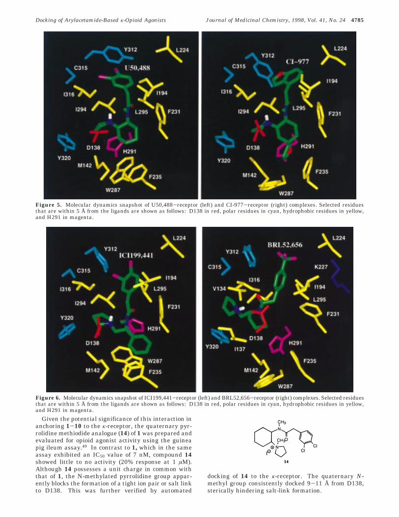
Given the potential significance of this interaction inanchoring 1-10 to the κ-receptor, the quaternary pyr-rolidine methiodide analogue (14) of 1 was prepared andevaluated for opioid agonist activity using the guineapig ileum assay.49 In contrast to 1, which in the sameassay exhibited an IC50 value of 7 nM, compound 14showed little to no activity (20% response at 1 µM).Although 14 possesses a unit charge in common withthat of 1, the N-methylated pyrrolidine group appar-ently blocks the formation of a tight ion pair or salt linkto D138. This was further verified by automated
docking of 14 to the κ-receptor. The quaternary N-methyl group consistently docked 9-11 Å from D138,sterically hindering salt-link formation.
Figure 5. Molecular dynamics snapshot of U50,488-receptor (left) and CI-977-receptor (right) complexes. Selected residuesthat are within 5 Å from the ligands are shown as follows: D138 in red, polar residues in cyan, hydrophobic residues in yellow,and H291 in magenta.
Figure 6. Molecular dynamics snapshot of ICI199,441-receptor (left) and BRL52,656-receptor (right) complexes. Selected residuesthat are within 5 Å from the ligands are shown as follows: D138 in red, polar residues in cyan, hydrophobic residues in yellow,and H291 in magenta.
Docking of Arylacetamide-Based κ-Opioid Agonists Journal of Medicinal Chemistry, 1998, Vol. 41, No. 24 4785

Among the other acidic residues in the TM helices(D105, D223, and E297), E297 in TM6 has been impli-cated in the binding of κ-antagonists such as norBNI.50
However, the rather long distance between the ligands(1-10) and the carboxylate group of E297 (Table 5) rulesout any possible nonbonded interaction. This interpre-tation is further supported by an experimental pointmutation (E297A) that revealed the binding affinity of5 and 7 to be less affected in the mutant receptor (Table1). Of the other possible stabilizing receptor-ligandinteractions, the carbonyl oxygen of U50,488 may forma H-bond with the histidyl (H291) N-H bond in TM6.Considering the δ-tautomer of H291 in the presentstudy, automated docking of 1 disfavored such a non-bonded interaction as the corresponding (δ-H291)N-H‚‚‚OdC(U50,488) distance was 7.4 Å on an average.A similar trend in the (δ-H291)N-H‚‚‚OdC(ligand)interaction was evident for the bound complexes of2-10. Proximity of the ε-nitrogen in δ-H291 to thecarbonyl oxygen (εN‚‚‚OdC) in 6-8 indicated the likeli-hood of a weak H-bond, if the ε-tautomer (ε-H291) wereconsidered. However, the influence of δ- or ε-H291 onligand binding properties appears to be modest asrevealed by site-directed mutagenesis. As opposed tothe carbonyl group in 1-10, the sp3 oxygen in 3 and 5(Figure 5) could form an H-bond with the N-H moietyin ε-H291 and influence the binding properties uponspecific mutation. This explains the 7-fold decrease inCI-977 binding upon H291A mutation and perhaps thecause for the increased binding affinity of 5 over 2.7,11
Unlike the µ-receptor which displays significant loss inopiate agonist binding upon histidine mutation inTM6,51 the present MD simulation results and availablesite-directed mutagenesis studies suggest that H291 isnot involved directly in ligand binding or recognitionin this series. This is not due to the lack of a hydroxylsubstituent on the aryl group (to mimic the tyraminemoiety in most opiate ligands) but is a consequence ofthe different binding orientations of the substitutedarylacetamides (1-10) and the benzomorphan deriva-tives such as ethylketocyclazocine. The experimentalKi values of racemic 9 (0.5 nM), 10 (0.8 nM), and 13(194.0 nM)10 which differ only in the aryl substitutionreveal that 13 with a hydroxyl substituent has a muchweaker binding affinity than 9 and 10 which is consis-tent with our binding site model. Our proposed bindingorientation for κ-agonists (1-10) is also supported byexperimental results that illustrate sulfhydryl alkylat-ing agents inhibit U50,488 binding,52 thereby indicatingthe presence of a sulfhydryl group near the binding site.Consistently, the MD simulations captured C315 of TM7close to the aryl group of 1.
Apart from these specific point interactions, theκ-agonists are flanked by hydrophobic residues (Table6) in the TM region. Consequently, mutation of any ofthese residues will have only a marginal effect on theligand binding properties, since free rotation around thesingle bonds of the ligand and receptor side chainswould allow torsional flexibility for the agonist toreadjust to the mutated environment. For instance, thearyl group in 1-10 benefits from the hydrophobicinteractions of I194, I294, L295, A298, and I316 resi-dues. Even though a simple I294A mutation does notaffect the κ-affinity (as in 5 and 7), mutation to a
charged residue (I294K) impairs CI-977 and ICI199,-441 binding significantly (Table 1). Despite the mutatedresidue (I294K) exerting similar effects on the bindingprofile of 5 and 7, the subtle differences in the relativeorientation of the ligands in the receptor may beresponsible for the uneven deviation in the magnitudeof the binding affinity.
The cyclohexyl ring in 1-5, the phenyl/isopropylgroup in 6-8, and the piperidine ring in 9 and 10 arealso stabilized by nonpolar aromatic residues (F231,F235, and W287) and M142 with additional contribu-tions from W183, A186, G190, and A234 for 6-8 andV232 and A234 for 9, respectively. Likewise, thepyrrolidine ring in 1-10 benefits from the hydrophobiccooperation of I294, L295, I316, and G319 of theκ-receptor. In fact, the importance of G319 (TM7) inligand binding has been demonstrated by site-directedmutagenesis studies (G319V) which weaken (by 28-fold)the binding affinity of 7. Therefore, mutation of G319with sterically bulkier groups should decrease thebinding affinity of many other κ-agonists in this series.In addition, the involvement of polar residues such asY139, Y312, and Y320 that are also part of the ligandbinding pocket can be probed by appropriate receptormutations.
Another test of the proposed docking mode can befound in the enantioselectivity of the κ-agonists for thereceptor. As the carboxylate group of D138 and theN-H bond in 1-10 may be involved in direct receptor-ligand interaction, any change in the ligand orientation(θ5) around this region should destabilize κ-agonistbinding. Thus, the enantiomers of 1-10 possessing agauche+ form for the θ5 torsion (by mirror symmetry ofthe ligands) orient the proton on the basic nitrogen awayfrom D138 leading to a very weak binding. This wasdemonstrated by docking the less active isomer (R,R)-1(Ki ) 299 nM)22 following the DOCK protocol describedabove. Out of the 144 best-docked configurations re-trieved from a total of 1027 orientations, none had theN-H bond of (R,R)-1 pointing toward the carboxylategroup of D138. The corresponding distance ranges from7 to 11 Å for most of the docked configurations and isobviously too far away for any possible ion-pairing orelectrostatic interaction. The same reasoning can beextended to explain the enantioselective binding of otherκ-agonists in this series.
ConclusionIn conclusion, the present study has revealed striking
similarities in the three-dimensional structure, dynam-ics, and binding modes of representative arylacetamideκ-agonists. The SPASMS relative energy trends of 1-10display systematic conformational preferences and showoverall agreement throughout this ligand series. The
Table 6. Key Residues Defining the κ-Opioid Receptor PocketInvolved in Agonist (1-10) Binding as Obtained from theReceptor-Ligand Snapshot (750-ps) Structure during the MDSimulation (κ-Receptor-Specific Residues are Underlined)
TM3: V134,a I135, I137,a D138, Y139, M142TM4: W183,b A186,b G190,b I194TM5: L224, F231, V232,a A234,a,b F235TM6: W287, H291, I294, L295, E297,c A298TM7: Y312, C315, I316, A317, G319, Y320
a Involved additionally in 9 and 10. b Involved additionally in6-8. c Not within 5-Å radii of 6-8.
4786 Journal of Medicinal Chemistry, 1998, Vol. 41, No. 24 Subramanian et al.

MD conformational sampling of 1 was consistent withthe conformations derived from X-ray crystal data andfrom NMR experiments, justifying the reliability of thesimulation results for other κ-ligands in this series. Inaddition, the simulations have also revealed that bicyclicaromatics reduce the θ2 torsional flexibility and indicatepossible leads for designing rigid κ-agonists (for ex-ample, bicyclic aromatics in 6-10).
As was evident from the automated docking procedureand MD simulations of the receptor-agonist complex,the binding orientations of 1-10 differed from that ofthe opiates. Even though the protonated nitrogen of thenon-peptide agonists associated with the κ-opioid recep-tor through ion pairing with the aspartate in TM3, thearyl ring in 1-10 does not mimic the binding environ-ment of the phenolic component of the tyramine moietyof the opiate ligands. Thus, the histidine in TM6 (H291)which may form “hydrogen bonds” with the hydroxylsubstituent on the phenolic moiety in many benzomor-phan derivatives is not involved in any similar interac-tion with the ligand atoms in this series. Since thecyclohexyl ring in 1-5 and the ethyl spacer in 6-10 areclose to H291 (Figures 5 and 6), incorporation offunctional groups containing electronegative atoms inthis region would provide insights into the role of H291toward ligand binding. We should point out that analternative role for H291 has been proposed in U50,-488 binding by Cappelli et al.53 While details of themolecular conformation and docking mode were notgiven in that paper, some differences in the receptor-ligand complex are evident, mainly in the orientationof H291 toward the carbonyl oxygen of U50,488. Thebinding site analysis of CI-977 and related ligandspresented above, however, supports an alternative rolefor this proton donor. Nevertheless, such an interactioncannot be entirely ruled out given the proximity of H291to the ligand binding site. The binding site modelpresented here also reveals several residues specific tothe κ-receptor that may be useful in ligand design.Although D223 and E297 residues are apparently notinvolved in ligand binding of arylacetamides, they areviable sites that could reinforce the receptor-ligandassociation. Appropriate substituents (like guanidine)attached to the meta/para positions of the aromatic ringpresumably may reach the acidic residues to increasethe ligand binding affinity.
In addition to the key electrostatic interaction stabi-lizing the receptor-agonist complex, the results indicatea network of surrounding hydrophobic residues thatmay strengthen ligand binding in this series. Nearlyone-third of the residues involved in ligand binding areunique to the κ-receptor, which may explain the dif-ferential µ/κ-selectivity of the U50,488 derivatives. Thissuggests a very different mechanism of recognitionwhen compared with the κ-opioid antagonist norBNI.Previous computational and experimental studies ofnorBNI binding to the κ-receptor have indicated E297(near the top of TM6) to be critical to selectivity.50 Ourstructural analysis, however, shows this acidic residueplays no part in U50,488 binding as noted above. Whileour model is consistent with available site-directedmutagenesis data, additional work is needed to fullydefine this putative binding pocket and how it differsfrom that of the µ- and δ-receptors.
Finally, it is important to point out that the resultsstrongly support the existence of multiple bindingepitopes for opioid receptors. In comparing the analysispresented here with previous docking studies involvingopiates and peptides, divergent models of recognitionand selectivity emerge.6,24 Although this further com-plicates both receptor and ligand modeling efforts,knowledge of additional recognition sites within the µ-,δ-, or κ-opioid receptors may lend new insight to thedevelopment of even more potent and selective ligands.Moreover, such sites may provide important clues as tothe structural basis of agonist versus antagonist recog-nition that would have a far reaching impact on GPCRresearch.
Acknowledgment. The authors thank NIH/NIDAfor financial support to D.M.F. and P.S.P. and Dr. ThueSchwartz for sharing his site-directed mutagenesisresults.
Supporting Information Available: Fragmentationscheme used for deriving RESP atomic charges; tables ofexperimental Ki/IC50 values; SPASMS conformational energiesof 2-10; in vacuo geometry-optimized coordinates (PDBformat) and MD simulation profiles of the solvated and dockedligands, along with the putative ligand binding pocket andbinding orientations generated from the automated dockingprocedure; and experimental details and characterization of14 (28 pages). Ordering information is given on any currentmasthead page.
References(1) (a) Martin, W. R. Pharmacology of Opioids. Pharmacol. Rev.
1983, 35, 283-323. (b) Millan, M. J. Multiple Opioid Systemsand Pain. Pain 1986, 27, 303-347. (c) Jaffe, J. H.; Martin, W.R. Opioid Analgesics and Antagonists. In The PharmacologicalBasis of Therapeutics, 8th ed.; Gilman, A. G., Rall, T. W., Nies,A. S., Taylor, P., Eds.; Pergamon Press: New York, 1990; pp485-521. (d) Satoh, M.; Minami, M. Molecular Pharmacologyof the Opioid Receptors. Pharmacol. Ther. 1995, 68, 343-364.(e) Kieffer, B. L. Recent Advances in Molecular Recognition andSignal Transduction of Active Peptides: Receptors for OpioidPeptides. Cell. Mol. Neurobiol. 1995, 15, 615-635.
(2) (a) Olson, G. A.; Olson, R. D.; Kastin, A. J.; Coy, D. H.Endogenous Opiates: 1981. Peptides 1982, 3, 1039-1072. (b)Romer, D.; Buscher, H. H.; Hill, R. C.; Maurer, R.; Petcher, T.J.; Zeugner, H.; Benson, W.; Finner, E.; Milkowsky, W.; Thies,P. W. An Opioid Benzodiazepine. Nature (London) 1982, 298,759-760. (c) Romer, D.; Buscher, H. H.; Hill, R. C.; Maurer, R.;Petcher, T. J.; Zeugner, H.; Benson, W.; Finner, E.; Milkowsky,W.; Thies, P. W. Unexpected Opioid Activity in a Known Classof Drug. Life Sci. 1982, 31, 1217-1220. (d) Kley, H.; Scheide-mantel, U.; Bering, B.; Muller, W. E. Reverse Stereoselectivityof Opiate and Benzodiazepine Receptors for the Opioid Benzo-diazepine Trifluadom. Eur. J. Pharmacol. 1983, 87, 503-504.(e) Wood, P. L. Opioid Receptor Affinities of Kappa Agonists,Agonist/Antagonists and Antagonists In Vitro and In Vivo. Prog.Neuro-Psychopharmacol. Biol. Psychiatry 1983, 7, 657-662. (f)Dhawan, B. N.; Cesselin, F.; Raghubir, R.; Reisine, T.; Bradley,P. B.; Portoghese, P. S.; Hamon, M. International Union ofPharmacology. XII. Classification of Opioid Receptors. Pharma-col. Rev. 1996, 48, 567-592.
(3) (a) Szmuszkovicz, J.; von Voigtlander, P. F. BenzeneacetamideAmines: Structurally Novel Non-µ Opioids. J. Med. Chem. 1982,25, 1125-1126. (b) Lahti, R. A.; von Voigtlander, P. F.; Barsuhn,C. Properties of a Selective Kappa Agonist, U-50,488H. Life Sci.1982, 31, 2257-2260. (c) von Voigtlander, P. F.; Lahti, R. A.;Ludens, J. H. U-50,488: A Selective and Structurally Novel Non-Mu (Kappa) Opioid Agonist. J. Pharmacol. Exp. Ther. 1983, 224,7-12. (d) de Costa, B. R.; Bowen, W. D.; Hellewell, S. B.; George,C.; Rothman, R. B.; Reid, A. A.; Walker, J. M.; Jacobson, A. E.;Rice, K. C. Alterations in the Stereochemistry of the κ-SelectiveOpioid Agonist U50,488 Result in High-Affinity σ Ligands. J.Med. Chem. 1989, 32, 1996-2002.
(4) Bellucci, F.; Dondio, G. Molecular Modelling Comparison ofPotent and Selective κ Agonists Belonging to Different ChemicalStructures: A First Attempt to κ Receptor Mapping. In Trends
Docking of Arylacetamide-Based κ-Opioid Agonists Journal of Medicinal Chemistry, 1998, Vol. 41, No. 24 4787

in QSAR and Molecular Modeling ‘92; Proceedings of the 9thEuropean Symposium on Structure-Activity Relationships:QSAR and Molecular Modeling; Vermuth, C. G., Ed.; ESCOM:Leiden, 1993; pp 461-462.
(5) (a) Portoghese, P. S.; Nagase, H.; Lipkowski, A. W.; Larson, D.L.; Takemori, A. E. Binaltorphimine-Related Bivalent Ligandsand Their κ-Opioid Receptor Antagonist Selectivity. J. Med.Chem. 1988, 31, 836-841. (b) Takemori, A. E.; Portoghese, P.S. Selective Naltrexone-Derived Opioid Receptor Antagonists.Annu. Rev. Pharmacol. Toxicol. 1992, 32, 239-269. (c) Lomize,A. L.; Pogozheva, I. D.; Mosberg, H. I. Development of a Modelfor the δ-Opioid Receptor Pharmacophore: 3. Comparison of theCyclic Tetrapeptide, tyr-c[D-Cys-Phe-D-Pen]OH with other con-formationally Constrained δ-Receptor Selective Ligands. Biopoly-mers 1996, 38, 221-234.
(6) Metzger, T. G.; Paterlini, M. G.; Portoghese, P. S.; Ferguson, D.M. Application of the Message-Address Concept of the Dockingof Naltrexone and Selective Naltrexone-Derived Opioid Antago-nists into Opioid Receptor Models. Neurochem. Res. 1996, 21,1287-1294.
(7) Halfpenny, P. R.; Hill, R. G.; Horwell, D. C.; Hughes, J.; Hunter,J. C.; Johnson, S.; Rees, D. C. Highly Selective κ-OpioidAnalgesics. 2. Synthesis and Structure-Activity Relationshipsof Novel N-[(2-Aminocyclohexyl)aryl]acetamide Derivatives. J.Med. Chem. 1989, 32, 1620-1626.
(8) Halfpenny, P. R.; Horwell, D. C.; Hughes, J.; Hunter, J. C.; Rees,D. C. Highly Selective κ-Opioid Analgesics. 3. Synthesis andStructure-Activity Relationships of Novel N-[2-(1-Pyrrolidinyl)-4- or -5-substituted-cyclohexyl]arylacetamide Derivatives. J.Med. Chem. 1990, 33, 286-291.
(9) Hayes, A. G.; Birch, P. J.; Hayward, N. J.; Sheehan, M. J.;Rogers, H.; Tyers, M. B.; Judd, D. B.; Scopes, D. I. C.; Naylor,A. A series of Novel, Highly Potent and Selective Agonists forthe κ-Opioid Receptor. Br. J. Pharmacol. 1990, 101, 944-948.
(10) Vecchietti, V.; Giordani, A.; Giardina, G.; Colle, R.; Clarke, G.D. (2S)-1-(Arylacetyl)-2-(aminomethyl)piperidine Derivatives:Novel, Highly Selective κ Opioid Analgesics. J. Med. Chem. 1991,34, 397-403.
(11) Giardina, G.; Clarke, G. D.; Dondio, G.; Petrone, G.; Sbacchi,M.; Vecchietti, V. Selective κ-Opioid Agonists: Synthesis andStructure-Activity Relationships of Piperidines Incorporatingan Oxo-Containing Acyl Group. J. Med. Chem. 1994, 37, 3482-3491.
(12) Vecchietti, V.; Clarke, G. D.; Colle, R.; Giardina, G.; Petrone,G.; Sbacchi, M. (1S)-1-(Aminomethyl)-2-(arylacetyl)-1,2,3,4-tet-rahydroisoquinoline and Heterocyclic-Condensed Tetrahydro-pyridine Derivatives: Members of a Novel Class of Very Potentκ Opioid Analgesics. J. Med. Chem. 1991, 34, 2624-2633.
(13) Clark, C. R.; Halfpenny, P. R.; Hill, R. G.; Horwell, D. C.; Hughes,J.; Jarvis, T. C.; Rees, D. C.; Schofield, D. Highly Selective κOpioid Analgesics. Synthesis and Structure-Activity Relation-ships of Novel N-[(2-Aminocyclohexyl)aryl]acetamide and N-[(2-Aminocyclohexyl)aryloxy]acetamide Derivatives. J. Med. Chem.1988, 31, 831-836.
(14) Lahti, R. A.; Mickelson, M. M.; McCall, J. M.; von Voigtlander,P. F. [3H]U-69593 A Highly Selective Ligand for the Opioid κReceptor. Eur. J. Pharmacol. 1985, 109, 281-284.
(15) (a) von Voigtlander, P. F.; Lewis, R. A. Analgesic and MechanisticEvaluation of Spiradoline, a Potent Kappa Opioid. J. Pharmacol.Exp. Ther. 1988, 246, 259-262. (b) Meecham, K. G.; Boyle, S.J.; Hunter, J. C.; Hughes, J. An in vitro Profile of Activity forthe (+) and (-) Enantiomers of Spiradoline and PD117302. Eur.J. Pharmacol. 1989, 173, 151-157.
(16) Hunter, J. C.; Leighton, G. E.; Meecham, K. G.; Boyle, S. J.;Horwell, D. C.; Rees, D. C.; Huges, J. CI-977, a Novel andSelective Agonist for the κ-Opioid Receptor. Br. J. Pharmacol.1990, 101, 183-189.
(17) Weerawarna, S. A.; Davis, R. D.; Nelson, W. L. Isothiocyanate-Substituted κ-Selective Opioid Receptor Ligands Derived fromN-Methyl-N-[(1S)-1-phenyl-2-(1-pyrrolidinyl)ethyl]phenylaceta-mide. J. Med. Chem. 1994, 37, 2856-2864.
(18) Costello, G. F.; Main, B. G.; Barlow, J. J.; Carroll, J. A.; Shaw,J. S. A Novel Series of Potent and Selective Agonists at theOpioid κ-Receptor. Eur. J. Pharmacol. 1988, 151, 475-478.
(19) (a) Nock, B.; Giordano, A. L.; Cicero, T. J. ICI 197067 is aSelective Ligand for the U-69593-sensitive κ Opiate Binding Site.Eur. J. Pharmacol. 1989, 162, 385-386. (b) Shaw, J. S.; Carroll,J. A.; Alcock, P.; Main, B. G. ICI 204448: a κ-Opioid Agonistwith Limited Access to the CNS. Br. J. Pharmacol. 1989, 96,986-992.
(20) Barlow, J. J.; Blackburn, T. P.; Costello, G. F.; James, R.; LeCount, D. J.; Main, B. G.; Pearce, R. J.; Russell, K.; Shaw, J. S.Structure/Activity Studies Related to 2-(3,4-Dichlorophenyl)-N-methyl-N-[2-(1-pyrrolidinyl)-1-substituted-ethyl]acetamides: ANovel Series of Potent and Selective κ-Opioid Agonists. J. Med.Chem. 1991, 34, 3149-3158.
(21) Naylor, A.; Judd, D. B.; Lloyd, J. E.; Scopes, D. I. C.; Hayes, A.G.; Birch, P. J. A Potent New Class of κ-Receptor Agonist:4-Substituted 1-(Arylacetyl)-2-[(dialkylamino)methyl]pipera-zines. J. Med. Chem. 1993, 36, 2075-2083.
(22) (a) Rothman, R. B.; Bykov, V.; Reid, A.; de Costa, B. R.; Newman,A.-H.; Jacobson, A. E.; Rice, K. C. A Brief Study of the Selectivityof Norbinaltorphimine, (-)-Cyclofoxy, and (+)-Cyclofoxy AmongOpioid Receptor Subtypes In Vitro. Neuropeptides 1988, 12,181-187. (b) Rothman, R. B.; France, C. P.; Bykov, V.; de Costa,B. R.; Jacobson, A. E.; Woods, J. H.; Rice, K. C. PharmacologicalActivities of Optically Pure Enantiomers of the κ-Opioid Agonist,U50,488, and its cis Diastereomer: Evidence for Three κReceptor Subtypes. Eur. J. Pharmacol. 1989, 167, 345-353.
(23) Costello, G. F.; James, R.; Shaw, J. S.; Slater, A. M.; Stutchbury,N. C. J. 2-(3,4-Dichlorophenyl)-N-methyl-N-[2-(1-pyrrolidinyl)-1-substituted-ethyl]acetamides: The Use of ConformationalAnalysis in the Development of a Novel Series of Potent Opioidκ Agonists. J. Med. Chem. 1991, 34, 181-189.
(24) Paterlini, G.; Portoghese, P. S.; Ferguson, D. M. MolecularSimulation of Dynorphin A(1-10) Binding to Extracellular Loop2 of the κ-Opioid Receptor. A Model for Receptor Activation. J.Med. Chem. 1997, 40, 3254-3262.
(25) Kong, H.; Raynor, K.; Reisine, T. Amino Acids in the ClonedMouse Kappa Receptor that are Necessary for High AffinityAgonist Binding but not Antagonist Binding. Reg. Pept. 1994,54, 155-156.
(26) (a) Thirstrup, K.; Elling, E. C.; Hjorth, S. A.; Schwartz, T. W.Construction of a High Affinity Zinc Switch in the κ-OpioidReceptor. J. Biol. Chem. 1996, 271, 7875-7878. (b) Hjorth, S.A.; Thirstrup, K.; Schwartz, T. W. Radioligand-dependent Dis-crepancy in Agonist Affinities Enhanced by Mutations in theκ-Opioid Receptor. Mol. Pharmacol. 1996, 50, 977-984.
(27) (a) Doi, M.; Ishida, T.; Inoue, M. Structure of κ-Agonist, U-50488Acta Crystallogr. 1990, C46, 676-678. (b) Doi, M.; Ishida, T.;Inoue, M. Conformational Characteristics of Opioid κ-receptoragonist: Crystal Structure of (5S,7S,8S)-(-)N-Methyl-N-[7-(1-pyrrolidinyl)-1-oxaspiro[4.5]dec-8-yl)benzeneacetamide (U69,-593), and Conformational Comparison with κ-Agonists. Chem.Pharm. Bull. 1990, 38, 1815-1818. (c) Chang, A.-C.; Takemori,A. E.; Ojala, W. H.; Gleason, W. B.; Portoghese, P. S. κ OpioidReceptor Selective Affinity Labels: Electrophilic Benzeneaceta-mides as κ-Selective Opioid Antagonists. J. Med. Chem. 1994,37, 4490-4498.
(28) Cieplak, P.; Cornell, W. D.; Bayly, C.; Kollman, P. A. Applicationof the Multimolecule and Multiconformational RESP Methodol-ogy to Biopolymers: Charge Derivation for DNA, RNA, andProteins. J. Comput. Chem. 1995, 16, 1357-1377.
(29) (a) Pearlman, D. A.; Case, D. A.; Caldwell, J. W.; Ross, W. S.;Cheatham, T. E., III; Debolt, S.; Ferguson, D. M.; Seibel, G. L.;Kollman, P. A. AMBER, a Package of Computer Programs forApplying Molecular Mechanics, Normal-Mode Analysis, Molec-ular Dynamics and Free Energy Calculations to Simulate theStructural and Energetic Properties of Molecules. Comput. Phys.Commun. 1995, 91, 1-41. (b) Pearlman, D. A.; Case, D. A.;Caldwell, J. W.; Ross, W. S.; Cheatham, T. E., III; Ferguson, D.M.; Seibel, G. L.; Singh, U. C.; Weiner, P. K.; Kollman, P. A.AMBER, Version 4.1; Department of Pharmaceutical Chemistry,University of California: San Francisco, CA, 1995.
(30) Cornell, W. D.; Cieplak, P.; Bayly, C. I.; Gould, I. R.; Merz, K.M., Jr.; Ferguson, D. M.; Spellmeyer, D. C.; Fox, T.; Caldwell,J. W.; Kollman, P. A. A Second Generation Force Field for theSimulation of Proteins, Nucleic Acids, and Organic Molecules.J. Am. Chem. Soc. 1995, 117, 5179-5197.
(31) Jorgensen, W. L.; Chandrasekhar, J.; Madura, J. D.; Impey, R.W.; Klein, M. L. Comparison of Simple Potential functions forSimulating Liquid Water. J. Chem. Phys. 1983, 79, 926-935.
(32) Berendsen, H. J. C.; Postma, J. P. M.; van Gunsteren, W. F.;DiNola, A.; Haak, J. R. Molecular Dynamics with Coupling toan External Bath. J. Chem. Phys. 1984, 81, 3684-3690.
(33) (a) Meng, E. C.; Shoichet, B. K.; Kuntz, I. D. Automated Dockingwith Grid-Based Energy Evaluation. J. Comput. Chem. 1992,13, 505-524. (b) Meng, E. C.; Gschwend, D. A.; Blaney, J. M.;Kuntz, I. D. Orientational Sampling and Rigid-Body Minimiza-tion in Molecular Docking. Proteins: Struct. Funct. and Genet.1993, 17, 266-278. (c) Connolly, M.; Gschwend, D. A.; Good, A.C.; Oshiro, C.; Kuntz, I. D. DOCK, Version 3.5; Department ofPharmaceutical Chemistry, University of California: San Fran-sisco, CA, 1995.
(34) Baldwin, J. M.; Schertler, G. F. X.; Unger, V. M. An Alpha-carbonTemplate for the Transmembrane helices in the RhodopsinFamily of G-protein-coupled Receptors. J. Mol. Biol. 1997, 272,144-164.
(35) Xue, J. C.; Chen, C.; Zhu, J.; Kunapuli, S.; DeRiel, J. K.; Yu, L.;Liu-Chen, L.-Y. Differential Binding Domains of Peptide andNon-peptide Ligands in the Cloned Rat κ-opioid Receptor. J. Biol.Chem. 1994, 269, 30195-30199.
4788 Journal of Medicinal Chemistry, 1998, Vol. 41, No. 24 Subramanian et al.
(36) (a) Connolly, M. L. Solvent-Accessible Surfaces of Proteins andNucleic Acids. Science 1983, 221, 709-713. (b) Connolly, M. L.Analytical Molecular Surface Calculation. J. Appl. Crystallogr.1983, 16, 548-558.
(37) Molecular Graphics Images were produced using the MidasPlusProgram from the Computer Graphics Laboratory, Universityof California, San Fransisco (supported by NIH RR-01081).Ferrin, T. E.; Huang, C. C.; Jarvis, L. E.; Langridge, R. TheMIDAS Display System. J. Mol. Graph. 1988, 6, 13-27.
(38) Young, L.; Topol, I. A.; Rashin, A. A.; Burt, S. K. BuildingMolecular Charge Distributions from Fragments: Applicationto HIV-1 Protease Inhibitors. J. Comput. Chem. 1997, 18, 522-532 and references therein.
(39) Foresman, J. B.; Frisch, Æ. Exploring Chemistry with ElectronicStructure Methods, 2nd ed.; Gaussian, Inc.: Pittsburgh, 1996.
(40) Frisch, M. J.; Trucks, G. W.; Schlegel, H. B.; Gill, P. M. W.;Johnson, B. G.; Robb, M. A.; Cheeseman, J. R.; Keith, T.;Petersson, G. A.; Montgomery, J. A.; Raghavachari, K.; Al-Laham, M. A.; Zakrzewski, V. G.; Ortiz, J. V.; Foresman, J. B.;Cioslowski, J.; Stefanov, B. B.; Nanayakkara, A.; Challacombe,M.; Peng, C. Y.; Ayala, P. Y.; Chen, W.; Wong, M. W.; Andres,J. L.; Replogle, E. S.; Gomperts, R.; Martin, R. L.; Fox, D. J.;Binkley, J. S.; Defrees, D. J.; Baker, J.; Stewart, J. J. P.; Head-Gordon, M.; Gonzalez, C.; Pople, J. A. Gaussian 94, Revision E2;Gaussian, Inc.: Pittsburgh, PA, 1995.
(41) Allinger, N. L. Conformational Analysis. 131. MM2. A Hydro-carbon Force Field Utilizing V1 and V2 Torsional Terms. J. Am.Chem. Soc. 1977, 99, 8127-8134; subsequent versions, e.g.,MM2-87, MM2-89, MM2-91.
(42) Veenstra, D. L.; Ferguson, D. M.; Kollman, P. A. How Transfer-able are Hydrogen parameters in Molecular Mechanics Calcula-tions? J. Comput. Chem. 1992, 13, 971-978.
(43) Froimowitz, M.; DiMeglio, C. M.; Makriyannis, A. Conforma-tional Preferences of the κ-Selective Opioid Agonist U50488. ACombined Molecular Mechanics and Nuclear Magnetic Reso-nance Study. J. Med. Chem. 1992, 35, 3085-3094.
(44) Cheng, C.-Y.; Lu, H.-Y.; Lee, F.-M.; Tam, S. W. Synthesis of (1′,2′-trans)-3-Phenyl-1-[2′-(N-pyrrolidinyl)cyclohexyl]pyrrolid-2-ones as κ-Selective Opiates. J. Pharm. Sci. 1990, 79, 758-762.
(45) Strahs, D.; Weinstein, H. Comparative Modeling and MolecularDynamics Studies of the δ, κ, and µ Opioid Receptors. ProteinEng. 1997, 10, 1019-1038.
(46) (a) Thirstrup, K.; Hjorth, S. A.; Schwartz, T. W. Investigationof the Binding Pocket in the Kappa Opioid Receptor by aCombination of Alanine Substitutions and Steric HindranceMutagenesis. 27th Meeting of the International NarcoticsResearch Conference (INRC) ′96, 1996; poster M30. (b) Thir-strup, K.; Hjorth, S. A.; Schwartz, T. W. Unpublished results.
(47) Independent automated docking of 2-8 following the standardDOCK protocols discussed in the text also revealed the bindingorientation of these ligands to be similar to that of 1.
(48) (a) Barlow, D. J.; Thornton, J. M. Ion-Pairs in Proteins. J. Mol.Biol. 1983, 168, 867-885. (b) Zheng, Y.-J.; Ornstein, R. L. WhatHappens to Salt-Bridges in Nonaqueous Environments: Insightsfrom Quantum Mechanics Calculations. J. Am. Chem. Soc. 1996,118, 11237-11243 and references therein.
(49) Rang, H. P. Stimulant Actions of Volatile Anaesthetics onSmooth Muscle. Br. J. Pharmacol. 1964, 22, 356-365.
(50) Hjorth, S. A.; Thirstrup, K.; Grandy, D. K.; Schwartz, T. W.Analysis of Selective Binding Epitopes for the κ-Opioid ReceptorAntagonist Nor-binaltorphimine. Mol. Pharmacol. 1995, 47,1089-1094.
(51) Spivak, C. E.; Beglan, C. L.; Seidleck, B. K.; Hirshben, L. D.;Blaschak, C. J.; Uhl, G. R.; Surratt, C. K. Naloxone Activationof µ-Opioid Receptors Mutated at a Histidine Residue Liningthe Opioid Binding Cavity. Mol. Pharmacol. 1997, 52, 983-992and references therein.
(52) Joseph, D. B.; Bidlack, J. M. The κ-Opioid Receptor Expressedon the Mouse Lymphoma Cell Line R1.1 Contains a SulfhydrylGroup at the Binding Site. Eur. J. Pharmacol. 1994, 267, 1-6.
(53) Cappelli, A.; Anzini, M.; Vomero, S.; Menziani, M. C.; DeBenedetti, P. G.; Sbacchi, M.; Clarke, G. D.; Mennuni, L.Synthesis, Biological Evaluation, and Quantitative ReceptorDocking Simulations of 2-[(Acylamino)ethyl]-1,4-benzodiazepinesas Novel Tifluadom-like Ligands with High Affinity and Selec-tivity for κ-Opioid Receptors. J. Med. Chem. 1996, 39, 860-872.
JM9803166
Docking of Arylacetamide-Based κ-Opioid Agonists Journal of Medicinal Chemistry, 1998, Vol. 41, No. 24 4789