conformational analysis of peptide analogues of silkmoth chorion protein segments using cd, nmr and...
TRANSCRIPT

Journal of Peptide ScienceJ. Peptide Sci. 10: 381–392 (2004)Published online 26 January 2004 in Wiley InterScience (www.interscience.wiley.com). DOI: 10.1002/psc.540
Conformational Analysis of Peptide Analoguesof Silkmoth Chorion Protein Segments Using CD,NMR and Molecular Modelling
DIMITRA C. BENAKI,a EMMANUEL MIKROSa* and STAVROS J. HAMODRAKASb
a Department of Pharmaceutical Chemistry, School of Pharmacy, University of Athens, GR 157 71, Athens,Greeceb Division of Cell Biology and Biophysics, Department of Biology, University of Athens, GR 157 71, Athens,Greece
Received 28 July 2003Accepted 12 September 2003
Abstract: Silkmoth proteins secreted from the follicular cells that surround the oocyte form a largeextracellular assembly which is important for protecting and sustaining the structure of the oocyte andthe developing embryo. These proteins have been classified into two major families (A and B). Sequenceanalysis showed conservation of a central domain containing long stretches of six amino acid residue repeatsin both families, which have been suggested to be organized in β-sheet structures. In this work NMR and CDspectra, as well as molecular calculations, have been used to investigate the conformational properties oftwo synthetic peptides (A and B), analogues of parts of the central domain of silkmoth chorion proteins of theA and B families, respectively. These peptides consist of three tandem repeats of the six-residue basic motif.Analysis of CD spectra of the two peptides in aqueous solutions and mixtures with organic solvents revealedβ-sheet and turn structural elements with a percentage higher than 40%. NOESY spectra at low temperatures(263–273 K) show sequential nOe connectivities (i, i + 1), indicative of a relative flexibility. The presenceof HNi-HNi+1 cross-peaks and medium Hαi-HNi+1 connectivities, chemical shift deviations and temperaturecoefficient data provide, for the first time, experimental evidence that local folded structures around Glyresidues occur in peptide segments of chorion proteins in solution. Simulated annealing calculations wereused to examine the conformational space of the peptides and to probe the initial steps of amyloid fibrilformation in the case of chorion proteins. Copyright 2004 European Peptide Society and John Wiley &Sons, Ltd.
Keywords: 3D-structure; CD; NMR; molecular modelling; peptide; silkmoth chorion proteins
INTRODUCTION
Chorion, the major part of insect eggshell, isa complex extracellular proteinaceous structuresurrounding the oocyte, which performs impor-tant physiological functions: it allows sperm
* Correspondence to: Dr Emmanuel Mikros, Department of Phar-maceutical Chemistry, School of Pharmacy, University of Athens,GR 157 71, Athens, Greece; e-mail: [email protected]/grant sponsor: EMBO.Contract/grant sponsor: SRMB.
entry–fertilization and exchange of the respiratorygases, and provides for thermal and mechanicalinsulation, waterproofing, exclusion of microorgan-isms and hatching (for reviews see [1–3]).
Silkmoth chorion is largely proteinaceous (over95% of its dry weight) and is the part of theeggshell that exhibits a helicoidal architecture.About 200 proteins have been detected [4] andhave been classified into two major classes, Aand B [1]. These are products of the chorion genesuperfamily, which has two branches: the α-branch
Copyright 2004 European Peptide Society and John Wiley & Sons, Ltd.

382 BENAKI, MIKROS AND HAMODRAKAS
and the β-branch [5]. The conservation and lengthinvariance of the central domain of chorion proteinsof the two branches [6] suggest that it adopts aprecise, functionally important, three-dimensionalentity [3].
A structural model has been proposed for chorionproteins combining data from amino acid sequencecomparisons, secondary structure prediction, anal-ysis of amino acid periodicities and modelling [7,8].According to this model an antiparallel β-pleatedsheet of consecutive two-residue turns, alternatingwith short four-residue β-strands is formed. Threerepetitions of this hexapeptide periodicity (Figure 1)provide an adequate length for a study of the confor-mational character of the consecutive hexapeptiderepeats as a minimal structural unit and to investi-gate its functional role in the chorion protein foldingprocess.
Thus, our interest was focused on synthesizing[9] and studying two peptides, parts of the Aand B class central domains (Figure 1). The aminoacid sequences of these peptides are as follows:A peptide, GELPVAGKTAVAGRVPII, and B peptide,GNLPFLGTAGVAGEFPTA. Peptide sequences wereselected on the basis of the following criteria:
(a) The A and B peptides are analogues ofthe segments of the central domain of proteinsbelonging to the α- and β-branches of the silkmothchorion protein superfamily, respectively. (b) The
Figure 1 Schematic representation of proteins belongingto the A (a) and B (b) classes of chorion proteins showingthe characteristic tripartite structure: two flanking arms(N- and C-terminal) and the central domain (box) conservedin both sequence and length. The separated areas of thecentral domain represent the hexapeptide periodicity andthe perpendicular lines the position of the Gly residuesconserved among protein sequences of the same class. Aand B peptide sequences are provided (one letter code)along with their relative position in the central domain.The conserved Gly residues in the peptide sequences aremarked with an asterisk.
hexapeptide periodicity is repeated three times. (c) Aand B peptide sequences contain Pro residues atpositions 4 and 16, which are highly conservedin the vast majority of silkmoth chorion proteinprimary structure.
Studies of these peptides with laser-Ramanand IR absorption spectroscopies suggested apreponderance of the antiparallel β-pleated sheetstructure, both in solution and in the solid state[9]. Recently, a β-sheet helix model (composedof a basic structural unit of three parallel β-strands folded into a coil [10]) has been proposedas an alternative plausible structure for silkmothchorion proteins. The proposed structure was basedon x-ray diffraction, spectroscopic data (FT-Ramanand ATR-FTIR) and theoretical studies for the Bpeptide [11] and for the 51-residue cA peptidewhich comprises the entire central domain ofthe A family of silkmoth chorion proteins. Itwas also shown that these peptides fold andself-assemble forming amyloid-like fibrils undera variety of conditions [11,12], suggesting thatsilkmoth chorion is a natural, protective amyloid.Understanding the mechanism of amyloid formationis of great importance since the deposition ofinsoluble protein fibrils is associated with numerousneurodegenerative diseases such as Alzheimer’s,Parkinson’s and spongiform encephalopathies andother types of dementia [13,14].
This work presents structural studies based onCD and NMR techniques, together with restrainedsimulated annealing calculations, of the two 18-residue synthetic peptides A and B to investigatefurther the propensity of these molecules to formfunctional structures, aimed at visualizing theirconformational preferences and at investigating theinitial steps of the chorion protein self-assemblyprocess.
MATERIALS AND METHODS
Peptide Synthesis
Peptides were synthesized by the solid-phasemethod using Boc chemistry [9].
Circular Dichroism Spectroscopy
CD spectra were recorded on a Jasco J-710instrument, calibrated using the ammonium salt of10-(+)-camphorsulfonic acid. Quartz cells of variouspathlengths were employed and maintained at 278 K
Copyright 2004 European Peptide Society and John Wiley & Sons, Ltd. J. Peptide Sci. 10: 381–392 (2004)
SILKMOTH CHORION PROTEIN SEGMENTS 383
using a Neslab RTE-100 water bath. Spectra wererecorded in the range 250–190 nm as an averageof 10 scans, with a bandwidth of 2 nm, 0.2 nmstep size and a time constant of 0.2 s. The spectrawere normalized for concentration and pathlength toobtain the mean residue ellipticity after subtractionof the buffer contribution. Where it was necessary,the spectra were smoothed using a third-orderpolynomial function.
CONTIN [15], Lincomb [16] and Chang andcoworkers’ [17] program analyses were used forestimation of secondary structure percentages.
Nuclear Magnetic Resonance
1H NMR experiments were performed on a 400 MHzBruker DRX-Avance spectrometer, with the probetemperature maintained using a BVT-3000 Brukercontrol unit.
The A peptide was studied in three differentsolutions: (a) in H2O/D2O (9 : 1) with 10 mM sampleconcentration at 280 and 293 K, (b) in CD3OD/H2O(2 : 1), and (c) in DMSO/H2O (1 : 1) solution. Thepeptide concentration in organic solvents rangedfrom 2 to 4 mM and the studies were performedat temperatures in the range 263–300 K. As the Bpeptide exhibited lower solubility and formed a gelin water and methanol solutions, its NMR studywas carried out at 2 mM in DMSO/H2O (1 : 1) attemperatures of 273–300 K.
1D spectra were acquired using 32K data pointsand zero-filled to 64K data points before Fouriertransformation. All 2D spectra were recorded inthe phase-sensitive mode using time proportionalphase incrementation (TPPI) [18]. NOESY spectra[19] were recorded with mixing times of 80, 100,120, 150, 200, 250, 300, 400 and 600 ms. In theROESY [20] experiments the spin lock time was100, 200 and 250 ms at 268 K. TOCSY spectra[21] were recorded using the MLEV-17 spin locksequence with mixing times of 70–80 ms. Typically,the sweep width was 12 ppm and the spectra werecollected into 2048 points in the t2 dimension(32 transients were co-added for each of 512 t1
points). The double quantum filtered correlationexperiment (DQF-COSY) [22] was recorded into8192 points in t2 and 2048 increments in t1, with64 scans per t1 experiment. The relaxation delayin all the experiments was fixed to 2 s. Solventsuppression was achieved, either by presaturationduring the relaxation and mixing period (NOESYand ROESY experiments), or by the WATERGATEgradient module [23,24].
2D spectra were Fourier transformed after apply-ing phase-shifted squared sine-bell functions inboth dimensions (the optimal phase shift was 60°). t1
data were zero filled to 1024 points (4096 points forthe DQF-COSY experiment). The polynomial base-line correction was applied in selected regions ofthe spectra. The chemical shifts were referencedto internal DSS at 0.0 ppm. Data were processedusing the standard XWIN-NMR Bruker program ona SGI Indy. The XWIN-PLOT 1.04 program, pro-vided in the XWIN-NMR package, was used for thepresentation of selected NMR spectral regions. Forthe determination of the amide proton temperaturecoefficients, the chemical shifts of the HN frequen-cies were obtained from TOCSY spectra acquired at263, 268, 272, 276, 280, 284, 288, 295 and 300 K.
Extraction of distance constraints from 2D NOESYspectra. Interproton distances were calculated fromnOe cross-peak intensities using the programMARDIGRAS [25] which incorporates all the effectsof network relaxation and multiple spin effects.MARDIGRAS determines a set of distances utilizingan initial guess of molecular structure; it recalcu-lates iteratively the relaxation rate matrix R startingwith the hybrid intensity matrix and iterativelyinserting experimental intensities. The process isrepeated until the error between the calculated andthe observed intensities reaches a minimum value.From the final relaxation matrix a set of distancescorresponding to measured cross peaks is obtained.An RMS factor for the comparison between the ini-tial model structure interproton distances and thedistances proposed by MARDIGRAS is also provided.An energy minimized extended-chain structure wasused as initial model generated by ‘the best geome-try extended structures protocol’ implemented in theCrystallographic & NMR System (CNS) software [26].An isotropic correlation time of 2 ns for all protonswas used [27,28].
Simulated Annealing Calculations
Restrained simulated annealing calculations wereperformed using the protocol implemented in theCNS 1.0 software [26]. The energy minimizedextended structure used in MARDIGRAS was furthersubjected to 2000 steps of energy minimization. Inthe first stage of the simulated annealing protocol,the system was subjected to 15 ps (1000 steps) oftorsion-angle molecular dynamics [29] at 50 000 K.The scale factor used for the nOe energy termduring the high temperature stage was 150, the van
Copyright 2004 European Peptide Society and John Wiley & Sons, Ltd. J. Peptide Sci. 10: 381–392 (2004)

384 BENAKI, MIKROS AND HAMODRAKAS
der Waals energy term 0.1, to facilitate rotationalbarrier crossings, and the dihedral angle term 100.The system was then subjected to a slow-coolingtorsion-angle molecular dynamics stage in whichthe temperature was reduced from 50 000 to 2000 K(temperature step: 250 K) over a period of 15 ps,while the van der Waals energy term was linearlyincreased from 0.1 to 1.0. The scale factor of thedihedral angle energy term was 200. The third stageof the protocol consisted of a slow-cooling stagefrom 2000 to 300 K for 15 ps of Cartesian moleculardynamics. During this stage of calculation the vander Waals energy term was linearly increased from1.0 to 4.0. Finally, the structure was subjected to10 cycles of 200 step of restraint Powell energyminimization, with a scale factor for the nOe term of75 and for the dihedral angle of 400.
The estimated by MARDIGRAS distance restrai-nts, (22 sequential, Hαi-HNi+1, Hβi-HNi+1, HNi-HNi+1
for the A and 38 for the B peptide, 15 intraresiduefor the A and 25 for the B peptide) were introducedto the CNS protocol and a group of 30 possibleconformations was generated. Several of thesestructures were used as starting models for a secondcycle through MARDIGRAS. The structures thatgave a minimum RMSD (∼0.6) were selected asstarting structures for a new simulated annealingcalculation cycle. This procedure was followed until200 structures were generated for each peptide.
The XCLUSTER 1.2 program [30], as implementedin the MACROMODEL 6.5 software [31], was usedfor the superposition of the generated structuresin order to determine families of conformers.XCLUSTER inputs the series of conformationsand computes the RMS difference between allpossible pairs of conformations. Structures ofthe input sequence are then reordered on thebasis of increasing RMS deviation. Structureswere displayed using Maestro 4.1 Schrodinger Inc.Software.
RESULTS AND DISCUSSION
Circular Dichroism
A peptide. The CD spectra of the A peptide inaqueous solutions at pH 4–9 (Figure 2a) werecharacterized by negative ellipticity values, witha negative maximum at 198 nm. Analysis of theCD spectra provided an insight of the structuralelements present in aqueous solutions (α-helix0%, β-sheet 20%–35%, turns 25% and remainder
Figure 2 CD spectra of A peptide at 278 K in aqueoussolutions at different pH (a): sodium acetate 50 mM, pH 4(�) and pH 5 (ž), MOPS 20 mM, pH 6 (�) and pH 7 (∆)
and CAPS 20 mM, pH 8 (°) and pH 9 (♦) and in solutionswith different concentration of methanol (b): 0% (�), 30%(ž), 50% (∆), 60% (�) and 80% (°). Sample concentrationwas 30 µM.
structures 55%–60%). Under strong denaturingconditions (6 M guanidinium chloride) a decreaseof the absolute value of the ellipticity (in the region213–250 nm) was detected, suggesting a shift ofthe conformational equilibrium in water to lessstructured states.
The CD spectra of A peptide in different TFE solu-tions showed that the helical secondary structurecomponent increased upon increasing the TFE per-centage (α-helix 15% at 50% TFE and α-helix 27% at100% TFE solution). It should be noted that peptideswith pronounced helical propensity often reach themaximum helical content at lower TFE concentra-tions (30%–50%) [32]. Consequently, the A peptideshould not have a marked tendency to adopt a heli-cal structure.
Copyright 2004 European Peptide Society and John Wiley & Sons, Ltd. J. Peptide Sci. 10: 381–392 (2004)

SILKMOTH CHORION PROTEIN SEGMENTS 385
In Figure 2b the CD spectra of the A peptidesolutions are shown in different methanol solutions(0, 30%, 50%, 60% and 80% CH3OH). As TFE,methanol promotes the structural organization ofthe specific peptide sequence: by increasing themethanol content a gradual shift of the negativemaximum to higher wavelengths was detected,consistent with an increase in regular structureelements. The α-helical content estimated by theanalysis programs [15–17] varied from 0 to 5%for 0–60% methanol solutions (at 80% methanolsolution it exhibited a higher percentage: 27%α-helix).
The β-sheet percentage varied from 15% to 30%for 0–60% methanol solutions, and increased to60% for the higher percentage methanol solutions.
B peptide. The B peptide exhibited similar behaviourto the A peptide in aqueous solutions of different pHvalues, with a negative maximum found at 198 nm.Secondary structure analysis of the CD spectrarevealed 25%–35% turns and 65%–75% of theremainder structures. The Jasco program (SSEAXJasco Corp) estimated a β-sheet percentage of theorder of 20%. Addition of a strong denaturing agent(6 M guanidinium chloride) resulted in a pronouncedshift of the negative maximum to lower values, atrend similar to that of the A peptide.
Nuclear Magnetic Resonance
A peptide. The assignment of proton resonanceswas achieved through the combined analysis of2D TOCSY experiments, in the range 263–300 K,and 2D NOESY experiments with different mixingtimes (80–400 ms), using the standard protocol forsequential assignment [33]. The NMR spectra ofthe A peptide in aqueous solution (H2O/D2O, 9 : 1),although fully assigned, were characterized by smallchemical shift dispersion and peak overlap (data notshown).
The A peptide was further studied in CD3OD/H2Oand DMSO/H2O solutions providing an environmentthat helps the structural organization of the peptide.Additionally, these solutions allowed us to work atsubzero temperatures and to ‘freeze’ the most stableconformations present in solution.
In both solutions the A peptide exhibited similarchemical shifts and nOe patterns (strong Hαi-HNi+1
connectivities and HNi-HNi+1 cross-peaks). However,the chemical shift dispersion in methanol solu-tion was relatively higher with less nOe overlap-ping. Thus, in the rest of the present work theCD3OD/H2O (2 : 1) solution is referred to.
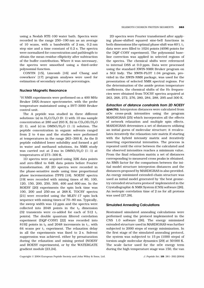
Chemical shift values of identified protons of Apeptide at 273 K are listed in Table 1. The fingerprintregion and the assignment of a NOESY spectrum(mixing time 200 ms), is shown in Figure 3a.
Table 1 Chemical Shifts of A Peptide in CD3OD/H2O (2 : 1) Solution at 273 K
Residue HN Hα Hβ Other protons Temperaturecoefficient
Gly 1 3.67Glu 2 8.66 4.28 1.94/1.76 γ2.17 4.0Leu 3 8.54 4.51 1.53/1.46 γ1.45 δ 0.85/0.81 7.1Pro 4 — 4.38 2.1/ γ1.92/1.83 δ 3.75/3.52Val 5 8.17 3.95 1.93 γ0.85 8.6Ala 6 8.52 4.12 1.28 9.0Gly 7 8.45 3.85/3.74 7.0Lys 8 8.11 4.30 1.77/1.67 γ1.36 δ1.57ε2.84 3.7Thr 9 8.00 4.19 4.09 γ1.01 5.8Ala 10 8.27 4.26 1.27 6.2Val 11 8.02 3.95 1.95 γ0.83 7.6Ala 12 8.35 4.13 1.29 7.6Gly 13 8.37 3.82/3.75 7.5Arg 14 7.99 4.35 1.74/1.64 γ1.49 δ 3.07ε7.15/6.65 3.3Val 15 8.13 4.30 1.99 γ0.87 7.5Pro 16 — 4.36 2.14/2.11 γ1.95/1.85 δ 3.78/3.58Ile 17 8.16 4.08 1.75 γ1.44/1.08 δ 0.83 8.6Ile 18 7.50 4.03 1.70 γ1.33 δ 0.77 4.4
Copyright 2004 European Peptide Society and John Wiley & Sons, Ltd. J. Peptide Sci. 10: 381–392 (2004)
386 BENAKI, MIKROS AND HAMODRAKAS
Figure 3 Fingerprint (a) and amide-amide proton(b) regions (Hα-HN and HN-HN) of NOESY spectrum of the Apeptide recorded at 273 K in CD3OD/H2O (2 : 1) solution.The mixing time was 200 ms.
The nOes detected concern protons of successiveresidues in the peptide sequence (i, i + 1). For thewhole temperature range studied (263–300 K) thenOe cross-peaks were negative.
Sequential Hαi-HNi+1 nOes were strong alongthe peptide chain and intraresidue Hαi-HNi nOeconnectivities were of medium intensity. ThedαN(i,i+1)/dαN(i,i) ratio was reversed in the case ofGly residues (positions 7 and 13 of the sequence)where a decrease of the interresidue connectivitywas observed. Two weak HNi-HNi+1 nOes for theresidues Val5-Ala6 and Gly7-Lys8 and an almost
complete series of weak cross-peaks between suc-cessive residues for the rest of the sequence (Thr9-Ile18) were detected (Figure 3b). No cis/trans iso-merism around the Xaa-Pro bonds was observed,as suggested by the absence of Hαi−1-Hαi nOes andthe presence of the Hαi−1-Hδi nOe peaks, even atNOESY experiments with a mixing time of 600 ms.Additionally, the lack of any ‘exchange’ peak in theROESY spectra, that could be attributed to differentconformers, slowly interconverting in the NMR timescale, is indicative that the two Pro residues are inthe trans conformation [34].
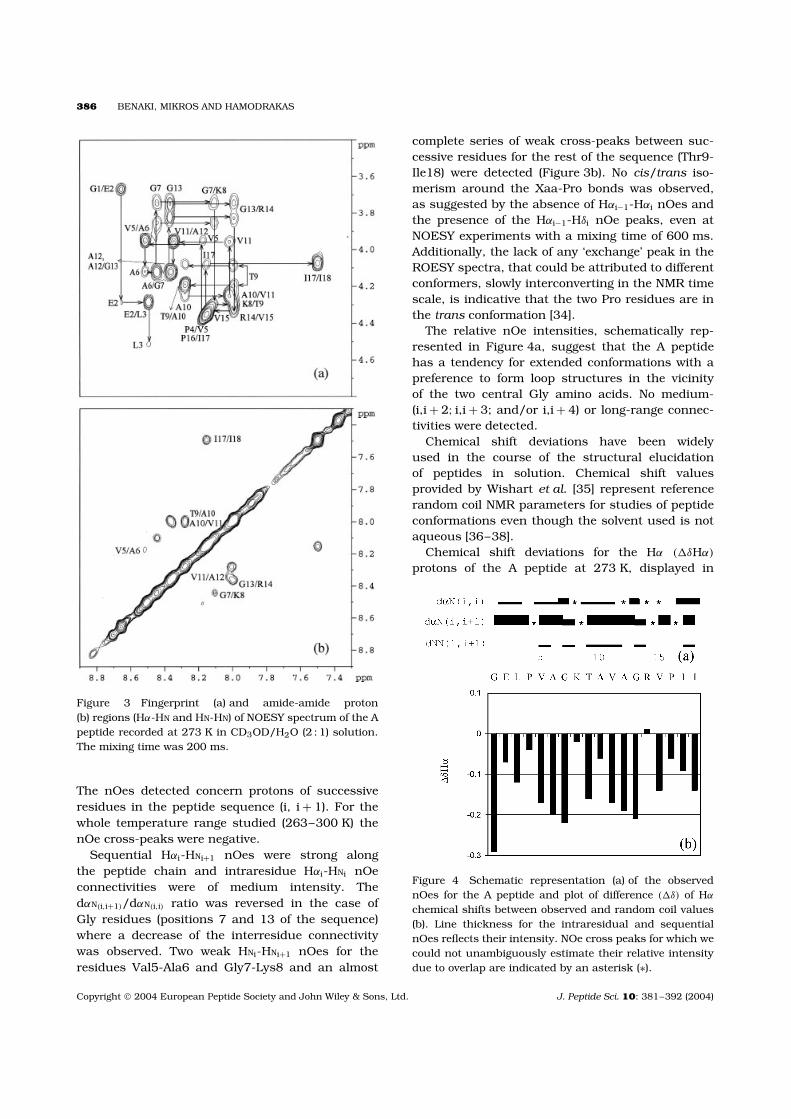
The relative nOe intensities, schematically rep-resented in Figure 4a, suggest that the A peptidehas a tendency for extended conformations with apreference to form loop structures in the vicinityof the two central Gly amino acids. No medium-(i,i + 2; i,i + 3; and/or i,i + 4) or long-range connec-tivities were detected.
Chemical shift deviations have been widelyused in the course of the structural elucidationof peptides in solution. Chemical shift valuesprovided by Wishart et al. [35] represent referencerandom coil NMR parameters for studies of peptideconformations even though the solvent used is notaqueous [36–38].
Chemical shift deviations for the Hα (�δHα)
protons of the A peptide at 273 K, displayed in
Figure 4 Schematic representation (a) of the observednOes for the A peptide and plot of difference (�δ) of Hα
chemical shifts between observed and random coil values(b). Line thickness for the intraresidual and sequentialnOes reflects their intensity. NOe cross peaks for which wecould not unambiguously estimate their relative intensitydue to overlap are indicated by an asterisk (∗).
Copyright 2004 European Peptide Society and John Wiley & Sons, Ltd. J. Peptide Sci. 10: 381–392 (2004)
SILKMOTH CHORION PROTEIN SEGMENTS 387
Figure 4b, had mainly negative values. Interestingly,a common pattern was observed in the vicinityof the two central Gly residues: considering Glyat the i position in the sequence, upfield shiftsbigger than 0.1 ppm were observed, with increasingvalues, from the i-2 to the i position. However,at the i + 1 amino acid (positively charged Lys8in the first segment, and Arg14 in the second) aclear inversion of the chemical shift deviation wasobserved in both segments. This pattern of the Hα
chemical shift deviations was indicative of a turnand confirms that Gly residues play important rolein the conformational behaviour of the peptide.
The temperature dependence of the amide pro-ton chemical shifts was determined over the range263 to 300 K (Table 1). The changes of the amideproton chemical shifts varied linearly with temper-ature (data not shown), suggesting that no majorconformational rearrangements occur in the tem-perature range studied. The coefficients for most ofthe residues were similar, varying from 6.5 to 8.0ppb/K. In the case of Glu2, Lys8 and Arg14 (i.e.the charged amino acids following the Gly residuesin the sequence) the coefficients were found to beconsiderably lower (3.9, 3.7 and 3.3 ppb/K, respec-tively), suggesting a relatively higher protection fromthe solvent and indicating that in a significant per-centage of the structures existing in solution theseamide protons might participate in intramolecularhydrogen bonds.
No additional information on the structuralcharacterization of the peptide was provided by the3JHNα coupling constants. Only for residues Leu3,Val15, Ile17 and Ile18 the coupling constant valueswere greater than 8 Hz, but their non sequentialposition in the peptide sequence did not allow anyconformational conclusion to be drawn.
In summary, the NMR characteristics of mostof the residues point to extended structures.However, a common pattern was observed aroundthe Gly residues: low intensity of the dαN(i,i+1)
parameters, low temperature coefficients of thevicinal polar amino acids, and upfield shifts forthe Hα nuclei. The segments of the peptide markedwith these characteristics should adopt backbonearrangements with dihedral angles in the α-helicalregion of the (φ,ψ) conformational space [39].
B peptide. The B peptide, although exhibitinghomology with the A peptide sequence, was onlysparingly soluble in water. At concentrations of1–3 mM it formed transparent solutions, which afew hours later (10–12 h) were characterized by
high viscosity. It has been argued that insolubilityof short peptides in water is an indicator of β-sheetstructure formation [40]. The B peptide bears onlyone charged residue (Glu), whereas the A peptidehas three (Glu, Lys and Arg) and one polar (Thr).In the B peptide the residues corresponding to Lysand Arg of the A peptide sequence are Thr and Glu.Moreover two Phe residues are also present in the Bpeptide. It is thus expected that the B peptide wouldbe less water soluble compared with the A peptide.A similar behaviour was observed in CD3OD/H2Osolutions at the concentrations required for the NMRexperiments [11].
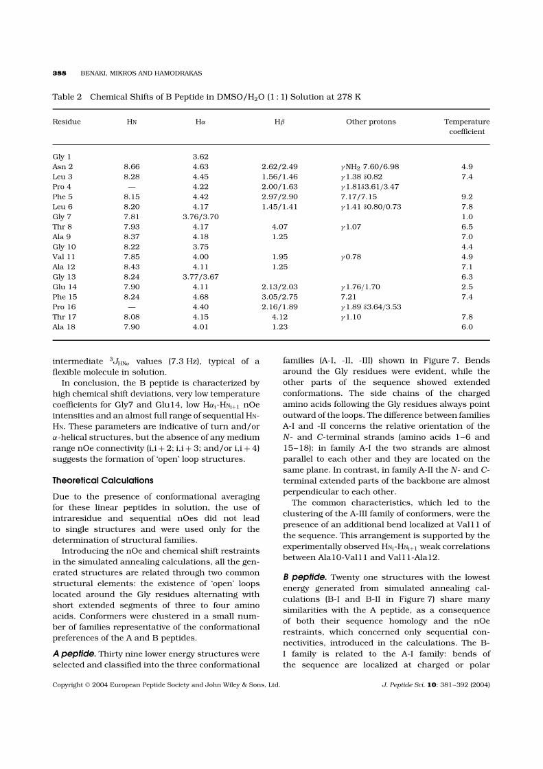
B peptide was thus studied in a DMSO/H2O (1 : 1)solution at a concentration of 2 mM and in thetemperature range from 273 to 300 K. The chemicalshift values are listed in Table 2.
The amino acid sequence was easily identifiedby a series of TOCSY and NOESY experimentsperformed at different temperatures. Part of theNOESY spectrum, illustrating the Hαi-HNi, Hαi-HNi+1
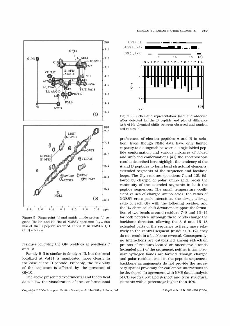
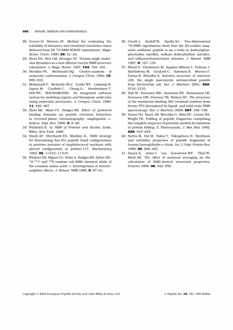
connectivities, is provided in Figure 5a. An almostfull series of medium to weak HNi-HNi+1 nOe cross-correlations were detected (Figure 5b). The relativenOe intensities of the sequential backbone protonsare depicted in Figure 6a. As in the case of the Apeptide, a decrease of the Hαi-HNi+1 intensity wasobserved at the Gly residues.
The two Pro residues (Pro4, Pro16) are both in thetrans conformation, as in the case of the A peptide.
Hα chemical shift deviations from the randomcoil (�δHα) were negative along the whole peptidesequence (Figure 6b), with differences suggestinga relatively higher structural stability and organi-zation. No medium- (i,i + 2; i,i + 3; and/or i,i + 4)or long-range nOe connectivities were observed.However, the rest of the structural characteristicsreferred to above provide evidence for the exis-tence of structured populations in solution (probably‘open’ loops).
Temperature coefficient data (Table 2) were moreinformative than those of the A peptide, as tworesidues (Gly7 and Glu14) exhibited considerablylow values (1 ppb/K and 2.5 ppb/K, respectively).
3JHNα values (Phe5, Thr8, Ala9, Val11, Glu14,Thr17), measured at 278 K from a DQF-COSYspectrum, were indicative of extended conformationsas they varied from 8.3 to 9.3 Hz. These valueswere larger compared with those of the A peptide,implying a higher structural stability. For the Asn2,Gly7, Gly10, Gly13 and Phe15 residues it wasnot possible to measure the 3JHNα values due tosignal overlapping. Leu3, Leu6 and Ala18 have
Copyright 2004 European Peptide Society and John Wiley & Sons, Ltd. J. Peptide Sci. 10: 381–392 (2004)
388 BENAKI, MIKROS AND HAMODRAKAS
Table 2 Chemical Shifts of B Peptide in DMSO/H2O (1 : 1) Solution at 278 K
Residue HN Hα Hβ Other protons Temperaturecoefficient
Gly 1 3.62Asn 2 8.66 4.63 2.62/2.49 γNH2 7.60/6.98 4.9Leu 3 8.28 4.45 1.56/1.46 γ1.38 δ0.82 7.4Pro 4 — 4.22 2.00/1.63 γ1.81δ3.61/3.47Phe 5 8.15 4.42 2.97/2.90 7.17/7.15 9.2Leu 6 8.20 4.17 1.45/1.41 γ1.41 δ0.80/0.73 7.8Gly 7 7.81 3.76/3.70 1.0Thr 8 7.93 4.17 4.07 γ1.07 6.5Ala 9 8.37 4.18 1.25 7.0Gly 10 8.22 3.75 4.4Val 11 7.85 4.00 1.95 γ0.78 4.9Ala 12 8.43 4.11 1.25 7.1Gly 13 8.24 3.77/3.67 6.3Glu 14 7.90 4.11 2.13/2.03 γ1.76/1.70 2.5Phe 15 8.24 4.68 3.05/2.75 7.21 7.4Pro 16 — 4.40 2.16/1.89 γ1.89 δ3.64/3.53Thr 17 8.08 4.15 4.12 γ1.10 7.8Ala 18 7.90 4.01 1.23 6.0
intermediate 3JHNα values (7.3 Hz), typical of aflexible molecule in solution.
In conclusion, the B peptide is characterized byhigh chemical shift deviations, very low temperaturecoefficients for Gly7 and Glu14, low Hαi-HNi+1 nOeintensities and an almost full range of sequential HN-HN. These parameters are indicative of turn and/orα-helical structures, but the absence of any mediumrange nOe connectivity (i,i + 2; i,i + 3; and/or i,i + 4)suggests the formation of ‘open’ loop structures.
Theoretical Calculations
Due to the presence of conformational averagingfor these linear peptides in solution, the use ofintraresidue and sequential nOes did not leadto single structures and were used only for thedetermination of structural families.
Introducing the nOe and chemical shift restraintsin the simulated annealing calculations, all the gen-erated structures are related through two commonstructural elements: the existence of ‘open’ loopslocated around the Gly residues alternating withshort extended segments of three to four aminoacids. Conformers were clustered in a small num-ber of families representative of the conformationalpreferences of the A and B peptides.
A peptide. Thirty nine lower energy structures wereselected and classified into the three conformational
families (A-I, -II, -III) shown in Figure 7. Bendsaround the Gly residues were evident, while theother parts of the sequence showed extendedconformations. The side chains of the chargedamino acids following the Gly residues always pointoutward of the loops. The difference between familiesA-I and -II concerns the relative orientation of theN- and C-terminal strands (amino acids 1–6 and15–18): in family A-I the two strands are almostparallel to each other and they are located on thesame plane. In contrast, in family A-II the N- and C-terminal extended parts of the backbone are almostperpendicular to each other.
The common characteristics, which led to theclustering of the A-III family of conformers, were thepresence of an additional bend localized at Val11 ofthe sequence. This arrangement is supported by theexperimentally observed HNi-HNi+1 weak correlationsbetween Ala10-Val11 and Val11-Ala12.
B peptide. Twenty one structures with the lowestenergy generated from simulated annealing cal-culations (B-I and B-II in Figure 7) share manysimilarities with the A peptide, as a consequenceof both their sequence homology and the nOerestraints, which concerned only sequential con-nectivities, introduced in the calculations. The B-I family is related to the A-I family: bends ofthe sequence are localized at charged or polar
Copyright 2004 European Peptide Society and John Wiley & Sons, Ltd. J. Peptide Sci. 10: 381–392 (2004)
SILKMOTH CHORION PROTEIN SEGMENTS 389
Figure 5 Fingerprint (a) and amide-amide proton (b) re-gions (Hα-HN and HN-HN) of NOESY spectrum (tm = 200ms) of the B peptide recorded at 278 K in DMSO/H2O(1 : 1) solution.
residues following the Gly residues at positions 7and 13.
Family B-II is similar to family A-III, but the bendlocalized at Val11 is manifested more clearly inthe case of the B peptide. Probably, the flexibilityof the sequence is affected by the presence ofGly10.
The above presented experimental and theoreticaldata allow the visualization of the conformational
(a)
(b)
* *
* * *
5 10 15
dαN(i,i)
dαN(i,i+1)
dNN(i,i+1)
Figure 6 Schematic representation (a) of the observednOes detected for the B peptide and plot of difference(�δ) of Hα chemical shifts between observed and randomcoil values (b).
preferences of chorion peptides A and B in solu-tion. Even though NMR data have only limitedcapacity to distinguish between a single folded pep-tide conformation and various mixtures of foldedand unfolded conformations [41] the spectroscopicresults described here highlight the tendency of theA and B peptides to form local structural elements:extended segments of the sequence and localizedloops. The Gly residues (positions 7 and 13), fol-lowed by charged or polar amino acid, break thecontinuity of the extended segments in both thepeptide sequences. The small temperature coeffi-cient values of charged amino acids, the ratios ofNOESY cross-peak intensities, the dαN(i,i+1)/dαN(i,i)
ratio of each Gly with the following residue, andthe Hα chemical shift deviations support the forma-tion of two bends around residues 7–8 and 13–14for both peptides. Although these bends change thebackbone direction, allowing the 3–6 and 15–18extended parts of the sequence to freely move rela-tively to the central segment (residues 9–12), theydo not result in a backbone reversal. Consequently,no interactions are established among side-chainprotons of residues located on successive strands(extended part of the sequence), neither intramolec-ular hydrogen bonds are formed. Though chargedand polar residues exist in the peptide sequences,backbone arrangements do not provide the neces-sary spatial proximity for coulombic interactions tobe developed. In agreement with NMR data, analysisof CD spectra revealed β-sheet and turn structuralelements with a percentage higher than 40%.
Copyright 2004 European Peptide Society and John Wiley & Sons, Ltd. J. Peptide Sci. 10: 381–392 (2004)

390 BENAKI, MIKROS AND HAMODRAKAS
Figure 7 Superposition of selected conformers of the A and B peptides (clustered into families), as a result of our theoreticalcalculations.
Experimental data from laser-Raman and FT-IRabsorption studies of the A and B peptides bothin solution and in the solid state [9], as well asx-ray diffraction, FT-Raman and ATR-FTIR data ofamyloid-like fibrils formed from chorion peptides[11,12] suggest the preponderance of the antipar-allel β-pleated sheet structure. Additionally, theleft-handed parallel β-sheet helix [10] model struc-ture has recently been proposed by Iconomidouet al. [11,12], as an attractive alternative to describethe folding and self-assembly of chorion peptides.According to this model, there is a central hydropho-bic core region forming a triangular prism-likehelix; the edges of the prism are occupied by Glyand charged or polar residues, while four-residueextended segments lie on the prism planes.
It has been anticipated that the hexapeptide peri-odicity and the presence of Gly residues dictate thesecondary structure of the fibrils in chorion pro-teins and peptide analogues [3]. The data reportedin this work are of considerable importance sincethey clearly establish, for the first time, experimen-tal evidence that local folded structures around theGly residues do occur in segments of the chorionproteins in solution. The structural characteristics
of both peptides in solution, emerging from theexperimental data, provide a better insight for theinitiation of the chorion protein folding process. Thestructures described in this work may representearly folding intermediates capable of directing sub-sequent folding events and the nucleation of the finalfunctionally important conformation, the amyloid-like fibrils. However, due to the short length of thepeptide sequences, plausible ‘triangular’ [10] and/orantiparallel β-sheet structures [9] are probably notstable enough. In any case, it remains to be seenwhich of the two models (left-handed parallel β-helixor ‘cross-β’ antiparallel β-sheet structure) actuallyrepresents the basic folding motif of the amyloid-likefibrils formed by chorion peptides.
Acknowledgements
D.B. was the recipient of short term EMBO andSRMB fellowships. Thanks are due to Dr S.T. Casefor the synthesis of peptides A and B. The authorsgratefully acknowledge Dr A. Pastore for continuingsupport during this work.
Copyright 2004 European Peptide Society and John Wiley & Sons, Ltd. J. Peptide Sci. 10: 381–392 (2004)

SILKMOTH CHORION PROTEIN SEGMENTS 391
REFERENCES
1. Regier JC, Kafatos FC. In Comprehensive InsectBiochemistry, Physiology and Pharmacology, Gilbert LI,Kerkut GA (eds). Pergamon Press: Oxford, 1985; 1:113–151.
2. Goldsmith MR, Kafatos FC. Developmentally regulatedgenes in silkmoths. Annu. Rev. Genet. 1984; 18:443–487.
3. Hamodrakas SJ. In Results and Problems in CellDifferentiation, Case ST (ed.). Springer: Berlin, 1992;19: 115–186.
4. Kafatos FC, Regier JC, Mazur GD, NadelMR, Blau HM, Petri WH, Wyman AR, Gelinas RE,Moore PB, Paul M, Efstratiadis A, Vournakis JN,Goldsmith MR, Hunsley JR, Baker B, Nardi J,Koehler M. In Results and Problems in CellDifferentiation, Beerman W (ed.). Springer: Berlin,1977; 8: 45–145.
5. Lecanidou R, Rodakis GC, Eickbush TH, Kafatos FC.Evolution of the silkmoth chorion gene superfamily:gene families CA and CB. Proc. Natl Acad. Sci. USA1986; 83: 6514–6518.
6. Hamodrakas SJ, Jones CW, Kafatos FC. Secondarystructure predictions for silkmoth chorion proteins.Biochim. Biophys. Acta 1982; 700: 42–51.
7. Hamodrakas SJ, Etmektzoglou T, Kafatos FC. Aminoacid periodicities and their structural implications forthe evolutionarily conservative central domain of somesilkmoth chorion proteins. J. Mol. Biol. 1985; 186:583–589.
8. Hamodrakas SJ, Bosshard HE, Carlson CN. Struc-tural models of the evolutionarily conservative centraldomain of silkmoth chorion proteins. Protein Eng.1988; 2: 201–207.
9. Benaki DC, Aggeli A, Chryssikos GD, Yiannopou-los YD, Kamitsos EI, Brumley E, Case ST, Boden N,Hamodrakas SJ. Laser-Raman and FT-IR spectro-scopic studies of peptide analogues of silkmoth chorionprotein segments. Int. J. Biol. Macromol. 1998; 23:49–59.
10. Yoder MD, Jurnak F. Protein motifs. 3. The parallelβ-helix and other coiled folds. FASEB J. 1995; 9:335–342.
11. Iconomidou VA, Chryssikos GD, Gionis V, Vriend G,Hoenger A, Hamodrakas SJ. Amyloid-like fibrils froman 18-residue peptide analogue of a part of the centraldomain of the B-family of silkmoth chorion proteins.FEBS Lett. 2001; 499: 268–273.
12. Iconomidou VA, Vriend G, Hamodrakas SJ. Amyloidsprotect the silkmoth oocyte and embryo. FEBS Lett.2000; 479: 141–145.
13. Sunde M, Blake CCF. From the globular to the fibrousstate: protein structure and structural conversion inamyloid formation. Quart. Rev. Biophys. 1998; 31:1–39.
14. Sunde M, Serpell LC, Bartlam M, Fraser PE, Pepys MB,Blake CCF. Common core structure of amyloid fibrilsby synchrotron x-ray diffraction. J. Mol. Biol. 1997;273: 729–739.
15. Provencher SW, Glockner J. Analysis of thecomponents present in kinetics (or titration) curves.J. Biochem. Biophys. Methods 1983; 7: 331–334.
16. Perczel A, Hollosi M, Tusnady G, Fasman GD. CON-VEX constraint analysis: a natural deconvolution ofcircular dichroism curves of proteins. Protein Eng.1991; 4: 669–679.
17. Chang CT, Wu CSC, Yang JT. Circular dichroicanalysis of protein conformation: inclusion of the β-turns. Anal. Biochem. 1978; 91: 13–31.
18. Bodenhausen G, Kogler H, Ernst RR. Selectionof coherence-transfer pathways in NMR pulseexperiments. J. Magn. Reson. 1984; 58: 370–388.
19. Kumar A, Ernst RR, Wuthrich K. A two-dimensionalnuclear Overhauser enhancement (2D NOE)experiment for the elucidation of complete proton-proton cross-relaxation networks in biologicalmacromolecules. Biochem. Biophys. Res. Commun.1980; 95: 1–6.
20. Bothner-By AA, Stephens RL, Lee JM, Warren CD,Jeanloz RW. Structure determination of a tetrasaccha-ride: transient nuclear Overhauser effects in the rotat-ing frame. J. Am. Chem. Soc. 1984; 106: 811–813.
21. Bax A, Davis DG. MLEV-17-based two-dimensionalhomonuclear magnetization transfer spectroscopy. JMagn. Reson. 1985; 65: 355–360.
22. Rance M, Sorensen OW, Bodenhausen G, Wagner G,Ernst RR, Wuthrich K. Improved spectral resolution inCOSY 1H-NMR spectra of proteins via double quantumfiltering. Biochem. Biophys. Res. Commun. 1983; 117:479–485.
23. Piotto M, Saudek V, Sklenar V. Gradient-tailoredexcitation for single-quantum NMR spectroscopy ofaqueous solutions. J. Biomol. NMR 1992; 2: 661–665.
24. Sklenar V, Piotto M, Leppik R, Saudek V. Gradient-tailored water suppression for 1H-15N HSQCexperiments optimized to retain full sensitivity. J.Magn. Reson., Ser. A 1993; 102: 241–245.
25. Borgias BA, James TL. MARDIGRAS A procedure formatrix analysis of relaxation for discerning geometryof an aqueous structure. J. Magn. Reson. 1990; 87:475–487.
26. Brunger AT, Adams PD, Clore GM, DeLano WL,Gros P, Grosse-Kunstleve RW, Jiang JS, Kuszewski J,Nilges M, Pannu NS, Read RJ, Rice LM, Simonson T,Warren GL. Crystallography and NMR system. Anew software suite for macromolecular structuredetermination. Acta Crystallogr. 1998; D 54: 905–921.
27. Palmer AG, Rance M, Wright PE. Intramolecularmotions of a zinc finger DNA-binding domainfrom XFIN characterized by proton-detected naturalabundance 12C heteronuclear NMR spectroscopy. J.Am. Chem. Soc. 1991; 113: 4371–4380.
Copyright 2004 European Peptide Society and John Wiley & Sons, Ltd. J. Peptide Sci. 10: 381–392 (2004)
392 BENAKI, MIKROS AND HAMODRAKAS
28. Genest D, Simorre JP. Method for evaluating thereliability of distances and rotational correlation timesdeduced from 2D 1H-NMR NOESY experiments. Magn.Reson. Chem. 1990; 28: 21–24.
29. Stein EG, Rice LM, Brunger AT. Torsion-angle molec-ular dynamics as a new efficient tool for NMR structurecalculation. J. Magn. Reson. 1997; 124: 154–164.
30. Shenkin PS, McDonald DQ. Cluster-analysis ofmolecular conformations. J. Comput. Chem. 1994; 15:899–916.
31. Mohamadi F, Richards NGJ, Guida WC, Liskamp R,Lipton M, Caufield C, Chang G, Hendrickson T,Still WC. MACROMODEL. An integrated softwaresystem for modeling organic and bioorganic moleculesusing molecular mechanics. J. Comput. Chem. 1990;11: 440–467.
32. Zhou NE, Mant CT, Hodges RS. Effect of preferredbinding domains on peptide retention behaviourin reversed-phase chromatography: amphipathic α-helices. Pept. Res. 1990; 3: 8–20.
33. Wuthrich K. In NMR of Proteins and Nucleic Acids.Wiley: New York, 1986.
34. Hinck AP, Eberhardt ES, Markley JL. NMR strategyfor determining Xaa-Pro peptide bond configurationsin proteins mutants of staphylococcal nuclease withaltered configuration at proline-117. Biochemistry1993; 32: 11 810–11 818.
35. Wishart DS, Bigam CG, Holm A, Hodges RS, Sykes BD.1H,13 C and 15N random coil NMR chemical shifts ofthe common amino acids. I. Investigations of nearest-neighbor effects. J. Biomol. NMR 1995; 5: 67–81.
36. Gesell J, Zasloff M, Opella SJ. Two-dimensional1H-NMR experiments show that the 23-residue mag-ainin antibiotic peptide is an α-helix in dodecylphos-phocholine micelles, sodium dodecylsulfate micelles,and trifluoroethanol/water solution. J. Biomol. NMR1997; 9: 127–135.
37. Blond A, Cheminant M, Segalas-Milazzo I, Peduzzi J,Barthelemy M, Goulard C, Salomon R, Moreno F,Farias R, Rebuffat S. Solution structure of microcinJ25, the single macrocyclic antimicrobial peptidefrom Escherichia coli. Eur. J. Biochem. 2001; 268:2124–2133.
38. Bak M, Sorensen MD, Sorensen ES, Rasmussen LK,Sorensen OW, Petersen TE, Nielsen NC. The structureof the membrane-binding 38C-terminal residues frombovine PP3 determined by liquid- and solid-state NMRspectroscopy. Eur. J. Biochem. 2000; 267: 188–199.
39. Dyson HJ, Sayre JR, Merutka G, Shin HC, Lerner RA,Wright PE. Folding of peptide fragments comprisingthe complete sequence of proteins: models for initiationof protein folding. 2. Plastocyanin. J. Mol. Biol. 1992;226: 819–835.
40. Narita M, Doi M, Nakai T, Takegahara H. Synthesisand solubility properties of peptide fragments ofhuman hemoglobulin α-chain. Int. J. Pept. Protein Res.1988; 32: 200–207.
41. Daura X, Antes I, van Gunsteren WF, Thiel W,Mark AE. The effect of motional averaging on thecalculation of NMR-derived structural properties.Proteins 1999; 36: 542–555.
Copyright 2004 European Peptide Society and John Wiley & Sons, Ltd. J. Peptide Sci. 10: 381–392 (2004)