conformational analysis of the endogenous μ-opioid agonist endomorphin-1 using nmr spectroscopy and...
TRANSCRIPT
Conformational analysis of the endogenous W-opioid agonistendomorphin-1 using NMR spectroscopy and molecular modeling
Brent L. Podlogar1;a, M. Germana Paterlinib, David M. Ferguson2;b, Gregory C. Leoa,David A. Demetera, Frank K. Browna, Allen B. Reitza
aDepartment of Medicinal Chemistry, The R.W. Johnson Pharmaceutical Research Institute, Route 202, P.O. Box 300, Raritan, NJ 08869, USAbDepartment of Medicinal Chemistry, University of Minnesota, 308 Harvard St. SE, Minneapolis, MN 55455, USA
Received 6 August 1998
Abstract Endomorphin-1 (Tyr-Pro-Trp-Phe-NH2) is a highlyselective and potent agonist of the WW-opioid receptor. To identifystructural attributes unique to this opioid peptide and potentialsites of recognition, a conformational analysis has beenperformed using multidimensional NMR and molecular modelingtechniques. The spectroscopic results, derived from experimentsin both DMSO and water, indicate that endomorphin-1 exists inthe cis- and trans-configuration with respect to the Pro-omegabond in approximately 25% and 75% populations, respectively.In DMSO, the cis-configuration adopts a compact sandwichconformation in which the Tyr and Trp aromatic rings packagainst the proline ring, whereas the trans-configuration adoptsan extended conformation. Although non-random structure wasnot observed in water, condensed phase molecular dynamicscalculations indicate that trans-isomers dominate the populationin this higher dielectric medium. Structural comparison of thecis- and trans-configurations with morphine and selective WW-peptide ligands PL-017 and D-TIPP, as well as the NN-selectivepeptide ligands TIPP (NN-antagonist, WW-agonist) and DPDPEwere also performed and suggest the trans-isomer is likely thebioactive form. A hypothesis is proposed to explain WW- and NN-selectivity based on the presence of spatially distinct selectivitypockets among these ligands.z 1998 Federation of European Biochemical Societies.
Key words: Opioid; Peptide structure; Conformationalanalysis ; Molecular dynamics; Multidimensional nuclearmagnetic resonance spectroscopy; Structure-activityrelationship
1. Introduction
Opioid receptors are important targets for pharmacologicalinteraction in acute pain therapy [1]. Recent work in this areaappears to indicate that the observed e¡ects of many opioidanalgesics are caused by combinations of opioid receptor typeactivation [2^5]. To unravel these interactions, detailed struc-tural models capable of di¡erentiating N, W and U receptortype selectivity and ultimately, receptor activation vs. inacti-vation are crucial. Structural models have been generally de-rived from small molecule opioid receptor agonists and antag-onists [6,7], and while they have advanced our thinking in thisarea, and have shed some light on separation of N bindingfrom U and W binding [8], they are neither predictive nor
complete. Although the small molecule ligands, e.g. opiatealkaloids, are less problematic to model, their small physicalsize as compared to the endogenous opioid ligands, e.g. enke-phalins, endomorphins, dynorphins, restricts ligand interac-tions to a small region of the receptor active site and mayfail to fully address the full complement of interactions re-sponsible for speci¢c receptor type activation [9,10]. Identi¢-cation and structural characterization of selective opioid pep-tide ligands for the N receptor (e.g. DPDPE [11], JOM-13 [12])and the U receptor (dynorphin [13,14]) have allowed modelingstudies to begin for these types, but to date, few such studiesfor W-selective peptide ligands (D-TIPP-NH2) have been re-ported [15].
Recently, two novel peptides, endomorphin-1 (YPWF-NH2) and endomorphin-2 (YPFF-NH2) were reported asthe endogenous W receptor ligands [16]. Endomorphin-1 showsremarkable a¤nity for the W receptor (360 pM) and selectivityof 4000- and 15 000-fold for the W receptor over the N and Ureceptors, respectively. The availability of the endomorphinpeptides makes possible the comparison with other W-selectivepeptides, such as D-TIPP (Y-D-Tic-FF-NH2) [15] and PL-017(YPF-D-P-NH2) [17]. We have studied the properties of endo-morphin-1 using NMR spectroscopy and molecular simula-tions, and the results have been complemented with molecularcomparisons of this peptide with D-TIPP and PL-017. Struc-tural comparisons with the N-agonist DPDPE [11] and thedual W-agonist/N-antagonist, TIPP-NH2 [18] have also beenperformed to rationalize structure-based selectivities amongthese ligands and opioid receptor types.
2. Materials and methods
2.1. Synthesis of endomorphin-1Endomorphin-1 was synthesized using the Merri¢eld solid-phase
method of peptide synthesis. t-BOC chemistry with HBTU [2-(1H-benzotriazol-1-yl)-1,1,3,3-tetramethyluronium hexa£uorophosphate]activation was employed for peptide elongation. The peptide-resinswere treated with 95% HF/5% anisole at 34³C for 1.5 h to generatefree peptides. The peptide-associated resin complexes were washedwith dimethyl ether to remove the anisole. The peptides were ex-tracted in 20% HOAc and puri¢ed on two 22.5U250 mm VydacC18 columns in tandem (5 Wm particle size, 300 Aî pore size) usinga gradient of 0^27% `B' in 27 min at a £ow rate of 30 ml/min. The `A'bu¡er was 0.1% TFA/water and the `B' bu¡er was 0.1% TFA/aceto-nitrile. The fractions containing the desired peptide were then com-bined and lyophilized. The dried peptides were characterized by ana-lytical RP-HPLC and electrospray MS.
2.2. NMR spectroscopyAll spectra were recorded at 600.13 MHz on a Bruker DMX spec-
trometer. Samples in water included 10% deuterium oxide as the locksolvent and TSP as the chemical shift reference. Spectra were recordedat 275 K or 278 K. Sample concentrations used were approximately
FEBS 20961 17-11-98 Cyaan Magenta Geel Zwart
0014-5793/98/$19.00 ß 1998 Federation of European Biochemical Societies. All rights reserved.PII: S 0 0 1 4 - 5 7 9 3 ( 9 8 ) 0 1 2 0 2 - 2
1Corresponding author. Current address: Bayer Corp., 400 MorganLane, West Haven, CT 06516, USA.E-mail: [email protected]
2Corresponding author.E-mail: [email protected]
FEBS 20961 FEBS Letters 439 (1998) 13^20
0.6 mM or 3.5 mM. TOCSY spectra were measured using a 50 msspin lock (10 000 Hz ¢eld strength) using either DIPSI-2 [19] orMLEV-17 [20] and magic angle gradient water suppression with the3-9-19 Watergate sequence [21,22]. Through-space correlations weredetected using both NOESY, [23] ROESY [24] (2550 Hz ¢eldstrength) and o¡-resonance ROESY [25] (10 000 Hz ¢eld strength,10 000 Hz o¡-resonance) experiments using mixing times rangingfrom 150 ms to 250 ms. Water suppression was accomplished usingmagic angle gradient 3-9-19 Watergate or low power, continuouswave pre-saturation. The sample in DMSO-d6 was approximately 7mM and TMS was used as the chemical shift reference. TOCSY [26](10 000 Hz spin lock ¢eld for 60 ms) and NOESY (250 ms mix time)spectra were recorded at 297 K. All 2D spectra were acquired in thephase sensitive mode (States-TPPI) and processed using a 90³ shifted,squared sine-bell apodization [27].
2.3. Restrained molecular dynamicsRestrained molecular dynamics simulations were conducted using
the Insight II/Discover 95 suite of software. All simulations wereconducted in vacuo using the CFF91 force¢eld3. The NOE distancedata were applied a generic distance constraints using a lower distancebound of 1.8 Aî , and a force constant value set to 10 kcal/mol Aî 2. Nodistance cuto¡s were used; a distance dependent dielectric of 3.5Urwas used. A 20 ps high temperature molecular dynamics simulations(700³C) was used to generate a total of 200 random starting geome-tries. The random structure were cooled by successive minimizationswith the force¢eld scaled at 0.5 (500 cycles of steepest descents); 0.75(500 cycles of steepest descents), 1.0 (1000 cycles of steepest descentfollowed by 1500 cycles of conjugate gradient minimization; conver-gence criterion set to 0.1 kcal/Aî .) All g bonds were forced to 180³ (or0³ for the cis-proline isomer) using a force constant of 100 kcal/Aî 2.Chiral centers were constrained to the starting values.
2.4. Systematic conformational searchingGeneral molecular modeling and conformational analysis was
conducted in Sybyl 6.34. Atomic point charges were calculated usingthe Gasteiger-Huëckel method [28^32]. Full geometry optimizationswere done using the Tripos force ¢eld [33,34] with the Powellminimizer and electrostatics. The following variable parameters wereset: termination_criterion = energy, min_energy_change = 0.00001kcal/mol, max_iterations = 1 000 000, dielectric_constant = 2.0, dielec-tric_function=distance. Default settings were used for all other varia-ble parameters.
Systematic conformational searches were done using the Triposforce ¢eld with the csearch algorithm, energies, and electrostatics.All rotatable bonds were searched from 0³ to 359³. An angle in-crement of 10³ was used for the Tyr i dihedral, 20³ for the Tic idihedral, and 10³ for all the backbone and sidechain rotatable bondsin the two Phe residues for a total of 10 rotatable bonds. The follow-ing variable parameters were set: reference_conformation = zeroed,van_der_Waals_scaling_factors (general = 0.90, 1^4 = 0.82, hbond =0.65), energy_cuto¡ = 9999.9 kcal/mol, dielectric_constant = 2.0, di-electric_function = distance. Default settings were used for all othervariable parameters.
Potential energy surface analyses were done in PESA 1.0 [35]. Thefollowing variable parameters were set: ¢nd minima, neighbors mini-ma noprint, statistics surface minima. Default settings were used forall other variable parameters.
Steric and electrostatic CoMFA ¢elds [36^38] were calculated withthe CoMFA regions de¢ned automatically using the molecular vol-umes option. Default settings were used for all other variable param-eters. Field correlations were calculated with the QSAR COMFAFIELD COMPARE command. The correlation coe¤cients of thesteric and electrostatic ¢elds were summed and the conformer withthe highest value was selected as the most similar. This conformer was
then minimized as described above and was ¢t to trans-endomorphin-1.
2.5. Molecular dynamics simulationsMD simulations were performed using the Amber 4.1 suite of pro-
grams with the Cornell et al. force ¢eld [39]. Simulations in aqueoussolutions were obtained by placing endomorphin-1 in a box of equi-librated water and then stripping any water molecule at a distance of2.0 Aî or less from the peptide. The dimensions of the water box were46U38U35 for a total of 1647 water molecules. An energy minimized,extended structure was used as the starting structure. Molecular dy-namics simulations were conducted at constant temperature and pres-sure using the Berendsen coupling scheme with a time constant of 0.2for temperature and pressure regulation. Bond lengths were con-strained using the SHAKE algorithm and an integration time stepof 2 fs was used. A 10 Aî cuto¡ was used for non-bonded interactionsand the non-bonded pairlist was updated every 50 fs. Equilibrium wasreached after about 600 ps as judged from the rmsd deviation fromthe initial structure which rose to 4.5 Aî .
3. Results
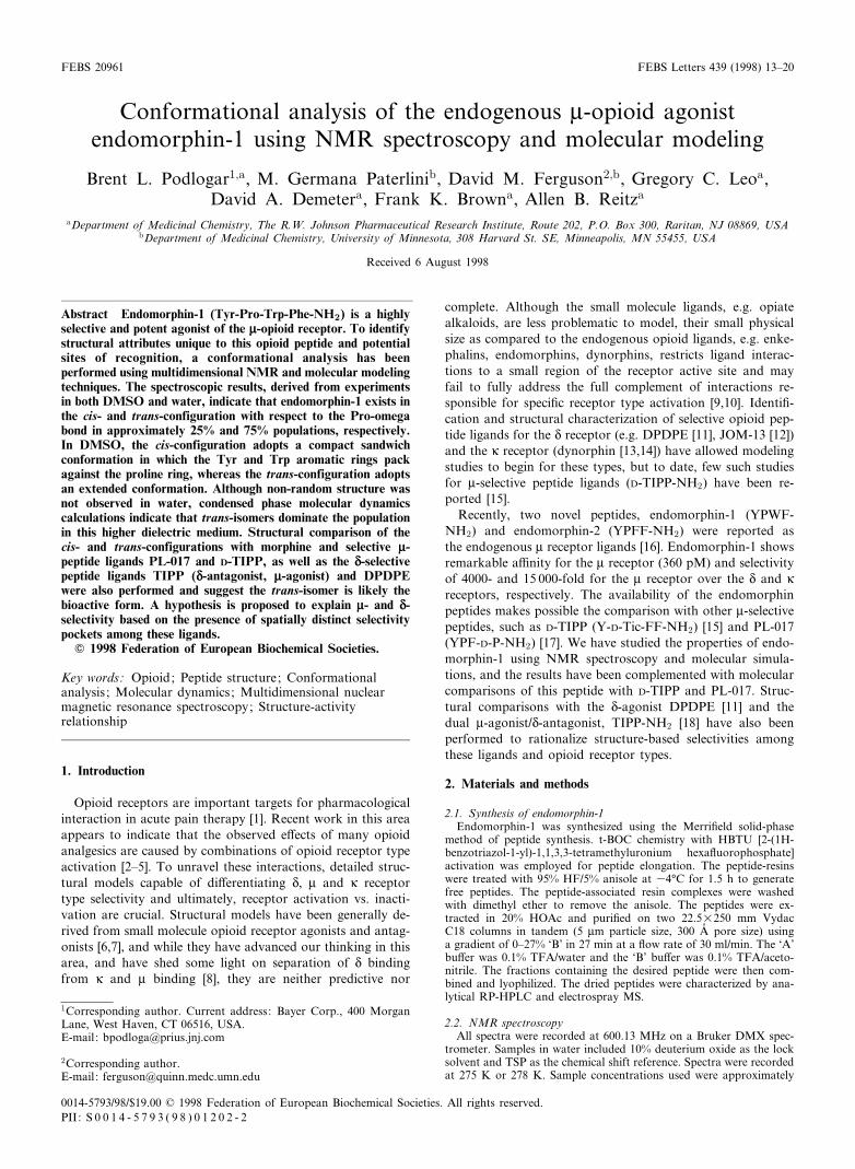
NMR experimentation was conducted using standard tech-niques (see Section 2) in H2O (4³C) and in DMSO-d6 (25³C).A variable temperature study was also conducted in DMSO-d6 to evaluate internal hydrogen bonding. The 1D spectrumof endomorphin-1 (Fig. 1) clearly highlights the existence ofcis- and trans-isomer populations.
The relative peak intensities from the 1D spectrum for boththe aqueous and DMSO media indicate that endomorphin-1resides in 25/75% populations of the cis-trans isomers.
The cis-trans peak assignments were con¢rmed by the char-acteristic sequential NOEs between the Pro and Tyr residues[40]. Non-sequential ROESY cross peaks were not observed inaqueous solution for endomorphin-1.
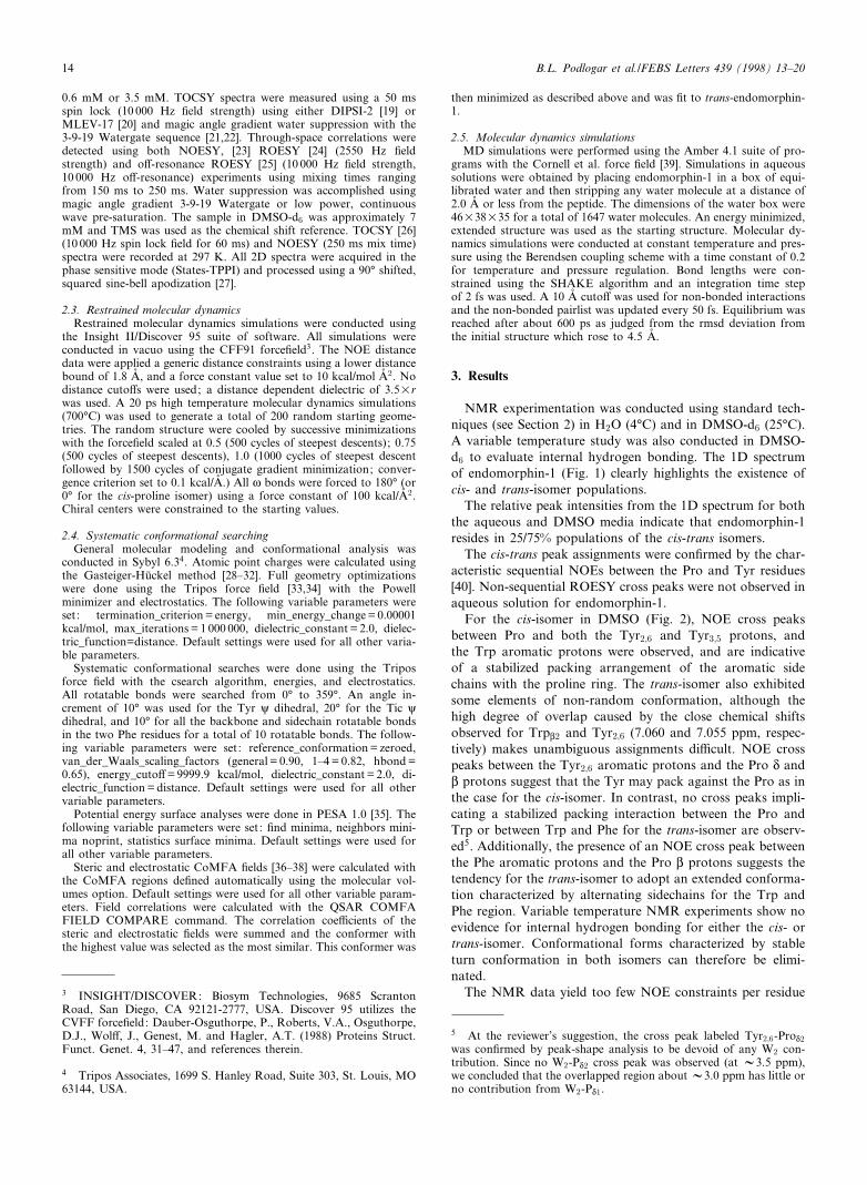
For the cis-isomer in DMSO (Fig. 2), NOE cross peaksbetween Pro and both the Tyr2;6 and Tyr3;5 protons, andthe Trp aromatic protons were observed, and are indicativeof a stabilized packing arrangement of the aromatic sidechains with the proline ring. The trans-isomer also exhibitedsome elements of non-random conformation, although thehigh degree of overlap caused by the close chemical shiftsobserved for TrpL2 and Tyr2;6 (7.060 and 7.055 ppm, respec-tively) makes unambiguous assignments di¤cult. NOE crosspeaks between the Tyr2;6 aromatic protons and the Pro N andL protons suggest that the Tyr may pack against the Pro as inthe case for the cis-isomer. In contrast, no cross peaks impli-cating a stabilized packing interaction between the Pro andTrp or between Trp and Phe for the trans-isomer are observ-ed5. Additionally, the presence of an NOE cross peak betweenthe Phe aromatic protons and the Pro L protons suggests thetendency for the trans-isomer to adopt an extended conforma-tion characterized by alternating sidechains for the Trp andPhe region. Variable temperature NMR experiments show noevidence for internal hydrogen bonding for either the cis- ortrans-isomer. Conformational forms characterized by stableturn conformation in both isomers can therefore be elimi-nated.
The NMR data yield too few NOE constraints per residue
FEBS 20961 17-11-98 Cyaan Magenta Geel Zwart
3 INSIGHT/DISCOVER: Biosym Technologies, 9685 ScrantonRoad, San Diego, CA 92121-2777, USA. Discover 95 utilizes theCVFF force¢eld: Dauber-Osguthorpe, P., Roberts, V.A., Osguthorpe,D.J., Wol¡, J., Genest, M. and Hagler, A.T. (1988) Proteins Struct.Funct. Genet. 4, 31^47, and references therein.
4 Tripos Associates, 1699 S. Hanley Road, Suite 303, St. Louis, MO63144, USA.
5 At the reviewer's suggestion, the cross peak labeled Tyr2;6-ProN2
was con¢rmed by peak-shape analysis to be devoid of any W2 con-tribution. Since no W2-PN2 cross peak was observed (at V3.5 ppm),we concluded that the overlapped region about V3.0 ppm has little orno contribution from W2-PN1.
B.L. Podlogar et al./FEBS Letters 439 (1998) 13^2014
for a rigorous structure determination. Nevertheless, applica-tion of the observed cross peaks via restrained molecular dy-namics was conducted to obtain plausible `NMR structures'for our modeling studies that re£ect the conformations whichcannot be ruled out by the NMR data. Low energy confor-mations consistent with the NMR data were determined usingNOE restraints for both the cis- and trans-isomers. A total of19 NOE restraints from the cis-isomer, and 10 NOE restraintsfrom the trans isomer were taken from the NOESY spectra.The restraints were calibrated to internal stationary backboneproton-proton distances (trans-TyrN-ProL and trans-PheN-ProL) and are shown in Tables 1 and 2.
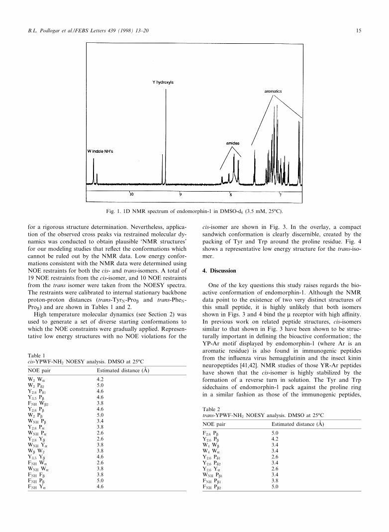
High temperature molecular dynamics (see Section 2) wasused to generate a set of diverse starting conformations towhich the NOE constraints were gradually applied. Represen-tative low energy structures with no NOE violations for the
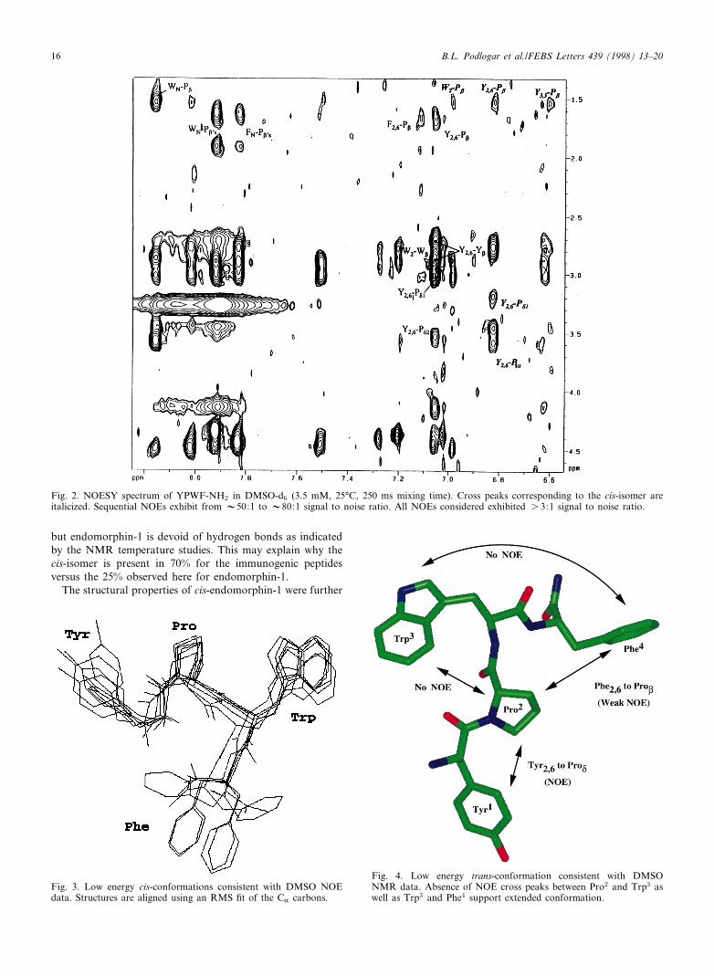
cis-isomer are shown in Fig. 3. In the overlay, a compactsandwich conformation is clearly discernible, created by thepacking of Tyr and Trp around the proline residue. Fig. 4shows a representative low energy structure for the trans-iso-mer.
4. Discussion
One of the key questions this study raises regards the bio-active conformation of endomorphin-1. Although the NMRdata point to the existence of two very distinct structures ofthis small peptide, it is highly unlikely that both isomersshown in Figs. 3 and 4 bind the W receptor with high a¤nity.In previous work on related peptide structures, cis-isomerssimilar to that shown in Fig. 3 have been shown to be struc-turally important in de¢ning the bioactive conformation; theYP-Ar motif displayed by endomorphin-1 (where Ar is anaromatic residue) is also found in immunogenic peptidesfrom the in£uenza virus hemagglutinin and the insect kininneuropeptides [41,42]. NMR studies of those YR-Ar peptideshave shown that the cis-isomer is highly stabilized by theformation of a reverse turn in solution. The Tyr and Trpsidechains of endomorphin-1 pack against the proline ringin a similar fashion as those of the immunogenic peptides,
FEBS 20961 17-11-98 Cyaan Magenta Geel Zwart
Table 1cis-YPWF-NH2 NOESY analysis. DMSO at 25³C
NOE pair Estimated distance (Aî )
W2 WK 4.2W2 PN2 5.0Y2;6 PN1 4.6Y3;5 PL 4.6FNH WL2 3.8Y2;6 PL 4.6W2 PL 5.0WNH PL 3.4Y2;6 PK 3.8WNH PK 2.6Y2;6 YL 2.6WNH YK 3.8WL W2 3.8Y3;5 YL 4.6FNH WK 2.6WNH WK 3.8FNH FL 3.8FNH PL 5.0FNH YK 4.6
Fig. 1. 1D NMR spectrum of endomorphin-1 in DMSO-d6 (3.5 mM, 25³C).
Table 2trans-YPWF-NH2 NOESY analysis. DMSO at 25³C
NOE pair Estimated distance (Aî )
F2;6 PL 5.0Y2;6 PL 4.2W4 WL 3.4W4 WK 3.4Y2;6 PN1 2.6Y2;6 PN2 3.4Y2;6 YK 2.6WNH PL1 3.4FNH PL1 3.8FNH PL2 5.0
B.L. Podlogar et al./FEBS Letters 439 (1998) 13^20 15
but endomorphin-1 is devoid of hydrogen bonds as indicatedby the NMR temperature studies. This may explain why thecis-isomer is present in 70% for the immunogenic peptidesversus the 25% observed here for endomorphin-1.
The structural properties of cis-endomorphin-1 were further
FEBS 20961 17-11-98 Cyaan Magenta Geel Zwart
Fig. 3. Low energy cis-conformations consistent with DMSO NOEdata. Structures are aligned using an RMS ¢t of the CK carbons.
Fig. 2. NOESY spectrum of YPWF-NH2 in DMSO-d6 (3.5 mM, 25³C, 250 ms mixing time). Cross peaks corresponding to the cis-isomer areitalicized. Sequential NOEs exhibit from V50:1 to V80:1 signal to noise ratio. All NOEs considered exhibited s 3:1 signal to noise ratio.
Fig. 4. Low energy trans-conformation consistent with DMSONMR data. Absence of NOE cross peaks between Pro2 and Trp3 aswell as Trp3 and Phe4 support extended conformation.
B.L. Podlogar et al./FEBS Letters 439 (1998) 13^2016
explored using molecular dynamics simulations in water andin vacuo with varying dielectric constants. In each case, thesimulations failed to reproduce the sidechain packing interac-tions predicted by our NOE data and observed in publishedreports [41,42]. Since the dielectric properties of DMSO andliquid water are signi¢cantly di¡erent (40 versus 78.5, respec-tively), this result is not surprising. Temussi et al. have alsoreported viscosity e¡ects on the structure of small peptides[43]. Similar to our results, they observed a pattern of struc-ture in DMSO and no structure in aqueous mediums. Since
structural elements were observed only in DMSO, which isnot a biologically relevant solvent, and that only 25% of thepeptide population for endomorphin-1 exists in the cis-form,we conclude that the compact structure observed for the cis-isomer is an artifact of the solution conditions.
To investigate the signi¢cance of the trans-isomer to peptidebinding, molecular dynamics simulations have also been per-formed on this conformer in water. An analysis of a 5 nstrajectory reveals the peptide exists in two stable structuralfamilies separated by a set of rotations about the i (Pro)
FEBS 20961 17-11-98 Cyaan Magenta Geel Zwart
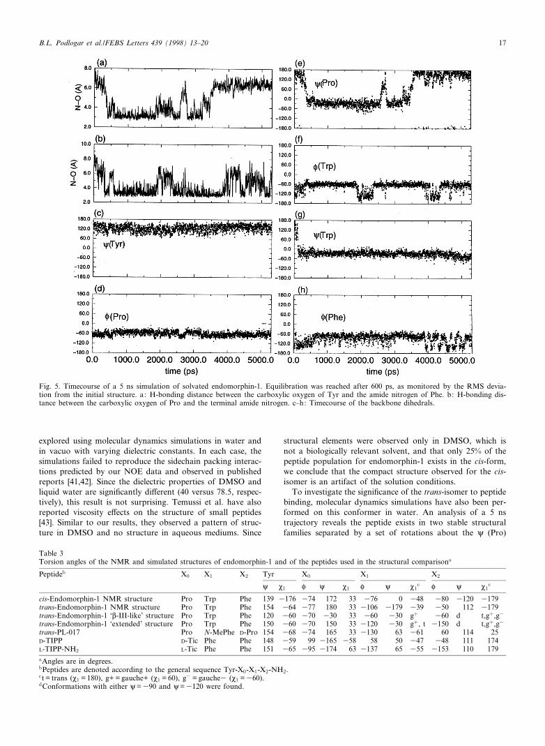
Fig. 5. Timecourse of a 5 ns simulation of solvated endomorphin-1. Equilibration was reached after 600 ps, as monitored by the RMS devia-tion from the initial structure. a: H-bonding distance between the carboxylic oxygen of Tyr and the amide nitrogen of Phe. b: H-bonding dis-tance between the carboxylic oxygen of Pro and the terminal amide nitrogen. c^h: Timecourse of the backbone dihedrals.
Table 3Torsion angles of the NMR and simulated structures of endomorphin-1 and of the peptides used in the structural comparisona
Peptideb X0 X1 X2 Tyr X0 X1 X2
i M1 P i M1 P i M1c P i M1
c
cis-Endomorphin-1 NMR structure Pro Trp Phe 139 3176 374 172 33 376 0 348 380 3120 3179trans-Endomorphin-1 NMR structure Pro Trp Phe 154 364 377 180 33 3106 3179 339 350 112 3179trans-Endomorphin-1 `L-III-like' structure Pro Trp Phe 120 360 370 330 33 360 330 g� 360 d t,g�,g3
trans-Endomorphin-1 `extended' structure Pro Trp Phe 150 360 370 150 33 3120 330 g�, t 3150 d t,g�,g3
trans-PL-017 Pro N-MePhe D-Pro 154 368 374 165 33 3130 63 361 60 114 25D-TIPP D-Tic Phe Phe 148 359 99 3165 358 58 50 347 348 111 174L-TIPP-NH2 L-Tic Phe Phe 151 365 395 3174 63 3137 65 355 3153 110 179aAngles are in degrees.bPeptides are denoted according to the general sequence Tyr-X0-X1-X2-NH2.ct = trans (M1 = 180), g+ = gauche+ (M1 = 60), g3 = gauche3 (M1 =360).dConformations with either i=390 and i=3120 were found.
B.L. Podlogar et al./FEBS Letters 439 (1998) 13^20 17
and P (Trp) dihedral angles (Fig. 5 and Table 3). One familyof states is similar to the L-III turn and is observed between0.5 and 1.8 ns and again between 2.5 and 3 ns. During theseintervals in time, Pro is in the K-conformation with hydrogenbonds occurring between the Tyr-carbonyl and the Phe-NHgroups. An additional hydrogen bond can also form betweenthe Pro-carbonyl and the terminal amine group in this com-pact structural form as shown in Fig. 5. After 3 ns, however,the peptide reverts to an extended form devoid of hydrogenbonds. This second conformational family is characterized byhaving relatively extended dihedral angles of 150³ and 3120³for i (Pro) and P (Trp), respectively.
Although our molecular dynamics time lengths are insu¤-cient to determine the statistical probability of these two fam-ilies occurring, the extended structure is consistent with theNOE data of the trans-isomer. In comparing the experimentaland calculated structures of trans-endomorphin-1, similarbackbone dihedral angles are evident, but di¡erences areseen in the sidechain conformations. While the NMR dataare compatible with either the Tyr sidechain packed againstthe Pro (M1V3150³) or away from Pro (M1V360³), Tyr M1
maintained a gauche (3) (M1 360³) throughout the simula-tion.
To settle this apparent inconsistency, we have capitalized onthe observation that many potent N, W and U opioid ligandspossess a tyramine (para-hydroxy phenethylamine) moietyburied within their molecular framework. In fact, the tyrosinering and the positively charged nitrogen are general require-ments for nearly all of the reported small molecule pharma-cophores [6]. Therefore, in our comparisons, we require thatthe tyramine moiety of the peptide ligands adopt the confor-mation as de¢ned by the small molecule ligands such as mor-phine.
Our use of the extended conformation of trans-endomor-phin-1 for the bioactive conformation becomes justi¢ed whenconsidering how it overlays with other selective W ligands, PL-017 and D-TIPP. PL-017, by virtue of its two proline rings andN-methyl amide, prohibits internal hydrogen bonding, is con-
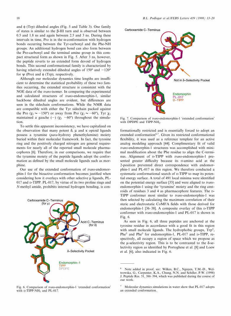
formationally restricted and is essentially forced to adopt anextended conformation6;7. Given its restricted conformational£exibility, it was used as a reference template for an activeanalog modeling approach [44]. Complimentary ¢t of validtrans-endomorphin-1 structures was accomplished with mini-mal modi¢cation about the Phe residue to align the C-termi-nus. Alignment of D-TIPP with trans-endomorphin-1 pre-sented greater di¤culty because its D-amino acid at the2-position prevented direct correspondence with endomor-phin-1 and PL-017 in this region. We therefore conducted asystematic conformational search of D-TIPP to map its poten-tial energy surface. A total of 491 local minima were identi¢edon the potential energy surface [35] and were aligned to trans-endomorphin-1 using the `tyramine' moiety and the ring cent-roids of residues 3 and 4 as pharmacophore features. The D-TIPP conformer most similar to trans-endomorphin-1 wasthen selected by calculating the maximum correlation of theirsteric and electrostatic CoMFA ¢elds with those derived forendomorphin-1 [36^38]. A composite overlay of this D-TIPPconformer with trans-endomorphin-1 and PL-017 is shown inFig. 6.
As seen in Fig. 6, all three peptides are anchored at thetyrosine residue in accordance with a good ¢t in this regionwith small molecule ligands. The hydrophobic groups, Trp3,Phe3 and Phe3 for endomorphin-1, PL-017 and D-TIPP, re-spectively, all occupy a region of space which we propose asthe W-selectivity region. This is to be contrasted to the N-se-lectivity region as identi¢ed by Portoghese et al. [8] and Loewet al. [6], also indicated in Fig. 6.
FEBS 20961 17-11-98 Cyaan Magenta Geel Zwart
Fig. 7. Comparison of trans-endomorphin-1 `extended conformation'with DPDPE and TIPP-NH2.
6 Note added in proof, see: Wilkes, B.C., Nguyen, T.M.-D., Wel-trowska, G., Carpenter, K.A., Chung, N.N. and Schiller, P.W. (1998)J. Peptide Res. 51, 386^394, which was published during the course ofour work.
7 Molecular dynamics simulations in water show that PL-017 adoptsan extended conformation.
Fig. 6. Comparison of trans-endomorphin-1 `extended conformation'with D-TIPP-NH2 and PL-017.
B.L. Podlogar et al./FEBS Letters 439 (1998) 13^2018
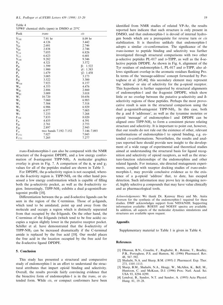
trans-Endomorphin-1 can also be compared with the NMRstructure of the N-agonist DPDPE, and a low energy confor-mation of N-antagonist TIPP-NH2. A molecular graphicsoverlay is given in Fig. 7. A comparison of the P, i and M1
values for all of the peptides studied are shown in Table 3.For DPDPE, the W-selectivity region is not occupied, where-
as the N-activity region is. TIPP-NH2 on the other hand pos-sessed a low energy conformation which is capable of ¢llingboth the W-selectivity pocket, as well as the N-selectivity re-gion. Interestingly, TIPP-NH2 exhibits a dual W-agonist/N-an-tagonist pro¢le [18].
Di¡erentiation between the W- and N-selective ligands can beseen in the region of the C-terminus. Those of W-ligands,which tend to be amidated, point up and away from themolecule and occupy a region which is distinctly separatedfrom that occupied by the N-ligands. On the other hand, theC-terminus of the N-ligands (which tend to be free acids) oc-cupies a region slightly lower in the putative receptor pocket.Schiller et al. have demonstrated that the N-selectivity ofTIPP-NH2 can be increased dramatically if the C-terminalamide is replaced by the free acid [18]. Our model placesthe free acid in the location occupied by the free acid forthe N-selective ligand DPDPE.
5. Conclusion
This study has presented a structural and comparativestudy of endomorphin-1 in an e¡ort to understand the struc-tural attributes that impart opioid binding and selectivity.Overall, the results provide fairly convincing evidence thatthe bioactive form of endomorphin-1 is in the trans, or ex-tended form. While cis, or compact conformers have been
identi¢ed from NMR studies of related YP-Ar, the resultsreported here indicate that such structure is only present inDMSO, and that endomorphin-1 is devoid of internal hydro-gen bonds which are a prerequisite for reverse turn or cisstabilization. It is therefore unlikely that endomorphin-1adopts a similar cis-conformation. The signi¢cance of thetrans-isomer to peptide binding and selectivity was furtherinvestigated through structural comparisons with two otherW-selective peptides PL-017 and D-TIPP, as well as the N-se-lective peptide DPDPE. As shown in Fig. 6, alignment of theTyr residues of endomorphin-1, PL-017 and D-TIPP, also al-lows signi¢cant overlap in the aromatic residues £anking Pro.In terms of the `message-address' concept forwarded by Por-toghese et al. [45,46], this secondary element may representthe `address' or site of selectivity for the W-opioid receptor.This hypothesis is further supported by structural alignmentsof endomorphin-1 and the N-agonist DPDPE, which showlittle or no overlap between the putative W-selectivity and N-selectivity regions of these peptides. Perhaps the most provo-cative result is seen in the structural comparison using thedual W-agonist/N-antagonist TIPP-NH2. In this case, boththe W and N `addresses', as well as the tyramine moieties oropioid `message' of endomorphin-1 and DPDPE can bealigned onto TIPP-NH2 to form a consistent picture relatingstructure and selectivity. It is important to point out, however,that our results do not rule out the existence of other, relevantconformations of endomorphin-1 to opioid binding, e.g. ex-tended cis-conformations. Nevertheless, the results and anal-yses reported here should provide new insight to the develop-ment of a wide range of experimental and theoretical studiesaimed at understanding the structural basis for ligand recog-nition and selectivity of opioid receptors, as well as the struc-ture-function relationships of the endomorphins and otherrelated ligands. For instance, site directed mutagenesis experi-ments, coupled with receptor docking studies of trans-endo-morphin-1, may provide conclusive evidence as to the exis-tence of a W-opioid `address' that, to date, has escapeddetection. Such information may prove key in designing nov-el, highly selective W compounds that may have value clinicallyand as pharmacological tools.
Acknowledgements: We thank Dr. Kenway Hoey and Ms. AnitaEverson for the synthesis of the endomorphin-1 required for thesestudies. DMF acknowledges support from NIDA/NIH. Supportinginformation available: ROESY and NOESY spectra are available.In addition, all aspects of the molecular dynamics simulations andstructures are available upon request.
Appendix
Supplementary material to Table 1 is given in Table 4.
References
[1] Dhawan, B.N., Cesselin, F., Raghubir, R., Reisine, T., Bradley,P.B., Portoghese, P.S. and Hamon, M. (1996) Pharmacol. Rev.48, 567^592.
[2] Shahabi, N.A. and Sharp, B.M. (1995) J. Pharmacol. Exp. Ther.273, 1105^1113.
[3] Sharp, B.M., Shahabi, N.A., Heagy, W., McAllen, K., Bell, M.,Huntoon, C. and McKean, D.J. (1996) Proc. Natl. Acad. Sci.USA 93, 8294^8299.
[4] Lendvai, B., Sandor, N.T. and Sandor, A. (1993) Acta Physiol.Hung. 81, 19^28.
FEBS 20961 17-11-98 Cyaan Magenta Geel Zwart
Table 4YPWF chemical shifts (ppm) in DMSO at 25³C
Peak trans cis
YNH 7.91 br 8.09 brYK 4.126 3.442YL1 2.681 2.746YL2 2.838 2.746Y2;6 7.055 6.826Y3;5 6.623 6.601YOH 9.282 9.346PK 4.321 3.572PL1 1.599 L : 1.522PL2 1.900 Q1: 1.383PQ 1.679 Q2: 1.458PN1 3.005 3.173PN2 3.522 3.300WNH 7.923 8.157WK 4.353 4.476WL1 2.886 2.860WL2 3.009 3.018W1 10.720 10.656W2 7.060 6.983W4 7.504 7.518W5 6.906 6.929W6 6.977 7.005W7 7.233 7.268FNH 7.833 8.020FK 4.435 4.391FL1 2.761 2.753FL2 2.897 2.943Far two bands 7.192^7.152 7.146^7.093FCT1 7.027 7.043FCT2 7.205 7.277
B.L. Podlogar et al./FEBS Letters 439 (1998) 13^20 19
[5] Suzuki, T., Mori, T., Tsuji, M., Misawa, M. and Nagase, H.(1997) Eur. J. Pharmacol. 331, 1^8, and references cited therein.
[6] Huang, P. and Loew, G. (1997) J. Comp.-Aided Mol. Des. 11,21^28.
[7] Fang, X., Larson, D.L. and Portoghese, P.S. (1997) J. Med.Chem. 40, 3064^3070.
[8] Portoghese, P.S. (1996) in: Perspective in Receptor Research(Giardina, D., Piergentili, A. and Pigini, M., Eds.), pp. 303^312, Elsevier Science, Amsterdam.
[9] Metzger, T.G. and Ferguson, D.M. (1995) FEBS Lett. 375, 1^4.[10] Varga, E.V., Li, X., Stropova, D., Zalewska, T., Landsman, R.S.,
Knapp, R.J., Malatynska, E., Kawai, K., Mizusura, A., Nagase,H., Calderon, S.N., Rice, K., Hruby, V.J., Roeske, W.R. andYamamura, H.I. (1996) Mol. Pharmacol. 50, 1619^1624.
[11] Hruby, V.J., Koa, L.-F., Pettitt, B.M. and Karplus, M. (1988)J. Am. Chem. Soc. 110, 3351^3359.
[12] Lomize, A.L., Pogozheva, I.D. and Mosberg, H.I. (1996) Biopol-ymers 38, 221^234.
[13] Paterlini, G., Portoghese, P.S. and Ferguson, D.M. (1997) J. Med.Chem. 40, 3263^3270.
[14] Tessmer, M.R., Meyer, J.-P., Hruby, V.J. and Kallick, D.A.(1997) J. Med. Chem. 40, 2148^2155.
[15] Flippen-Anderson, J.L., Deschamps, J.R., George, C., Reddy,P.A., Lewin, A.H., Brine, G.A., Sheldrick, G. and Nikiforovich,G. (1997) J. Peptide Res. 49, 384^393.
[16] Zadina, J.E., Hackler, L., Ge, L.-J. and Kastin, A.J. (1997) Na-ture 383, 499^502.
[17] Chang, K.J., Killian, A., Hazum, E., Cuatrecasas, P. and Chang,J.K. (1981) Science 212, 75^77.
[18] Schiller, P.W., Nguyen, T.M.-D., Weltrowska, G., Wilkes, B.C.,Marsden, B.J., Lemieux, C. and Chung, N.N. (1992) Proc. Natl.Acad. Sci. USA 89, 11871^11875.
[19] Shaka, A.J., Lee, C.J. and Pines, A. (1988) J. Magn. Reson. 77,274^293.
[20] Bax, A. and Davis, D. (1985) J. Magn. Reson. 65, 355^360.[21] van Zijl, P.C.M., O'Neil-Johnson, M., Mori, S. and Hurd, R.E.
(1995) J. Magn. Reson. A 113, 265^270.[22] Sklenar, V., Piotto, M., Leppik, R. and Saudek, V. (1993)
J. Magn. Reson. A 102, 241^245.[23] Jeener, J., Meier, B.H., Bachmann, P. and Ernst, E.E.J. (1979)
Chem. Phys. 71, 4546^4556.[24] Bothner-By, A.A., Stephens, B.L., Lee, J., Warren, C.D. and
Jeanloz, R.W. (1984) J. Am. Chem. Soc. 106, 811^813.[25] Desvaux, H., Berthault, P., Birlirakis, N., Goldman, M. and
Piotto, M. (1995) J. Magn. Reson. A 113, 47^52.[26] Braunschweiler, L. and Ernst, R.R. (1983) Magn. Reson. 53,
521^528.[27] Marion, D., Ikura, M., Tschudin, R. and Bax, A. (1985) J. Magn.
Reson. 85, 393^399.[28] Gasteiger, J. and Marsili, M. (1980) Tetrahedron 36, 3219^3228.
[29] Marsili, M. and Gasteiger, J. (1980) Croat. Chem. Acta 53, 601^614.
[30] Gasteiger, J. and Marsili, M. (1981) Organ. Magn. Reson. 15,353^360.
[31] Streitwieser, A. (1961) Molecular Orbital Theory for OrganicChemists, Wiley, New York.
[32] Purcel, W.P. and Singer, J.A. (1967) J. Chem. Eng. Data 12, 235^246.
[33] Clark, M., Cramer III, R.D. and Van Opdenbosch, N. (1989)J. Comp. Chem. 10, 982^1012.
[34] Sybyl 6.0 Theory Manual, Appendix 2.2, pp. 2421^2423, October1992.
[35] Demeter, D.A. (1994) Development and Application of PotentialEnergy Surface Analysis and the Local Minima Method of Phar-macophore Determiniation, Avail. Univ. Micro¢lms Int., OrderNo. DA9502554., 361 pp. From: Diss. Abstr. Int. B 1995, 55,3896.
[36] Cramer III, R.D., Patterson, D.E. and Bunce, J.D. (1988) J. Am.Chem. Soc. 110, 5959^5967.
[37] Marshall, G. and Cramer III, R.D. (1988) Trends Pharmacol.Sci., 9, 285^289.
[38] Allen, M.S., Yan, Y.C., Trudell, M.L., Narayanan, K., Schin-dler, L.R., Martin, M.J., Schultz, C., Hagen, T.J., Koehler, K.F.,Codding, P.W., Skolnick, P. and Cook, J.M. (1990) J. Med.Chem. 33, 2343^2357.
[39] Case, D.A., Pearlman, D.A., Caldwell, J.W., Cheatham, T.E.,Ross, W.S., Simmerling, C.L., Darden, T.A., Merz, K.M., Stan-ton, R.V., Cheng, A.L., Vincent, J.J., Crowley, M., Ferguson,D.M., Radmer, R.J., Seibel, G.L., Singh, U.C., Weiner, P.K.and Kollman, P.A. (1997) AMBER 5, University of California,San Fransisco, CA.
[40] Wuëthrich, K. (1986) NMR of Proteins and Nucleic Acids, JohnWiley and Sons, New York.
[41] Yao, J., Dyson, J. and Wright, P.E. (1994) J. Mol. Biol. 243,754^766.
[42] Roberts, V.A., Nachman, R.J., Coast, G.M., Hariharan, M.,Chung, J.S., Holman, G.M., Williams, H. and Tainor, J.A.(1997) Curr. Biol. 4, 105^117.
[43] Temussi, P.A., Picone, D., Saviano, G., Amodeo, P., Motta, A.,Tancredi, T., Salvadori, S. and Tomatis, R. (1992) Biopolymers32, 367^372.
[44] Marshall, G.R., Barry, B.E., Bosshard, H.E., Dammkoehler,R.A. and Dunn, D.A. (1979) in: Computer Assisted Drug Design(Olson, E.C., Christo¡erson, R.E., Eds.), Vol. 112, pp. 205^226,ACS Symposium Series, Washington, DC.
[45] Takemori, A.E. and Portoghese, P.S. (1992) Annu. Rev. Phar-macol. Toxicol. 32, 239^269.
[46] Metzger, T.G., Paterlini, M.G., Portoghese, P.S. and Ferguson,D.M. (1996) Neurochem. Res. 21, 1287^1294.
FEBS 20961 17-11-98 Cyaan Magenta Geel Zwart
B.L. Podlogar et al./FEBS Letters 439 (1998) 13^2020