conformational changes of rbti from buckwheat upon...
TRANSCRIPT

Conformational Changes of rBTI from Buckwheat uponBinding to Trypsin: Implications for the Role of the P89Residue in the Potato Inhibitor I FamilyLongfei Wang1,2, Fei Zhao3, Mei Li1, Hongmei Zhang1, Yu Gao1, Peng Cao1, Xiaowei Pan1, Zhuanhua
Wang3*, Wenrui Chang1*
1 National Laboratory of Biomacromolecules, Institute of Biophysics, Chinese Academy of Sciences, Beijing, People’s Republic of China, 2 Graduate University of the
Chinese Academy of Sciences, Beijing, People’s Republic of China, 3 Key Laboratory of Chemical Biology and Molecular Engineering of the Ministry of Education, Institute
of Biotechnology, Shanxi University, Taiyuan, People’s Republic of China
Abstract
BWI-1 (buckwheat trypsin inhibitor), a member of the potato inhibitor I family, suppresses the growth of T-acutelymphoblastic leukemia cells and induces apoptosis in human solid tumor cell lines. Here, we report the crystal structureof rBTI (recombinant buckwheat trypsin inhibitor), a recombinant protein of BWI-1, at 1.84 A resolution and the structureof rBTI in complex with bovine trypsin at 2.26 A resolution. A conformational change of Trp53 at the P89 position in rBTIwas observed upon its binding to trypsin, which is not seen in other members of the potato inhibitor I family reportedpreviously. The role of the P89 residue in the potato inhibitor I family was examined by measuring the association anddissociation rates of four rBTI mutants with different substitutions at the P2 and P89 positions when binding to trypsin.One of the mutants, P44T, was found to be a much stronger inhibitor than wild-type rBTI, with a picomolar (pM)dissociation constant. Our results could provide valuable insights for designing a new rBTI-based antitumor drug in thefuture.
Citation: Wang L, Zhao F, Li M, Zhang H, Gao Y, et al. (2011) Conformational Changes of rBTI from Buckwheat upon Binding to Trypsin: Implications for the Roleof the P89 Residue in the Potato Inhibitor I Family. PLoS ONE 6(6): e20950. doi:10.1371/journal.pone.0020950
Editor: Haiwei Song, Institute of Molecular and Cell Biology, Singapore
Received April 4, 2011; Accepted May 13, 2011; Published June 15, 2011
Copyright: � 2011 Wang et al. This is an open-access article distributed under the terms of the Creative Commons Attribution License, which permitsunrestricted use, distribution, and reproduction in any medium, provided the original author and source are credited.
Funding: This work was supported by the 973 Project (Grant No. 2011CBA00902), the National Natural Science Foundation of China (Grant No. 31021062) andthe Knowledge Innovation Program of the Chinese Academy of Sciences (Grant No. KSCX2-YW-R-123 and KSCX2-EW-J-3). The funders had no role in study design,data collection and analysis, decision to publish, or preparation of the manuscript.
Competing Interests: The authors have declared that no competing interests exist.
* E-mail: [email protected] (WC); [email protected] (ZW)
Introduction
Canonical inhibitors of serine protease function according to the
standard mechanism of protease inhibition in which they bind
tightly in the active site of a cognate protease in a substrate-like
manner (substrate residues of protease inhibitors surrounding the
cleavage site are designated by the nomenclature of Schechter and
Berger [1]. The scissile bond is the starting point. In the direction
of the N terminus, substrate residues are numbered P1, P2, P3 and
so on, and in the direction of the C terminus, residues are num-
bered P19, P29, P39 and so on.) [2]. However, unlike substrates,
canonical inhibitors cannot be easily hydrolyzed by proteases,
which is attributed to the rigidity of their convex binding loop [3].
The protein core of a canonical inhibitor serves as a scaffold for
the binding loop and is responsible for maintaining the binding
loop stability. A previous study revealed that an inhibitor could
quickly form an acyl-enzyme intermediate with a protease but was
hydrolyzed very slowly. Thus, a clogged gutter mechanism was
proposed to underscore two key factors in protease inhibition: the
intramolecular hydrogen-bonding network and the correct orien-
tation of the religating amide [4].
The potato inhibitor I family belongs to the canonical inhibitors,
and their P2, P19, P69, and P89 residues are highly conserved due to
their importance in the formation of the internal hydrogen-bonding
network between the binding loop and protein core. Mutations of
either P2 Thr or P19 Glu in CI-2 (chymotrypsin inhibitor 2) result in
a dramatic increase of the dissociation constant between CI-2 and
chymotrypsin [5]. P69 and P89 mutants of CMTI-V (cucurbita
maxima trypsin inhibitor V) have been proven to be very unstable.
The P69 mutant, in particular, can be easily hydrolyzed by trypsin
[6].
Recently, attentions have been drawn to another member of the
potato inhibitor I family from buckwheat seeds, BWI-1 (Buck-
wheat Inhibitor 1). BWI-1 was sequenced and characterized in
buckwheat seeds soon after its discovery [7,8,9]. A previous cyto-
biology study revealed that BWI exhibits suppression activity
against human T-Acute lymphoblastic leukemia cell lines [10]. In
the past few years, Wang and her colleagues has focused on the
antitumor activity of the BWI-1 recombinant protein rBTI
(recombinant buckwheat trypsin inhibitor) [11] and has investi-
gated its effects on the induction of apoptosis in several human
solid tumor cell lines (EC907, HepG2 and HeLa) [12]. Addi-
tionally, the resistance of tobacco and potatoes to biotic stress can
be improved by introducing the BWI-1 encoding gene [13].
Interestingly, BWI-1 has an uncommon binding loop sequence
with a Pro at the P2 position and Trp at the P89 position, sugge-
sting a unique mode of intramolecular interactions between the
binding loop and the protein core. Because the inhibition acti-
PLoS ONE | www.plosone.org 1 June 2011 | Volume 6 | Issue 6 | e20950

vity of certain canonical inhibitors is strongly affected by their
intramolecular hydrogen-bonding network [4], it is logical to
propose that BWI-1 inhibits proteases in an unusual way.
Here, we report the crystal structure of rBTI at 1.84 A reso-
lution and the structure of rBTI-trypsin complex at 2.26 A reso-
lution. Curiously, structural superposition revealed a significant
conformational change of P89 Trp in rBTI upon binding to
trypsin. Several rBTI mutants were constructed to mimic different
binding loop conformations of potato inhibitor I family members.
Their association and dissociation rates upon binding to bovine
trypsin were determined, allowing us to correlate several binding
loop conformations with their inhibition abilities in the potato
inhibitor I family. Out of our expectations, one of the mutants,
P44T, was found to be a much stronger inhibitor compared to
the wild-type with a picomolar (pM) dissociation constant. These
results allow us to propose a detailed model for the structural basis
of protease inhibition of the potato Inhibitor I family.
Results and Discussion
Overall structure of native rBTI and its complex withbovine trypsin
The structure of rBTI is composed of 69 amino acid residues.
Its main structural elements comprise a single a-helix (a1, residues
18 to 28), a central parallel b-sheet consisting of two strands (b1,
residues 30 to 38; b2, residues 51 to 56), a binding loop (residues
39 to 50) and two irregular structures at the N-terminus (residues 3
to 15) and C-terminus (residues 61 to 69)(Figure 1A). A hydro-
phobic core is formed in rBTI among a1, b1, b2 and two short
loops (side-chains of Trp10, Ile25, Val32, Val52 and Pro66), as
Figure 1. (A) Cartoon representation of the overall structure of rBTI. Different structural elements are shown in different colors, and the disulfidebridge is indicated. (B) A view of the hydrogen-bonding network and the hydrophobic core in rBTI. rBTI is shown in a cartoon presentation in green.Residues involved in hydrogen-bonding network and hydrophobic core are shown as ball-and-stick models. The grey sphere indicates a watermolecule. Hydrogen bonds are indicated by black dashes and hydrophobic interactions are indicated by dotted clouds. (C) Overview of the structureof rBTI-trypsin complexes within an asymmetric unit. rBTIs are shown in yellow and magenta; trypsins are shown in green and cyan. The calcium ionsin trypsin are shown as light-blue spheres. (D) Superposition of trypsin-bound rBTI and free rBTI. The binding loop of trypsin-bound rBTI (cyan) isshifted by a small distance from that of free rBTI (grey). The RMSD value calculated by superposition of trypsin-bound rBTI’s and free rBTI’s bindingloops is 0.26 A.doi:10.1371/journal.pone.0020950.g001
Structures of rBTI and rBTI-Trypsin Complex
PLoS ONE | www.plosone.org 2 June 2011 | Volume 6 | Issue 6 | e20950
shown in Figure 1B. The Cys4-Cys49 disulfide bond stabilizes the
binding loop by connecting it with the N-terminus. The binding
loop of rBTI is a convex loop sandwiched between b1 and b2.
Within the loop, the P1 residue, Arg45, is an ideal substrate of
trypsin, which ensures the inhibitor’s tight binding to trypsin. As
in other canonical inhibitors, the binding loop of rBTI forms a
hydrogen-bonding network with the protein core [6,14,15]
(Figure 1B). This hydrogen-bonding network is one of the key fac-
tors that causes inhibitors to be hydrolyzed at a very slow rate [3].
In the crystal structure of the rBTI-trypsin complex, one cry-
stallographic asymmetric unit contains two rBTI-trypsin complex-
es, that is, two rBTIs and two trypsins, as shown in Figure 1C. The
structure of bovine trypsin in complex with rBTI aligns well with
other trypsin structures deposited in the PDB database. Trypsin-
bound rBTI has an overall structure similar to that of free rBTI,
with both consisting of one a-helix and a central parallel b-sheet.
By superposing trypsin-bound rBTI over free rBTI, we found that
the Arg45 at the P1 position was buried deeply into the binding
pocket of trypsin, leading to a small but noticeable shift of the
binding loop towards trypsin (RMSD 0.26 A, Figure 1D). This
movement disrupts several hydrogen bonds between the binding
loop and protein core, indicating that a significant conformational
change of the binding loop occurs upon binding to trypsin.
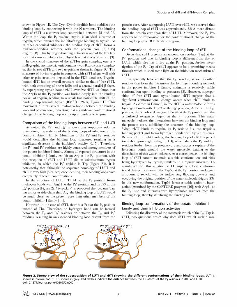
Comparison of the binding loops between rBTI and LUTIAs noted, the P69 and P89 residues play important roles in
maintaining the stability of the binding loops of inhibitors in the
potato inhibitor I family. Mutations of the P69 and P89 residues
would destabilize the binding loop structure, resulting in a
significant decrease in the inhibitor’s activity [6,15]. Therefore,
the P69 and P89 residues are highly conserved among members of
the potato inhibitor I family. Almost all reported structures in the
potato inhibitor I family exhibit an Arg at the P89 position, with
the exception of rBTI and LUTI (linum usitatissimum trypsin
inhibitor), in which the P89 residue is Trp (Figure S1). It is
noteworthy that although the sequence homology of LUTI and
rBTI is very high (58% sequence identity), their binding loops have
completely different conformations.
In the structure of LUTI, Thr44 at the P2 position forms
hydrogen bonds with Arg51 at the P69 position and Trp53 at the
P89 position (Figure 2). Cierpicki et al. proposed that because Trp
has a shorter side-chain than Arg, the binding loop of LUTI would
be much closer to the protein core than other members of the
potato inhibitor I family [14].
However, in the case of rBTI, there is a Pro at the P2 position
instead of Thr. Therefore, no hydrogen bond can be formed
between the P2 and P89 residues or between the P2 and P69
residues, resulting in an extended binding loop distant from the
protein core. After superposing LUTI over rBTI, we observed that
the binding loop of rBTI was approximately 3.5 A more distant
from the protein core than that of LUTI. Moreover, the P2 Pro
appears to be responsible for the conformational change of the
binding loop after rBTI binds to trypsin.
Conformational change of the binding loop of rBTIGiven that rBTI presents an uncommon residues (Trp) at the
P89 position and that its binding loop is different from that of
LUTI, which also has a Trp at the P89 position, further inves-
tigation of the P89 Trp of rBTI appears to be a promising means
through which to shed some light on the inhibition mechanism of
rBTI.
It is generally believed that the P89 residue, as well as other
residues that form the intramolecular hydrogen-bonding network
in the potato inhibitor I family, maintains a relatively stable
conformation upon binding to proteases [3]. However, superpo-
sition of free rBTI and trypsin-bound rBTI suggests that a
significant conformational change occurs when rBTI binds to
trypsin. As shown in Figure 3, in free rBTI, a water molecule forms
hydrogen bonds with Trp53 at the P89 position, Arg51 at the P69
position, the a carbonyl oxygen of Pro44 at the P2 position and the
a carbonyl oxygen of Asp46 at the P19 position. This water
molecule mediates the interactions between the binding loop and
the protein core, stabilizing the structure of the binding loop.
When rBTI binds to trypsin, its P1 residue fits into trypsin’s
binding pocket and forms hydrogen bonds with trypsin residues.
Because of this tight binding, the binding loop of rBTI is pulled
towards trypsin slightly (Figure 1D), which shifts the P2 and P19
residues further from the protein core and causes a rupture of the
hydrogen bonds around the water molecule, leading to the
dissociation of this water molecule. As a consequence, the binding
loop of rBTI cannot maintain a stable conformation and risks
being hydrolyzed by trypsin, similarly to a regular substrate. To
counteract with this situation, rBTI employs a local conforma-
tional change mechanism: the Trp53 at the P89 position undergoes
a rotameric switch, with its indole ring flipping upwards and
occupying the original position of the water molecule (Figure S3).
In this new conformation, Trp53 forms a stable cation-p inter-
action (examined by the CaPTURE program [16]) with Arg51 at
the P69 site and interacts with hydrophobic residues from the
binding loop, thereby stabilizing the binding loop.
Binding loop conformations of the potato inhibitor Ifamily and their inhibition activities
Following the discovery of the rotameric switch of the P89 Trp in
rBTI, two questions arose: why does rBTI exhibit such a rare
Figure 2. Stereo view of the superposition of LUTI and rBTI showing the different conformations of their binding loops. LUTI isshown in brown, and rBTI is shown in grey. Red dashes indicate the distance between the Ca atoms of the P1 residues in rBTI and LUTI.doi:10.1371/journal.pone.0020950.g002
Structures of rBTI and rBTI-Trypsin Complex
PLoS ONE | www.plosone.org 3 June 2011 | Volume 6 | Issue 6 | e20950

inhibition mechanism; and what roles do the P89 and P2 residues
play in its inhibition activity? To address these questions, we
referred to the structure of several classical members of the potato
inhibitor I family and designed four rBTI mutants. These mutants
each include substitutions at the P2 and P89 positions to mimic
different binding loop conformations of the potato inhibitor I
family members. Interactions of wild-type rBTI and rBTI mutants
with bovine trypsin were investigated by means of an optical
biosensor using the surface plasmon resonance (SPR) effect. Their
association rate (ka) and dissociation rate (kd) and the dissociation
constant (KD) were determined (Table 1, Figure S2).
Both rBTI mutants with an Arg substitution at the P89 position
(W53R, W53R/P44T) show elevated association rates compared
to the wild-type rBTI for tryspin. As proposed previously, the
closer packing of the binding loop of LUTI against the protein
core compared to CI-2 and eglin C is attributed to the shorter
side-chain of Trp compared to Arg [14]. We speculated that it is
the same in rBTI: because Arg has a longer side-chain than Trp or
Phe, mutants with Arg at the P89 position (W53R, W53R/P44T)
exhibit more extended binding loops than other forms of rBTI
(wild-type rBTI, P44T, W53F) (Figure 4A). Thus, the binding
loops of W53R and W53R/P44T are more accessible to the
substrate pocket of trypsin, resulting in elevated association rates.
As noted earlier, for wild-type rBTI, a water molecule mediates
the interaction between the binding loop and protein core,
suggesting that the binding loop of wild-type rBTI is also extended.
This could explain why rBTI exhibits a third higher association
rate among the wild-type rBTI and all rBTI mutants.
We also found that mutants that are able to form hydrogen
bonds between the P2 and P89 residues (P44T, W53R/P44T)
present significantly lower dissociation rates compared to wild-type
for trypsin. The most surprising outcome from our kinetic analyses
Figure 3. Structural differences between free rBTI and trypsin-bound rBTI at the local region around the P89 position. (A) Interactionsbetween the P2 and P89 residues in free rBTI. (B) Interactions between the P2 and P89 residues in trypsin-bound rBTI. Side-chains of residues in thebinding loop are shown in green, while those in the protein core are shown in cyan. The grey sphere indicates a water molecule. Trypsin is shownas surface. Hydrophobic interactions are indicated by dotted clouds. Part of the side chain of Arg45 in free rBTI is missing due to poor electrondensities.doi:10.1371/journal.pone.0020950.g003
Table 1. Association rates (ka), dissociation rates (kd) and dissociation constants (KD) for the interactions of wild-type rBTI and itsmutants with bovine trypsin.
Inhibitor P89 residue P2 residue Speculated interaction Reference Model ka, M21s21 kd, s21 KD(kd/ka), M
WT rBTI Trp53 Pro44 Hydrophobic force rBTI 4.626105 1.2561023 2.6961029
P44T Trp53 Thr44 Hydrogen bond LUTI 3.466105 5.2461027 1.52610212
W53R/P44T Arg53 Thr44 Hydrogen bond CI-2 6.796105 5.5461024 8.15610210
W53F Phe53 Pro44 Hydrophobic force - 3.996105 2.4661023 6.1661029
W53R Arg53 Pro44 - - 6.596105 9.9761023 1.5161028
doi:10.1371/journal.pone.0020950.t001
Structures of rBTI and rBTI-Trypsin Complex
PLoS ONE | www.plosone.org 4 June 2011 | Volume 6 | Issue 6 | e20950
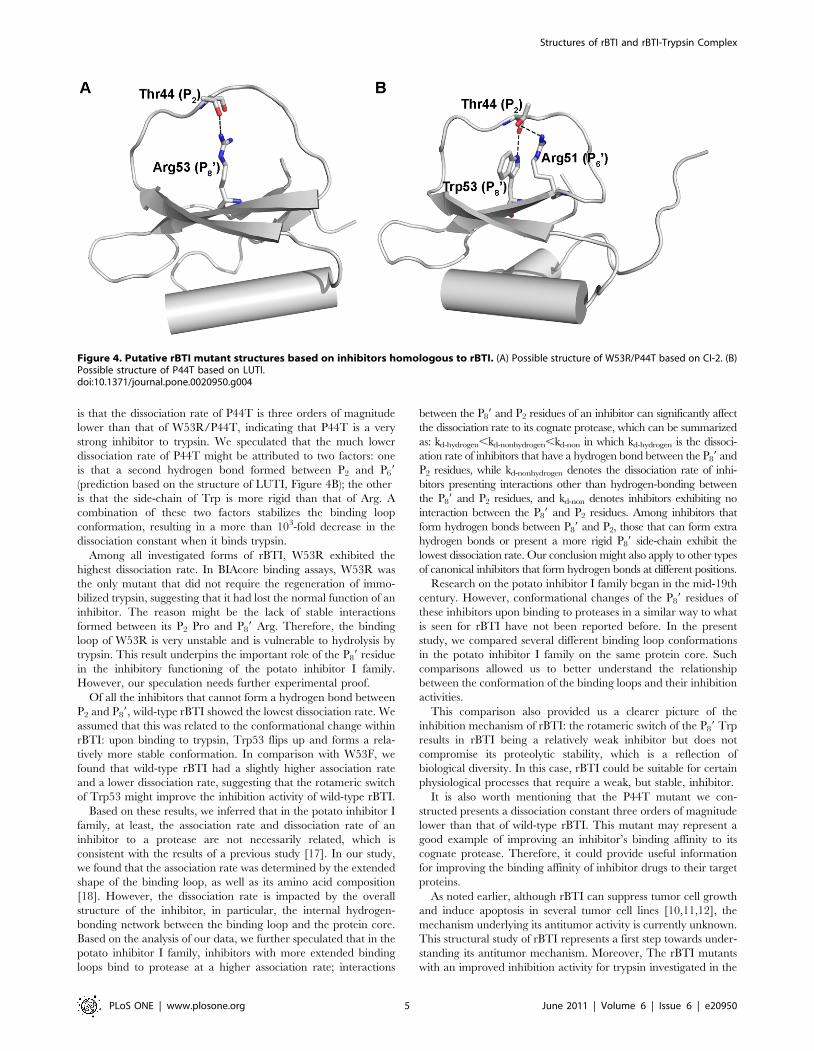
is that the dissociation rate of P44T is three orders of magnitude
lower than that of W53R/P44T, indicating that P44T is a very
strong inhibitor to trypsin. We speculated that the much lower
dissociation rate of P44T might be attributed to two factors: one
is that a second hydrogen bond formed between P2 and P69
(prediction based on the structure of LUTI, Figure 4B); the other
is that the side-chain of Trp is more rigid than that of Arg. A
combination of these two factors stabilizes the binding loop
conformation, resulting in a more than 103-fold decrease in the
dissociation constant when it binds trypsin.
Among all investigated forms of rBTI, W53R exhibited the
highest dissociation rate. In BIAcore binding assays, W53R was
the only mutant that did not require the regeneration of immo-
bilized trypsin, suggesting that it had lost the normal function of an
inhibitor. The reason might be the lack of stable interactions
formed between its P2 Pro and P89 Arg. Therefore, the binding
loop of W53R is very unstable and is vulnerable to hydrolysis by
trypsin. This result underpins the important role of the P89 residue
in the inhibitory functioning of the potato inhibitor I family.
However, our speculation needs further experimental proof.
Of all the inhibitors that cannot form a hydrogen bond between
P2 and P89, wild-type rBTI showed the lowest dissociation rate. We
assumed that this was related to the conformational change within
rBTI: upon binding to trypsin, Trp53 flips up and forms a rela-
tively more stable conformation. In comparison with W53F, we
found that wild-type rBTI had a slightly higher association rate
and a lower dissociation rate, suggesting that the rotameric switch
of Trp53 might improve the inhibition activity of wild-type rBTI.
Based on these results, we inferred that in the potato inhibitor I
family, at least, the association rate and dissociation rate of an
inhibitor to a protease are not necessarily related, which is
consistent with the results of a previous study [17]. In our study,
we found that the association rate was determined by the extended
shape of the binding loop, as well as its amino acid composition
[18]. However, the dissociation rate is impacted by the overall
structure of the inhibitor, in particular, the internal hydrogen-
bonding network between the binding loop and the protein core.
Based on the analysis of our data, we further speculated that in the
potato inhibitor I family, inhibitors with more extended binding
loops bind to protease at a higher association rate; interactions
between the P89 and P2 residues of an inhibitor can significantly affect
the dissociation rate to its cognate protease, which can be summarized
as: kd-hydrogen,kd-nonhydrogen,kd-non in which kd-hydrogen is the dissoci-
ation rate of inhibitors that have a hydrogen bond between the P89 and
P2 residues, while kd-nonhydrogen denotes the dissociation rate of inhi-
bitors presenting interactions other than hydrogen-bonding between
the P89 and P2 residues, and kd-non denotes inhibitors exhibiting no
interaction between the P89 and P2 residues. Among inhibitors that
form hydrogen bonds between P89 and P2, those that can form extra
hydrogen bonds or present a more rigid P89 side-chain exhibit the
lowest dissociation rate. Our conclusion might also apply to other types
of canonical inhibitors that form hydrogen bonds at different positions.
Research on the potato inhibitor I family began in the mid-19th
century. However, conformational changes of the P89 residues of
these inhibitors upon binding to proteases in a similar way to what
is seen for rBTI have not been reported before. In the present
study, we compared several different binding loop conformations
in the potato inhibitor I family on the same protein core. Such
comparisons allowed us to better understand the relationship
between the conformation of the binding loops and their inhibition
activities.
This comparison also provided us a clearer picture of the
inhibition mechanism of rBTI: the rotameric switch of the P89 Trp
results in rBTI being a relatively weak inhibitor but does not
compromise its proteolytic stability, which is a reflection of
biological diversity. In this case, rBTI could be suitable for certain
physiological processes that require a weak, but stable, inhibitor.
It is also worth mentioning that the P44T mutant we con-
structed presents a dissociation constant three orders of magnitude
lower than that of wild-type rBTI. This mutant may represent a
good example of improving an inhibitor’s binding affinity to its
cognate protease. Therefore, it could provide useful information
for improving the binding affinity of inhibitor drugs to their target
proteins.
As noted earlier, although rBTI can suppress tumor cell growth
and induce apoptosis in several tumor cell lines [10,11,12], the
mechanism underlying its antitumor activity is currently unknown.
This structural study of rBTI represents a first step towards under-
standing its antitumor mechanism. Moreover, The rBTI mutants
with an improved inhibition activity for trypsin investigated in the
Figure 4. Putative rBTI mutant structures based on inhibitors homologous to rBTI. (A) Possible structure of W53R/P44T based on CI-2. (B)Possible structure of P44T based on LUTI.doi:10.1371/journal.pone.0020950.g004
Structures of rBTI and rBTI-Trypsin Complex
PLoS ONE | www.plosone.org 5 June 2011 | Volume 6 | Issue 6 | e20950

present study may facilitate designing inhibitors with higher
antitumor activities and are of potential therapeutic value.
Materials and Methods
Expression, purification and crystallization of rBTI and therBTI-trypsin complex
rBTI was prepared as described previously [11]. The rBTI
crude sample was then applied to Superdex75 (GE Healthcare).
The elution buffer used was 25 mM Tris-HCl (pH 8.0) and
50 mM NaCl. The fractions containing rBTI were collected and
concentrated to 20 mg/ml. rBTI was crystallized by vapor
diffusion. Crystals were grown at 18uC in hanging drops over a
reservoir of 24% (w/v) PEG MME2000, 220 mM (NH4)2SO4,
100 mM NaAc (pH 4.4) and 100 mM NaI. Drops were prepared
by mixing equal volumes of protein and reservoir solutions. After
two weeks, thin and rod-like crystals were harvested, soaked in a
cryoprotectant mixture (paraffin oil and NVH oil in a ratio of 7:3)
and flash-frozen in liquid nitrogen.
rBTI and bovine trypsin (AppliChem) were mixed in a 1:1.3
stoichiometric molar ratio and incubated at room temperature
for a half an hour to form complexes. The incubation buffer
contained 50 mM Tris-HCl (pH 8.0) and 200 mM NaCl. After
incubation, the incubation sample was applied to Superdex75 to
remove excessive rBTI. The elution buffer used was 50 mM Tris
pH 8.0 and 20 mM NaCl. The complex of rBTI with bovine
trypsin was also crystallized by vapor diffusion. Crystals were
grown at 18uC in hanging drops over a reservoir of 15% (w/v)
PEG3350, 200 mM MgCl2 and 100 mM Tris-HCl (pH 9.0).
Drops were prepared by mixing equal volumes of protein and
reservoir solutions. Rod-like crystals grew over the course of two
weeks. They were then harvested, soaked in a cryoprotectant
solution (100 mM Tris-HCl pH 9.0, 20% (w/v) PEG3350, 20%
(v/v) glycerol and 200 mM MgCl2) and flash-frozen in liquid
nitrogen.
X-ray data collection and processingFor rBTI, synchrotron X-ray data were collected from a single
crystal at 100 K using a MAR555 CCD detector at beamline
1W2B, BSRF (Beijing Synchrotron Radiation Facility). For rBTI
in complex with bovine trypsin, X-ray data were collected from a
single crystal at 100 K using a Raxis4 IP detector at the Institute of
Microbiology, Chinese Academy of Science. All data were
processed and scaled with the HKL2000 software suite [19].
PhasingThe rBTI-trypsin complex was the first to yield diffraction
quality crystals. An incomplete structure containing only trypsin
was solved by molecular replacement using the program Phaser
[20]. The search model was derived from a previous structure of
trypsin (PDB entry 2CMY). After failed attempts at molecular
replacement using two search models (LUTI, linum usitatissimum
trypsin inhibitor, PDB entry 1DWM; CMTI-V, cucurbita maxima
trypsin inhibitor-V, PDB entry 1 HYM) that share the highest
sequence homology with rBTI, we used MrBump [21] to perform
a search for homologous structures. Then, one of the two rBTIs in
the asymmetric unit was solved using a search model of BGTI
(Bitter Gourd Trypsin Inhibitor, PDB entry 1VBW). The other
rBTI was solved by superposing one rBTI-trypsin complex over
the other trypsin using trypsin as the reference structure in the
asymmetric unit.
The rBTI structure was solved by molecular replacement using
the program Phaser [20] with the solved trypsin-bound rBTI
structure as a search model.
Model RefinementModels were rebuilt using the model-building module of the
PHENIX software suite [22]. Cycles of manual rebuilding in
COOT [23] were alternated with automated refinement using the
refinement module of PHENIX. Test sets comprised 5% of the
total reflections were excluded from refinement to allow the
calculation of the free R-factors. Composite omit maps generated
by the CNS software suite [24] and prime and switch maps
generated by Resolve [25] were used as reference maps in manual
rebuilding. Model validations were carried out using PRO-
CHECK [26]. Superpositions were performed using COOT, and
all figures representing structures were created using the gra-
phics software PyMOL. A summary of the data collection and
refinement statistics is presented in Table 2. The coordinates and
structure factors of rBTI were deposited into RCSB Protein Data
Bank with accession code 3RDY. The coordinates and structure
factors of rBTI-trypsin complex were deposited with accession
code 3RDZ.
Expression and purification of the mutantsThe rBTI expression construct was mutagenized using the
PCR-based QuickChange method (Stratagene). We designed four
mutants based on the structure of the homologous inhibitors of
rBTI. They were P44T, W53F, W53R and W53R/P44T double
mutant. The mutants of rBTI were expressed and purified in the
same way as wild-type rBTI.
Table 2. Summary of Data Collection and RefinementStatistics.
rBTI-trypsin complex rBTI
Wavelength (A) 1.5418 1.00
Space group P21 P43212
Resolution rangea (A) 12.022.26(2.3422.26) 15.021.84(1.9121.84)
Unique Reflections 26279 8344
Unit Cell (a,b,c) (A) 66.7, 50.2, 84.5 62.7,62.7,45.9
Completenessa (%) 99.8(97.7) 99.8(100)
Redundencya 3.7(3.6) 24.1(23.2)
Averageab I/s 22.6(4.9) 46.4(4.7)
Rmergea (%) 5.5(25.3) 8.0(50)
a.s.u content
No. trypsin 2 -
No. rBTI 2 1
No. Non-hydrogen atoms 4247 494
No. Ca2+ 2 -
No. water molecules 300 94
R factor and Rfree (%) 18.2/22.6 19.1/21.6
r.m.s deviations:
Bond length (A) 0.0075 0.012
Bond angles (deg) 1.116 1.390
B-factors (A2):
Protein 30.9 28.8
Main-chain 29.5 25.7
Side-chain and water 32.4 31.3
aOuter shell values are given in parentheses.bI is the intensity; s is the standard deviation.doi:10.1371/journal.pone.0020950.t002
Structures of rBTI and rBTI-Trypsin Complex
PLoS ONE | www.plosone.org 6 June 2011 | Volume 6 | Issue 6 | e20950

BIAcore Binding AssaysInteractions of wild-type rBTI and its mutants with bovine
trypsin (AppliChem) were measured using the optical biosensor
BIAcore 3000 and CM5 optical chips. Carboxymethlated dextran
on the chip surface was activated with the mixture 0.2 M EDC/
0.05 M NHS. The subsequent immobilization of bovine trypsin
was carried out by injecting a trypsin solution (20 mg/ml in
10 mM acetate buffer, pH 5.5) at a flow rate of 5 ml/min over the
activated sensor surface. The residual active groups of dextran
were blocked by 1 M ethanolamine.
Interactions of different inhibitors with immobilized trypsin
were studied using concentrations of 7.4 nM, 22.2 nM, 66.7 nM
and 200 nM (at a flow rate of 30 ml/min for 1 min) in running
buffer containing 100 mM NaCl, 10 mM Na2HPO4:NaH2PO4
(pH 8.0) (Figure S2). After the injection of each inhibitor sample,
except W53R, the chip was regenerated by the injection of 10 mM
Glycine-HCl (pH 3.0). A channel without immobilized protein
was used as a reference. Running buffers without inhibitors were
used to generate the baseline. Kinetic parameters were calculated
using the program BIAevaluation, and the mathematical model
was 1:1 Langmuir binding.
Supporting Information
Figure S1 Sequence alignment of several members ofthe potato inhibitor I family. The binding loop are marked
with grey alpha boxes.
(TIF)
Figure S2 Sensograms of the interaction of wild-typerBTI and rBTI mutants with immobilized bovine tryp-sin. (A) Wild-type rBTI. (B) W53R/P44T double mutant. (C)
W53F. (D) P44T. (E) W53R.
(TIF)
Figure S3 (A and B) P89 Trp omit maps of free rBTI (A) and
trypsin-bound rBTI (B). The 2Fo-Fc map is shown in blue, and the
Fo-Fc map is shown in green and red. The positive electron
density is shown in green, and the negative density is shown in red.
(C and D) 2Fo-Fc (blue) and Fo-Fc (red and green) maps of P89
Trp with incorrect conformations in free rBTI (C) and trypsin-
bound rBTI (D). The positive electron density is shown in green,
and the negative density is shown in red.
(TIF)
Acknowledgments
We thank the Beijing Synchrotron Radiation Facility for their help with
data collection. We also acknowledge Prof. Zhenfeng Liu and Prof. Tao
Jiang for their helpful suggestions and Yuanyuan Chen for her kind help
with the BIAcore assays.
Author Contributions
Conceived and designed the experiments: WC ZW. Performed the
experiments: LW FZ. Analyzed the data: LW ML. Contributed
reagents/materials/analysis tools: HZ YG PC XP. Wrote the paper: LW
WC.
References
1. Schechte I, Berger A (1967) On Size of Active Site in Proteases .I. Papain.Biochemical and Biophysical Research Communications 27: 157.
2. Laskowski M, Kato I (1980) Protein Inhibitors of Proteinases. Annual Review of
Biochemistry 49: 593–626.3. Krowarsch D, Cierpicki T, Jelen F, Otlewski J (2003) Canonical protein
inhibitors of serine proteases. Cell Mol Life Sci 60: 2427–2444.4. Radisky ES, Koshland DE, Jr. (2002) A clogged gutter mechanism for protease
inhibitors. Proc Natl Acad Sci U S A 99: 10316–10321.
5. Jackson SE, Fersht AR (1994) Contribution of Residues in the Reactive-SiteLoop of Chymotrypsin Inhibitor-2 to Protein Stability and Activity. Biochem-
istry 33: 13880–13887.6. Cai M, Huang Y, Prakash O, Wen L, Dunkelbarger SP, et al. (1996) Differential
modulation of binding loop flexibility and stability by Arg50 and Arg52 inCucurbita maxima trypsin inhibitor-V deduced by trypsin-catalyzed hydrolysis
and NMR spectroscopy. Biochemistry 35: 4784–4794.
7. Belozersky MA, Dunaevsky YE, Musolyamov AX, Egorov TA (1995) Completeamino acid sequence of the protease inhibitor from buckwheat seeds. FEBS Lett
371: 264–266.8. Dunaevsky YE, Pavlukova EB, Belozersky MA (1996) Isolation and properties of
anionic protease inhibitors from buckwheat seeds. Biochem Mol Biol Int 40:
199–208.9. Dunaevsky YE, Gladysheva IP, Pavlukova EB, Beliakova GA, Gladyshev DP,
et al. (1997) The anionic protease inhibitor BWI-1 from buckwheat seeds.Kinetic properties and possible biological role. Physiologia Plantarum 101:
483–488.10. Park SS, Ohba H (2004) Suppressive activity of protease inhibitors from
buckwheat seeds against human T-Acute lymphoblastic leukemia cell lines.
Applied Biochemistry and Biotechnology 117: 65–74.11. Zhang Z, Li Y, Li C, Yuan J, Wang Z (2007) Expression of a buckwheat trypsin
inhibitor gene in Escherichia coli and its effect on multiple myeloma IM-9 cellproliferation. Acta Biochim Biophys Sin (Shanghai) 39: 701–707.
12. Li YY, Zhang Z, Wang ZH, Wang HW, Zhang L, et al. (2009) rBTI induces
apoptosis in human solid tumor cell lines by loss in mitochondrialtransmembrane potential and caspase activation. Toxicology Letters 189:
166–175.13. Khadeeva NV, Kochieva EZ, Tcherednitchenko MY, Yakovleva EY,
Sydoruk KV, et al. (2009) Use of buckwheat seed protease inhibitor gene for
improvement of tobacco and potato plant resistance to biotic stress.Biochemistry-Moscow 74: 260–267.
14. Cierpicki T, Otlewski J (2000) Determination of a high precision structure of a
novel protein, Linum usitatissimum trypsin inhibitor (LUTI), using computer-
aided assignment of NOESY cross-peaks. J Mol Biol 302: 1179–1192.
15. Cai M, Gong YX, Wen L, Krishnamoorthi R (2002) Correlation of binding-loop
internal dynamics with stability and function in potato I inhibitor family: relative
contributions of Arg(50) and Arg(52) in Cucurbita maxima trypsin inhibitor-V as
studied by site-directed mutagenesis and NMR spectroscopy. Biochemistry 41:
9572–9579.
16. Gallivan JP, Dougherty DA (1999) Cation-pi interactions in structural biology.
Proceedings of the National Academy of Sciences of the United States of
America 96: 9459–9464.
17. Salameh MA, Soares AS, Navaneetham D, Sinha D, Walsh PN, et al. (2010)
Determinants of affinity and proteolytic stability in interactions of Kunitz family
protease inhibitors with mesotrypsin. J Biol Chem 285: 36884–36896.
18. Grzesiak A, Helland R, Smalas AO, Krowarsch D, Dadlez M, et al. (2000)
Substitutions at the P(1) position in BPTI strongly affect the association energy
with serine proteinases. J Mol Biol 301: 205–217.
19. Otwinowski Z, Minor W (1997) Processing of X-ray diffraction data collected in
oscillation mode. Macromolecular Crystallography, Pt A 276: 307–326.
20. McCoy AJ, Grosse-Kunstleve RW, Adams PD, Winn MD, Storoni LC, et al.
(2007) Phaser crystallographic software. J Appl Crystallogr 40: 658–674.
21. Keegan RM, Winn MD (2007) Automated search-model discovery and
preparation for structure solution by molecular replacement. Acta
Crystallogr D Biol Crystallogr 63: 447–457.
22. Adams PD, Afonine PV, Bunkoczi G, Chen VB, Davis IW, et al. (2010)
PHENIX: a comprehensive Python-based system for macromolecular structure
solution. Acta Crystallogr D Biol Crystallogr 66: 213–221.
23. Emsley P, Lohkamp B, Scott WG, Cowtan K (2010) Features and development
of Coot. Acta Crystallographica Section D-Biological Crystallography 66:
486–501.
24. Brunger AT, Adams PD, Clore GM, DeLano WL, Gros P, et al. (1998)
Crystallography & NMR system: A new software suite for macromolecular
structure determination. Acta Crystallogr D Biol Crystallogr 54: 905–921.
25. Terwilliger TC (2000) Maximum-likelihood density modification. Acta
Crystallogr D Biol Crystallogr 56: 965–972.
26. Laskowski RA, Macarthur MW, Moss DS, Thornton JM (1993) Procheck - a
Program to Check the Stereochemical Quality of Protein Structures. Journal of
Applied Crystallography 26: 283–291.
Structures of rBTI and rBTI-Trypsin Complex
PLoS ONE | www.plosone.org 7 June 2011 | Volume 6 | Issue 6 | e20950