congenital anomalies of the genitourinary tract as cause
TRANSCRIPT

CONGENITAL ANOMALIES OF THE GENITOURINARY TRACT AS CAUSE OF OBSCURE DISEASE
W. J. ENGEL, M.D. and J. G. WARDEN, M.D.* Department of Urology
CO N G E N I T A L anomalies of the genitourinary tract are relatively com-mon and are varied in type. They may be associated with symptoms
directly related to the urinary tract and thus come to medical attention. How-ever, they may be responsible for obscure symptoms and should be considered in differential diagnosis.
A detailed discussion of the embryology of the genitourinary tract is not necessary for clinical appreciation of these conditions. However a brief ex-planation may help to orient our thinking. The genital and urinary organs are closely related in embryologic development. Since both arise from the meso-derm of the intermediate cell mass which is the primordium of the urogenital system, there is a common relation of ducts of the urinary and genital organs with the primitive cloaca.
The metanephros or kidney consists of two portions: (1) the secretory or glandular, and (2) the collecting system. The glandulotubular portion which comprises the glomerulus and the other portions of the nephron or secretory unit arises from the metanephrogenic blastema; the collecting portion or ureter is an outgrowth from the primary excretory duct. Growing dorsocranially, it finally presses into the metanephrogenic blastema where, by division, it forms the renal pelvis, calyces, and collecting tubules of the kidney; finally, the tissue of the renal blastema forms its nephrons and the lumen of these becomes continuous with those of the collecting tubules.
Primitively the ureters open on the dorsal wall of the mesonephric duct. As development progresses, each acquires an independent opening into the vesi-courethral primordium which becomes the bladder and urethra. If two ureteral buds develop simultaneously they may persist to form complete ureteral re-duplication in which both ureteral orifices will open within the bladder. How-ever, if a second ureteral bud develops at a later stage, it may be incorporated in that portion of the mesonephric duct which develops below the vesical out-let. In this way it may come to open into the urethra, ejaculatory duct, seminal vesical or vas deferens in the male or the urethra or vestibule in the female. Embryologists are a bit uncertain how the ureters may come to open into the vagina and uterus, although there are well established cases of this anomaly. It should be noted, also, that embryologically an ectopic ureter always goes to the upper portion of a duplex kidney. In the same manner, when two ureteral orifices are present within the bladder, the lowermost orifice always leads to the upper part of a duplex kidney.
*Now practicing in Hagerstown, Maryland.
2 8 9
only. All other uses require permission. on February 4, 2022. For personal usewww.ccjm.orgDownloaded from

ENGEL AND W A R D E N
With these facts in mind, we may set down a working classification of the anomalies of the upper urinary tract.
Kidney Position —
Malrotation Ectopia
F o r m -Fusion—horseshoe, crossed, etc. Infantile —aplastic, multicystic Congenital absence Duplex — triplex
Vascular — Aberrant artery Anomalous vessels
Ureter Division —
Reduplication (partial, complete) To upper renal pelvis Ending blindly
Triple ureters Failure of developments (neuromuscular?)
Megaloureter, unilateral, bilateral Congenital diverticulum
Ectopic opening — Male: urethra, seminal vesicle, vas deferens Female: urethra, vagina, cervix, uterus
Diagnosis
One must of course suspect the possibility of a congenital anomaly. A care-ful analysis of the history may present the first clue. Physical examination may yield few significant positive findings and perhaps none which will establish the final diagnosis. If there is pus in the urine, attention will be directed to the urinary tract. However, a negative urine does not exclude an anomaly. Like-wise, other laboratory studies may be of limited value.
Adequate x-ray examination is essential. A plain roentgenogram of the abdomen should be made first and, following this, an intravenous urogram. This alone may establish the diagnosis, or it may be valuable in directing sub-sequent examinations. In viewing the urogram, there are certain things which should cause suspicion. Absence of dye in one kidney indicates further study. Sometimes there may be a larger renal shadow than can be supplied by the visible pelvis, an observation which may suggest duplex kidney, renal cyst, or tumor. Traces of dye should be looked for in abnormal locations and renal fusions of various types usually can be detected by a urogram. Dilated ureters or an atypical cystographic shadow may be noted also.
2 9 0
only. All other uses require permission. on February 4, 2022. For personal usewww.ccjm.orgDownloaded from
CONGÉNITAL ANOMALIES
A careful cystourethroscopic examination should be included, and ureteral catheterization when indicated. When in search of a congenital anomaly, particularly an ectopic ureteral orifice, a detailed cystoscopic inspection is necessary. Frequently repeated examinations are required before a diagnosis can finally be made. In a suspected case a good rule is to probe every suspicious opening in the bladder or urethra with a ureteral catheter. Sometimes insig-nificant looking "dimples" will be the key to the correct diagnosis.
In some of the anomalies it may not be possible to establish a preoperative diagnosis and the exact nature of a suspected lesion may be disclosed only by surgical exploration.
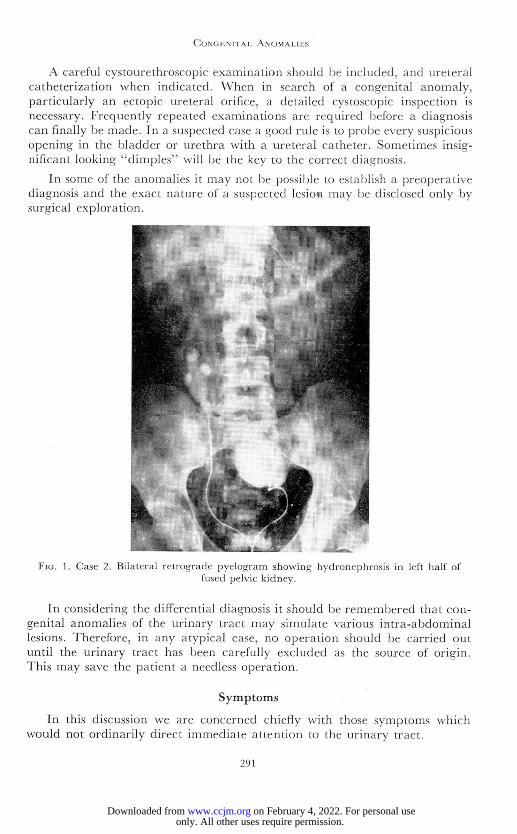
FIG. 1. Case 2. Bilateral retrograde pyelogram showing hydronephrosis in left half of fused pelvic kidney.
In considering the differential diagnosis it should be remembered that con-genital anomalies of the urinary tract may simulate various intra-abdominal lesions. Therefore, in any atypical case, no operation should be carried out until the urinary tract has been carefully excluded as the source of origin. This may save the patient a needless operation.
Symptoms
In this discussion we are concerned chiefly with those symptoms which would not ordinarily direct immediate attention to the urinary tract.
2 9 1
only. All other uses require permission. on February 4, 2022. For personal usewww.ccjm.orgDownloaded from

ENGEL AND WARDEN
FIG. 2. Case 4. Showing location of ectopic ureter opening into vagina. Ureteral catheter has been passed into anomalous ureter.
Pain is an extremely variable symptom and there is no pain which can be said to be typical of any urinary tract anomaly. However, with any discomfort in the flank, lateral abdomen, or pelvis which is not characteristic of any other known disease and in which the entire clinical picture is not clear-cut, urinary tract investigation should be considered. The pain of a pelvic kidney may be confusing, especially in the woman, where it is usually mistaken for disease of the pelvic organs. In one of our patients the pain was thought to be due to a diverticulitis of the colon. The pain associated with certain ureteral anomalies may simulate almost any intra-abdominal lesion. If on the right side, the patient usually has had an appendectomy or a pelvic laparotomy before the true
FIG. 3. Case 4. Gross specimen removed at operation.
2 9 2
only. All other uses require permission. on February 4, 2022. For personal usewww.ccjm.orgDownloaded from

CONGÉNITAL ANOMALIES
nature of the condition is recognized. There being no characteristic pain, and since these patients almost never represent surgical emergencies, the careful physician, with the patient's interest in mind, will consider the possibility of a urinary tract anomaly and take measures to exclude this possibility.
Case Reports
Case 1. A man aged 34 complained of pain in the right groin and right side of the abdomen which had been associated with constipation. He had also observed that taking of an enema appeared to aggravate the abdominal pain. Colon x-ray disclosed no organic disease. Intravenous urogram revealed a normally functioning left kidney, but there was no dye in the region of the right kidney. There were traces of dye observed overlying the bones of the pelvis. Cystoscopy revealed a normal bladder. Retrograde right pyelogram disclosed the presence of a typical pelvic kidney with slight hydrone-phrosis. The patient was managed conservatively for a period of some 6 months, but the pain continued and became even worse. A transperitoneal nephrectomy was carried out. This resulted in relief of his symptoms. It should be notecf that, in this case, the original urinalysis was entirely normal.
Case 2. A young man aged 28 was first observed complaining of attacks of sudden acute abdominal pain, beginning in the back and radiating through to the deep pelvic region. The attacks occurred about every 2 months at the onset, but became more frequent and severe. Recently he had experienced fever and sweating. Some intra-abdominal disease was suspected. He had had his appendix removed when a child of 12 because of indefinite lower abdominal pain.
On physical examination considerable tenderness was evident in the lower part of the abdomen, with an indefinite rounded mass perceptible in the midline. Intravenous urogram showed prompt function from the right kidney, which was low-lying and of atypical pelvic outline. No dye could be seen in the region of the left kidney. A retro-grade pyelogram revealed the presence of a hydronephrosis in the left pelvic kidney which appeared to be fused to the right kidney (fig. 1).
Nephrectomy of the hydronephrotic left half of this fused pelvic kidney completely relieved the patient's symptoms. He later developed an obstructing stone in the right ureter, which was removed by ureterolithotomy. Since that time the patient has experi-enced no further difficulty.
Case 3. A young woman, 29 years of age, presented the complaint of recurrent severe attacks of pain in the lower right abdomen associated with intense vomiting. The onset had been 6 years previous to the present examination. An initial diagnosis of appendi-citis had been made and appendectomy performed. Her symptoms recurred and because of the presence of a slight icterus, gall bladder disease was suspected and a cholecystec-tomy performed. This afforded no relief of her symptoms and at a later date pelvic laparotomy was done and a salpingo-oophorectomy performed for a supposed ovarian cyst. Her symptoms continued, however, with recurring bouts of pain, vomiting, and considerable weight loss and she was referred to the Clinic for further study.
Urinary tract investigation was carried out. Intravenous pyelogram showed the left kidney to be normal. The right kidney revealed a small pelvis which, in other respects, was normal. Cystoscopy examination disclosed the presence of two ureteral orifices on the right. The upper orifice was catheterized and retrograde pyelogram filled the small renal pelvis, lying in normal position. The lower ureteral orifice was catheterized, but the catheter could be passed only 3 or 4 cm. at which point the patient complained
2 9 3
only. All other uses require permission. on February 4, 2022. For personal usewww.ccjm.orgDownloaded from

ENGEL AND WARDEN
m e t r i c •"> -
j . ' I , , , , ! , . . ? ! . , , , ) , , . 3 ! 111j [ in1111[11 |q11[1111[ i 6 | j | |
FIG. 4. Case 5. Gross specimen removed at operation showing congenital multicystic kidney.
, *
(a) (b) FIG. 5. Case 6. (a) Intravenous urogram showing essentially normal renal pelves. How-ever, left kidney appears smaller than right, (b) Ureteral catheter passed into vaginal opening with injection of pyelographic media showing dilated tortuous ectopic ureter on
left side. Infection was present.
2 9 4
only. All other uses require permission. on February 4, 2022. For personal usewww.ccjm.orgDownloaded from
CONGÉNITAL ANOMALIES
of pain similar to the type she had been experiencing. Attempts to fill this ureter were unsuccessful because of the intense pain. Operation was performed and an anomalous accessory ureter was found which ended blindly at the level of the hilus of the kidney. Removal of this accessory ureter completely relieved the patient of her symptoms. This case has been previously reported.1
Case 4. A woman of 31 came to the Clinic complaining of severe right lower quad-rant pain which was referred to the right lumbar region and the inner surface of the right thigh. The history revealed that since the age of 11 she had experienced attacks of right lower quadrant pain, occasionally associated with nausea and vomiting. An appendec-tomy had been performed 11 years previously because of a severe bout of right lower quadrant pain accompanied by fever, nausea, and vomiting. However, the pain was not relieved and she continued to experience similar attacks. Later she underwent a pelvic laparotomy and uterine suspension was done with curettage and conization of the cervix. Because her symptoms continued, a left oophorectomy was performed without relief of symptoms, and 2 years previous to this examination a complete hysterectomy, which failed to relieve her pain. Careful questioning revealed the fact that she had periodic vaginal discharge and she believed that this discharge increased in amount following her attacks of pain. There was also a history of severe dyspareunia.
(a) ( b )
FIG. 6. Case 7. (a) Retrograde left pyelogram showing apparently normal left kidney pelvis, (b) Catheter passed into ectopic ureter opening into urethra. Injection of Pyelo-graphie dye shows marked hydroureter and hydronephrosis of upper pelvis of duplex
kidney.
2 9 5
only. All other uses require permission. on February 4, 2022. For personal usewww.ccjm.orgDownloaded from

ENGEL AND WARDEN
As part of the investigation, intravenous urography was done, revealing no function from the right kidney. The left kidney functioned normally and presented some com-pensatory hypertrophy. The patient had considerable vaginal discharge and on careful examination a small reddened area was found in the right anterior vaginal wall about 2.5 cm. back of the urethral meatus. After several attempts a small No. 4 ureteral catheter passed into this opening and progressed for a distance of about 20 cm. (fig. 2). Injection of radiopaque dye revealed an atypical smudge of dye extending between the transverse process of the second and third lumbar vertebrae. At operation the anomalous ureter was identified and on tracing it upward, it was found to end in a small mass of infected atrophic and partially cystic renal tissue. The patient was relieved of her symp-toms (fig. 3).
Fever of unexplained origin with or without chills should always direct attention to the urinary tract. When shown to be the source of the difficulty pyuria will usually be present although not necessarily. In the case of an in-fected ectopic ureter the opening may be outside the bladder, permitting the urine to remain clear. Also, as will be demonstrated in a subsequent case, an infected cyst or multicystic kidney may produce fever and yet reveal little if anything indicative in the urine.
Case 5. A child of 32 months was brought to the Clinic because of intermittent periods of fever of undetermined origin. The present illness was of some 9 days' duration, during which time she had had daily temperature elevation to 102 and 103 F. The patient had become listless although there were no localizing symptoms. As a part of the general survey, intravenous urography was done which showed a normally functioning left kidney but no dye visible on the right side.
The urine analysis had been essentially negative. However, further investigation was indicated because of the absence of function of the right kidney.
A cystoscopic examination was carried out, which showed the bladder to be essen-tially normal. A ureteral orifice was present on the right side. A No. 4 catheter entered the orifice but would not pass beyond approximately 6 cm. An attempt to inject pyelo-graphy media showed no dye at all in the kidney and all dye refluxed into the bladder.
Exploration of the right kidney was carried out. At operation a congenital multi-cystic type of kidney with atresia of the upper ureter was found (fig. 4). Following removal the patient had an uneventful convalescence, and has experienced no recur-rence of fever. She has remained well since operation.
Case 6. A married woman of 34 was admitted to the Clinic with the complaint of recurrent episodes of chills and fever associated with left flank pain. While she had experienced this discomfort for the past 10 years, episodes appeared to be occurring with increasing frequency. Especially significant was the fact that, subsequent to these epi-sodes, there was a noticeable increase of vaginal discharge, from which Trichomonas had been isolated.
Intravenous urogram revealed an apparently normal upper urinary tract. Careful vaginal inspection disclosed the presence of a small reddened area well up on the left lateral vaginal wall, close to the cervix. A minute opening was finally located with a lacrimal duct probe, and after dilation a No. 4 catheter was passed into this opening for a distance of 25 cm. Injection of radiopaque media disclosed the presence of a dilated tortuous ectopic ureter, which ended blindly at the level of the upper pole of the left kidney (fig. 5). Of interest is the fact that material obtained from this ureter was pus-laden and contained many trichomonads.
Surgical removal of this lesion has afforded this patient complete relief of symptoms.
2 9 6
only. All other uses require permission. on February 4, 2022. For personal usewww.ccjm.orgDownloaded from
CONGÉNITAL ANOMALIES
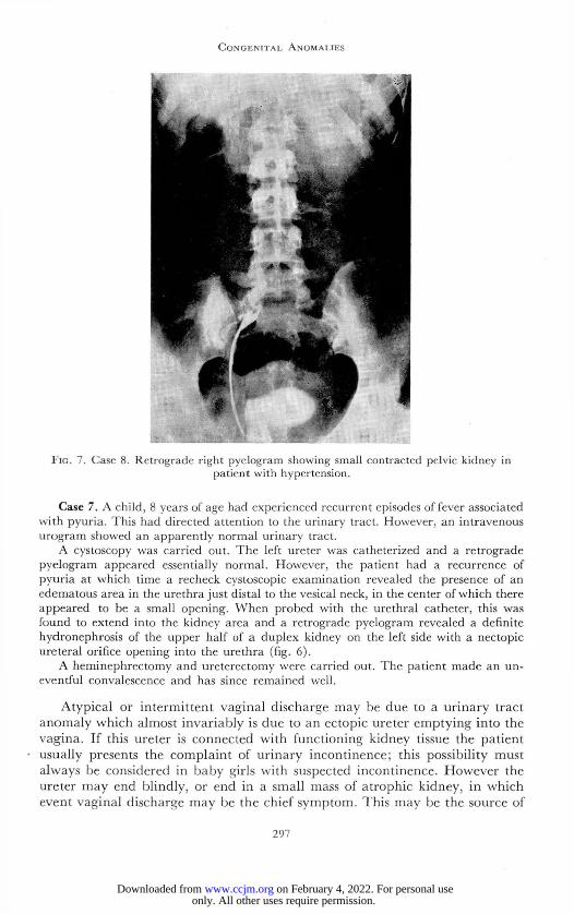
FIG. 7. Case 8. Retrograde right pyelogram showing small contracted pelvic kidney in patient with hypertension.
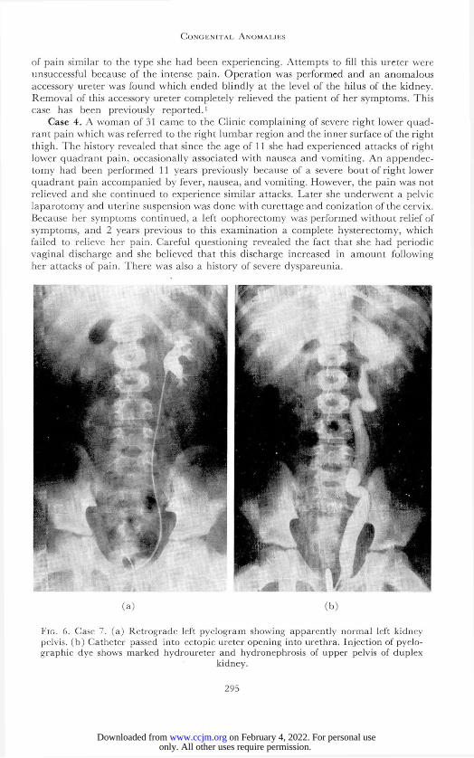
Case 7. A child, 8 years of age had experienced recurrent episodes of fever associated with pyuria. This had directed attention to the urinary tract. However, an intravenous urogram showed an apparently normal urinary tract.
A cystoscopy was carried out. The left ureter was catheterized and a retrograde pyelogram appeared essentially normal. However, the patient had a recurrence of pyuria at which time a recheck cystoscopic examination revealed the presence of an edematous area in the urethra just distal to the vesical neck, in the center of which there appeared to be a small opening. When probed with the urethral catheter, this was found to extend into the kidney area and a retrograde pyelogram revealed a definite hydronephrosis of the upper half of a duplex kidney on the left side with a nectopic ureteral orifice opening into the urethra (fig. 6).
A heminephrectomy and ureterectomy were carried out. The patient made an un-eventful convalescence and has since remained well.
A typ ica l or i n t e r m i t t e n t vag ina l d i s cha rge m a y be d u e to a u r i n a r y t r ac t a n o m a l y w h i c h a lmost i n v a r i a b l y is d u e to a n ec topic u r e t e r e m p t y i n g in to t he vag ina . If this u r e t e r is c o n n e c t e d wi th f u n c t i o n i n g k idney tissue the p a t i e n t usual ly p resen ts t he c o m p l a i n t of u r i n a r y i n c o n t i n e n c e ; this possibil i ty mus t a lways be cons idered in b a b y girls w i th suspected incon t inence . H o w e v e r t he u re t e r m a y e n d bl indly , or end in a smal l mass of a t r o p h i c k idney , in w h i c h event vag ina l d i scha rge m a y be t h e chief s y m p t o m . T h i s m a y be t h e source of
2 9 7
only. All other uses require permission. on February 4, 2022. For personal usewww.ccjm.orgDownloaded from

ENGEL AND W A R D E N
recurrent trichomonas vaginitis or other types of infections, as illustrated by the case previously presented.
Obscure perineal and rectal pain are symptoms which deserve careful urologic study. We have observed a case of a large congenital type of a cyst of the seminal vesical into which a blindly ending ureter emptied (reported case).2
Occasionally a complete survey for hypertension will disclose previously unsuspected urinary tract lesions. An intravenous urogram has become an almost routine examination in the study of hypertension and this examination should never be omitted in young people. The following case illustrates its importance.
Case 8. A young woman, 20 years of age, entered the Clinic with a history of hyper-tension, known to have been present for 4 months. There were no other complaints. The most significant finding on physical examination was the blood pressure, which varied f rom 210/115 to 174/104. The intravenous urogram showed a normally func-tioning left kidney. There was no dye visible on the right side in any of the films of the urogram series. A cystoscopy was carried out but showed a normal bladder. The right ureter was catheterized, but the catheter was passed only about 15 cm. A retrograde pyelogram revealed the presence of a small aplastic contracted pelvic kidney (fig. 7). A transperitoneal nephrectomy was carried out. The patient made an uneventful recovery following operation and since surgery the blood pressure has been maintained at nearly normal levels, ranging from 135/80 to 160/92 on postoperative visits.
Summary and Conclusions
In any obscure abdominal complaint a congenital anomaly of the geni-tourinary tract should be considered in the differential diagnosis. Careful urologic investigation should be carried out before subjecting the patient to a needless operation. Illustrative cases have been presented to call attention to some of these lesions and the varied symptomatology they may present. Many other types of congenital anomalies may be encountered.
References
1. Engel, W. J . : Aberrant ureters ending blindly. J . Urol. 42:674 (Nov.) 1939. 2. Idem: Ureteral ectopia opening into seminal vcsicle. J . Urol. 60:46 (July) 1948.
2 9 8
only. All other uses require permission. on February 4, 2022. For personal usewww.ccjm.orgDownloaded from