congenital heart disease (chd)
TRANSCRIPT

Congenital Heart Disease (CHD)
Mr. Fadi J. Zaben R.N M.S.N
IMET2000, Ramallah
April, 2013
IMET2000 1

Outline:
• Overview of fetal circulation.
• Definition of CHD.
• Types of CHD.
• Pathophysiology and Etiology.
• Diagnosis and Clinical Manifestations.
• Treatment.
• Nursing Care and Interventions.
IMET2000 2

Fetal Circulation:
Main Blood Flow:
• Placenta Umbilical Vein Liver Ductus Venosus Inferior Vena Cava
• Vena Cava Right Atrium Foramen Ovale Left Atrium Left Ventricle
• Aorta Body
IMET2000 3

Secondary Route:
• Right Atrium
• Right Ventricle
• Pulmonary Artery
• Ductus Arteriosus
(so does not go to lungs)
• Aorta
• Body
Fetal Circulation:
IMET2000 4

Third route of blood flow:
• Right Atrium
• Right Ventricle
• Pulmonary Artery
• Lungs (needs to perfuse the lungs
and upper body with oxygen)
• Left Atrium
• Left Ventricle
• Aorta
• Body
Fetal Circulation:
IMET2000 5

IMET2000 6

Transition from Fetal Circulation to Pulmonary circulation
• The umbilical arteries and vein and the ductus venosus become non-functional.
• Decreased pulmonary vascular resistance and increased pulmonary blood flow
• Increase pressure of the left atrium, decrease pressure in right atrium, causing closure of foramen ovale.
• Pulmonary resistance is less than systematic resistance so there is left-to-right shunting resulting in closure of the ductus arteriosus.
IMET2000 7

IMET2000 8

CONGENITAL HEART DISEASE (CHD)
IMET2000 9

Introduction: • Congenital heart disease (or defects) is one of the
most common forms of congenital anomalies.
• It involves the chambers, valves, and great vessels arising from the heart.
• In most cases, the cause of CHD is not known.
• Some infants and children with CHD may appear perfectly healthy, whereas others may be critically ill.
• Most infants and children with CHD can be successfully managed with medications and surgeries.
IMET2000 10

ETIOLOGY AND INCIDENCE:
CHD affects 8 to 12 of every 1,000 neonates.
Exact cause of CHD is unknown in 90% of cases.
The heart begins as a single cell and develops into a four-chambered pumping system during the third to eighth weeks of gestation.
Girls: ASD, and PDA.
Boys: PS, AS, TGA, and CoA.
In the USA:
~ 32,000 children born/year with CHD
~ 11,000/year with “Critical” CHD
IMET2000 11

Causes: Mostly unknown.
Genetic.
Chromosomal aberrations (5%).
Multifactorial inheritance (90%).
Envirnomental Factor (3%).
Fetal or maternal infection during the first trimester (rubella).
Drugs in ANC: thalidomide, anticonvlusion, alcohol, PG E, and Radiation.
Maternal insulin-dependent diabetes.
IMET2000 12

Neonatal Diagnosis:
45 % diagnosed prior to day of discharge.
20 % presented before 6 weeks of age.
10 % at 6wk NB exam.
24 % diagnosed later in 1st year of life.
1 % die of heart disease undiagnosed.
IMET2000 13

Neonatal Diagnosis:
Screening for CHD with Pulse Oximetry
If sat less than 95 % ; echocardiogram should be done.
This method of screening will only catch cyanotic lesions such as… Pulmonary Atresia
Transposition of Great Arteries
Total Anomalous Pulmonary Veins
Tricuspid Atresia
Will not catch coarctation or Aortic Stenosis, VSD, ASD.
IMET2000 14

Syndromes and CHD: Marfan's syndrome: mitral valve prolapse (MVP), dilated aortic root. Turner's syndrome: aortic valve stenosis (AVS), coarctation of the aorta (CoA). Noonan's syndrome: dysplastic pulmonary valve. William's syndrome: supravalvular pulmonary stenosis (PS). DiGeorge syndrome: interrupted aortic arch (IAA), truncus arteriosus, transposition of great arteries (TGA), tetralogy of Fallot (TOF). Down syndrome (trisomy 21): atrioventricular (AV) canal defect, ventricular septal defect (VSD). About 50% of children with Down syndrome have a CHD.
IMET2000 15

CHD Classifications:
Congenital heart defects can be classified into three categories:
1. Obstruction systemic blood flow.
2. Increased pulmonary blood flow (acyanotic lesions).
3. Decreased pulmonary blood flow (cyanotic lesions).
OR:::: 1. Mild: resolve by themselves
2. Non life threatening: but require treatment
3. Severe: multiple operations , lifetime medication
IMET2000 16

Aortic Stenosis (AS).
Coarctation of the Aorta (CoA).
Defects Obstructing Systemic Blood Flow:
IMET2000 17

Aortic Stenosis (AS):
• Congenital AS is narrowing or stricture of the aortic valve.
• AS causing resistance to blood flow in the Lt. Ventricle, and decrease cardiac output.
• AS is the most common form of left ventricular outflow tract obstruction.
• It accounts for 3% to 6% of congenital heart defects.
• AS may occur at any age, and it occurs more commonly in boys than in girls.
IMET2000 18

IMET2000 19

Clinical Manifestations:
Neonate:
Severe congestive heart failure (CHF).
Metabolic acidosis.
Tachypnea.
Faint peripheral pulses, poor perfusion, poor capillary refill, & cool skin.
Poor feeding and feeding intolerance.
Child and Adolescent:
Chest pain on exertion, decreased exercise tolerance.
Dyspnea, fatigue, shortness of breath.
Syncope, light-headedness.
Palpitations.
Sudden death.
IMET2000 20

Management:
Neonate:
• Stabilize with prostaglandin E1 (PGE1) infusion to maintain cardiac output through the PDA.
• Cardiac catheterization: aortic balloon valvuloplasty or aortic balloon angioplasty.
• Surgical: valvotomy, or myectomy/myotomy.
• Intubation and ventilation as needed.
• Infective endocarditis prophylaxis (lifelong).
IMET2000 21

Management……………. Child and Adolescent
Child and Adolescent:
• Medical management with close follow-up.
• Restrict strenuous exercise and anaerobic exercise (eg, weight lifting), and competitive sports.
• Aortic balloon valvuloplasty or aortic balloon angioplasty.
• Infective endocarditis prophylaxis (lifelong).
• Surgical intervention. – Surgical valvotomy, or myectomy/ myotomy.
– Aortic valve replacement.
IMET2000 22

COARCTATION OF THE AORTA (CoA)
IMET2000 23

IMET2000 24

COARCTATION OF THE AORTA:
• CoA is a discrete narrowing of the aortic arch, usually in the insertion of ductus arterious position.
• It accounts for 8% to 10% of congenital heart defects.
• The discrete narrowing increases the workload of the left ventricle (increased LV systolic pressure).
IMET2000 25

Clinical Manifestations: The neonate with critical CoA:
Asymptomatic until the PDA begins to close.
After PDA closure: severe CHF, poor lower body perfusion, tachypnea, acidosis, absent femoral and pedal pulses.
The child or adolescent with CoA: Usually asymptomatic normal growth and development.
Hypertension in the upper extremities, with absent or weak femoral pulses.
Nosebleeds, headaches, leg cramps.
IMET2000 26

Management: • Medical management:
– Resuscitation and stabilization with PGE1 infusion, usually perform during the neonate period; monitor for complications related to PGE1 therapy (fever, hypotention, and apnea).
– Intubation and ventilation as needed.
• Surgical intervention:
– End-to-end anastomosis.
– Balloon angioplasty
– Dacron patch.
• Medical management for hypertension (beta-adrenergic blockers).
• Infective endocarditis prophylaxis (lifelong).
IMET2000 27

Defects that Increase Pulmonary Blood Flow:
Patent Ductus Arterious (PDA).
Atrial Septal Defect (ASD).
Ventricle Septal Defect (VSD).
IMET2000 28

Patent Ductus Arterious (PDA): • The ductus arteriosus is a normal fetal connection
between the left PA and the descending aorta. • During fetal life, blood flow is shunted away from
the lungs through the ductus arteriosus and directly into the systemic circulation.
• PDAs are common in premature neonates who weigh less than 1,500 g.
• They account for 5% to 10% of CHDs. • It is associated with poor weight gain, failure to
thrive, feeding difficulties and tachypnea, and frequent respiratory tract infections.
• Long term caused lung and heart damage (CHF).
IMET2000 29

Pathophysiology and Etiology:
After birth, the ductus arteriosus is no longer needed. Functional closure usually occurs within 48 hours after birth. Anatomic closure is completed by age 2 to 3 weeks.
When the ductus arteriosus fails to close, blood from the aorta (high pressure) flows into the low-pressure PA, resulting in pulmonary overcirculation.
Increased pulmonary blood flow leads to a volume-loaded LV.
IMET2000 30

Management:
1. In the symptomatic premature neonate; indomethacin given I.V.
2. Surgical management through PDA ligation. 3. Medical management:
o Monitor growth and development. o Reassess for spontaneous PDA closure. o Increase caloric intake as needed for normal weight
gain. o Diuretics: furosemide (Lasix), spironolactone
(Aldactone).
4. Cardiac catheterization: o For small PDAs coil occlusion. o For larger PDAs a closure device may be used.
IMET2000 31

ATRIAL SEPTAL DEFECT (ASD):
• ASD is an abnormal communication between the left and right atrias.
• Hole between the tow atrias.
• Foramen ovale fails to close.
• ASDs account for 9% of CHDs.
• It is usually asymptomatic.
• infant with ASD have frequent upper respiratory infections (URIs), poor weight gain.
IMET2000 32

ASD Types:
• There are three types:
1) Ostium secundum ASD: the most common type of ASD; abnormal opening in the middle of the atrial septum.
2) Ostium primum ASD: abnormal opening at the bottom of the atrial septum.
3) Sinus venosus ASD: abnormal opening at the top of the atrial septum.
IMET2000 33

Pathophysiology and Etiology:
• Blood flows from the higher-pressure left atrium across the ASD into the lower-pressure right atrium (left-to-right shunt).
• Increased blood return to the right heart leads to right ventricular volume overload and right ventricular dilation.
• Increased pulmonary blood flow leads to elevated pulmonary artery pressures congestive heart failure. IMET2000 34

Treatment:
Medical Management:
Medications – digoxin
Cardiac Catheterizaton: septal occluder
Open heart Surgery
IMET2000 35

VENTRICULAR SEPTAL DEFECT: • A VSD is an abnormal communication
between the right and left ventricles.
• Hole between two ventricles.
• It is the most common type of congenital heart defect (25% of all CHDs).
• Small VSDs usually asymptomatic.
• Large VSDs. – Frequent URIs.
– Poor weight gain, failure to thrive.
– Feeding difficulties.
– CHF: tachypnea, tachycardia.
IMET2000 36

Pathophysiology and Etiology:
• Blood flows from the high-pressure left ventricle across the VSD into the low-pressure right ventricle and into the PA, resulting in pulmonary overcirculation.
• A left-to-right shunt because of a VSD results in increased right ventricular pressure and increased PA pressure.
• The increased pulmonary venous return to the left side of the heart results in left atrial dilation.
IMET2000 37

Management:
• Medical management (anticongestive therapy).
• Cardiac catheterization for placement of a ventricular occlusion device for muscular defects.
• Surgical intervention is usually not necessary.
– Usually repaired before age 1.
– One-stage approach: preferred surgical
plan; patch closure of VSD.
– Two-stage approach: first surgery is to
band the PA to restrict pulmonary
blood flow; second surgery is to patch
close the VSD and remove the PA band.
IMET2000 38

Defects that Decrease Pulmonary Blood Flow:
TETRALOGY OF FALLOT (TOF).
TRANSPOSITION OF THE GREAT ARTERIES (TGA).
TRICUSPID ATRESIA (TA).
IMET2000 39

TETRALOGY OF FALLOT (TOF):
TOF is the most common complex congenital heart defect.
It accounts for 6% to 10% of all CHDs.
IMET2000 40

Continue…….TOF
The four abnormalities of TOF include the following: 1. VSD.
2. Aortic override.
3. Pulmonary stenosis (right ventricular outflow tract obstruction).
4. Right ventricular hypertrophy.
IMET2000 41
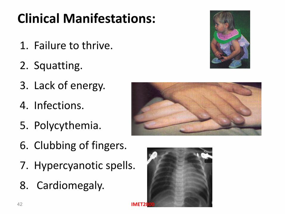
Clinical Manifestations:
1. Failure to thrive.
2. Squatting.
3. Lack of energy.
4. Infections.
5. Polycythemia.
6. Clubbing of fingers.
7. Hypercyanotic spells.
8. Cardiomegaly.
IMET2000 42

Management:
• Surgical interventions
– Blalock – Taussig or Potts procedure – increases blood flow to the lungs.
– Open heart surgery
IMET2000 43

TRANSPOSITION OF THE GREAT ARTERIES (TGA):
• Transposition of the great arteries (TGA) occurs when the PA arises off the left ventricle and the aorta arises off the
right ventricle.
• It accounts for 5% to 10%
of CHDs.
• It is associated lesions
include VSD, ASD, PDA,
PS, and CoA.
IMET2000 44

Clinical Manifestations:
Symptoms evident soon after birth:
– Cyanosis.
– Tachypnea.
– Metabolic acidosis.
– CHF.
– Feeding difficulties.
IMET2000 45

Management:
1. Medical Management.
2. Stabilize with PGE1 infusion.
3. Cardiac catheterization.
4. Surgical Management.
IMET2000 46
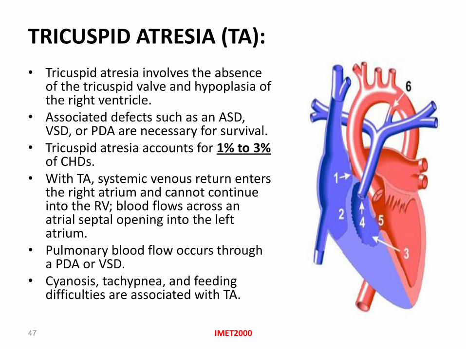
TRICUSPID ATRESIA (TA):
• Tricuspid atresia involves the absence of the tricuspid valve and hypoplasia of the right ventricle.
• Associated defects such as an ASD, VSD, or PDA are necessary for survival.
• Tricuspid atresia accounts for 1% to 3% of CHDs.
• With TA, systemic venous return enters the right atrium and cannot continue into the RV; blood flows across an atrial septal opening into the left atrium.
• Pulmonary blood flow occurs through a PDA or VSD.
• Cyanosis, tachypnea, and feeding difficulties are associated with TA.
IMET2000 47

Truncus Arteriosus:
Single great vessel arising from of base on the heart.
It serving as a pulmonary artery and aorta.
Systolic murmur is heard.
IMET2000 48

Outcomes of CHD: Mortality
Source: Boneva et al. (CDC) Circulation 2001;103:2376-81
• 1995-97: CHD contributed to 5822 deaths/year
Tal Geva 2/04
IMET2000 49

Outcomes of CHD: Mortality
Source: Boneva et al. (CDC) Circulation 2001;103:2376-81
• 51% of deaths in infants; 7% in 1-4 years
• ~19% higher mortality in blacks compared
with whites
Tal Geva 2/04
IMET2000 50

Nursing Diagnoses:
Impaired Gas Exchange related to altered pulmonary blood flow or pulmonary congestion
Decreased Cardiac Output related to decreased myocardial function
Activity Intolerance related to hypoxia or decreased myocardial function
Imbalanced Nutrition: Less Than Body Requirements related to excessive energy demands required by increased cardiac workload
Risk for Infection related to chronic illness
Fear and Anxiety related to life-threatening illness
IMET2000 51

Nursing Interventions:
Relieving Respiratory Distress: • Record vital signs and oxygen saturations. • Assess skin color, mucous membranes, and
extremities pulses. • Position the child in a reclining, semi-upright
position. • Suction oral and nasal secretions as needed. • Administer oxygen as prescribed. • Administer prescribed medications (Diuretics,
Bronchodilators)and document response. • May need to change oral feedings to nasogastric
feedings because of increased risk of aspiration with respiratory distress.
IMET2000 52

Continue……
Improving Cardiac Output:
• Organize nursing care and medication schedule to provide periods of uninterrupted rest.
• Provide play or educational activities that can be done in bed with minimal exertion.
• Maintain normothermia.
• Administer medications as prescribed (lasix, digoxin, and beta blockers)
IMET2000 53

Continue……
Providing Adequate Nutrition: For the infant:
Small, frequent feedings. Limit oral feeding time to 15 to 20 minutes. Supplement oral feeds with nasogastric feedings as needed to
provide weight gain (ie, continuous nasogastric feedings at night with mouth feeds during the day).
For the child: Small, frequent meals. High-calorie, nutritional supplements. Determine child's likes and dislikes and plan meals accordingly. Allow the parents to bring the child's favorite foods to the hospital.
Document daily weight (same time of day, same scale, same clothing).
Record accurate inputs and outputs; assess for fluid retention.
IMET2000 54

Continue…… Preventing Infection:
Maintain routine childhood immunization schedule. Administer yearly influenza vaccine.
Prevent exposure to communicable diseases.
Good hand washing.
Report fevers.
Report signs of URI: runny nose, cough, increase in nasal secretions.
Report signs of GI illness: diarrhea, abdominal pain, irritability.
IMET2000 55

Continue.......
Instruct the family in necessary measures to maintain the child's health:
Complete immunization.
Adequate diet and rest.
Prevention and control of infections.
Regular medical and dental checkups. The child should be protected against infective endocarditis when undergoing certain dental procedures.
Regular cardiac checkups.
IMET2000 56

Continue.......
Teach the family about the defect and its treatment: Provide patients and families with written and verbal
information regarding the CHD.
Offer appropriate Internet resources for information about CHD and medical and surgical treatment options.
Signs and symptoms of CHF.
Signs of hypercyanotic spells associated with cyanotic defects and need to place child in knee-chest position.
Need to prevent dehydration, which increases risk of thrombotic complications.
Emergency precautions related to hypercyanotic spells.
Special home care equipment, monitors, oxygen. IMET2000
57

Continue.......
Encourage the parents and other people (teachers, peers) to treat the child in as normal a manner as possible. Avoid overprotection and overindulgence.
Avoid rejection.
Promote growth and development with modifications. Facilitate performance of the usual developmental tasks within the limits of the child's physiologic state.
Prevent adults from projecting their fears and anxieties onto the child.
Help family deal with its anger, guilt, and concerns related to the disabled child.
IMET2000 58

IMET2000 59