congenital laryngeal anomalies - welcome to utmb … … · congenital laryngeal anomalies ......
TRANSCRIPT

Congenital Laryngeal
Anomalies
Jean Paul Font, MD
Faculty Advisor: Seckin Ulualp, MD
The University of Texas Medical Branch
Department of Otolaryngology
Grand Rounds Presentation
November 2, 2005
Outline
Laryngeal Anatomy, Embryology & Function
Laryngomalacia
Laryngoceles & Saccular Cyst
Vocal Cord Paralysis
Congenital Laryngeal Web & Atresia
Congenital Subglottic Stenosis
Laryngeal & laryngotracheoesophageal clefts
Subglottic Hemangiomas
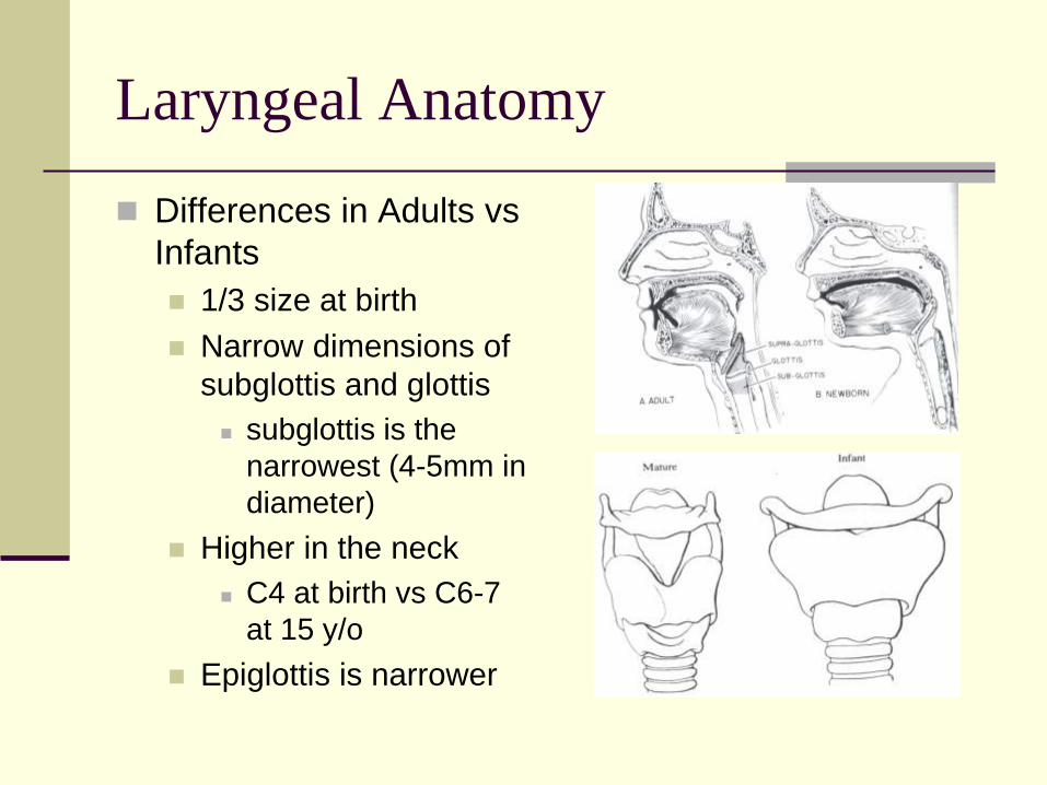
Laryngeal Anatomy
Differences in Adults vs
Infants
1/3 size at birth
Narrow dimensions of
subglottis and glottis
subglottis is the
narrowest (4-5mm in
diameter)
Higher in the neck
C4 at birth vs C6-7
at 15 y/o
Epiglottis is narrower
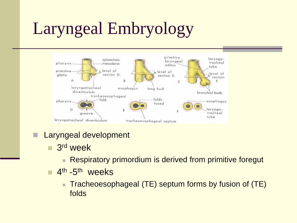
Laryngeal Embryology
Laryngeal development
3rd week
Respiratory primordium is derived from primitive foregut
4th -5th weeks
Tracheoesophageal (TE) septum forms by fusion of (TE)
folds
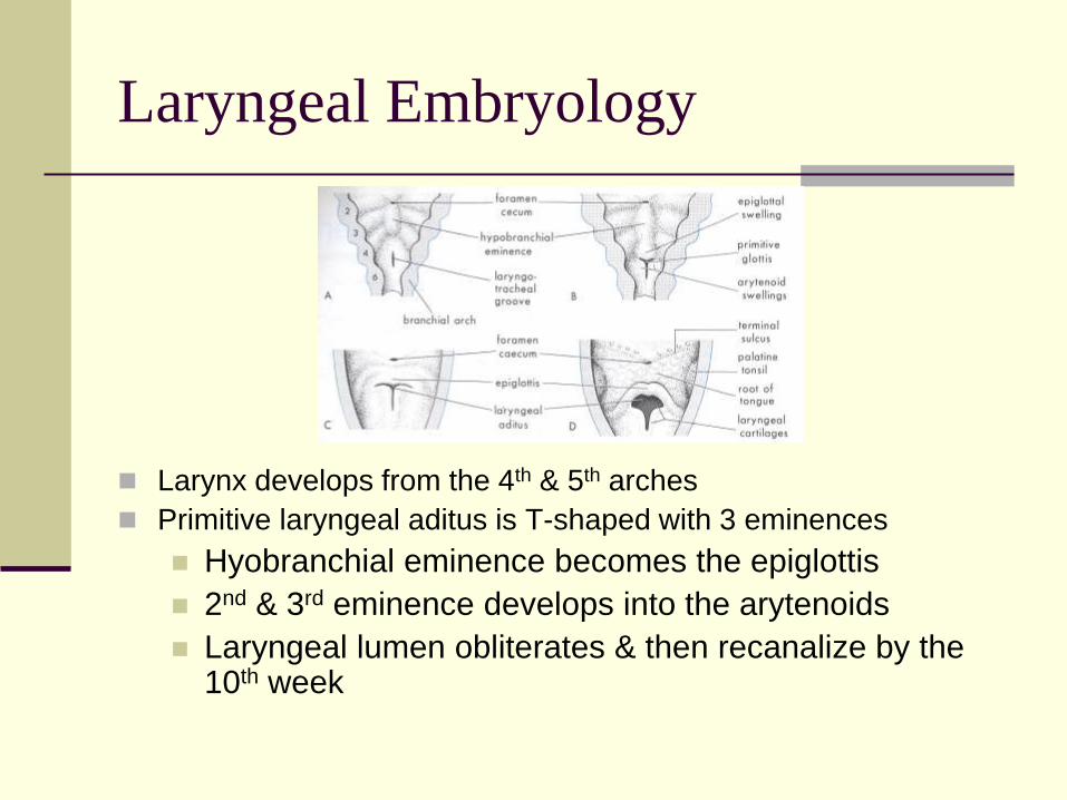
Laryngeal Embryology
Larynx develops from the 4th & 5th arches
Primitive laryngeal aditus is T-shaped with 3 eminences
Hyobranchial eminence becomes the epiglottis
2nd & 3rd eminence develops into the arytenoids
Laryngeal lumen obliterates & then recanalize by the 10th week
Laryngeal Function
Laryngeal Function
Breathing Passage
Airway protection
Aid in the clearance of secretion
Vocalization
Symptoms of Laryngeal Anomalies
Airway obstruction
Feeding difficulties
Abnormalities of Phonation
Airway Obstruction
Symptoms
Stridor
Increase work of breathing with retraction, nasal flaring & tachypnea
apnea episodes, cyanosis & sudden death
Stridor
Inspiratory stridor (Supraglottic & glottic)
Collapse during negative inspiratory pressure
Biphasic stridor (Subglottic)
Expiratory stridor (lower tracheobronchial tree)
Airway protection
First level- Epiglottis, aryepiglottic folds & arytenoids
Second level- False vocal folds
Third level- True vocal folds
Anomalies of any of this structures lead to aspiration and swallowing dysfunction
Symptoms- coughing, choking and gagging episodes, stasis of secretion, and recurrent pneumonia
Phonatory abnormality
Dependent on the level of abnormality
Muffled cry suggest supraglottic obstruction
High pitch or absent cry is associated with
glottic abnormalities
Laryngomalacia
Most common congenital laryngeal anomaly
(50-75%)
Most frequent cause of stridor in children
Male predominance 2:1
Flaccidity of supraglottic laryngeal tissues
Characterized by inward collapse of
supraglottic structures during inspiration
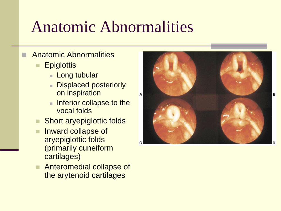
Anatomic Abnormalities
Anatomic Abnormalities
Epiglottis
Long tubular
Displaced posteriorly on inspiration
Inferior collapse to the vocal folds
Short aryepiglottic folds
Inward collapse of aryepiglottic folds (primarily cuneiform cartilages)
Anteromedial collapse of the arytenoid cartilages
Laryngomalacia Symptoms
Airway obstruction
Mild to moderate obstruction
Stridor exacerbated by exertion
Crying, agitation, feeding or supine position
Severe obstruction
Substernal retraction
Pectus excavatum with chronic severe obstruction
Other complications
Feeding difficulties
GERD
Failure to thrive
Cyanosis, cardiac failure & death
Stridor in Laryngomalacia
Inspiratory stridor
Intermittent low-pitched
Starts within first two weeks of birth
Worsens in the first few months followed by
gradual improvement
Peak at 6 months and most are symptom free
by 18 to 24 months (75%)

Laryngomalacia Pathophysiology
The cause of the collapse is unknown
Theories
Derangement of supraglottic anatomy, histology
or neurologic function
Laryngeal cartilage immaturity
Incidence of laryngomalacia is not increased in
premature infants
Histopathology- normal microanatomy
Subepithelial edema
Laryngomalacia Pathophysiology
Neurologic involvement
Associated with central apnea, hypotonia,
mental retardation and early speech delay
Abnormal Neuromuscular Control
Muscular dilation of supraglottic structures
Stylopharyngeus, Palatopharyngeus, Hyoglossus &
Digastric
Gastroesophageal reflux
>50% of patients with laryngomalacia
Airway edema contributes to airway
compromise
Pathophysiology
Increased negative intrathoracic pressure with
collapsed supraglottic leads to retrograde
gastric contents
Edema and/or erythema of posterior
supraglottic structures
Diagnosis of Laryngomalacia
Awake flexible fiberoptic laryngoscopy
Visualize supraglottic anatomy and collapse
Fluoroscopy
Direct laryngoscopy and bronchoscopy-
evaluate for synchronous lesions (27%)
Treatment of Laryngomalacia
Observation- most cases resolve spontaneously
Medical management for GERD
Surgical management- severe symptoms
In 1922, Iglauer amputation of epiglottic redundant tissue with a wire snare
Supraglottoplasty (CO2 laser, microlaryngeal scissors, microdebrider)
Trim redundant tissue from:
Lateral edges of the epiglottis
Aryepiglottic folds
Arytenoids
Corniculate cartilages
Tracheotomy
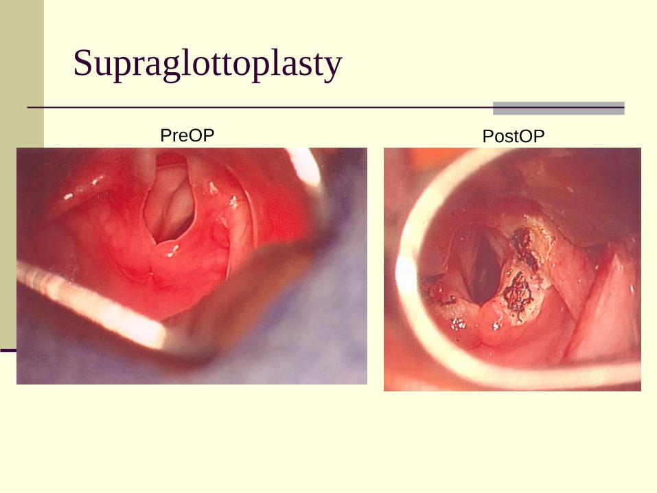
Supraglottoplasty
PreOP PostOP
Laryngomalacia
Supraglottoplasty complications
Aggressive approach
supraglottic stenosis
exacerbation of dysphagia with aspiration
Rare- massive collapse of supraglottic framework
needing tracheotomy placement
Conservative excision minimizes the
probability of postoperative complications
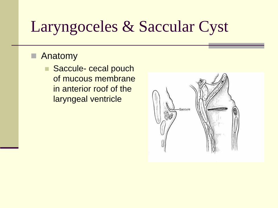
Laryngoceles & Saccular Cyst
Anatomy
Saccule- cecal pouch
of mucous membrane
in anterior roof of the
laryngeal ventricle
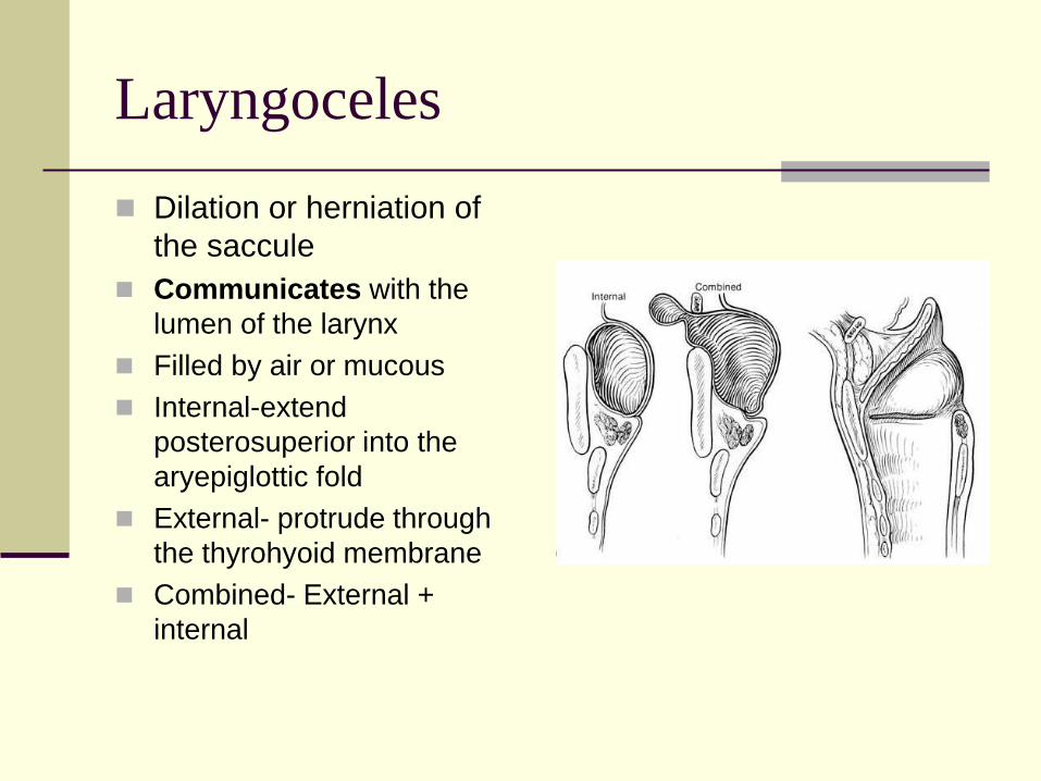
Laryngoceles
Dilation or herniation of
the saccule
Communicates with the
lumen of the larynx
Filled by air or mucous
Internal-extend
posterosuperior into the
aryepiglottic fold
External- protrude through
the thyrohyoid membrane
Combined- External +
internal
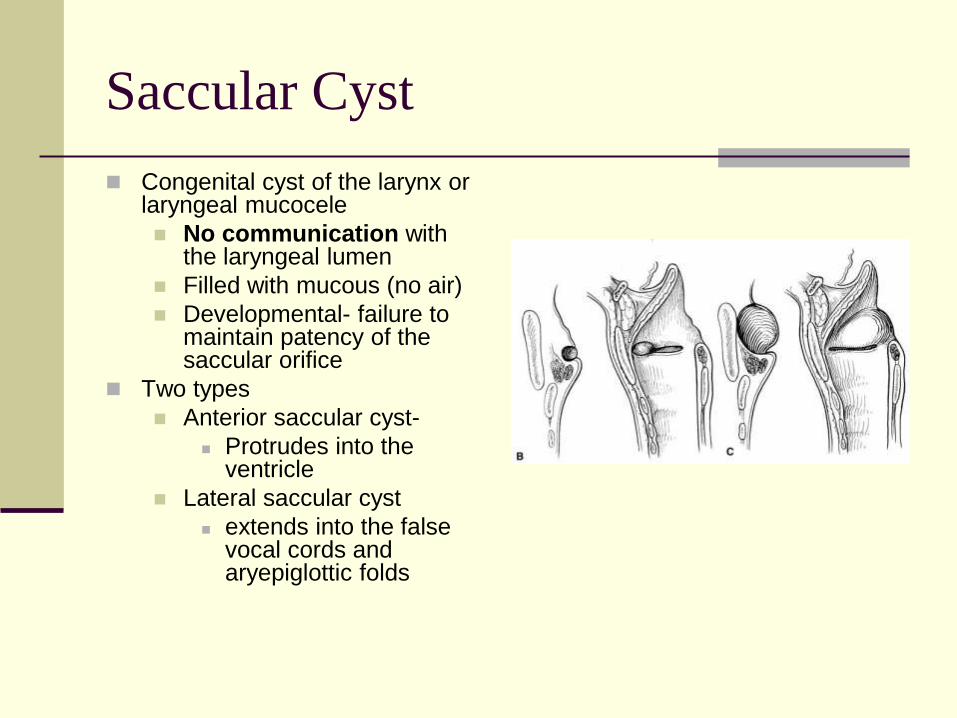
Saccular Cyst
Congenital cyst of the larynx or laryngeal mucocele
No communication with the laryngeal lumen
Filled with mucous (no air)
Developmental- failure to maintain patency of the saccular orifice
Two types
Anterior saccular cyst-
Protrudes into the ventricle
Lateral saccular cyst
extends into the false vocal cords and aryepiglottic folds

Laryngoceles & Saccular Cyst
Acquired Laryngoceles
Increased pressure on the laryngeal lumen
(player of wind instruments)
Acquired saccular cyst
Occlusion of the saccular orifice (inflammation,
trauma or tumors)
Laryngopyocele
Infected laryngocele or saccular cyst
Symptoms
Laryngocele
Intermittent hoarseness and dyspnea
Weak cry or aphonia
Saccular cyst
respiratory distress with inspiratory stridor
inaudible or muffle cry
occasionally dysphagia
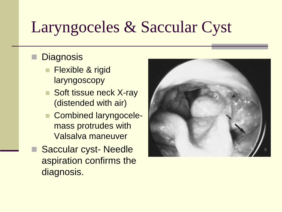
Laryngoceles & Saccular Cyst
Diagnosis
Flexible & rigid
laryngoscopy
Soft tissue neck X-ray
(distended with air)
Combined laryngocele-
mass protrudes with
Valsalva maneuver
Saccular cyst- Needle
aspiration confirms the
diagnosis.
Laryngoceles & Saccular Cyst
Treatment
Saccular cyst- aspiration or unroofing with cup forceps
or CO2 laser (recurs)
Endoscopic excision
Removing remnants CO2 laser
Open procedures for recurrence
Lateral cervical approach incising the thyrohyoid
membrane
Protect the superior laryngeal nerve
Intubation may be needed until edema subsides
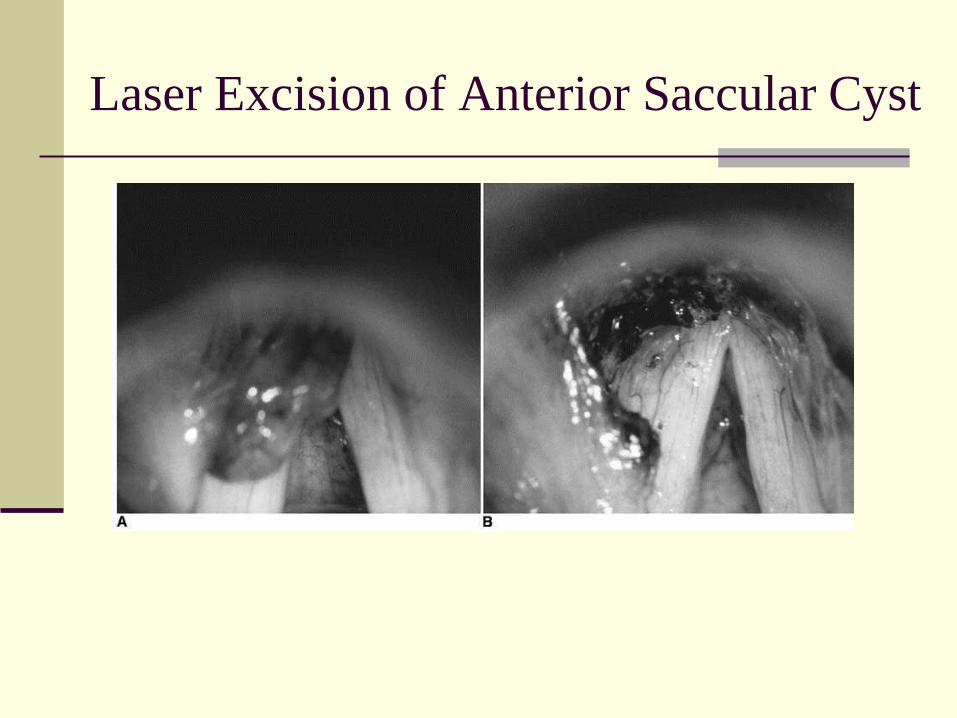
Laser Excision of Anterior Saccular Cyst
Vocal Cord Paralysis
Third most common congenital laryngeal anomaly producing stridor
Unilateral & Bilateral (1:1)
50% are associated to other anomalies
Acquired paralysis 70% association to congenital neurologic abnormalities
or neurosurgical procedure to treat them (Meningocele, Arnold Chiari Malformation and
Hydrocephalus)
Unilateral are associated to cardiovascular anomalies (PDA) and left side is more common
Vocal Cord Paralysis
Symptoms
Bilateral
High-pitched inspiratory stridor
Inspiratory cry
Paradoxical function (pressure changes)
close during inspiration and open during expiration
Unilateral (less symptoms)
weak cry and occasional breathy
Feeding difficulties secondary to laryngeal
penetration and aspiration
Vocal Cord Paralysis Diagnosis
Awake flexible fiberoptic laryngoscopy
record for slow motion replay
Direct laryngoscopy
Palpation of the glottis
Laryngeal EMG
Imaging of head (MRI) and chest to evaluate for
associated abnormalities (Neurologic & CV)
Unilateral VC Paralysis Treatment
Watchful waiting
70% of idiopathic unilateral VC paralysis resolve spontaneously
Most within 6 month
Feeding difficulties manage by thickening of liquids
Speech therapy consult
Rare surgical management
Increased Intracranial Pressure
early shunting or posterior fossa decompression (better outcome)
Bilateral VC Paralysis Treatment
Tracheotomy may be necessary (50%)
Lateralizing one or both paralyzed vocal
cords
Injurious to the developing larynx
Excisional procedure
Tissue removed from posterior glottis
Endoscopic technique with laser
More consistent results are achieved by external
approach
Congenital Laryngeal Web-Atresia
Uncommon
Failure of laryngeal recanalization
Most are glottic (75%)
Symptoms
Vocal dysfunction
Hoarseness
Aphonia if severe
Airway obstruction
Complete laryngeal atresia is incompatible with life and need emergent tracheostomy
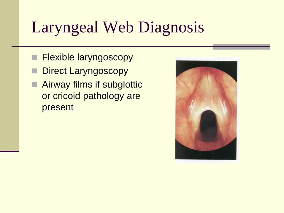
Laryngeal Web Diagnosis
Flexible laryngoscopy
Direct Laryngoscopy
Airway films if subglottic
or cricoid pathology are
present
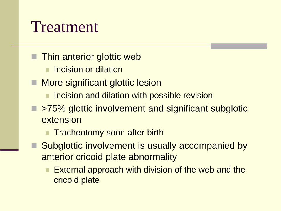
Treatment
Thin anterior glottic web
Incision or dilation
More significant glottic lesion
Incision and dilation with possible revision
>75% glottic involvement and significant subglotic
extension
Tracheotomy soon after birth
Subglottic involvement is usually accompanied by
anterior cricoid plate abnormality
External approach with division of the web and the
cricoid plate

Congenital Subglottic Stenosis
Second most common cause of stridor in neonates, infants and children
Incomplete laryngeal lumen recanalization
Newborn larynx <4 mm (premature <3mm)
Congenital less severe than acquired
Two types
Membranous vs cartilagenous
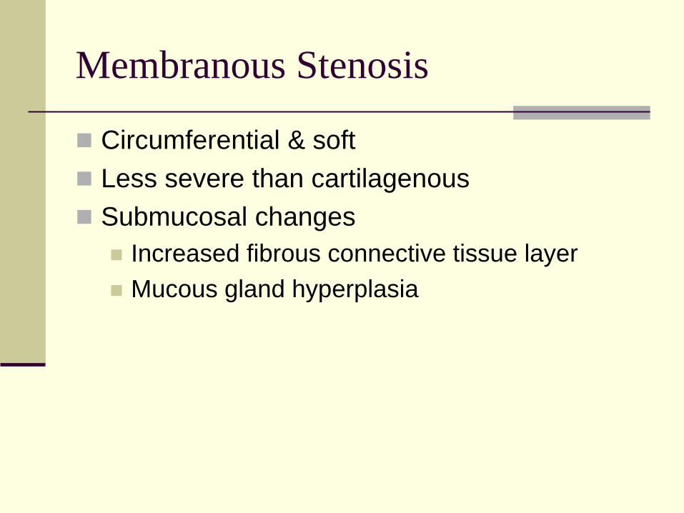
Membranous Stenosis
Circumferential & soft
Less severe than cartilagenous
Submucosal changes
Increased fibrous connective tissue layer
Mucous gland hyperplasia
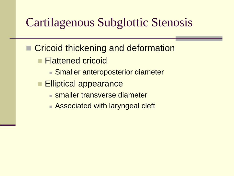
Cartilagenous Subglottic Stenosis
Cricoid thickening and deformation
Flattened cricoid
Smaller anteroposterior diameter
Elliptical appearance
smaller transverse diameter
Associated with laryngeal cleft
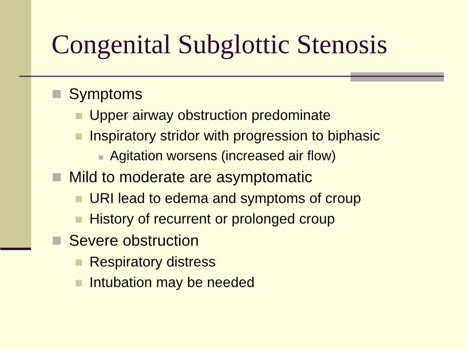
Congenital Subglottic Stenosis
Symptoms
Upper airway obstruction predominate
Inspiratory stridor with progression to biphasic
Agitation worsens (increased air flow)
Mild to moderate are asymptomatic
URI lead to edema and symptoms of croup
History of recurrent or prolonged croup
Severe obstruction
Respiratory distress
Intubation may be needed
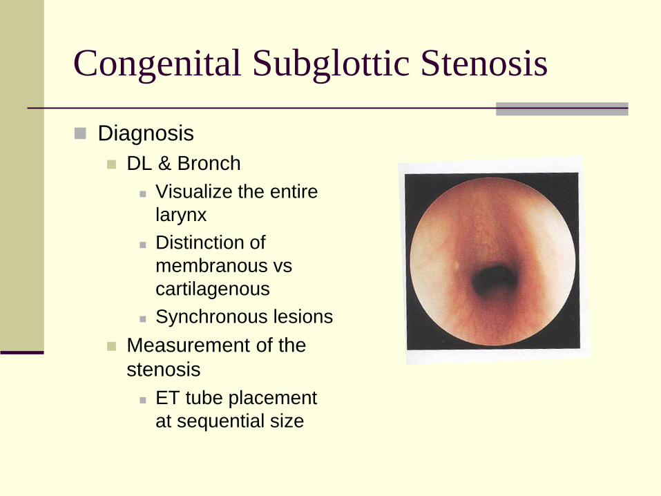
Congenital Subglottic Stenosis
Diagnosis
DL & Bronch
Visualize the entire
larynx
Distinction of
membranous vs
cartilagenous
Synchronous lesions
Measurement of the
stenosis
ET tube placement
at sequential size
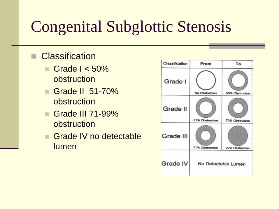
Congenital Subglottic Stenosis
Classification
Grade I < 50%
obstruction
Grade II 51-70%
obstruction
Grade III 71-99%
obstruction
Grade IV no detectable
lumen

Congenital Subglottic Stenosis
Treatment of Grade I
Watchful waiting for growth
>50% obstruction may require some
intervention
Soft tissue acquired lesions
Dilation & laser (CO2 & KTP) are sometimes
effective
Most congenital stenosis are cartilagenous
Laser & dilation are not useful
Congenital Subglottic Stenosis
Grade II-III treatment
Multiple failed extubation
Tracheostomy may be needed
Until cricoid grows for decannulation
Anterior cricoid split
Successful extubation in 66-78%
Decannulation rate 75-78%
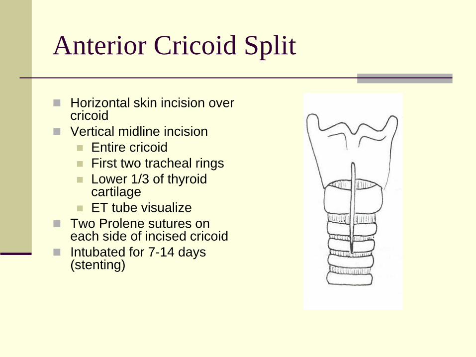
Anterior Cricoid Split
Horizontal skin incision over cricoid
Vertical midline incision
Entire cricoid
First two tracheal rings
Lower 1/3 of thyroid cartilage
ET tube visualize
Two Prolene sutures on each side of incised cricoid
Intubated for 7-14 days (stenting)
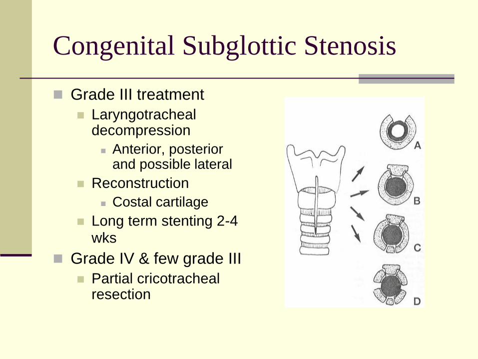
Congenital Subglottic Stenosis
Grade III treatment
Laryngotracheal decompression
Anterior, posterior and possible lateral
Reconstruction
Costal cartilage
Long term stenting 2-4 wks
Grade IV & few grade III
Partial cricotracheal resection
Laryngeal & Larygotracheoesophageal
clefts
rare, incidence of <0.1%
Incomplete development of TE septum
Communication of posterior larynx and
esophagus
Strong association with other anomalies
(56%)
TE fistula in 25%
Laryngeal & larygoesophageal Clefts
Laryngeal Clefting
Interarytenoids only
Partial or complete cricoid
Laryngotracheoesophageal clefts
Cervical or intrathoracic trachea

Laryngeal & Laryngotracheoesophageal Clefts
Symptoms
Proportional to the length
Can be asymptomatic (minor)
Inspiratory stridor
Feeding problems aspiration
Cyanotic episodes
Recurrent pneumonia
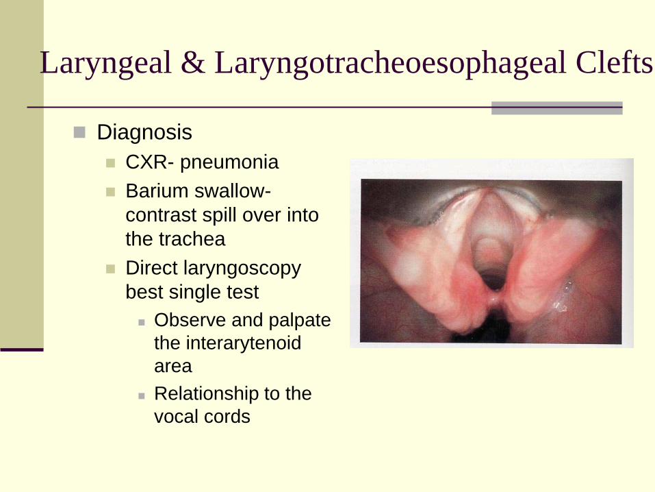
Diagnosis
CXR- pneumonia
Barium swallow-
contrast spill over into
the trachea
Direct laryngoscopy
best single test
Observe and palpate
the interarytenoid
area
Relationship to the
vocal cords
Laryngeal & Laryngotracheoesophageal Clefts
Treatment
Supraglottic larynx
Conservative management
Swallowing therapy to prevent aspiration
GERD evaluation and treatment
Surgical approach
80% success rate with Endoscopic repair
Extension below the vocal cords
Surgical repair is required
Laryngeal & Laryngotracheoesophageal Clefts
Mortality
Laryngeal clefts, rate of 11% and 46%
other anomalies
Delay in diagnosis
Intrathoracic laryngotracheoesophageal is as
high as 93%
Laryngeal & Laryngotracheoesophageal Clefts
Subglottic Hemangiomas
Benign vascular malformations
Histological- endothelial hyperplasia
Female predominance 2:1
Asymptomatic at birth
Stridor presents by 6 months (85%)
Associated cutaneous hemangioma (50%)
Subglottic Hemangiomas
Rapid growth phase in the 1st year followed
by slow resolution
Most have complete resolution by 5 years
30-70% mortality rate if untreated
Priority is to maintain the airway while
minimizing potential long term sequelae
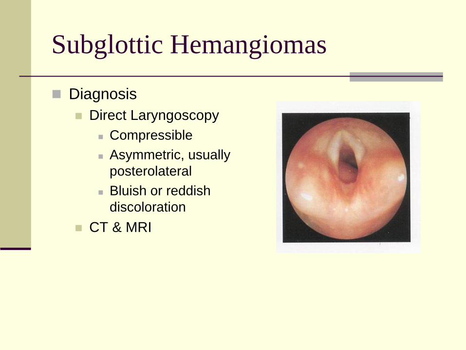
Subglottic Hemangiomas
Diagnosis
Direct Laryngoscopy
Compressible
Asymmetric, usually
posterolateral
Bluish or reddish
discoloration
CT & MRI
Treatment of Subglottic hemangiomas
Systemic steroids (principal)
Partial regression in most patients (82-97%)
Risk of growth retardation and increase susceptibility to infection
Risk is reduced by alternate-day dosing regimen in the smallest doses
Also intralesion corticosteroids has been employed with successful avoidance of tracheotomy
Interferon alpha-2a
50% or greater regression of lesion in 73% of patients
It requires prolonged therapy, blocks various steps of angiogenesis
Side effects neuromuscular impairment, skin slough, fever and liver enzyme elevation
Treatment of Subglottic hemangiomas
Tracheotomy
Bypass the obstructing lesion
Waiting for the expected involution
risks of tracheostomy as well as delay in
speech and language
Laser CO2 and KTP
associated with a significant risk of inducing
subglottic stenosis in up to 20%
Treatment of Subglottic Hemangiomas
Surgical excision
Decannulation shortly after surgery
Avoiding tracheostomy in 85% of patients
Laryngeal distortion or damage