congenital pial arteriovenous fistula in the temporal ... · traumatic carotid cavernous fitulas...
TRANSCRIPT

497Korean J Radiol 14(3), May/Jun 2013kjronline.org
INTRODUCTION
Intracranial pial arteriovenous fistula (AVFs) are rare cerebrovascular lesions of the brain that have recently been recognized as a distinct pathological entity, differing from the cerebral arteriovenous malformations (AVMs) in that they lack a true nidus. According to a series reported by Halbach et al. (1), intracranial pial AVFs account for 1.6% of all intracranial vascular malformations. However, congenital AVFs draining into the cavernous sinus in infants are very rare. Here we present the first case of a congenital pial AVF between the temporal branch of the left
Congenital Pial Arteriovenous Fistula in the Temporal Region Draining into Cavernous Sinus: A Case ReportZiyin Zhang, MD1,2, Kun Yang, MD2, Chaohua Wang, MD1, Changwei Zhang, MD1, Xiaodong Xie, MD1, Jianjian Tang, MD2
1Department of Neurosurgery, West China Hospital of Sichuan University, Chengdu, Sichuan Province, 610041, China; 2Department of Neurosurgery, Affiliated Hospital of Hainan Medical College, Haikou, Hainan Province, 570102, China
This report concerns a 4-month-old infant with progressive prominent and redness of his left eye since birth. This report concerns a 4-month-old infant with progressive prominent redness of his left eye since birth. Angiography revealed a congenital pial arteriovenous fistula between the temporal branch of the left posterior cerebral artery and left cavernous sinus through the sphenoparietal sinus, a condition not reported in the literature. The fistula was successfully occluded with two micro-coils by vertebrobasilar approach.Index terms: Pial arteriovenous fistula; Embolization; Infant; Cavernous sinus
Received October 25, 2012; accepted after revision December 10, 2012.Corresponding author: Xiaodong Xie, MD, Department of Neurosurgery, West China Hospital of Sichuan University, Chengdu, Sichuan Province, 610041, China. • Tel: (86137) 0800-5179 • Fax: (8628) 8542-2600• E-mail: [email protected] is an Open Access article distributed under the terms of the Creative Commons Attribution Non-Commercial License (http://creativecommons.org/licenses/by-nc/3.0) which permits unrestricted non-commercial use, distribution, and reproduction in any medium, provided the original work is properly cited.
Case Report | Neurointervention
Korean J Radiol 2013;14(3):497-500
posterior cerebral artery (PCA) and the left cavernous sinus through the sphenoparietal sinus, successfully treated by a vertebrobasilar approach with the use of two detachable micro-coils.
CASE REPORT
A 4-month-old infant was admitted to the department of ophthalmology in our hospital with progressive proptosis and redness of his left eye since birth. He was uneventfully delivered at full term, weighing 3.1 kg, and from a healthy mother. There was no history of birth trauma, and the neurologic examination was normal. There were no signs of irritability, fever or disturbance of swallowing, and weight gain was good. Blood pressure and heart rate were within normal limits. However, the physical examination revealed engorged conjunctiva, tortuous episcleral vessels, and swollen left eyelids. A slight intermittent systolic bruit was heard over the left global, whose rhythm was consistent with the heartbeat. The ophthalmologist strongly suspected cerebrovascular pathology, so the baby was transferred to the department of neurosurgery.
An MRI examination revealed orbital edema, proptosis,
http://dx.doi.org/10.3348/kjr.2013.14.3.497pISSN 1229-6929 · eISSN 2005-8330
Korean J Radiol 14(3), May/Jun 2013 kjronline.org498
Zhang et al.
was kept at approximately 80% of the basal blood pressure for 72 hours after the embolization. The ocular symptoms completely disappeared after four days. The patient was discharged five days after the intervention without any neurological deficit. The MRI examination at nine months showed permanent obliteration of the fistula.
DISCUSSION
Intracranial pial AVFs are rare cerebrovascular lesions, representing only 1.6% of all brain AVMs in larger reported series (1). They have recently been recognized as a distinct pathological entity from other intracranial AVMs. Unlike the cerebral AVMs and dural AVFs, pial AVFs are comprised of one or more arterial connections to a single venous channel without any intervening nidus of vessels or capillaries. They can be congenital or result from trauma. Congenital pial AVFs are very rare, and little is known regarding their pathophysiological mechanisms. Abnormal angiogenesis, cytokines and associated vascular growth factors may play a role in the formation of congenital pial AVFs, and it is possible that an embryological misstep could produce these lesions (2).
This case is a congenital pial AVF between the temporal branch of the left posterior cerebral artery and the left cavernous sinus through the sphenoparietal sinus. Anatomically, temporal poles can drain into the sphenoparietal sinus and cavernous sinus (3). Thus, this type of AVF can occur, although no case like this has been reported. Similar to the carotid cavernous fistulas, a congenital pial arteriovenous fistula draining into cavernous sinus can cause bruit, proptosis, conjunctiva chemosis and symptoms referable to the optic and trigeminal nerves. Fistulas in newborns and infants manifest as enlarged head circumference, heart failure, seizures or intracranial hemorrhage (4-7). In our case, the infant presented only with progressive proptosis and redness of his left eye, so he was admitted to the department of ophthalmology initially. If this infant was continually misdiagnosed as an eye disease, the outcome may have been intracranial hemorrhage or heart failure.
Little research concerning the natural history of these lesions has been published because they are so rare. There are a few reports regarding spontaneous resolution of a congenital pial AVF draining into the cavernous sinus (2, 8). According to one report (9), conservative management of pial AVF was associated with mortality in five (63%) of
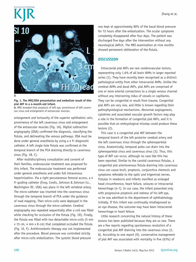
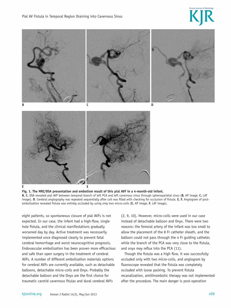
enlargement and tortuosity of the superior ophthalmic vein, prominence of the left cavernous sinus and enlargement of the extraocular muscles (Fig. 1A). Digital subtraction angiography (DSA) confirmed the diagnosis, classifying the fistula, and delineating the venous pathways. DSA must be done under general anesthesia by using a 4 Fr diagnostic catheter. A left single hole fistula was confirmed at the temporal branch of the PCA draining directly to cavernous sinus (Fig. 1B, C).
After multidisciplinary consultation and consent of their families, endovascular treatment was proposed for this infant. The endovascular treatment was performed under general anesthesia and under full intravenous heparinization. Via a right percutaneous femoral access, a 4 Fr guiding catheter (Evoy, Cordis, Johnson & Johnson Co., Washington DC, USA) was place in the left vertebral artery. The micro-catheter was inserted into the cavernous sinus through the temporal branch of PCA under the guidance of road mapping. Then micro-coils were deployed in the cavernous sinus through the micro-catheter. Cerebral angiography was repeated sequentially after a coil was filled while checking for occlusion of the fistula (Fig. 1D). Finally, the fistula was filled with two detachable micro-coils (5 mm x 15 cm, 4 mm x 8 cm) that completed occluded the fistula (Fig. 1E, F). Antithrombotic therapy was not implemented after the procedure. Blood pressure was controlled strictly after micro-coils embolization. The systolic blood pressure
Fig. 1. The MRI/DSA presentation and embolism result of this pial AVF in a 4-month-old infant.A. MRI revealed that proptosis of left eye, prominence of left cavern-ous sinus and enlargement of extraocular muscles.
A
Korean J Radiol 14(3), May/Jun 2013kjronline.org 499
Pial AV Fistula in Temporal Region Draining into Cavernous Sinus
eight patients, so spontaneous closure of pial AVFs is not expected. In our case, the infant had a high-flow, single hole fistula, and the clinical manifestations gradually worsened day by day. Active treatment was necessarily implemented once diagnosed clearly to prevent fatal cerebral hemorrhage and worst neurocognitive prognosis. Endovascular embolization has been proven more efficacious and safe than open surgery in the treatment of cerebral AVFs. A number of different embolization materials options for cerebral AVFs are currently available, such as detachable balloons, detachable micro-coils and Onyx. Probably the detachable balloon and the Onyx are the first choice for traumatic carotid cavernous fitulas and dural cerebral AVFs
(2, 9, 10). However, micro-coils were used in our case instead of detachable balloon and Onyx. There were two reasons: the femoral artery of the infant was too small to allow the placement of the 8 Fr catheter sheath, and the balloon could not pass through the 4 Fr guiding catheter, while the branch of the PCA was very close to the fistula, and onyx may reflux into the PCA (11).
Though the fistula was a high flow, it was successfully occluded only with two micro-coils, and angiogram by fluoroscope revealed that the fistula was completely occluded with loose packing. To prevent fistula recanalization, antithrombotic therapy was not implemented after the procedure. The main danger is post-operation
DB
E E
C
Fig. 1. The MRI/DSA presentation and embolism result of this pial AVF in a 4-month-old infant.B, C. DSA revealed pial AVF between temporal branch of left PCA and left cavernous sinus through sphenoparietal sinus (B. AP image, C. LAT image). D. Cerebral angiography was repeated sequentially after coil was filled with checking for occlusion of fistula. E, F. Angiogram of post-embolization revealed fistula was entirely occluded by using only two micro-coils (E. AP image, F. LAT image).

Korean J Radiol 14(3), May/Jun 2013 kjronline.org500
Zhang et al.
cerebral hemorrhage of the feeding arteries, which must be controlled by strict blood pressure monitoring while maintaining the systolic blood pressure at approximately 80% of the basal blood pressure for 72 hours after embolization. Four days after embolization, the symptoms and signs entirely disappeared. The stable occlusion of the fistula has been demonstrated by MRI examination at 9 months.
REFERENCES
1. Halbach VV, Higashida RT, Hieshima GB, Norman D. Normal perfusion pressure breakthrough occurring during treatment of carotid and vertebral fistulas. AJNR Am J Neuroradiol 1987;8:751-756
2. Hoh BL, Putman CM, Budzik RF, Ogilvy CS. Surgical and endovascular flow disconnection of intracranial pial single-channel arteriovenous fistulae. Neurosurgery 2001;49:1351-1363; discussion 1363-1364
3. Oka K, Rhoton AL Jr, Barry M, Rodriguez R. Microsurgical anatomy of the superficial veins of the cerebrum. Neurosurgery 1985;17:711-748
4. Amaral FT, Machado HR, Almeida S. [Congenital cerebral arteriovenous fistula. Diagnostic peculiarity and surgical
repair in an infant with heart failure]. Arq Bras Cardiol 1994;63:207-209
5. Haase J. Congestive heart failure secondary to cerebral arteriovenous fistula. Childs Nerv Syst 1988;4:75
6. Berant M, Tadmor R, Blieden L, Deutsch V, Neufeld HN. Cerebral arteriovenous fistula causing congestive heart failure in infancy. Angiology 1977;28:684-686
7. Sasamori T, Hida K, Yano S, Asano T, Iwasaki Y. Cervical perimedullary arteriovenous fistula in an infant presenting with subarachnoid hemorrhage--case report. Neurol Med Chir (Tokyo) 2008;48:409-413
8. Lee JY, Son YJ, Kim JE. Intracranial pial arteriovenous fistulas. J Korean Neurosurg Soc 2008;44:101-104
9. Nelson K, Nimi Y, Lasjaunias P, Berenstein A. Endovascular embolization of congenital intracranial pial arteriovenous fistulas. Neuroimaging Clin N Am 1992;2:309-317
10. Paramasivam S, Toma N, Niimi Y, Berenstein A. De novo development of dural arteriovenous fistula after endovascular embolization of pial arteriovenous fistula. J Neurointerv Surg 2012 [Epub ahead of print]
11. Rivera R, Blanc R, Piotin M, Spelle L, Moret J. Single hole cerebral arteriovenous fistula between the anterior choroidal artery and the basal vein of Rosenthal in a child. Childs Nerv Syst 2009;25:1521-1523