congenital sick sinus syndrome caused by recessive mutations in
TRANSCRIPT
IntroductionSick sinus syndrome (SSS) was described nearly 40years ago as a complicating arrhythmia following car-dioversion (1). The disorder is characterized by inap-propriate sinus bradycardia, sinus arrest, or chrono-tropic incompetence (2, 3). SSS has been attributed todysfunction of the sinoatrial (SA or sinus) node, butdespite extensive efforts to define SSS in terms ofabnormal automaticity, exit block, or impaired intraa-trial conduction and excitability, it has remainedlargely an electrocardiographic diagnosis. Episodes ofatrial tachycardias coexisting with sinus bradycardia(tachycardia-bradycardia syndrome) are also commonin this disorder.
Although primarily a disease of the elderly, SSS is animportant clinical problem in pediatric patients,because treatment of associated exercise intolerance,presyncope or syncope, usually requires lifelong car-diac pacing. In children a previous history of cardiacsurgery for congenital heart malformation is noted inapproximately 80% of SSS cases (4–7). SSS is also seenin the fetus, infant, and child without heart disease orother contributing factors, however (5, 6, 8–13). In thissetting, a congenital origin is presumed and bradycar-dia progressing to atrial inexcitability or standstill hasbeen noted, a phenomenon highly unusual in SSSassociated with postoperative heart disease (11, 14).Familial cases consistent with autosomal dominantinheritance with reduced penetrance or recessiveinheritance have been observed (6, 8, 10–12, 14–16).Familial SSS is grouped with congenital absence ofsinus rhythm and familial sinus node disease underthe term “nodal rhythm” in the online MendelianInheritance of Man database (OMIM 163800).
Based on prior associations with disorders of car-diac rhythm and conduction, we screened the α sub-unit of the cardiac sodium channel (SCN5A) as a can-didate gene in ten pediatric patients from sevenfamilies who were diagnosed with congenital SSS dur-ing the first decade of life. Compound heterozygousSCN5A mutations were found in five individuals fromthree families, indicating that congenital SSS may, insome families, segregate as a recessive disorder of thecardiac sodium channel.
The Journal of Clinical Investigation | October 2003 | Volume 112 | Number 7 1019
Congenital sick sinus syndrome caused by recessivemutations in the cardiac sodium channel gene (SCN5A)
D. Woodrow Benson,1 Dao W. Wang,2 Macaira Dyment,1 Timothy K. Knilans,1
Frank A. Fish,3 Margaret J. Strieper,4 Thomas H. Rhodes,5 Alfred L. George, Jr.2,5
1Department of Pediatrics, Cincinnati Children’s Hospital, Cincinnati, Ohio, USA2Department of Pharmacology, and3Department of Pediatrics, Vanderbilt University School of Medicine, Nashville, Tennessee, USA4Sibley Heart Center-Cardiology, Children’s Healthcare of Atlanta, Atlanta, Georgia, USA5Department of Medicine, Vanderbilt University School of Medicine, Nashville, Tennessee, USA
Sick sinus syndrome (SSS) describes an arrhythmia phenotype attributed to sinus node dysfunctionand diagnosed by electrocardiographic demonstration of sinus bradycardia or sinus arrest. Althoughfrequently associated with underlying heart disease and seen most often in the elderly, SSS may occurin the fetus, infant, and child without apparent cause. In this setting, SSS is presumed to be congen-ital. Based on prior associations with disorders of cardiac rhythm and conduction, we screened theα subunit of the cardiac sodium channel (SCN5A) as a candidate gene in ten pediatric patients fromseven families who were diagnosed with congenital SSS during the first decade of life. Probands fromthree kindreds exhibited compound heterozygosity for six distinct SCN5A alleles, including two muta-tions previously associated with dominant disorders of cardiac excitability. Biophysical characteri-zation of the mutants using heterologously expressed recombinant human heart sodium channelsdemonstrate loss of function or significant impairments in channel gating (inactivation) that pre-dict reduced myocardial excitability. Our findings reveal a molecular basis for some forms of con-genital SSS and define a recessive disorder of a human heart voltage-gated sodium channel.
J. Clin. Invest. 112:1019–1028 (2003). doi:10.1172/JCI200318062.
Received for publication February 6, 2003, and accepted in revised formJuly 22, 2003.
Address correspondence to: D. Woodrow Benson, Division ofCardiology, ML7042, Children’s Hospital Medical Center, 3333 Burnet Avenue, Cincinnati, Ohio 45229-3039, USA. Phone: (513) 636-7716; Fax: (513) 636-5958; E-mail: [email protected]. Woodrow Benson and Dao W. Wang contributed equally tothis work.Conflict of interest: The authors have declared that no conflict ofinterest exists.Nonstandard abbreviations used: sick sinus syndrome (SSS);human heart sodium channel (hH1); human β1 subunit (hβ1);viral internal ribosome entry site (IRES); current (I);amplification-refractory mutation system analysis (ARMS); wild-type hH1 (WT-hH1); human β3 subunit (hβ3);hyperpolarization-activated cyclic nucleotide-gated (HCN4).
1020 The Journal of Clinical Investigation | October 2003 | Volume 112 | Number 7
Methods
Clinical studies
Probands with symptomatic sinus bradycardia leadingto pacemaker implantation and their family memberswere invited to participate in the study. Informed con-sent was obtained from all participants in accordancewith the Cincinnati Children’s Hospital Medical Cen-ter Institutional Review Board. Participants were eval-uated by history, review of medical records, and 12-leadelectrocardiogram. Clinical studies were performedwithout knowledge of genotype.
Molecular genetic methods
Genomic DNA was isolated from blood of study par-ticipants, and the PCR was used to amplify the codingregion and flanking intronic sequence of SCN5A, asdescribed previously (17). Sequencing reactions wereperformed in the presence of fluorescence-labeleddideoxynucleotides and additional primer for exon-specific sequencing in both sense and antisense direc-tion on isolated PCR product.
Biophysical characterization of SCN5A mutants
Mutagenesis and heterologous expression of Na channels. Allsix mutations (T220I, P1298L, G1408R, delF1617,R1623X, and R1632H) were engineered in a recombi-nant human heart sodium channel cDNA (hH1) usingrecombinant PCR. Final constructs were assembled inthe mammalian expression plasmid pRc/CMV-hH1and then sequenced to verify creation of the mutationand exclusion of polymerase errors.
Cells (tsA201) were transiently transfected with pRc-CMV-hH1, T220I, P1298L, delF1617, or R1632H usingSuperFect (QIAGEN Inc., Santa Clarita, California,USA) in combination with a plasmid (pGFP-IRES-hβ1)encoding both GFP and the human β1 subunit (hβ1)under the control of a single CMV promoter with thetwo coding regions separated by a viral internal ribo-some entry site (IRES). This approach ensures that cellsexhibiting green fluorescence have been successfullytransfected with the β1-encoding plasmid. Transfec-tions were performed with 1 µg of channel-encodingplasmid DNA and 1 µg of pGFP-IRES-hβ1. In someexperiments, we cotransfected hH1 with pGFP-IRES-hβ3 encoding the human sodium channel β3 subunitrather than the β1 subunit. Transiently transfectedcells were transferred to a chamber (Warner InstrumentCorp., Hamden, Connecticut, USA) for electrophysio-logical measurements 24–72 hours after transfection.
Electrophysiology. Sodium currents were recorded usingthe whole-cell patch-clamp technique as described previ-ously (18). Electrode resistance ranged from 0.8 to 1.5MΩ. Data acquisition was carried out using an Axopatch200B patch-clamp amplifier and pCLAMP 8.0 software(Axon Instruments Inc., Foster City, California, USA).Currents were acquired at 20–50 kHz, filtered at 5 kHz(–3 dB, four-pole Bessel filter), and digitized using an ana-logue-to-digital interface (Digidata 1320A; Axon Instru-
ments Inc.). Recordings from cells exhibiting peak cur-rent amplitudes less than 0.8 nA were excluded fromanalysis to avoid potential endogenous channel contam-ination. Cells exhibiting very large whole-cell currentswere also excluded if voltage control was compromised.Typically, fewer than 20% of cells were excluded fromanalyses. Whole-cell capacitance and access resistancewere determined by integrating capacitive transients ofvoltage steps from –120 mV to –110 mV filtered at 10–50kHz. Series resistance compensation ensured that thecommand potential was achieved on a microsecond timescale with voltage errors less than 3 mV. The holdingpotential was –120 mV for all experiments, except whenotherwise indicated (details of each pulse protocol aregiven schematically in the figures and explained inResults or figure legends). The time between sequentialvoltage protocols was 10 seconds for all experiments. Allexperiments were performed at 22°C. The bath solutioncontained 145 mM NaCl, 4 mM KCl, 1.8 mM CaCl2, 1mM MgCl2, 10 mM HEPES, and 10 mM glucose, pH 7.35(adjusted with NaOH). The pipette solution (intracellu-lar solution) contained 10 mM NaF, 110 mM CsF, 20mM CsCl, 10 mM EGTA, and 10 mM HEPES, pH 7.35(adjusted with CsOH). Osmolarity was adjusted to 310milliosmoles with sucrose for all solutions.
Data analysis. At least two independent clones weretested for each mutant, and the data were pooled. Alldata were analyzed using pCLAMP 8.0 (Axon Instru-ments Inc.), and figures were prepared using SigmaPlot2002 (SPSS Science, Chicago, Illinois, USA). The timecourse of inactivation was fit with a two-exponentialfunction: I(t)/Imax = A1 × exp(–t/τ1) + A2 × exp(–t/τ2)where values for A and τ refer to amplitudes and timeconstants, respectively. I refers to current and t refers totime. Steady-state availability was fit with the Boltz-mann equation, I/Imax = [1 + exp((V – V1/2)/k)] – 1, todetermine the membrane potential for half-maximalinactivation (V1/2) and the slope factor k. Recovery frominactivation was analyzed by fitting data with the two-exponential function: I(t)/Imax = Af × [1 – exp(–t/τf)] + As
× [1 – exp(–t/τs)]. Curve fitting was performed using theMarquardt-Levenberg algorithm. A coefficient of deter-mination greater than 0.985, standard error of eachparameter less than 0.3, and coefficients of variation lessthan 2% were considered evidence of the best fit. Thevoltage-dependence of channel activation was estimat-ed by measuring peak sodium current during a variabletest potential from a holding potential of –120 mV. Thecurrent at each membrane potential was divided by theelectrochemical driving force for sodium ions, Vm – ENa
(Vm is the test potential and ENa represents the sodiumequilibrium potential) and normalized to the maxi-mum sodium conductance. All data were fit using anonlinear least-squares minimization method. Resultsare presented as means plus or minus standard error,and the statistical comparisons were made using theunpaired Student’s t test. Statistical significance wasassumed for P values less than 0.05. In some figures, thestandard error bars are smaller than the data symbols.
The Journal of Clinical Investigation | October 2003 | Volume 112 | Number 7 1021
ResultsClinical characteristics. Sinus bradycardia was diagnosedin ten infants and children during evaluation ofabnormal heart rate; none had a history of cardiacsurgery. In four cases (three families) the initial diag-nosis had been made in utero (20–38 weeks gestation)during Doppler-echocardiographic evaluation ofirregular fetal heart rate, and in six cases the diagno-sis was made by standard electrocardiogram betweenthe ages of two and nine years during evaluation ofirregular heart rate, presyncope or syncope. A con-genital heart defect, including aortic stenosis (2), pul-monary stenosis (1), or heterotaxy syndrome (1) wasidentified in four subjects in whom congenital SSShad been diagnosed in utero. Except for probands andtheir siblings, there was no family history of a cardiacrhythm disorder or other forms of cardiovascular dis-ease in the young. Nine subjects were white and onewas African-American.
All ten patients with SSS underwent pacemakerimplantation. Intracardiac electrophysiology studiesperformed in three subjects demonstrated atrial inex-citability (no spontaneous atrial depolarization,inability to capture with pacing) and prolonged His-ventricle conduction time (60 milliseconds). Atrialinexcitability was observed in two additional patients
at the time of pacemaker implant. One subjectwith heterotaxy syndrome died during thefirst month of life, but other subjects havedone well for the follow-up of 2–15 years.
Compound heterozygous SCN5A mutations identi-fied in five individuals. Based on prior associa-
tions with disorders of cardiac rhythm and conduction,we considered SCN5A as a candidate gene for congeni-tal SSS. During systematic survey of all SCN5A-codingexons, compound heterozygous nucleotide changeswere identified in five individuals from three kindreds(Figure 1). The age of SSS diagnosis in these five indi-viduals was 2 years to 9 years; none had other evidenceof heart disease. None of the nucleotide sequencechanges were identified in over 150 chromosomes fromnormal, unrelated control subjects.
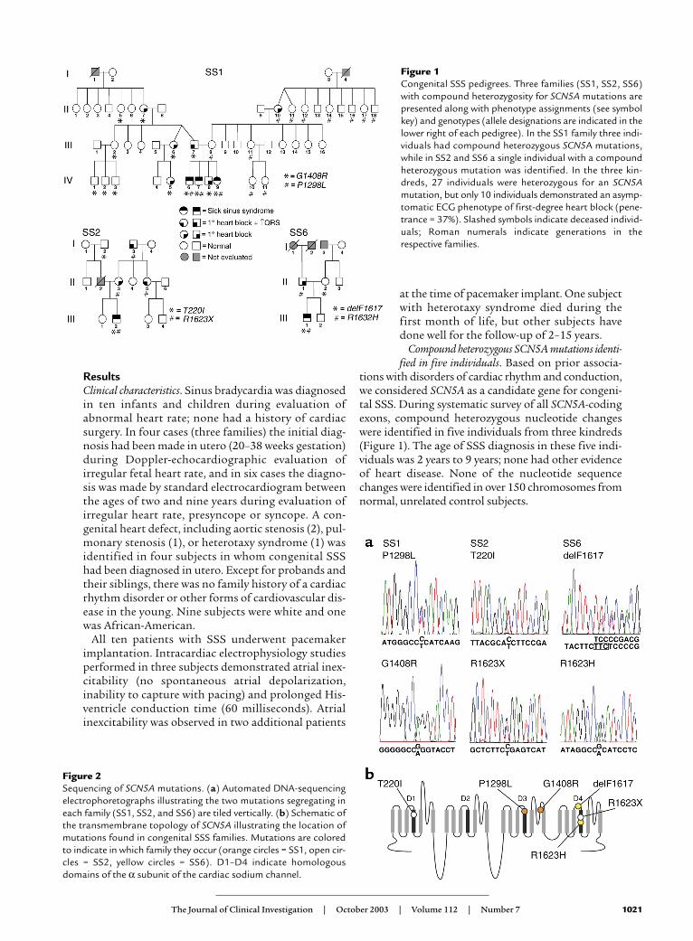
Figure 1Congenital SSS pedigrees. Three families (SS1, SS2, SS6)with compound heterozygosity for SCN5A mutations arepresented along with phenotype assignments (see symbolkey) and genotypes (allele designations are indicated in thelower right of each pedigree). In the SS1 family three indi-viduals had compound heterozygous SCN5A mutations,while in SS2 and SS6 a single individual with a compoundheterozygous mutation was identified. In the three kin-dreds, 27 individuals were heterozygous for an SCN5Amutation, but only 10 individuals demonstrated an asymp-tomatic ECG phenotype of first-degree heart block (pene-trance = 37%). Slashed symbols indicate deceased individ-uals; Roman numerals indicate generations in therespective families.
Figure 2Sequencing of SCN5A mutations. (a) Automated DNA-sequencingelectrophoretographs illustrating the two mutations segregating ineach family (SS1, SS2, and SS6) are tiled vertically. (b) Schematic ofthe transmembrane topology of SCN5A illustrating the location ofmutations found in congenital SSS families. Mutations are coloredto indicate in which family they occur (orange circles = SS1, open cir-cles = SS2, yellow circles = SS6). D1–D4 indicate homologousdomains of the α subunit of the cardiac sodium channel.
1022 The Journal of Clinical Investigation | October 2003 | Volume 112 | Number 7
In family SS1, three individuals with congenital SSS(Figure 1, IV-6, IV-7, and IV-9) were found to haveinherited two distinct missense mutations (Figure 2).The maternal allele is a previously undescribed C → Ttransition at nucleotide 3893 (nucleotide numberingbased upon the open reading frame sequence of Gen-Bank accession number M77235) and encoding leucinein place of proline 1298 (designated P1298L). A G → Atransition of nucleotide 4222 was transmitted from thefather and resulted in substitution of arginine forglycine at codon 1408 (G1408R). This mutation, previ-ously designated as G1406R, was reported in a largeFrench family segregating both Brugada syndrome andisolated cardiac conduction system disease in a domi-
nant manner (19). The nucleotide changes 3893C → Tand 4222G → A abolish a BanII site and create a BstXIrestriction endonuclease site, respectively, allowingindependent confirmation of P1298L and G1408R.
In family SS2 we inferred transmission to theproband (III-2) of a previously undescribed missenseallele (659C → T, T220I) from the paternal lineage andinheritance of a novel nonsense mutation (4867C → T,R1623X) from his asymptomatic mother (Figure 2).Interestingly, two distinct missense mutations affect-ing codon 1623 (R1623Q, R1623L) have been previ-ously associated with congenital long QT syndrome(20, 21). Amplification-refractory mutation systemanalysis (ARMS) was used to confirm the presence orabsence of 659C → T (T220I) (Table 1) (22). Thenucleotide change 4867C → T creates a DdeI restric-tion endonuclease site allowing independent confir-mation of R1623X.
The proband in family SS6 (III-1) was found to havecompound heterozygosity for a previously reported in-frame deletion of codon 1617 (4849–4851delTTC,delF1617) inherited from the mother and a novel mis-sense mutation (4895G → A, R1632H) transmittedfrom the father (Figure 1). Deletion of codon 1617(del4849–4851) eliminates a single phenylalanineresidue in the short extracellular loop between segmentsS3 and S4 in the fourth-repeat domain (D4); delF1617
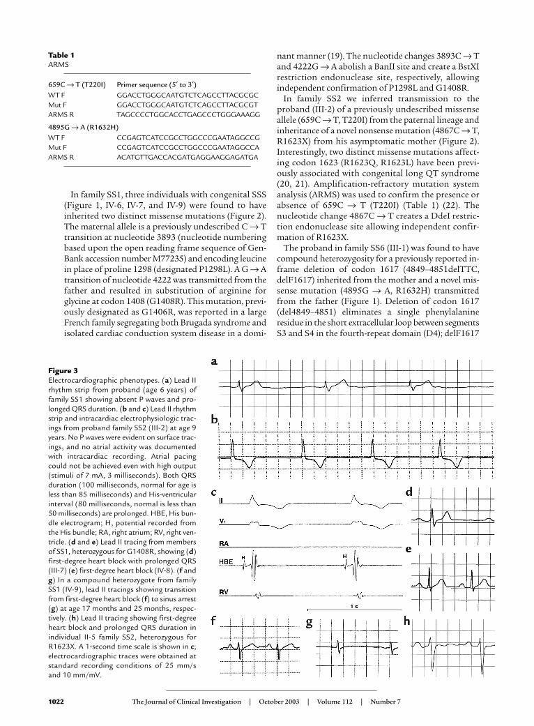
Figure 3Electrocardiographic phenotypes. (a) Lead IIrhythm strip from proband (age 6 years) offamily SS1 showing absent P waves and pro-longed QRS duration. (b and c) Lead II rhythmstrip and intracardiac electrophysiologic trac-ings from proband family SS2 (III-2) at age 9years. No P waves were evident on surface trac-ings, and no atrial activity was documentedwith intracardiac recording. Atrial pacingcould not be achieved even with high output(stimuli of 7 mA, 3 milliseconds). Both QRSduration (100 milliseconds, normal for age isless than 85 milliseconds) and His-ventricularinterval (80 milliseconds, normal is less than50 milliseconds) are prolonged. HBE, His bun-dle electrogram; H, potential recorded fromthe His bundle; RA, right atrium; RV, right ven-tricle. (d and e) Lead II tracing from membersof SS1, heterozygous for G1408R, showing (d)first-degree heart block with prolonged QRS(III-7) (e) first-degree heart block (IV-8). (f andg) In a compound heterozygote from familySS1 (IV-9), lead II tracings showing transitionfrom first-degree heart block (f) to sinus arrest(g) at age 17 months and 25 months, respec-tively. (h) Lead II tracing showing first-degreeheart block and prolonged QRS duration inindividual II-5 family SS2, heterozygous forR1623X. A 1-second time scale is shown in c;electrocardiographic traces were obtained atstandard recording conditions of 25 mm/sand 10 mm/mV.
Table 1ARMS
659C → T (T220I) Primer sequence (5′ to 3′)WT F GGACCTGGGCAATGTCTCAGCCTTACGCGCMut F GGACCTGGGCAATGTCTCAGCCTTACGCGTARMS R TAGCCCCTGGCACCTGAGCCCTGGGAAAGG
4895G → A (R1632H)WT F CCGAGTCATCCGCCTGGCCCGAATAGGCCGMut F CCGAGTCATCCGCCTGGCCCGAATAGGCCAARMS R ACATGTTGACCACGATGAGGAAGGAGATGA
The Journal of Clinical Investigation | October 2003 | Volume 112 | Number 7 1023
was described previously in association with congenitallong QT syndrome, although no specific clinical orgenetic information was provided (21). The nucleotidechange 4867C → T abolishes a BsaXI restrictionendonuclease site allowing independent confirmationof del1617F. ARMS was used to confirm the presence orabsence of 4895G → A (R1632H) (Table 1) (22).
Clinical electrophysiologic phenotype of SCN5A mutationcarriers. There was no family history of cardiac arrest orsudden death in any of the three families. Among 27heterozygotes, no individual exhibited ECG evidence ofBrugada syndrome. The mean QTc in heterozygoteswas 400 ± 17 milliseconds while that in mutation-neg-ative family members (n = 27) was 423 ± 16 millisec-onds (P = NS by Student’s t test).
In family SS1, electrocardiographic evaluation ofbradycardia detected in the proband (IV-7) at age 6years demonstrated absent P waves, pauses up to 5 sec-onds, and an escape rhythm at 42 beats per minutewith a prolonged QRS waveform (Figure 3a). Intracar-diac electrophysiologic study demonstrated a pro-longed His-ventricle conduction time (72 millisec-onds), absent atrial depolarizations and atrialinexcitability (not shown). Subsequent evaluation ofhis seven-year-old brother (IV-6) revealed similar find-ings. During serial observations in a younger sister (IV-9), congenital SSS was first detected at age 25months (Figure 3, f and g). Eleven heterozygous carri-ers of P1298L were asymptomatic and largely withoutelectrocardiographic abnormalities, except the mater-nal grandmother (II-10), who had first-degree heartblock (Figure 1). Of the ten G1408R heterozygotes infamily SS1 ranging in ages from 3 to 68 years, five indi-viduals exhibited asymptomatic first-degree heartblock (Figure 3, d and e), including oneindividual (III-7) with associated prolon-gation of QRS duration; the remainingfive individuals had no electrocardio-graphic abnormalities.
During evaluation for syncope at nineyears of age, the proband in family SS2demonstrated absent P waves, pauses upto 15 seconds, and an escape rhythm at50 beats per minute; intracardiac elec-trophysiologic study demonstrated pro-longation of QRS duration and His-ven-tricle conduction time (68 milliseconds),absent atrial depolarizations, and atrialinexcitability (Figure 3, b and c). Fol-lowing pacemaker implantation, heexperienced exertional syncope. Anintracardiac cardioverter defibrillatorwas implanted; during 10 years of treat-ment with β-adrenergic blockade, he hashad a single discharge for an idiopathicirregular monomorphic ventriculartachycardia at 210 bpm. Three asymp-tomatic carriers of R1623X exhibitedfirst-degree heart block; intraventricular
conduction delay (I-3) or right bundle branch blockand left anterior fascicular block (II-5, Figure 3h) wasalso present in one individual each. The single T220Iheterozygote (I-2) in this family was clinically andelectrocardiographically normal.
In family SS6, the diagnosis of congenital SSS wasdetermined in the proband (III-1) at age 5 years. Asnoted in other probands, bradycardia, absent atrialdepolarizations, atrial inexcitability, prolonged QRS,and prolonged His-ventricle conduction time (60 mil-liseconds) were observed. The single heterozygous car-rier of delF1617 (II-2) had a normal electrocardiogram.The single heterozygous carrier for R1632H (II-1)demonstrated electrocardiographic features of first-degree heart block.
Biophysical characterization of SCN5A mutants. To deter-mine the functional consequences of the SCN5A muta-tions associated with congenital SSS, we engineeredfour missense mutations (T220I, P1298L, G1408R,R1632H), the in-frame deletion (delF1617), and thenonsense mutation (R1623X) in the recombinanthuman cardiac voltage-gated sodium channel α sub-unit (designated hH1). We then expressed each muta-tion heterologously in a cultured mammalian cell line(tsA201) with the sodium channel accessory hβ1 andused whole-cell patch-clamp recording to characterizeresultant sodium currents. The nonsense mutation(R1623X) was suspected to encode nonfunctionalchannels, and mutation G1408R had previously beendemonstrated to disable sodium channel function (19);our experiments confirmed these predictions (data notshown). Expression of the remaining four mutationsexhibited measurable sodium channel activity intsA201 cells and were further characterized.
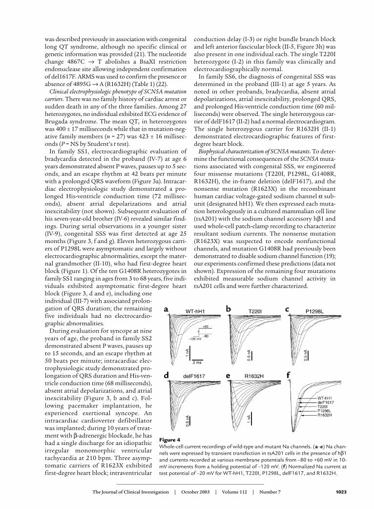
Figure 4Whole-cell current recordings of wild-type and mutant Na channels. (a–e) Na chan-nels were expressed by transient transfection in tsA201 cells in the presence of hβ1and currents recorded at various membrane potentials from –80 to +60 mV in 10-mV increments from a holding potential of –120 mV. (f) Normalized Na current attest potential of –20 mV for WT-hH1, T220I, P1298L, delF1617, and R1632H.
1024 The Journal of Clinical Investigation | October 2003 | Volume 112 | Number 7
Figure 4 illustrates representative whole-cell currenttracings from cells expressing either wild-type hH1(WT-hH1), T220I, P1298L, delF1617, or R1632H. Allrecombinant alleles exhibited rapid activation andinactivation in response to a series of depolarizing test
potentials typical of voltage-gated sodiumchannels. The mutants differed from WT-hH1,however, in the time course of inactivation.Three of the mutations (T220I, P1298L,delF1617) shared a similar mild degree of inac-tivation delay while R1632H exhibited a moresevere impairment (Figure 4f). Given the simi-larity of the biophysical disturbance exhibitedby T220I, P1298L, and delF1617, we presentthe functional characterization of these muta-tions separately from that of R1632H.
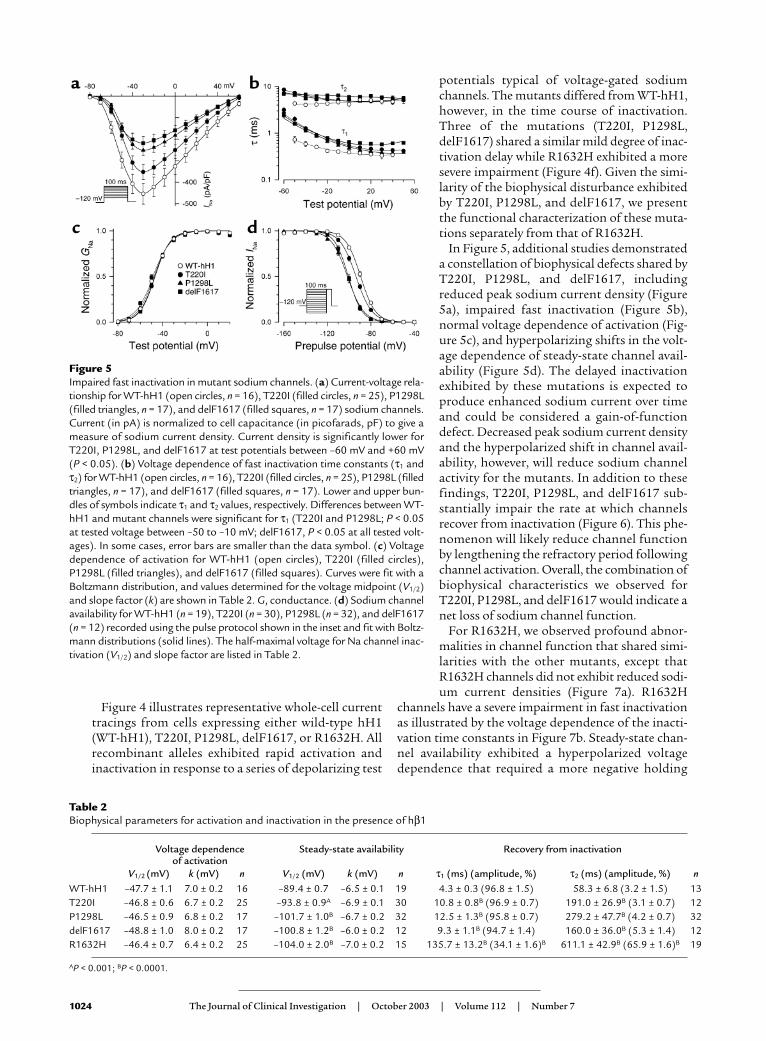
In Figure 5, additional studies demonstrateda constellation of biophysical defects shared byT220I, P1298L, and delF1617, includingreduced peak sodium current density (Figure5a), impaired fast inactivation (Figure 5b),normal voltage dependence of activation (Fig-ure 5c), and hyperpolarizing shifts in the volt-age dependence of steady-state channel avail-ability (Figure 5d). The delayed inactivationexhibited by these mutations is expected toproduce enhanced sodium current over timeand could be considered a gain-of-functiondefect. Decreased peak sodium current densityand the hyperpolarized shift in channel avail-ability, however, will reduce sodium channelactivity for the mutants. In addition to thesefindings, T220I, P1298L, and delF1617 sub-stantially impair the rate at which channelsrecover from inactivation (Figure 6). This phe-nomenon will likely reduce channel functionby lengthening the refractory period followingchannel activation. Overall, the combination ofbiophysical characteristics we observed forT220I, P1298L, and delF1617 would indicate anet loss of sodium channel function.
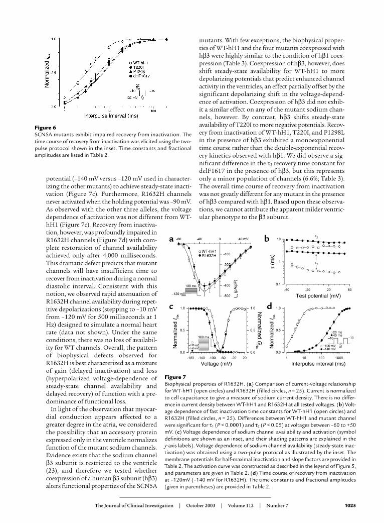
For R1632H, we observed profound abnor-malities in channel function that shared simi-larities with the other mutants, except thatR1632H channels did not exhibit reduced sodi-um current densities (Figure 7a). R1632H
channels have a severe impairment in fast inactivationas illustrated by the voltage dependence of the inacti-vation time constants in Figure 7b. Steady-state chan-nel availability exhibited a hyperpolarized voltagedependence that required a more negative holding
Figure 5Impaired fast inactivation in mutant sodium channels. (a) Current-voltage rela-tionship for WT-hH1 (open circles, n = 16), T220I (filled circles, n = 25), P1298L(filled triangles, n = 17), and delF1617 (filled squares, n = 17) sodium channels.Current (in pA) is normalized to cell capacitance (in picofarads, pF) to give ameasure of sodium current density. Current density is significantly lower forT220I, P1298L, and delF1617 at test potentials between –60 mV and +60 mV(P < 0.05). (b) Voltage dependence of fast inactivation time constants (τ1 andτ2) for WT-hH1 (open circles, n = 16), T220I (filled circles, n = 25), P1298L (filledtriangles, n = 17), and delF1617 (filled squares, n = 17). Lower and upper bun-dles of symbols indicate τ1 and τ2 values, respectively. Differences between WT-hH1 and mutant channels were significant for τ1 (T220I and P1298L; P < 0.05at tested voltage between –50 to –10 mV; delF1617, P < 0.05 at all tested volt-ages). In some cases, error bars are smaller than the data symbol. (c) Voltagedependence of activation for WT-hH1 (open circles), T220I (filled circles),P1298L (filled triangles), and delF1617 (filled squares). Curves were fit with aBoltzmann distribution, and values determined for the voltage midpoint (V1/2)and slope factor (k) are shown in Table 2. G, conductance. (d) Sodium channelavailability for WT-hH1 (n = 19), T220I (n = 30), P1298L (n = 32), and delF1617(n = 12) recorded using the pulse protocol shown in the inset and fit with Boltz-mann distributions (solid lines). The half-maximal voltage for Na channel inac-tivation (V1/2) and slope factor are listed in Table 2.
Table 2Biophysical parameters for activation and inactivation in the presence of hβ1
Voltage dependence Steady-state availability Recovery from inactivationof activation
V1/2 (mV) k (mV) n V1/2 (mV) k (mV) n τ1 (ms) (amplitude, %) τ2 (ms) (amplitude, %) nWT-hH1 –47.7 ± 1.1 7.0 ± 0.2 16 –89.4 ± 0.7 –6.5 ± 0.1 19 4.3 ± 0.3 (96.8 ± 1.5) 58.3 ± 6.8 (3.2 ± 1.5) 13T220I –46.8 ± 0.6 6.7 ± 0.2 25 –93.8 ± 0.9A –6.9 ± 0.1 30 10.8 ± 0.8B (96.9 ± 0.7) 191.0 ± 26.9B (3.1 ± 0.7) 12P1298L –46.5 ± 0.9 6.8 ± 0.2 17 –101.7 ± 1.0B –6.7 ± 0.2 32 12.5 ± 1.3B (95.8 ± 0.7) 279.2 ± 47.7B (4.2 ± 0.7) 32delF1617 –48.8 ± 1.0 8.0 ± 0.2 17 –100.8 ± 1.2B –6.0 ± 0.2 12 9.3 ± 1.1B (94.7 ± 1.4) 160.0 ± 36.0B (5.3 ± 1.4) 12R1632H –46.4 ± 0.7 6.4 ± 0.2 25 –104.0 ± 2.0B –7.0 ± 0.2 15 135.7 ± 13.2B (34.1 ± 1.6)B 611.1 ± 42.9B (65.9 ± 1.6)B 19
AP < 0.001; BP < 0.0001.
The Journal of Clinical Investigation | October 2003 | Volume 112 | Number 7 1025
potential (–140 mV versus –120 mV used in character-izing the other mutants) to achieve steady-state inacti-vation (Figure 7c). Furthermore, R1632H channelsnever activated when the holding potential was –90 mV.As observed with the other three alleles, the voltagedependence of activation was not different from WT-hH1 (Figure 7c). Recovery from inactiva-tion, however, was profoundly impaired inR1632H channels (Figure 7d) with com-plete restoration of channel availabilityachieved only after 4,000 milliseconds.This dramatic defect predicts that mutantchannels will have insufficient time torecover from inactivation during a normaldiastolic interval. Consistent with thisnotion, we observed rapid attenuation ofR1632H channel availability during repet-itive depolarizations (stepping to –10 mVfrom –120 mV for 500 milliseconds at 1Hz) designed to simulate a normal heartrate (data not shown). Under the sameconditions, there was no loss of availabil-ity for WT channels. Overall, the patternof biophysical defects observed forR1632H is best characterized as a mixtureof gain (delayed inactivation) and loss(hyperpolarized voltage-dependence ofsteady-state channel availability anddelayed recovery) of function with a pre-dominance of functional loss.
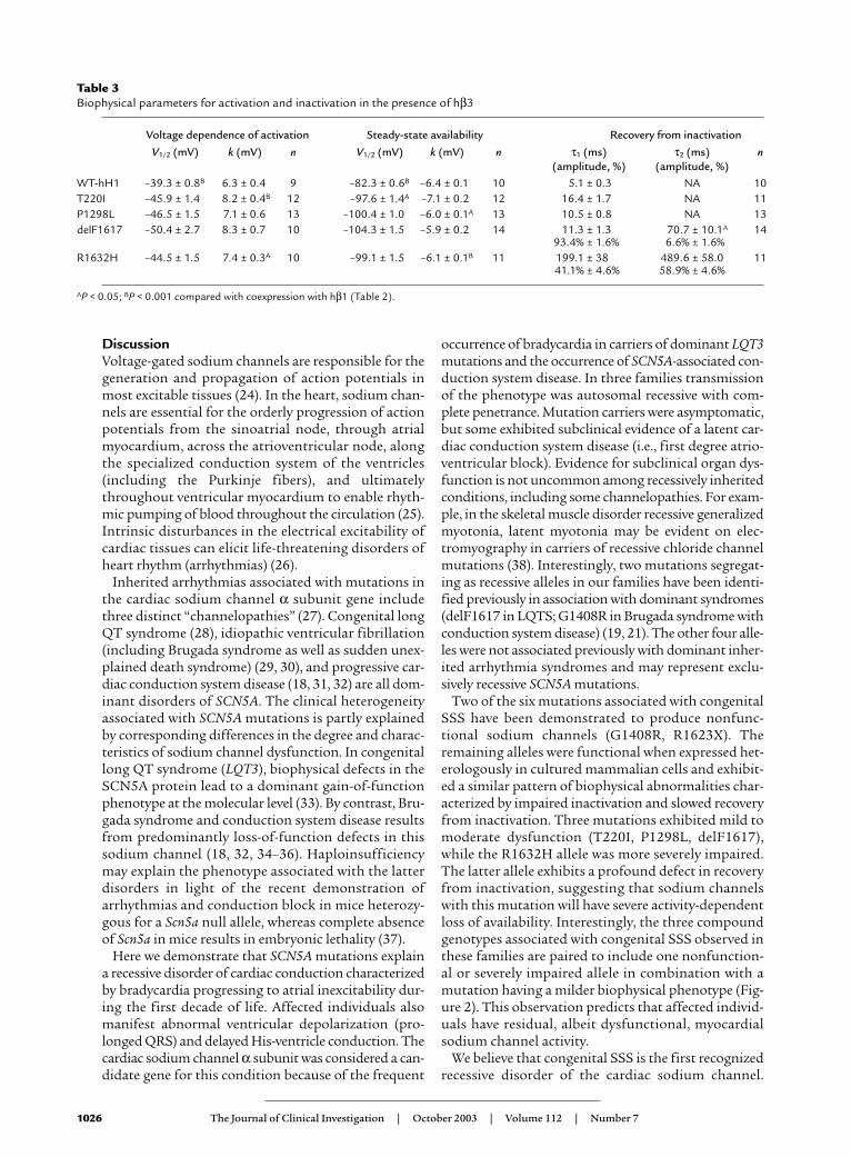
In light of the observation that myocar-dial conduction appears affected to agreater degree in the atria, we consideredthe possibility that an accessory proteinexpressed only in the ventricle normalizesfunction of the mutant sodium channels.Evidence exists that the sodium channelβ3 subunit is restricted to the ventricle(23), and therefore we tested whethercoexpression of a human β3 subunit (hβ3)alters functional properties of the SCN5A
mutants. With few exceptions, the biophysical proper-ties of WT-hH1 and the four mutants coexpressed withhβ3 were highly similar to the condition of hβ1 coex-pression (Table 3). Coexpression of hβ3, however, doesshift steady-state availability for WT-hH1 to moredepolarizing potentials that predict enhanced channelactivity in the ventricles, an effect partially offset by thesignificant depolarizing shift in the voltage-depend-ence of activation. Coexpression of hβ3 did not exhib-it a similar effect on any of the mutant sodium chan-nels, however. By contrast, hβ3 shifts steady-stateavailability of T220I to more negative potentials. Recov-ery from inactivation of WT-hH1, T220I, and P1298Lin the presence of hβ3 exhibited a monoexponentialtime course rather than the double-exponential recov-ery kinetics observed with hβ1. We did observe a sig-nificant difference in the τ2 recovery time constant fordelF1617 in the presence of hβ3, but this representsonly a minor population of channels (6.6%; Table 3).The overall time course of recovery from inactivationwas not greatly different for any mutant in the presenceof hβ3 compared with hβ1. Based upon these observa-tions, we cannot attribute the apparent milder ventric-ular phenotype to the β3 subunit.
Figure 6SCN5A mutants exhibit impaired recovery from inactivation. Thetime course of recovery from inactivation was elicited using the two-pulse protocol shown in the inset. Time constants and fractionalamplitudes are listed in Table 2.
Figure 7Biophysical properties of R1632H. (a) Comparison of current-voltage relationshipfor WT-hH1 (open circles) and R1632H (filled circles, n = 25). Current is normalizedto cell capacitance to give a measure of sodium current density. There is no differ-ence in current density between WT-hH1 and R1632H at all tested voltages. (b) Volt-age dependence of fast inactivation time constants for WT-hH1 (open circles) andR1632H (filled circles, n = 25). Differences between WT-hH1 and mutant channelwere significant for τ1 (P < 0.0001) and τ2 (P < 0.05) at voltages between –60 to +50mV. (c) Voltage dependence of sodium channel availability and activation (symboldefinitions are shown as an inset, and their shading patterns are explained in the y-axis labels). Voltage dependence of sodium channel availability (steady-state inac-tivation) was obtained using a two-pulse protocol as illustrated by the inset. Themembrane potentials for half-maximal inactivation and slope factors are provided inTable 2. The activation curve was constructed as described in the legend of Figure 5,and parameters are given in Table 2. (d) Time course of recovery from inactivationat –120mV (–140 mV for R1632H). The time constants and fractional amplitudes(given in parentheses) are provided in Table 2.
1026 The Journal of Clinical Investigation | October 2003 | Volume 112 | Number 7
DiscussionVoltage-gated sodium channels are responsible for thegeneration and propagation of action potentials inmost excitable tissues (24). In the heart, sodium chan-nels are essential for the orderly progression of actionpotentials from the sinoatrial node, through atrialmyocardium, across the atrioventricular node, alongthe specialized conduction system of the ventricles(including the Purkinje fibers), and ultimatelythroughout ventricular myocardium to enable rhyth-mic pumping of blood throughout the circulation (25).Intrinsic disturbances in the electrical excitability ofcardiac tissues can elicit life-threatening disorders ofheart rhythm (arrhythmias) (26).
Inherited arrhythmias associated with mutations inthe cardiac sodium channel α subunit gene includethree distinct “channelopathies” (27). Congenital longQT syndrome (28), idiopathic ventricular fibrillation(including Brugada syndrome as well as sudden unex-plained death syndrome) (29, 30), and progressive car-diac conduction system disease (18, 31, 32) are all dom-inant disorders of SCN5A. The clinical heterogeneityassociated with SCN5A mutations is partly explainedby corresponding differences in the degree and charac-teristics of sodium channel dysfunction. In congenitallong QT syndrome (LQT3), biophysical defects in theSCN5A protein lead to a dominant gain-of-functionphenotype at the molecular level (33). By contrast, Bru-gada syndrome and conduction system disease resultsfrom predominantly loss-of-function defects in thissodium channel (18, 32, 34–36). Haploinsufficiencymay explain the phenotype associated with the latterdisorders in light of the recent demonstration ofarrhythmias and conduction block in mice heterozy-gous for a Scn5a null allele, whereas complete absenceof Scn5a in mice results in embryonic lethality (37).
Here we demonstrate that SCN5A mutations explaina recessive disorder of cardiac conduction characterizedby bradycardia progressing to atrial inexcitability dur-ing the first decade of life. Affected individuals alsomanifest abnormal ventricular depolarization (pro-longed QRS) and delayed His-ventricle conduction. Thecardiac sodium channel α subunit was considered a can-didate gene for this condition because of the frequent
occurrence of bradycardia in carriers of dominant LQT3mutations and the occurrence of SCN5A-associated con-duction system disease. In three families transmissionof the phenotype was autosomal recessive with com-plete penetrance. Mutation carriers were asymptomatic,but some exhibited subclinical evidence of a latent car-diac conduction system disease (i.e., first degree atrio-ventricular block). Evidence for subclinical organ dys-function is not uncommon among recessively inheritedconditions, including some channelopathies. For exam-ple, in the skeletal muscle disorder recessive generalizedmyotonia, latent myotonia may be evident on elec-tromyography in carriers of recessive chloride channelmutations (38). Interestingly, two mutations segregat-ing as recessive alleles in our families have been identi-fied previously in association with dominant syndromes(delF1617 in LQTS; G1408R in Brugada syndrome withconduction system disease) (19, 21). The other four alle-les were not associated previously with dominant inher-ited arrhythmia syndromes and may represent exclu-sively recessive SCN5A mutations.
Two of the six mutations associated with congenitalSSS have been demonstrated to produce nonfunc-tional sodium channels (G1408R, R1623X). Theremaining alleles were functional when expressed het-erologously in cultured mammalian cells and exhibit-ed a similar pattern of biophysical abnormalities char-acterized by impaired inactivation and slowed recoveryfrom inactivation. Three mutations exhibited mild tomoderate dysfunction (T220I, P1298L, delF1617),while the R1632H allele was more severely impaired.The latter allele exhibits a profound defect in recoveryfrom inactivation, suggesting that sodium channelswith this mutation will have severe activity-dependentloss of availability. Interestingly, the three compoundgenotypes associated with congenital SSS observed inthese families are paired to include one nonfunction-al or severely impaired allele in combination with amutation having a milder biophysical phenotype (Fig-ure 2). This observation predicts that affected individ-uals have residual, albeit dysfunctional, myocardialsodium channel activity.
We believe that congenital SSS is the first recognizedrecessive disorder of the cardiac sodium channel.
Table 3Biophysical parameters for activation and inactivation in the presence of hβ3
Voltage dependence of activation Steady-state availability Recovery from inactivation
V1/2 (mV) k (mV) n V1/2 (mV) k (mV) n τ1 (ms) τ2 (ms) n(amplitude, %) (amplitude, %)
WT-hH1 –39.3 ± 0.8B 6.3 ± 0.4 9 –82.3 ± 0.6B –6.4 ± 0.1 10 5.1 ± 0.3 NA 10T220I –45.9 ± 1.4 8.2 ± 0.4B 12 –97.6 ± 1.4A –7.1 ± 0.2 12 16.4 ± 1.7 NA 11P1298L –46.5 ± 1.5 7.1 ± 0.6 13 –100.4 ± 1.0 –6.0 ± 0.1A 13 10.5 ± 0.8 NA 13delF1617 –50.4 ± 2.7 8.3 ± 0.7 10 –104.3 ± 1.5 –5.9 ± 0.2 14 11.3 ± 1.3 70.7 ± 10.1A 14
93.4% ± 1.6% 6.6% ± 1.6%R1632H –44.5 ± 1.5 7.4 ± 0.3A 10 –99.1 ± 1.5 –6.1 ± 0.1B 11 199.1 ± 38 489.6 ± 58.0 11
41.1% ± 4.6% 58.9% ± 4.6%
AP < 0.05; BP < 0.001 compared with coexpression with hβ1 (Table 2).
The Journal of Clinical Investigation | October 2003 | Volume 112 | Number 7 1027
Lupoglazoff et al., however, described a child homozy-gous for a missense SCN5A allele (V1777M) who exhib-ited LQTS with rate-dependent atrioventricular con-duction block (39). The child’s parents and oneheterozygous sibling had borderline QT interval pro-longation but were asymptomatic. Biophysical studiesof the V1777M variant demonstrated noninactivatingsodium current resembling the defect observed withmost LQTS-associated SCN5A mutations. These dataare most consistent with recessive inheritance of anotherwise dominant mutation exhibiting reduced pen-etrance in the heterozygous parents and sibling.
The current study and two recent reports clearlyestablish the genetic heterogeneity associated withfamilial sinus bradycardia syndromes. For example,atrial bradycardia, atrial standstill, prolonged His-ven-tricle conduction time, and age of onset in the third orfourth decade in a small Dutch kindred was attributedto the coinheritance of a heterozygous SCN5A muta-tion (D1275N) and a specific haplotype in the connex-in-40 promoter (40). Failure of the SCN5A allele aloneto confer a clinical phenotype supported an argumentfavoring digenic inheritance. The biophysical proper-ties of D1275N, however, were distinct from thatobserved in the four functional alleles we studied.These mutants exhibited small differences in the volt-age-dependence of activation observed in the absenceof the β1 accessory subunit and acceleration of recov-ery from inactivation. Schulze-Bahr et al. reported amutation in a component of If current (hyperpolariza-tion-activated cyclic nucleotide gated, or HCN4) in asporadic case of pacemaker dysfunction characterizedby bradyarrhythmia, chronotropic incompetence, andintermittent atrial fibrillation with age of onset in thefifth decade (41). Biophysical studies suggested a dom-inant negative effect of the HCN4 mutant on wild-typechannels; If-like currents with altered biophysical prop-erties were identified. Taken together, these findingssuggest that distinct genetic and molecular mecha-nisms exist to explain sinus bradycardia.
We can speculate on the pathogenesis of congenitalSSS associated with recessive SCN5A mutations. Pri-mary dysfunction of the sinoatrial node seems unlike-ly because action potentials in this tissue are largely cal-cium channel dependent or are associated withtetrodotoxin-sensitive (TTX-sensitive) sodium currentin contrast to the TTX-resistant current arising fromthe sodium channel encoded by SCN5A (42). Sinusnode dysfunction caused by failure of impulses to con-duct into adjacent atrial myocardium (exit block) hasbeen suggested as a cause of acquired SSS (43, 44) andis a plausible mechanism to explain this SCN5A-linkeddisorder. The clinical progression from bradycardia toatrial inexcitability further suggests that factors asso-ciated with postnatal development may contribute tothe emergence of the complete phenotype. Althoughatrial action potentials progressively disappear in thisdisorder and intraventricular conduction is abnormal,a ventricular escape rhythm was evident in affected
members of these families. This implies that actionpotential generation and/or propagation is moreseverely impaired in the atria than ventricles. It is con-ceivable that intrinsic differences between atrial andventricular myocardium, such as liminal length foraction potential propagation or sodium current densi-ties, may underlie the more severe atrial phenotype. Wecan also speculate that the generation of ventricularaction potentials in this disorder might depend uponsodium channel isoforms other than SCN5A expressedin ventricular tissue (45, 46). Alternatively, ventricle-specific metabolic or signal transduction events andalternative processing of SCN5A mRNA or accessoryproteins other than β1 and β3 subunits may enablepartial rescue of mutant SCN5A phenotypes in the ven-tricle but not the atrial myocardium.
AcknowledgmentsThe authors are grateful to the families who participat-ed in this study. The studies were facilitated by the assis-tance of Linda Boehm, Burt I. Bromberg, Kerry Howell,Connie Jones, James Joyce, and Peter Karpawich in col-lecting patient material. We also thank the VanderbiltDNA Sequencing Facility and the Cincinnati Children’sHospital Medical Center Genomics Core Facility. Thiswork was supported by the NIH (grant HD-39946 toD.W. Benson, grant NS-32387 to A.L. George, Jr.) andthe American Heart Association Southeast Affiliate(grant AHA-0160241B to D.W. Wang). We thank DanRoden, John D. Kugler, and Frank Zimmerman for crit-ical reading of the manuscript.
1. Lown, B. 1967. Electrical reversion of cardiac arrhythmias. Br. Heart J.29:469–489.
2. Fish, F.A., and Benson, D.W. 2001. Disorders of cardiac rhythm and con-duction. In Moss and Adam’s heart disease in infants, children and adolescents.H.D. Allen, E.B. Clark, H.P. Gutgesell, and D.J. Driscoll, editors. Lippin-cott, Williams & Wilkins. Philadelphia, Pennsylvania, USA. 482–533.
3. Martin, A.B., and Kugler, J.D. 1999. Sinus node dysfunction. In Clinicalpediatric arrhythmias: electrophysiology and pacing. P.C. Gillette and A.J. Gar-son, editors. W.B. Saunders. Philadelphia, Pennsylvania, USA. 51–62.
4. Yabek, S.M., Swensson, R.E., and Jarmakani, J.M. 1977. Electrocardio-graphic recognition of sinus node dysfunction in children and youngadults. Circulation. 56:235–239.
5. Yabek, S.M., and Jarmakani, J.M. 1978. Sinus node dysfunction in chil-dren, adolescents, and young adults. Pediatrics. 61:593–598.
6. Radford, D.J., and Izukawa, T. 1975. Sick sinus syndrome. Symptomaticcases in children. Arch. Dis. Child. 50:879–885.
7. Kugler, J.D., Gillette, P.C., Mullins, C.E., and McNamara, D.G. 1979.Sinoatrial conduction in children: an index of sinoatrial node function.Circulation. 59:1266–1276.
8. Ector, H., and Van der Hauwaert, L.G. 1980. Sick sinus syndrome inchildhood. Br. Heart J. 44:684–691.
9. Beder, S.D., Gillette, P.C., Garson, A., Jr., Porter, C.B., and McNamara,D.G. 1983. Symptomatic sick sinus syndrome in children and adoles-cents as the only manifestation of cardiac abnormality or associated withunoperated congenital heart disease. Am. J. Cardiol. 51:1133–1136.
10. Guillerm, F., et al. 1989. Sick sinus syndrome in children with a “healthyheart”. Apropos of 2 cases with direct endocavitory tracing of the sino-atrial block [in French.]. Ann. Cardiol. Angeiol. (Paris). 38:143–146.
11. Nordenberg, A., Varghese, P.J., and Nugent, E.W. 1976. Spectrum ofsinus node dysfunction in two siblings. Am. Heart J. 91:507–512.
12. Scott, O., Macartney, F.J., and Deverall, P.B. 1976. Sick sinus syndromein children. Arch. Dis. Child. 51:100–105.
13. Yabek, S.M., Dillon, T., Berman, W., Jr., and Niland, C.J. 1982. Sympto-matic sinus node dysfunction in children without structural heart dis-ease. Pediatrics. 69:590–593.
14. Ward, D.E., Ho, S.Y., and Shinebourne, E.A. 1984. Familial atrial stand-still and inexcitability in childhood. Am. J. Cardiol. 53:965–967.

1028 The Journal of Clinical Investigation | October 2003 | Volume 112 | Number 7
15. Spellberg, R.D. 1971. Familial sinus node disease. Chest. 60:246–251.16. Bharati, S., Surawicz, B., Vidaillet, H.J., Jr., and Lev, M. 1992. Familial con-
genital sinus rhythm anomalies: clinical and pathological correlations.Pacing Clin. Electrophysiol. 15:1720–1729.
17. Wang, Q., Li, Z., Shen, J., and Keating, M.T. 1996. Genomic organizationof the human SCN5A gene encoding the cardiac sodium channel.Genomics. 34:9–16.
18. Wang, D.W., Viswanathan, P.C., Balser, J.R., George, A.L., Jr., and Benson,D.W. 2002. Clinical, genetic, and biophysical characterization of SCN5Amutations associated with atrioventricular conduction block. Circulation.105:341–346.
19. Kyndt, F., et al. 2001. Novel SCN5A mutation leading either to isolatedcardiac conduction defect or Brugada syndrome in a large French fami-ly. Circulation. 104:3081–3086.
20. Makita, N., et al. 1998. A de novo missense mutation of human cardiacNa+ channel exhibiting novel molecular mechanisms of long QT syn-drome. FEBS Lett. 423:5–9.
21. Splawski, I., et al. 2000. Spectrum of mutations in long-QT syndromegenes. KVLQT1, HERG, SCN5A, KCNE1, and KCNE2. Circulation.102:1178–1185.
22. Little, S. 1995. Current protocols in human genetics. John Wiley and Sons.New York, New York, USA. 9.8.1–9.8.6.
23. Fahmi, A.I., et al. 2001. The sodium channel beta-subunit SCN3b modu-lates the kinetics of SCN5a and is expressed heterogeneously in sheepheart. J. Physiol. 537:693–700.
24. Catterall, W.A. 2000. From ionic currents to molecular mechanisms: thestructure and function of voltage-gated sodium channels. Neuron.26:13–25.
25. Balser, J.R. 1999. Structure and function of the cardiac sodium channels.Cardiovasc. Res. 42:327–338.
26. Roberts, R., and Brugada, R. 2003. Genetics and arrhythmias. Annu. Rev.Med. 54:257–267.
27. Marban, E. 2002. Cardiac channelopathies. Nature. 415:213–218.28. Wang, Q., et al. 1995. Cardiac sodium channel mutations in patients with
long QT syndrome, an inherited cardiac arrhythmia. Hum. Mol. Genet.4:1603–1607.
29. Wang, Q., et al. 1995. SCN5A mutations associated with an inherited car-diac arrhythmia, long QT syndrome. Cell. 80:805–811.
30. Vatta, M., et al. 2002. Genetic and biophysical basis of sudden unex-plained nocturnal death syndrome (SUNDS), a disease allelic to Bruga-da syndrome. Hum. Mol. Genet. 11:337–345.
31. Schott, J.J., et al. 1999. Cardiac conduction defects associate with muta-tions in SCN5A. Nat. Genet. 23:20–21.
32. Tan, H.L., et al. 2001. A sodium-channel mutation causes isolated cardiacconduction disease. Nature. 409:1043–1047.
33. Bennett, P.B., Yazawa, K., Makita, N., and George, A.L., Jr. 1995. Molecu-lar mechanism for an inherited cardiac arrhythmia. Nature. 376:683–685.
34. Dumaine, R., et al. 1999. Ionic mechanisms responsible for the electro-cardiographic phenotype of the Brugada syndrome are temperaturedependent. Circ. Res. 85:803–809.
35. Deschenes, I., et al. 2000. Electrophysiological characterization of SCN5Amutations causing long QT (E1784K) and Brugada (R1512W andR1432G) syndromes. Cardiovasc. Res. 46:55–65.
36. Wang, D.W., Makita, N., Kitabatake, A., Balser, J.R., and George, A.L., Jr.2000. Enhanced Na(+) channel intermediate inactivation in Brugada syn-drome. Circ. Res. 87:E37–E43.
37. Papadatos, G.A., et al. 2002. Slowed conduction and ventricular tachy-cardia after targeted disruption of the cardiac sodium channel geneScn5a. Proc. Natl. Acad. Sci. U. S. A. 99:6210–6215.
38. Becker, P.E. 1979. Heterozygote manifestation in recessive generalizedmyotonia. Hum. Genet. 46:325–329.
39. Lupoglazoff, J.M., et al. 2001. Homozygous SCN5A mutation in long-QTsyndrome with functional two-to-one atrioventricular block. Circ. Res.89:E16–E21.
40. Groenewegen, W.A., et al. 2003. A cardiac sodium channel mutationcosegregates with a rare connexin 40 genotype in familial atrial standstill.Circ. Res. 92:14–22.
41. Schulze-Bahr, E., et al. 2003. Pacemaker channel dysfunction in a patientwith sinus node disease. J. Clin. Invest. 111:1537–1545. doi:10.1172/JCI200316387.
42. Gellens, M.E., et al. 1992. Primary structure and functional expression ofthe human cardiac tetrodotoxin-insensitive voltage-dependent sodiumchannel. Proc. Natl. Acad. Sci. U. S. A. 89:554–558.
43. Asseman, P., et al. 1983. Persistent sinus nodal electrograms duringabnormally prolonged postpacing atrial pauses in sick sinus syndromein humans: sinoatrial block vs. overdrive suppression. Circulation.68:33–41.
44. Asseman, P., et al. 1991. Postextrasystolic sinoatrial exit block in humansick sinus syndrome: demonstration by direct recording of sinus nodeelectrograms. Am. Heart J. 122:1633–1643.
45. Maier, S.K., et al. 2002. An unexpected role for brain-type sodium chan-nels in coupling of cell surface depolarization to contraction in the heart.Proc. Natl. Acad. Sci. U. S. A. 99:4073–4078.
46. Maier, S.K., et al. 2003. An unexpected requirement for brain-type sodi-um channels for control of heart rate in the mouse sinoatrial node. Proc.Natl. Acad. Sci. U. S. A. 100:3507–3512.