congential and ethanol-induced disorders of n-linked ... · zurich open repository and archive...
TRANSCRIPT

Zurich Open Repository andArchiveUniversity of ZurichMain LibraryStrickhofstrasse 39CH-8057 Zurichwww.zora.uzh.ch
Year: 2013
Congential and ethanol-induced disorders of N-linked protein glycosylation
Welti, Michael
Abstract: Protein glycosylation is an essential protein modification existing in all domains of life. About2% of the human genome is involved in the glycosylation machinery and 50% of the human proteinsare modified with glycans. In the endoplasmic reticulum, N-linked protein glycosylation begins with theassembly of an oligosaccharide precursor by step-wise transfer of sugar building blocks to a membrane-embedded, reduced polyprenol anchor called dolichol-phosphate. The fully assembled N-linked glycanprecursor is then transferred co- or post-translationally to defined asparagine residues of target proteins.Considering the abundance of N-glycosylated proteins throughout all human cell types, inherited glycosy-lation defects called congenital disorders of glycosylation (CDG) show multi-systemic involvement due todevelopmental defects in children. Most CDG involve a neurological component resulting in psychomo-tor retardation. Among the most common symptoms are epilepsy, hypotonia, hyporeflexia, strabismus,retinitis pigmentosa, polyneuropathy, myopathy, and cerebellar hypotrophy/hypoplasia. A commonmarker for CDG is the underglycosylation of blood serum proteins such as transferrin. Depending on thetype of CDG, the N-linked glycans differ in their structure and can be detected in carbohydrate-deficienttransferrin (CDT). Interestingly, alcoholic liver disease (ALD) is accompanied by a N-glycosylation de-fect as well. Besides a similar pattern of CDT, certain forms of CDG and ALD share another commonsymptom: liver fibrosis. Chronic alcoholism is estimated to be responsible for 4% of global death and theliver being the primary site of ethanol metabolism is particularly affected. Despite the long history ofCDT in ALD, glycosylation deficiency in ALD has not yet been characterized at the molecular level. Sofar, treatment of CDG is very restricted. In MPI-CDG, the mannose-phosphate isomerase is defective, anenzyme responsible for the conversion of fructose-6-phosphate to mannose-6-phosphate, thereby provid-ing mannose for the glycosylation machinery. A simple supplementation of nutrition with mannose wasshown to attenuate the manifestations of MPI-CDG. In the first part of this thesis, we tested the potentialof a cholesterol-lowering drug, Zaragozic acid A, to improve N-glycosylation in DPM1-CDG. DPM1 isa subunit of the dolichol-phosphate-mannose synthase which produces dolichol-phosphate-mannose, animportant mannose donor for N-linked glycosylation. Zaragozic acid A inhibits an enzyme at a bifur-cation of the anabolic pathway common to dolichol and cholesterol synthesis. At this bifurcation, themetabolic flux either goes towards cholesterol or dolichol synthesis. By blocking the enzyme for cholesterolsynthesis, N-linked protein glycosylation improved as observed by increased dolichol-phosphate-mannoselevels, increased dolichol-phosphate levels, normalized dolichol- and N-linked oligosaccharide distribu-tion. The restored dolichol-phosphate-mannose pool also resulted in better GPI-anchor availability. Wecould compensate the lower dolichol-phopshate-mannose synthase activity by increasing substrate avail-ability. Thus Zaragozic acid represents a possibility for drug treatment of DPM1-CDG. The secondpart of the thesis is focusing on ethanol-induced N-glycosylation deficiency. We studied the effect ofethanol on N-glycosylation in two hepatoma cell lines. The two cell lines VA-13 and HepaRG are dis-tinct because they express alcohol dehydrogenase and cytochrome P450 2E1, respectively, which confersethanol-metabolizing properties usually lost in cultured hepatocytes. We found lower dolichol levels inethanol-treated cells. Moreover, the dolichol-linked oligosaccharide pattern was disturbed with a lowerfraction of the final N-glycan precursor. The changes in glycosylation were accompanied by transcriptionalchanges. DPM1 was downregulated while RPN2 was upregulated. The transcriptional changes could beregulatory responses since we observed an increased DPM synthase activity despite the lower DPM1transcription. In conclusion, we explored CDG and acquired deficiency in N-linked protein glycosylation
by using a therapeutic approach to treat DPM1-CDG and by the characterization of N-glycosylationin an ethanol-induced glycosylation deficiency model. With this work we contributed to the search fortreatment possibilities for CDG and promoted the understanding of ethanol-induced N-glycosylationdeficiency.
Posted at the Zurich Open Repository and Archive, University of ZurichZORA URL: https://doi.org/10.5167/uzh-92872Dissertation
Originally published at:Welti, Michael. Congential and ethanol-induced disorders of N-linked protein glycosylation. 2013, Uni-versity of Zurich, Faculty of Medicine.
2
Zurich Open Repository and Archive
University of Zurich
Main Library
Winterthurerstrasse 190
CH-8057 Zurich
www.zora.uzh.ch
Year: 2013
Congential and ethanol-induced disorders of N-linked
protein glycosylation
Michael Welti
Posted at the Zurich Open Repository and Archive, University of Zurich
http://dx.doi.org/10.5167/uzh-92872
Originally published at:
Welti, Michael. Congential and ethanol-induced disorders of N-linked protein glycosylation. 2013, University
of Zurich, Faculty of Medicine.

Congenital and Ethanol-induced Disorders
of N-linked Protein Glycosylation
Dissertation
zur
Erlangung der naturwissenschaftlichen Doktorwürde
(Dr. sc. nat.)
vorgelegt der
Mathematisch-naturwissenschaftlichen Fakultät
der
Universität Zürich
von
Michael Andreas Welti
von
Berikon AG
Promotionskomitee
Prof. Dr. Thierry Hennet (Vorsitz)
Dr. Andreas Hülsmeier
PD Dr. Lubor Borsig
Prof. Dr. Matthias Baumgartner
Zürich, 2013

2
Table of contents
3
TABLE OF CONTENTS
Summary .............................................................................................................................................. 7
Zusammenfassung ......................................................................................................................... 10
Abbreviations.................................................................................................................................. 13
Introduction .................................................................................................................................... 16
Monosaccharides: Building blocks of glycosylation ...................................................... 16
Major glycan classes in eukaryotic cells ............................................................................ 18
N-linked protein glycosylation ............................................................................................. 19
Types of N-linked glycans ................................................................................................................. 19
Biosynthesis of the N-linked glycan precursor in the Endoplasmic Reticulum .......... 20
Processing of the N-linked glycan ................................................................................................. 21
Functions of N-linked protein glycosylation ............................................................................. 23
Congenital disorders in N-linked glycosylation .............................................................. 28
Alcoholic liver disease and N-linked glycosylation deficiency .................................. 30
References .................................................................................................................................... 33
Publications ..................................................................................................................................... 39
Improvement of dolichol-linked oligosaccharide biosynthesis by the squalene
synthase inhibitor Zaragozic acid ........................................................................................ 39
Abstract ................................................................................................................................................... 40
Introduction ........................................................................................................................................... 41
Experimental Procedures ................................................................................................................. 43
Results ...................................................................................................................................................... 46
Discussion ............................................................................................................................................... 53
Footnotes ................................................................................................................................................ 55
Acknowledgements ............................................................................................................................. 55
References .............................................................................................................................................. 56
Glycoprotein maturation and the UPR ............................................................................... 60

Table of contents
4
Abstract ................................................................................................................................................... 61
1. Introduction ...................................................................................................................................... 62
2. N-glycosylation ................................................................................................................................ 63
2.1. Dolichol phosphate analysis .................................................................................................... 63
2.2. Dolichol-linked oligosaccharide analysis ........................................................................... 68
2.3. N-glycosylation Site Occupancy ............................................................................................. 72
3. O-glycosylation................................................................................................................................. 76
3.1. Release of O-glycans by the が-elimination reaction ....................................................... 76
3.2. Release of O-glycans by hydrazinolysis .............................................................................. 79
Acknowledgments ............................................................................................................................... 81
References .............................................................................................................................................. 83
Regulation of dolichol-linked glycosylation .................................................................... 86
Abstract ................................................................................................................................................... 87
Dolichol biosynthesis and its role in N-linked glycosylation .............................................. 88
Deficiency of dolichol biosynthesis ‒ a new family of CDG ................................................. 89
Therapeutics targeting dolichol ..................................................................................................... 94
Outlook..................................................................................................................................................... 96
References .............................................................................................................................................. 97
Ethanol-induced impairment in the biosynthesis of N-linked glycosylation ..... 101
Abstract .................................................................................................................................................102
Highlights ..............................................................................................................................................103
Keywords ..............................................................................................................................................103
Abbreviations ......................................................................................................................................103
1. Introduction ....................................................................................................................................104
2. Materials and Methods ................................................................................................................106
3. Results ...............................................................................................................................................110
4. Discussion ........................................................................................................................................116
Table of contents
5
5. Conclusion ........................................................................................................................................117
References ............................................................................................................................................118
GENERAL DISCUSSION ........................................................................................................................ 122
The challenge of uncovering regulatory mechanisms ................................................ 123
Therapeutics for glycosylation disorders ....................................................................... 125
Future directions ..................................................................................................................... 127
References .................................................................................................................................. 128
ACKNOWLEDGEMENTS ....................................................................................................................... 132
CURRICULUM VITAE ........................................................................................................................... 135

6
Summary
7
SUMMARY
Protein glycosylation is an essential protein modification existing in all domains of life.
About 2% of the human genome is involved in the glycosylation machinery and 50% of
the human proteins are modified with glycans. In the endoplasmic reticulum, N-linked
protein glycosylation begins with the assembly of an oligosaccharide precursor by step-
wise transfer of sugar building blocks to a membrane-embedded, reduced polyprenol
anchor called dolichol-phosphate. The fully assembled N-linked glycan precursor is then
transferred co- or post-translationally to defined asparagine residues of target proteins.
Considering the abundance of N-glycosylated proteins throughout all human cell types,
inherited glycosylation defects called congenital disorders of glycosylation (CDG) show
multi-systemic involvement due to developmental defects in children. Most CDG involve
a neurological component resulting in psychomotor retardation. Among the most
common symptoms are epilepsy, hypotonia, hyporeflexia, strabismus, retinitis
pigmentosa, polyneuropathy, myopathy, and cerebellar hypotrophy/hypoplasia. A
common marker for CDG is the underglycosylation of blood serum proteins such as
transferrin. Depending on the type of CDG, the N-linked glycans differ in their structure
and can be detected in carbohydrate-deficient transferrin (CDT). Interestingly, alcoholic
liver disease (ALD) is accompanied by a N-glycosylation defect as well. Besides a similar
pattern of CDT, certain forms of CDG and ALD share another common symptom: liver
fibrosis. Chronic alcoholism is estimated to be responsible for 4% of global death and
the liver being the primary site of ethanol metabolism is particularly affected. Despite
the long history of CDT in ALD, glycosylation deficiency in ALD has not yet been
characterized at the molecular level.
So far, treatment of CDG is very restricted. In MPI-CDG, the mannose-phosphate
isomerase is defective, an enzyme responsible for the conversion of fructose-6-
phosphate to mannose-6-phosphate, thereby providing mannose for the glycosylation
machinery. A simple supplementation of nutrition with mannose was shown to
attenuate the manifestations of MPI-CDG.
In the first part of this thesis, we tested the potential of a cholesterol-lowering drug,
Zaragozic acid A, to improve N-glycosylation in DPM1-CDG. DPM1 is a subunit of the
dolichol-phosphate-mannose synthase which produces dolichol-phosphate-mannose, an
important mannose donor for N-linked glycosylation. Zaragozic acid A inhibits an
Summary
8
enzyme at a bifurcation of the anabolic pathway common to dolichol and cholesterol
synthesis. At this bifurcation, the metabolic flux either goes towards cholesterol or
dolichol synthesis. By blocking the enzyme for cholesterol synthesis, N-linked protein
glycosylation improved as observed by increased dolichol-phosphate-mannose levels,
increased dolichol-phosphate levels, normalized dolichol- and N-linked oligosaccharide
distribution. The restored dolichol-phosphate-mannose pool also resulted in better GPI-
anchor availability. We could compensate the lower dolichol-phopshate-mannose
synthase activity by increasing substrate availability. Thus Zaragozic acid represents a
possibility for drug treatment of DPM1-CDG.
The second part of the thesis is focusing on ethanol-induced N-glycosylation deficiency.
We studied the effect of ethanol on N-glycosylation in two hepatoma cell lines. The two
cell lines VA-13 and HepaRG are distinct because they express alcohol dehydrogenase
and cytochrome P450 2E1, respectively, which confers ethanol-metabolizing properties
usually lost in cultured hepatocytes. We found lower dolichol levels in ethanol-treated
cells. Moreover, the dolichol-linked oligosaccharide pattern was disturbed with a lower
fraction of the final N-glycan precursor. The changes in glycosylation were accompanied
by transcriptional changes. DPM1 was downregulated while RPN2 was upregulated. The
transcriptional changes could be regulatory responses since we observed an increased
DPM synthase activity despite the lower DPM1 transcription.
In conclusion, we explored CDG and acquired deficiency in N-linked protein
glycosylation by using a therapeutic approach to treat DPM1-CDG and by the
characterization of N-glycosylation in an ethanol-induced glycosylation deficiency
model. With this work we contributed to the search for treatment possibilities for CDG
and promoted the understanding of ethanol-induced N-glycosylation deficiency.

9

Zusammenfassung
10
ZUSAMMENFASSUNG
Die Glykosylierung von Proteinen ist eine essentielle Proteinmodifikation, die in allen
Domänen des Lebens vorkommt. Etwa 2% des humanen Genoms kodiert Proteine, die
Teil der Glykosylierungsmaschinerie sind, und 50% der humanen Proteine sind
glykosyliert. Die N-Glykosylierung von Proteinen beginnt im endoplasmatischen
Retikulum, wo zuerst der N-Glykan Precursor schrittweise aufgebaut wird, indem
einzelne Zuckerbausteine auf einem membran-gebundenen, reduzierten
Polyprenolanker, dem Dolichol-phosphat, zusammengefügt werden. Der angefertigte N-
Glykan Precursor wird dann co- oder posttranslational vom Dolichol-phosphat auf
spezifische Asparaginseitenketten von Zielproteinen übertragen.
Aufgrund der Abundanz der N-Glykosylierung von menschlichen Proteinen in allen
Zelltypen weisen vererbte N-Glykosylierungsdefekte ein multi-systemisches
Krankheitsbild auf. Vererbte Glykosylierungsdefekte (CDG) beeinträchtigen meist die
pränatale Entwicklung des Menschen. Die häufigsten CDG-Symptome sind Epilepsie,
Hypotonie, Hyporeflexie, Strabismus, Retinitis pigmentosa, Polyneuropathie, Myopathie
und Hypotrophie/Hypoplasie des Kleinhirns. Die Unterglykosylierung von
Serumproteinen, wie z.B. Transferrin, wird als Marker für CDG benutzt. Abhängig vom
CDG-Typ unterscheiden sich die N-Glykane auf Transferrin, weshalb verschiedene
Formen von Desialotransferrin (CDT) detektiert werden können. Die alkoholische
Leberkrankheit (ALD) wird interessanterweise von einem N-Glykosylierungsdefekt
begleitet. Neben einem ähnlichen CDT-Bild zeigen gewisse CDG-Typen und ALD auch ein
anderes gemeinsames Symptom: die Leberfibrose. Die Leber als primärer Ort des
Ethanolabbaus ist besonders den Effekten des Alkoholismus betroffen. Obwohl der
Zusammenhang zwischen CDT und ALD schon lange bekannt ist, wurde die
Glykosylierungsdefizienz in ALD noch nicht vollständig auf molekularer Ebene
charakterisiert.
Die medikamentöse Behandlung von CDG ist sehr beschränkt. Ein Beispiel ist MPI-CDG,
in der das Enzym, das für die Konversion von Fruktose-6-phosphat zu Mannose-6-
phosphat, defekt ist. Diese Reaktion ist wichtig, um Mannose für die
Glykosylierungsreaktionen zur Verfügung zu stellen. Eine einfache Supplementierung
der Nahrung mit Mannose ist in diesem Fall genug, um die Krankheitserscheinungen zu
mildern.
Zusammenfassung
11
Im ersten Teil dieser Dissertation untersuchten wir das Potenzial eines cholesterin-
senkenden Stoffs, Zaragozic acid A, die N-Glykosylierung in DPM1-CDG zu verbessern.
DPM1 ist eine Untereinheit der Dolichol-phosphate-mannose Synthase, die als Komplex
Dolichol-phosphate-mannose, einen wichtigen Mannose-donor für die N-Glykosylierung,
produziert. Zaragozic acid A inhibiert ein Enzym an einer Verzweigung eines
Synthesewegs, über den sowohl die Dolichol- als auch Cholesterinproduktion stattfindet.
Bei der genannten Verzweigung geht der metabolische Fluss entweder in Richtung
Dolichol- oder Cholesterinsynthese. Mit der Blockade des Enzyms, das die abzweigende
Reaktion zur Cholesterinsynthese katalysiert, wurde die N-Glykosylierung verbessert, so
dass erhöhtes Dolichol-phosphate und eine verbesserte Verteilung der dolichol- und N-
assozierten Oligosaccharide gemessen werden konnte. Ausserdem hatte die auf
Normwerte erhöhte Dolichol-phosphat-mannose zur Folge, dass die Verfügbarkeit von
GPI-Ankern verbessert wurde. Wir konnten also die reduzierte Aktivität der Dolichol-
phosphate-mannose Synthase mittels verbesserter Substratverfügbarkeit
kompensieren. Daher stellt Zaragozic acid A ein potenzielles Medikament zur
Behandlung von DPM1-CDG dar.
Der zweite Teil der Dissertation fokussiert auf die ethanol-induzierte N-
Glykosylierungsdefizienz. Dazu haben wir den Effekt von Ethanol auf die N-
Glykosylierung in zwei Leberkrebszelllinien untersucht. Die VA-13 und die HepaRG
Zelllinien sind gut dazu geeignet, weil sie im Gegensatz zu anderen kultivierten
Leberzellen die Alkoholdehydrogenase und das Zytochrom P450 2E1 exprimieren, was
die Metabolisierung von Ethanol ermöglicht. Wir konnten nach der Behandlung mit
Ethanol niedrigeres Dolichol-phosphat sowie eine niedrigere Verfügbarkeit des N-
Glykan Precursors feststellen. Diese Glykosylierungsstörung wurde von
transkriptionalen Veränderungen begleitet. DPM1 war runterreguliert während RPN2
hochreguliert wurde. Diese Veränderungen der Transkription könnten regulatorische
Mechanismen darstellen, da die DPM Synthase Aktivität erhöht war, obwohl die DPM1-
Untereinheit transkriptional runterreguliert war.
Zusammenfassend haben wir uns mit einem vererbten wie auch einem erworbenen N-
Glykosylierungsdefekt befasst. Wie haben einen therapeutischen Ansatz zur Milderung
des N-Glykosylierungsdefekts in DPM1-CDG Fibroblasten erfoglsversprechend getestet
und den ethanol-induzierten N-Glykosylierungsdefekt in zwei Leberzelllinien
charakterisiert.

12

Abbreviations
13
ABBREVIATIONS
9-ADM 9-anthryldiazomethane
ADH Alcohol dehydrogenase
ALD Alcoholic liver disease
ALG Asparagine-linked glycosylation
CDG Congenital Disorders of Glycosylation
CDT Carbohydrate-deficient transferrin
CYP2E1 Cytochrome P450 2E1
DLO Dolichol-linked oligosaccharide
DMSO Dimethyl sulfoxide
DNA Desoxyribonucleic acid
Dol Dolichol
Dol-P Dolichol-phosphate
Dol-PP Dolichol-pyrophosphate
DPM Dolichol-phosphate mannose (synthase)
ER Endoplasmic Reticulum
ERAD Endoplasmic reticulum-associated degradation
FBS Fetal bovine serum
Gal Galactose
GDP Guanosine diphosphate
Glc Glucose
GlcNAc N-acetlyglucosamine
GPI Glycophosphatidylinositol
HMG-CoA 3-hydroxy-3-methyl-glutaryl-coenzyme A
HPLC High performance liquid chromatography
IEF Isoelectric focusing
LLO Lipid-linked oligosaccharide
Man Mannose
MPI Mannose-phosphate isomerase
Abbreviations
14
mRNA Messenger RNA
NAD(H) が-Nicotineamide adenine dinucleotide (reduced)
NLO N-linked oligosaccharide
OST Oligosaccharyltransferase
PCR Polymerase chain reaction
PMM Phosphomannomutase
RNA Ribonucleic acid
UDP Uridine disphosphate
ZGA Zaragozic acid A
Fuc Fucose

15
Introduction
16
INTRODUCTION
Post-translational modifications of proteins are crucial for maintaining proper
functioning of cells and whole organisms. Among the most important protein
modifications in animals are phosphorylation, lipid conjugation, ubiquitination, and
glycosylation. Phosphorylation is essential in many cellular processes and mostly
regulates activity of the modified proteins (Hunter, 2000) while ubiquitination marks
proteins for the major pathway of proteasomal protein degradation (Hochstrasser,
2009). Lipid modifications serve as membrane anchors to restrict proteins to the ER,
Golgi apparatus, or the plasma membrane (Nadolski and Linder, 2007). Glycosylation is
the most frequent protein and lipid modification which is apparent when looking at the
complex and dense layer of glycans that covers the surface of all living organisms
including viruses (Larkin and Imperiali, 2011; Spiro, 2002). About 2% of the human
genes are involved in glycan formation as enzymes, transporters, or cofactors (Freeze et
al., 2012; Varki et al., 2008). Glycans are the most diverse and versatile biomolecules in
terms of target substrates, chemical linkages, and non-linear branching. In humans, 50%
of all proteins are glycosylated underlining the significance of protein glycosylation.
However, defining functions of glycosylation is difficult due to its inherent complexity
and requirement for advanced technology and methodology. The process of
glycosylation describes the attachment of a single saccharide or polysaccharide from a
donor substrate to an acceptor substrate. The donor substrate consists of a sugar
building block activated by a carrier via phospho-diester or phospho-ester bond. Donor
as well as the acceptor substrates are recognized by enzymes termed
glycosyltransferases which attach the sugar building block to the acceptor substrate in a
linkage-specific manner.
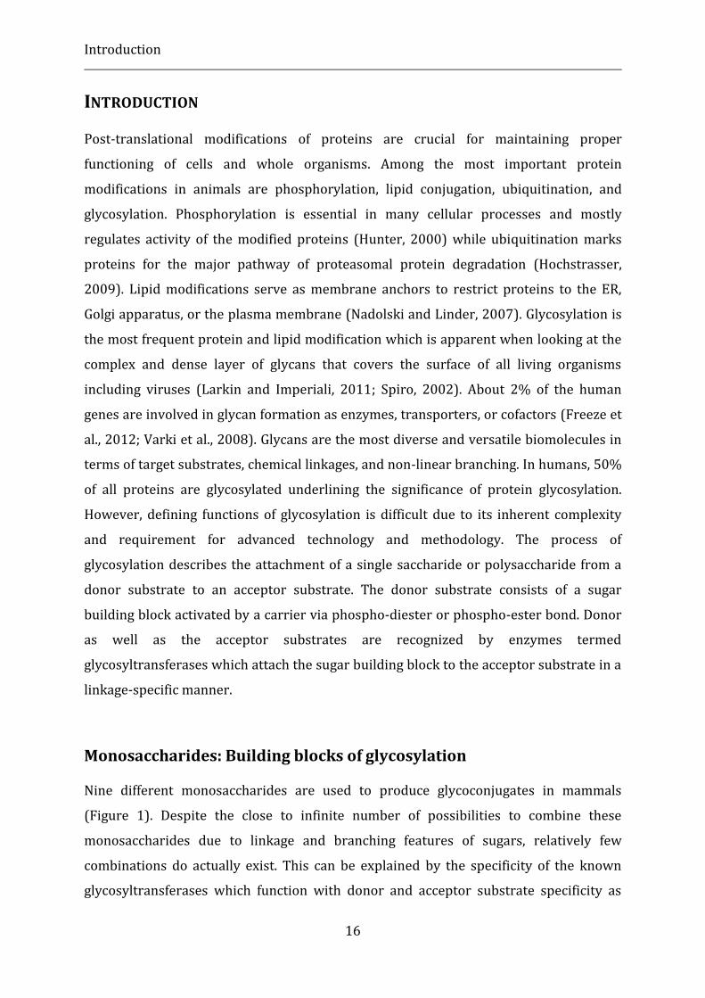
Monosaccharides: Building blocks of glycosylation
Nine different monosaccharides are used to produce glycoconjugates in mammals
(Figure 1). Despite the close to infinite number of possibilities to combine these
monosaccharides due to linkage and branching features of sugars, relatively few
combinations do actually exist. This can be explained by the specificity of the known
glycosyltransferases which function with donor and acceptor substrate specificity as

Introduction
17
well as linkage specificity. Therefore, unlimited and functional glycan diversity would
require a huge number of glycosyltransferases which themselves would need regulation.
Another level of complexity is added by modifications of glycans. Sulfation, acetylation,
and methylation and epimerization of GlcA to iduronic acid can occur on the conjugated
glycan (Silbert and Sugumaran, 2002; Varki et al., 2009). These glycan modifications
require activated substrates, i.e. ぬ旺-phosphoadenyl-の旺-phosphosulfate for sulfation,
acetyl-CoA for acetylation, and S-adenosylmethionine for methylation. Activated
substrates are produced in the cytoplasm and therefore depend on membrane
transporters to reach the ER-Golgi pathway.
Figure 1 Structural representations of the nine monosaccharides used in mammalian glycosylation. The names of the sugars are indicated below the structure including the abbreviations used in this thesis. (adapted from (Varki et al., 2008))
The complexity of glycans is major challenge for structural elucidation even with the
most advanced approaches using mass spectrometry (Kolarich et al., 2013; Mechref et
al., 2012; Zaia, 2010; Zaia, 2013).
Introduction
18
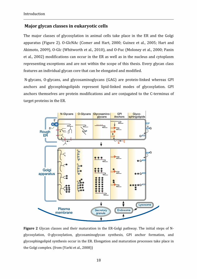
Major glycan classes in eukaryotic cells
The major classes of glycosylation in animal cells take place in the ER and the Golgi
apparatus (Figure 2). O-GlcNAc (Comer and Hart, 2000; Guinez et al., 2005; Hart and
Akimoto, 2009), O-Glc (Whitworth et al., 2010), and O-Fuc (Moloney et al., 2000; Panin
et al., 2002) modifications can occur in the ER as well as in the nucleus and cytoplasm
representing exceptions and are not within the scope of this thesis. Every glycan class
features an individual glycan core that can be elongated and modified.
N-glycans, O-glycans, and glycosaminoglycans (GAG) are protein-linked whereas GPI
anchors and glycosphingolipids represent lipid-linked modes of glycosylation. GPI
anchors themselves are protein modifications and are conjugated to the C-terminus of
target proteins in the ER.
Figure 2 Glycan classes and their maturation in the ER-Golgi pathway. The initial steps of N-
glycosylation, O-glycosylation, glycosaminoglycan synthesis, GPI anchor formation, and
glycosphingolipid synthesis occur in the ER. Elongation and maturation processes take place in
the Golgi complex. (from (Varki et al., 2008))

Introduction
19
N-linked protein glycosylation
The N-linked glycan core originates from an N-glycan precursor in the ER (Aebi, 2013).
There, N-linked protein glycosylation occurs on Asn residues co- or post-translationally.
When a protein with an N-glycan consensus sequence Asn-X-Ser/Thr (X can be any
amino acid except Pro) is translated into the ER, an oligosaccharide structure termed N-
glycan precursor is transferred from dolichol-pyrophosphate (Dol-PP) to the Asn of the
consensus site (Marshall, 1974). The N-glycan thereafter can assist in the folding of the
modified protein in the ER. After trimming the original N-glycan, the N-glycosylated
protein follows the secretory pathway to the Golgi apparatus where further trimming
and subsequent addition of various monosaccharides can occur. Furthermore, glycans
can undergo a maturation process involving chemical modifications. Finally, the
glycosylated protein is secreted or transported to its target membrane.
Types of N-linked glycans
Three types of N-linked glycans are distinguished: High mannose type, complex type,
and hybrid type (Figure 3). They all originate from the N-glycan precursor after passing
through the glycosylation machinery in the ER and Golgi complex.
Figure 3 N-glycan classes. The three N-glycan classes share a common core (Man3GlcNAc2).
Blue squares represent GlcNAc, green circles Man, yellow circles Gal, purple diamonds sialic
acid, and red triangle Fuc. (from (Varki et al., 2008))

Introduction
20
Biosynthesis of the N-linked glycan precursor in the Endoplasmic Reticulum
The first part of N-linked protein glycosylation requires the assembly of the N-glycan
precursor Glc3Man9GlcNAc2 on Dol-PP, a lipid carrier residing in the ER membrane
(Figure 4). The formation of dolichol (Dol) occurs in the mevalonate pathway and is
described and discussed in detail in the section ╉Regulation of Dolichol-linked glycosylation╊ (Ramachandran and Melnykovych, 1986). The substrate for the initial
glycosyltransferases is dolichol-P (Dol-P) that is supplied by dolichol kinase (DK)
phosphorylating Dol (red rectangle in Fig. 4). The precursor is built by the addition of
single sugar blocks in a sequence-, linkage-, and conformation-specific manner. The first
seven steps occur at the outer leaflet of the ER. Two GlcNAc (blue squares) are
sequentially added to Dol-P by DPGAT1 and ALG13/14, respectively. Both enzymes
require UDP-GlcNAc as a substrate but DPGAT1 links P-GlcNAc to Dol-P resulting in
GlcNAc-PP-Dol and UMP. ALG13/14 produces GlcNAc2-PP-Dol and UDP. ALG1 catalyzes
the addition of the first Man residues (green circles) from GDP-Man to GlcNAc2-PP-Dol.
Thereafter, ALG2 adds two Man from GDP-Man to ManGlcNAc2-PP-Dol. The last two
steps at the outer leaflet of the ER are catalyzed by ALG11 which adds two Man to -
Man3GlcNAc2-PP-Dol resulting in the intermediate Dol-linked oligosaccharide (DLO)
Man5GlcNAc2-PP-Dol. RFT1 flips this DLO across the ER membrane into the lumen by a
yet unresolved mechanism. Four further Man are added by the indicated enzymes using
Dol-P-Man as source of Man. Notably, ALG9 recognizes both Man6GlcNAc2-PP-Dol as well
as Man8GlcNAc2-PP-Dol as substrates for mannosylation. Three Glc sugars are
transferred from Dol-P-Glc to the DLO by the sequential action of ALG6, ALG8, and
ALG10. Finally, the completed N-glycan precursor Glc3Man9GlcNAc2 (Figure 4) is
transferred en bloc from Dol-PP to the amide group of Asn side chains in the consensus
sequence Asn-X-Ser/Thr by the oligosaccharyl-transferase complex (OST). Site
occupancy analyses of different glycoproteins revealed a preference for N-X-T over N-X-
S sites (Gavel and von Heijne, 1990). Dol-PP is recycled by dephosophorylation to Dol-P
and subsequent flipping to the cytosolic leaflet (Cantagrel and Lefeber, 2011;
Ramachandran and Melnykovych, 1986; Rush et al., 2008). Recycling critically
contributes to Dol-P availability but the molecular details of Dol-P recycling remain
elusive.

Introduction
21
Figure 4 Biosynthesis of dolichol-linked oligosaccharides (DLO) dolichol-activated
monosaccharides. The N-glycan precursor is assembled by the stepwise addition of
monosaccharides. On the cytosolic leaflet of the ER, two GlcNAc and five Man are transferred to
Dol-P using nucleotide-activated monosaccharides as substrates. Dol-PP-GlcNAc2Man5 is flipped
into the ER lumen where the DLO is extended with four Man and three Glc by glycosyl-
transferases using Dol-P-activated monosaccharides as substrates. The finished Dol-PP-linked
N-glycan precursor can then be transferred to a target protein. Red labels indicate proteins
associated with congenital disorders of glycosylation. Blue labels indicate proteins that have not
been found in congenital disorders of glycosylation so far. (adapted from (Haeuptle and Hennet,
2009))
Processing of the N-linked glycan
After transfer to target proteins, the N-linked glycan can assist the protein in folding
properly. This cyclic process is termed ER quality control and involves the chaperone
proteins calnexin and calreticulin which can recognize N-glycans on unfolded proteins
(Williams, 2006)┻ )mmediately after the glycan transfer┸ ゎ-glucosidase I removes the
terminal ゎ1‒2-linked Glc and glucosidase II then removes the two ゎ1-3-linked Glc
sugars. The resulting oligosaccharide is taking part in the ER folding quality control.
Iterative reglucosylation by UGGT1 and deglucosylation during the folding process

Introduction
22
ensure retention of improperly folded proteins in the so-called calnexin/calreticulin
cycle ゅsee section ╉ER quality control of protein folding╊ and (Helenius and Aebi, 2004)).
Many de-glucosylated glycans on properly folded proteins are processed by ER ゎ-mannosidase ) before exiting the ER which removes the ゎな-2-linked Man from the
central branch of the glycan resulting in a Man8GlcNAc2-Asn isomer. Most glycans exit
the ER with eight or nine Man residues but some escape the quality control processing
and can carry a terminal Glc when entering the Golgi apparatus.
Figure 5 Schematic structure of the N-linked glycan
precursor. It consists of two GlcNAc (blue squares),
nine Man (green circles), and three Glc (blue circles).
The residues are labeled alphabetically for cross-
referencing. Linkage specificities are indicated at the
connections between linked sugars.
In the Golgi, incompletely processed GlcMan8GlcNAc2-Asn is acted on by endo-ゎ-mannosidase which cleaves the disaccharide Glcゎな-3Man off the glycan resulting in a
Man8GlcNAc2-Asn different from the one produced by ER ゎ-mannosidase I. Further trimming occurs by ゎな-2 mannosidases IA, IB, and IC in the cis-Golgi resulting the
important intermediate glycan Man5GlcNAc2-Asn. From Man5GlcNAc2-Asn, hybrid and
complex N-glycans can arise from remodeling but some of these structures escape
remodeling. Glycans that evade these early de-mannosylation steps will not be further
processed and together with un-processed GlcNAc2Man5-Asn represent oligomannose
glycans of the type Man5-9GlcNAc2-Asn on mature glycoproteins. Most glycoproteins
carry some unprocessed, oligomannose glycans.
The initial step in hybrid and complex glycan formation occurs in the medial-Golgi and is
catalyzed by the がな-2 N-acetylglucosaminyltransferase GnT-I which adds an GlcNAc to position Cに of the ゎな-3-linked Man in the core of Man5GlcNAc2-Asn (cp. Figure 4). Only

Introduction
23
after GnT-) action┸ the glycan is recognized by ゎ-mannosidase II which removes terminally attached ゎな-ぬ and ゎな-6 Man. The resulting GlcNAcMan3GlcNAc2-Asn is
further modified by GnT-II giving rise to the precursor of all biantennary, complex N-
glycans. If ゎ-mannosidase II is not acting on the glycan, the result are hybrid N-glycans
still carrying the unmodified terminal ゎな-ぬ and ゎな-6 Man in the mature glycoprotein. From glycans that are incompletely processed by ゎ-mannosidase II, another hybrid of
the type GlcNAcMan4GlcNAc2-Asn emerges. Another enzyme┸ ゎ-mannosidase IIx, also
cleaves GlcNAcMan5GlcNAc2-Asn generated by GlcNAcT-I. Mouse double knockouts
missing ゎ-mannosidase II and IIx totally lack complex N-glycans (Moremen, 2002).
Branching of complex N-glycans starts with GlcNAcylation of Man giving rise to complex
glycans with up to four branches in mammals and five branches in fish and birds.
In the trans-Golgi, hybrid and branched N-glycans undergo further modifications which
is referred to as maturation. Three types of maturation can occur: sugar additions to the
core glycan, sugar additions to elongate branching GlcNAc, and capping of the elongated
branches. In vertebrates, fucosylation by FucT-VIII is the most common core
modification. Very rarely, modification of the core with GlcNAc can occur. Elongation typically takes place by initial addition of が-linked N-acetyllactosamine to the initiating
GlcNAc. If lactose is added to the C4 position of the initiating GlcNAc, the most frequent -Galがな-4GlcNAc- repeat termed type-2 N-acetyllactosamine is produced. If lactose is
linked to position C3, the less frequent type-1 N-acetyllactosamine, -Galがな-3GlcNAc-,
arises. Further alternate elongation by GlcNAc and Gal yield a type-2 or type-な ╉LacNAc╊ sequence┻ The last and most important modification is the ╉capping╊ or ╉decorating╊ of glycan branches. Capping encompasses the addition of terminal sialic acid, fucose,
galactose, GlcNAc, and sulfate. Interestingly, such capping structures are shared by N-
and O-glycans as well as glycolipids┻ The ゎ-linkage of the terminal sugars contrasts with the common が-linkages within the branches and confers better accessibility for lectins or antibodies due to the protrusion away from the が-linked ribbon-like branches.
Functions of N-linked protein glycosylation
The most prominent and obvious functions of N-linked protein glycosylation are linked
to the early steps of protein glycosylation. Early functions of N-linked glycans
encompass recruitment of molecular chaperones for facilitated protein folding, proper
Introduction
24
processing and targeting of the N-glycosylated protein, and enhancing solubility and
protein stability (Hebert et al., 1997; Hurtley and Helenius, 1989; Klausner and Sitia,
1990; Olden et al., 1982). In addition to structural and regulatory properties, glycans are
involved in more specific recognition events which are dependent on carbohydrate-
binding proteins, so-called lectins. With increasing complexity of the N-glycans in the
elongation and maturation process, less global but more specific functions of the glycan
unfold. Functions of glycans which require specific recognition involve carbohydrate-
binding proteins termed lectins. Moreover, glycosaminoglycan-binding proteins and
carbohydrate-binding antibodies are defined classes of glycan-binding proteins.
Different types of lectins are defined based on their sequence and structural homology.
Generally, a function of a specific glycan for a specific glycoprotein is difficult to assess.
Moreover, the same glycan structure can have different functions depending on the
protein it is attached to, on the location within an organism, and on the developmental
state of an organism or life cycle of a single cell (Varki, 1993; Varki et al., 2008).
Functions of glycans are difficult to address especially since an introduced defect in
isolated cells often may or may not show a marked phenotype but still may have
catastrophic consequences on the whole, functional organism. Below, some functions of
N-glycans are presented. More extensive discussion of N-glyan functions are described
elsewhere (Varki, 1993; Varki et al., 2008).
Quality control of protein folding in the Endoplasmic Reticulum
Folding and subsequent processing and secretion of some proteins is highly dependent
on N-linked glycans. Notably, blocked N-glycosylation can result in different outcomes
ranging from partial loss of folding and impaired secretion efficiency to temperature
sensitivity of the affected proteins (Helenius, 1994). The importance of N-glycosylation
for folding usually correlates with the number of N-glycosylation sites in a protein
(Helenius and Aebi, 2004). The effects of a glycan on protein folding can be of
biophysical nature by restricting the conformational space of the polypeptide chain, by promoting the formation of が-turns through the interaction of the N-acetyl groups of the
first GlcNAc residues with the polypeptide chain, or by just globally enhancing solubility
of folding peptide chains (Imberty and Perez, 1995; Imperiali and O'Connor, 1999;
O'Connor and Imperiali, 1996; Wormald and Dwek, 1999). The indirect effects of N-
glycans on protein folding are dependent on a complex machinery of

Introduction
25
glucosyltransferase, glucosidase, lectins (carbohydrate-binding proteins), and
associated factors as for instance thiol-disulfide oxidoreductases.
The central enzyme of protein folding quality control is the glucosyl-transferase UGGT1.
It can recognize and bind hydrophobic patches of unfolded proteins and re-glucosylate
Man9GlcNAc2-Asn. As long as an N-glycosylated protein remains unfolded, the
oligosaccharide is subjected to iterative de- and re-glucosylation by glucosidase and
UGGT1, respectively (Suh et al., 1989). Unfolded proteins carrying monoglucosylated
glycans enter the so-called calnexin/calreticulin cycle and are bound by calnexin
(membrane bound) or calreticulin (soluble ER protein) which retain the protein in the
ER (Hammond and Helenius, 1993; Hammond and Helenius, 1994; Peterson et al.,
1995). Binding to calcium-dependent lectins calnexin or calreticulin prevents unfolded
proteins from aggregation or premature degradation thereby assisting in folding
together with other chaperones and protein disulfide isomerases (e.g. BiP, Grp94,
ERp57, PDI). Monoglucosylated Man structures on unfolded proteins also interacts with
degradation enhancing ゎ-mannosidase-like protein (EDEM) targeting terminally
misfolded proteins to proteasomal degradation.
Endoplasmic Reticulum-associated degradation (ERAD)
Degradation of proteins from the ER require translocation to the cytosol due to the
cytoplasmic location of the proteasomal degradation machinery. Terminally misfolded
proteins from the ER are recognized, targeted to the ER membrane, and
retrotranslocated to the cytosol in a multi-step process. Numerous ERAD factors act on
target peptides which function in substrate recognition, targeting, extraction from the
ER, ubiquitination, and finally degradation (Helenius and Aebi, 2004; Nakatsukasa and
Brodsky, 2008).
The basis of ERAD is given by the competition for unfolded, N-glycosylated proteins by
calnexin/calreticulin and binding of EDEM. This competition depends on N-glycan
trimming and is time-dependent. Immediate removal of the terminal Glc sugars after the
transfer of the glycan precursor on a target protein ensures entering into the
calnexin/calreticulin cycle for assisted folding. The longer the time an unfolded protein
spends in the ER, the more likely it will be degraded via ERAD. This mechanism is
possible through the trimming enzyme mannosidase I which removes the terminal Man

Introduction
26
from the mid branch of the N-linked glycan to yield Man8GlcNAc2-Asn. Mannosidase I
seems to act as a timer due to its relative inefficiency to act on N-glycans. Once
mannosidase I action is complete, unfolded target proteins are recognized by EDEM 30-
90 minutes after translation. The recognition by EDEM depends on a bipartite signal
consisting of an N-glycan trimmed by mannosidase I and an unfolded peptide (Xie et al.,
2009). Interestingly, Man removal slows down the calnexin/calreticulin cycle because of
decreased efficiency of glucosidase II and UGGT1 upon mannosidase I action. Other
mannosidases cleaving the terminal Man from the other two branches have been found
but their roles in ERAD are not yet fully understood.
Intracellular transport and targeting to lysosomes
Protein sorting and targeting is often based on signals within the amino acid sequence of
a protein. Specific glycan structures can target proteins to different compartments of a
cell as well. Targeting to the Golgi complex for instance is achieved by P-type lectins that
bind mannose-6-phosphate (M6P). Two different M6P receptors exist and both bind to
M6P with high affinity (Ghosh et al., 2003; Munier-Lehmann et al., 1996).
Lysosomal enzymes carry oligomannose N-glycans with one or two phosphate
modifications on the C6 position of various Man. Selected Man are not directly
phosphorylated by kinases but are acted on by UDP-GlcNAc-dependent GlcNAc-1-
phosphotransferase (GlcNAc-P-T) (Gelfman et al., 2007; Ghosh and Kornfeld, 2004). The resulting GlcNAcゎな-P-6-Manゎな-(N-glycan)-Asn is then processed by a Golgi-resident
enzyme removing GlcNAc and uncovering the M6P recognition marker. The generation
of the M6P recognition marker must be very specific in order to deliver lysosomal
enzymes reliably to the lysosome. Since GlcNAc-P-T target proteins do not share any
amino acid sequence similarity, substrate specificity is given by secondary and tertiary
structures of target proteins. As few as two lysine residues in proper orientation
towards each other and towards the oligomannose N-glycan are sufficient to be
recognized by GlcNAc-P-T. Additional amino acid residues or recognition domains can
enhance interaction with GlcNAc-P-T (Braulke et al., 2008).
The M6P receptors mainly localize to the trans-Golgi network and late endosomes from
where they cycle between early sorting endosomes, recycling endosomes, and the cell
membrane. M6P receptor trafficking is achieved by sorting signals in their cytoplasmic

Introduction
27
tails. Once a lysosomal protein is recognized by a receptor in the trans-Golgi network,
the receptor-protein complex is recruited to clathrin-coated pits. Arrival at the early
endosomes triggers the release of the lysosomal protein from the M6P receptors due to
the decrease in pH. Interestingly, accidentally secreted lysosomal proteins can be
captured by one of the two receptors at the cell surface and internalized by clathrin-
mediated endocytosis.
Hepatic asialoglycoprotein receptor
The hepatic asialoglycoprotein receptor, ASGPR, was the first C-type (Ca2+ dependent)
lectin to be discovered (Irie and Tavassoli, 1986; Tavassoli, 1985). ASPGR assists in the
clearance of blood proteins by binding desialylated blood proteins. Studies
demonstrated a preference of ASPGR for terminal GalNAc residues but also binding to
Gal. Many pituitary protein hormones and parasite-derived proteins contain terminal
GalNAc residues potentially recognized by ASPGR. Protein stability mediated by glycans
is therefore not only a direct effect of enhanced solubility or protection against
proteases but also by regulatory possibilities by marking desialylated proteins for
clearance. This mechanism can also be observed in immune cells that recognize glycans
as virulent factors and mediate endocytosis.
N-glycans and integrin functions
Integrins are plasma membrane proteins with a lectin portion in the extracellular space
and function as adhesion proteins binding the extracellular matrix as well as in
transducing signals from the extracellular space into the cell (Gu et al., 2012; Hynes,
2002). As a consequence, aberrant integrin function has been associated with tumor
invasion (Bellis, 2004; Dennis et al., 2002). Changes in N-glycosylation of integrins were
shown to potentially regulate cell adhesion (Isaji et al., 2010). The N-
acetylglucosaminyltransferase III (GnT-III) adds the ╉bi-secting╊ GlcNAc to the core Man
(c, Figure 4) of the N-linked glycan thereby suppressing further branching of the glycan.
Overexpression of GnT-III resulted in decreased branching of integrin N-glycans and
inhibition of integrin-mediated spreading and migration of cancer cells. Conversely,
GnT-III knockdown resulted in more N-glycan branching and increased cell migration.
Accordingly, diverse cancer cell lines were reported to have increased glycan branching
Introduction
28
(Blomme et al., 2012a; Blomme et al., 2012b; Blomme et al., 2013; Blomme et al., 2009;
Dennis, 1991).
Congenital disorders in N-linked glycosylation
Congenital disorders of glycosylation (CDG) are a rapidly growing family of diseases
affecting different glycosylation pathways (Eklund and Freeze, 2006; Freeze, 2007;
Grunewald, 2007; Jaeken, 2013). Genetic defects can affect all classes of glycosylation
presently adding up to more than 80 affected genes. The most frequently affected
pathway in CDG is N-linked glycosylation (Freeze and Aebi, 2005; Haeuptle and Hennet,
2009). Among these CDG, defects in the assembly of the N-glycan precursor in the ER
(Figure 4) and defects in N-glycan maturation and processing in the Golgi complex are
distinguished.
Individuals with CDG typically show diverse dysfunctions in several organ systems
reflecting the importance and abundance of functions of glycosylation. Most
prominently, patients show strong neurological defects or at least a neurological
component among the symptoms. The most frequent symptoms are psychomotor
retardation, epilepsy, hypotonia, hyporeflexia, strabismus, retinitis pigmentosa,
polyneuropathy, myopathy, and cerebellar hypotrophy/hypoplasia. This broad range of
symptoms hampers recognition of CDG and likely lead to under-diagnosis. Additionally,
the severity of CDG often leads to death in early infancy making diagnosis more difficult
(Funke et al., 2013). Decreased occupancy of N-glycosylation sites are a common feature
of most CDG. Therefore, diagnosis of CDG is done by carbohydrate-deficient transferrin
(CDT) analysis using isoelectric focusing or mass spectrometry-based approaches.
Transferrin is a blood serum protein with two Fe(III) binding sites produced and
secreted by the liver. Serum transferrin takes part in iron homeostasis by binding Fe(III)
in the blood and transporting it to target sites where it is absorbed by receptor-
mediated endocytosis. Reaching early endosomes, the drop in pH triggers the release of
iron without breaking the association of transferrin to its receptor. After iron release,
receptor-bound transferrin is recycled into the blood stream where it can again bind
iron. Transferrin normally carries two bi-antennary N-glycans and therefore has four
capping sialic acids in total. Besides this so-called tetrasialo-transferrin, 10-15% of
transferrin carry one or two tri-antennary N-glycans giving rise to pentasialo- and

Introduction
29
hexasialo-transferrin. CDG can be distinguished from other metabolic diseases with
similar manifestations by assessing the glycoform distribution of transferrin. Blood
plasma from CDG patients contains hypoglycosylated transferrin. Deficiencies in the
early steps of the glycan precursor assembly (former CDG-I type) on Dol result in
increased occurrence of disialo- and asialo-transferrin while defects in later stage of N-
glycosylation (former CDG-II type) result in a higher proportion of trisialo- and
monosialo-transferrin. Notably, a missing hypoglycosylation phenotype of transferrin
does not rule out CDG. In adults, a hypoglycosylation phenotype can arise from alcohol
abuse that results in perturbed N-glycosylation in the liver leading to CDT. With the
discovery of vesicular transport defects impairing glycosylation, the former distinction
between CDG-I and CDG-II types was replaced by the name of the deficient gene instead,
e.g. CDG-ALG6 (Jaeken et al., 2008).
The identification and characterization of CDG of N-linked glycosylation has shown that
basically every single gene along the pathway can be affected (Figure 4). However, the
severity of a given CDG at the systemic level is difficult to predict. A totally dysfunctional
enzyme is likely to lead to embryonic death and observed gene defects in CDG seem to
retain residual enzyme activity of the affected protein. Yeast models have played a
pivotal role in identifying gene defects in CDG (Aebi and Hennet, 2001). Typically, a
patient diagnosed with CDG based on CDT analysis has an aberrant DLO pattern that can
be defined from skin fibroblasts. Comparison of a specific accumulation of DLO with
yeast mutants is used to identify affected genes. Cloning of the human gene and
complementation analysis in the yeast mutant can confirm the putative CDG. The
development of whole-exome sequencing methods has enabled screening of patients
with psychomotor retardation for mutated genes and recently has led to the
identification of new forms of CDG (Freeze et al., 2012; Hennet, 2012; Matthijs et al.,
2013).
Recently, a novel type of CDG affecting the biosynthesis of Dol-P has emerged. Dol is not
only involved in N-glycosylation but also in O-mannosylation, C-mannosylation and GPI-
anchor biosynthesis. This group of CDG is discussed in the section ╉Regulation of Dol-linked glycosylation╊ (Welti, 2013).
Introduction
30
Alcoholic liver disease and N-linked glycosylation deficiency
Alcoholic liver disease (ALD) is a consequence of chronic and excessive alcohol intake.
Since the liver is the primary site of ethanol degradation, side-effects of ethanol and its degradation products acetaldehyde and acetate along with ╉by-products╊ like excess NADH and reactive oxygen species have considerable impact on liver function. An
estimated 50% of liver cirrhosis with lethal outcome is attributable to alcohol
consumption (WHO, 2011).
A major cause of ethanol-induced liver injury is oxidative stress exerted by the products
of ethanol degradation. Many symptoms of liver injury have been described involving
disruption of various liver functions. Transcription factors are activated (NF-だB┸ Nrf-2,
AP-1) and the expression of antioxidant enzymes (glutathione S-transferase, catalase,
heme oxigenase-1) is upregulated (Dey and Cederbaum, 2006). Moreover, ethanol
impairs the ubiquitin-proteasome system, leading to the promotion of apoptosis through
pathways depending on the degradation of inhibitory factors (Donohue et al., 2007).
Degradation of ethanol occurs in two major pathways. Alcohol dehydrogenase (ADH) is
constitutively expressed in hepatocytes and converts ethanol to acetaldehyde under
expenditure of NAD. The second pathway depends on the inducible cytochrome P450
system, originally discovered as microsomal ethanol-oxidizing system and occurs in the
smooth ER. Many isoforms of these microsomal cytochromes exist but only cytochrome
P450 II E1 (CYP2E1) is induced upon ethanol consumption (Koivisto et al., 1996; Oneta
et al., 2002; Salmela et al., 1998; Takahashi et al., 1993). Acetaldehyde produced by ADH
and CYP2E1 is further oxidized by acetaldehyde dehydrogenase to yield acetate. Acetate
can be fueled into the citric acid cycle or used for the biosynthesis of a variety of
carbohydrate-based molecules (Berg et al., 2007).
Initial effects of chronic alcohol abuse are of metabolic nature and result in inhibition of
glycolysis due to excess reductive compound NADH. Glycolysis is a major pathway for
energy production in which Glc yields 2 molecules of ATP, 2 molecules of NADH, and 2
molecules of pyruvate. Pyruvate can then be fueled into the citric acid cycle which
produces 15 ATP molecules per Glc. The conversion of ethanol to acetaldehyde and
eventually acetate produces 2 molecules of NADH. Acetate is used as a substrate for the
citric acid cycle. Excess NADH from ethanol metabolism from chronic alcohol abuse
induces a negative feedback loop and disrupts the chemical equilibrium of reactions in
Introduction
31
glycolysis and the citric acid cycle┻ Additionally┸ NAD( accumulation inhibits が-oxidation
of lipids thus inhibiting lipid breakdown. As a consequence, fatty acid synthesis is
prompted and lipid storage marks the beginning of fatty liver disease (Sozio and Crabb,
2008). Sustained ethanol abuse leads to chronic inflammation of the liver eventually
leading to fibrosis and cirrhosis.
Protein glycosylation deficiency in the context of ALD is a poorly investigated area
despite the fact that ethanol uptake correlates with carbohydrate-deficient blood
proteins (Stibler, 1991). The blood serum protein transferrin for instance is
carbohydrate-deficient in both, CDG and alcoholism. Interestingly, liver fibrosis is not
only frequent in ALD but also in some forms of CDG (Jaeken et al., 1998; Pelletier et al.,
1986). The detection of carbohydrate-deficient transferrin has been widely used as a
tool to detect alcoholism and CDG (Jaeken and Matthijs, 2007). The detection of
carbohydrate-deficient transferrin is based on the detection of the different glycoforms.
Transferrin contains two consensus sequences for N-linked glycosylation. Potentially
each N-linked glycan can carry three sialic acids, each conferring a negative charge to
the protein. Isoelectric focusing separates the distinct glycoforms according to their
isoelectric point that is dependent on the number of carboxyl functional groups. In
healthy individuals, the tetrasialo-transferrin is predominant. Pentasialo-, trisialo-, and
disialo-transferrin can be detected to a lower extent. The glycoform distribution is
shifted in individuals suffering from chronic alcoholism. Still, the tetrasialo-transferrin is
the predominant form but more disialo-transferrin can be detected. In addition,
monosialo- and asialo-transferrin are present (Helander et al., 2004; Stibler, 1991). A
commonly used model is the human hepatocellular carcinoma cell line HepG2 which was
isolated from a child and shows active plasma protein secretion (Knowles et al., 1980).
HepG2 cells treated with 50-100 mM ethanol were shown to have a decreased
transcription of the alpha-2,6-sialyltransferase (2,6-ST). This transferase is responsible
for the sialylation of transferrin at the ends of the attached sugar chains. Lower 2,6-ST
and increased activity of plasma and plasma membrane sialidases were observed in the
liver from chronically ethanol-fed rats and humans (Cottalasso et al., 1996; Ghosh et al.,
1993; Xin et al., 1995). Additionally, the 2,6-ST mRNA is destabilized by a ぬ╆-untranslated region-specific binding protein as could be demonstrated by depletion
experiments (Garige et al., 2006; Garige et al., 2005; Rao and Lakshman, 1997; Rao and
Lakshman, 1999). Notably, the downregulation of 2,6-ST does not necessarily need to be

Introduction
32
the only cause of carbohydrate-deficient transferrin. Furthermore, lower GlcNAc
transferase activity was found in livers of ethanol-fed rats (Xin et al., 1995). As
illustrated in Figure 4, several intermediates might be affected as well, potentially
leading to asialo-transferrin or a shift in glycoforms. In CDG patients suffering from very
severe forms the disorder, the occupancy of the second N-glycosylation site can be
reduced to 41%. In contrast, the occupancy of the first site was maximally reduced to
71%. CDT from chronic alcohol abuse exhibits a site occupancy of more than 90% for
both N-glycosylation sites (Hülsmeier et al., 2007). The presence of sialic acid-deficient
transferrin was mainly attributed to the loss of an entire oligosaccharide in another
study (Peter et al., 1998). Another defect induced by chronic alcohol treatment is an
impaired synthesis of Dol and Dol-P (Cottalasso et al., 1998; Cottalasso et al., 1996).

Introduction
33
References
Aebi, M. 2013. N-linked protein glycosylation in the ER. Biochimica et biophysica acta Aebi, M., and T. Hennet. 2001. Congenital disorders of glycosylation: genetic model systems lead
the way. Trends Cell Biol 11:136-141. Bellis, S.L. 2004. Variant glycosylation: an underappreciated regulatory mechanism for beta1
integrins. Biochimica et biophysica acta 1663:52-60. Berg, J.M., J.L. Tymoczko, and L. Stryer. 2007. Biochemistry. Freeman, New York. 1026 pp. Blomme, B., E. Fitzpatrick, A. Quaglia, R. De Bruyne, A. Dhawan, and H. Van Vlierberghe. 2012a.
Serum protein N-glycosylation in paediatric non-alcoholic fatty liver disease. Pediatr
Obes 7:165-173. Blomme, B., S. Francque, E. Trepo, L. Libbrecht, D. Vanderschaeghe, A. Verrijken, P. Pattyn, Y.V.
Nieuwenhove, D.V. Putte, A. Geerts, I. Colle, J. Delanghe, C. Moreno, L.V. Gaal, N. Callewaert, and H.V. Vlierberghe. 2012b. N-glycan based biomarker distinguishing non-alcoholic steatohepatitis from steatosis independently of fibrosis. Dig Liver Dis 44:315-322.
Blomme, B., F. Heindryckx, J.M. Stassen, A. Geerts, I. Colle, and H. Van Vlierberghe. 2013. Serum protein N-glycan alterations of diethylnitrosamine-induced hepatocellular carcinoma mice and their evolution after inhibition of the placental growth factor. Mol Cell Biochem 372:199-210.
Blomme, B., C. Van Steenkiste, N. Callewaert, and H. Van Vlierberghe. 2009. Alteration of protein glycosylation in liver diseases. J Hepatol 50:592-603.
Braulke, T., S. Pohl, and S. Storch. 2008. Molecular analysis of the GlcNac-1-phosphotransferase. J Inherit Metab Dis 31:253-257.
Cantagrel, V., and D.J. Lefeber. 2011. From glycosylation disorders to dolichol biosynthesis defects: a new class of metabolic diseases. J Inherit Metab Dis 34:859-867.
Comer, F.I., and G.W. Hart. 2000. O-Glycosylation of nuclear and cytosolic proteins. Dynamic interplay between O-GlcNAc and O-phosphate. J Biol Chem 275:29179-29182.
Cottalasso, D., A. Bellocchio, M.A. Pronzato, C. Domenicotti, N. Traverso, M.V. Gianelli, U.M. Marinari, and G. Nanni. 1998. Effect of ethanol administration on the level of dolichol in rat liver microsomes and Golgi apparatus. Alcoholism, clinical and experimental research 22:730-737.
Cottalasso, D., P. Gazzo, D. Dapino, C. Domenicotti, M.A. Pronzato, N. Traverso, A. Bellocchio, G. Nanni, and U.M. Marinari. 1996. Effect of chronic ethanol consumption on glycosylation processes in rat liver microsomes and Golgi apparatus. Alcohol and alcoholism (Oxford,
Oxfordshire) 31:51-59. Dennis, J.W. 1991. N-linked oligosaccharide processing and tumor cell biology. Semin Cancer Biol
2:411-420. Dennis, J.W., J. Pawling, P. Cheung, E. Partridge, and M. Demetriou. 2002. UDP-N-
acetylglucosamine:alpha-6-D-mannoside beta1,6 N-acetylglucosaminyltransferase V (Mgat5) deficient mice. Biochimica et biophysica acta 1573:414-422.
Dey, A., and A.I. Cederbaum. 2006. Alcohol and oxidative liver injury. Hepatology (Baltimore, Md 43:S63-74.
Donohue, T.M., Jr., A.I. Cederbaum, S.W. French, S. Barve, B. Gao, and N.A. Osna. 2007. Role of the proteasome in ethanol-induced liver pathology. Alcoholism, clinical and experimental
research 31:1446-1459. Eklund, E.A., and H.H. Freeze. 2006. The congenital disorders of glycosylation: a multifaceted
group of syndromes. NeuroRx 3:254-263. Freeze, H.H. 2007. Congenital Disorders of Glycosylation: CDG-I, CDG-II, and beyond. Curr Mol
Med 7:389-396. Freeze, H.H., and M. Aebi. 2005. Altered glycan structures: the molecular basis of congenital
disorders of glycosylation. Curr Opin Struct Biol 15:490-498. Freeze, H.H., E.A. Eklund, B.G. Ng, and M.C. Patterson. 2012. Neurology of inherited glycosylation
disorders. Lancet Neurol 11:453-466.

Introduction
34
Funke, S., T. Gardeitchik, D. Kouwenberg, M. Mohamed, S.B. Wortmann, E. Korsch, M. Adamowicz, L. Al-Gazali, R.A. Wevers, A. Horvath, D.J. Lefeber, and E. Morava. 2013. Perinatal and early infantile symptoms in congenital disorders of glycosylation. Am J Med Genet A 3:578-584.
Garige, M., M. Gong, and M.R. Lakshman. 2006. Ethanol destabilizes liver Gal beta l, 4GlcNAc alpha2,6-sialyltransferase, mRNA by depleting a 3'-untranslated region-specific binding protein. The Journal of pharmacology and experimental therapeutics 318:1076-1082.
Garige, M., M. Gong, M.N. Rao, Y. Zhang, and M.R. Lakshman. 2005. Mechanism of action of ethanol in the down-regulation of Gal(beta)1, 4GlcNAc alpha2,6-sialyltransferase messenger RNA in human liver cell lines. Metabolism: clinical and experimental 54:729-734.
Gavel, Y., and G. von Heijne. 1990. Sequence differences between glycosylated and non-glycosylated Asn-X-Thr/Ser acceptor sites: implications for protein engineering. Protein
Eng 3:433-442. Gelfman, C.M., P. Vogel, T.M. Issa, C.A. Turner, W.S. Lee, S. Kornfeld, and D.S. Rice. 2007. Mice
lacking alpha/beta subunits of GlcNAc-1-phosphotransferase exhibit growth retardation, retinal degeneration, and secretory cell lesions. Invest Ophthalmol Vis Sci 48:5221-5228.
Ghosh, P., N.M. Dahms, and S. Kornfeld. 2003. Mannose 6-phosphate receptors: new twists in the tale. Nat Rev Mol Cell Biol 4:202-212.
Ghosh, P., and S. Kornfeld. 2004. The GGA proteins: key players in protein sorting at the trans-Golgi network. Eur J Cell Biol 83:257-262.
Ghosh, P., C. Okoh, Q.H. Liu, and M.R. Lakshman. 1993. Effects of chronic ethanol on enzymes regulating sialylation and desialylation of transferrin in rats. Alcoholism, clinical and
experimental research 17:576-579. Grunewald, S. 2007. Congenital disorders of glycosylation: rapidly enlarging group of
(neuro)metabolic disorders. Early Hum Dev 83:825-830. Gu, J., T. Isaji, Q. Xu, Y. Kariya, W. Gu, T. Fukuda, and Y. Du. 2012. Potential roles of N-
glycosylation in cell adhesion. Glycoconjugate journal 29:599-607. Guinez, C., W. Morelle, J.C. Michalski, and T. Lefebvre. 2005. O-GlcNAc glycosylation: a signal for
the nuclear transport of cytosolic proteins? The international journal of biochemistry &
cell biology 37:765-774. Haeuptle, M.A., and T. Hennet. 2009. Congenital disorders of glycosylation: an update on defects
affecting the biosynthesis of dolichol-linked oligosaccharides. Hum Mutat 30:1628-1641. Hammond, C., and A. Helenius. 1993. A chaperone with a sweet tooth. Curr Biol 3:884-886. Hammond, C., and A. Helenius. 1994. Folding of VSV G protein: sequential interaction with BiP
and calnexin. Science 266:456-458. Hart, G.W., and Y. Akimoto. 2009. The O-GlcNAc Modification. Hebert, D.N., J.X. Zhang, W. Chen, B. Foellmer, and A. Helenius. 1997. The number and location of
glycans on influenza hemagglutinin determine folding and association with calnexin and calreticulin. J Cell Biol 139:613-623.
Helander, A., J. Bergstrom, and H.H. Freeze. 2004. Testing for congenital disorders of glycosylation by HPLC measurement of serum transferrin glycoforms. Clinical chemistry 50:954-958.
Helenius, A. 1994. How N-linked oligosaccharides affect glycoprotein folding in the endoplasmic reticulum. Mol Biol Cell 5:253-265.
Helenius, A., and M. Aebi. 2004. Roles of N-linked glycans in the endoplasmic reticulum. Annual
review of biochemistry 73:1019-1049. Hennet, T. 2012. Diseases of glycosylation beyond classical congenital disorders of glycosylation.
Biochimica et biophysica acta 9:9. Hochstrasser, M. 2009. Origin and function of ubiquitin-like proteins. Nature 458:422-429. Hülsmeier, A.J., P. Paesold-Burda, and T. Hennet. 2007. N-glycosylation site occupancy in serum
glycoproteins using multiple reaction monitoring liquid chromatography-mass spectrometry. Mol Cell Proteomics 6:2132-2138.
Hunter, T. 2000. Signaling--2000 and beyond. Cell 100:113-127.
Introduction
35
Hurtley, S.M., and A. Helenius. 1989. Protein oligomerization in the endoplasmic reticulum. Annu
Rev Cell Biol 5:277-307. Hynes, R.O. 2002. Integrins: bidirectional, allosteric signaling machines. Cell 110:673-687. Imberty, A., and S. Perez. 1995. Stereochemistry of the N-glycosylation sites in glycoproteins.
Protein Eng 8:699-709. Imperiali, B., and S.E. O'Connor. 1999. Effect of N-linked glycosylation on glycopeptide and
glycoprotein structure. Curr Opin Chem Biol 3:643-649. Irie, S., and M. Tavassoli. 1986. Liver endothelium desialates ceruloplasmin. Biochem Biophys Res
Commun 140:94-100. Isaji, T., Y. Kariya, Q. Xu, T. Fukuda, N. Taniguchi, and J. Gu. 2010. Functional roles of the bisecting
GlcNAc in integrin-mediated cell adhesion. Methods Enzymol 480:445-459. Jaeken, J. 2013. Congenital disorders of glycosylation. Handb Clin Neurol 113:1737-1743. Jaeken, J., T. Hennet, H.H. Freeze, and G. Matthijs. 2008. On the nomenclature of congenital
disorders of glycosylation (CDG). J Inherit Metab Dis 31:669-672. Jaeken, J., and G. Matthijs. 2007. Congenital disorders of glycosylation: a rapidly expanding
disease family. Annual review of genomics and human genetics 8:261-278. Jaeken, J., G. Matthijs, J.M. Saudubray, C. Dionisi-Vici, E. Bertini, P. de Lonlay, H. Henri, H. Carchon,
E. Schollen, and E. Van Schaftingen. 1998. Phosphomannose isomerase deficiency: a carbohydrate-deficient glycoprotein syndrome with hepatic-intestinal presentation. Am J
Hum Genet 62:1535-1539. Klausner, R.D., and R. Sitia. 1990. Protein degradation in the endoplasmic reticulum. Cell 62:611-
614. Knowles, B.B., C.C. Howe, and D.P. Aden. 1980. Human hepatocellular carcinoma cell lines secrete
the major plasma proteins and hepatitis B surface antigen. Science 209:497-499. Koivisto, T., V.M. Mishin, K.M. Mak, P.A. Cohen, and C.S. Lieber. 1996. Induction of cytochrome P-
4502E1 by ethanol in rat Kupffer cells. Alcoholism, clinical and experimental research 20:207-212.
Kolarich, D., E. Rapp, W.B. Struwe, S.M. Haslam, J. Zaia, R. McBride, S. Agravat, M.P. Campbell, M. Kato, R. Ranzinger, C. Kettner, and W.S. York. 2013. The minimum information required for a glycomics experiment (MIRAGE) project: improving the standards for reporting mass-spectrometry-based glycoanalytic data. Mol Cell Proteomics 12:991-995.
Larkin, A., and B. Imperiali. 2011. The expanding horizons of asparagine-linked glycosylation. Biochemistry 50:4411-4426.
Marshall, R.D. 1974. The nature and metabolism of the carbohydrate-peptide linkages of glycoproteins. Biochem Soc Symp 17-26.
Matthijs, G., D. Rymen, M.B. Millon, E. Souche, and V. Race. 2013. Approaches to homozygosity mapping and exome sequencing for the identification of novel types of CDG. Glycoconjugate journal 30:67-76.
Mechref, Y., Y. Hu, A. Garcia, and A. Hussein. 2012. Identifying cancer biomarkers by mass spectrometry-based glycomics. Electrophoresis 33:1755-1767.
Moloney, D.J., V.M. Panin, S.H. Johnston, J. Chen, L. Shao, R. Wilson, Y. Wang, P. Stanley, K.D. Irvine, R.S. Haltiwanger, and T.F. Vogt. 2000. Fringe is a glycosyltransferase that modifies Notch. Nature 406:369-375.
Moremen, K.W. 2002. Golgi alpha-mannosidase II deficiency in vertebrate systems: implications for asparagine-linked oligosaccharide processing in mammals. Biochimica et biophysica
acta 1573:225-235. Munier-Lehmann, H., F. Mauxion, and B. Hoflack. 1996. Function of the two mannose 6-
phosphate receptors in lysosomal enzyme transport. Biochem Soc Trans 24:133-136. Nadolski, M.J., and M.E. Linder. 2007. Protein lipidation. FEBS J 274:5202-5210. Nakatsukasa, K., and J.L. Brodsky. 2008. The recognition and retrotranslocation of misfolded
proteins from the endoplasmic reticulum. Traffic 9:861-870. O'Connor, S.E., and B. Imperiali. 1996. Modulation of protein structure and function by
asparagine-linked glycosylation. Chem Biol 3:803-812.
Introduction
36
Olden, K., J.B. Parent, and S.L. White. 1982. Carbohydrate moieties of glycoproteins. A re-evaluation of their function. Biochimica et biophysica acta 650:209-232.
Oneta, C.M., C.S. Lieber, J. Li, S. Ruttimann, B. Schmid, J. Lattmann, A.S. Rosman, and H.K. Seitz. 2002. Dynamics of cytochrome P4502E1 activity in man: induction by ethanol and disappearance during withdrawal phase. J Hepatol 36:47-52.
Panin, V.M., L. Shao, L. Lei, D.J. Moloney, K.D. Irvine, and R.S. Haltiwanger. 2002. Notch ligands are substrates for protein O-fucosyltransferase-1 and Fringe. J Biol Chem 277:29945-29952.
Pelletier, V.A., N. Galeano, P. Brochu, C.L. Morin, A.M. Weber, and C.C. Roy. 1986. Secretory diarrhea with protein-losing enteropathy, enterocolitis cystica superficialis, intestinal lymphangiectasia, and congenital hepatic fibrosis: a new syndrome. J Pediatr 108:61-65.
Peter, J., C. Unverzagt, W.D. Engel, D. Renauer, C. Seidel, and W. Hosel. 1998. Identification of carbohydrate deficient transferrin forms by MALDI-TOF mass spectrometry and lectin ELISABiochim Biophys Acta 1998 Aug 24;1381(3):356. Biochimica et biophysica acta 1380:93-101.
Peterson, J.R., A. Ora, P.N. Van, and A. Helenius. 1995. Transient, lectin-like association of calreticulin with folding intermediates of cellular and viral glycoproteins. Mol Biol Cell 6:1173-1184.
Ramachandran, C.K., and G. Melnykovych. 1986. Differential regulation of the synthesis of mannosylphosphoryl derivatives of dolichol and retinol in HeLa cells. Biochimica et
biophysica acta 877:96-103. Rao, M.N., and M.R. Lakshman. 1997. Chronic ethanol downregulates Gal-beta-1,4GlcNAc alpha
2,6-sialyltransferase and Gal-beta-1,3GlcNAc alpha 2,3-sialyltransferase mRNAs in rat liver. Alcoholism, clinical and experimental research 21:348-351.
Rao, M.N., and M.R. Lakshman. 1999. Chronic ethanol consumption leads to destabilization of rat liver beta-galactoside alpha2,6-sialyltransferase mRNA. Metabolism: clinical and
experimental 48:797-803. Rush, J.S., N. Gao, M.A. Lehrman, and C.J. Waechter. 2008. Recycling of dolichyl monophosphate
to the cytoplasmic leaflet of the endoplasmic reticulum after the cleavage of dolichyl pyrophosphate on the lumenal monolayer. J Biol Chem 283:4087-4093.
Salmela, K.S., I.G. Kessova, I.B. Tsyrlov, and C.S. Lieber. 1998. Respective roles of human cytochrome P-4502E1, 1A2, and 3A4 in the hepatic microsomal ethanol oxidizing system. Alcoholism, clinical and experimental research 22:2125-2132.
Silbert, J.E., and G. Sugumaran. 2002. Biosynthesis of chondroitin/dermatan sulfate. IUBMB Life 54:177-186.
Sozio, M., and D.W. Crabb. 2008. Alcohol and lipid metabolism. Am J Physiol Endocrinol Metab 295:E10-16.
Spiro, R.G. 2002. Protein glycosylation: nature, distribution, enzymatic formation, and disease implications of glycopeptide bonds. Glycobiology 12:43R-56R.
Stibler, H. 1991. Carbohydrate-deficient transferrin in serum: a new marker of potentially harmful alcohol consumption reviewed. Clinical chemistry 37:2029-2037.
Suh, K., J.E. Bergmann, and C.A. Gabel. 1989. Selective retention of monoglucosylated high mannose oligosaccharides by a class of mutant vesicular stomatitis virus G proteins. J
Cell Biol 108:811-819. Takahashi, T., J.M. Lasker, A.S. Rosman, and C.S. Lieber. 1993. Induction of cytochrome P-4502E1
in the human liver by ethanol is caused by a corresponding increase in encoding messenger RNA. Hepatology (Baltimore, Md 17:236-245.
Tavassoli, M. 1985. Liver endothelium binds, transports, and desialates ceruloplasmin which is then recognized by galactosyl receptors of hepatocytes. Trans Assoc Am Physicians 98:370-377.
Varki, A. 1993. Biological roles of oligosaccharides: all of the theories are correct. Glycobiology 3:97-130.
Varki, A., R. Cummings, J. Esko, H.H. Freeze, P. Stanley, C.R. Bertozzi, G.W. Hart, and M.E. Etzler. 2009. Essentials of Glycobiology, 2nd edition. Cold Spring Harbor Laboratory Press
Introduction
37
Varki, A., R.D. Cummings, J. Esko, H.H. Freeze, P. Stanley, C.R. Bertozzi, G.W. Hart, and M.E. Etzler. 2008. Essentials of glycobiology, 2nd ed. Cold Spring Harb Laboratory Press
Welti, M. 2013. Regulation of dolichol-linked glycosylation. Glycoconjugate journal 30:51-56. Whitworth, G.E., W.F. Zandberg, T. Clark, and D.J. Vocadlo. 2010. Mammalian Notch is modified
by D-Xyl-alpha1-3-D-Xyl-alpha1-3-D-Glc-beta1-O-Ser: implementation of a method to study O-glucosylation. Glycobiology 20:287-299.
WHO, W.H.O. 2011. Global status report on alcohol and health. Williams, D.B. 2006. Beyond lectins: the calnexin/calreticulin chaperone system of the
endoplasmic reticulum. J Cell Sci 119:615-623. Wormald, M.R., and R.A. Dwek. 1999. Glycoproteins: glycan presentation and protein-fold
stability. Structure 7:R155-160. Xie, W., K. Kanehara, A. Sayeed, and D.T. Ng. 2009. Intrinsic conformational determinants signal
protein misfolding to the Hrd1/Htm1 endoplasmic reticulum-associated degradation system. Mol Biol Cell 20:3317-3329.
Xin, Y., J.M. Lasker, and C.S. Lieber. 1995. Serum carbohydrate-deficient transferrin: mechanism of increase after chronic alcohol intake. Hepatology (Baltimore, Md 22:1462-1468.
Zaia, J. 2010. Mass spectrometry and glycomics. OMICS 14:401-418. Zaia, J. 2013. Glycosaminoglycan glycomics using mass spectrometry. Mol Cell Proteomics
12:885-892.

38

Publications ‒ Improvement of DLO biosynthesis by ZGA
39
PUBLICATIONS
Improvement of dolichol-linked oligosaccharide biosynthesis by the
squalene synthase inhibitor Zaragozic acid
Micha A. Haeuptle1,3, Michael Welti1,3, Heinz Troxler2, Andreas J. Hülsmeier1, Timo
Imbach1, and Thierry Hennet1
1Institute of Physiology, University of Zürich, Zürich, Switzerland; 2Division of Clinical
Chemistry and Biochemistry, Children Hospital Zurich, Switzerland
3These authors contributed equally
Running head: Zaragozic acid improves N-glycosylation
The Journal of Biological Chemistry. 2011 Feb 25;286(8):6085-91.
Corresponding author:
Thierry Hennet
Institute of Physiology
University of Zürich
Winterthurerstrasse 190
CH-8057 Zürich
Phone: +41-44-635-5080
Fax: +41-44-635-6814
E-mail: [email protected]
Publications ‒ Improvement of DLO biosynthesis by ZGA
40
Abstract
The majority of Congenital Disorders of Glycosylation (CDG) are caused by defects of
dolichol (Dol)-linked oligosaccharide assembly, which lead to under-occupancy of N-
glycosylation sites. Most mutations encountered in CDG are hypomorphic, thus leaving
residual activity to the affected biosynthetic enzymes. We hypothesized that increased
cellular levels of Dol-linked substrates might compensate for the low biosynthetic
activity and thereby improve the output of protein N-glycosylation in CDG. To this end,
we have investigated the potential of the squalene synthase inhibitor zaragozic acid to
redirect the flow of the poly-isoprene pathway towards Dol by lowering cholesterol
biosynthesis. The addition of zaragozic acid to CDG fibroblasts with a Dol-P-Man
synthase defect led to the formation of longer Dol-P species and to increased Dol-P-Man
levels. This treatment was shown to decrease the pathologic accumulation of incomplete
Dol-PP-GlcNAc2Man5 in Dol-P-Man synthase deficient fibroblasts. Zaragozic acid
treatment also decreased the amount of truncated protein N-linked oligosaccharides in
these CDG fibroblasts. The increased cellular levels of Dol-P-Man in zaragozic acid-
treated cells also led to increased availability of the glycosylphosphatidylinositol-anchor
as shown by the elevated cell surface expression of the CD59 protein. The present study
shows that manipulation of the cellular Dol pool, as achieved by zaragozic acid addition,
may represent a valuable approach aimed at improving N-linked glycosylation in CDG
cells.

Publications ‒ Improvement of DLO biosynthesis by ZGA
41
Introduction
Congenital Disorders of Glycosylation (CDG)a are a group of inherited defects of protein
glycosylation (Freeze, 2006). Mutations in genes encoding either proteins involved in
biosynthesis of lipid-linked oligosaccharide (LLO) required for N-glycosylation (Jaeken
and Matthijs, 2007) or proteins involved in glycan processing (De Praeter et al., 2000;
Hansske et al., 2002) or transport of N-glycoproteins (Foulquier, 2008) form the
molecular basis of CDG. The majority of CDG encompass disorders affecting the
assembly of the LLO precursor dolichol-pyrophosphate (Dol-PP)-GlcNAc2Man9Glc3,
which leads to under-occupancy of N-glycosylation sites (Haeuptle and Hennet, 2009).
The stepwise biosynthesis of the LLO precursor begins at the cytosolic side of the
endoplasmic reticulum (ER) membrane by transfer of GlcNAc-P to dolichol-P (Dol-P)
and completes at the luminal side of the ER membrane. Dol-P does not only serve as
carrier of maturing LLO, but also as lipid component of Dol-P-Man and Dol-P-Glc, both
donor substrates for luminally acting mannosyl- and glucosyltransferases (Kornfeld and
Kornfeld, 1985).
The symptoms associated to CDG are principally of neurologic nature, such as
psychomotor retardation, ataxia and hypotonia but also include hormonal alterations
and coagulopathies (Leroy, 2006). The clinical severity of CDG mainly depends on the
degree of N-glycosylation site under-occupancy (Hülsmeier et al., 2007), which itself
depends on the available pool of complete LLO Dol-PP-GlcNAc2Man9Glc3. To date, only
two forms of CDG can be successfully treated by oral carbohydrate supplementation.
The glycosylation defects resulting from deficiency of Man-P isomerase (MPI) can be
corrected by Man supplementation (Niehues et al., 1998; Westphal et al., 2001), whereas
Fuc uptake has been shown to rescue the deficiency of GDP-Fuc transport (Marquardt et
al., 1999). Considering the involvement of Dol throughout the LLO biosynthetic pathway,
we made the hypothesis that increased cellular levels of Dol and Dol-P based substrates
may increase the formation of Dol-PP-GlcNAc2Man9Glc3 in CDG.
Dol biosynthesis follows the sterol pathway up to the formation of the C15-intermediate
farnesyl-PP (Swiezewska and Danikiewicz, 2005) (Fig. 1A). Instead of squalene
formation by head to head assembly of two farnesyl-PP molecules (Muscio et al., 1974),
the consecutive condensation of isopentenyl-PP units leads to the diverging synthesis of
polyprenyl-PP, a pre-stage of Dol-P. The enzyme squalene synthase catalyzes the first
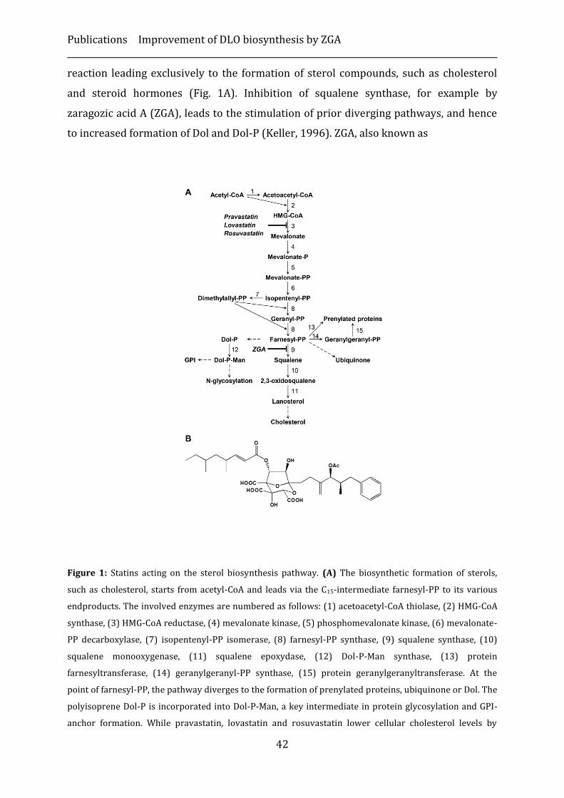
Publications ‒ Improvement of DLO biosynthesis by ZGA
42
reaction leading exclusively to the formation of sterol compounds, such as cholesterol
and steroid hormones (Fig. 1A). Inhibition of squalene synthase, for example by
zaragozic acid A (ZGA), leads to the stimulation of prior diverging pathways, and hence
to increased formation of Dol and Dol-P (Keller, 1996). ZGA, also known as
Figure 1: Statins acting on the sterol biosynthesis pathway. (A) The biosynthetic formation of sterols,
such as cholesterol, starts from acetyl-CoA and leads via the C15-intermediate farnesyl-PP to its various
endproducts. The involved enzymes are numbered as follows: (1) acetoacetyl-CoA thiolase, (2) HMG-CoA
synthase, (3) HMG-CoA reductase, (4) mevalonate kinase, (5) phosphomevalonate kinase, (6) mevalonate-
PP decarboxylase, (7) isopentenyl-PP isomerase, (8) farnesyl-PP synthase, (9) squalene synthase, (10)
squalene monooxygenase, (11) squalene epoxydase, (12) Dol-P-Man synthase, (13) protein
farnesyltransferase, (14) geranylgeranyl-PP synthase, (15) protein geranylgeranyltransferase. At the
point of farnesyl-PP, the pathway diverges to the formation of prenylated proteins, ubiquinone or Dol. The
polyisoprene Dol-P is incorporated into Dol-P-Man, a key intermediate in protein glycosylation and GPI-
anchor formation. While pravastatin, lovastatin and rosuvastatin lower cellular cholesterol levels by

Publications ‒ Improvement of DLO biosynthesis by ZGA
43
inhibiting the pathway at the stage of the HMG-CoA reductase, ZGA achieves this by inhibiting the
squalene synthase. The chemical structure of ZGA is depicted in (B).
squalestatin I, was discovered by screening metabolites of filamentous fungi for
cholesterol lowering activity (Bergstrom et al., 1995). Detailed analysis disclosed that
ZGA acts as a competitive inhibitor of the squalene synthase by mimicking the farnesyl-
PP substrate or the stable intermediate presqualene-PP with its bicyclic, highly acidic
core (Fig. 1B). In contrast to statins acting on common steps of both Dol and cholesterol
biosynthesis, such as the 3-hydroxy-3-methyl-glutaryl coenzyme A (HMG-CoA)
reductase inhibitors pravastatin (Koga et al., 1990), lovastatin (Alberts et al., 1980) or
rosuvastatin (McTaggart et al., 2001), ZGA does not negatively affect the diverging
pathways generating Dol, ubiquinone or prenylated proteins (Fig. 1A). In the present
study, we have investigated the ability of the squalene synthase inhibitor to increase Dol
biosynthesis and thereby the output of N-glycosylation in CDG fibroblasts.
Experimental Procedures
Materials - ZGA was a gift from Merck & Co., Inc. (Rahway, NJ). The C80-polyprenyl-P
standards were purchased from Larodan Fine Chemicals (Sweden), and broad range
mammalian Dol-P standards were from Sigma-Aldrich (Switzerland). Acetonitrile
(Scharlau, Spain), dichloromethane (Sigma-Aldrich) and water (Sigma-Aldrich) were of
HPLC grade, other chemicals were of analytical grade.
Cholesterol analysis Ȃ Human primary skin fibroblasts (4 x 10⁷ cells) cultured in DMEM
(Sigma-Aldrich) with 10% fetal calf serum (Bioconcept, Switzerland) at 37°C were treated for ばに h with either などど づM ZGA or DMSO alone as negative control┻ Cells were harvested by trypsinization, washed in PBS and centrifuged at 1000 x g for 5 min. To the
cell pellet 1 ml of 4 % (w/v) KOH in 90 % ethanol was added. After vortexing, the
solution was incubated for 10 min in a ultrasonic bath and transferred to a glass tube.
After addition of 80 µl of internal standard (0.1 mg/ml epicoprostanol (Sigma-Aldrich)
in pyridine), the solution was saponified for 60 min at 60°C, mixed with 1 ml of water
and extracted three times with 2 ml heptane. The pooled heptane extracts were dried
under nitrogen and derivatized with 75 µl BSTFA (Machery-Nagel, Oensingen,
Switerzland) in 75 µl pyridine at 60°C for 60 min. For GC/MS analysis, the derivative
mixture was diluted 5-fold with heptane and 1 µl was injected (injector temperature:
280°C; splitless injection). A Restek RTX-1MS (BGB Analytik AG, Böckten, Switzerland)
Publications ‒ Improvement of DLO biosynthesis by ZGA
44
column (15 m, ID = 0.25 mm) was used for chromatographic separation of the sterols.
The carrier gas was helium at a constant flow of 1.5 ml/min. After a dwell time of 3 min
at 90°C, the oven temperature was raised to 200°C at 20°C/min, then to 260°C at
1.5°C/min, and finally held at 260°C for 10 min. Mass spectrometry was performed on a
Finnigan PolarisQ ion trap mass spectrometer. Mass spectra were acquired in the mass
range of m/z = 50 - 550.
Dol-P analysis Ȃ Fibroblasts (4 x 10⁷ cells) were harvested and centrifuged at 1000 x g
for 5 min. Cell pellets were dissolved in 12 ml of water/methanol (1:1). Prior to alkaline
hydrolysis (Elmberger et al., 1989)┸ なの づg of C80-polyprenyl-P standards were added.
The extracted Dol and Dol-P were purified on a C18 Sep Pak column (Waters, USA) and
subsequently separated on a Silica Sep Pak column (Waters) (Haeuptle et al., 2010). The
Dol-P were dimethylated using diazomethane generated in an Aldrich diazomethane
generator system (Sigma-Aldrichょ according to the manufacturer╆s instructions┻ The resulting Dol-P-Me2 were selectively demethylated by overnight incubation in tert-
butylamine (Sigma-Aldrich) at 70°C (Yamada et al., 1986). Monomethylated Dol-P
samples were dissolved in a saturated solution of 9-anthryldiazomethane (Sigma-
Aldrich) in diethylether and incubated for 6 h on ice in the dark. Anthracene labeled Dol-
P were separated from non-reacted labeling agent by organic extraction (Yamada et al.,
1986). The purified products were dissolved in acetonitrile/dichloromethane (3:2) and
subjected to HPLC on an Inertsil ODS-ぬ column ゅの づm┸ ね┻は x にのど mm┹ GL Sciences )nc┻┸ Japan) equipped with a precolumn. Isocratic elution in acetonitrile/dichloromethane
(3:2) and 0.01% diethylamine (Sigma-Aldrich) was performed at a flow rate of 1 ml/min
(Yamada et al., 1986). Fluorescent labeled Dol-P were detected by excitation at 365 nm
and emission at 412 nm. Defined amounts of internal standard C80-polyprenyl-P were
used for quantification.
Determination of Dol-P-Man - Approximately 4 x 10⁷ fibroblasts were grown for 72 h in
DMEM containing low Glc (5 mM; Sigma-Aldrich), supplemented with 2% fetal calf serum and などど づM ZGA or DMSO as negative control┻ Low Glc medium was utilized to achieve improved Man incorporation (Korner et al., 1998). Cellular Dol-P-Man was
metabolically labeled, extracted and purified according to the protocol of Körner and co-
workers (Korner et al., 1998). Briefly, the fibroblasts were labeled by incubation in
DMEM containing 0.5 mM Glc and 125 µCi [3H]-Man (Hartmann Analytic, Germany) for
30 min. Dol-P-Man and short LLO were extracted once with chloroform/methanol (2:1)

Publications ‒ Improvement of DLO biosynthesis by ZGA
45
and twice with chloroform/methanol (3:2). The combined organic phases were dried
and washed. Thin-layer chromatography on Silica gel 60 plates was performed in
chloroform/methanol/water (65:25:4). The plates were analyzed by radiography after
signal enhancement with an EN3HANCE spray (PerkinElmer, USA) and the area
containing Dol-P-Man were scraped and counted in a TRI‒CARB 2900 TR liquid
scintillation analyzer (Packard, USA).
CD59 flow cytometry - Fibroblasts (2 x 10⁵ cells) were harvested by trypsinization,
washed once in PBS containing 2% fetal calf serum and incubated with a FITC
conjugated mouse anti-human CD59 antibody (BD Pharmingen, USA) diluted 1:100 in
PBS containing 2% fetal calf serum for 20 min on ice (Imbach et al., 2000). Fibroblasts
were washed and analyzed on a BD FACSCanto II flow cytometer (BD Biosciences, USA)
equipped with BD FACSDiva software (BD Biosciences).
LLO and N-linked oligosaccharide (NLO) analysis - The LLO profiles of 1.5 x 10⁷ fibroblasts treated for ばに h with either などど づM ZGA or DMSO were analyzed as described (Haeuptle et al., 2008). The cells were starved for 45 min in fetal calf serum-
and Glc-free DMEM (Invitrogen) and metabolically labeled for 60 min by addition of 150 づCi い3H]-Man. LLO were extracted from cell pellets and oligosaccharides were released
by mild acid hydrolysis in 0.1 N HCl. Glycoproteins recovered from the LLO extraction
were denatured and NLO were released by overnight incubation with PNGaseF
endoglycosidase (New England BioLabs, USA) (Grubenmann et al., 2002).
Oligosaccharides were purified by ion-exchange chromatography on AG1-X2 and
AG50W-X8 resins (Bio-Rad, USA) and by hydrophobic chromatography on Supelclean
ENVI-Carb 120/400 beads (Supelco, USA) and C18 Sep Pak columns and subjected to
HPLC analysis.
Statistics - Results were expressed as mean ± SEM. The one-way ANOVA test with Bonferroni╆s multiple comparison post-test was applied to confirm differences between
groups. Significance was accepted for p<0.05.

Publications ‒ Improvement of DLO biosynthesis by ZGA
46
Results
The toxicity of ZGA was first determined by incubating human primary skin fibroblasts
for ten days with increasing concentrations from 10 to 500 µM. ZGA was tolerated by
fibroblasts up to a concentration of 125 µM. Above that concentration, the rate of cell
proliferation slowed down and cell morphology was altered (data not shown). The
addition of 100 µM ZGA to healthy control and CDG fibroblasts led to a moderate
decrease of cellular cholesterol levels by 15% and 30%, respectively (Fig. 2).
Figure 2: Analysis of cholesterol in healthy control and DPM1-CDG fibroblasts. Steady-state cholesterol
levels were determined in fibroblasts by GC/MS after organic extraction. Cholesterol levels relative to the
protein amounts of the extracted mock treated cells are shown as white bars and cholesterol levels of ZGA
treated fibroblasts are shown as gray bars. Data are represented by mean ± SEM of three independent
experiments, * p<0.05.
The effect of ZGA on glycosylation was determined by measuring cellular levels of Dol-P,
Dol-P-Man, a glycosylphosphatidylinositol (GPI)-anchored model protein, LLO and NLO.
The impact of ZGA on Dol-P levels was first addressed on healthy control fibroblasts.
After labeling with the fluorochrome 9-anthryldiazomethane, Dol-P levels were
quantitated after HPLC separation (Haeuptle et al., 2010). The resulting fluorescent
HPLC profiles of untreated cells (Fig. ぬAょ and of cells treated with などど づM ZGA for ばに h (Fig. 3B) were compared. The pattern of Dol-P species changed upon ZGA treatment,
where the amount of longer C100- and C105-Dol-P increased in ZGA treated fibroblasts
(Fig. 3C).
To address the effect of ZGA on various parameters of LLO biosynthesis in CDG, we
chose fibroblasts with a deficiency of Dol-P-Man synthase-1 (DPM1) (Imbach et al.,
2000), which present low Dol-

Publications ‒ Improvement of DLO biosynthesis by ZGA
47
Figure 3: Analysis and quantification of Dol-P levels in mock and ZGA treated control fibroblasts. (A)
Control cells were grown in the presence of DMSO. Cellular Dol-P were labeled with the fluorophore 9-
anthryldiazomethane and separated by HPLC. The enlarged section in the fluorescent profile highlights
the separation of the particular Dol-P species. The elution times of mammalian Dol-P standards ranging
from C90- to C105-Dol-P are indicated below the section. The internal C80-polyprenol-P standard utilized for
quantification eluted at a retention time of 33.5 min. (B) The separation of Dol-P from ZGA treated control
fibroblasts is presented in the enlarged section, and the respective Dol-P species were assigned like in (A).
Quantification was accomplished by comparison with the internal standard hexadecaprenyl-P eluting at
34.2 min. (C) The Dol-P levels from four independent HPLC runs were calculated and normalized to 107
fibroblasts. White bar represent Dol-P originating from mock treated and gray bars Dol-P from ZGA
treated cells. Data are represented by mean ± SEM of four independent experiments, * p<0.05.
P-Man levels, an accumulation of the LLO Dol-PP-GlcNAc2Man5 and the corresponding
GlcNAc2Man5 glycan structure on glycoproteins. Dol-P-Man production was measured
after labeling cells with [3H]-Man. The [3H]-Dol-P-Man pool determined in DPM1-
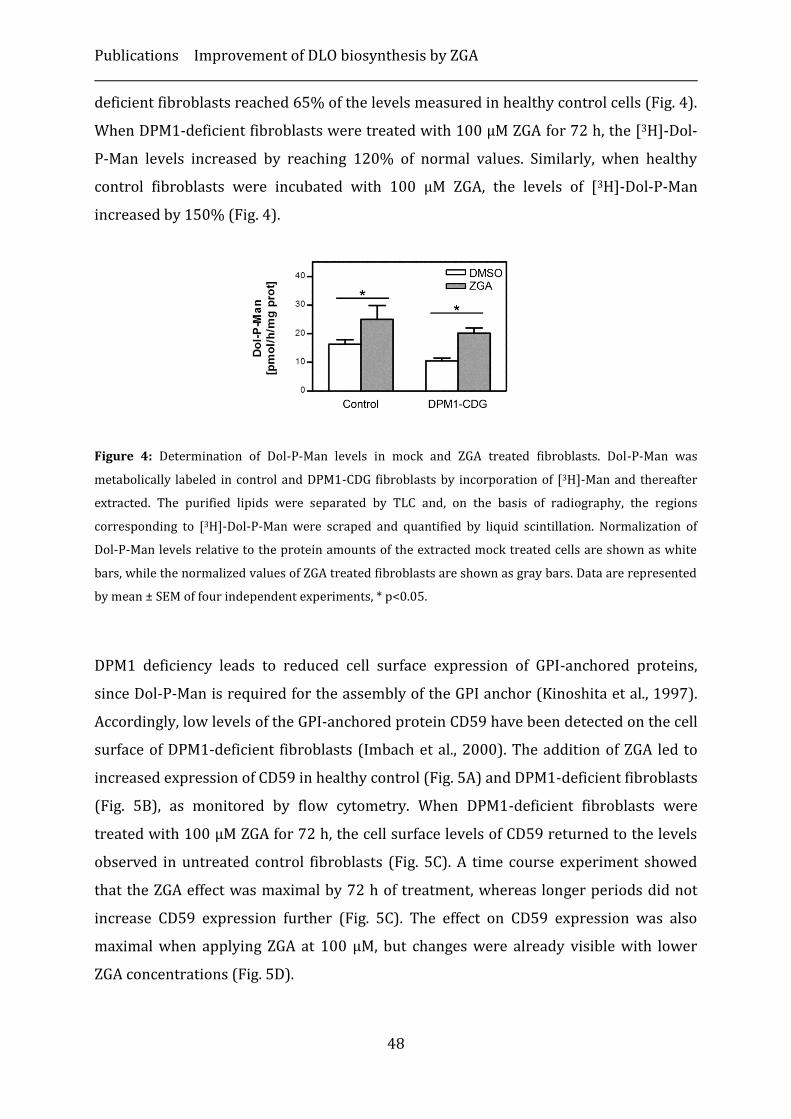
Publications ‒ Improvement of DLO biosynthesis by ZGA
48
deficient fibroblasts reached 65% of the levels measured in healthy control cells (Fig. 4).
When DPM1-deficient fibroblasts were treated with 100 µM ZGA for 72 h, the [3H]-Dol-
P-Man levels increased by reaching 120% of normal values. Similarly, when healthy
control fibroblasts were incubated with 100 µM ZGA, the levels of [3H]-Dol-P-Man
increased by 150% (Fig. 4).
Figure 4: Determination of Dol-P-Man levels in mock and ZGA treated fibroblasts. Dol-P-Man was
metabolically labeled in control and DPM1-CDG fibroblasts by incorporation of [3H]-Man and thereafter
extracted. The purified lipids were separated by TLC and, on the basis of radiography, the regions
corresponding to [3H]-Dol-P-Man were scraped and quantified by liquid scintillation. Normalization of
Dol-P-Man levels relative to the protein amounts of the extracted mock treated cells are shown as white
bars, while the normalized values of ZGA treated fibroblasts are shown as gray bars. Data are represented
by mean ± SEM of four independent experiments, * p<0.05.
DPM1 deficiency leads to reduced cell surface expression of GPI-anchored proteins,
since Dol-P-Man is required for the assembly of the GPI anchor (Kinoshita et al., 1997).
Accordingly, low levels of the GPI-anchored protein CD59 have been detected on the cell
surface of DPM1-deficient fibroblasts (Imbach et al., 2000). The addition of ZGA led to
increased expression of CD59 in healthy control (Fig. 5A) and DPM1-deficient fibroblasts
(Fig. 5B), as monitored by flow cytometry. When DPM1-deficient fibroblasts were
treated with 100 µM ZGA for 72 h, the cell surface levels of CD59 returned to the levels
observed in untreated control fibroblasts (Fig. 5C). A time course experiment showed
that the ZGA effect was maximal by 72 h of treatment, whereas longer periods did not
increase CD59 expression further (Fig. 5C). The effect on CD59 expression was also maximal when applying ZGA at などど づM┸ but changes were already visible with lower ZGA concentrations (Fig. 5D).

Publications ‒ Improvement of DLO biosynthesis by ZGA
49
Figure 5: Cell surface expression of GPI-anchored CD59. (A) Control fibroblasts were stained with a FITC conjugated ゎ-human CD59 antibody and analysed by flow cytometry. The fluorescent histogram shows
unstained cells as gray solid line, mock treated cells as black solid line and ZGA treated cells as black
dotted line. (B) CD59 expression on DPM1-CDG patient fibroblasts was monitored by flow cytometry.
Experimental set up and specimen allocations were like for control cells in (A). (C) Control (white bars)
and DPM1-CDG fibroblasts (gray bars) were incubated with 100 µM ZGA for varying time periods and the
detected CD59 expressions were normalized to CD59 levels of untreated control fibroblasts. (D) Different
concentrations of ZGA were administered to control (white bars) or DPM1-CDG (gray bars) patient
fibroblasts for 72 hours. As in (C), the measured CD59 expressions were normalized to untreated control
cells. Data are represented by mean ± SEM of three independent experiments.
The biosynthesis of the LLO Dol-PP-GlcNAc2Man9Glc3 requires Dol as carrier of the
growing oligosaccharide and as donor substrate for Dol-P-Man and Dol-P-Glc (Kornfeld
and Kornfeld, 1985). In normal cells, only the complete LLO Dol-PP-GlcNAc2Man9Glc3
can be detected (Fig. 6A). DPM1 deficiency
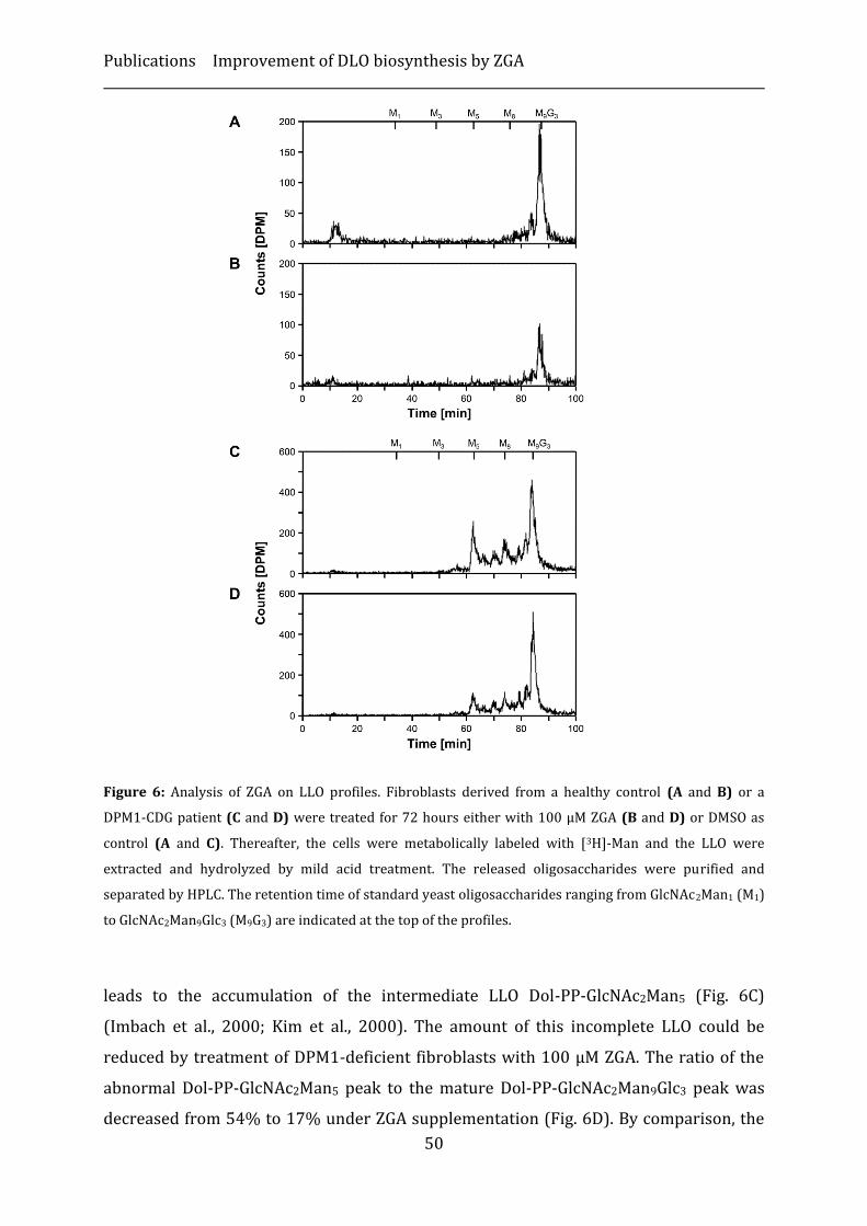
Publications ‒ Improvement of DLO biosynthesis by ZGA
50
Figure 6: Analysis of ZGA on LLO profiles. Fibroblasts derived from a healthy control (A and B) or a
DPM1-CDG patient (C and D) were treated for 72 hours either with 100 µM ZGA (B and D) or DMSO as
control (A and C). Thereafter, the cells were metabolically labeled with [3H]-Man and the LLO were
extracted and hydrolyzed by mild acid treatment. The released oligosaccharides were purified and
separated by HPLC. The retention time of standard yeast oligosaccharides ranging from GlcNAc2Man1 (M1)
to GlcNAc2Man9Glc3 (M9G3) are indicated at the top of the profiles.
leads to the accumulation of the intermediate LLO Dol-PP-GlcNAc2Man5 (Fig. 6C)
(Imbach et al., 2000; Kim et al., 2000). The amount of this incomplete LLO could be
reduced by treatment of DPM1-deficient fibroblasts with 100 µM ZGA. The ratio of the
abnormal Dol-PP-GlcNAc2Man5 peak to the mature Dol-PP-GlcNAc2Man9Glc3 peak was
decreased from 54% to 17% under ZGA supplementation (Fig. 6D). By comparison, the

Publications ‒ Improvement of DLO biosynthesis by ZGA
51
addition of 100 µM ZGA to control fibroblasts did not influence the quality of the LLO
profile (Fig. 6B).
After transfer from LLO to proteins, the NLO GlcNAc2Man9Glc3 is trimmed by
glucosidases and mannosidases in the ER (Helenius and Aebi, 2004). The analysis of NLO
after [3H]-Man labeling of control fibroblasts showed, as expected, GlcNAc2Man8,
GlcNAc2Man9 and GlcNAc2Man9Glc1 as main oligosaccharide structures (Fig. 7A). In
DPM1-deficient fibroblasts, several intermediary oligosaccharide structures ranging
from GlcNAc2Man4 to GlcNAc2Man7 were found, whereas the normal oligosaccharides
GlcNAc2Man8, GlcNAc2Man9 and GlcNAc2Man9Glc1 were under-represented (Fig. 7C).
While ZGA treatment did not affect the NLO profile of control fibroblasts (Fig. 7B), it
decreased the occurrence of abnormal N-linked glycans GlcNAc2Man4 to GlcNAc2Man7
and increased the amounts of the normal NLO GlcNAc2Man8, GlcNAc2Man9 and
GlcNAc2Man9Glc1 in DPM1-deficient fibroblasts (Fig. 7D). In these cells, the ratio of the
pathologic GlcNAc2Man5 peak to the normal GlcNAc2Man9 peak was 422% and this ratio
was decreased to 143% after addition of ZGA (Fig. 7D). This normalization of the NLO
profile demonstrated the beneficial effect of ZGA treatment on N-glycosylation.
Taken together, the present study showed that treatment of human CDG fibroblasts with
the squalene synthase inhibitor ZGA stimulated Dol-P biosynthesis and thereby
improved the level of N-glycosylation in conditions of limited substrate availability as
encountered in DPM1-deficiency. In addition, the supplementation with ZGA resulted in
the normalization of the expression of the GPI-anchored protein CD59 on DPM1-
deficient fibroblasts.

Publications ‒ Improvement of DLO biosynthesis by ZGA
52
Figure 7: Protein N-glycosylation in ZGA treated fibroblasts. NLO from control (A and B) and DPM1-CDG
patient fibroblasts (C and D) were separated by HPLC. The protein linked oligosaccharides were prepared
by PNGaseF endoglycosidase release from metabolically labeled cells previously treated for 72 hours
either with 100 µM ZGA (B and D) or DMSO as control (A and C). The retention times of GlcNAc2Man1 (M1)
to GlcNAc2Man9Glc3 (M9G3) are marked at the top of the profiles.

Publications ‒ Improvement of DLO biosynthesis by ZGA
53
Discussion
Successful treatment of biosynthetic CDG is restricted so far to oral administration of
Man to Man-P isomerase deficient (MPI-CDG) patients (Niehues et al., 1998; Westphal et
al., 2001). The applied Man could be phosphorylated by hexokinase and allows
functional bypassing of the defective isomerization of fructose-6-P to Man-6-P. In the
group of Golgi-associated CDG, a deficiency of the Golgi GDP-Fuc transporter (SLC35C1-
CDG or leukocyte adhesion deficiency type II) (Lübke et al., 2001) could also be treated
by simple supplementation of a monosaccharide. Oral Fuc administration induced the
expression of fucosylated glycoproteins and within short terms, the clinical symptoms of
a SLC35C1-CDG patient could be relieved (Marquardt et al., 1999). Moreover, a few
elementary attempts have been conducted to treat phosphomannomutase-2 deficiency
(PMM2-CDG), which forms by far the largest CDG subtype (Freeze, 2009). Mutations in
the PMM2 gene lead to disrupted conversion of Man-6-P to Man-1-P, which acts as
precursor of GDP-Man (Matthijs et al., 1997). However, Man-1-P is neither able to diffuse
through biological membranes nor does a transport system exist. Thus, it needs
chemical modification prior to direct administration to overcome this obstacle (Eklund
et al., 2005; Hardre et al., 2007; Rutschow et al., 2002). Membrane permeable Man-1-P
analogues were successful in restoring LLO biosynthesis in CDG patient cells, but such
prodrugs were toxic, very unstable and had to be applied in high concentrations (Eklund
et al., 2005).
Shang and Lehrman discovered a metformin-stimulated Man specific transport activity
in human fibroblasts (Shang and Lehrman, 2004). The resulting increased Man uptake
was shown to be able to correct artificially induced defects in LLO biosynthesis and
protein N-glycosylation in control and PMM2-CDG fibroblasts. However, the observation
that the clinical phenotype of PMM2 deficiency could not be treated with dietary Man,
which was actually effective in cellular models (Freeze, 2009), renders a therapeutic
administration of metformin questionable.
Our approach to increase Dol-P biosynthesis by inhibition of the squalene synthase
offers a therapeutic perspective for inherited biosynthetic N-glycosylation defects
featuring residual enzymatic activity. Conveniently, ZGA is well tolerated in animal
experiments (Baxter et al., 1992; Bergstrom et al., 1993) compared to classical statins,
which interfere with various isoprenoid-based biosynthesis pathways and are
Publications ‒ Improvement of DLO biosynthesis by ZGA
54
associated to adverse effects (Silva et al., 2007). We could show that ZGA decreased
cellular cholesterol levels, although only by 15-30%, indicating that membrane
properties were likely unaltered due to this treatment. However, we cannot exclude an
effect of the lowered cholesterol levels on the formation of glycolipid rafts, in which GPI-
anchored proteins partition. Accordingly, the increased expression of the GPI-anchored
protein CD59 observed in ZGA-treated cells could be caused by increased Dol-P
availability and altered raft distribution. By contrast, the positive effect of ZGA
supplementation on Dol-P-Man levels and LLO patterns in DPM1-CDG fibroblasts are
unlikely to be related to decreased cholesterol biosynthesis since the ER membrane is
low in cholesterol levels (Liscum and Underwood, 1995). Nevertheless, the effects of
ZGA on protein modifications, such as farnesylation and geranyl-geranylation, have not
been investigated, so that we cannot dismiss an impact of such modifications on the N-
glycosylation pathway at this stage.
The present study was based on DPM1-deficient fibroblasts because this form of CDG
shows several measurable abnormalities along the biosynthesis of N-glycans and GPI-
anchored proteins. By contrast, PMM2-deficiency that represents the largest group of
CDG cases (Haeuptle and Hennet, 2009), does not feature accumulating LLO
intermediates. Consequently, incomplete oligosaccharides on glycoproteins are usually
not encountered in PMM2-deficiency. However, the up-regulation of Dol-P dependent
substrates by ZGA may be beneficial in normalizing the glycosylation disorders caused
by deficiency of glycosyltransferases catalyzing the attachment of the last four Man and
three Glc of the LLO precursor (Kornfeld and Kornfeld, 1985).
Over the last decade, knockout mouse models for PMM2-, MPI- and DPAGT1-deficiency
have been generated (DeRossi et al., 2006; Marek et al., 1999; Thiel et al., 2006). These
works clearly showed that disruption of LLO biosynthesis lead to early embryonic
lethality. In this context, it would be interesting to investigate whether the embryonic
lethality of the homozygotes could be lagged to a later stage of development by treating
pregnant heterozygous mice with ZGA according to Keller (Keller, 1996). In addition, the
generation of viable CDG mouse models, for instance by introduction of selected point
mutations, would allow detailed analysis of organ specific effects of ZGA in the context of
inherited N-glycosylation deficiencies. This seems to be of particular interest,
considering that Keller reported an enormous effect of ZGA on Dol and Dol-P pools in rat
livers, but not in other organs such as brain, kidney, intestine or testis (Keller, 1996),
Publications ‒ Improvement of DLO biosynthesis by ZGA
55
which was similarly observed in different brain cells (Crick et al., 1995). Subcutaneously
administered ZGA might preferentially be taken up by mammalian livers via a specific
hepatic transport mechanism, which was likewise proposed in other studies (Baxter et
al., 1992; Bergstrom et al., 1993). Considering the frequent impairment of liver function
in CDG patients (Leroy, 2006), the proposed hepatic transport system for ZGA would
suggest that liver N-glycosylation may benefit from treatment with the compound. Along
this line, it would be of interest to address the effect of ZGA supplementation in hepatic
cell lines, since fibroblasts do not represent suitable cell models to study glycoprotein
secretion. The generation of hepatic cell lines with inactivated glycosylation genes will
represent an opportunity to study the regulation of N-glycosylation by ZGA and similar
compounds.
In conclusion, this study opens new perspectives in developing treatments of
glycosylation deficiency by showing that manipulation of the dolichol biosynthesis
pathway, as shown here by ZGA administration, represents a valid option.
Footnotes
a The abbreviations used are: CDG, Congenital Disorders of Glycosylation; Dol,
dolichol; GPI, glycosylphosphatidylinositol; HMG-CoA, 3-hydroxy-3-methyl-
glutaryl Coenzyme A; LLO, lipid-linked oligosaccharides; NLO, N-linked
oligosaccharides; ZGA, zaragozic acid A
Acknowledgements
We are grateful to Merck & Co., Inc. for the generous providing of zaragozic acid A and to
Eric G. Berger for critically reading the manuscript. This work was supported by the
Swiss National Science Foundation Grant 31003A-116039 to T. Hennet.

Publications ‒ Improvement of DLO biosynthesis by ZGA
56
References
Alberts, A.W., J. Chen, G. Kuron, V. Hunt, J. Huff, C. Hoffman, J. Rothrock, M. Lopez, H. Joshua, E. Harris, A. Patchett, R. Monaghan, S. Currie, E. Stapley, G. Albers-Schonberg, O. Hensens, J. Hirshfield, K. Hoogsteen, J. Liesch, and J. Springer. 1980. Mevinolin: a highly potent competitive inhibitor of hydroxymethylglutaryl-coenzyme A reductase and a cholesterol-lowering agent. Proc. Natl. Acad. Sci. U. S. A. 77:3957-3961.
Baxter, A., B.J. Fitzgerald, J.L. Hutson, A.D. McCarthy, J.M. Motteram, B.C. Ross, M. Sapra, M.A. Snowden, N.S. Watson, R.J. Williams, and C. Wright. 1992. Squalestatin 1, a potent inhibitor of squalene synthase, which lowers serum cholesterol in vivo. J. Biol. Chem. 267:11705-11708.
Bergstrom, J.D., C. Dufresne, G.F. Bills, M. Nallin-Omstead, and K. Byrne. 1995. Discovery, biosynthesis, and mechanism of action of the zaragozic acids: potent inhibitors of squalene synthase. Annu. Rev. Microbiol. 49:607-639.
Bergstrom, J.D., M.M. Kurtz, D.J. Rew, A.M. Amend, J.D. Karkas, R.G. Bostedor, V.S. Bansal, C. Dufresne, F.L. VanMiddlesworth, O.D. Hensens, J.M. Liesch, D.L. Zink, K.E. Wilson, J. Onishi, J.A. Milligan, G. Bills, L. Kaplan, M. Nallin-Omstead, R.G. Jenkins, L. Huang, M.S. Meinz, L. Quinn, R.W. Burg, Y.L. Kong, S. Mochales, M. Mojena, I. Martin, F. Pelaez, M.T. Diez, and A.W. Alberts. 1993. Zaragozic acids: a family of fungal metabolites that are picomolar competitive inhibitors of squalene synthase. Proc. Natl. Acad. Sci. U. S. A. 90:80-84.
Crick, D.C., J. Suders, C.M. Kluthe, D.A. Andres, and C.J. Waechter. 1995. Selective inhibition of cholesterol biosynthesis in brain cells by squalestatin 1. J. Neurochem. 65:1365-1373.
De Praeter, C.M., G.J. Gerwig, E. Bause, L.K. Nuytinck, J.F. Vliegenthart, W. Breuer, J.P. Kamerling, M.F. Espeel, J.J. Martin, A.M. De Paepe, N.W. Chan, G.A. Dacremont, and R.N. Van Coster. 2000. A novel disorder caused by defective biosynthesis of N-linked oligosaccharides due to glucosidase I deficiency. Am. J. Hum. Genet. 66:1744-1756.
DeRossi, C., L. Bode, E.A. Eklund, F. Zhang, J.A. Davis, V. Westphal, L. Wang, A.D. Borowsky, and H.H. Freeze. 2006. Ablation of mouse phosphomannose isomerase (Mpi) causes mannose 6-phosphate accumulation, toxicity, and embryonic lethality. J. Biol. Chem. 281:5916-5927.
Eklund, E.A., N. Merbouh, M. Ichikawa, A. Nishikawa, J.M. Clima, J.A. Dorman, T. Norberg, and H.H. Freeze. 2005. Hydrophobic Man-1-P derivatives correct abnormal glycosylation in Type I congenital disorder of glycosylation fibroblasts. Glycobiology 15:1084-1093.
Elmberger, P.G., I. Eggens, and G. Dallner. 1989. Conditions for quantitation of dolichyl phosphate, dolichol, ubiquinone and cholesterol by HPLC. Biomed. Chromatogr. 3:20-28.
Foulquier, F. 2008. COG defects, birth and rise! Biochim. Biophys. Acta 1792:896-902. Freeze, H.H. 2006. Genetic defects in the human glycome. Nat. Rev. Genet. 7:537-551. Freeze, H.H. 2009. Towards a therapy for phosphomannomutase 2 defeciency, the defect in CDG-
Ia patients. Biochim. Biophys. Acta 1792:835-840. Grubenmann, C.E., C.G. Frank, S. Kjaergaard, E.G. Berger, M. Aebi, and T. Hennet. 2002. ALG12
mannosyltransferase defect in congenital disorder of glycosylation type lg. Hum. Mol.
Genet. 11:2331-2339. Haeuptle, M.A., and T. Hennet. 2009. Congenital Disorders of Glycosylation: an Update on Defects
Affecting the Biosynthesis of Dolichol-linked Oligosaccharides. Hum. Mutat. 30:1628-1641.
Haeuptle, M.A., A.J. Hülsmeier, and T. Hennet. 2010. HPLC and mass spectrometry analysis of dolichol-phosphates at the cell culture scale. Anal. Biochem. 396:133-138.
Haeuptle, M.A., F.M. Pujol, C. Neupert, B. Winchester, A.J. Kastaniotis, M. Aebi, and T. Hennet. 2008. Human RFT1 deficiency leads to a disorder of N-linked glycosylation. Am. J. Hum.
Genet. 82:600-606. Hansske, B., C. Thiel, T. Lübke, M. Hasilik, S. Honing, V. Peters, P.H. Heidemann, G.F. Hoffmann,
E.G. Berger, K. von Figura, and C. Körner. 2002. Deficiency of UDP-galactose:N-

Publications ‒ Improvement of DLO biosynthesis by ZGA
57
acetylglucosamine beta-1,4-galactosyltransferase I causes the congenital disorder of glycosylation type IId. J. Clin. Invest. 109:725-733.
Hardre, R., A. Khaled, A. Willemetz, T. Dupre, S. Moore, C. Gravier-Pelletier, and Y. Le Merrer. 2007. Mono, di and tri-mannopyranosyl phosphates as mannose-1-phosphate prodrugs for potential CDG-Ia therapy. Bioorg. Med. Chem. Lett. 17:152-155.
Helenius, A., and M. Aebi. 2004. Roles of N-linked glycans in the endoplasmic reticulum. Annu.
Rev. Biochem. 73:1019-1049. Hülsmeier, A.J., P. Paesold-Burda, and T. Hennet. 2007. N-glycosylation site occupancy in serum
glycoproteins using multiple reaction monitoring liquid chromatography-mass spectrometry. Mol Cell Proteomics 6:2132-2138.
Imbach, T., B. Schenk, E. Schollen, P. Burda, A. Stutz, S. Grunewald, N.M. Bailie, M.D. King, J. Jaeken, G. Matthijs, E.G. Berger, M. Aebi, and T. Hennet. 2000. Deficiency of dolichol-phosphate-mannose synthase-1 causes congenital disorder of glycosylation type Ie. J.
Clin. Invest. 105:233-239. Jaeken, J., and G. Matthijs. 2007. Congenital Disorders of Glycosylation: A Rapidly Expanding
Disease Family. Annu. Rev. Genomics Hum. Genet. 8:261-278. Keller, R.K. 1996. Squalene synthase inhibition alters metabolism of nonsterols in rat liver.
Biochim. Biophys. Acta 1303:169-179. Kim, S., V. Westphal, G. Srikrishna, D.P. Mehta, S. Peterson, J. Filiano, P.S. Karnes, M.C. Patterson,
and H.H. Freeze. 2000. Dolichol phosphate mannose synthase (DPM1) mutations define congenital disorder of glycosylation Ie (CDG-Ie). J. Clin. Invest. 105:191-198.
Kinoshita, T., K. Ohishi, and J. Takeda. 1997. GPI-anchor synthesis in mammalian cells: genes, their products, and a deficiency. J. Biochem. 122:251-257.
Koga, T., Y. Shimada, M. Kuroda, Y. Tsujita, K. Hasegawa, and M. Yamazaki. 1990. Tissue-selective inhibition of cholesterol synthesis in vivo by pravastatin sodium, a 3-hydroxy-3-methylglutaryl coenzyme A reductase inhibitor. Biochim. Biophys. Acta 1045:115-120.
Korner, C., L. Lehle, and K. von Figura. 1998. Abnormal synthesis of mannose 1-phosphate derived carbohydrates in carbohydrate-deficient glycoprotein syndrome type I fibroblasts with phosphomannomutase deficiency. Glycobiology 8:165-171.
Kornfeld, R., and S. Kornfeld. 1985. Assembly of asparagine-linked oligosaccharides. Annu. Rev.
Biochem. 54:631-664. Leroy, J.G. 2006. Congenital disorders of N-glycosylation including diseases associated with O- as
well as N-glycosylation defects. Pediatr. Res. 60:643-656. Liscum, L., and K.W. Underwood. 1995. Intracellular cholesterol transport and
compartmentation. J Biol Chem 270:15443-15446. Lübke, T., T. Marquardt, A. Etzioni, E. Hartmann, K. von Figura, and C. Körner. 2001.
Complementation cloning identifies CDG-IIc, a new type of congenital disorders of glycosylation, as a GDP-fucose transporter deficiency. Nat. Genet. 28:73-76.
Marek, K.W., I.K. Vijay, and J.D. Marth. 1999. A recessive deletion in the GlcNAc-1-phosphotransferase gene results in peri-implantation embryonic lethality. Glycobiology 9:1263-1271.
Marquardt, T., K. Luhn, G. Srikrishna, H.H. Freeze, E. Harms, and D. Vestweber. 1999. Correction of leukocyte adhesion deficiency type II with oral fucose. Blood 94:3976-3985.
Matthijs, G., E. Schollen, E. Pardon, M. Veiga-Da-Cunha, J. Jaeken, J.J. Cassiman, and E. Van Schaftingen. 1997. Mutations in PMM2, a phosphomannomutase gene on chromosome 16p13, in carbohydrate-deficient glycoprotein type I syndrome (Jaeken syndrome). Nat.
Genet. 16:88-92. McTaggart, F., L. Buckett, R. Davidson, G. Holdgate, A. McCormick, D. Schneck, G. Smith, and M.
Warwick. 2001. Preclinical and clinical pharmacology of Rosuvastatin, a new 3-hydroxy-3-methylglutaryl coenzyme A reductase inhibitor. Am. J. Cardiol. 87:28B-32B.
Muscio, F., J.P. Carlson, L. Kuehl, and H.C. Rilling. 1974. Prequalene pyrophosphate. A normal intermediate in squalene biosynthesis. J. Biol. Chem. 249:3746-3749.
Niehues, R., M. Hasilik, G. Alton, C. Körner, M. Schiebe-Sukumar, H.G. Koch, K.P. Zimmer, R. Wu, E. Harms, K. Reiter, K. von Figura, H.H. Freeze, H.K. Harms, and T. Marquardt. 1998.
Publications ‒ Improvement of DLO biosynthesis by ZGA
58
Carbohydrate-deficient glycoprotein syndrome type Ib. Phosphomannose isomerase deficiency and mannose therapy. J. Clin. Invest. 101:1414-1420.
Rutschow, S., J. Thiem, C. Kranz, and T. Marquardt. 2002. Membrane-permeant derivatives of mannose-1-phosphate. Bioorg. Med. Chem. 10:4043-4049.
Shang, J., and M.A. Lehrman. 2004. Metformin-stimulated mannose transport in dermal fibroblasts. J. Biol. Chem. 279:9703-9712.
Silva, M., M.L. Matthews, C. Jarvis, N.M. Nolan, P. Belliveau, M. Malloy, and P. Gandhi. 2007. Meta-analysis of drug-induced adverse events associated with intensive-dose statin therapy. Clin. Ther. 29:253-260.
Swiezewska, E., and W. Danikiewicz. 2005. Polyisoprenoids: structure, biosynthesis and function. Prog. Lipid Res. 44:235-258.
Thiel, C., T. Lübke, G. Matthijs, K. von Figura, and C. Körner. 2006. Targeted disruption of the mouse phosphomannomutase 2 gene causes early embryonic lethality. Mol. Cell. Biol. 26:5615-5620.
Westphal, V., S. Kjaergaard, J.A. Davis, S.M. Peterson, F. Skovby, and H.H. Freeze. 2001. Genetic and metabolic analysis of the first adult with congenital disorder of glycosylation type Ib: long-term outcome and effects of mannose supplementation. Mol. Genet. Metab. 73:77-85.
Yamada, K., S. Abe, T. Suzuki, K. Katayama, and T. Sato. 1986. A high-performance liquid chromatographic method for the determination of dolichyl phosphates in tissues. Anal.
Biochem. 156:380-385.

59

Publications ‒ Glycoprotein maturation and the UPR
60
Glycoprotein maturation and the UPR
Andreas J. Hülsmeier, Michael Welti and Thierry Hennet
Institute of Physiology, University of Zürich, Winterthurerstrasse 190, CH-8057 Zürich,
Switzerland
Running title: N- and O-glycosylation analysis
Methods in Enzymology. 2011;491:163-82.
Corresponding author:
Thierry Hennet
Institute of Physiology
University of Zürich
Winterthurerstrasse 190
CH-8057 Zürich
Tel: +41 44 635 5080
Fax: +41 44 635 6814
E-mail: [email protected]

Publications ‒ Glycoprotein maturation and the UPR
61
Abstract
Glycosylation is a complex form of protein modification occurring in the secretory
pathway. The addition of N- and O-glycans affects intracellular processes like the folding
and trafficking of most glycoproteins. To better understand the impact of glycosylation
in protein folding and maturation, parameters like glycosylation site occupancy and
oligosaccharide structure must be measured quantitatively. In the present chapter, we
describe current methods enabling the determination of N-glycosylation by assessment
of cellular dolichol-phosphate levels, dolichol-linked oligosaccharides and the occupancy
of N-glycosylation sites. We also provide detailed methods for the analysis of O-
glycosylation, whose role in intracellular protein maturation is often overlooked.

Publications ‒ Glycoprotein maturation and the UPR
62
1. Introduction
Glycosylation is a widespread and complex form of modification, which adds signals and
specific functions to glycoproteins. N-linked and O-linked glycosylation, which are the
main forms of protein glycosylation, are structurally and functionally distinct. N-linked
glycosylation is initiated in the endoplasmic reticulum (ER), where the oligosaccharide
GlcNAc2Man9Glc3 is transferred co-translationally to selected asparagine residues of
nascent glycoproteins. While still in the ER, oligosaccharides on glycoproteins are
trimmed by glucosidase and mannosidase enzymes to GlcNAc2Man8. Later in the Golgi
apparatus N-linked oligosaccharides undergo further trimming and elongation steps,
which contribute to the structural diversity of N-linked glycosylation. In contrast to the
co-translational beginning of N-glycosylation, O-linked glycosylation takes place on
folded proteins and is initiated by the transfer of monosaccharides to serine and
threonine residues. Some forms of O-glycosylation like O-fucosylation (Harris and
Spellman, 1993) and O-mannosylation (Lommel and Strahl, 2009) begin in the ER,
whereas mucin-type O-glycosylation (Tian and Ten Hagen, 2009) and the biosynthesis of
glycosaminoglycan chains (Bishop et al., 2007) begin in the Golgi apparatus.
N-glycosylation is important for the folding and trafficking of many glycoproteins
(Helenius and Aebi, 2004). The inhibition of N-linked glycosylation, for example by
tunicamycin, results in the accumulation of misfolded proteins in the ER, which is a
strong stimulus of the UPR (Shamu et al., 1994). After trimming by glucosidase-I in the
ER, N-linked oligosaccharides are bound by the chaperone proteins calnexin and
calreticulin, which contribute to the folding of glycoproteins (Ellgaard et al., 1999).
Furthermore, N-linked oligosaccharides are also recognized by the EDEM protein, which
redirects misfolded proteins from the ER lumen to the cytosol for proteasome-mediated
degradation. Another important signal carried by N-linked oligosaccharides is the
mannose-6-phosphate epitope, which mediates the recognition and transfer of
lysosomal proteins to their target organelle (Dahms et al., 1989).
The most common form of O-glycosylation, i.e. mucin-type glycosylation, does not
contribute to glycoprotein folding since it occurs in the Golgi apparatus. However, the
trafficking and secretion of some glycoproteins requires proper mucin-type
glycosylation, as demonstrated in the case of the hormone FGF23. Deficient initiation of
mucin-type O-glycosylation alters the susceptibility of FGF23 to Golgi-localized

Publications ‒ Glycoprotein maturation and the UPR
63
proteases, thereby inhibiting the maturation of active FGF23. Mucin-type glycosylation
of FGF23 is initiated by the polypeptide N-acetylgalactosaminyltransferase-3 enzyme.
Loss of this enzymatic activity leads to the disease tumoral calcinosis, which is also
caused by FGF23 deficiency (Chefetz and Sprecher, 2009).
Another example of glycosyltransferase-assisted trafficking is given by the OFUT1 O-
fucosyltransferase enzyme. This ER-localized glycosyltransferase adds fucose to the
EGF-like repeats of proteins such as Notch and its ligands. Independent from its
glycosyltransferase activity, OFUT1 also acts as a chaperone, which is essential for the
transfer of Notch from the ER to the Golgi apparatus (Okajima et al., 2005). The
contribution of O-linked glycosylation to glycoprotein folding and secretion is much less
documented than in the case of N-linked glycosylation. This limited knowledge is
certainly related to the technically difficult investigation of O-linked glycan structures. In
fact, O-linked glycans are often densely clustered and these glycans cannot be released
without significantly altering the polypeptide backbone. The O-glycanase enzyme from
Streptococcus pneumoniae has a specificity limited to the O-linked disaccharide Ga -
3)GalNAc-O. By contrast, N-linked glycans can be conveniently cleaved at the first
GlcNAc unit by the N-glycosidase F.
2. N-glycosylation
Proper N-linked glycosylation is required for folding and intracellular trafficking of
glycoproteins. Deficiency in the biosynthesis of the precursor of N-glycans or in the
processing of N-glycans in the ER can alter glycoprotein folding and induce UPR. The
following methods can be applied to identify defects of oligosaccharide assembly and of
N-glycosylation site occupancy in target cells.
2.1. Dolichol phosphate analysis
The oligosaccharide core consisting of GlcNAc2Man9Glc3 is first assembled on the
polyisoprenoid carrier dolichol (Dol). Moreover, dolichol-phosphate (Dol-P) is also part
of the glycosylation substrates Dol-P-Man and Dol-P-Glc, which account for the final
extension of the oligosaccharide core in the ER. Considering the low levels of Dol-P
based structures in cells, a sensitive Dol-P detection can be achieved by labeling with the
Publications ‒ Glycoprotein maturation and the UPR
64
fluorescent compound 9-anthryldiazomethane. The procedure was originally described
for tissue sample extraction (Elmberger et al., 1989) and we have adapted the method
for cultured cells (Haeuptle et al., 2009). Detection of unlabeled Dol-P can also be
achieved by negative ion electrospray mass spectrometry (ESI-MS), which, in
combination to precursor ion fragmentation, provides detailed information on Dol-P
composition and on the saturation state of the ゎ-isoprene unit.
2.1.1. Required materials
Devices and materials
- LaChrom D-7000 HPLC system (Merck), equipped with an Inertsil ODS-3 column ゅの づm┸ ね┻は x にのど mm┹ GL Sciences )nc┻┸ Japanょ with a precolumn and a LaChrom 7485 fluorescence detector.
- Nano-flow ESI-ion trap MS (Eksigent nano-LC, 3200 QTRAP-MS, AB/SCIEX)
- C18 Sep-Pak and Silica Sep-Pak cartridges (Waters)
Other reagents
- 9-anthryldiazomethane (ADAM) (Sigma-Aldrich)
- C80-polyprenol and C95-Dol-P standards (Larodan Fine Chemicals, Sweden)
- Diazald (Sigma-Aldrich)
- Carbitol (Sigma-Aldrich)
2.1.2. Extraction and purification of Dol and Dol-P
Adherent cells (about 3 x 10⁸ cells) are washed in PBS after trypsinization and
resuspended in 6 ml of water and then fixed by addition of 6 ml of methanol. The
resulting suspension is transferred to a round bottom glass flask and 3 ml of 15 M
potassium hydroxide are added for alkaline hydrolysis. An aliquot of the C80 polyprenyl-
phosphate (e.g. 15 µg) can be added as internal standard for subsequent quantification.
Publications ‒ Glycoprotein maturation and the UPR
65
The round bottom flask is attached to a reflux cooler and incubated in a preheated oil
bath at 100 °C for 1 h. This alkaline hydrolysis removes carbohydrates from the Dol-P
carrier. The resulting lysate is transferred to a glass tube and methanol is added to a
total volume of 15 ml. The suspension is mixed with 30 ml of
methanol/dichloromethane (1:4, v:v) and incubated at 40 °C for 1 h. Discard the upper
phase (methanol) and transfer the lower phase (dichloromethane) containing the
extracted lipids to a glass tube suitable for centrifugation. Wash the organic phase with
10 ml of dichloromethane/methanol/water (3:48:47, v:v:v). For phase separation,
centrifuge at 6800 x g at room temperature for 10 min and discard the upper phase.
Repeat this washing step four times, which ensures the removal of free sugars and salt
from the sample. The organic phase is then dried under nitrogen gas.
Dol and Dol-P are purified in two steps using C18- and silica-based chromatography
resins. First, equilibrate the C18 Sep-Pak cartridge with 5 ml of methanol, followed by 5
ml of chloroform/methanol (2:1, v:v) and 10 ml of methanol/water (98:2, v:v,
supplemented with 20 mM phosphoric acid). The dried lipids from the extraction step are dissolved in ねどど づl of chloroform【methanol ゅに┺な┸ v┺vょ and diluted by adding など ml of methanol/water (98:2, v:v) supplemented with 20 mM phosphoric acid. Load the
solution onto the C18 Sep-Pak cartridge and wash with 20 ml of methanol/water (98:2,
v:v, supplemented with 20 mM phosphoric acid). To remove phosphoric acid from the
cartridge, wash again with 10 ml of methanol/water (98:2, v:v) devoid of phosphoric
acid. Dol and Dol-P can be eluted with 20 ml of chloroform/methanol (2:1, v:v) and collected in a new glass tube┻ The eluate should be neutralized by adding などど づl of にのガ ammonium hydroxide.
The second purification step through Silica Sep-Pak cartridges allows the separation of
Dol from Dol-P. First, equilibrate the cartridge with 10 ml of
chloroform/methanol/water (10:10:3, v:v:v) and 40 ml of chloroform/methanol (2:1,
v:v, in 0.5% ammoniumhydroxide). Place a new glass tube to collect the dolichol
fraction, which will be in the flowthrough of the chromatography. Apply the neutralized
eluate from the previous step onto the Silica Sep-Pak cartridge. Rinse the glass tube of
the eluate with 10 ml chloroform/methanol (2:1, v:v, in 0.5% ammoniumhydroxide) and
load it onto the Silica cartridge. Repeat the process with10 ml chloroform/methanol
(2:1, v:v, in 0.5% ammonium hydroxide). Elute Dol-P into another glass tube by applying

Publications ‒ Glycoprotein maturation and the UPR
66
30 ml chloroform/methanol/water (10:10:3, v:v:v) onto the Silica cartridge. Dry the Dol
and Dol-P fractions under nitrogen gas at 50 °C.
2.1.3. Anthryldiazomethane labeling of Dol-P
Dol-P labeling is made in two steps. First, Dol-P is dimethylated and then selectively
demethylated to produce mono methylated <mono-methylated, monomethylated?> Dol-
P (Dol-P-Me). The procedure yields an enhanced fluorescence signal when labeling Dol-
P-Me with 9-anthryl derivatives (Yamada et al., 1986). The second step is the
derivatization of Dol-P-Me with 9-anthryldiazomethane (ADAM). We use the
diazomethane generator system (Sigma-Aldrich) for the dimethylation of Dol-P1. It
consists of two glass tubes: a small internal tube placed inside a larger outer tube. The
dried Dol-P fraction is first dissolved in 1 ml of diethylether and transferred into the
large outer glass tube of the diazomethane generator. The glass that contained the Dol-P
fraction is washed twice with 1 ml of diethylether, which is added to the outer tube of
the diazomethane generator. To produce diazomethane, add 0.367 g Diazald (explosive!)
to the inner tube of the diazomethane generator system. Dissolve Diazald by addition of
1 ml of Carbitol. Assemble the two parts of the diazomethane generator system and
immerse it into an ice bath. Make sure that the generator system is sealed. Using a
syringe, carefully add 1.5 ml of 37% potassium hydroxide dropwise to the inner tube of
the diazomethane generator system. Add few drops first and wait until the reaction
starts, as seen by formation of gaseous diazomethane in the Diazald solution. In this way,
the content of the inner tube is prevented from spilling into the outer tube. Then,
carefully add the rest of the potassium hydroxide. Gently mix the reactants and incubate
the reaction for 1 hour in the ice bath. The diethylether phase containing Dol-P should
turn yellow by then, indicating an excess of diazomethane. After disassembly of the
diazomethane generator, leftover reactants can be neutralized by addition of 0.15 g of
silicic acid to the inner tube.
The outer tube should be sealed with parafilm and left for 2 h at room temperature. The
Dol-P-Me2 solution is transferred to a glass extraction tube and dried under nitrogen
gas. To selectively demethylate Dol-P-Me2, dissolve it in 4 ml of tert-butylamine and
1 Diazomethane is very toxic in contact with eye, skin or by inhalation. Diazomethane may explode in contact with sharp edges and when heated beyond 100 °C.
Publications ‒ Glycoprotein maturation and the UPR
67
incubate at 70 °C for 14 h. Make sure to seal the extraction tube completely to prevent
the loss of tert-butylamine. Then, evaporate tert-butylamine under nitrogen gas and
dissolve Dol-P-Me in 1.5 ml of 0.1 M hydrochloric acid adjusted to pH 3 with sodium
hydroxide. Extract Dol-P-Me twice with 3 ml of diethylether and dry the combined
extracts under nitrogen gas. Labeling is performed in 1 ml of diethylether saturated with
ADAM. Prepare the ADAM solution in a separate glass tube by gradually adding the reddish ADAM powder to ど┻ば ml of diethylether until saturation┻ Take はどど づl of the yellowish ADAM solution and add it to the dried Dol-P-Me fraction. Incubate the labeling
reaction at 4 °C in the dark for 8h. After drying under nitrogen gas, dissolve the reaction
products in 2.4 ml of n-hexane. Wash the hexane phase three times with 1.8 ml of
acetonitrile. Discard each time the acetonitrile lower phase. Dry the n-hexane phase
under nitrogen gas. The ADAM labeled Dol-P-Me fraction can be dissolved in
acetonitrile/dichloromethane (3:2, v:v) for HPLC analysis.
2.1.4. HPLC analysis of fluorescently labeled Dol-P
ADAM-derivatized Dol-P are separated by HPLC using an Intertsil ODS-3 column and
applying isocratic elution according to Yamada et al. (Yamada et al., 1986). The mobile
phase consists of acetonitrile/dichloromethan (3:2, v:v) supplemented with 0.01%
diethylamine. ADAM fluorescence is detected at 412 nm using an excitatory wavelength
of 365 nm.
2.1.5. Negative ion electrospray mass spectrometry of Dol-P
Purified Dol-P can also be directly analyzed by ESI-MS (Haeuptle et al., 2009) without
derivatization. The dried Dol-P sample is first dissolved in 90% acetonitrile, 10% n-
hexane, containing 0.01% diethylamine, and then vortexed and centrifuged to eliminate
any particulate matter. The nano-flow system consists of an injector fitted with a 10 µl
fused silica capillary loop, coupled in-line to a nano-flow pump and emitter tip of a nano-
electrospray ion source via 20 µm ID fused silica capillary tubing. An aliquot of the
sample is loaded into the injector loop and switched in-line to the nano-flow system,
introducing the sample directly into the ion source with the dissolution solvent at a flow
rate of 400 nl/min. Using a 3200 AB/Sciex QTRAP MS, all mass spectra are acquired
manually in the Enhanced MS scan mode (ion trapping mode), scanning from m/z 1000
Publications ‒ Glycoprotein maturation and the UPR
68
to m/z 1700 at a scan rate of 1000 amu/sec and a step size of 0.06 amu. The curtain gas
flow is maintained at 10 psi, collision gas pressure at 4 x 10e-5 torr (High), interface
heater temperature at 150 °C, declustering potential at -200V, entrance potential at -
10V, collision energy at -10V and the ion spray voltage is varied between -2400 and -
4500 V, depending on the condition of the emitter needle. Fragment ion spectra of
selected Dol-P precursor masses are acquired in the Enhanced Product Ion mode (EPI
mode, fragmentation in ion trapping mode) with the collision energy set to -100V and
linear scanning starting at m/z 100. Extensive rinsing of the nano-flow system in
between sample injections is necessary to avoid cross contamination.
2.2. Dolichol-linked oligosaccharide analysis
Defects of Dol-linked oligosaccharide (DLO) assembly decrease the availability of the
mature Dol-PP-GlcNAc2Man9Glc3 core for glycosylation of acceptor Asn sites on nascent
glycoproteins (Hülsmeier et al., 2007). Such a DLO shortage leads to the non-occupancy
of N-glycosylation sites, which impacts on protein folding and trafficking (Helenius and
Aebi, 2004). DLOs are conveniently analyzed either by metabolic labeling using [3H]-
mannose, or by derivatization with the fluorochrome 2-aminobenzamide (2AB). The
extraction, hydrolysis, and purification procedure of DLOs is common to both methods.
2.2.1. Required materials
Chemicals
In addition to the solvents mentioned in section 0, you need the reagents listed below.
- 3H-labeled D-mannose, 5 mCi (Hartmann Analytics, Germany)
- 2-Propanol (Sigma-Aldrich)
- Trizma base (Sigma-Aldrich)
- 2-Aminobenzamide (Sigma-Aldrich)

Publications ‒ Glycoprotein maturation and the UPR
69
- Sodium cyanoborohydride NaBH3CN, (Sigma-Aldrich)
Devices and other materials
- Dowex AG 1 x 4, Dowex AG 50 x 8 (Bio-Rad)
- ENVI-C18 and ENVI-Carb resin (Supelco)
- Ultrafree-MC centrifugal filter devices (Millipore, Cat. No.: UFC30LH25)
- Supelcosil LC-NH2 column┸ の づm particle size┸ にのど x ね┻は mm ゅSupelcoょ
- Scintillation flow monitor FLO-ONE A-525 (Packard)
2.2.2. [3H]-labeling of DLOs
Approximately 2 x 10⁸ are washed three times with PBS to remove Glucose from the
medium. Cells are then incubated in DMEM without serum and glucose at 37 °C for 45
min. After this starving step, [3H]-mannose ゅなにの づCi 【 など8 cells) is added to the cells,
which are incubated further at 37 °C for 1 h.
2.2.3. Extraction of DLOs
The labeling medium is aspirated and cells are washed quickly with 13 ml of ice-cold
PBS. Cells are then fixed by adding 11 ml of ice-cold methanol/0.1 M Tris-HCl (8:3, v:v)
and collected by scraping when dealing with adherent cells. The cell suspension is
transferred to a 50 ml conical tube and 12 ml of chloroform are added. After vigorous
mixing using a vortex, the suspension is centrifuged at 6000 x g for 10 min. The upper
methanol phase and the lower chloroform phases are discarded, while the interface
precipitate is kept for further extraction of DLOs. To this end, add 3 ml of
chloroform/methanol/water (10:10:3, v:v:v), vortex for 2 min and spin at 6000 x g for
10 min. Transfer the supernatant to a glass extraction tube. Repeat the extraction three
times using 2 ml of chloroform/methanol/water (10:10:3, v:v:v). Then dry the extracted
DLOs under nitrogen gas.

Publications ‒ Glycoprotein maturation and the UPR
70
2.2.4. Hydrolysis and purification of oligosaccharides
Acid hydrolysis is used to release the oligosaccharide from the lipid carrier. For this,
incubate the extracted DLOs in 2 ml of 0.1 M hydrochloric acid (in 50% 2-propanol) at
50 °C for 1 h, then neutralize by adding 1 ml of 0.2 M NaOH. To purify oligosaccharides,
prepare a disposable chromatography column with 1ml of Dowex AG 1x4 resin, followed
by 0.6 ml of Dowex AG 50x8 resin. Equilibrate the column with 20 ml of 30% 2-propanol
and apply the 3 ml hydrolyzate, then wash with 3 ml of 30% 2-propanol. Collect the
flowthrough (6 ml total) into a new glass tube and evaporate under nitrogen gas. A
second column system is used to further purify the DLOs. Prepare a column with 0.2 ml
of ENVI-C18 resin and 1 ml of ENVI-Carb 120/400 resin. Place a C18 Sep-Pak cartridge on
top of the prepared column. Wash the system with 5 ml of 100% methanol, 5 ml of
100% acetonitrile and 5 ml of water/acetonitrile/0.1 M ammonium acetate. Finally,
equilibrate with 9 ml of 2% acetonitrile/0.1 M ammonium acetate. Resuspend the dried
oligosaccharide sample in 1 ml of acetonitrile/0.1 M ammonium acetate and apply it
onto the column. Wash the sample vial with 1 ml of acetonitrile/0.1 M ammonium
acetate, which is then loaded onto the column. Make sure that the lower ENVI resin
column does not run dry during the loading process. Wash the column system with 9 ml
of acetonitrile/0.1 M ammonium acetate. To elute DLOs, add 6 ml of water/acetonitrile
(3:1, v:v) and collect the eluate in a new glass tube. The pure oligosaccharides are then
dried under nitrogen gas.
2.2.5. HPLC analysis of [3H]-labeled oligosaccharides
A Supelco LC-NH2 normal phase column (including a LC-NH2 guard column) and a two
solvent system are used for HPLC separation (Zufferey et al., 1995). Prepare
acetonitrile/water (7:3, v:v) and acetonitrile/water (1:1) as mobile phases. Dissolve the
purified oligosaccharides in 50-などど づl water for injection┻ The separation is carried out by running a gradient from acetonitrile/water (7:3, v:v) to acetonitrile/water (1:1) over
75 min at a flow rate of 1 ml/min. The [3H]-labeled oligosaccharides are detected using a
flow scintillation detector. When the level of [3H]-incorporation is too low for detection
by flow scintillation, fractions of the HPLC run can be collected and measured separately
in a scintillation counter (Müller et al., 2005). While more time consuming, the latter
detection yields a superior signal to noise ratio.

Publications ‒ Glycoprotein maturation and the UPR
71
2.2.6. 2-aminobenzamide labeling of DLOs
The labeling of oligosaccharides with 2-aminobenzamide (2AB) is performed according
to Bigge et al. (Bigge et al., 1995). For 2AB labeling, DLOs are extracted as described
under section 2.2.3 and oligosaccharides are purified as outlined under section 2.2.4. Dried oligosaccharides are resuspended in にどど づl water【acetonitrile ゅぬ┺な┸ v┺vょ and transferred to a 1.5 ml, screw cap Eppendorf tube, where they are dried again. The 2AB labeling reagent is prepared by dissolving にね mg にAB in のどど づl acetic acid【DMSO ゅぬ┺ば┸ v:v) and adding 31 mg sodium cyanoborohydride, thereby achieving a 0.35 M 2AB and 1
M sodium cyanoborohydride solution┻ Add にど づl of the labeling reagent to the dried oligosaccharides, mix by vortexing and incubate at 65 °C for 2 h. After cooling, add 380 づl acetonitrile to the sample┻ Excess labeling reagent is removed using the paper disk clean up procedure (Müller et al., 2005).To this end, punch paper disks from a Whatman
3MM paper using an office hole puncher. Place two paper disks in a centrifugal filter device and wash with ねのど づl of water by spinning ぬど s at にどどど x g┻ Wash the filter twice with ねのど づl 95% acetonitrile and load the labeled samples. Let the sample pass through the paper disks without spinning the filter device┻ Wash six times with ねのど づl ひのガ acetonitrile followed by a quick spin at 2000 x g. Elute the labeled oligosaccharides by
applying three times のど づl of water and spinning briefly at にどどど x g┻
2.2.7. HPLC analysis of 2AB-labeled oligosaccharides
To separate 2AB-labeled oligosaccharides, we have adapted the procedure of Royle et al.
(Royle et al., 2002) using a three buffer solvent system (Grubenmann et al., 2004). We
use a GlykoSep-N column maintained at 30 °C. Solvent A consists of 80% acetonitrile
with 10 mM formic acid (pH 4.4) and solvent B of 40% acetonitrile with 30 mM formic
acid (pH 4.4). The 50 mM formic acid stock solution is adjusted to pH 4.4 with
ammonium hydroxide. Solvent C is 0.5% formic acid (pH is not adjusted). The gradient is
run from 100% solvent A to 100% solvent B over 160 min at a flow rate of 0.4 ml/min. A
transition from 100% solvent B to 100% solvent C follows within 2 min at a flow rate of
1 ml/min.

Publications ‒ Glycoprotein maturation and the UPR
72
2.3. N-glycosylation Site Occupancy
Most defects of N-glycosylation result in partial occupancy of N-glycosylation sites on
proteins. The quantitative analysis of glycosylation site occupancy is not trivial because
glycosylated glycosylation sites usually ionize less efficiently than native peptides and it
is not feasible to synthesize adequate standard glycopeptides for quantification
purposes. Due to the structural microheterogenity of glycopeptides the relatively low
sensitivity in detection is further compromised. Therefore, we developed a strategy to
simplify the analysis and quantification of glycosylation site occupancy by
deglycosylating the N-linked glycopeptides with N-glycosidase F. As an example, we
describe here the analysis of human serum transferrin, which carries two N-glycans at
Asn413 and Asn611. We showed previously that Asn611 glycosylation is most sensitive to
cellular stress imposed by congenital disorders of glycosylation or alcohol abuse
(Hülsmeier et al., 2007). Serum transferrin is immune-
proteolytically digested and N-glycans are removed by digestion with N-glycosidase F,
which converts the corresponding glycosylated Asn to Asp. N-glycosidase F digestion in
buffer constituted with isotopic water (H218O) leading to the incorporation of 18O into
the respective Asp, which allows to monitor for potential spontaneous deamination of
unoccupied Asn prior to digestion with N-glycosidase F. The inclusion of isotopic labeled
standard peptide enables sensitive and accurate quantitation of deglycosylated versus
unoccupied glycosylation sites. The generated glycosylation sequon peptides are then
analyzed by liquid chromatography multiple reaction monitoring mass spectrometry
(LC-MRM-MS)2. We are using a nanoflow LC (Eksigent) coupled to a hybrid type QTRAP-
MS (ABSciex 3200 QTRAP-MS) equipped with a nano-electrospray ionization source.
With this instrumentation the triple quadrupole mode MRM-MS can be combined
sequentially with product ion scanning in ion trapping mode, or in a MRM signal induced
product ion scanning mode. MRM has the advantage that selected fragment transitions
are measured resulting in high specificity, sensitivity and reproducible signals for
quantitation over a wide range of signal intensities. In our setup, the product ion
scanning serves as an additional scanning mode to confirm the sequence of the peptide
ion selected in the first quadrupole ion filter.
2 MRM is also referred to as single reaction monitoring mass spectrometry (SRM MS) and can be considered synonymous to MRM.

Publications ‒ Glycoprotein maturation and the UPR
73
2.3.1. Required devices and materials
- Multiple Affinity Removal LC Column, 4.6 x 50 mm, with proprietary buffer system ゅMARS LC column ╉(uman は╊ or custom made ╉(uman ぬ╊ for )gG┸ transferrin and antitrypsin, Agilent Technologies)
- Iodoacetamide (Sigma Ultra grade)
- Endoproteinase Asp-N (Roche Diagnostics GmbH)
- Trypsin (Roche Diagnostics GmbH, proteomics grade)
- N-Glycosidase F (Roche Diagnostics GmbH)
- Aprotenin (Roche Diagnostics GmbH)
- 20 mM ammonium bicarbonate, dissolved in 97 atom % H218O, Cambridge
Isotope Laboratories Inc.)
- Custom synthesized┸ isotope labeled peptides ゅセひどガ purity┸ (eavyPeptide Basic kit, Thermo Electron Corp.)
- Multiple reaction monitoring- (MRM-) capable nano-LC-MS system
- C18 PepMap 100 trap column (300-づm inner diameter ケ の mm┸ Dionexょ
- C18 PepMap100 column (75-づm inner diameter ケ なの cm┸ ぬ µm, 100 Å, Dionex)

Publications ‒ Glycoprotein maturation and the UPR
74
2.3.2. Purification of transferrin
A sample aliquot containing transferrin corresponding to 5 µl human serum is diluted
ten times with Agilent buffer A3 and loaded onto the MARS column according to the
manufacturer instructions. Transferrin will be eluted from the column in one step with
100% Agilent buffer B. The chromatography can be monitored by UV absorption at 280
nm and the transferrin containing peak fraction is collected. The eluate is desalted and
concentrated by TCA precipitation. The samples are cooled on ice and adjusted to 12%
TCA by adding 0.136 volumes of 100% TCA. The samples are kept on ice for 30 min or
ideally at -20°C overnight and centrifuged for 30 min at 14000 x g. The supernatants are
discarded and the pellet is washed twice with 300 µl ice-cold (-20 °C) acetone.
Centrifuge for 15 min at 4 °C and 14000 x g each time after adding the acetone.
2.3.3. Proteolytic digestion and deglycosylation of glycopeptides
The TCA precipitated protein pellet is dissolved by sonication in 250 µl of 0.57 M Tris-
HCl, pH 8.5, 50 mM DTT (1M DTT diluted 20-fold with 0.6 M Tris-HCl, pH 8.5). Proteins
are incubated for 5 min at 80°C, cooled to room temperature and cysteine residues are
alkylated by adding 63 µl of 1M iodoacetamide (5-fold excess over DTT, freshly
prepared). The samples are incubated at room temperature in the dark for 40 min and
subsequently desalted by TCA precipitation (see section 2.3.2). The alkylated proteins
are re-dissolved in 50 µl of 20 mM ammonium bicarbonate, containing 10% acetonitrile.
Then 0.5 µg of endoproteinase Asp-
incubated at 37°C for 16 h. Proteolysis is minimized by heating the sample for 5 min at
80°C, adding 0.5 µmol EDTA and 100 µg aprotinin. Then 8 pmol of each standard peptide
is added and the sample is lyophilized overnight. The sample is re-dissolved in 50 µl of
20 mM ammonium bicarbonate in 95 atom % H218O, 1.5 units of N-Glycosidase F
(dissolved in 95 atom % H218O) is added and the sample is digested at 37°C for 8 h. The
duration of the N-Glycosidase F digestion was empirically optimized and it is
recommended to keep these conditions to avoid sample degradation or incomplete N-
Glycosidase F digestion. The reaction is stopped by heat inactivation at 80 °C for 5 min.
3 Buffers A and B are supplied by Agilent together with the MARS column and their composition is proprietary. Therefore, we name these buffers “Agilent buffer A” and “Agilent buffer B”, respectively.

Publications ‒ Glycoprotein maturation and the UPR
75
One tenth of the sample is adjusted to 20 µl of 0.1% formic acid in 2% acetonitrile and
filtered through a 0.22 µm centrifugal filter device.
2.3.4. Liquid chromatography multiple reaction monitoring mass spectrometry The sample is loaded on the trap column and the column is washed with はど づl of ど┻なガ formic acid, 2% acetonitrile. After washing, the trap column is switched in line to the
PepMap column and a binary gradient at 250 nl/min is applied with Buffer A 0.1%
formic acid, and buffer B 80% ACN containing 0.1% formic acid. Buffer B is held at 14%
for 3 min, increased to 54% over 102 min and to 100% over 8 min, held at 100% for 12
min, and decreased to 14% over 12 min, and the column is re-equilibrated at 14% buffer
B for 16 min. The Asn413 peptides elute between 8 and 14 minutes and the Asn611
peptides elute between 22 and 34 minutes retention time. The transferrin peptides
containing N-glycosylation sites can be detected by multiple reaction monitoring mass
spectrometry using the parameters listed under table 1.
2.3.5. Calculation of the N-glycosylation site occupancies
The average counts per second (cps) for each MRM transition are calculated by
integrating the counts over the time of peak elution with the instrument acquisition
software. The molar relative response factor (MRRF) for each peptide sequence is
calculated by dividing the cps of the target peptide to the cps of the corresponding
standard peptide. 警迎迎繋 噺 潔喧嫌鐸叩嘆巽奪担 丹奪丹担辿辰奪潔喧嫌託担叩樽辰叩嘆辰 丹奪丹担辿辰奪
The percentage of site occupancies is calculated as follows:
ガ 欠建 畦嫌券滞怠怠 噺 警迎迎繋帖蝶脹警迎迎繋帖蝶脹 髪 警迎迎繋朝蝶脹 抜 などど
ガ 欠建 畦嫌券替怠戴 噺 警迎迎繋帖懲 髪 警迎迎繋帖懲聴警迎迎繋帖懲 髪 警迎迎繋帖懲聴 髪 警迎迎繋朝懲 髪 警迎迎繋朝懲聴 抜 などど
Publications ‒ Glycoprotein maturation and the UPR
76
3. O-glycosylation
Mucin-type glycosylation is initiated in the Golgi apparatus and is therefore not directly
involved in the UPR. However, mucin-type O-glycosylation is involved in the
intracellular processing of glycoproteins and contributes to their stability at the cell
surface.
3.1. Release of O-glycans by the が-elimination reaction
O-glycans linked to the hydroxyamino acids Ser or Thr can be released by mild alkali
treatment. This mild alkali treatment induces the が-elimination reaction, releasing O-
linked carbohydrates from the が-carbon of Ser or Thr and leads to the formation of 2-
aminopropenoic acid or 2-amino-2-butenocic acid, respectively (Figure 1). A decrease of
Ser or Thr after mild alkali treatment can be monitored by amino acid analysis,
providing further information about the presence of O-glycosylation and the
hydroxyamino acid involved in the O-glycosyl linkage (Hülsmeier et al., 2010; Hülsmeier
et al., 2002). The released O-glycans are unstable under alkaline conditions and undergo
stepwise degradation reactions termed ╉peeling reaction╊┻ A second が-elimination
reaction occurs at the reducing end GalNAc residue, resulting in the formation of a furanosyl compound┸ the Morgan Elson chromogen ゅFigure なょ┻ The ╉peeling reaction╊ can be minimized by including sodium borohydride (NaBH4) to the reaction mixture,
thereby reducing the aldehydic group of GalNAc to the corresponding primary alcohol
N-acetylgalactosaminitol (GalNAc-ol). GalNAc-ol is stable under alkaline conditions.
Further GalNAc can be isotopically labeled by using either sodium borodeuterite
(NaBD4) or sodium borotritiate as reducing agents. A 2:8(mol:mol) mixture of NaBH4
and NaBD4 can be used┸ which introduces a ╉fingerprint╊ isotope distribution into the O-
glycans, facilitating the identification of O-glycans in MALDI-MS and alleviating the
assignments of fragmentation ion spectra by marking the reducing end C1 carbohydroxy
group (Hülsmeier et al., 2002).
3.1.1. Required devices and materials
- Sodium borohydride, sodium borodeuterite (Sigma)

Publications ‒ Glycoprotein maturation and the UPR
77
- Sodium hydroxide, purest available grade (Sigma)
- Clean glass rod and glass tube
- Methyl iodide (ReagentPlus, 99.5%, Sigma-Aldrich)
- sodium thiosulphate (Sigma)
- 2,5-dihydroxybenzoic acid (Fluka)
ぬ┻な┻に┻ Reductive が-elimination
The glycoprotein sample (1 mg or less) is lyophilized in a 1.5 ml screw cap
polypropylene vial and dissolved in 200 µl freshly prepared 0.1 M sodium hydroxide,
containing 0.2 M NaBH4 and 0.8 M NaBD4 (prepared by mixing 1M NaBH4 and 1M NaBD4
solutions 2:8 by volume). The sample is incubated at 37°C for 24 h. Then further 100 µl
of 0.1 M sodium hydroxide, containing 0.2 M NaBH4 and 0.8 M NaBD4 are added and
incubation continues for further 48 h at 37°C. The reaction is stopped by carefully
adding acetic acid until gas development ceases. The sample will be acidified to
approximately pH 4.
3.1.3. Purification of the が-eliminated O-glycans
A C18 Sep-Pak cartridge is conditioned with 5 ml methanol, 5 ml propanol and
equilibrated with 2 times 5 ml 1% acetic acid. A 0.6 ml Dowex AG50W-X12 column is
conditioned with 3 times 5 ml 4 M HCl and washed with water until the flow through
becomes neutral. The Sep-Pak cartridge is mounted on top of the Dowex column and
both columns are equilibrated with 3 times 5 ml of 1% acetic acid. The が-elimination
products are passed through the combined columns and the columns are washed twice
with 2 ml of 1% acetic acid. The flow through is collected into a glass vial and dried
under vacuum evaporation. Residual boric acid is removed by co-evaporation twice with
250 µl 1% acetic acid in methanol and twice with 250 µl methanol. Residual acetic acid
is removed by two evaporations with 50 µl of toluene.

Publications ‒ Glycoprotein maturation and the UPR
78
3.1.4. Permethylation
The permethylation derivatization is carried out according to the NaOH method
(Ciucanu and Kerel, 1984). The purified O-glycans are dried from 100 µl of 10 mM
triethylamine to improve the solubility of negatively charged glycans as triethylamine
salts in dimethyl sulfoxide (DMSO). Then, 50 µl DMSO is added and the reaction vial is
agitated for 20 min. A 120 mg/ml slurry of NaOH in DMSO is prepared by crushing NaOH
pellets with a glass rod in a glass tube. The NaOH will not dissolve completely and
precipitate. Mixing of the slurry is required prior application to the derivatization
reaction. A 50 µl aliquot of the NaOH/DMSO slurry is added to the reaction vial
containing the O-glycans and the vial is shaken for additional 20 min. Then, 10 µl of
methyl iodide are added twice, and the vial is agitated for 10 min after each addition,
followed by a final addition of 20 µl of methyl iodide and agitation of the vial for further
20 minutes. Permethylated glycans are extracted by adding 250 µl of chloroform and
500 µl of 1 M sodium thiosulphate. The vial is agitated thoroughly and the liquid phases
are separated by centrifugation. The aqueous phase is discarded and the chloroform
phase, containing the permethylated O-glycans is washed 10 times with 1 ml of water.
The chloroform phase is vacuum dried and the permethylated O-glycans are re-
dissolved in 50 % acetonitrile for application to MALDI-mass spectrometry (MADLI-MS).
3.1.5. MALDI-MS
The MALDI matrix is prepared by dissolving 10 mg 2,5-dihydroxybenzoic acid (DHB) in
1 ml of 50 % acetonitrile, containing 1 mM sodium chloride (Hülsmeier et al., 2010).
Aliquots of the permethylated O-glycans are mixed on the MALDI plate with DHB matrix
1:1 (v:v) and allowed to dry at room temperature. The dried spots are re-crystallized by
applying less than 0.1 µl of ethanol. A 10 µl pipette tip is dipped into ethanol and some
solvent will be taken up by capillary force. Ethanol outside the tip is evaporated by
waving the tip in the air for a few seconds. Then the tip is placed over the dried DHB
spot, that the ethanol solvent just touches the MALDI plate surface. The DHB crystals
will dissolve quickly and are left to air dry. As a result an even layer of DHB crystals will
be formed, permitting sensitive detection of the analytes. The re-crystallization can be
repeated, if necessary. Additional DHB matrix can be added, if difficult samples are to be
analyzed. MALDI mass spectra are recorded in positive ion mode and glycans are mainly
Publications ‒ Glycoprotein maturation and the UPR
79
detected as their sodium ion adducts, due the presence of 1 mM sodium chloride in the
DHB matrix.
3.2. Release of O-glycans by hydrazinolysis
Releasing O-glycans by hydrazinolysis has the advantage, that the glycans are liberated
with a free reducing end saccharide. This allows subsequent labeling with fluorophores
like 2AB or anthranilic acid, facilitating high resolution chromatographic separation
combined with unparalleled sensitive fluorescence detection (see section 2.2.6-2.2.7,
Figure 2). It is important for this reaction to occur under anhydrous conditions and that
the sample is free of salt, metal ions, detergent, dyes and stains. The reaction mechanism
during hydrazinolysis has not been elucidated so far. However, an initial formation of a
hydrazone derivative with concomitant release of water seems to be likely. After re-N-
acetylation of the released glycans, acetohydrazone derivatives are formed and O-
glycans in unreduced form can be recovered after passage through cation exchange
resin and the addition of Copper-(II)-acetate in mild acid (Patel et al., 1993).
3.2.1. Required devices and materials
- Water and methanol rinsed, oven dried 250 µl glass syringe
- Glass reaction vials
- Lyophilizer
- Pure anhydrous hydrazine (Ludger Ltd, UK)
- Ice cold, saturated sodium bicarbonate solution
- Acetic acid anhydride (Sigma)
- Dowex AG50 [+H-form] resin (Bio-Rad)
Publications ‒ Glycoprotein maturation and the UPR
80
- Copper-(II)-acetate (Sigma)
- Supelcleanl ENV)-18 resin (Sigma)
3.2.2. Hydrazinolysis
The glycoprotein sample is dialyzed against 0.1% TFA and transferred to a clean glass
reaction vial. The sample is lyophilized extensively for one to three days, depending on
the amount of protein (up to 5 mg protein). Then, anhydrous hydrazine is added
immediately using a glass syringe, pre-rinsed with hydrazine. Hydrazine is added in
excess to the sample to give a less than 5 mg protein per ml hydrazine solution. The
reaction vessel is capped securely and mixed gently to bring the majority of sample into
solution. The sample is transferred in a heating block at 60 °C and incubated for 5 h. The
sample is cooled to room temperature and vacuum dried or lyophilized. Residual
hydrazine can be removed from the sample by three times re-drying from 100 µl
methanol and finally evaporation from 50 µl toluene. The vial is placed on ice and 100 µl
of ice-cold saturated sodium-bicarbonate solution is added, followed by the addition of
twice 10 µl acetic acid anhydride4. The sample is mixed and incubated at room
temperature for 10 min. Then, a second aliquot of acetic acid anhydride is added and the
incubation proceeds for further 20 min. The sample is passed through a 3 ml Dowex
AG50 [+H-form] column, followed by 4 ml of water. The eluate is collected, dried and re-
dissolved in 2 ml of 1 mM Copper-(II)-acetate in 1 mM acetic acid. The sample is
incubated at room temperature for 1 h and the O-glycans are purified by passage
through a column of 2 ml ENVI-18 resin over 1 ml Dowex AG50 [+H-form] resin, eluted
with water. The O-glycans are now ready for 2AB-derivatisation and HPLC analysis (see
sections 2.2.6 and 2.2.7). 2AB-labeled glycan can also be subjected to permethylation for
analysis by MALDI-MS. In our hands, permethylation of 2AB-glycans leads to significant
higher signal intensities in MALDI-MS compared to permethylation of alkaline
borohydride reduced glycans.
4 From our experience, re-N-acetylation with sodium bicarbonate can lead to partial O-acetylation reactions. O-acetylation might are not evident in HPLC analyses, but can be detected in MALDI-MS. Here it can serve as an indicator for the presence of saccharide in precursor ion scanning experiments. Saccharides would be detected in MALDI with a characteristic satellite peak increment of 42 Da.

Publications ‒ Glycoprotein maturation and the UPR
81
Acknowledgments
This work was supported by the Swiss National Foundation grant 31003A-116039 to
TH.

Publications ‒ Glycoprotein maturation and the UPR
82
Table 1: Peptides used for transferrin glycosylation sites Asn413 and Asn611. Amino acids
containing the isotope label are marked bold: F, +10 Da; L, +7 Da; A, +4 Da. Due to a
misscleavage at Lys414 two sets of peptides are required. Two Asn413 sequon variants are
generated at approximately equal abundance in the reaction mixture. The fragmentation
conditions for each peptide were optimized empirically for maximum signal intensities.
Dwell time, time per transition; DP, declustering potential; EP, entrance potential; CEP,
collision cell entrance potential; CE, collision energy; CXP, collision cell exit potential.
Designation Sequence m/z value Q3 ion
Standard peptide Target peptide
Asn413-NK CGLVPVLAENYNK 749.4 742.4 y9
Asn413-NKS CGLVPVLAENYNKS 792.9 785.9 y10
Asn413-DK CGLVPVLAENYDK 755.9 744.9 y9
Asn413-DKS CGLVPVLAENYDKS 799.4 788.4 y10
Asn611-NVT QQQHLFGSNVT 640.3 633.3 b5-NH3
Asn611-DVT QQQHLFGSDVT 645.3 635.3 b9
Designation Dwell time DP EP CEP CE CXP
(ms) (V) (V) (V) (V) (V)
Asn413-NK 100 40 9.0 40 41 45
Asn413-NKS 100 41 9.5 43 44 48
Asn413-DK 100 41 9.0 40 41 45
Asn413-DKS 100 41 9.5 43 44 48
Asn611-
NVT
150 40 9.0 35 37 43
Asn611-DVT 150 40 9.0 35 37 44

Publications ‒ Glycoprotein maturation and the UPR
83
References
Bigge, J.C., T.P. Patel, J.A. Bruce, P.N. Goulding, S.M. Charles, and R.B. Parekh. 1995. Nonselective and efficient fluorescent labeling of glycans using 2-amino benzamide and anthranilic acid. Anal Biochem 230:229-238.
Bishop, J.R., M. Schuksz, and J.D. Esko. 2007. Heparan sulphate proteoglycans fine-tune mammalian physiology. Nature 446:1030-1037.
Chefetz, I., and E. Sprecher. 2009. Familial tumoral calcinosis and the role of O-glycosylation in the maintenance of phosphate homeostasis. Biochim Biophys Acta 1792:847-852.
Ciucanu, I., and F. Kerel. 1984. A simple and rapid method for the permethylation of carbohydrates. Carbohydr. Res. 131:209-217.
Dahms, N.M., P. Lobel, and S. Kornfeld. 1989. Mannose 6-phosphate receptors and lysosomal enzyme targeting. J Biol Chem 264:12115-12118.
Ellgaard, L., M. Molinari, and A. Helenius. 1999. Setting the standards: quality control in the secretory pathway. Science 286:1882-1888.
Elmberger, P.G., I. Eggens, and G. Dallner. 1989. Conditions for quantitation of dolichyl phosphate, dolichol, ubiquinone and cholesterol by HPLC. Biomed Chromatogr 3:20-28.
Grubenmann, C.E., C.G. Frank, A.J. Hülsmeier, E. Schollen, G. Matthijs, E. Mayatepek, E.G. Berger, M. Aebi, and T. Hennet. 2004. Deficiency of the first mannosylation step in the N-glycosylation pathway causes congenital disorder of glycosylation type Ik. Human
molecular genetics 13:535-542. Haeuptle, M.A., A.J. Hülsmeier, and T. Hennet. 2009. HPLC and mass spectrometry analysis of
dolichol-phosphates at the cell culture scale. Analytical biochemistry Harris, R.J., and M.W. Spellman. 1993. O-linked fucose and other post-translational modifications
unique to EGF modules. Glycobiology 3:219-224. Helenius, A., and M. Aebi. 2004. Roles of N-linked glycans in the endoplasmic reticulum. Annual
review of biochemistry 73:1019-1049. Hülsmeier, A.J., P. Deplazes, S. Naem, N. Nonaka, T. Hennet, and P. Köhler. 2010. An Echinococcus
multilocularis coproantigen is a surface glycoprotein with unique O-glycosylation. Glycobiology 20:127-135.
Hülsmeier, A.J., P.M. Gehrig, R. Geyer, R. Sack, B. Gottstein, P. Deplazes, and P. Kohler. 2002. A major Echinococcus multilocularis antigen is a mucin-type glycoprotein. J Biol Chem 277:5742-5748.
Hülsmeier, A.J., P. Paesold-Burda, and T. Hennet. 2007. N-glycosylation site occupancy in serum glycoproteins using multiple reaction monitoring liquid chromatography-mass spectrometry. Mol Cell Proteomics 6:2132-2138.
Lommel, M., and S. Strahl. 2009. Protein O-mannosylation: conserved from bacteria to humans. Glycobiology 19:816-828.
Müller, R., A.J. Hülsmeier, F. Altmann, K. Ten Hagen, M. Tiemeyer, and T. Hennet. 2005. Characterization of mucin-type core-1 beta1-3 galactosyltransferase homologous enzymes in Drosophila melanogaster. FEBS J 272:4295-4305.
Okajima, T., A. Xu, L. Lei, and K.D. Irvine. 2005. Chaperone activity of protein O-fucosyltransferase 1 promotes notch receptor folding. Science 307:1599-1603.
Patel, T., J. Bruce, A. Merry, C. Bigge, M. Wormald, A. Jaques, and R. Parekh. 1993. Use of hydrazine to release in intact and unreduced form both N- and O-linked oligosaccharides from glycoproteins. Biochemistry 32:679-693.
Royle, L., T.S. Mattu, E. Hart, J.I. Langridge, A.H. Merry, N. Murphy, D.J. Harvey, R.A. Dwek, and P.M. Rudd. 2002. An analytical and structural database provides a strategy for sequencing O-glycans from microgram quantities of glycoproteins. Anal Biochem 304:70-90.
Shamu, C.E., J.S. Cox, and P. Walter. 1994. The unfolded-protein-response pathway in yeast. Trends Cell Biol 4:56-60.
Tian, E., and K.G. Ten Hagen. 2009. Recent insights into the biological roles of mucin-type O-glycosylation. Glycoconj J 26:325-334.

Publications ‒ Glycoprotein maturation and the UPR
84
Yamada, K., S. Abe, T. Suzuki, K. Katayama, and T. Sato. 1986. A high-performance liquid chromatographic method for the determination of dolichyl phosphates in tissues. Anal
Biochem 156:380-385. Zufferey, R., R. Knauer, P. Burda, I. Stagljar, S. te Heesen, L. Lehle, and M. Aebi. 1995. STT3, a
highly conserved protein required for yeast oligosaccharyl transferase activity in vivo. EMBO J 14:4949-4960.

85
Publications ‒ Regulation of dolichol-linked glycosylation
86
Regulation of dolichol-linked glycosylation
Michael Welti
Institute of Physiology, University of Zürich, Zürich, Switzerland
Glycoconjugate Journal. 2013 Jan;30(1):51-6.
Corresponding author:
Michael Welti
Institute of Physiology
University of Zürich
Winterthurerstrasse 190
CH-8057 Zürich
Phone: +41-44-635-5072
Fax: +41-44-635-6814
E-mail: [email protected]
Publications ‒ Regulation of dolichol-linked glycosylation
87
Abstract
In the majority of congenital disorders of glycosylation, the assembly of the glycan
precursor GlcNAc2Man9Glc3 on the polyprenol carrier dolichyl-pyrophosphate is
compromised. Because N-linked glycosylation is essential to life, most types of
congenital disorders of glycosylation represent partial losses of enzymatic activity.
Consequently, increased availability of substrates along the glycosylation pathway can
be beneficial to increase product formation by the compromised enzymes. Recently, we
showed that increased dolichol availability and improved N-linked glycosylation can be
achieved by inhibition of squalene biosynthesis. This review summarizes the current
knowledge on the biosynthesis of dolichol-linked glycans with respect to deficiencies in
N-linked glycosylation. Additionally, perspectives on therapeutic treatments targeting
dolichol and dolichol-linked glycan biosynthesis are examined.

Publications ‒ Regulation of dolichol-linked glycosylation
88
Dolichol biosynthesis and its role in N-linked glycosylation
N-linked glycosylation occurs in all domains of life and the same basic molecular
principles underlie this post-translational modification (Larkin and Imperiali, 2011;
Schwarz and Aebi, 2011). Briefly, an oligosaccharide precursor is assembled on a lipid
carrier before the oligosaccharide is transferred to a target protein. In eukaryotes, N-
linked protein glycosylation requires the assembly of the oligosaccharide precursor at
both the outer and inner leaflet of the ER membrane (Veneselli et al., 1998). The
precursor is assembled on Dol-PP starting at the cytoplasmic side. The intermediate Dol-
PP-GlcNAc2Man5 is then flipped to the luminal side of the ER where the assembly
proceeds to Dol-PP-GlcNAc2Man9Glc3. Note worthily, Dol serves as lipid carrier in
eukaryotes and archaea whereas another isoprenoid alcohol, bactoprenol, is used in
bacteria. Apart from its role as carrier of the oligosaccharide GlcNAc2Man9Glc3, Dol is
used to form the activated monosaccharides Dol-P-Man and Dol-P-Glc, which are
substrates for glycosyltransferases involved in N-glycosylation, O-mannosylation, C-
mannosylation and GPI-anchor biosynthesis.
Dol biosynthesis begins at the mevalonate pathway (Fig. 1) (Armstrong et al., 1993;
Raucy et al., 1991), which produces essential isoprenoids. Isoprenoids are divided into
two classes: sterol isoprenoids, such as cholesterol and steroid hormones, and non-
sterol isoprenoids, encompassing polyprenols, ubiquinone, and Dol. Starting from
acetyl-CoA via acetoacetyl-CoA, 3-hydroxy-3-methylglutaryl-CoA (HMG-CoA) is first
produced. The reduction from HMG-CoA to mevalonate is catalyzed by the rate-limiting
HMG-CoA reductase (HMGCR) enzyme. Mevalonate is then phosphorylated to
mevalonate-P by mevalonate kinase (MVK). A second phosphorylation step and
subsequent decarboxylation leads to isopentenyl-5-pyrophosphate (IPP), which
represents an important building block for isoprenoids. IPP is an activated isoprene unit
of five carbon atoms and is used to form farnesyl-pyrophosphate. At this point, the
pathway diverges to feed on the one hand the biosynthesis of cholesterol and on the
other hand the biosynthesis of non-sterol isoprenoids. To obtain Dol, dehydrodolichyl
diphosphate synthase (DHDDS) catalyzes the stepwise head-to-tail cis addition of IPP to
farnesyl-pyrophosphate, thus giving rise to polyprenyl-pyrophosphates of 15 to 19
isoprene units. These polyprenyl-pyrophosphates are then dephosphorylated by pyro- or monophosphatases┻ The resulting polyprenols are reduced at the ゎな position to become Dol of different lengths. Dol is then phosphorylated by Dol kinase (DOLK). Dol-P
Publications ‒ Regulation of dolichol-linked glycosylation
89
can then be utilized as the carrier for the oligosaccharide GlcNAc2Man9Glc3 or as a
carrier for Man and Glc. Dol-P-Man is produced by the Dol-P-Man synthase (DPM1-3)
that transfers Man from GDP-Man to Dol-P while Dol-P-Glc is produced by the
glucosyltransferase ALG5 that transfers Glc from UDP-Glc to Dol-P.
Importantly, Dol levels at the cytoplasmic leaflet of the ER are not only maintained by de
novo synthesis but also depend on recycling of discharged Dol-PP and Dol-P. Once the
oligosaccharide precursor is transferred to a target protein, Dol-PP is released and
dephosphorylated to Dol-P by the luminal phosphatase DOLPP1 (Rush et al., 2002). Dol-
P is then flipped across the membrane to the cytoplasmic leaflet by a yet unknown
mechanism (Rush et al., 2008). Recycling of Dol-P and Dol-PP contributes significantly to
the Dol pool available glycosylation in the ER. Consequently, a defect of the DOLPP1
orthologue in yeast leads to impaired N-linked glycosylation (van Berkel et al., 1999).
Deficiency of dolichol biosynthesis ‒ a new family of CDG
Several forms of congenital disorders of glycosylation (CDG) caused by Dol biosynthesis
defects have been characterized recently. The long sought polyprenol reductase has
been identified though the description of SRD5A3-CDG (Cantagrel and Lefeber, 2011)
and DHDDS deficiency was discovered in a subgroup of retinitis pigmentosa patients
(Ghosh et al., 1993a). Moreover, the clinical picture of Dol kinase deficiency was
extended through the description of new cases presenting with dilated cardiomyopathy
(Jung et al., 1999).
Proximal to Dol, a defect along the mevalonate pathways has also been linked to
diseases. Mutations in the mevalonate kinase gene (MVK) (Fig. 1) cause of two different
forms: mevalonic aciduria (MVA) (Barone et al., 1999) and hyperimmunoglobulinemia D
syndrome (HIDS) (Valnot et al., 1999). In MVA, MVK activity is reduced to 0-4% of the
normal values. Patients die in early childhood. Typcial symptoms (Table 1) are
psychomotor retardation, failure to thrive, progressive cerebellar ataxia, dysmorphic
features, progressive visual impairment and frequent febrile attacks (Raucy et al., 1991;
Sturla et al., 2003; Valliere-Douglass et al., 2009). Psychomotor retardation, failure to
thrive, dysmorphic features, and visual impairment are also frequently found in CDG,
thus suggesting possible glycosylation problems in MVA. HIDS is a milder form of the
disease correlating with the residual MVK activity of 5-15% of normal values. In HIDS,

Publications ‒ Regulation of dolichol-linked glycosylation
90
febrile attacks and skin rashes start in early childhood and can be triggered by diverse
events, such as vaccinations or minor infections. The febrile disposition is believed to be
linked to insufficient levels of anti-inflammatory isoprenylated proteins. Low
geranylgeranyl-PP in HIDS leads to caspase-1 activation and IL-なが secretion (Parfait et
al., 1999; van der Knaap et al., 1999). Other symptoms found in HIDS (Table 1) are
mental retardation, ataxia, ocular symptoms and epilepsy (Kim et al., 2008). Diagnosing
HIDS involves the detection of increased mevalonic acid in urine and increased serum
immunoglobulin D and A. Apart from febrile attacks, the clinical features of MVA and
HIDS are reminiscent of CDG. Indeed, metabolic labelling experiments with
[14C]galactose in MVK deficient fibroblasts revealed lower secretion of the radio-labelled
macromolecules indicating impaired glycosylation, which is possibly due to decreased
formation of Dol and Dol-P (Gebauer et al., 2008). However, systematic analysis of
glycosylation in HIDS has not been performed yet.
Recently, DHDDS deficiency (Fig. 1) has been linked to inherited retinitis pigmentosa
(Ghosh et al., 1993a), a disorder causing retinal degeneration with an estimated
incidence of 1 in 3,000-4,500. The single mutation identified was autosomal-recessive
and present in a very small subgroup of retinitis pigmentosa patients (Haldar et al.,
1993). Interestingly, DHDDS patients did not show typical CDG symptoms despite the
central role of DHDDS in Dol production (Table 1). This fact suggests that DHDDS
mutations associated with retinitis pigmentosa only partially impair polyprenol-PP
formation and that only few photoreceptor-specific proteins may be sensitive to a
reduced polyprenol-PP pool. Note worthily, inhibition of N-linked glycosylation by
tunicamycin also leads to retinal degeneration in Xenopus (Jenisch et al., 1999; Wimmer-
Greinecker et al., 1999). Retinitis pimentosa is also associated with MVK deficiency and
with cases of phosphomannomutase 2 deficiency in PMM2-CDG (Grunewald, 2009).
The third disorder of Dol biosynthesis is a recently described type of CDG, in which the
SRD5A3 gene (Fig. 1) is affected (Fu and van Halbeek, 1992; Hart et al., 1988). SRD5A3 encodes the steroid のゎ reductase type ぬ protein┸ which reduces the ゎ-isoprene in
polyprenol to form Dol. Symptoms of SRD5A3-CDG are typical for glycosylation
disorders and encompass ocular malformations, cerebellar vermis hypoplasia, skin
lesions, psychomotor retardation, and facial dysmorphism (Table 1). Strikingly, these
multisystemic manifestations strongly differ from those found in DHDDS-CDG despite
the proximity of the two enzymes in the Dol pathway. SRD5A3-CDG features loss of
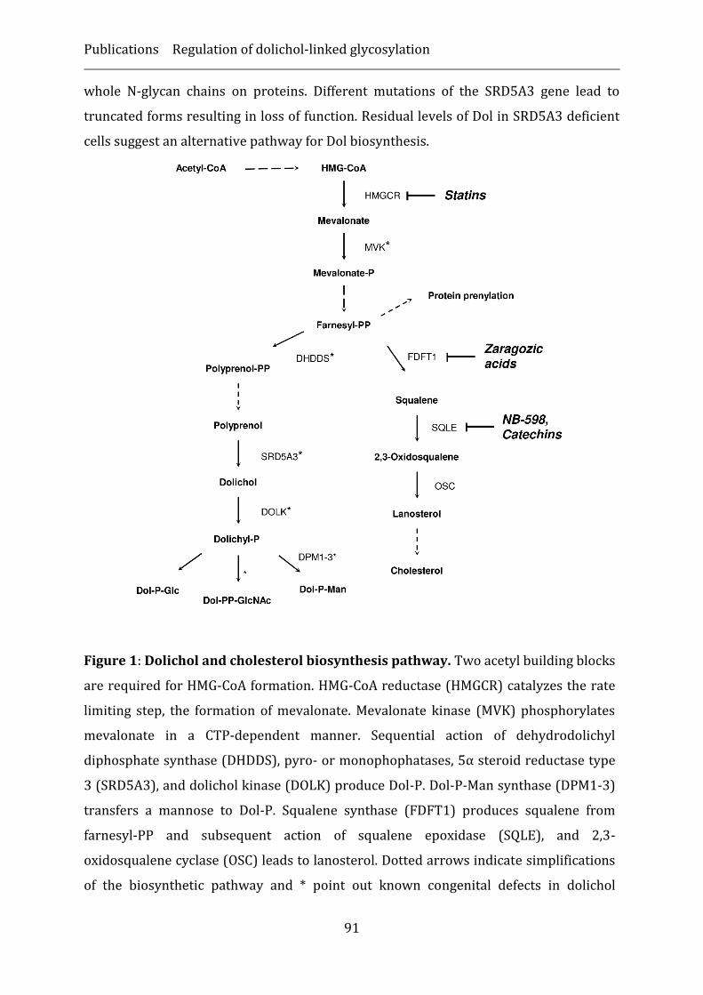
Publications ‒ Regulation of dolichol-linked glycosylation
91
whole N-glycan chains on proteins. Different mutations of the SRD5A3 gene lead to
truncated forms resulting in loss of function. Residual levels of Dol in SRD5A3 deficient
cells suggest an alternative pathway for Dol biosynthesis.
Figure 1: Dolichol and cholesterol biosynthesis pathway. Two acetyl building blocks
are required for HMG-CoA formation. HMG-CoA reductase (HMGCR) catalyzes the rate
limiting step, the formation of mevalonate. Mevalonate kinase (MVK) phosphorylates
mevalonate in a CTP-dependent manner. Sequential action of dehydrodolichyl
diphosphate synthase (DHDDS), pyro- or monophophatases, 5ゎ steroid reductase type
3 (SRD5A3), and dolichol kinase (DOLK) produce Dol-P. Dol-P-Man synthase (DPM1-3)
transfers a mannose to Dol-P. Squalene synthase (FDFT1) produces squalene from
farnesyl-PP and subsequent action of squalene epoxidase (SQLE), and 2,3-
oxidosqualene cyclase (OSC) leads to lanosterol. Dotted arrows indicate simplifications
of the biosynthetic pathway and * point out known congenital defects in dolichol
Publications ‒ Regulation of dolichol-linked glycosylation
92
biosynthesis of the respective enzymes.
Dol kinase deficiency, characterizing DOLK-CDG, impairs the phosphorylation of Dol to
Dol-P (Fig. 1). DOLK-CDG presents with severe phenotypes including hypotonia, skin
disorders, and the loss of hair (Schindler et al., 1987). Moreover, cardiomyopathy,
seizures, hypoglycemia, microcephaly, and visual impairment can occur as well (Table
1). Due to the severity of the symptoms, most DOLK-CDG patients have died in early
childhood. Reduced DOLK activity and the resulted low availability of Dol-P impair the
assembly Dol-PP-GlcNAc2Man9Glc3 and hence N-linked glycosylation. Recently, 11
children with dilated cardiomyopathy (DCM), a disease possibly linked to sudden
cardiac death and heart failure, were found to have a novel mutation in DOLK (Jung et
al., 1999). Besides DCM, only few of the patients exhibit additional symptoms like
ichtyosis, failure to thrive, and mild neurological involvement. Biochemical analysis of
Dol-P dependent glycosylation pathways of the DCM heart tissue indicated reduced O-mannosylation of ゎ-dystroglycan (Ijland et al., 1999). Remarkably, abnormal N-
glycosylation of serum transferrin was equally pronounced in both forms of DOLK-CDG.
The phenotypes of DCM strongly contrast with the originally described features of
DOLK-CDG, thus suggesting a possible tissue-specific regulation of Dol-P dependent
glycosylation.
Human Dol-P-Man synthase (DPM1-3, Fig. 1) consists of 3 subunits. DPM1 is the
catalytic subunit and DPM2/3 are regulatory and membrane anchoring proteins
residing in the ER membrane (Maeda and Kinoshita, 2008). So far, two forms of Dol-P-
Man synthase deficiency have been described featuring mutations in the DPM1 and
DPM3 genes. DPM1-CDG is characterized by recurrent seizures, hypotonia,
developmental delay, dysmorphic features, microcephaly, visual impairment, and in
some cases ataxia and coagulopathy (Table 1) (Chen et al., 2005; Kreishman et al., 1972;
Palamarczyk and Janczura, 1977; Tao et al., 2006). Lower Dol-P-Man levels lead to
impaired Dol-PP-GlcNAc2Man9Glc3 assembly, abnormal N-linked protein glycosylation,
and decreased formation of GPI anchored proteins. DPM3-CDG appears to be a milder
form of Dol-P-Man synthase deficiency, for which only one patient has been found so far
(Zaia, 2013). The symptoms included mild myopathy, a dilated cardiomyopathy,
moderate muscular dystrophy, and a single stroke-like episode. The adult patient is able
to live a practically normal life. N-glycosylation of serum transferrin is only slightly
abnormal. In DPM1-CDG and DPM3-CDG, the severity seems to reflect the molecular

Publications ‒ Regulation of dolichol-linked glycosylation
93
functions of the affected subunits. So, a deficient catalytic subunit, DPM1, leads to a
much more severe phenotype than a deficient tethering subunit, DPM3.
Table 1: Disorders of dolichol biosynthesis and their respective symptoms.
Symptoms are distinguished between ** for typical/dominant symptoms and * for
sporadic symptoms or symptoms of moderate severity.
a) Classical DOLK-CDG, b) DOLK defect in dilated cardiomyopathy.
MVA HIDS DHDDS-
CDG
SRD5A3-
CDG
DOLK-
CDGa)
DOLK-
CDGb)
DPM1-
CDG
DPM3-
CDG
Cardiomyopathy
* ** **
*
Cerebellar ataxia /
malformation ** **
**
**
Coagulopathy
**
**
Dysmorphic features **
** **
Failure to thrive **
Febrile attacks ** **
Hypoglycemia
**
Hypotonia
**
**
Microcephaly ** *
**
**
Muscular dystrophy
** *
Myopathy
** *
Ocular malfunctions **
** ** **
**
Psychomotor retardation ** **
** ** * **
Seizures ** *
**
**
Skin disorders ** **
* ** *
In general, the clinical severity of Dol-related diseases does not correlate with the
relative position of the deficient enzymes along the biosynthesis pathway. Defects of Dol
biosynthesis are generally expected to be severe since not only the assembly of Dol-PP-

Publications ‒ Regulation of dolichol-linked glycosylation
94
GlcNAc2Man9Glc3 is impaired, but also GPI anchor formation and O-mannosylation are
affected. This notion is confirmed in SRD5A3-CDG that present with severe
multisystemic phenotypes. However, the discovery of DHDDS-CDG with a very specific
phenotype and lack of the typical CDG symptoms suggests a complex regulation of Dol-
dependent glycosylation. Future efforts addressing the regulation of Dol biosynthesis
should provide a better understanding of the question and open the way to novel
therapeutic perspectives.
Therapeutics targeting dolichol
To date, only two forms of CDG are treatable. MPI-CDG can be successfully treated with
oral mannose supplementation (Liu et al., 2006; Tissot et al., 2007). In GFTP-CDG, GDP-
fucose transporter deficiency can be compensated with nutritional fucose
supplementation (Marquardt et al., 1999). The hypomorphic nature of most CDG
mutations allows for a compensatory approach, for instance by providing more Dol to
overcome the compromised assembly of Dol-PP-GlcNAc2Man9Glc3. Along this line,
inhibition of squalene synthase (FDFT1, Fig. 1) was shown to improve N-linked protein
glycosylation and GPI-anchored protein expression in DPM1-CDG fibroblasts (Haeuptle
et al., 2011). We tested potential drugs such as clofibrate for its ability to increase GPI
anchor availability in DPM1-CDG fibroblasts. Clofibrate is a fibrate known to decrease
cholesterol levels by targeting lipid metabolism via activation of peroxisome
proliferator-activated receptors, especially PPARゎ (Abourbih et al., 2009; Forman et al.,
1997). Indeed, GPI anchor availability was improved upon clofibrate treatment as
observed by an increased surface expression of the GPI anchored protein CD59
(unpublished data). In another approach to increase the Dol pool, we attempted to
increase DHDDS activity by treatment of DPM1-CDG fibroblasts with the AMP cyclase
activator forskolin (Losfeld et al., 2012). DHDDS activity was previously shown to
increase with higher cyclic AMP levels (Konrad and Merz, 1994; Konrad and Merz,
1996). Treatment of DPM1-CDG fibroblasts with forskolin led to higher levels of surface
CD59 as well.
New therapeutic possibilities arise for CDG with the advance of non-statin cholesterol
lowering drugs, which target the late pathway of cholesterol biosynthesis. Presently,
statins are the prevalent medication to lower cholesterol and decrease cardiovascular
Publications ‒ Regulation of dolichol-linked glycosylation
95
disease (Hoenger et al., 1993). Statins are inhibitors of the HMG-CoA reductase (Fig. 1),
the rate limiting enzyme at the beginning of the mevalonate pathway. The effect of
statins on Dol levels has not been investigated systematically. Hela cells treated with
pravastatin showed a decrease in Dol-P by 65% (Kornfeld et al., 1985). However, rats
treated with lovastatin did not show a change of total Dol-P levels but increase of Dol-P
in the liver (Low et al., 1992). The potential of different statins to upregulate Dol could
be investigated in a screen. For instance, CDG fibroblasts treated with an array of statins
could be screened for changes of Dol levels.
Inhibition of the first committed enzyme of cholesterol biosynthesis, squalene synthase
(FDFT1, Fig. 1), has proven effective in lowering cholesterol production (Dhar et al.,
1993). Prominent among these inhibitors are zaragozic acids, which were identified in a
screen of fungal compounds for cholesterol lowering activity (Bergstrom et al., 1995).
Changes in Dol-P patterns were observed in human fibroblasts treated with zaragozic
acid A. Also, DPM1-CDG fibroblasts showed improved N-linked glycosylation upon
treatment with zaragozic acid A (Takahashi et al., 1993). Moreover, the expression of the
GPI-anchored CD59 protein could be normalized in DPM1-CDG fibroblasts after
treatment. Since zaragozic acid is well tolerated in animals (Baxter et al., 1992;
Bergstrom et al., 1993), this class of FDFT1 inhibitors might be considered for clinical
testing in some cases of CDG.
Squalene epoxidase (SQLE) catalyzes the next committed step of cholesterol
biosynthesis after FDFT1 (Fig. 1). Therefore, inhibition of SQLE is considered to
specifically lower cholesterol. Inhibition of SQLE by the compound NB-598 ((E)N-ethyl-
N-(6,6-dimethyl-2-hepten-4-ynyl)-3-[(3,3'-bith iophen-5-yl)methoxy]benzene-
methanamine) successfully reduced cholesterol levels (Ghosh et al., 1993c; Hope et al.,
1993), although no changes in Dol levels were detected in treated HepG2 cells (Ghosh et
al., 1993b). In spite of this early finding, newly developed SQLE inhibitors should be
considered as promising candidates for an upregulation of Dol in CDG cells. Moreover,
green tea has been attributed a cholesterol lowering effect, which is linked to SQLE
inhibition (Abe et al., 2000). In a recent study, oral supplementation with the main green
tea catechin epigallocatechin gallate was shown to lower LDL-cholesterol (Wu et al.,
2012). The effects of epigallocatechin gallate on Dol have not been tested yet. Catechins
might represent another option to upregulate Dol in CDG cells by inhibition of SQLE.

Publications ‒ Regulation of dolichol-linked glycosylation
96
Oxidosqualene cyclase (OSC) inhibitors interfere with the conversion of 2,3-
oxidosqualene to lanosterol (Fig. 1). Due to adverse effects, the development of such
inhibitors has been stopped (Hope et al., 1993). Unless novel OSC inhibitors without
adverse effects are discovered, the potential of such drugs to increase the Dol pool is not
worth further investigation.
In conclusion, inhibitors of late cholesterol biosynthesis have a potential increasing
effect on Dol levels and should be accordingly tested in CDG cells. Much effort is invested
in the development of cholesterol lowering drugs in the context of cardiovascular
diseases. Therefore, potential CDG therapies could profit from the discovery of novel
cholesterol lowering drugs in the future.
Outlook
The recent identification of tissue-restricted symptoms in DHDDS-CDG and of a novel
pathology associated to DOLK deficiency suggests a pronounced tissue specificity of Dol-
related biology. Accordingly, additional diseases are likely to be associated with local
alterations of Dol biosynthesis in the near future. The study of these tissue-restricted
diseases represents valuable models to better understand the regulation of Dol
biosynthesis and its impact on various types of glycosylation. Novel therapeutic
approaches may be deduced from such insights. The treatment of CDG is still very
limited and merits further attention. We believe that interference of cholesterol
production and thus upregulation of Dol biosynthesis represents a valuable approach to
improve glycosylation in CDG. High-throughput screenings of existing cholesterol
lowering drugs that upregulate Dol are relatively straightforward and could open novel
therapeutic opportunities.

Publications ‒ Regulation of dolichol-linked glycosylation
97
References
Abe, I., T. Seki, K. Umehara, T. Miyase, H. Noguchi, J. Sakakibara, and T. Ono. 2000. Green tea polyphenols: novel and potent inhibitors of squalene epoxidase. Biochem Biophys Res
Commun 268:767-771. Abourbih, S., K.B. Filion, L. Joseph, E.L. Schiffrin, S. Rinfret, P. Poirier, L. Pilote, J. Genest, and M.J.
Eisenberg. 2009. Effect of fibrates on lipid profiles and cardiovascular outcomes: a systematic review. Am J Med 122:962 e961-968.
Armstrong, S.J., R.A. Read, P. Ghosh, and D.M. Wilson. 1993. Moderate exercise exacerbates the osteoarthritic lesions produced in cartilage by meniscectomy: a morphological study. Osteoarthritis Cartilage 1:89-96.
Barone, R., L. Pavone, A. Fiumara, R. Bianchini, and J. Jaeken. 1999. Developmental patterns and neuropsychological assessment in patients with carbohydrate-deficient glycoconjugate syndrome type IA (phosphomannomutase deficiency). Brain Dev 21:260-263.
Baxter, A., B.J. Fitzgerald, J.L. Hutson, A.D. McCarthy, J.M. Motteram, B.C. Ross, M. Sapra, M.A. Snowden, N.S. Watson, R.J. Williams, and et al. 1992. Squalestatin 1, a potent inhibitor of squalene synthase, which lowers serum cholesterol in vivo. J Biol Chem 267:11705-11708.
Bergstrom, J.D., C. Dufresne, G.F. Bills, M. Nallin-Omstead, and K. Byrne. 1995. Discovery, biosynthesis, and mechanism of action of the zaragozic acids: potent inhibitors of squalene synthase. Annu Rev Microbiol 49:607-639.
Bergstrom, J.D., M.M. Kurtz, D.J. Rew, A.M. Amend, J.D. Karkas, R.G. Bostedor, V.S. Bansal, C. Dufresne, F.L. VanMiddlesworth, O.D. Hensens, and et al. 1993. Zaragozic acids: a family of fungal metabolites that are picomolar competitive inhibitors of squalene synthase. Proc Natl Acad Sci U S A 90:80-84.
Cantagrel, V., and D.J. Lefeber. 2011. From glycosylation disorders to dolichol biosynthesis defects: a new class of metabolic diseases. J Inherit Metab Dis 34:859-867.
Chen, B., B.G. Zhang, J. Zhang, Y.Q. Gu, J.X. Li, H.X. Yu, and Z.G. Wang. 2005. [Primary experimental study on the construction of tissue engineering blood vessel]. Zhonghua Wai Ke Za Zhi 43:1271-1274.
Dhar, S., A.J. Kanwar, and S. Ghosh. 1993. Leucotrichia in nevus depigmentosus. Pediatr Dermatol 10:198-199.
Forman, B.M., J. Chen, and R.M. Evans. 1997. Hypolipidemic drugs, polyunsaturated fatty acids, and eicosanoids are ligands for peroxisome proliferator-activated receptors alpha and delta. Proc Natl Acad Sci U S A 94:4312-4317.
Fu, D., and H. van Halbeek. 1992. N-glycosylation site mapping of human serotransferrin by serial lectin affinity chromatography, fast atom bombardment-mass spectrometry, and 1H nuclear magnetic resonance spectroscopy. Anal Biochem 206:53-63.
Gebauer, J.M., S. Muller, F.G. Hanisch, M. Paulsson, and R. Wagener. 2008. O-glucosylation and O-fucosylation occur together in close proximity on the first epidermal growth factor repeat of AMACO (VWA2 protein). J Biol Chem 283:17846-17854.
Ghosh, K., S. Ramakrishnan, and G. Chandra. 1993a. Resistivity and susceptibility studies on UPd2-xPtxAl3 (x=0, 0.1, 0.2, 0.3, and 0.5). Phys Rev B Condens Matter 47:8305-8307.
Ghosh, P., R. Read, S. Armstrong, D. Wilson, R. Marshall, and P. McNair. 1993b. The effects of intraarticular administration of hyaluronan in a model of early osteoarthritis in sheep. I. Gait analysis and radiological and morphological studies. Semin Arthritis Rheum 22:18-30.
Ghosh, P., R. Read, Y. Numata, S. Smith, S. Armstrong, and D. Wilson. 1993c. The effects of intraarticular administration of hyaluronan in a model of early osteoarthritis in sheep. II. Cartilage composition and proteoglycan metabolism. Semin Arthritis Rheum 22:31-42.
Grunewald, S. 2009. The clinical spectrum of phosphomannomutase 2 deficiency (CDG-Ia). Biochimica et biophysica acta 1792:827-834.

Publications ‒ Regulation of dolichol-linked glycosylation
98
Haeuptle, M.A., M. Welti, H. Troxler, A.J. Hülsmeier, T. Imbach, and T. Hennet. 2011. Improvement of dolichol-linked oligosaccharide biosynthesis by the squalene synthase inhibitor zaragozic acid. J Biol Chem 286:6085-6091.
Haldar, K.K., B. Sahadalal, and E.L. Ghosh. 1993. Transferable drug resistance in Salmonella senftenberg. Indian J Pathol Microbiol 36:81-83.
Hart, G.W., G.D. Holt, and R.S. Haltiwanger. 1988. Nuclear and cytoplasmic glycosylation: novel saccharide linkages in unexpected places. Trends Biochem Sci 13:380-384.
Hoenger, A., R. Ghosh, C.A. Schoenenberger, U. Aebi, and A. Engel. 1993. Direct in situ structural analysis of recombinant outer membrane porins expressed in an OmpA-deficient mutant Escherichia coli strain. J Struct Biol 111:212-221.
Hope, N., P. Ghosh, T.K. Taylor, D. Sun, and R. Read. 1993. Effects of intraarticular hyaluronan on matrix changes induced in the lateral meniscus by total medial meniscectomy and exercise. Semin Arthritis Rheum 22:43-51.
Ijland, S.A., M.J. Jager, B.M. Heijdra, J.R. Westphal, and R. Peek. 1999. Expression of angiogenic and immunosuppressive factors by uveal melanoma cell lines. Melanoma Res 9:445-450.
Jenisch, S., S. Koch, T. Henseler, R.P. Nair, J.T. Elder, C.E. Watts, E. Westphal, J.J. Voorhees, E. Christophers, and M. Kronke. 1999. Corneodesmosin gene polymorphism demonstrates strong linkage disequilibrium with HLA and association with psoriasis vulgaris. Tissue
Antigens 54:439-449. Jung, H., P. Carmel, M.S. Schwartz, J.W. Witkin, K.H. Bentele, M. Westphal, J.H. Piatt, M.E. Costa, A.
Cornea, Y.J. Ma, and S.R. Ojeda. 1999. Some hypothalamic hamartomas contain transforming growth factor alpha, a puberty-inducing growth factor, but not luteinizing hormone-releasing hormone neurons. J Clin Endocrinol Metab 84:4695-4701.
Kim, M.L., K. Chandrasekharan, M. Glass, S. Shi, M.C. Stahl, B. Kaspar, P. Stanley, and P.T. Martin. 2008. O-fucosylation of muscle agrin determines its ability to cluster acetylcholine receptors. Mol Cell Neurosci 39:452-464.
Konrad, M., and W.E. Merz. 1994. Regulation of N-glycosylation. Long term effect of cyclic AMP mediates enhanced synthesis of the dolichol pyrophosphate core oligosaccharide. J Biol
Chem 269:8659-8666. Konrad, M., and W.E. Merz. 1996. Long-term effect of cyclic AMP on N-glycosylation is caused by
an increase in the activity of the cis-prenyltransferase. Biochem J 316 ( Pt 2):575-581. Kornfeld, P., E.P. Ambinder, R. Matta, A. Bender, A.E. Papatestas, H. Gross, and G. Genkins. 1985.
Azathioprine experience in severe, generalized, recalcitrant myasthenia gravis. Mt Sinai J
Med 52:347-352. Kreishman, G.P., J.T. Witkowski, R.K. Robins, and M.P. Schweizer. 1972. The use of proton and
carbon-13 nuclear magnetic resonance for assignment of the glycosylation site in 3- and 5-substituted 1- -D-ribofuranosyl-1,2,4-triazoles. J Am Chem Soc 94:5894-5896.
Larkin, A., and B. Imperiali. 2011. The expanding horizons of asparagine-linked glycosylation. Biochemistry 50:4411-4426.
Liu, X., D.J. McNally, H. Nothaft, C.M. Szymanski, J.R. Brisson, and J. Li. 2006. Mass spectrometry-based glycomics strategy for exploring N-linked glycosylation in eukaryotes and bacteria. Anal Chem 78:6081-6087.
Losfeld, M.E., F. Soncin, B.G. Ng, I. Singec, and H.H. Freeze. 2012. A sensitive green fluorescent protein biomarker of N-glycosylation site occupancy. FASEB J 26:4210-4217.
Low, P., M. Andersson, C. Edlund, and G. Dallner. 1992. Effects of mevinolin treatment on tissue dolichol and ubiquinone levels in the rat. Biochimica et biophysica acta 1165:102-109.
Maeda, Y., and T. Kinoshita. 2008. Dolichol-phosphate mannose synthase: structure, function and regulation. Biochimica et biophysica acta 1780:861-868.
Marquardt, T., K. Luhn, G. Srikrishna, H.H. Freeze, E. Harms, and D. Vestweber. 1999. Correction of leukocyte adhesion deficiency type II with oral fucose. Blood 94:3976-3985.
Palamarczyk, G., and E. Janczura. 1977. Lipid mediated glycosylation in yeast nuclear membranes. FEBS Lett 77:169-172.
Parfait, B., P. de Lonlay, J.C. von Kleist-Retzow, V. Cormier-Daire, D. Chretien, A. Rotig, D. Rabier, J.M. Saudubray, P. Rustin, and A. Munnich. 1999. The neurogenic weakness, ataxia and

Publications ‒ Regulation of dolichol-linked glycosylation
99
retinitis pigmentosa (NARP) syndrome mtDNA mutation (T8993G) triggers muscle ATPase deficiency and hypocitrullinaemia. Eur J Pediatr 158:55-58.
Raucy, J.L., J.M. Lasker, J.C. Kraner, D.E. Salazar, C.S. Lieber, and G.B. Corcoran. 1991. Induction of cytochrome P450IIE1 in the obese overfed rat. Molecular pharmacology 39:275-280.
Rush, J.S., S.K. Cho, S. Jiang, S.L. Hofmann, and C.J. Waechter. 2002. Identification and characterization of a cDNA encoding a dolichyl pyrophosphate phosphatase located in the endoplasmic reticulum of mammalian cells. J Biol Chem 277:45226-45234.
Rush, J.S., N. Gao, M.A. Lehrman, and C.J. Waechter. 2008. Recycling of dolichyl monophosphate to the cytoplasmic leaflet of the endoplasmic reticulum after the cleavage of dolichyl pyrophosphate on the lumenal monolayer. J Biol Chem 283:4087-4093.
Schindler, M., M. Hogan, R. Miller, and D. DeGaetano. 1987. A nuclear specific glycoprotein representative of a unique pattern of glycosylation. J Biol Chem 262:1254-1260.
Schwarz, F., and M. Aebi. 2011. Mechanisms and principles of N-linked protein glycosylation. Curr Opin Struct Biol 21:576-582.
Sturla, L., R. Rampal, R.S. Haltiwanger, F. Fruscione, A. Etzioni, and M. Tonetti. 2003. Differential terminal fucosylation of N-linked glycans versus protein O-fucosylation in leukocyte adhesion deficiency type II (CDG IIc). J Biol Chem 278:26727-26733.
Takahashi, T., J.M. Lasker, A.S. Rosman, and C.S. Lieber. 1993. Induction of cytochrome P-4502E1 in the human liver by ethanol is caused by a corresponding increase in encoding messenger RNA. Hepatology (Baltimore, Md 17:236-245.
Tao, L., L.W. Wagner, P.E. Rouviere, and Q. Cheng. 2006. Metabolic engineering for synthesis of aryl carotenoids in Rhodococcus. Appl Microbiol Biotechnol 70:222-228.
Tissot, B., N. Gasiunas, A.K. Powell, Y. Ahmed, Z.L. Zhi, S.M. Haslam, H.R. Morris, J.E. Turnbull, J.T. Gallagher, and A. Dell. 2007. Towards GAG glycomics: analysis of highly sulfated heparins by MALDI-TOF mass spectrometry. Glycobiology 17:972-982.
Valliere-Douglass, J.F., L.J. Brady, C. Farnsworth, D. Pace, A. Balland, A. Wallace, W. Wang, M.J. Treuheit, and B. Yan. 2009. O-fucosylation of an antibody light chain: characterization of a modification occurring on an IgG1 molecule. Glycobiology 19:144-152.
Valnot, I., J. Kassis, D. Chretien, P. de Lonlay, B. Parfait, A. Munnich, J. Kachaner, P. Rustin, and A. Rotig. 1999. A mitochondrial cytochrome b mutation but no mutations of nuclearly encoded subunits in ubiquinol cytochrome c reductase (complex III) deficiency. Hum
Genet 104:460-466. van Berkel, M.A., M. Rieger, S. te Heesen, A.F. Ram, H. van den Ende, M. Aebi, and F.M. Klis. 1999.
The Saccharomyces cerevisiae CWH8 gene is required for full levels of dolichol-linked oligosaccharides in the endoplasmic reticulum and for efficient N-glycosylation. Glycobiology 9:243-253.
van der Knaap, M.S., C. Jakobs, G.F. Hoffmann, M. Duran, A.C. Muntau, S. Schweitzer, R.I. Kelley, F. Parrot-Roulaud, J. Amiel, P. De Lonlay, D. Rabier, and O. Eeg-Olofsson. 1999. D-2-hydroxyglutaric aciduria: further clinical delineation. J Inherit Metab Dis 22:404-413.
Veneselli, E., R. Biancheri, M. Di Rocco, and S. Tortorelli. 1998. Neurophysiological findings in a case of carbohydrate-deficient glycoprotein (CDG) syndrome type I with phosphomannomutase deficiency. Eur J Paediatr Neurol 2:239-244.
Wimmer-Greinecker, G., G. Matheis, S. Dogan, T. Aybek, P. Kessler, K. Westphal, and A. Moritz. 1999. Complications of port-access cardiac surgery. J Card Surg 14:240-245.
Wu, A.H., D. Spicer, F.Z. Stanczyk, C. Tseng, C.S. Yang, and M.C. Pike. 2012. Effect of 2-month controlled green tea intervention on lipoprotein cholesterol, glucose, and hormonal levels in healthy postmenopausal women. Cancer Prev Res (Phila)
Zaia, J. 2013. Glycosaminoglycan glycomics using mass spectrometry. Mol Cell Proteomics 12:885-892.

100

Publications ‒ Ethanol-induced N-glycosylation deficiency
101
Ethanol-induced impairment in the biosynthesis of N-linked
glycosylation
Michael Welti and Andreas J. Hülsmeier
Institute of Physiology, University of Zürich, Zürich, Switzerland
Manuscript accepted in the Journal of Cellular Biochemistry 2013
Corresponding author:
Andreas J. Hülsmeier
Institute of Physiology
University of Zürich
Winterthurerstrasse 190
CH-8057 Zürich
Phone: +41-44-635-5104
Fax: +41-44-635-6814
E-mail: [email protected]

Publications ‒ Ethanol-induced N-glycosylation deficiency
102
Abstract
Background
Deficiency in N-linked protein glycosylation is a long-known characteristic of alcoholic
liver disease and congenital disorders of glycosylation. Previous investigations of
ethanol-induced glycosylation deficiency demonstrated perturbations in the early steps
of substrate synthesis and in the final steps of capping N-linked glycans in the Golgi. The
significance of the biosynthesis of N-glycan precursors in the endoplasmic reticulum,
however, has not yet been addressed in alcoholic liver disease.
Methods
Ethanol-metabolizing hepatoma cells were treated with increasing concentrations of
ethanol. Transcript analysis of genes involved in the biosynthesis of N-glycans, activity
assays of related enzymes, quantification of dolichol-phosphates, and analysis of
dolichol-linked oligosaccharides were performed.
Results
Upon treatment of cells with ethanol, we found a decrease in the final N-glycan
precursor Dol-PP-GlcNAc2Man9Glc3 and in C95- and C100-dolichol-phosphate levels.
Transcript analysis of genes involved in N-glycosylation showed a 17% decrease in
expression levels of DPM1, a subunit of the dolichol-phosphate-mannose synthase, and a
8% increase in RPN2, a subunit of the oligosaccharyl transferase.
Conclusions
Ethanol treatment decreases the biosynthesis of dolichol-phosphate. Consequently, the
formation of N-glycan precursors is affected, resulting in an aberrant precursor
assembly. Messenger RNA levels of genes involved in N-glycan biosynthesis are not
affected by ethanol treatment, indicating that the assembly of N-glycan precursors is not
regulated at the transcriptional level.
General Significance
This study confirms that ethanol impairs N-linked glycosylation by affecting dolichol
biosynthesis leading to impaired dolichol-linked oligosaccharide assembly. Together our
data help to explain the underglycosylation phenotype observed in alcoholic liver
disease and congenital disorders of glycosylation.

Publications ‒ Ethanol-induced N-glycosylation deficiency
103
Highlights
- Dolichol-phosphate levels are reduced after ethanol treatment
- Assembly of the N-glycan precursor is impaired after ethanol treatment
- Transcript levels of genes involved in N-glycosylation are slightly affected after treatment
Keywords
N-linked glycosylation, alcoholic liver disease, dolichol, dolichol-linked oligosaccharides
Abbreviations
ALD, alcoholic liver disease; CDG, congenital disorder of glycosylation; CDT,
carbohydrate-deficient transferrin; Dol-P, dolichol-phosphate; Dol-PP, dolichol-
pyrophosphate; DPM1-3, dolichol-phosphate-mannose synthase subunits 1-3; RPN2,
ribophorin 2; Glc, glucose; GlcNAc, N-acetylglucosamine; Man, mannose; ADH, alcohol
dehydrogenase; CYP2E1; cytochrome P450 2E1

Publications ‒ Ethanol-induced N-glycosylation deficiency
104
1. Introduction
Alcoholic liver disease (ALD) displays a broad spectrum of symptoms reflecting the
diverse functions of the liver. Among the most prominent characteristics of ALD are fatty
liver, hepatitis, and liver fibrosis. Hepatocytes are responsible for detoxification in the
liver and thus for alcohol metabolism. Major metabolic changes in hepatocytes occur due
to the constant need for ethanol oxidation. Ethanol conversion to acetate leads to a shift
in the NAD/NADH ratio impairing glycolysis and lipolysis and initiating fatty acid
synthesis. The inversion of glycolytic flux eventually results in alcoholic fatty liver
(Purohit et al., 2009). Oxidative stress is evoked by reactive oxygen species from ethanol
breakdown in hepatocytes and toll-like receptor 4 activation by LPS leaking from the gut
into the blood stream (Wang et al., 2012; Zhu et al., 2012). This oxidative stress
contributes to liver inflammation and can lead to liver fibrosis and eventually cirrhosis.
Another clinical feature in ALD receiving less attention is alcohol-induced deficiency in
N-linked protein glycosylation although it has been used as a marker for alcohol abuse
(Stibler, 1991).
ALD and congenital disorders of glycosylation (CDG) share some symptoms. Typically,
CDG display systemic deficiencies like psychomotor retardation with variable
neuromuscular involvement and additional features like hormonal abnormalities and
coagulopathies (Leroy, 2006). Importantly, fatty liver, hepatitis, and liver fibrosis are
also found in CDG indicating a possible role of glycosylation in ALD pathology. Indeed,
CDG was linked to a fibrotic response in fibroblasts involving the insulin-like growth
factor-binding protein 5 (Lecca et al., 2011). As in CDG, blood serum proteins originating
from the liver are not glycosylated properly in ALD. For instance, the distribution of
carbohydrate-deficient transferrin (CDT) glycoforms found in ALD and CDG are similar
(Bean and Peter, 1993; de Jong et al., 1992; Petren et al., 1987; Vigo and Adair, 1982).
The two glycosylation sites of transferrin are normally occupied with carbohydrates
adding up to four branches carrying terminal sialic acids. Besides tetrasialo-transferrin,
disialo-transferrin is the most prevalent glycoform of transferrin in blood serum. In ALD
and CDG, asialo- and disialo-transferrin become more apparent and tetrasialo-
transferrin levels are reduced (Hülsmeier et al., 2007). In CDG, such a CDT pattern
indicates a deficiency in the assembly of the N-linked glycan precursor in the ER
(Filipovic and Menzel, 1981).
Publications ‒ Ethanol-induced N-glycosylation deficiency
105
So far, glycosylation deficiency in ALD has been assessed at the levels of Golgi
glycosylation and dolichol metabolism. However, N-linked glycosylation starts in the ER
and requires the synthesis of an oligosaccharide precursor on dolichol-phosphate (Dol-
P). On the outer leaflet of the ER, one phospho-GlcNAc, one GlcNAc, and five Man are
sequentially transferred to Dol-P before flipping the resulting Dol-PP-GlcNAc2-Man5 into
the ER lumen. After the addition of four Man and three Glc residues, the complete N-
glycan precursor, Dol-PP-GlcNAc2-Man9-Glc3 is transferred en bloc to target proteins.
Microsomes isolated from rat livers of chronically ethanol-fed animals showed a
decrease in total dolichol by 36% (Cottalasso et al., 1998; Cottalasso et al., 1996). Acute
treatment with ethanol of rats resulted in a reduction of total dolichol in liver
microsomes by up to 52%. In a cell culture model, HepG2 cells treated with 50-100 mM
ethanol showed decreased transcription of the 2,6-sialyltransferase(Garige et al., 2006;
Garige et al., 2005; Rao and Lakshman, 1997; Rao and Lakshman, 1999). This enzyme is
responsible for terminal sialylation of sugar chains of transferrin in the Golgi apparatus.
The same effect was observed in the liver from chronically ethanol-fed rats. Additionally,
the 2,6-sialyltransferase mRNA is destabilized by a ぬ╆-untranslated region-specific
binding protein. Notably, besides downregulation of 2,6-sialyltransferase other effects
could cause CDT. Several intermediates along the biosynthesis of the N-glycan precursor
might be affected, potentially leading to an altered distribution of glycoforms. In one
study the site occupancy of serum protein glycosylation in an alcohol abusing subject
was reported to be reduced to levels observed in patients with mild forms of CDG
(Hülsmeier et al., 2007). Another study reported the loss of one or both N-glycans as the
cause for CDT in patients with severe alcohol abuse (Peter et al., 1998).
To understand the mechanisms underlying alcohol-induced glycosylation deficiency, we
investigated the effects of ethanol exposure on the early steps of N-linked protein
glycosylation in two cell models. Cultured hepatocytes usually lose the expression of
drug metabolizing enzymes (Carter and Wands, 1988; Gapp et al., 2012; Luo et al., 2006;
Utesch et al., 1992; Van Kaick et al., 1983). Therefore, we used two cell lines expressing
ethanol metabolizing enzymes in our study. First, the HepaRG cell line derived from a
human hepatocellular carcinoma with an inducible cytochrome p450 system which
renders the cell line suitable as a model for metabolism of xenobiotics in the human liver
(Crick and Carroll, 1987; Eggens et al., 1990; Eggens and Elmberger, 1990; Keller, 1986;
Stoll et al., 1988; Yokoyama et al., 1989). Ethanol degradation via cytochrome p450 2E1

Publications ‒ Ethanol-induced N-glycosylation deficiency
106
(CYP2E1) is a major pathway of ethanol metabolism and can be induced by DMSO
treatment in HepaRG cells (Koop and Tierney, 1990; Lieber, 1999; Ohnishi and Lieber,
1977). The second cell line, VA-13, is derived from HepG2 cells, which expresses murine
alcohol dehydrogenase 1 (ADH) (Clemens et al., 2002). Ethanol degradation via alcohol
dehydrogenase is the second major pathway of ethanol metabolism in the liver. We
tested the effect of ethanol metabolism in hepatocytes on the N-linked glycosylation
pathway in the ER and found that N-linked glycosylation is affected at several stages
along the biosynthetic pathway.
2. Materials and Methods
2.1 Cell lines and experimental conditions
HepaRG cells were kindly provided by Dr. N. Zitzmann (Department of Biochemistry,
University of Oxford, UK). HepaRG cells were seeded at a density of 2.6 x 10⁴ cells/cm2 and cultured in Williams╆ Medium E ゅSigma-Aldrich) supplemented with 10% FBS, 5
g/ml insulin, 2 mM L-glutamine, and 50 M hydrocortisone hemisuccinate for two
weeks. To induce maximal induction of the P450 enzymes, HepaRG cells were cultured
for two additional weeks in the same medium added with 2% DMSO (Sigma) (Gripon et
al., 2002). On day 24 after seeding, ethanol was added to the medium and culture flasks
were sealed with Parafilm to prevent evaporation of ethanol. HepaRG cells were
harvested and analyzed after 28 days in culture. VA-13 cells were a kind gift from Dr.
D.L. Clemens (Department of Internal Medicine, University of Nebraska Medical Center,
USA). VA-13 cells are HepG2 cells stably transfected with an expression plasmid
carrying the murine alcohol dehydrogenase gene, Adh-1 (Clemens et al., 2002). VA-13
cells were seeded at a density of 1.7 x 10⁴ cells/cm2 and ethanol treatment was started 6
h after seeding. VA-13 cells were harvested and analyzed 4 days after treatment start.
2.2 Isoelectric focusing gel electrophoresis and immunoblotting
Supernatant from cultured hepatoma cells was saturated with 0.4 mM ferric citrate in
presence of 20 mM sodium hydrogen carbonate for 30 min. Samples were analyzed
using pre-cast Novex pH 3-7 IEF gels (Invitrogen). Gels were run at 7 mA (500 V
maximum voltage) for 4 h on ice. Proteins were transferred to a nitrocellulose
membrane by Western blotting. Transferrin isoforms were visualized using polyclonal
Publications ‒ Ethanol-induced N-glycosylation deficiency
107
rabbit anti-human transferrin primary antibody (DakoCytomation, Denmark) and
peroxidase-coupled goat anti-rabbit secondary antibody (Vector Laboratories, USA).
2.3 Alcohol dehydrogenase activity assay
Alcohol dehydrogenase (ADH) activity was measured in cell lysates as previously
described (Clemens et al., 1995). Activity was determined in an assay using ethanol and
NAD as substrates. ADH activity was indirectly determined from the conversion of NAD
by measuring NADH photometrically at 340 nm.
2.4 Cytochrome P450 2E1 activity assay
Cytochrome p450 2E1 activity was determined as described previously (Wu and
Cederbaum, 2008). Microsomal fractions were prepared and assayed for CYP2E1
activity using p-nitrophenol and NADPH as substrates.
2.5 Quantitative PCR for transcript analysis
Transcript analysis of the glycosylation pathway were quantified with an adapted
method for quantitative PCR in mice (Nairn et al., 2010). Mann-Whitney statistical
analysis was done using the InStat software (GraphPad). Significance was accepted for p
< 0.05.
2.6 Dolichol-phosphate-mannose synthase activity assay
Cells were grown on 300 cm2 plates and collected by trypsinization. Prior to cell lysis,
cells were washed twice in ice-cold PBS. The cells were lysed in 25 mM Tris-HCl, 150
mM KCl, 1% Triton X-100, and cell debris was removed by centrifugation. Protein
concentration was determined by BCA (Pierce). Dolichol-phosphate-mannose (Dol-P-
Man) synthase activity was assayed using 30 ml of cell lysate in 100 ml of reaction
buffer, with the addition of 40 mg/ml Dol-P (Sigma-Aldrich Co.) and 17 mM GDP-[14C]
mannose (Amersham Pharmacia Biotech). The reaction buffer contained 50 mM HEPES
(pH 7.4), 25 mM KCl, 5 mM MgCl2, 5 mM MnCl2, and 0.2% Triton X-100. Reaction
mixtures were incubated for 5 min at 37°C. Dol-P-Man was isolated by organic

Publications ‒ Ethanol-induced N-glycosylation deficiency
108
extraction with chloroform/methanol (2:1 vol/vol), and radioactivity was determined in
a beta counter (Beckman Coulter Inc., Fullerton, California, USA) (McLachlan and Krag,
1994). Statistical significance was determined by paired student t-test. Significance was
accepted for p < 0.05.
2.7 Fluorescent labeling and analysis of dolichol-phosphate
Extraction, labeling, and analysis of Dol-P were adapted from previous studies
(Elmberger et al., 1989; Haeuptle et al., 2010). Briefly, approximately 3 x 10⁸ cells were
collected by trypsinization, counted, and washed once in PBS. For quantification, 10 g
C80-polyprenol-phosphate (Larodan Fine Chemicals, Sweden) was added as internal
standard. After addition of 6 ml methanol and 3 ml 15 M KOH, the sample was
hydrolyzed for 1 h at 100 °C. Dolichol and Dol-P were extracted with
chloroform/methanol (2:1 vol/vol) and the chloroform phase was dried under nitrogen.
The sample was dissolved in methanol/water (98:2 vol/vol) supplemented with 20 mM
phosphoric acid and applied to a C18 SepPak column (Waters, USA). Dolichol and Dol-P
were eluted in chloroform/methanol (2:1 vol/vol) followed by separation on a Silica
SepPak column (Waters, USA). Dol-P was eluted in chloroform/methanol/water
(10:10:3 vol/vol/vol). Dol-P was fluorescently labeled with 9-anthryldiazomethane
(Sigma-Aldrich) in a multi-step procedure described elsewhere (Haeuptle et al., 2010).
Samples were analyzed by using a LaChrom D-7000 HPLC system (Merck, Germany)
equipped with an Inertsil ODS-3 column (5 m, 4.6 x 250 mm; GL Sciences Inc., Japan).
Fluorescence was detected using a LaChrom L-7485 fluorescence detector using 365 nm
as excitation wavelength and 412 nm as emission wavelength. Separation was done by
applying isocratic elution with acetonitrile/dichloromethane (3:2 vol/vol)
supplemented with 0.01% diethylamine (Sigma‒Aldrich). Flow was held constant at 1
ml/min at 30 °C. Statistical analysis was done by paired student t-test. Significance was
accepted for p < 0.05.
2.8 Radioactive labeling and analysis of dolichol-linked oligosaccharides
After ethanol exposure, cells were washed with PBS and incubated in serum- and
glucose-free DMEM (Invitrogen) for 45 min at 37°C. Metabolic labeling of dolichol-linked
oligosaccharide was performed by addition of 150 づCi [3H]mannose (Hartmann,

Publications ‒ Ethanol-induced N-glycosylation deficiency
109
Germany) for 1 h at 37°C. Cells were washed twice with PBS and scraped in 11 ml
methanol and 0.1 mM Tris, pH 7.4 (8:3 vol/vol). After the addition of 12 ml chloroform
and vortexing, cells were pelleted by centrifugation at 5,000 x g for 5 min. Dolichol-
linked oligosaccharides were extracted as described previously and were analyzed by
HPLC (Burda et al., 1998; Zufferey et al., 1995). Data were subjected to paired t-test for
statistical analysis. Significance was accepted for p < 0.05.
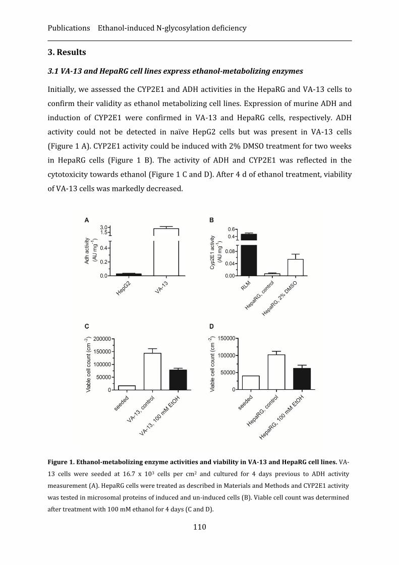
Publications ‒ Ethanol-induced N-glycosylation deficiency
110
3. Results
3.1 VA-13 and HepaRG cell lines express ethanol-metabolizing enzymes
Initially, we assessed the CYP2E1 and ADH activities in the HepaRG and VA-13 cells to
confirm their validity as ethanol metabolizing cell lines. Expression of murine ADH and
induction of CYP2E1 were confirmed in VA-13 and HepaRG cells, respectively. ADH
activity could not be detected in naïve HepG2 cells but was present in VA-13 cells
(Figure 1 A). CYP2E1 activity could be induced with 2% DMSO treatment for two weeks
in HepaRG cells (Figure 1 B). The activity of ADH and CYP2E1 was reflected in the
cytotoxicity towards ethanol (Figure 1 C and D). After 4 d of ethanol treatment, viability
of VA-13 cells was markedly decreased.
Figure 1. Ethanol-metabolizing enzyme activities and viability in VA-13 and HepaRG cell lines. VA-
13 cells were seeded at 16.7 x 10³ cells per cm2 and cultured for 4 days previous to ADH activity
measurement (A). HepaRG cells were treated as described in Materials and Methods and CYP2E1 activity
was tested in microsomal proteins of induced and un-induced cells (B). Viable cell count was determined
after treatment with 100 mM ethanol for 4 days (C and D).

Publications ‒ Ethanol-induced N-glycosylation deficiency
111
VA-13 sensitivity towards ethanol has been previously described (Clemens et al., 2002).
Compared to rat liver microsomes, the microsomal fractions of induced HepaRG cells
showed a 8.6-fold reduced activity of CYP2E1. Induced HepaRG cells showed a similar
sensitivity to ethanol as the VA-13 cells and viability was reduced by 39% upon ethanol
treatment.
3.2 Transferrin glycosylation is unaffected by ethanol treatment
To investigate the production of CDT, hepatocytes were exposed to different
concentrations of ethanol for 4 days and supernatants were analyzed. Supernatants
were subjected to isoelectric focusing and immunoblotting against transferrin.
Transferrin glycoform distribution was different in supernatants of cultured
hepatocytes compared to blood serum (Figure 2). In contrast to blood serum,
supernatants showed more prominent asialo- up to trisialo-transferrin bands instead of
tetrasialo- and disialo-transferrin, which were prominent in healthy control serum. This
pattern could be observed both cell lines and reflects the aberrant N-glycosylation found
in hepatic carcinoma conditions (Kamiyama et al., 2013). Moreover, no effect on
glycoform distribution was visible using concentrations between 25 to 200 mM ethanol.
With higher ethanol concentrations, less transferrin was detected. The decrease in
Figure 2. Carbohydrate-deficient transferrin as marker for glycosylation deficiency. Transferrin
was analyzed 4 days after ethanol exposure using isoelectric focusing and subsequent immunoblotting.
As a comparison, blood serum from a phosphomannomutase 2-CDG patient was analyzed by the same
procedure.

Publications ‒ Ethanol-induced N-glycosylation deficiency
112
transferrin signal reflected low cell numbers due to ethanol toxicity or may indicate
impaired protein secretion.
3.3 Transcript analysis reveals potential regulatory changes along the N-
glycosylation pathway
To get more detailed information of changes occurring in the N-linked glycosylation
pathway, we analyzed the transcripts of different glycosylation genes that are involved
in key steps of the Dol-linked oligosaccharide assembly by quantitative PCR after 25 mM
and 100 mM ethanol treatment for 4 days (Figure 3 A). No significant changes could be
observed for the analyzed transcripts upon 25 mM ethanol treatment. After treatment
with 100 mM ethanol, however, the mRNA levels of two genes were slightly changed.
DPM1, a subunit of DPM synthase, was downregulated by 17%. DPM synthase works as
a heterotrimer (DPM1-DPM3) transferring a Man-1-P from GDP-mannose to Dol-P. The
resulting Dol-P-Man serves as a substrate for mannosyl transferases in the ER lumen.
The second transcript RPN2 was upregulated by 8%. RPN2 is a ribophorin taking part in
the recruitment of the oligosaccharyl transferase complex to the ER-coupled ribosomes.
We observed a dose dependency on ethanol concentration as a trend in all the tested
transcripts. For instance, DPM1 mRNA was decreased at 25 mM ethanol but only at 100
mM ethanol the change was significant between control and ethanol-treated cells.
DPM synthase activity was measured to determine whether the lower transcript level of
DPM1 would affect DPM synthase activity. We found a marked increase in DPM synthase
activity in cell lysates from ethanol treated VA-13 cells (Figure 3 B). The increase in DPM
synthase activity stands in contrast with the minor decrease in DPM1 transcript levels.
Notably, the catalytic subunit of DPM synthase was not affected at the transcriptional
level. The moderate downregulation in DPM1 transcript together with the 36% increase
in DPM synthase activity might point to a regulatory mechanism.
3.4 Dolichol-phosphate pool is decreased after ethanol treatment
Next, we assessed the most prevalent Dol-P species consisting of 90, 95, and 100 carbon
atoms in hepatoma cells. Treatment of HepaRG cells with ethanol resulted in a decrease
of Dol-P levels (Figure 4). The strongest decrease was detected for the C95-Dol-P, which
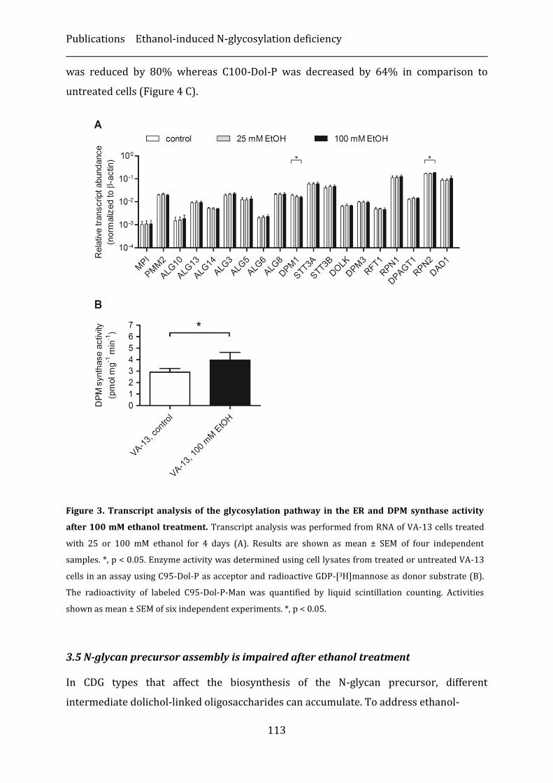
Publications ‒ Ethanol-induced N-glycosylation deficiency
113
was reduced by 80% whereas C100-Dol-P was decreased by 64% in comparison to
untreated cells (Figure 4 C).
Figure 3. Transcript analysis of the glycosylation pathway in the ER and DPM synthase activity
after 100 mM ethanol treatment. Transcript analysis was performed from RNA of VA-13 cells treated
with 25 or 100 mM ethanol for 4 days (A). Results are shown as mean ± SEM of four independent
samples. *, p < 0.05. Enzyme activity was determined using cell lysates from treated or untreated VA-13
cells in an assay using C95-Dol-P as acceptor and radioactive GDP-[3H]mannose as donor substrate (B).
The radioactivity of labeled C95-Dol-P-Man was quantified by liquid scintillation counting. Activities
shown as mean ± SEM of six independent experiments. *, p < 0.05.
3.5 N-glycan precursor assembly is impaired after ethanol treatment
In CDG types that affect the biosynthesis of the N-glycan precursor, different
intermediate dolichol-linked oligosaccharides can accumulate. To address ethanol-

Publications ‒ Ethanol-induced N-glycosylation deficiency
114
Figure 4. Dolichol-phosphate levels after 100 mM ethanol treatment. Dol-P levels were determined
in HepaRG cells after 4 d of ethanol treatment. C90, C95, and C100 peaks were identified using Dol-P
standards and quantitative determination was done with using C80-Dol-P as internal standard.
Representative HPLC profiles are shown (A, B). Peak areas were determined and analyzed (C). Data
represent mean values ±SEM of three independent experiments. *, p < 0.05.
induced changes in N-glycan precursor assembly, we metabolically labeled dolichol-
linked oligosaccharides with [3H]mannose and subsequently separated them by HPLC
coupled to a flow scintillation analyzer. The dolichol-linked oligosaccharide profile
underwent a shift, displaying a smaller fraction of the final oligosaccharide precursor
when cells were treated with 100 mM ethanol (Figure 5 A , B). A reduction of the
GlcNAc2-Man9-Glc3 peak by 27% was observed in HepaRG cells indicating either a lower
availability of dolichol leading to a general decrease is dolichol-linked oligosaccharide or
a defect in a later stage of the N-glycan biosynthesis in the ER. Typically, defects more

Publications ‒ Ethanol-induced N-glycosylation deficiency
115
early in the biosynthesis lead to an accumulation or a uniform decrease of dolichol-
linked oligosaccharide precursors (Figure 5 C). The same trend of a reduced GlcNAc2-
Man9-Glc3 peak could be observed in VA-13 cells (Figure 5 D).
Figure 5. Dolichol-linked oligosaccharide analysis. VA-13 and HepaRG cells were metabolically
labeled with [3H]mannose to analyze dolichol-linked oligosaccharide precursors. Representative control
and 100 mM ethanol treated sample profiles are shown (A and B). Relative peak areas of the
GlcNAc2Man9Glc3 to total area were calculated (C and D). Data represent mean values ±SEM of three
independent experiments. *, p < 0.05.
Publications ‒ Ethanol-induced N-glycosylation deficiency
116
4. Discussion
We investigated the effect of ethanol exposure on N-linked protein glycosylation and
observed lower Dol-P levels, in particular C95-Dol-P in our HepaRG model. Lower
dolichol levels have been reported previously in murine model systems upon ethanol
treatment (Cottalasso et al., 1998; Cottalasso et al., 1996). Dol-P is critical for N-
glycosylation in two ways. First, it serves as a carrier for the N-glycan precursor. Second,
Dol-P coupled to Man or Glc is essential for glycosyltransferases located in the ER lumen.
Accordingly, low availability of Dol-P-Man in DPM1-CDG was previously demonstrated
to be attenuated by increasing Dol-P thereby compensating the low activity of DPM
synthase with better substrate availability (Haeuptle et al., 2011). The strategy of
increasing dolichol levels to attenuate glycosylation deficiency in ALD would be
interesting but might not be sufficient due to the complexity of ethanol-induced N-
glycosylation deficiency.
This is the first study showing ethanol-induced effects on the biosynthesis of the N-
glycan precursors, namely a reduction of Dol-PP-GlcNAc2Man9Glc3 compared to
untreated cells. Interestingly, while general decrease in dolichol-linked oligosaccharide
is well defined in CDG, such a reduction of the final dolichol-linked precursor has not
been observed yet, neither in inherited nor in acquired glycosylation deficiencies. The
dolichol-linked oligosaccharide pattern suggests a deficiency in the ER luminal assembly
of the N-glycan precursor or could be a consequence of lower dolichol levels. CDG
research has shown that deficient enzymes of the early, cytosolic steps of N-glycan
precursor biosynthesis preferentially lead to an accumulation of Dol-P precursors, such
as Dol-PP-GlcNAc2-Man5 in DPM1-CDG (Imbach et al., 2000). The shortage in dolichol
could also explain the lower Dol-PP-GlcNAc2Man9Glc3 levels with decreased availability
of the N-glycan precursors, leading to drainage of the dolichol-linked oligosaccharide
pool. CDG with deficient dolichol or Dol-P biosynthesis typically result in reduced
dolichol-linked oligosaccharide but do not alter the distribution of the dolichol-linked
precursors as observed in DOLK-CDG (Kranz et al., 2007). In SRD5A3-CDG, the
polyprenol reductase along the dolichol biosynthetic pathway is non-functional but does
not lead to decreased dolichol levels and fibroblasts show a healthy dolichol-linked
oligosaccharide distribution (Grundahl et al., 2012). Symptoms on the systemic level are
not shared by CDG affecting dolichol biosynthesis and ALD. The tendency of CDG for
liver malformation or disease is apparent in other forms of CDG with defects in later
Publications ‒ Ethanol-induced N-glycosylation deficiency
117
steps of the N-linked glycosylation pointing towards a more complex mechanism in N-
glycosylation deficiency in ALD.
Transcriptional regulation of N-glycosylation genes could explain ethanol-induced N-
glycosylation deficiency. We found a trend for transcript alteration upon ethanol
treatment but only to a marginal extent. DPM1, a subunit of the DPM synthase, was
transcriptionally downregulated by 17% indicating a regulatory mechanism. This notion
is supported by our observation that DPM synthase activity was increased by 36%. More
work is needed to elucidate possible regulatory mechanisms for the N-glycosylation
pathway in ALD. Since reduced Dol-PP-GlcNAc2Man9Glc3 might be due to dysfunction of
the later, luminal steps of the N-glycan precursor assembly, a deregulation of the
oligosaccharyl transferase (OST) offers an explanation. Indeed, we found an indication
for potential OST deregulation. RPN2, a part of the OST complex, was slightly
upregulated. RPN2 was shown to promote glycosylation of P-glycoprotein, an ABC
transporter of the multidrug resistance family (Honma et al., 2008; Kerb et al., 2001).
Knock-down of RPN2 resulted in reduced glycosylation as well as reduced membrane
localization of P-glycoprotein. We speculate that RPN2 upregulation upon ethanol-
treatment induces changes in N-linked glycosylation of RPN2 target proteins
contributing to an aberrant dolichol-linked oligosaccharide pattern. It would be
interesting to further investigate the expression and function of OST subunits in order to
get a notion of the impact of ethanol on OST activity.
5. Conclusion
In conclusion, we found that decreased Dol-P and altered dolichol-linked
oligosaccharide profiles contribute to ethanol-induced N-linked glycosylation deficiency.
Furthermore, altered transcription of genes along the N-linked glycosylation pathway
was found indicating a deregulation in N-glycosylation. Together with previous findings
of lower sialyltransferase and higher sialidase activities, ethanol-induced glycosylation
deficiency proves to be a result of metabolic interferences along the N-glycosylation
biosynthetic pathway starting from the early steps of dolichol synthesis, continuing with
the biosynthesis of the N-linked glycan precursor, and concluding with the final steps of
glycan capping in the Golgi apparatus.

Publications ‒ Ethanol-induced N-glycosylation deficiency
118
References
Bean, P., and J.B. Peter. 1993. A new approach to quantitate carbohydrate-deficient transferrin isoforms in alcohol abusers: partial iron saturation in isoelectric focusing/immunoblotting and laser densitometry. Alcohol Clin Exp Res 17:1163-1170.
Burda, P., L. Borsig, J. de Rijk-van Andel, R. Wevers, J. Jaeken, H. Carchon, E.G. Berger, and M. Aebi. 1998. A novel carbohydrate-deficient glycoprotein syndrome characterized by a deficiency in glucosylation of the dolichol-linked oligosaccharide. J Clin Invest 102:647-652.
Carter, E.A., and J.R. Wands. 1988. Ethanol-induced inhibition of liver cell function: I. Effect of ethanol on hormone stimulated hepatocyte DNA synthesis and the role of ethanol metabolism. Alcohol Clin Exp Res 12:555-562.
Clemens, D.L., A. Forman, T.R. Jerrells, M.F. Sorrell, and D.J. Tuma. 2002. Relationship between acetaldehyde levels and cell survival in ethanol-metabolizing hepatoma cells. Hepatology 35:1196-1204.
Clemens, D.L., C.M. Halgard, R.R. Miles, M.F. Sorrell, and D.J. Tuma. 1995. Establishment of a recombinant hepatic cell line stably expressing alcohol dehydrogenase. Arch Biochem
Biophys 321:311-318. Cottalasso, D., A. Bellocchio, M.A. Pronzato, C. Domenicotti, N. Traverso, M.V. Gianelli, U.M.
Marinari, and G. Nanni. 1998. Effect of ethanol administration on the level of dolichol in rat liver microsomes and Golgi apparatus. Alcoholism, clinical and experimental research 22:730-737.
Cottalasso, D., P. Gazzo, D. Dapino, C. Domenicotti, M.A. Pronzato, N. Traverso, A. Bellocchio, G. Nanni, and U.M. Marinari. 1996. Effect of chronic ethanol consumption on glycosylation processes in rat liver microsomes and Golgi apparatus. Alcohol and alcoholism (Oxford,
Oxfordshire) 31:51-59. Crick, D.C., and K.K. Carroll. 1987. Extraction and quantitation of total cholesterol, dolichol and
dolichyl phosphate from mammalian liver. Lipids 22:1045-1048. de Jong, G., W.L. van Noort, and H.G. van Eijk. 1992. Carbohydrate analysis of transferrin
subfractions isolated by preparative isoelectric focusing in immobilized pH gradients. Electrophoresis 13:225-228.
Eggens, I., T.J. Ekstrom, and F. Aberg. 1990. Studies on the biosynthesis of polyisoprenols, cholesterol and ubiquinone in highly differentiated human hepatomas. J Exp Pathol
(Oxford) 71:219-232. Eggens, I., and P.G. Elmberger. 1990. Studies on the polyisoprenoid composition in
hepatocellular carcinomas and its correlation with their differentiation. APMIS 98:535-542.
Elmberger, P.G., I. Eggens, and G. Dallner. 1989. Conditions for quantitation of dolichyl phosphate, dolichol, ubiquinone and cholesterol by HPLC. Biomed Chromatogr 3:20-28.
Filipovic, I., and B. Menzel. 1981. Action of low-density lipoprotein and compactin, a competitive inhibitor of 3-hydroxy-3-methylglutaryl-CoA reductase, on the synthesis of dolichol-linked oligosaccharides and low-density-lipoprotein receptor in human skin fibroblasts. Biochem J 196:625-628.
Gapp, I.W., C.R. Congreve, and B.S. Lieberman. 2012. Unraveling the phylogenetic relationships of the Eccoptochilinae, an enigmatic array of ordovician cheirurid trilobites. PLoS One 7:e49115.
Garige, M., M. Gong, and M.R. Lakshman. 2006. Ethanol destabilizes liver Gal beta l, 4GlcNAc alpha2,6-sialyltransferase, mRNA by depleting a 3'-untranslated region-specific binding protein. The Journal of pharmacology and experimental therapeutics 318:1076-1082.
Garige, M., M. Gong, M.N. Rao, Y. Zhang, and M.R. Lakshman. 2005. Mechanism of action of ethanol in the down-regulation of Gal(beta)1, 4GlcNAc alpha2,6-sialyltransferase messenger RNA in human liver cell lines. Metabolism 54:729-734.
Publications ‒ Ethanol-induced N-glycosylation deficiency
119
Gripon, P., S. Rumin, S. Urban, J. Le Seyec, D. Glaise, I. Cannie, C. Guyomard, J. Lucas, C. Trepo, and C. Guguen-Guillouzo. 2002. Infection of a human hepatoma cell line by hepatitis B virus. Proc Natl Acad Sci U S A 99:15655-15660.
Grundahl, J.E., Z. Guan, S. Rust, J. Reunert, B. Muller, I. Du Chesne, K. Zerres, S. Rudnik-Schoneborn, N. Ortiz-Bruchle, M.G. Hausler, J. Siedlecka, E. Swiezewska, C.R. Raetz, and T. Marquardt. 2012. Life with too much polyprenol: polyprenol reductase deficiency. Mol
Genet Metab 105:642-651. Haeuptle, M.A., A.J. Hülsmeier, and T. Hennet. 2010. HPLC and mass spectrometry analysis of
dolichol-phosphates at the cell culture scale. Anal Biochem 396:133-138. Haeuptle, M.A., M. Welti, H. Troxler, A.J. Hülsmeier, T. Imbach, and T. Hennet. 2011. Improvement
of dolichol-linked oligosaccharide biosynthesis by the squalene synthase inhibitor zaragozic acid. J Biol Chem 286:6085-6091.
Honma, K., K. Iwao-Koizumi, F. Takeshita, Y. Yamamoto, T. Yoshida, K. Nishio, S. Nagahara, K. Kato, and T. Ochiya. 2008. RPN2 gene confers docetaxel resistance in breast cancer. Nat
Med 14:939-948. Hülsmeier, A.J., P. Paesold-Burda, and T. Hennet. 2007. N-glycosylation site occupancy in serum
glycoproteins using multiple reaction monitoring liquid chromatography-mass spectrometry. Mol Cell Proteomics 6:2132-2138.
Imbach, T., B. Schenk, E. Schollen, P. Burda, A. Stutz, S. Grunewald, N.M. Bailie, M.D. King, J. Jaeken, G. Matthijs, E.G. Berger, M. Aebi, and T. Hennet. 2000. Deficiency of dolichol-phosphate-mannose synthase-1 causes congenital disorder of glycosylation type Ie. J Clin
Invest 105:233-239. Kamiyama, T., H. Yokoo, J. Furukawa, M. Kurogochi, T. Togashi, N. Miura, K. Nakanishi, H.
Kamachi, T. Kakisaka, Y. Tsuruga, M. Fujiyoshi, A. Taketomi, S. Nishimura, and S. Todo. 2013. Identification of novel serum biomarkers of hepatocellular carcinoma using glycomic analysis. Hepatology 57:2314-2325.
Keller, R.K. 1986. The mechanism and regulation of dolichyl phosphate biosynthesis in rat liver. J Biol Chem 261:12053-12059.
Kerb, R., S. Hoffmeyer, and U. Brinkmann. 2001. ABC drug transporters: hereditary polymorphisms and pharmacological impact in MDR1, MRP1 and MRP2. Pharmacogenomics 2:51-64.
Koop, D.R., and D.J. Tierney. 1990. Multiple mechanisms in the regulation of ethanol-inducible cytochrome P450IIE1. Bioessays 12:429-435.
Kranz, C., C. Jungeblut, J. Denecke, A. Erlekotte, C. Sohlbach, V. Debus, H.G. Kehl, E. Harms, A. Reith, S. Reichel, H. Grobe, G. Hammersen, U. Schwarzer, and T. Marquardt. 2007. A defect in dolichol phosphate biosynthesis causes a new inherited disorder with death in early infancy. Am J Hum Genet 80:433-440.
Lecca, M.R., C. Maag, E.G. Berger, and T. Hennet. 2011. Fibrotic response in fibroblasts from congenital disorders of glycosylation. J Cell Mol Med 15:1788-1796.
Leroy, J.G. 2006. Congenital disorders of N-glycosylation including diseases associated with O- as well as N-glycosylation defects. Pediatr Res 60:643-656.
Lieber, C.S. 1999. Microsomal ethanol-oxidizing system (MEOS): the first 30 years (1968-1998)--a review. Alcoholism, clinical and experimental research 23:991-1007.
Luo, Y., A. Nita-Lazar, and R.S. Haltiwanger. 2006. Two distinct pathways for O-fucosylation of epidermal growth factor-like or thrombospondin type 1 repeats. J Biol Chem 281:9385-9392.
McLachlan, K.R., and S.S. Krag. 1994. Three enzymes involved in oligosaccharide-lipid assembly in Chinese hamster ovary cells differ in lipid substrate preference. J Lipid Res 35:1861-1868.
Nairn, A.V., M. dela Rosa, and K.W. Moremen. 2010. Transcript analysis of stem cells. Methods
Enzymol 479:73-91. Ohnishi, K., and C.S. Lieber. 1977. Reconstitution of the microsomal ethanol-oxidizing system.
Qualitative and quantitative changes of cytochrome P-450 after chronic ethanol consumption. J Biol Chem 252:7124-7131.

Publications ‒ Ethanol-induced N-glycosylation deficiency
120
Peter, J., C. Unverzagt, W.D. Engel, D. Renauer, C. Seidel, and W. Hosel. 1998. Identification of carbohydrate deficient transferrin forms by MALDI-TOF mass spectrometry and lectin ELISABiochim Biophys Acta 1998 Aug 24;1381(3):356. Biochimica et biophysica acta 1380:93-101.
Petren, S., O. Vesterberg, and H. Jornvall. 1987. Differences among five main forms of serum transferrin. Alcohol Clin Exp Res 11:453-456.
Purohit, V., B. Gao, and B.J. Song. 2009. Molecular mechanisms of alcoholic fatty liver. Alcohol Clin
Exp Res 33:191-205. Rao, M.N., and M.R. Lakshman. 1997. Chronic ethanol downregulates Gal-beta-1,4GlcNAc alpha
2,6-sialyltransferase and Gal-beta-1,3GlcNAc alpha 2,3-sialyltransferase mRNAs in rat liver. Alcoholism, clinical and experimental research 21:348-351.
Rao, M.N., and M.R. Lakshman. 1999. Chronic ethanol consumption leads to destabilization of rat liver beta-galactoside alpha2,6-sialyltransferase mRNA. Metabolism: clinical and
experimental 48:797-803. Stibler, H. 1991. Carbohydrate-deficient transferrin in serum: a new marker of potentially
harmful alcohol consumption reviewed. Clinical chemistry 37:2029-2037. Stoll, J., A.G. Rosenwald, and S.S. Krag. 1988. A Chinese hamster ovary cell mutant F2A8 utilizes
polyprenol rather than dolichol for its lipid-dependent asparagine-linked glycosylation reactions. J Biol Chem 263:10774-10782.
Utesch, D., B. Diener, E. Molitor, F. Oesch, and K.L. Platt. 1992. Characterization of cryopreserved rat liver parenchymal cells by metabolism of diagnostic substrates and activities of related enzymes. Biochem Pharmacol 44:309-315.
Van Kaick, G., D. Lieberman, D. Lorenz, W.J. Lorenz, H. Luhrs, K.E. Scheer, H. Wesch, H. Muth, A. Kaul, H. Immich, G. Wagner, and K. Wegener. 1983. Recent results of the German Thorotrast study--epidemiological results and dose effect relationships in Thorotrast patients. Health Phys 44 Suppl 1:299-306.
Vigo, C., and W.L. Adair, Jr. 1982. In vivo biosynthesis of the saturated isoprene unit of dolichyl phosphate. Biosci Rep 2:835-840.
Wang, H.J., B. Gao, S. Zakhari, and L.E. Nagy. 2012. Inflammation in alcoholic liver disease. Annu
Rev Nutr 32:343-368. Wu, D., and A.I. Cederbaum. 2008. Development and properties of HepG2 cells that constitutively
express CYP2E1. Methods Mol Biol 447:137-150. Yokoyama, K., H. Mizuguchi, Y. Araki, S. Kaya, and E. Ito. 1989. Biosynthesis of linkage units for
teichoic acids in gram-positive bacteria: distribution of related enzymes and their specificities for UDP-sugars and lipid-linked intermediates. J Bacteriol 171:940-946.
Zhu, H., Z. Jia, H. Misra, and Y.R. Li. 2012. Oxidative stress and redox signaling mechanisms of alcoholic liver disease: updated experimental and clinical evidence. J Dig Dis 13:133-142.
Zufferey, R., R. Knauer, P. Burda, I. Stagljar, S. te Heesen, L. Lehle, and M. Aebi. 1995. STT3, a highly conserved protein required for yeast oligosaccharyl transferase activity in vivo. EMBO J 14:4949-4960.

121

Discussion
122
GENERAL DISCUSSION
Considering the immense abundance of glycans on cell surfaces and secreted proteins,
countless essential biological functions depend on glycosylation. However, glycobiology
has been a comparatively small field of research so far. Although glycobiologists are very
active in promoting their field, glycobiology has received relatively little attention in
other areas of biological sciences. In part, this was due to the gap of knowledge about the
significance of glycosylation but also due to restricted possibilities regarding efficient
methods to analyze the almost unmanageable complexity of sugar structures. With the
advent of advanced technologies such as high-throughput mass spectrometry in
conjunction with powerful computational software, glycans have moved within reach of
researchers other than glycobiologists (Kolarich et al., 2013; Mechref et al., 2012; Zaia,
2010; Zaia, 2013). Additionally, progress in glycobiology prompted other scientists to
consider glycobiological aspects in their field of research. Consequently, the significance
of glycosylation is being discovered in many fields including cancer research,
immunology, virology, and developmental biology. This development is highly beneficial
for the understanding of glycans and their functions.
Initially, glycans were merely considered decorating structures on cell surfaces of all
kind of cell types in all the domains of life. The significance of these ╉decorations╊ for humans was first realized with the discovery of the ABO blood groups in 1900
(Landsteiner, 1900). At the time, the nature of the determinants was totally elusive but
the consequences for blood transfusion were immense. Nearly 60 years later, Morgan
and Watkins defined sugars to be the blood group determinants (Morgan and Watkins,
1959; Watkins and Morgan, 1959). Later, the first carbohydrate-deficient glycoprotein
syndrome was discovered (Jaeken et al., 1980). The patients suffered from mental
retardation, muscular hypotonia, failure to thrive, inverted nipples, and abnormal fat
deposits. Carbohydrate-deficient transferrin (CDT) was defined as a marker for CDG
using isoelectric focusing of blood serum. Only fifteen years after its discovery, a
phosphomannomutase deficiency was defined as the molecular basis of this CDG (Van
Schaftingen and Jaeken, 1995). In the meantime, a CDG with a different CDT profile was
discovered with a defect in GnT-II. It was termed CDG-II (Jaeken et al., 1994; Ramaekers
et al., 1991). The discovery rate of novel forms of CDG was rapidly increasing and greatly
contributed to the understanding of the glycosylation pathways. Yeast models have been
Discussion
123
instrumental by comparison of DLO profiles of yeast mutants with DLO profiles of CDG
patients thereby identifying genes of the glycosylation machinery in yeast and
subsequently confirmation of the mutation in CDG patients by gene sequencing (Aebi
and Hennet, 2001). Considering the 2% fraction of the genome that is involved in
glycosylation, it can be speculated that the majority of CDG types is yet to be discovered.
Recently discovered forms of CDG surprisingly do not have the typical symptomatic
picture but show tissue- or organ-specific impact. In DHDDS-CDG, for instance, retinal
degeneration is the single apparent symptom (Zelinger et al., 2011). DHDDS is the
dehydrodolichyl diphosphate synthase which catalyzes the synthesis of polyprenol-PP
from two molecules of farnesyl-PP. Thus, DHDDS is crucial for Dol biosynthesis and the
implications of a defective DHDDS would seem to cause a much broader spectrum of
symptoms. Either the discovered DHDDS-CDG cases have enough residual polyprenol-PP
production from mutated DHDDS or another enzyme can compensate for the loss in
DHDDS activity. Retinis pigmentosa is a manifestation well known in CDG as it occurs
also in mevalonate kinase deficiency and PMM2-CDG (Goldfinger, 2009; Grunewald,
2009).
The challenge of uncovering regulatory mechanisms
The pathways of glycosylation in humans are well defined and all the N-linked
glycosylation steps are assigned to enzymes (Haeuptle and Hennet, 2009; Jaeken,
2010b). However, parallel pathways to the ones already discovered are more difficult to
identify partly because of the more complex human genome compared to model systems
like yeast. Some of the pathways are specific to vertebrates or even restricted to
mammals (Dell et al., 2010). Moreover, deficient enzymes involved in more peripheral
functions such as regulatory enzymes or tissue specific glycosylation-associated genes
render a characterization more difficult because of the difficulty to detect such defects
unlike the strong phenotype of classical CDG. Generally, the regulation of the
glycosylation machinery is elusive. Importantly, pathways with global contributions to
an organism are highly regulated as can be seen in metabolic pathways but also signal
transduction or gene regulation (Alberts, 2008). Regulation of glycosylation seems likely
to be tightly regulated especially during development of the embryo since CDG patients
have severe, multisystemic defects in early childhood (Haltiwanger and Lowe, 2004;

Discussion
124
Jaeken, 2010a). Additionally, mechanisms of development of CDG have not been defined
yet. Investigations in this direction are difficult, though. An promising approach is a
switch system in which genes can be turned on and off at certain points in development
using mouse models.
We encountered a possible regulatory mechanism for N-linked glycosylation in liver
cells exposed to ethanol. ALD is unique due a non-systemic ethanol-induced N-
glycosylation deficiency of liver proteins. We tested the effect of ethanol on N-linked
glycosylation in a cell culture model using two hepatic carcinoma cell models to define
N-linked glycosylation deficiency in ALD. Ethanol exposure led to decreased Dol-P levels
supporting previous experiments with ethanol-fed rats and particularly the C95 species
was dramatically decreased (Cottalasso et al., 1998; Cottalasso et al., 1996). As a
possible consequence, we found a decreased amount of the N-glycan precursor. This was
the first characterization of Dol-linked oligossacharide profiles in ethanol-induced N-
glycosylation deficiency. To investigate gene regulation at the transcriptional level, we
tested the glycosylation genes taking part in ER glycosylation steps. Interestingly, we
found a dose dependency of transcription on ethanol concentration as a trend.
Transcription of two genes was significantly changed. DPM1, a component of the
trimeric DPM synthase, was downregulated whereas RPN2, a regulatory subunit of the
OST, was upregulated. To obtain a hint whether the transcriptional changes might be of
regulatory nature, we tested the activity of DPM synthase and found a strong
upregulation of enzymatic activity. How this potential regulatory mechanism is
governed has yet to be investigated for instance by determination of the dose-
dependency of DPM synthase activity and DPM1 transcriptional downregulation on
ethanol concentration and by looking at the correlation of activity and transcript level.
In case of RPN2, we can only speculate about the effect of this upregulation due to the
complexity of the OST which can consist of different subunits possibly targeting
different sets of proteins or even individual proteins (Vleugels et al., 2009).
Identification of RPN2-specific N-glycosylation targets could provide a readout for our
findings.
Discussion
125
Therapeutics for glycosylation disorders
While novel forms of CDG are being discovered, efforts to treat the known forms of CDG
have been undertaken. However, treatment for CDG is principally limited to post-natal
patients because of the lack of pre-natal tests for CDG (Funke et al., 2013). Glycosylation
is crucial for development and proper functioning of a variety of organ systems,
particularly for the nervous system evident from the neurological component of most
CDG. Unfortunately, neurological deficits in CDG are not treatable with present-day
medicine. In order to find therapeutic targets, identifying mutations in the genome is not
enough. High-throughput sequencing methods like whole-exome sequencing enable
screening of patients with inherited diseases to pinpoint mutations (Freeze, 2013).
However, biochemical read-outs are required to confirm candidate genes and potential
treatments need to be validated for improvements of biochemical parameters.
MPI-CDG is a special case of CDG due to the usual lack of neurological symptoms
(Westphal et al., 2001). This lack indicates that the defect does not manifest already
during development but is important for maintaining the function of developed organs.
However, the liver and gastrointestinal tract are affected. Symptoms include vomiting,
diarrhea, gastrointestinal bleeding, protein-losing enteropathy, hepatomegaly, and liver
fibrosis during childhood and they improve in adulthood. Severe cases show
coagulopathy, hypoglycemia, and thrombosis. Nutritional mannose supplementation has
been successful in relieving many of the symptoms (Babovic-Vuksanovic et al., 1999; de
Lonlay et al., 1999; Niehues et al., 1998; Penel-Capelle et al., 2003; Westphal et al., 2001).
Besides MPI-CDG, only two other CDG were reported to respond to treatment (Jaeken,
2010a). In SLC35C1-CDG, the deficiency in the GDP-Fuc transporter in the Golgi network
can be compensated with fucose supplementation in some cases (Hellbusch et al., 2007).
In PIGM-CDG, histone histone acetylation at the PIGM promotor is impaired and thus
lowers PIGM-dependent mannosyltransferase transcription (Almeida et al., 2006). Using
butyrate to inhibit histone deacetylase increased PIGM transcription and stopped
seizures. Considering yet undiscovered CDG with non-developmental symptoms,
medical treatment could help to attenuate manifestations caused by impaired
glycosylation. Delivery of the medication is critical of course and in case of mannose
supplementation an easy task. Recently, prenatal mannose treatment of mice with an
engineered PMM2 defect has proven successful. Embryonic lethality of the hypomorphic
PMM2 mutation could be overcome by mannose administration to the mother
Discussion
126
(Schneider et al., 2011). The mannose-treated PMM2-defective embryos showed no
manifestations of CDG. These examples demonstrate the main difficulties when it comes
to CDG treatment. First, CDG affecting development, which include most CDG, require
pre-natal treatment and need to be identified by methods that are not yet available.
Second, pre-natal treatment is more difficult because treatment usually is applied on the
mother and is restricted by the placenta barrier (Giaginis et al., 2012; Vahakangas and
Myllynen, 2009). In case of non-developmental effects, the efficiency of medication
greatly depends on sufficient residual activity of the affected enzyme. Importantly,
development of medication for rare diseases is financially unattractive and therefore
mannose treatment for MPI-CDG treatment has been a lucky exception due to its low
cost and easy application.
In this work, we tested a therapeutic approach to treat DPM1-CDG. We exploited the
action of Zaragozic acid, a cholesterol-lowering drug that differs from the classical
statins. Zaragozic acid was discovered in a screen for cholesterol-lowering compounds
in the context of cardiovascular disease treatment (Bergstrom et al., 1995). It inhibits
squalene synthase which is active at a bifurcation in the mevalonate pathway. The
bifurcation divides into cholesterol biosynthesis ‒ through the action of squalene
synthase ‒ and into Dol biosynthesis (Welti, 2013). We could show that supplying
fibroblasts with the squalene synthase inhibitor Zaragozic acid improved N-linked
protein glycosylation and GPI-anchor formation in DPM1-CDG (Haeuptle et al., 2011). As
expected, inhibition of squalene synthase resulted in lower cholesterol and higher Dol-P
levels. We speculated that higher Dol-P levels could compensate for the insufficient DPM
synthase activity. Indeed, Dol-P-Man levels could be restored. Moreover, Dol-linked
oligosaccharide and N-linked oligosaccharide profiles were partly normalized and were
reminiscent of healthy oligosaccharide profiles. Moreover, GPI anchor availability was
improved as assessed by the expression of the GPI-anchored membrane surface protein
CD59. With this work, we provided a first step in DPM1-CDG therapy. We used an
available drug that was shown to be well tolerated at least in rats and confirmed the
treatment success by measuring biochemical readouts (Baxter et al., 1992; Bergstrom et
al., 1993). Whether such a treatment could attenuate DPM1-CDG manifestations at the
level of an entire organism, needs further confirmation for instance using an animal
model.
Discussion
127
Future directions
Considering the 2% of genes that are predicted to be involved in glycosylation
processes, it is safe to speculate that glycobiology plays an important ‒ even if not yet
fully recognized and defined ‒ part in other diseases like cancer, immune disorders, or
infectious diseases (Freeze et al., 2012). We showed, for instance, that glycosylation is
perturbed at the level of Dol-linked oligosaccharide assembly in ethanol-treated
hepatoma cells. This is likely to be associated with lower Dol levels. The specific
consequences of underglycosylation of blood proteins as found in ALD are difficult to
estimate due to the impact of chronic alcohol consumption on multiple organ systems
(Wang et al., 2010). Future studies might assess the contribution of glycosylation
deficiency to the clinical picture of ALD and other consequences of alcoholism.
Importantly, connecting glycobiology to other fields of research is beneficial in many
ways. Awareness of glycan functions can be raised, more knowledge about mechanisms
in regulation of glycosylation can be gained, and possibly novel therapeutic targets in
CDG could be identified. In this way, glycobiologists can profit from the attention and
knowledge of other fields as we demonstrated by using cholesterol-lowering Zaragozic
acid found in the fight against cardiovascular disease to improve glycosylation in DPM1-
CDG. In ALD, liver fibrosis is a well-known characteristic as it is in certain forms of CDG
(Jaeken et al., 1998; Pelletier et al., 1986). Efforts to uncover mechanisms how liver
fibrosis develops in CDG might help to understand liver fibrosis in ALD given the
common deficiency in N-glycosylation. Vice versa, understanding the development of
ethanol-induced glycosylation deficiency might reveal regulatory mechanisms for N-
linked glycosylation.
Discussion
128
References
Aebi, M., and T. Hennet. 2001. Congenital disorders of glycosylation: genetic model systems lead the way. Trends Cell Biol 11:136-141.
Alberts, B. 2008. Molecular biology of the cell. Garland Science, New York. 1268 pp. Almeida, A.M., Y. Murakami, D.M. Layton, P. Hillmen, G.S. Sellick, Y. Maeda, S. Richards, S.
Patterson, I. Kotsianidis, L. Mollica, D.H. Crawford, A. Baker, M. Ferguson, I. Roberts, R. Houlston, T. Kinoshita, and A. Karadimitris. 2006. Hypomorphic promoter mutation in PIGM causes inherited glycosylphosphatidylinositol deficiency. Nat Med 12:846-851.
Babovic-Vuksanovic, D., M.C. Patterson, W.F. Schwenk, J.F. O'Brien, J. Vockley, H.H. Freeze, D.P. Mehta, and V.V. Michels. 1999. Severe hypoglycemia as a presenting symptom of carbohydrate-deficient glycoprotein syndrome. J Pediatr 135:775-781.
Baxter, A., B.J. Fitzgerald, J.L. Hutson, A.D. McCarthy, J.M. Motteram, B.C. Ross, M. Sapra, M.A. Snowden, N.S. Watson, R.J. Williams, and et al. 1992. Squalestatin 1, a potent inhibitor of squalene synthase, which lowers serum cholesterol in vivo. J Biol Chem 267:11705-11708.
Bergstrom, J.D., C. Dufresne, G.F. Bills, M. Nallin-Omstead, and K. Byrne. 1995. Discovery, biosynthesis, and mechanism of action of the zaragozic acids: potent inhibitors of squalene synthase. Annu Rev Microbiol 49:607-639.
Bergstrom, J.D., M.M. Kurtz, D.J. Rew, A.M. Amend, J.D. Karkas, R.G. Bostedor, V.S. Bansal, C. Dufresne, F.L. VanMiddlesworth, O.D. Hensens, and et al. 1993. Zaragozic acids: a family of fungal metabolites that are picomolar competitive inhibitors of squalene synthase. Proc Natl Acad Sci U S A 90:80-84.
Cottalasso, D., A. Bellocchio, M.A. Pronzato, C. Domenicotti, N. Traverso, M.V. Gianelli, U.M. Marinari, and G. Nanni. 1998. Effect of ethanol administration on the level of dolichol in rat liver microsomes and Golgi apparatus. Alcoholism, clinical and experimental research 22:730-737.
Cottalasso, D., P. Gazzo, D. Dapino, C. Domenicotti, M.A. Pronzato, N. Traverso, A. Bellocchio, G. Nanni, and U.M. Marinari. 1996. Effect of chronic ethanol consumption on glycosylation processes in rat liver microsomes and Golgi apparatus. Alcohol and alcoholism (Oxford,
Oxfordshire) 31:51-59. de Lonlay, P., M. Cuer, S. Vuillaumier-Barrot, G. Beaune, P. Castelnau, M. Kretz, G. Durand, J.M.
Saudubray, and N. Seta. 1999. Hyperinsulinemic hypoglycemia as a presenting sign in phosphomannose isomerase deficiency: A new manifestation of carbohydrate-deficient glycoprotein syndrome treatable with mannose. J Pediatr 135:379-383.
Dell, A., A. Galadari, F. Sastre, and P. Hitchen. 2010. Similarities and differences in the glycosylation mechanisms in prokaryotes and eukaryotes. Int J Microbiol 148178:27.
Freeze, H.H. 2013. Understanding human glycosylation disorders: biochemistry leads the charge. J Biol Chem 288:6936-6945.
Freeze, H.H., E.A. Eklund, B.G. Ng, and M.C. Patterson. 2012. Neurology of inherited glycosylation disorders. Lancet Neurol 11:453-466.
Funke, S., T. Gardeitchik, D. Kouwenberg, M. Mohamed, S.B. Wortmann, E. Korsch, M. Adamowicz, L. Al-Gazali, R.A. Wevers, A. Horvath, D.J. Lefeber, and E. Morava. 2013. Perinatal and early infantile symptoms in congenital disorders of glycosylation. Am J Med Genet A 3:578-584.
Giaginis, C., S. Theocharis, and A. Tsantili-Kakoulidou. 2012. Current toxicological aspects on drug and chemical transport and metabolism across the human placental barrier. Expert
Opin Drug Metab Toxicol 8:1263-1275. Goldfinger, S. 2009. The inherited autoinflammatory syndrome: a decade of discovery. Trans Am
Clin Climatol Assoc 120:413-418. Grunewald, S. 2009. The clinical spectrum of phosphomannomutase 2 deficiency (CDG-Ia).
Biochimica et biophysica acta 1792:827-834. Haeuptle, M.A., and T. Hennet. 2009. Congenital disorders of glycosylation: an update on defects
affecting the biosynthesis of dolichol-linked oligosaccharides. Hum Mutat 30:1628-1641.

Discussion
129
Haeuptle, M.A., M. Welti, H. Troxler, A.J. Hülsmeier, T. Imbach, and T. Hennet. 2011. Improvement of dolichol-linked oligosaccharide biosynthesis by the squalene synthase inhibitor zaragozic acid. J Biol Chem 286:6085-6091.
Haltiwanger, R.S., and J.B. Lowe. 2004. Role of glycosylation in development. Annual review of
biochemistry 73:491-537. Hellbusch, C.C., M. Sperandio, D. Frommhold, S. Yakubenia, M.K. Wild, D. Popovici, D. Vestweber,
H.J. Grone, K. von Figura, T. Lubke, and C. Korner. 2007. Golgi GDP-fucose transporter-deficient mice mimic congenital disorder of glycosylation IIc/leukocyte adhesion deficiency II. J Biol Chem 282:10762-10772.
Jaeken, J. 2010a. Congenital disorders of glycosylation. Ann N Y Acad Sci Jaeken, J. 2010b. Congenital disorders of glycosylation. Ann N Y Acad Sci 1214:190-198. Jaeken, J., G. Matthijs, J.M. Saudubray, C. Dionisi-Vici, E. Bertini, P. de Lonlay, H. Henri, H. Carchon,
E. Schollen, and E. Van Schaftingen. 1998. Phosphomannose isomerase deficiency: a carbohydrate-deficient glycoprotein syndrome with hepatic-intestinal presentation. Am J
Hum Genet 62:1535-1539. Jaeken, J., H. Schachter, H. Carchon, P. De Cock, B. Coddeville, and G. Spik. 1994. Carbohydrate
deficient glycoprotein syndrome type II: a deficiency in Golgi localised N-acetyl-glucosaminyltransferase II. Arch Dis Child 71:123-127.
Jaeken, J., M. Vanderschueren-Lodeweyckx, P. Casaer, L. Snoeck, L. Corbeel, E. Eggermont, and R. Eeckels. 1980. Familial psychomotor retardation with markedly fluctuating serum prolactin, FSH and GH levels, partial TBG-deficiency, increased serum arylsulphatase A and increased CSF protein: a new syndrome[quest]: 90. Pediatr Res 14:179-179.
Kolarich, D., E. Rapp, W.B. Struwe, S.M. Haslam, J. Zaia, R. McBride, S. Agravat, M.P. Campbell, M. Kato, R. Ranzinger, C. Kettner, and W.S. York. 2013. The minimum information required for a glycomics experiment (MIRAGE) project: improving the standards for reporting mass-spectrometry-based glycoanalytic data. Mol Cell Proteomics 12:991-995.
Landsteiner, K. 1900. Zur Kenntnis der antifermentativen, lytischen und agglutinierenden Wirkungen des Blutserums und der Lymphe. Zentralbl Bak 27:357-362.
Mechref, Y., Y. Hu, A. Garcia, and A. Hussein. 2012. Identifying cancer biomarkers by mass spectrometry-based glycomics. Electrophoresis 33:1755-1767.
Morgan, W.T., and W.M. Watkins. 1959. Some aspects of the biochemistry of the human blood-group substances. Br Med Bull 15:109-113.
Niehues, R., M. Hasilik, G. Alton, C. Korner, M. Schiebe-Sukumar, H.G. Koch, K.P. Zimmer, R. Wu, E. Harms, K. Reiter, K. von Figura, H.H. Freeze, H.K. Harms, and T. Marquardt. 1998. Carbohydrate-deficient glycoprotein syndrome type Ib. Phosphomannose isomerase deficiency and mannose therapy. J Clin Invest 101:1414-1420.
Pelletier, V.A., N. Galeano, P. Brochu, C.L. Morin, A.M. Weber, and C.C. Roy. 1986. Secretory diarrhea with protein-losing enteropathy, enterocolitis cystica superficialis, intestinal lymphangiectasia, and congenital hepatic fibrosis: a new syndrome. J Pediatr 108:61-65.
Penel-Capelle, D., D. Dobbelaere, J. Jaeken, A. Klein, M. Cartigny, and J. Weill. 2003. Congenital disorder of glycosylation Ib (CDG-Ib) without gastrointestinal symptoms. J Inherit Metab
Dis 26:83-85. Ramaekers, V.T., H. Stibler, J. Kint, and J. Jaeken. 1991. A new variant of the carbohydrate
deficient glycoproteins syndrome. J Inherit Metab Dis 14:385-388. Schneider, A., C. Thiel, J. Rindermann, C. DeRossi, D. Popovici, G.F. Hoffmann, H.J. Grone, and C.
Korner. 2011. Successful prenatal mannose treatment for congenital disorder of glycosylation-Ia in mice. Nat Med 18:71-73.
Vahakangas, K., and P. Myllynen. 2009. Drug transporters in the human blood-placental barrier. Br J Pharmacol 158:665-678.
Van Schaftingen, E., and J. Jaeken. 1995. Phosphomannomutase deficiency is a cause of carbohydrate-deficient glycoprotein syndrome type I. FEBS Lett 377:318-320.
Vleugels, W., E. Schollen, F. Foulquier, and G. Matthijs. 2009. Screening for OST deficiencies in unsolved CDG-I patients. Biochem Biophys Res Commun 390:769-774.

Discussion
130
Wang, H.J., S. Zakhari, and M.K. Jung. 2010. Alcohol, inflammation, and gut-liver-brain interactions in tissue damage and disease development. World J Gastroenterol 16:1304-1313.
Watkins, W.M., and W.T. Morgan. 1959. Possible genetical pathways for the biosynthesis of blood group mucopolysaccharides. Vox Sang 4:97-119.
Welti, M. 2013. Regulation of dolichol-linked glycosylation. Glycoconjugate journal 30:51-56. Westphal, V., S. Kjaergaard, J.A. Davis, S.M. Peterson, F. Skovby, and H.H. Freeze. 2001. Genetic
and metabolic analysis of the first adult with congenital disorder of glycosylation type Ib: long-term outcome and effects of mannose supplementation. Mol Genet Metab 73:77-85.
Zaia, J. 2010. Mass spectrometry and glycomics. OMICS 14:401-418. Zaia, J. 2013. Glycosaminoglycan glycomics using mass spectrometry. Mol Cell Proteomics
12:885-892. Zelinger, L., E. Banin, A. Obolensky, L. Mizrahi-Meissonnier, A. Beryozkin, D. Bandah-Rozenfeld, S.
Frenkel, T. Ben-Yosef, S. Merin, S.B. Schwartz, A.V. Cideciyan, S.G. Jacobson, and D. Sharon. 2011. A missense mutation in DHDDS, encoding dehydrodolichyl diphosphate synthase, is associated with autosomal-recessive retinitis pigmentosa in Ashkenazi Jews. Am J Hum Genet 88:207-215.

131
Acknowledgements
132
ACKNOWLEDGEMENTS
I anticipated the excitement of discovering new things but also the struggles of going
through PhD studies before starting my project. However, I did not anticipate how much
I would rely on people to support me in various ways. Many thanks to all of you!
First, I would like to thank Prof. Dr. Thierry Hennet for giving me the opportunity to
work in his lab on an exciting project. His guidance and support through the project
were crucial and taught me a lot not only about hard-core glycobiology but also about
project management, publishing, and funding. Many thanks to Dr. Andreas Hülsmeier for
his supervision and constant support in science, methods, writing, and other problems.
Further, I would like to thank my PhD committee members PD Dr. Lubor Borsig and
Prof. Dr. Matthias Baumgartner for their scientific support and guidance through my
PhD studies.
One other thing I also failed to anticipate when I started my PhD studies was that I
would find so many dear friends and colleagues. I thank Dr. Nikunj Shah (my fellow
Asian no. 1 for keeping the lab socialized), Dr. Adrienne Weiss (our very kind dancing
queen for being my personal baker), Jürg Cabalzar (our interim dancing king and Sola manager for many scientific and other discussionsょ┸ Eddie ╉The Dragon╊ (uang ゅmy fellow Asian no. 2 for his perfect sense of timing to drop his dry humor), Stephan
Baumann (our 1000% guy for starting our trip habit and for inspirational discussions),
Anna Rommel (our cupcake and style expert for new fitness excercises), Nina Hochhold
(the in many ways promising start of a new generation of PhD students), Dr. Kelvin
Luther (for his never-failing grumpiness), Christoph Rutschmann (for always being
willing to help), Sacha Schneeberger (my fellow Asian no. 3 for expanding my martial art
experience), and Giovanna Roth (our supersecretary for taking good care of all of us).
Further, many thanks to the former members of the group: Micha Häuptle (for our joint
project and introducing me to lab methods), Andrea Fuhrer (for motivational
discussions), Charlotte Maag, Adriano Guetg, Cinzia Bernardi (my only and dearest
bench neighbor).
I would also like to thank the people from the L-floor and the whole Institute of
Physiology for providing a good work atmosphere and non-work-related activities. I
thank Dr. Katya Kurakevich, Dr. Alexandra Hoos, Carole Oertli, Irina Häuselmann, Jesus
Glaus (for the most manly hugs), Marko Roblek, Darya Protsyuk, Sonia Youhanna, Jenny

Acknowledgements
133
Kürth, Alkaly Gassama, Dr. Bob Claudemans, Dr. Aurelia Lelli (the most disciplined of my
favourite Ju-jitsu students), Melroy Miranda (the least disciplined of my favourite Ju-
Jitsu students), Dr. Sara Santambrogio (the most kind of my favourite Ju-Jitsu students),
Elisa Randi (clearly the strongest of my favourite Ju-Jitsu students), and Heidi Preisig.
Special thanks to Hung Nguyen who ensured that I could take my mind off work in the
many Ju-Jitsu trainings during my PhD studies.
I want to express my profound gratitude to my parents, Brenda and Hans Welti, for their
unfailing support without which none of this would have been possible. I am deeply
grateful to Manuela Gloor for constantly supporting me, pushing me if needed, and
believing in me.

134

Curriculum Vitae
136
Publications
M. Welti and A.J. Hülsmeier ╉Ethanol-induced impairment in the biosynthesis of N-linked glycosylation╊
Manuscript accepted in J Cell Biochem (2013)
M. Welti ╉Regulation of dolichol-linked glycosylation╊
Glycoconjugate journal, 30 (2013) 51-56.
A.J. Hülsmeier, M. Welti, T. Hennet ╉Glycoprotein maturation and the UPR╊
Methods Enzymol, 491 (2011) 163-182.
M.A. Häuptle, M. Welti, H. Troxler, A.J. Hülsmeier, T. Imbach, T. Hennet ╉Improvement of dolichol-linked oligosaccharide biosynthesis by the squalene synthase
inhibitor zaragozic acid╊
J Biol Chem, 286 (2011) 6085-6091.