connection of european particle therapy centers and generation of
TRANSCRIPT

Kessel et al. Radiation Oncology 2012, 7:115http://www.ro-journal.com/content/7/1/115
RESEARCH Open Access
Connection of European particle therapy centersand generation of a common particle databasesystem within the European ULICE-frameworkKerstin A Kessel1, Nina Bougatf1,3, Christian Bohn2, Daniel Habermehl1, Dieter Oetzel1, Rolf Bendl3,Uwe Engelmann2, Roberto Orecchia4, Piero Fossati4, Richard Pötter5, Manjit Dosanjh6, Jürgen Debus1
and Stephanie E Combs1*
Abstract
Background: To establish a common database on particle therapy for the evaluation of clinical studies integratinga large variety of voluminous datasets, different documentation styles, and various information systems, especially inthe field of radiation oncology.
Methods: We developed a web-based documentation system for transnational and multicenter clinical studies inparticle therapy. 560 patients have been treated from November 2009 to September 2011. Protons, carbon ions ora combination of both, as well as a combination with photons were applied. To date, 12 studies have beeninitiated and more are in preparation.
Results: It is possible to immediately access all patient information and exchange, store, process, and visualize textdata, any DICOM images and multimedia data. Accessing the system and submitting clinical data is possible forinternal and external users. Integrated into the hospital environment, data is imported both manually andautomatically. Security and privacy protection as well as data validation and verification are ensured. Studies can bedesigned to fit individual needs.
Conclusions: The described database provides a basis for documentation of large patient groups with specific andspecialized questions to be answered. Having recently begun electronic documentation, it has become apparentthat the benefits lie in the user-friendly and timely workflow for documentation. The ultimate goal is asimplification of research work, better study analyses quality and eventually, the improvement of treatmentconcepts by evaluating the effectiveness of particle therapy.
Keywords: Particle therapy, Multicenter clinical studies, Documentation system
IntroductionParticle therapy as an innovative and relatively new tech-nique is of increasing interest in radiation oncology.Compared to standard radiation therapy (RT) withphotons, the main advantages lie in the distinct physicalcharacteristics of particles enabling a more precise dosedelivery to the target and thereby sparing of normal tis-sue and organs at risk [1,2]. Heavier ions, such as carbonions, additionally offer distinct biological characteristics
* Correspondence: [email protected] of Radiation Oncology, Heidelberg University Hospital, ImNeuenheimer Feld 400, 69120 Heidelberg, GermanyFull list of author information is available at the end of the article
© 2012 Kessel et al.; licensee BioMed Central LCommons Attribution License (http://creativecreproduction in any medium, provided the or
leading to an increase in relative biological effectiveness(RBE) [3]: for example, it has been shown, that for glio-blastoma cell lines, the RBE lies between 3 and 5 de-pending on cell type and endpoint [4].For several further indications, clinical results of particle
therapy have been shown to be beneficial [5,6]. Particletherapy, however, has been available only in a limitednumber of institutions. While proton therapy is morewidespread, especially in the United States, carbon ionradiotherapy was only available in Japan and Germany.Beginning in 1997, treatment was performed at the Gesell-schaft für Schwerionenforschung (GSI) in Darmstadt,Germany and since November 2009, treatment has become
td. This is an Open Access article distributed under the terms of the Creativeommons.org/licenses/by/2.0), which permits unrestricted use, distribution, andiginal work is properly cited.

Kessel et al. Radiation Oncology 2012, 7:115 Page 2 of 9http://www.ro-journal.com/content/7/1/115
available within clinical routine at the Heidelberg Ion-BeamTherapy Center (HIT) in Heidelberg, Germany. In the nearfuture, other European Centers will take up clinical oper-ation. Recently, the CNAO (National Centre for Onco-logical Hadrontherapy) in Pavia, Italy, started patienttreatment with particle therapy.Since several European particle therapy initiatives are
underway, and since clinical data, especially with respectto randomized clinical studies, still remains scarce [7],the logical consequence is to combine all efforts in thefield of particle therapy and to generate a common plat-form for all patients treated with particle beams. There-fore, within the transnational access (TNA) pillar of theULICE project (Union of Light Ions Centers in Europe)funded by the European Commission, the generation ofa common database has been a main focus.Researchers at the HIT started developing this com-
mon database and documentation system to conductinternational, multicenter clinical studies where all pa-tient data is gathered by the participating institutions.During this process, increasingly large amounts of pa-tient data must be analyzed. In general, analyzing clinicalstudies, in particular retrospectively, which contain largepatient groups, is rather difficult, because of size andheterogeneity of the data and the different documenta-tion style within different departments. Especially radi-ation oncology as an interdisciplinary field must dealwith a large variety of voluminous datasets from variousinformation systems. This demands special coordinationin data management. Therefore, we primarily need adocumentation and management system integrated inthe clinical environment, but preferably an additional,built-in possibility for immediate analysis of the col-lected data.Nowadays, in the age of modern technology, the first
choice exists in using Internet technologies for trans-national access. It is easiest and most common to worktogether by using the web. It has not only the advantageof having a high user acceptance and intuitive usability,but also to be platform-independent. Especially in health-care, it is crucial to have all patient information on hand- even on mobile devices [8] - particularly in radiotherapywhere one is always involved with imaging information.In this paper, we describe our approach and first steps
to achieving an international web-based documentationsystem in particle therapy. Our main aim is to transferresults and experiences with treatment concepts andideas to potential new ion centers about to be set upworldwide.
MethodsGeneral database conceptThe database is constructed to assemble clinical, bio-logical, and physical data of all patients treated with
particle therapy within the ULICE framework. To valid-ate and establish the workflow in the database system, atfirst patients treated at the leading TNA institution HIT,have been included.From November 2009 to September 2011 560 patients
received treatment at the HIT. Protons (H1), carbon ions(C12) or a combination of both, as well as a combinationwith photons were applied to the target with the intensity-modulated rasterscanning technique [1,9,10]. When thecenter is fully operational, about 1300 patients will be trea-ted every year using two treatment rooms with simplehorizontal beam control and one with a gantry providingthe opportunity for a 360° beam direction.The next step will be to allow patients treated in the
other particle therapy centers, firstly CNAO in Pavia, Italy,to also be documented in the database system. Using web-based access, referring physician and co-researcherswithin the ULICE project can access “their” data and pro-vide additional information as well as follow-up data.The system will offer the unique possibility to docu-
ment specific clinical protocols or rather, studies, assummarized in Table 1.
3-Phase plan to attain a common databaseIt is still not unusual for clinical study documentation tobe achieved with collections of paper-based case reportforms (CRFs), excel sheets and local copies of medicalimages. It is not necessary to explain the disadvantages ofsuch unstructured and distributed documentation. Themain goal of our approach is to provide a central, web-based database and documentation system, with interfacesto the main existing information systems of the hospitalfor data import, to avoid double entries of patient andclinical data wherever possible.To accomplish this goal, we designed a 3-phase plan.
Phase 1 consists of an overall analysis of the patient work-flow during a radiation therapy at the HIT; choosing abasic documentation system, which can be easily adaptedand altered as necessary; and designing and implementingthe basic modules for overall documentation.The main part of the second phase is to connect the
mandatory information systems of the hospital and theimplementation of HL7 and DICOM interfaces for dataimport. The main input consists of overall treatmentand follow-up data, physical data such as treatmentplans, total dose and dose distribution as well as bio-logical, molecular and pathological information. Securityand data protection measures are implemented to fulfilllegal requirements. Furthermore, the first specific clin-ical studies are designed and generated.In phase 3, all existing clinical studies, particularly mul-
ticenter studies, are included in the database system.Therefore, the web-based access as an international plat-form for joint clinical research will be realized. In addition,

Table 1 Studies being initiated at the Heidelberg Ion Therapy center (HIT)
Study Indication Treatment concept
CLEOPATRA Primary glioblastoma H1 vs. C12 Boost
CINDERELLA Recurrent or progressive glioma C12 vs. standard therapy
CHONDROSARCOMA Skull base chondrosarcoma H1 vs. C12
CHORDOMA Skull base chordoma H1 vs. C12
MARCIE Atypical meningioma grad 3 and 4 C12 Boost
COSMIC Salivary gland tumor C12 Boost
ACCEPT Adenoid cystic carcinoma C12 Boost
PROMETHEUS Hepatocellular carcinoma C12
OSCAR Inoperable osteosarcoma H1+C12
IPI Prostate carcinoma C12 Boost
PANDORA Rectal carcinoma C12
TPF Head-neck tumor C12 Boost
PHOENIX-01 Pancreatic cancer C12
PINOCCHIO Low-Grade meningioma Photon vs. H1 vs. C12
Kessel et al. Radiation Oncology 2012, 7:115 Page 3 of 9http://www.ro-journal.com/content/7/1/115
the system will be used as a referring system. Partners de-siring to use this new technology for their patients are ableto request particle therapy and receive assistance with or-ganizing complex treatment sequences.Naturally, it is necessary to be able to follow the
course of treatment at all times in order to provide opti-mal patient care [11]. Especially for external patients,knowledge of previous treatment is vital for the respon-sible oncologist in order to plan ion radiation. Therefore,all data for evaluating the case must be uploaded.Understandably, the referring physician himself is inter-ested in the progress of treatment, while both partieswill be awaiting the follow-up results.
ResultsAfter a period of eight months, we completed phase 1 inMay of 2010 and are now intensively working on phase2. We plan to start phase 3 in early 2012 and finish theproject by the end of 2013. Electronic documentationwas started in May 2011, and all patients are beingdocumented. The following sections describe our resultsfrom the first two phases and the current progress.
General principles and architectureThe basis of the documentation system is built with anopen source PostgreSQL database with standard inter-faces to the PACS world. It is based on the DICOM datamodel and can be dynamically extended with additionaldata structures. Interfaces allow the exchange andprocess of DICOM data as well as other information viaHL7 messages.A telemedicine record [12] functioning as an extension
is added with the characteristic of an electronic patientrecord (EPR) and a professional DICOM viewer (Class
IIb; according the European Medical Devices Directive).It allows the user to exchange, store, process, andvisualize text data, all types of DICOM images and othermultimedia data.This general infrastructure was originally developed by
the CHILI GmbH, a company specialized in radiologysystems with whom we are privileged to maintain astrong cooperation and to whom we attribute the tech-nical know-how and experience [13]. Based on our vi-sion for the ULICE project, we have planned andimplemented additional functionality and customizedthe general setting, thus creating a specialized studydocumentation system.
Data presentation and storageOn the one hand, data can be imported to the systemautomatically via the mentioned standard DICOM andHL7 interfaces. On the other hand, the user can importit manually. This is done by entering single values intothe interactive documentation modules or by web-upload of any multimedia documents through an inter-face, which has been implemented as a Java appletrunning in any Internet browser. Furthermore, a long-term archive (6 TB) is available to store and backup allDICOM, multimedia and documentation data.The web-based graphical user interface is independent
both from the running operation system (e.g. MAC OS,Linux, MS-Windows) and the used browser (e.g. Safari,Mozilla, Internet Explorer). Presentation and access topatient information is always patient-oriented. First, afterlogging in, the system shows the list of all patientsincluded in that particular study, for whom the user isauthorized. The data shown in the list can be configured

Kessel et al. Radiation Oncology 2012, 7:115 Page 4 of 9http://www.ro-journal.com/content/7/1/115
for each study as needed. In addition, the list can besorted and filtered individually.After selecting one patient record, basic patient informa-
tion is shown in the header as well as the created moduleentries for this patient. Figure 1 shows the view of a singlepatient with integrated radiation information. With a clickon an imaging thumbnail, the DICOM-RT viewer isopened displaying the corresponding examination (seeFigure 2 and Figure 3). This viewer is also web-based, canbe executed from every workstation in the hospital, andwill even be available for external users. Currently, we aredeveloping the extension for visualizing the dose distribu-tion and dose-volume histogram (DVH).
Workflow integrationThe system is connected with the Hospital InformationSystem (HIS), the Laboratory Information System (LIS),the Picture Archiving and Communication Systems(PACS) and the Oncology Information System (OIS) inorder to acquire automatic input via HL7 messages andDICOM (see Figure 4). Interfaces to the HIS provide theinitial setting of patients with HL7-ADT messages. La-boratory findings such as blood results are automatically
Figure 1 Screenshot showing a patient record with radiation informa
imported via HL7-ORU messages from the LIS, and inter-faces to the PACS reveal radiation data by DICOM RTand DICOM RT ion (e.g. DICOM RT image/structure set/plan/dose/treatment record, CT and MR imaging). TheOIS supplies further radiation information such as firstand last day of irradiation, radiation method, and the ap-plied particle-type over HL7-DFT messages. These mes-sages are also used to trigger a DICOM Q/R (Query/Retrieve) on the different PACS to automatically importradiation data into the documentation system and map itto the corresponding patient (see Figure 1).One external user is, for example, a referring physician
possibly from a collaborating partner from abroad. Hecan upload all existing imaging information and detailedtreatment data (e.g. surgery findings, blood results) fromthe patient’s previous treatments into the documentationsystem over the Internet. The physician at the HIT willreview the case and decide if an indication for particletherapy is given and possibly a study inclusion. If the pa-tient is accepted for an irradiation in Heidelberg, all fur-ther essential data is documented in the system. Afterthe particle therapy, the referring physician continuesthe common documentation during the follow-up period
tion linked to the study documentation.
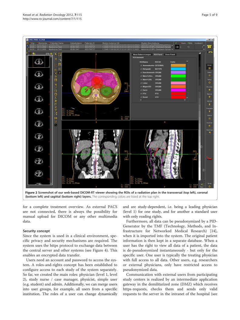
Figure 2 Screenshot of our web-based DICOM-RT viewer showing the ROIs of a radiation plan in the transversal (top left), coronal(bottom left) and sagittal (bottom right) layers. The corresponding colors are listed at the top right.
Kessel et al. Radiation Oncology 2012, 7:115 Page 5 of 9http://www.ro-journal.com/content/7/1/115
for a complete treatment overview. As external PACSare not connected, there is always the possibility formanual upload for DICOM or any other multimediadata.
Security conceptSince the system is used in a clinical environment, spe-cific privacy and security mechanisms are required. Thesystem uses the https protocol to exchange data betweenthe central server and other systems (see Figure 4). Thisenables an encrypted data transfer.Users need an account and password to access the sys-
tem. A roles-and-rights concept has been established toconfigure access to each study of the system separately.So far, we created the main roles: physician (level 1, level2), study nurse / case manager, physicist, simple user(e.g. student) and admin. Additionally, we can merge usersinto user groups, for example, all users from a specificinstitution. The roles of a user can change dynamically
and are study-dependent, i.e. being a leading physician(level 1) for one study, and for another a standard userwith only reading rights.Furthermore, all data can be pseudonymized by a PID-
Generator by the TMF (Technology, Methods, and In-frastructure for Networked Medical Research) [14],when it is imported into the system. The original patientinformation is then kept in a separate database. When auser has the right to view all data of a patient, the datais de-pseudonymized instantaneously - but only for thespecific user. One user is typically the treating physicianwith full access to all data. Other users, e.g. researchersor external physicians, only have restricted access topseudonymized data.Communication with external users from participating
study centers is realized by an intermediate applicationgateway in the demilitarized zone (DMZ) which receiveshttps-requests, checks them and sends only validrequests to the server in the intranet of the hospital (see

Figure 3 Screenshot of our web-based DICOM-RT viewer showing the dose distribution of a radiation plan in the transversal (top left),coronal (bottom left) and sagittal (bottom right) layers. The corresponding colors are listed at the top right.
Figure 4 Connected systems and used protocols.
Kessel et al. Radiation Oncology 2012, 7:115 Page 6 of 9http://www.ro-journal.com/content/7/1/115

Kessel et al. Radiation Oncology 2012, 7:115 Page 7 of 9http://www.ro-journal.com/content/7/1/115
Figure 4). Additionally, client certificates are used toprovide more host-to-host security and to verify author-ized browsers of external project partners.
Study and module designIn principle, each clinical study is designed and adminis-tered separately, consisting of several single modules.However, once a module is designed, it can be reusedagain. To this end, the documentation system has agraphical advanced administration tool, which includes aform generator for designing and adjusting specific mod-ules for the individual clinical studies. It covers both thegeneration of the data structures and the correspondinggraphical user interface. A major advantage is that thistool can be used by the local system administrators anddoes not need new developments by computer experts.Aside from that, a test system has been set up wherenew modules can be created, validated, and tested beforethey are used in the productive environment.Very early, we decided not only to document patients
treated within specific focused study protocols, but alsopatients not participating in clinical studies (non-studypatients), meaning all patients ever treated with particletherapy, because, of course, not every patient fulfills the in-clusion and exclusion criteria of a particular study. Thus, adefault study is available where all patients are documentedinitially, until it is decided that they take part in a clinicalstudy. In that case, a patient can be assigned to the studyaccordingly within the documentation system.This can only be achieved by similar basic documenta-
tion for each type of patient (study or non-study) and ledus to develop three different kinds of modules. Table 2summarizes all existing modules. Basic modules are usedfor all patients and include vital patient information. Themodule for the treatment overview contains informationsuch as diagnosis (ICD-O), TNM classification, tumor
Table 2 Documentation modules divided into basicmodules and modules for non-study and study patients
Basic modules Non-study modules Study modules
Basic patient data Region module Screening
Treatment overview Head / neck Inclusion / exclusioncriteria
Case management Brain / skull base Pre-study treatment
RT images Upper GI Study treatment
RT documentation Lower GI Last examination
Spine Follow-up
Pelvis AE (Adverse Event)
Extremities SAE (SeriousAdverse Event)
Recurrence /metastasis
Death
The study modules are tailored to fit the CLEOPATRA study.
region and planned radiation therapy as well as previousoncological therapies. Radiation data, such as DICOM-RT,CTs and MRIs are stored in the RT images module; thecorresponding precise details (e.g. dose information, num-ber of fields, time and organs at risk) are mapped into theRT documentation module. The case management mod-ule is used for organizational data management, i.e. thehealth insurance, payment status, etc.For non-study patients, depending on the location of
the tumor region, a specific region module must be usedfor documentation during screening, treatment andfollow-up periods. These modules contain informationthat is again similar for all region types, for example kar-nofsky index, acquisition date of imaging, tumor diam-eter and response. However, corresponding symptomsand side-effects are documented individually. The recur-rence / metastasis module documents the location andtreatment method of the recurrence / metastasis; thedeath module the date and cause of death.Furthermore, each clinical study has its own modules
specifically designed to document the parameters thatare required by the study protocol and / or CRF.
UsabilityClinical documentation and analyses are crucial for an op-timal patient care and medical research. However, this isnot a particularly popular task. For this reason, we aim toreplace manual input with automatic documentation wher-ever possible. Several features are implemented to supportthe documentation process and prevent double entries pa-tient information. Certain modules are only selectable ifpredefined conditions are fulfilled. Some modules are onlyselectable once for each patient or depending on a previ-ously created file entry. So-called listeners are implementedto fill data fields automatically, such as the time intervalbetween surgery and the particular day or the age at studyentry. Links to existing file entries within the documenta-tion system help to switch between corresponding infor-mation. A patient-related link accessing the HIS from thedocumentation system makes it easy to search for add-itional patient information that is not part of the documen-tation system. Furthermore, a web-based ICD-O selector isimplemented as an add-on feature for a standard docu-mentation of diagnoses only allowing the search and inser-tion of valid encodings. We added a consultation featurefor physicians to review findings or to obtain a secondopinion. With the lock-option, a module-entry, a group ofentries or even a whole case can be locked and thereafternot changed again by anyone. This is relevant for monitor-ing studies.
AnalysesThis task is still work in progress. Presently, data entriesfrom the database can be exported as an Excel sheet. Thus,

Kessel et al. Radiation Oncology 2012, 7:115 Page 8 of 9http://www.ro-journal.com/content/7/1/115
physicians can produce statistical reports for treatment-related questions within seconds - always up-to-date withthe current status of information. To allow this, a querybuilder has been implemented, which supports the user togenerate individual queries very intuitively. Additionally,these can be saved and reused any time for a continuousoverview on the data. Case managers are using this func-tionality to monitor upcoming patient visits and to ensurecomplete and correct documentation.
DiscussionThe present manuscript describes the generation of acommon European database and documentation systemon particle therapy developed within the ULICE project.The goal is to summarize all biological, physical, and clin-ical data in one European system to generate a broad data-set on particle therapy. Hence, new treatment standardswill be developed and tested on the basis of further clinicalstudies within ULICE. Each study can be designed andadjusted as needed within the system. By documentationof all patients ever treated with particle therapy, we canevaluate single studies prospectively and gain the possibil-ity for overall large-scale, retrospective studies.Different information systems such as the HIS, OIS
and PACS have been connected successfully, allowingdocumentation and evaluation of different analyses.All access to the documentation system takes place via
the web. Thus, no software needs to be installed on clin-ical computers, as a web-browser is standard, and there-fore the system can be immediately accessed everywhereon the Internet. The only prerequisite is an installed JavaRuntime Environment (JRE). The web-based approachwith its strong security measures allows the usage of thesystem for multicenter studies within the ULICE projectand enables the essential patient referral functionality.Moreover, this will play a crucial role for the follow-updocumentation. With the web-based access we can dir-ectly receive outcome reports by the physicians and evenpatient-reported-outcome reports according to theprotocol, assessed in the vicinity of the patient’s home.The usage of the documentation system is very simple
and user-friendly as proven by first feedback from studynurses, case managers and physicians. The interactive docu-mentation modules with their many features prevent wrongdata input and guarantee data validation and verification.In the environment of radiotherapy, it is essential that
any DICOM RT data can be processed and visualized byall systems. Processing and display of RT data today isnot yet a standard functionality of PACS or teleradiologysystems. However, our system is able to exchange andstore all kinds of DICOM RT data. The innovative, inte-grated and web-based DICOM viewer ensures examin-ation of radiation plans from every single computer inthe hospital. This enables physicians to quickly review
images without having to go to a PACS or even a radi-ation therapy planning station.Many others have already said there is no “one-size-
fits-all” solution for web-based documentation of clinicalstudies or patient data per se [15]. The large number ofrequirements and circumstances for the system make anindividual approach necessary. Our solution differs fromother systems, which either only manage and organizepatient treatment within a single department [16,17] orother numerous approaches only focused on electronic-ally documenting a single clinical study [18-21]. Wecombine both. On the one hand, we developed a com-mon platform that allows us to coordinate clinical stud-ies in radiation oncology even across departments, andon the other hand, we linked it all to the mandatory in-formation systems to manage a complete treatmentoverview with more detailed information that might beproven to be relevant in retrospect.
ConclusionThe major benefit of this system lies in the fact that im-aging information, i.e. RT, CT and MRI data is directlylinked to the rest of the study, or rather treatment docu-mentation (see Figure 1) and can be simply and quicklyaccessed with standard web-browsers. This, in turn, sim-plifies the process of conducting multicenter studies dis-tributed all over Europe. It gives us the opportunity toextend the analysis functionality to a more complexlevel. With the main aim to reduce the effort for futureclinical studies, we are planning a separate functionalityfor prospective and retrospective data analyses. It willnot only be able to answer simple statistical questions,but also considering imaging information. MR imagingand dose distribution are to be compared before andafter treatment and thus reveal their direct correlationwith clinical endpoints (e.g. overall survival, disease-freesurvival, recurrence location).In conclusion, the documentation system of today sim-
plifies the research work, ensures a better quality ofstudy analyses, and ultimately improves patient treat-ment concepts and supports the evaluation of the roleand effectiveness of particle therapy.
Competing interestsThere is no conflict of interest to report for this article.
Authors’ contributionsKAK, NB, CB, DO, RB, UE, RO, PF, RP, JD and SEC designed the commonparticle database. KAK, NB, CB and UE developed the system. KAK draftedand wrote the manuscript. DH, DO, JD and SEC were responsible for patienttreatment and care. NB, CB, DH, UE, MD and SEC reviewed/revised themanuscript. All authors read and approved the final manuscript.
AcknowledgementsULICE is co-funded by the European Commission within the FrameworkProgram 7 Capacities Specific Program, under Grant Agreement Number228436. The authors gratefully thank Dr. Sabrina Renfro-Kohl for her helpfulcomments and writing assistance.

Kessel et al. Radiation Oncology 2012, 7:115 Page 9 of 9http://www.ro-journal.com/content/7/1/115
Author details1Department of Radiation Oncology, Heidelberg University Hospital, ImNeuenheimer Feld 400, 69120 Heidelberg, Germany. 2CHILI GmbH,Friedrich-Ebert-Str. 2, 69221 Dossenheim, Germany. 3Department of MedicalInformatics, Heilbronn University, Max-Planck-Str. 39, 74081 Heilbronn,Germany. 4National Centre for Oncological Hadrontherapy, via Caminadella16, 20123 Milano, Italy. 5Medical University of Vienna, Department ofRadiotherapy, 18-20 Währinger Gürtel, 1090 Vienna, Austria. 6CERN, 1211,Geneva 23, Switzerland.
Received: 6 May 2012 Accepted: 24 July 2012Published: 24 July 2012
References1. Combs SE, Jäkel O, Haberer T, Debus J: Particle therapy at the Heidelberg
Ion Therapy Center (HIT) – Integrated research-driven university-hospital-based radiation oncology service in Heidelberg, Germany.Radiother Oncol 2010, 95(1):41–44.
2. Welsh JS: Basics of particle therapy: introduction to hadrons. Am J ClinOncol 2008, 31(5):493–495.
3. Combs SE, Kalbe A, Nikoghosyan A, et al: Carbon ion radiotherapyperformed as re-irradiation using active beam delivery in patients withtumors of the brain, skull base and sacral region. Radiother Oncol 2011,98(1):63–67.
4. Combs SE, Bohl J, Elsasser T, et al: Radiobiological evaluation andcorrelation with the local effect model (LEM) of carbon ion radiationtherapy and temozolomide in glioblastoma cell lines. Int J Radiat Biol2009, 85(2):126–137.
5. Schulz-Ertner D, Tsujii H: Particle Radiation Therapy Using Proton andHeavier Ion Beams. J Clin Oncol 2007, 25(8):953–964.
6. Mendenhall NP, Malyapa RS, Su Z, Yeung D, Mendenhall WM, Li Z:Proton therapy for head and neck cancer: rationale, potentialindications, practical considerations, and current clinical evidence. ActaOncol 2011, 50(6):763–771.
7. Nystroem H, Blomqvist E, Høyer M, et al: Particle therapy - a next logical stepin the improvement of radiotherapy. Acta Oncol 2011, 50(6):741–744.
8. Modahl M: Tablets set to change medical practice. Quantia MD 2011, 1–10.9. Combs SE, Ellerbrock M, Haberer T, et al: Heidelberg Ion Therapy Center
(HIT): initial clinical experience in the first 80 patients. Acta Oncol 2010,49(7):1132–1140.
10. Rieken S, Habermehl D, Nikoghosyan A, et al: Assessment of early toxicityand response in patients treated with proton and carbon ion therapy atThe Heidelberg Ion Therapy Center using the raster scanning technique.Int J Radiat Oncol Biol Phys 2011, 81(5):e793–e801.
11. Ratib O, Swiernik M, Mccoy JM: From PACS to integrated EMR. ComputMed Imaging Graph 2003, 27(2–3):207–215.
12. Engelmann U, Münch H, Schröter A, Bohn C, Meinzer HP: A DICOM basedtelemedicine record. Int J CARS 2008, 3(Suppl.1):158–160.
13. Münch H, Engelmann U, Schröter A, Meinzer HP: The integration ofmedical images with the electronic patient record and their web-baseddistribution. Acad Radiol 2004, 11(6):661–668.
14. Pommerening K, Reng M, Debold P, Semler S: Pseudonymization inmedical research -the generic data protection concept of the TMF. GMSMed Inform Biom Epidemiol 2005, 1(3):Doc17.
15. Reboussin D, Espeland MA: The science of web-based clinical trialmanagement. Clin Trials 2005, 2(1):1–2.
16. Heinemann F, Röhner F, Schmucker M, et al: Departmentandpatientmanagement in radiotherapy. The Freiburg model. StrahlentherOnkol 2009, 185(3):143–154.
17. Palta JR, Frouhar VA, Dempsey JF: Web-based submission, archive, andreview of radiotherapy data for clinical quality assurance: a newparadigm. Int J Radiat Oncol Biol Phys 2003, 57(5):1427–1436.
18. Pozamantir A, Lee H, Chapman J, Prohovnik I: Web-based multi-centerdata management system for clinical neuroscience research. J Med Syst2010, 34(1):25–33.
19. Buchsbaum R, Kaufmann P, Barsdorf AI, et al: Web-based datamanagement for a phase II clinical trial in ALS. Amyotroph Lateral Scler2009, 10(5–6):374–377.
20. Durkalski V, Zhao W, Dillon C, Kim J: A web-based clinical trialmanagement system for a sham-controlled multicenter clinical trial indepression. Clin Trials 2010, 7(2):174–182.
21. Pavlovic I, Miklavcic D: Web-based electronic data collection system tosupport electrochemotherapy clinical trial. IEEE Trans Inf Technol Biomed2007, 11(2):222–230.
doi:10.1186/1748-717X-7-115Cite this article as: Kessel et al.: Connection of European particle therapycenters and generation of a common particle database system withinthe European ULICE-framework. Radiation Oncology 2012 7:115.
Submit your next manuscript to BioMed Centraland take full advantage of:
• Convenient online submission
• Thorough peer review
• No space constraints or color figure charges
• Immediate publication on acceptance
• Inclusion in PubMed, CAS, Scopus and Google Scholar
• Research which is freely available for redistribution
Submit your manuscript at www.biomedcentral.com/submit