connective tissue disorders. terminology collagen vascular diseases auto immune diseases...
TRANSCRIPT

Connective Tissue Disorders

Terminology• Collagen vascular diseases• Auto immune diseases• Rheumatology = study & treatment of diseases of
immune system• Examples:
– SLE – JRA – Dermatomyositis– Vasculitides – Ankylosing spondylitis

Immunological tolerance
• Individual is incapable of developing an immune response against a specific antigen
• Self tolerance = lack of immune responsiveness to one’s own tissue antigens
• Necessary for our tissues to survive and live harmoniously with the body’s own lymphocytes
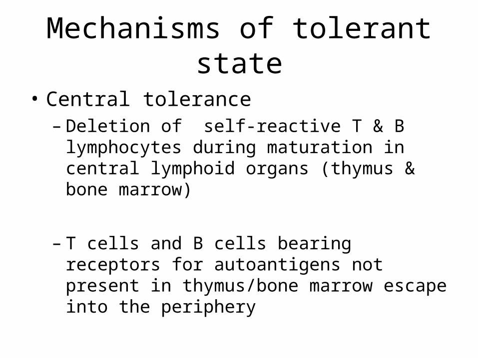
Mechanisms of tolerant state
• Central tolerance– Deletion of self-reactive T & B lymphocytes during
maturation in central lymphoid organs (thymus & bone marrow)
– T cells and B cells bearing receptors for autoantigens not present in thymus/bone marrow escape into the periphery
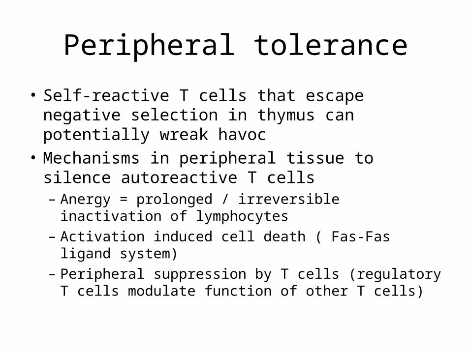
Peripheral tolerance
• Self-reactive T cells that escape negative selection in thymus can potentially wreak havoc
• Mechanisms in peripheral tissue to silence autoreactive T cells– Anergy = prolonged / irreversible inactivation of
lymphocytes– Activation induced cell death ( Fas-Fas ligand system) – Peripheral suppression by T cells (regulatory T cells
modulate function of other T cells)

Mechanisms of autoimmune disease
• FAILURE OF TOLERANCE– failure of activation-induced cell death– breakdown of T cell anergy– bypass of B cell requirement for T cell Help – failure of T cell mediated suppression– molecular mimicry (infectious agents share
epitopes with self antigen)– Polyclonal lymphocyte activation– Release of sequestered antigens (spermatozoa)

Infection in autoimmunity
• Microbes may trigger autoimmunity as follows :– Share cross-reacting epitopes with self–antigens– Microbial antigens & autoantigens may become
associated →immunogenic units →bypass T cell tolerance
– Infections→ tissue necrosis & inflammation → upregulation of co-stimulatory molecules on resting APCs→ breakdown of T cell anergy

Genetic factors
• Predisposition to autoimmune diseases– Familial clustering
– Linkage of autoimmune disease to HLA II antigens
– Induction of autoimmune disease in transgenic rats

SLE
Revised Diagnostic Criteria 1. Malar rash2. Discoid rash3. Photosensitivity4. Oral ulcers5. Arthritis6. Serositis7. Renal disorder

SLE
8. Haematologic disorder9. Neurologic disoder10. Immunologic disoder11. Antinuclear antibody
Diagnose if 4 or more of the 11 criteria are present, serially or simultaneously, during any interval of observation

Model for pathogenesis of SLE
Adapted from Pathologic basis of disease 7th edition
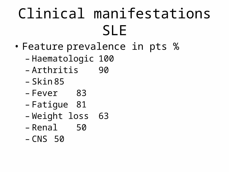
Clinical manifestations SLE
• Feature prevalence in pts %– Haematologic 100– Arthritis 90– Skin 85– Fever 83– Fatigue 81– Weight loss 63– Renal 50– CNS 50

Clinical SLE
• Feature prevalence in pts %– Pleuritis 46– Myalgia 33– Pericarditis 25– GIT 21– Raynaud’s phenomenon 20– Ocular 15– Peripheral neuropathy 14
Adapted from pathologic basis of disease 7th edition

Clinical manifestations SLE

Skin Manifestations of SLE
Increased nailbed capillaries

Malar rash


Lupus Nephritis
• WHO has described 5 classes of renal disease in SLE

Lupus nephritis
• WHO class I lupus nephritis• Minimal changes: <5% of biopsies • Light microscopy is normal • Immunoperoxidase staining will show
extensive C1q (associated with IgG and C3) throughout mesangial areas.

Lupus nephritis
• WHO class II lupus nephritis• Mesangial disease • 10–25% of renal biopsies.• Mesangial expansion but little increase in tuft
cellularity and the peripheral capillary walls are normal.

WHO II severe
• Extensive mesangial IgG deposits demonsstrated with immunoperoxidase staining
• Aggregates just begin to invade peripheral capillary walls.
• This represents the most severe changes in this class.

WHO III
• WHO class III lupus nephritis • Focal proliferative changes are seen on biopsy• Accounts for 20–35% of renal biopsies

WHO IV
• WHO class IV lupus nephritis• Diffuse proliferative changes• Accounts for 35–60% of biopsies• The tuft is increased in size by both a diffuse
increase in matrix and an excess of cells• Capillary walls are irregularly thickened when
stained with hematoxylin/eosin

WHO V
• WHO class V membranous • Accounts for 10–15% of biopsies• The predominant aggregates are along the
outside of the capillary wall• Focal and diffuse proliferative lesions are
absent

Vascular damage in nephritis
• 'Thrombus' may occlude a glomerular capillary loop in more severe disease
• Such a 'thrombus' contains platelets and cross–linked fibrin as well as immunoglobulins and thus has some characteristics of a TRUE thrombus

Management goals
• Early diagnosis• Appropriate therapy• Preserving kidney function• Preventing undue side effects• Thus early biopsy
– Diagnosis– Prognosis– Guide therapy

Treatment recommendations• Class I & II no specific therapy• Class III
– mild → glucocorticoids– Moderate → glucocorticoids + MMF(mycophenolate
mofetil) – Severe as for class IVClass IV induction (6 months) IV CYC or MMF
maintenance: MMF or AZA or quarterly IV CYC
Class V Glucocorticoid+/- (ciclosporin,AZA,MMF,CYC )Class VI NO SPECIFIC THERAPY

Current recommendations
• Immune therapy for Lupus– Anticytokines – TNF + IFN gamma pathway inhibition
– Targeting B cells (CD20 –Rituximab)
– Inhibiting co-stimulation- (anti-CD40L antibodies to prevent 2nd signal for T cell activation)
Better safety profile than traditional immunosuppressive agents

Rituximab
• Genetically engineered• Chimeric murine/human monoclonal antibody• Binds to cell surface marker CD20 which is
mainly expressed on B cells• Used in > half a million patients since 1996• Used successfully in patients with therapy
resistant SLE

Vasculitis treatment
• Current therapy induces remission effectivelyBUT
• Significant & serious side effects• ? Long term maintenance regimens• ? Therapy resistant patients
NOVEL THERAPEUTIC APPROACHES
NEW
• New approaches using standard therapeutic agents
• New immunosuppressive agents• IVIG and Plasma exchange

New look at old drugs
• No evidence yet- still undergoing trials• Steroids ↑dose + cyclophosphamide-
induction• Steroids ↓dose + Azathioprine – maintenance• Oral vs IV cyclophosphamide• PE+ oral CYC+ prednisolone instead of methyl
prednisolone → benefit for 12months• MMF

Mycofenolatemofetil(MMF)
• Potency to induce complete remission• Superior to pulsed IV CYC for induction of
lupus nephritis• Fewer side effects• Alternative choice for maintenance therapy of
severe LN

New immunomodulatory strategies& biologic agents
• Blockade of circulating mediators & surface receptors
• Lymphocyte depletion &immunoablation– TNF-alpha blockade using infliximab & etanercept→ improved control of refractory vasculitis→allowed lower steroid dosing in induction therapy→benefit as induction therapy in refractory WG

Immunoablation & autologus stem cell transplant
• Life saving• Costly• Benefit in severe auto immune disease

Conclusion
• Early diagnosis• Early biopsy in renal disease• May still utilize tried & tested agents initially
BUT in regimens that minimise adverse effects• Newer immunosuppressive &
immunomodulatory strategies are becoming more popular

References• Kumar .Abbas. Fausto. Robbins and Cotran Pathologic basis of disease 7th
edition• Richard johnson. John Feehally. CLINICAL NEPHROLOGY 2nd edition• Michael J. Dillon Vasculitis treatment-new therapeutic approaches.
Review. Eur J Peditr (2006)• Sandra Navarra. Immune therapy of lupus: what is on the horizon?
Nephrol Dial Transplant (2006)• Annette Friederike Jansson. Uwe Wintergerst et al.Rituximab-induced
long-term remission in two children with SLE. Short report . Eur J Pediatr (2007)
• Geoffrey R. Bihl, Micchelle Petri & Derick M. Fine. Kidney biopsy in lupus nephritis : look before you leap. Nephrol Dial Transplant (2006)