connective tissues by prof. dr. ashraf mahmoud. characters of c.t: mesodermal. blood vessels &...
TRANSCRIPT

Characters of C.T:Mesodermal.
Blood vessels & nerve can penetrate it. Cells are widely separated. Formed of: Cells, fibres and soft ground
substance.
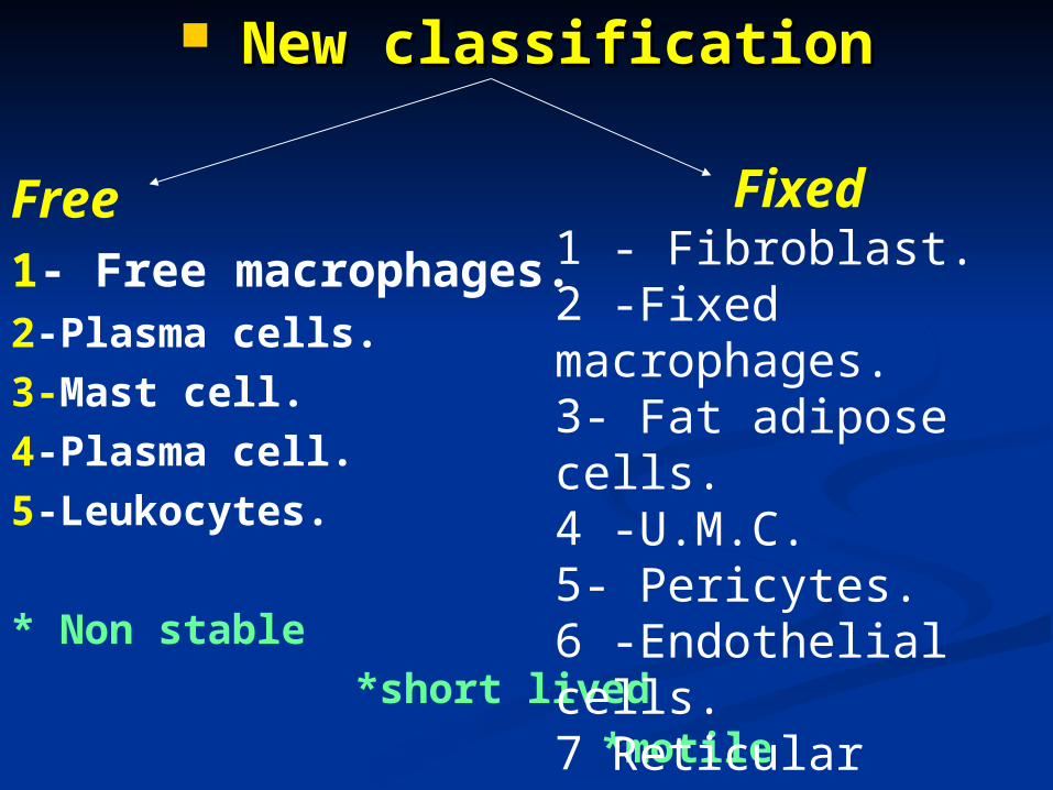
New classificationNew classification
Free1- Free macrophages.2-Plasma cells.3-Mast cell.4-Plasma cell.5-Leukocytes.
* Non stable *short lived *motile
Fixed1 - Fibroblast.2 -Fixed macrophages.3- Fat adipose cells.4 -U.M.C.5- Pericytes.6 -Endothelial cells.7 Reticular cells.*stable *long lived *non motile
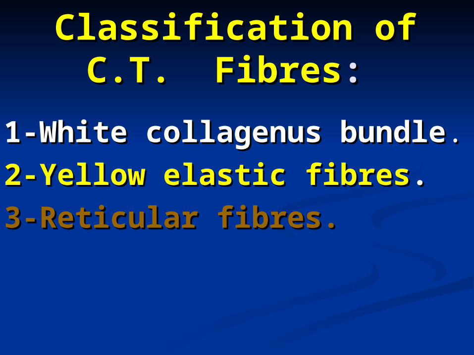
Classification of C.T. Classification of C.T. FibresFibres: :
1-White collagenus bundle1-White collagenus bundle..
2-Yellow elastic fibres2-Yellow elastic fibres..
3-Reticular fibres.3-Reticular fibres.

((cc) Different types of C.T. ) Different types of C.T. properproper::
1-1-Loose areolar C.T.Loose areolar C.T.
2-2-White fibrous C.T.White fibrous C.T.
3-3-Elastic C.T.Elastic C.T.
4-4-Reticular C.T.Reticular C.T.
5-5-Mucoid C.T.Mucoid C.T.6-Fatty adipose C.T..
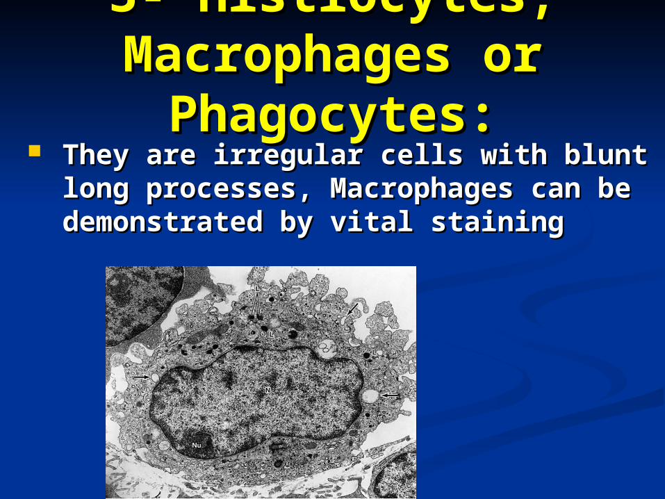
3- Histiocytes, 3- Histiocytes, Macrophages or Macrophages or
Phagocytes:Phagocytes: They are irregular cells with blunt They are irregular cells with blunt
long processes, Macrophages can be long processes, Macrophages can be demonstrated by vital staining demonstrated by vital staining

Function:Function: They engulf dead neutrophilic They engulf dead neutrophilic
leucocytes and bacteria by a leucocytes and bacteria by a phagocytes surrounding the phagocytes surrounding the object forming multinucleated object forming multinucleated foreign body giant cell.foreign body giant cell.
Marcophages increase in number Marcophages increase in number in chronic inflammation by:in chronic inflammation by:
1- Multiplication of already present 1- Multiplication of already present macrophages.macrophages.
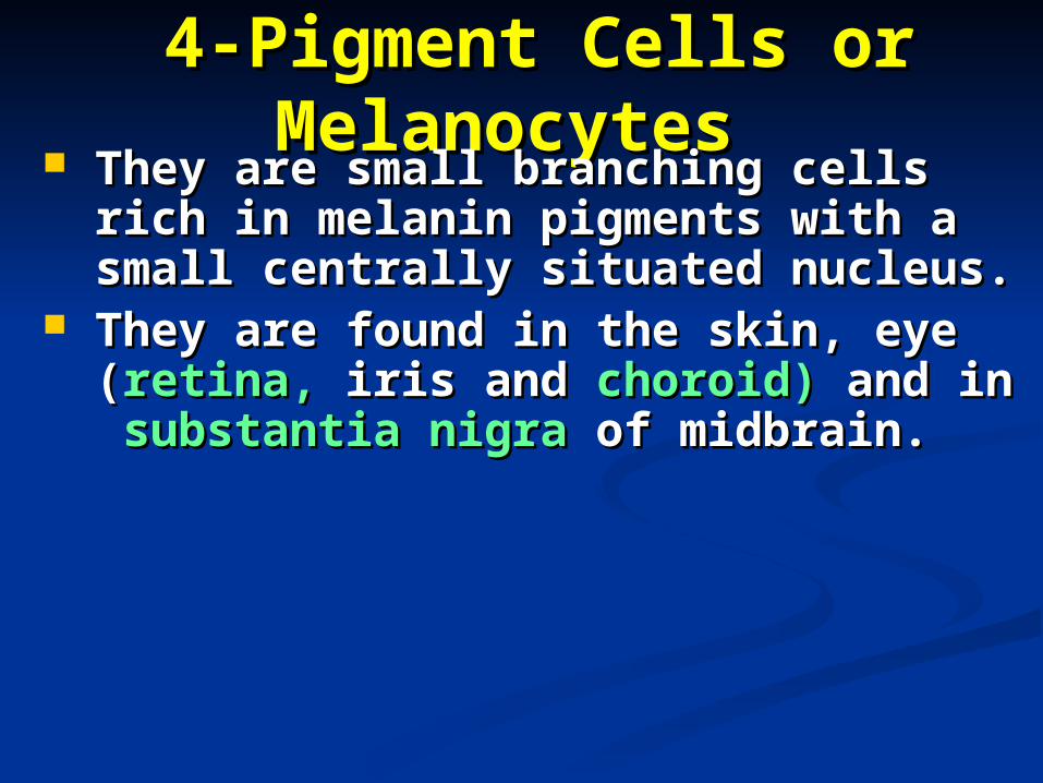
4-Pigment Cells or 4-Pigment Cells or MelanocytesMelanocytes
They are small branching cells They are small branching cells rich in melanin pigments with a rich in melanin pigments with a small centrally situated small centrally situated nucleus.nucleus.
They are found in the skin, eye They are found in the skin, eye ((retina,retina, iris and iris and choroid)choroid) and in and in substantia nigrasubstantia nigra of midbrain. of midbrain.

Function:Function: They carry melanin pigment They carry melanin pigment
which protect the body from which protect the body from the injurious effect of the sun.the injurious effect of the sun.

[A] Undifferentiated [A] Undifferentiated cellscells
1) U.M.C.1) U.M.C. 2) Pericytes2) Pericytes 3) Reticular 3) Reticular cellscells
SiteSite C.T. of embryo.C.T. of embryo. Around the Around the capillariescapillaries
Stroma of different Stroma of different organorgan
ShapShapee
Small branched Small branched cell.cell.
- N:- N: large, pale large, pale & oval.& oval.
- C:- C: pale pale basophilicbasophilic
Branched cell withBranched cell with
- N:- N: large, pale & large, pale & ovaloval
- C:- C: pale with few pale with few organellesorganelles
Branched cells; Branched cells; together with together with reticular fibres form reticular fibres form reticulum.reticulum.
functfunctionion
1)1) Can Can differentiate into differentiate into other C.T. cells.other C.T. cells.
2)2) In bone In bone marrow form all marrow form all blood cells.blood cells.
1)1) Same function Same function of U.M.C. but of U.M.C. but after birth.after birth.
2)2) 2)2) Their Their contraction → contraction → vasoconstrictiovasoconstriction n
1) 1) Support organs Support organs & glands.& glands.
2) 2) On need has On need has phagocytic functionphagocytic function
3)3) In bone marrow In bone marrow give blood cells. give blood cells.

B- Rounded C.T. B- Rounded C.T. CellsCells
1-Plasma cells1-Plasma cells They are irregularly ovoid in They are irregularly ovoid in
shape, small and centrically shape, small and centrically nucleus. nucleus.
The nuclear chromatin is The nuclear chromatin is concentrated towards the nuclear concentrated towards the nuclear membrane in a regular manner membrane in a regular manner giving it a “giving it a “cartwheelcartwheel” ” appearance. appearance.
The cytoplasm is basophilic with a The cytoplasm is basophilic with a pale area representing Golgi pale area representing Golgi apparatus “apparatus “Negative Golgi image”. Negative Golgi image”.
The cytoplasm of the plasma cells The cytoplasm of the plasma cells contains acidophilic inclusions contains acidophilic inclusions known as known as RUSSEL BODIES.RUSSEL BODIES.


Functions:Functions:Plasma cells are originated from Plasma cells are originated from B-lymphocytes:B-lymphocytes:B-lymphocytes B-lymphocytes antigen_antigen_ plasmablasts plasmablasts plasma cells. plasma cells.Plasma cells produce the Plasma cells produce the circulating antibodies and they are circulating antibodies and they are responsible for immune response.responsible for immune response.
2-Mast Cells2-Mast Cells They are large ovoid cells with ovoid They are large ovoid cells with ovoid
nuclei.nuclei. Cytoplasm is full of basophilic granules. Cytoplasm is full of basophilic granules. These granules are water soluble and These granules are water soluble and
are are
metachromatically stained by metachromatically stained by toluidine toluidine blueblue, so they are stained , so they are stained purple purple
Mast cells are present in loose C.T. in Mast cells are present in loose C.T. in relation to blood vessels.relation to blood vessels.

Functions:Functions: -Secrete heparin (anticoagulant), -Secrete heparin (anticoagulant), -Secrete histamine, which participates -Secrete histamine, which participates
in antigen antibody reaction.in antigen antibody reaction. -May have a role in serotonin -May have a role in serotonin
secretion which, is a vasoconstrictor secretion which, is a vasoconstrictor substance.substance.

3- Fat Cells3- Fat Cells They are rounded or oval cells with They are rounded or oval cells with
flattened peripheral nuclei and thin flattened peripheral nuclei and thin rim of cytoplasm, containing large fat rim of cytoplasm, containing large fat droplets. droplets.
In H&E sections, the fat dissolved In H&E sections, the fat dissolved leaving an empty space and giving leaving an empty space and giving the appearance of a “signet”. the appearance of a “signet”.
The fat cells can be stained The fat cells can be stained orangeorange with Sudan III andwith Sudan III and black black with Sudan with Sudan black.black.

Function:Function:They support vital organs as the They support vital organs as the kidney.kidney.They store fat, which is the main They store fat, which is the main source of energy.source of energy.They decrease loss of heat from They decrease loss of heat from the skin.the skin.

MacrophagesMacrophages Plasma cellsPlasma cells Mast cellsMast cells LeucocyteLeucocyte
OriginOrigin MonocytesMonocytes Β- Β- lymphocytes lymphocytes
U.M.CU.M.C Migrated Migrated from bloodfrom blood
SiteSite Loose C.T.Loose C.T. Lymphatic Lymphatic tissuetissue
Around blood Around blood vessel vessel
ShapeShape * Large branched * Large branched cells with cells with pseudopodiapseudopodia
- N:- N: small,dark & small,dark & kidney shape.kidney shape.
- C:- C: basophilic, basophilic, non-clear (rich in non-clear (rich in lysosomes & lysosomes & phagocytic phagocytic vesicles). vesicles).
* Small oval cell * Small oval cell withwith
-- N: N: eccentric eccentric round with cart round with cart wheel or clock wheel or clock fore fore appearance. appearance.
- C:- C: deeply deeply basophilic rich basophilic rich in: in: mit.,r-ER &mit.,r-ER &
* Oval cell * Oval cell with central with central round round nucleus.nucleus.
-C:-C: basophilicbasophilic
& have & have granule granule which are which are metachromatmetachromaticlly iclly
stained stained Toluidine Toluidine blue.blue.
. .
NeutrophilsNeutrophils appear in appear in acute acute infection.infection.
Eosinophils Eosinophils ↑↑in allergic ↑↑in allergic & parasitic & parasitic diseasedisease
lymphocytes lymphocytes & monocytes& monocytes
↑↑ ↑↑ in in chronic chronic infection. infection.
FunctionFunction Phagocytose any Phagocytose any foreign Clean foreign Clean wounds from wounds from Antigen Antigen presenting presenting cells. cells.
Secrete Secrete Antibodies.Antibodies.
Secrete Secrete heparin heparin anticoaganticoagulan.ulan.
Secrete Secrete histaminhistaminee
allergyallergy

FibersFibersConnective TissuesConnective Tissues

Classification of C.T. Classification of C.T. fibresfibres: :
1-White collagen us bundle1-White collagen us bundle..
2-Yellow elastic fibres2-Yellow elastic fibres..
3-Reticular fibres.3-Reticular fibres.
A-White or Collagen A-White or Collagen FibersFibers AboutAbout 1-20 µm formed of thick 1-20 µm formed of thick
backed fibres forming groups of backed fibres forming groups of bundles closely arranged together.bundles closely arranged together.
Collagenous bundlesCollagenous bundles are elastic - are elastic -long- cylindrical appearance.long- cylindrical appearance.
Mainly locatedMainly located inin Tendon.Tendon.Joint capsule.Joint capsule.Deep fascia.Deep fascia.


B-Yellow or Elastic B-Yellow or Elastic FibresFibres
--They are thinner than collagen They are thinner than collagen fibres, refractile.can branch, fibres, refractile.can branch, anastmose and are highly elastic.anastmose and are highly elastic.
-They occur as fenestrated -They occur as fenestrated lamellae in arteries, and they can lamellae in arteries, and they can be stainedbe stained brown brown withwith orcein orcein, , blue with Mallory and blue with Mallory and yellowyellow with with van Geisonvan Geison..

C-Reticular FibresC-Reticular Fibres--They are fine branching fibres stain They are fine branching fibres stain
brownish black with brownish black with silversilver. . - Function:Function:
- They participate in the They participate in the stromastroma formation of formation of
- many organs as the many organs as the kidneykidney, , liver liver and and lung.lung.

Comparison between Comparison between the different types of the different types of
C.T. fibersC.T. fibers

CollagenoCollagenous fibersus fibers
Elastic Elastic fibersfibers
Reticular Reticular fibersfibers
DiametDiameter er
Fiber 1-20 umFiber 1-20 um
fibril 75nm fibril 75nm Fibrils14 nm Fibrils14 nm 0.5-2um 0.5-2um
Shape Shape Closely packed Closely packed Irregular Irregular networks networks
Form extensive Form extensive networks networks
Stains Stains 1-Eosin= Pink1-Eosin= Pink
2-allory=Blue 2-allory=Blue 1-Orcein=Brown 1-Orcein=Brown 1-Silver= dark1-Silver= dark
brownbrown
ChemicChemicallall
Collagen type I Collagen type I Collagen type I Collagen type I Collagen type III Collagen type III
Boiling Boiling Form soft Form soft Gelatin Gelatin
Form soft Gelatin Form soft Gelatin
DigestiDigestion on
Form soft Form soft Gelatin Gelatin
Digested by Digested by elastaseelastase
Main Main sites sites
Tendon and Tendon and ligament ligament
Blood vessels,lungBlood vessels,lung Body organs Body organs
ContainContains s
Glycine,ProlineGlycine,Proline Desmosine and Desmosine and isodesmosine isodesmosine

II- C.T. ProperII- C.T. Proper

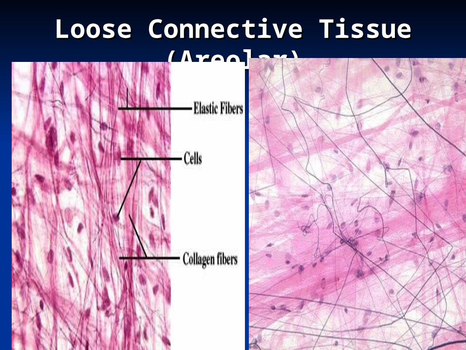
1-Loose Areolar 1-Loose Areolar C.T. C.T.
--It contains all the elements of C.T.It contains all the elements of C.T.
-Cells mainly fibroblasts and -Cells mainly fibroblasts and histiocytes.histiocytes.
Fibres mainly collagen and ground Fibres mainly collagen and ground substance.substance.
found in :found in : -Dermis of skin.-Dermis of skin.
-Adventitia of blood vessels. -Adventitia of blood vessels.
-Under epithelial membranes.-Under epithelial membranes.
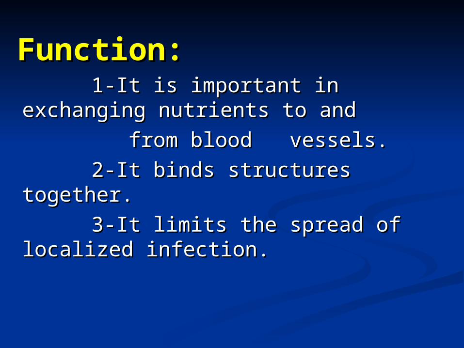
Function:Function: 1-It is important in exchanging 1-It is important in exchanging
nutrients to and nutrients to and
from blood vessels.from blood vessels.
2-It binds structures together.2-It binds structures together.
3-It limits the spread of localized 3-It limits the spread of localized infection.infection.
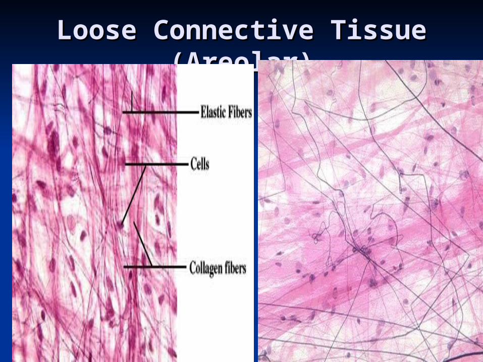
Loose Connective Tissue Loose Connective Tissue (Areolar)(Areolar)

Mucoid C.TMucoid C.T (Wharton’s (Wharton’s Jelly):Jelly):
PresentPresent mainly in the embryo.mainly in the embryo.
Formed ofFormed of::
Mucoid cells = U.M.C. or fibroblast Mucoid cells = U.M.C. or fibroblast joined by their processor.joined by their processor.
MatrixMatrix:: jelly like rich in mucin thin jelly like rich in mucin thin collagenfibres.collagenfibres.
SitesSites:: In embryo: umbilical cord. In embryo: umbilical cord.
In adult: In adult: Vitreous humour of Vitreous humour of eye.eye.
Pulp of growing teeth.Pulp of growing teeth.

Dense collagen Dense collagen C.T.:C.T.:- Either - Either regularregular or or irregular.irregular.
I-The regular dense collagenI-The regular dense collagen C.TC.T
is characterized by close packing of fibres is is characterized by close packing of fibres is found in found in tendons,tendons, aponeuroses, aponeuroses, ligaments ligaments and and cornea of the eye. cornea of the eye.
II-The irregular dense collagen C.TII-The irregular dense collagen C.T is found in is found in dermis dermis, , capsules capsules of some of some
organs, organs, perichondriumperichondrium and and sclera of the sclera of the eye.eye.
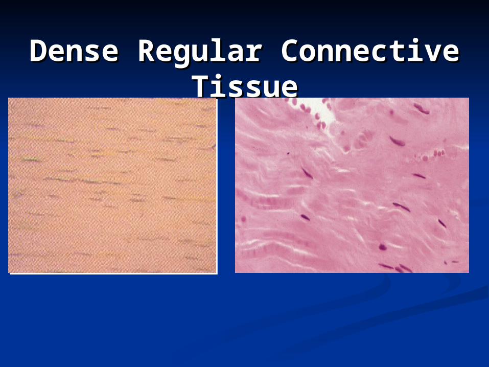
Dense Regular Dense Regular Connective TissueConnective Tissue


Yellow elastic C.T:Yellow elastic C.T:Formed Formed of condensed elastic fibres of condensed elastic fibres
separated by fibroblast.separated by fibroblast.
Function:Function: stretchable. stretchable.
Sites:Sites: Aorta & large arteries. Aorta & large arteries.
Ranched tree.Ranched tree.
Vocal cords.Vocal cords.
Ligamentum nuchae (neck).Ligamentum nuchae (neck).
Ligamentum flavum (between Ligamentum flavum (between vertebrae).vertebrae).
Loose Connective Tissue Loose Connective Tissue (Areolar)(Areolar)

Reticular C.T.Reticular C.T.-It is formed of reticular cells and -It is formed of reticular cells and
fibres. The cells are stellate in fibres. The cells are stellate in shape with processes. shape with processes.
-This tissue stains brownish black -This tissue stains brownish black with silverwith silver
--The main sitesThe main sites
-Lymphoid tissues-Lymphoid tissues
-Bone marrow-Bone marrow
-Stroma of body organs-Stroma of body organs

Adipose C.T.Adipose C.T. -It is formed of lobes and lobules of fat cells -It is formed of lobes and lobules of fat cells
separated by loose C.T. separated by loose C.T. -It is richly supplied with blood vessels. -It is richly supplied with blood vessels. -According to the vascularity and function, it is -According to the vascularity and function, it is
divided into white and brown adipose C.T. divided into white and brown adipose C.T.
The white type (Unilocular)The white type (Unilocular)isis distributed in :distributed in :
* * Adults subcutaneousAdults subcutaneous tissue and tissue and yellow bone yellow bone marrowmarrow..
--The brown type (Multilocular)The brown type (Multilocular) is is present :present :
* * Foetus Foetus and and newborn infantsnewborn infants..

Adipose (Fat)Adipose (Fat)

Adipose (Fat)Adipose (Fat)