conseuro seville 2009 - welcome to the e.f.c.d · conseuro seville 2009 1 conseuro seville 2009...
TRANSCRIPT
Conseuro Seville 2009
1
Conseuro Seville 2009
List of authors and corresponding n°'s Abalos, C.: OP 089.
About, I.: MAT 184.
Abreu Rodriguez, R.: MAT 164.
Abuin, M.: STUD 174*, STUD 175.
Ahmed, S.S.: OP 029.
Ahmetaj, A.: END 136, END 137.
Akincioglu, A.: MAT 009.
al Omari, Q.: OP 012*.
Alba, A.: STUD 087.
Alegria, G.: OP 069, OP 070.
Ali, A.: CLIN 045.
Almeida, P.J.: CLIN 139.
Alonso, J.: STUD 132.
Alpaslan, T.P.: OP 157.
Alpiste Illueca, F.: OP 122.
Amaral, J.: OP 071.
Amengual, J.: CLIN 015, OP 017, OP 019.
Amorim, A.: OP 185.
Antoniadou, M.: STUD 027.
Arias Moliz, M.T.: END 178*.
Arias Paniagua, A.M.: END 173.
Arocha, M.: CLIN 045.
Asensio, R.: CLIN 045.
Attin, T.P.: OP 105.
Austin, R.: END 180.
Aytac, F.: OP 001, OP 004, OP 025.
Azabal, M.: END 173.
Baca, P.: END 178.
Bachanek, T.: CLIN 028.
Bae, K.S.: MAT 031.
Baguena Gomez, J.C.: MAT 148, MAT 149, MAT 154,
MAT 155, MAT 162, MAT 163, PREV 062, STUD 113,
STUD 166.
Bahillo, J.: END 156.
Bailon Sanchez, E.: END 176*, END 177.
Baldissara, P.: MAT 134.
Banerjee, A.: CLIN 049, CLIN 050, CLIN 051.
Baracco, B.: MAT 057*.
Barciala Castro, N.: OP 058.
Barneji, S.: OP 094.
Bascones, J.: MAT 172.
Basilio, J.: OP 086.
Basso, M.: CLIN 049, CLIN 050, CLIN 051.
Bayer, T.: OP 064.
Beccio, R.: STUD 118.
Beckedorf, A.: MAT 079.
Bedini, R.: END 096, MAT 095.
Behrend, D.: END 187.
Benavides, R.: CLIN 045.
Benbachir, N.: OP 129.
Bentolila, O.: OP 129*.
Bergandi, L.: STUD 118.
Bernard, C.: MAT 147.
Bernardino, P.: END 107.
Berutti, E.: OP 116, PREV 097, PREV 098,
STUD 117, STUD 118.
Betoret, M.: STUD 132.
Blanco Mendez, J.: END 171.
Blanquer, M.: END 120.
Bleda, P.: END 120.
Blique, M.: CLIN 049, CLIN 050, CLIN 051.
Bobba, V.: STUD 117.
Bolaños Carmona, M.V.: END 159, END 161*,
Madureira, R.: END 077, END 107, END 115,
OP 106.
Magan, A.: END 037.
Magni, E.: MAT 081.
Mancebo, J.C.: END 073.
Mancini, M.: OP 021, OP 022.
Mangani, F.: OP 021, OP 022.
Manhart, M.J.: END 008.
Manzanares Cespedes, M.C.: MAT 074.
Marcantoni, C.: END 096.
Marigo, L.: END 061*, MAT 088.
Marin Altuve, E.: END 159, END 161.
Marques, D.: OP 071, OP 072.
Marques, J.: OP 071, OP 072.
Martin Biedma, B.: END 043, END 054,
END 059, END 067, END 156, END 171,
MAT 055, OP 058, STUD 060, STUD 083,
STUD 174,
Martinez Rodriguez, A.M.: PREV 062.
Martinez Rus, F.: MAT 033, MAT 041.
Martinez, S.: END 120.
Masian, P.: OP 069, OP 070.
Masini, M.: OP 021, OP 022*.
Mata, A.: OP 071, OP 072.
Mate Sanchez, J.E.: MAT 148, MAT 149,
MAT 154, MAT 155, MAT 162, MAT 163,
MAT 165, STUD 145, STUD 166, STUD 168.
Mateos Palacios, R.: OP 100*.
Mausberg, R.F.: OP 013.
Mayoral, J.: OP 086, OP 129.
Manzanares Cespedes, M.C.: MAT 075.
Mazzitelli, C.: MAT 119, MAT 135*, MAT 138.
McConnell, R.: CLIN 186.
Melo, M.: OP 122, OP 169.
Melo, P.: OP 185, PREV 046.
Mercade, M.: CLIN 045, OP 102.
Meseguer, L.: END 120.
Mestre, A.: MAT 074.
Mico Muñoz, P.: CLIN 112.
Migliaretti, G.: OP 116, STUD 117.
Miletic, I.: CLIN 049, CLIN 050, CLIN 051.
Millar, B.: MAT 092, MAT 093, OP 094.
Millen, C.S.: MAT 048*.
Mitropoulus, P.: OP 036*.
Molina, A.: STUD 132.
Monaco, C.: MAT 134.
Monticelli, F.: MAT 119, MAT 135.
Montouris, G.: OP 140.
Moraleda, J.M.: END 120.
Morales Canovas, M.A.: PREV 062.
Moreno, J.A.: STUD 142.
Mrasori, S.H.: END 136, END 137.
Mujdeci, A.: OP 002, OP 026.
Navajas, J.M.: OP 100.
Nicolas Silvente, A.I.: MAT 148, MAT 149,
MAT 154, MAT 155, MAT 162, MAT 163,
MAT 165, PREV 062, STUD 142, STUD 166.
Nocca, G.: MAT 088.
Novoa, X.R.: END 043.
Oh, T.S.: END 032, MAT 031.
Olivares Rueda, A.: STUD 167.
Oñate, R.: END 120.
Orlowski, M.: CLIN 028.

Conseuro Seville 2009
2
END 170, MAT 160.
Bollino, D.: END 061.
Bonet, J.: STUD 103.
Bonilla, V.: END 038, END 042.
Borracchini, A.: MAT 138.
Bote, A.: OP 169.
Briones Lujan, M.: END 170.
Buchalla, W.: OP 105.
Blunck, U.: OP 110.
Busch, M.: OP 030.
Cabezos Garcia, J.C.: STUD 158.
Cabrera, E.: MAT 078.
Cabrerizo Vilchez, M.A.: MAT 151.
Cabrita, J.: OP 071.
Caeiro, J.R.: PREV 016.
Calvo Guirado, J.L.: MAT 148, MAT 149, MAT 154,
MAT 155, MAT 162.
Can Karabulut, D.C.: MAT 009*, PREV 010.
Candel, E.: STUD 103.
Cano Gonzalvez, M.: END 141.
Cano, M.: STUD 087.
Cañadas, D.: END 073.
Cardinali, S.: END 061.
Cardoso, J.A.: CLIN 139*.
Carollo, M.: PREV 097*.
Carvalho Lobato, P.: MAT 074, MAT 075.
Cascales Moya, F.: END 101.
Castelo, P.: END 067, STUD 083.
Castro, L.: OP 106.
Casucci, A.: MAT 135, MAT 138.
Ceballos, L.: MAT 057, MAT 063, MAT 143,
MAT 152, STUD 076.
Celik, C.: OP 144.
Cengiz, E.: PREV 010.
Cernadas Lago, P.: END 054*.
Cerutti, A.: OP 021, OP 022.
Cezon Muñoz, L.: STUD 076.
Chalas, R.: CLIN 028.
Chang, S.W.: END 032, MAT 031.
Chiva Garcia, F.: END 101, MAT 148, MAT 149,
MAT 154, MAT 155, MAT 162, MAT 163, MAT 165,
PREV 062, STUD 142, STUD 158.
Comlekoglu, M.E.: PREV 121*.
Costa, M.: END 077*.
Cuenca Abela, A.M.: END 141.
Dahan, L.: MAT 130*.
Dalmazzo, A.: STUD 118*.
de la Macorra, J.C.: END 173, MAT 078, MAT 172.
de Palma, F.: MAT 088.
Deb, S.: MAT 092, MAT 093.
Degrange, M.: MAT 130, MAT 147.
del Rio, F.: MAT 033.
Delgado Ruiz, R.A.: END 101, END 141, MAT 148,
MAT 149, MAT 154, MAT 155, MAT 162, MAT 163,
MAT 165, STUD 158, STUD 167,
Derbanne, M.A.: MAT 147.
Dermici, M.: OP 133.
Diaz Mancha, A.: MAT 055.
Dimitrouli, M.: END 126.
Domejean Orliaguet, S.: CLIN 049,
CLIN 050, CLIN 051, OP 007.
Dorter, C.: OP 104.
Dundar, M.: MAT 124, OP 123, PREV 121.
Eden, E.: OP 029.
Efes Guray, B.: OP 052.
el Daraa, E.: OP 012.
Osorio, E.: MAT 138.
Osorio, R.: MAT 135, MAT 138.
Oteo Calatayud, C.: OP 068,
OP 069, OP 070, OP 080.
Oteo Calatayud, J.: OP 068,
OP 069, OP 070, OP 080.
Oteo Calatayud, M.D.: OP 068.
Oteo Zaccagnini, A.: OP 068.
Oteo, A.: OP 080.
Ozcan, M.: MAT 124, OP 123, PREV 121.
Ozgiour, F.: STUD 027.
Ozsoy, A.: OP 056.
Ozyegin, S.: MAT 009.
Pahncke, D.: END 187.
Palaniappan, S.: MAT 150*.
Pale, M.: CLIN 045.
Palomares, M.: END 018, OP 014.
Pameijer, C.H.: MAT 095.
Pamir, T.: OP 029.
Papacchini, F.: MAT 138.
Papacchini, P.: MAT 119.
Papazoglou, A.: OP 140.
Papazoglou, E.: OP 111.
Park, D.S.: END 032, MAT 031.
Park, J.W.: OP 082.
Park, S.: OP 039.
Parra Candel, S.: STUD 113.
Parres, R.: END 101.
Pascual Moscardo, A.: MAT 164,
OP 122, OP 169.
Pasqualini, D.: OP 116, PREV 097,
PREV 098, STUD 117, STUD 118.
Paximada, H.: OP 047.
Pchalek, G.: OP 030.
Pecci, M.: STUD 142.
Pecci, R.: END 096.
Pedrosa Torres, L.: STUD 060.
Pelaez, J.: CLIN 109, MAT 125.
Peñarrocha, D.: STUD 132*.
Peñate, L.: OP 102.
Pera, F.: OP 116, PREV 098.
Perez Albacete, C.: STUD 142.
Perez Estevez, A.J.: END 171.
Perez Gomez, B.: STUD 168.
Perez Gomez, M.M.: MAT 131.
Perez Sanchez, C.: PREV 062.
Perez, A.: OP 169.
Perez, J.C.: STUD 174, STUD 175.
Perez, M.C.: END 043.
Peschke, A.: END 066.
Peumans, M.: MAT 150.
Pinho, A.: CLIN 139.
Plotino, G.: END 096, MAT 095.
Polydorou, O.: OP 064.
Poplawska, A.: PREV 153.
Porlan Costa, M.: STUD 179.
Pradies, G.: MAT 041.
Prados, C.: END 037.
Puentes, M.: OP 169.
Pulgar Encinas, R.: MAT 131,
OP 100, STUD 108.
Putignano, A.: OP 021, OP 022.
Rahiotis, C.: OP 036.
Rahmer, H.: MAT 079.
Redondo, J.: END 101.
Reich, E.: CLIN 049, CLIN 050,

Conseuro Seville 2009
3
Enriques, H.: OP 185.
Erdermir, U.: OP 053, OP 056.
Erdilek, D.: OP 035, OP 052.
Eren, M.: OP 056.
Ersoz, E.: OP 001, OP 004, OP 005, OP 025.
Fahd, J.C.: CLIN 186.
Farge, P.: MAT 183.
Faus Llacer, V.J.: CLIN 023, CLIN 085,
CLIN 112, STUD 044.
Faus Matoses, I.: CLIN 023*.
Faus Matoses, V.J.: CLIN 023, CLIN 085, STUD 044.
Faus, M.: OP 169, STUD 103*, STUD 103.
Fayos Soler, T.: CLIN 112.
Federlin, M.: END 090, OP 114.
Fenoy, P.: OP 181.
Fernandez, A.: CLIN 040.
Ferrari, M.: MAT 081, MAT 119, MAT 135, MAT 138.
Ferrer Luque, C.M.: END 176, END 177, END 178.
Fessi, H.: MAT 184.
Flores Dorado, F.: STUD 076.
Flores, H.: CLIN 045.
Forner, L.: CLIN 015, END 018, END 077,
END 107, END 115, OP 014, OP 017,
OP 019, OP 106, PREV 016*, PREV 020,
Franquin, J.C.: OP 065.
Fuentes, M.V.: MAT 057, MAT 063, MAT 152.
Fygueroa Garcia, A.: OP 122.
Galvan Salazar, M.M.: END 141, STUD 168.
Garcia Barbero, A.E.: MAT 182.
Garcia Barbero, E.: END 037, MAT 182.
Garcia Barbero, J.: MAT 182.
Garcia Caballero, T.: END 156.
Garcia Cruz, E.: MAT 162, STUD 113.
Garcia Peñalver, V.: STUD 113, STUD 166.
Garcia Rielo, M.M.: END 043.
Garcia, J.A.: STUD 087.
Garrido, M.A.: MAT 143, MAT 152.
Gaucher, C.: CLIN 049, CLIN 050, CLIN 051.
Gea Ros, C.M.: STUD 158.
Geobaldo, F.: OP 116.
Geurtsen, W.: END 126, MAT 079, OP 127, OP 128.
Ghigo, D.: STUD 118.
Gil Loscos, F.: OP 122.
Gil Montoya, J.A.: OP 100.
Gimenez, A.: CLIN 015.
Giraldez, I.: MAT 143*, STUD 076.
Gokay, O.: OP 002, OP 026.
Gokce, B.: OP 123.
Goldberg, F.: OP 058.
Gomar Vercher, S.: STUD 044, CLIN 085*.
Gomec, Y.: OP 035, OP 052, OP 104.
Gomez Cogolludo, P.: MAT 125.
Gomez de Malla, M.A.: PREV 062.
Gomez, P.: CLIN 109.
Gomez, Z.: END 067, STUD 083.
Gonulol, N.: OP 005, OP 025.
Gonzalez Bahillo, J.: END 043, END 054,
END 059, END 156, MAT 055, OP 058,
STUD 060, STUD 174, STUD 175.
Gonzalez Castillo, S.: END 176, END 177*.
Gonzalez Gonzalez, H.: STUD 179.
Gonzalez Gonzalez, T.: END 059.
Gonzalez Lopez, S.: END 159*, END 161,
END 170, MAT 057, MAT 063, MAT 160.
Gonzalez Moreira, H.: MAT 151.
Gonzalez Rodriguez, M.P.: END 176,
CLIN 051.
Rinke, S.: OP 013.
Robinson, B.: END 180.
Rodriguez Espin, A.B.: MAT 148,
MAT 149, MAT 155, MAT 162,
MAT 163, MAT 165, STUD 145,
STUD 167, STUD 179.
Rodriguez Navarro, A.: MAT 160.
Rodriguez Priego, E.: END 170*.
Rodriguez, A.E.: MAT 154.
Rodriguez, F.J.: END 120*.
Rodriguez, J.: MAT 143, MAT 152.
Roh, B.: OP 084.
Roig Ferrando, D.: STUD 158.
Roig, M.: CLIN 045, OP 086,
OP 102, OP 129, OP 181, STUD 099.
Roldan, J.: CLIN 045*.
Roman Richon, S.: CLIN 085,
STUD 044.
Ros Clemente, A.: STUD 167.
Rosales Leal, J.I.: MAT 151*.
Rossary, C.: MAT 183.
Rossi Fedele, G.: CLIN 091*.
Rota, R.: STUD 117*.
Roulet, J.F.: END 066.
Roussel, F.: CLIN 049, CLIN 050,
CLIN 051.
Rubio, D.: MAT 182.
Rueger, C.: OP 030.
Ruiz Piñon, M.: END 054, END 059,
END 171, MAT 055, OP 058.
Ruiz, A.: OP 102.
Rumi, G.: MAT 088.
Sabbagh, J.: CLIN 186.
Salat, A.: STUD 099*.
Salido, M.P.: MAT 033.
Salmeron, M.: OP 014.
Salom Fontana, A.: CLIN 112.
Sampaio Fernandes, J.: CLIN 139.
Sanchez Guirao, E.: STUD 113.
Sanchez Sanchez, M.P.: MAT 160.
Sanchez Serna, I.: STUD 145.
Sanchez, G.: END 101.
Santana Diaz, J.: MAT 131.
Saorin, P.: STUD 142.
Sar Sancakli, H.: OP 053.
Sauco, J.J.: END 038.
Schmalz, G.: END 090, OP 114.
Schneider, H.: OP 030.
Scotti, N.: OP 116*, PREV 097,
STUD 117, STUD 118.
Sen, B.H.: PREV 121.
Sener, B.: OP 105.
Seo, D.: OP 084.
Serrano, E.: PREV 020.
Sheibat Othman, N.: MAT 184.
Silva, C.L.: CLIN 139.
Silveira, J.: OP 071, OP 072.
Sintes Zydowicz, N.: MAT 183.
Solak, H.: PREV 010*.
Soler Gimenez, A.: STUD 166.
Somma, F.: END 061, END 096,
MAT 088, MAT 095.
Soultis, T.: OP 140.
Stamatakis, C.: OP 036.
Stokowska, W.: PREV 153.

Conseuro Seville 2009
4
END 177, END 178.
Graetz, C.: END 003*.
Graetz, N.: END 003.
Grande, N.M.: END 096, MAT 095.
Gritsch, K.: MAT 183.
Grosgogeat, B.: MAT 183.
Guerrero Girones, J.: STUD 166.
Guerrero, E.: OP 089.
Guhr, S.: OP 127, OP 128.
Guilherme, N.: OP 072.
Guler, E.: OP 001, OP 005*, OP 025.
Gunay, H.: OP 127, END 126.
Guray Efes, B.: OP 035, OP 104.
Gurgan, S.: OP 157*.
Hahn, P.: OP 034, OP 064.
Hakes, P.: OP 034.
Hap, B.: MAT 041*.
Heintze, S.: OP 006.
Henriques, M.: PREV 046*.
Hernandez Maset, M.: CLIN 112*.
Herrera Maldonado, L.J.: MAT 131.
Herrera, M.: END 038, END 042.
Hickel, R.: MAT 024, MAT 081.
Hidalgo, J.J.: END 173.
Hiller, A.: OP 114.
Hwang, S.W.: OP 082.
Ibañez Parraga, A.: OP 058.
Ibarra, G.: MAT 079.
Ilie, N.: MAT 024*, MAT 081.
Inan, U.: END 011*.
Iniesta, F.: END 120.
Insuasti, C.: END 120.
Ioppolo, P.: MAT 095.
Irshaed, A.: OP 080.
Jane, L.: OP 181, STUD 099.
Jelen, E.: MAT 024.
Jentsch, H.: OP 030.
Jimena España, J.: STUD 145.
Jimenez Castellanos, E.: END 073.
Jimenez Contreras, E.: STUD 108.
Jimenez Fernandez, I.: STUD 108*.
Jimenez Planas, A.: END 042, OP 089.
Kakaboura, A.: OP 036, OP 047,
OP 111, OP 140.
Kalaji, N.: MAT 184.
Kamberi, B.: END 136, END 137.
Kampouropoulos, D.: OP 047.
Kang, HM.: END 032.
Karabulut, B.: MAT 009, PREV 010.
Karabulut, E.: OP 144.
Keogh, T.P.: CLIN 040.
Keum, H.J.: MAT 031.
Khandelwal, P.: CLIN 049, CLIN 050,
CLIN 051.
Kim, G.M.: OP 030.
Kiremitci, A.: OP 157.
Kocani, F.: END 136, END 137.
Koniaris, A.: STUD 027*.
Korkmaz, Y.: OP 144.
Korner, J.: OP 114.
Koubi, G.: OP 065.
Krah, M.: OP 034.
Krejci, I.: OP 129.
Krifka, S.: OP 114*.
Krol, B.U.: PREV 153*.
Kwon, O.: OP 039.
Stuffer, F.: STUD 118.
Suarez, M.J.: CLIN 109, MAT 033,
MAT 125.
Tadeu, F.: END 115.
Tallon Walton, V.J.: MAT 074,
MAT 075.
Tamagnone, L.: PREV 097, PREV 098,
STUD 117.
Taubock, T.T.: OP 105*.
Tessore, C.: PREV 098*.
Thonemann, B.: END 090.
Tiryaki, M.: OP 104.
Toledano, M.: MAT 135, MAT 138.
Topcu, F.: OP 056.
Torrado, A.: STUD 174, STUD 175*.
Torres Chaves, F.: STUD 060.
Torres Paya, L.: STUD 168.
Torres Rodriguez, C.: MAT 160*.
Trives Vivancos, M.: STUD 168.
Tsiliagkou, A.: OP 111*.
Tuncer, S.: OP 133.
Turbert Jeannin, S.: OP 007.
Uguz, O.: OP 002.
Uysal, O.: OP 133.
Valandro, L.F.: MAT 134.
Valdivia Gandur, I.: MAT 074,
MAT 075.
Valles, M.: OP 102.
van Meerbeek, B.: MAT 150.
Varela Patiño, P.: END 043,
END 054, END 059, END 067,
END 156, END 171, MAT 055, OP 058,
STUD 060, STUD 083.
Vazquez, I.: CLIN 109.
Vera, V.: END 037, MAT 182.
Vicente Hernandez, A.: MAT 165.
Vilaseca, L.: OP 169.
Villar Romero, L.: STUD 060.
Villora Morcillo, E.: STUD 113.
Volk, J.: MAT 079.
Vougiouklakis, G.: OP 036, OP 111.
Watzke, R.: END 066.
Wolkewitz, M.: OP 034.
Wu, T.: MAT 092.
Yalcin Cakir, F.: OP 157.
Yaman, B.C.: OP 035, OP 052, OP 053,
OP 104.
Yazicioglu, O.: OP 035, OP 052.
Yi, Y.: OP 084.
Yildiz, E.: OP 053, OP 056.
Yilmaz, F.: OP 001, OP 004, OP 005,
OP 025.
Yoo, H.M.: END 032, MAT 031.
Yoon, M.: OP 039.
Yucel, A.C.: OP 001, OP 004,
OP 005.
Yucel, T.: OP 053.
Zalba, J.: CLIN 049, CLIN 050,
CLIN 051.
Zaslansky, P.: OP 110.
Ziebolz, D.: OP 013.
Ziemann, D.C.: MAT 079.
Zimm
Conseuro Seville 2009
5
la Torre, G.: END 061, MAT 088.
Lambrechts, P.: MAT 150.
Lang, H.: END 187.
Lange, R.: OP 064.
Laurent, P.: MAT 184.
Lavoix, L.: CLIN 049, CLIN 050, CLIN 051.
le Coutre, F.: END 187.
le Goff, S.: MAT 147.
Lefever, D.: OP 086*.
Leyhausen, G.: MAT 079.
Lillywhite, G.R.R.: MAT 048.
Llamas, R.: END 038, END 042.
Llambes, G.: CLIN 015, OP 019.
Llena, C.: END 018, OP 014, OP 017,
OP 019, PREV 016, PREV 020.
Lopez Garcia, J.M.: MAT 131*.
Lopez Lago, M.: CLIN 040*.
Lopez Lozano, J.F.: MAT 125.
Lopez Mari, L.: MAT 148.
Lopez Tortosa, J.: MAT 149, STUD 168,
STUD 179.
Lopez, M.D.: STUD 087.
Loukidis, M.: OP 047.
Lozano de Luaces, V.: MAT 074, MAT 075.
Lozano, E.: OP 017.
Ludi, V.: STUD 174, STUD 175.
Luhrs, A.K.: END 126, OP 127, OP 128*.
Lupi, A.: MAT 088.
Luque, A.: END 037.
Luzi, A.: END 018*, PREV 020.
Conseuro Seville 2009
6
OP 001. Effects of curing tip distance on Vickers hardness value of different composite resins
E.ERSÖZ*, E. GÜLER, F. YILMAZ, F. AYTAÇ, AÇ. YÜCEL
Ankara University and Ondokuz Mayis University, Turkey
Introduction and objectives: The purpose of this study was to evaluate the Vickers hardness value
(VHV) of different composite restorative materials upon exposure to different distance between the
light source and resin surface. Material and methods: Twenty samples (thickness was 2mm and
diameter was 5mm.) were prepared from each types of composite resin (Filtek Silorane , Filtek
Supreme, Premise, Grandio, Inten-S) and divided into 5 groups for curing with different light source
distances [0 mm (surface contact), 3mm, 6mm and 9mm]. Surface hardness was evaluated by
Vickers Microhardness Test. Data were analyzed with 2-way-ANOVA and Paired t test. Results and
discussion: According to the 2-way-ANOVA, the restorative materials and distance between the
light source and resin surface were statistically significant (P=.0001) for the bottom and the top
surface of the specimens. Their interaction was statistically significant (P=.0001) for the bottom
surface. All composite resin material groups, the lowest hardness was observed in 9mm. The highest
VHV was observed in Grandio group. When comparing the top and bottom surfaces, there were
statistically significant differences in all study groups (p<.05). VHV of the resin composite materials
is related to distance between the light source and resin composite for the top and bottom surfaces.
Conclusions: An increase in distance of the light source from resin composite surface promoted a
decreased in the microhardness values. Bottom side hardness is substantially lower than top surface
hardness at any distance of the light tip.
OP 002. Evaluation of color stabilities of a silorane based resin composite and a methacrylate-
based resin composite
O. UGUZ*, O. GOKAY, A. MUJDECİ University of Ankara Faculty of Dentistry, Department of Operative Dentistry
Introduction and objectives: Aim of this study is to assess the stainabilitiy of a silorane based
(SBC) and a nanohybrid methacrylate-based resin composite (MBC) against two different staining
agents. Materials and methods: 24 disc-shaped specimens (10×2mm) were prepared for each of
SBC (Filtek Silorane, 3M/ESPE) and MBC (Grandio, VOCO) groups using teflon moulds.
Specimens were polished using polishing discs and they were stored in distilled water at 37ºC for
24h. Both restorative materials were divided into 3 groups (n=8) and stored for 24h at 37ºC in three
different solutions: coffee with artificial creamer and sugar, black tea and distilled water (control
group). Color measurements were performed before and after storage with a colorimeter and color
changes of each group were calculated. Results were analyzed statistically. Results and discussion:
In all of the solutions color stability of SBC was better than the tested MBC with statistically
significant difference (p<0.05). In both of the composite groups lowest ΔE measurements were
observed in coffee with artificial creamer and sugar solution and highest ΔE results were observed in
distilled water. Conclusions: Although SBC was marketed as a posterior composite for its low
polymerization shrinkage and stress, in our study SBC was found significantly more color stable than
the tested MBC

Conseuro Seville 2009
7
END 003. A human in vitro investigation of a new system of intraosseous anaesthesia
C. GRAETZ*, N. GRAETZ, C.E. DÖRFER Christian-Albrechts-University at Kiel, Clinic for Conservative Dentistry and Periodontology and
School of Dental Medicine, Germany
Introduction and objectives: To evaluate the temperature changes with a newly developed drilling
system to penetrate the cortical substance of human bone and apply anaesthetics into the cancellous
bone. Materials and methods: A humane male preparation was used to measure the increase of
temperature during penetrating the bone (n=11) and the deformation of the rotating needle (n=24).
Measurements were taken with a temperature probe close to the penetration spot. The needle tips
were morphologically analysed by a light microscope (60-fold magnification) and graded (0=no
deformation, 1=0°Results and discussion: During drilling (time =5s) the temperature increased on
average by 3.18±4.05K (p<0.001). This related to a temperature increase of 0.62K/s (curve fit: linear,
p<0.001). Needle defor-mations appeared in 83% (degree of deformation: 1.9±1.5, maximum torsion
=450°). In 14% the application of the anaesthetics was not possible. Drilling into the roots of teeth
resulted in needle deformation in 100%. Histological examinations showed metal fil-ings from the
needle tips and injuries of the roots. Conclusions: The results proved the IOA to be safe if used in
the bone. However the contact to the root surface should be avoided. Acknowledgements: This study
was supported by a grant from W&H.
OP 004. In vitro comparison of different prophylaxis pastes on laser fluorescence
measurements for caries detection
E.ERSÖZ, F. AYTAÇ*, F. YILMAZ, AÇ. YÜCEL
Ankara University and Ondokuz Mayis University, Turkey
Introduction and objectives: DIAGNOdent has been tested by several researchers on occlusal and
smooth surfaces and compared with visual inspection, histology, radiography and quantitative light-
induced fluorescence. The aim of this study was to test the effects of prophylaxis pastes on
DIAGNOdent readings. Materials and methods: A total of 42 extracted molar teeth were used in
this study. Before DIAGNOdent (KaVo) values (DV) measurements, saliva was spread on the tooth
surface and dried for 3s. After that 500 mg prophylaxis pastes (Nupro-Dentsply, Prophy Pearls-
KaVo, Topex-Sultan, Enamel Pro-Premier, Alpha Pro-Dental Technologies, Total 12 toothpaste-
Colgate) was taken and the occlusal surface cleaned for 10s with a slow rotating contra-angle
handpiece. The paste was then rinsed off with the 3-in-1 syringe for 3s using water with air. The DV
measurements were taken again and the rinsing for 3s and fluorescence measurements were
repeated. Results and discussion: The Duncan test showed that the lowest DV was observed in
Nupro and Prophy Pearl group, which were not significantly different from each other in all time
intervals. When comparing of rinsing time, there was statistically significantly difference in 3s
groups. There was no statistically differences between first DV and 3s + 3s groups. Industry is
formulating prophylaxis pastes with fluorescence and as a possible consequence with a high inherent
DV. Conclusions: With modern methods supporting the dentist in the detection, monitoring and
diagnosis of carious lesions the possible influences of prophylactic products used in practice and in
home care must be known to dentists and auxiliary personnel.

Conseuro Seville 2009
8
OP 005. Effects of different drinks on color stability of resin composite restorative materials
E. GÜLER*, E. ERSÖZ, N. GÖNÜLOL, F. YILMAZ, A.Ç. YÜCEL
Ondokuz Mayıs University and Ankara University, Türkiye
Introduction & objectives: Discoloration of restorative materials may lead to patient dissatisfaction
and additional expense for replacement. The purpose of this study was to evaluate the color stability
of different composite resins upon exposure to different drinks. Materials & methods: Twenty-five
cylindrical specimens (15 × 2 mm) were prepared for each of composite restorative materials (Filtek
Silorane, Aelite, Quixfill, Ceram X Mono, Grandio, IntenS). The specimens were wet ground with
1000 grit silicon carbide abrasive paper for 10 seconds. The 6 restorative material specimens were
divided into 5 groups (n = 5) for storage for 48 hours at 37°C in different types of solutions: water,
coffee, tea, cola, and red wine. Color of all specimen groups were measured before and after
exposure with a colorimeter (Minolta CR-300) using CIE L*a*b* relative then color changes (∆E*)
were calculated. The statistical analysis of the color variation data included a 2-way analysis of
variance (ANOVA). Then the means were compared by Tukey HSD test (α=.05). Results &
discussion: Composite resins, staining agents, and their interaction were statistically significant
(P=.0001). For the 6 restorative materials tested, the lowest ∆E* values were observed in the water
group. The highest color difference for all restorative materials was observed in the red wine and
coffee. When comparing the 6 different restorative materials, Aelite and Silorane demonstrated
significantly less color change than the other materials tested. The highest color difference was
observed in Ceram X Mono, IntenS, and Quixfill groups. Conclusions: Aelite and Silorane were
found more color stable than Ceram X Mono, IntenS, and Quixfill. However, when one of these
materials is used, the patient should be informed about the color change that is caused by red wine,
and coffee.
OP 006. Fracture rate of IPS Empress all ceramic crowns - A systematic review
S. HEINTZE*
R&D, Ivoclar Vivadent
Introduction and objectives: to evaluate the clinical fracture rate of crowns made with the pressable
leucite-reinforced ceramic material IPS Empress and relate the results to the restored tooth
type. Materials and methods: The database Medline has been searched for clinical studies with
crowns made of IPS Empress. Additionally, raw data of two studies could be used for the analysis.
Only crowns with an observation period of at least 2 years were included in the analysis. Results
and discussion: Seven clinical studies were identified with 1361 crowns being adhesively luted
(mean observation time 4.8±1.4 years) and 30 crowns being luted with zinc phosphate (mean
observation time 2.5±0.5 years). Fifty-seven of the adhesively luted crowns fractured (=4.2%) with
the majority of fractures (62%) having occurred within the 3rd and 6th year. Molar crowns showed a
statistically significantly higher fracture rate than premolar and incisor crowns (Kaplan Meier
cumulative survival: molars 90%, incisors/premolars 96%; Log-rank test, p<0.05). One molar crown
fractured in the zinc phosphate group after 1.2 years. Conclusions: Adhesively luted Empress
crowns showed a low fracture rate on incisors and premolars and a somewhat higher rate on molars
and canines. The sample size of the conventionally luted crowns was too small and the observation
too short to draw meaningful conclusions.
Conseuro Seville 2009
9
OP 007. Re-intervention in conservative dentistry
S. DOMEJEAN-ORLIAGUET*, S. TUBERT-JEANNIN
EA 3847, School of Dentistry, Clermont-Ferrand, France
Introduction and objectives: The aim of this study was to examine the characteristics of re-
interventions (RI) related to defective restorations on vital teeth in everyday practice in Auvergne
(France). Materials and methods: A random sample of private practitioners (n=100) was asked to
record characteristics of 35 consecutive preventive or restorative treatments on vital permanent teeth,
including patient characteristics, dental history, detection method, tooth treated, treatment (new
placement, replacement, repair, prevention) or material used. Results and discussion: 26 dentists
returned 921 forms. Of all therapies, 34% were RI, of which 95.2% were re-restorations, 2.9%
polishing/refurbishing and333 1.9% other options (2 partial replacements, 2 restoration extensions, 1
sealant and 1 other). The ratio “RI/all therapies performed” varied from 4.8% to 62.9% among the 26
participants. The primary reason for RI was the presence of secondary caries whatever the initial
material considered (amalgams: 35.6%; tooth-colored restorations: 24.7%). Visual inspection was
used for 47.6% of the RI and was associated with the use of an explorer for 35.8%. Radiographs
were taken for 16.8% of the RI (14 panoramics, 33 periapicals, 6 bitewings). Conclusions: Non-
invasive therapies and appropriate detection tools were rarely used. This shows the need for
development and large diffusion of validated guidelines about the assessment and the management of
defective restorations.
END 008. Calcium method of osteo – endo – cystic therapy
M.J. MANHART*
Calcium Therapy Institute, Nebraska, USA
Introduction and objectives: The “diastema” is no longer a dilemma. The Calcium Method of
Osteo-Endo-Cystic Therapy now resolves this discrepancy at the midline. Spacing of the maxillary
central incisors is commonly viewed as a cosmetic deviation treated with jaw surgery, orthodontics,
restoration, and endodontia. Convention ascribes no infectious or odontogenic connection to the
diastema and there seems is no cyst. Yet, evidence confirms that it is a complex pathological
condition. A nasopalatine cyst and a necrotic central become a major cause of periodontal disease,
calculus, decay, and extensive dental breakdown. Materials and methods: Studies show that the
cyst evolves from maxillary epithelial cells that proliferate into a nasopalatine duct at the midline and
spill into the oral cavity. Often the duct is invaded by oral bacteria and cystic formation begins. The
cyst expands, destroys bone, displaces teeth while its toxins drain into the mouth, and a low-grade,
long-term dental breakdown ensues in adulthood. This entire process is reversed with site - specific,
non-surgical osteopathic - endodontic - cystic therapy with calcium materials. Results and
discussion: Over years of bacterial invasion, toxic drainage of the nasopalatine cyst between the
centrals causes midline deviations, a diastema, and necrosis of an incisor. An innocent diastema
becomes an oral disaster which is inadvertently overlooked, and then, is hidden with surgeries,
restorations, orthodontic closure, and endodontia. The infectious dilemma is not resolved. Even
surgical removal of the cyst or the tooth, or endodontic therapy on the tooth alone does not prevent
cystic regeneration. Conclusions: The direct relation of bone, tooth and cyst does allow for rapid
healing when both the cyst and the tooth are treated non-surgically with calcium materials. The
treatment is based on long-standing calcium therapies.

Conseuro Seville 2009
10
MAT 009. Influence of experimental temporary cements on bond strength of composite to
dentin.
D. C. CAN-KARABULUT*1, A. AKINCIOĞLU1, S. ÖZYEĞİN2, B. KARABULUT1
1 Department of Operative Dentistry, Faculty of Dentistry, Near East University, Mersin 10, Turkey,
2 Department of Dental Technology, Marmara University, İstanbul, Turkey.
Table 1- Information regarding dental filling materials
Material Manufacturer Main Components
Clearfil tri-S Bond Kuraray,
Germany
MDP, bis-GMA, HEMA, initiator, ethanol, stabilizer,
filler, water
Clearfil Majesty
Posterior
Kuraray,
Germany
A light-cured nano-superfilled radioopaque restorative
composite resin with a filler loading of 92wt% (82vol%).
Zeolite containing
temporary cement Experimental
50% ZnO, 30% Ca(OH)2, 10% TiO2, 10% zeolite
(clinoptilolite), 0.33 ml. linoleic acid.
BHA containing
temporary cement Experimental
60% ZnO, 25% Ca (OH)2, 7.5% TiO2, 7.5% BHA,
0.33ml. linoleic acid
Introduction and objectives: The lack of sufficient biocompatibility to dentin of current temporary
cements appeared to be the major driving force for the development of experimental materials.
Zeolite, linoleic acid, hydroxyapatite derived from bovine bone (BHA) demonstrated improved
biocompatibility. The aim of this study was to prepare temporary cements containing zeolite, BHA
by the saponification of linoleic acid, to evaluate these cements’ effects on the bond strengths to
dentin of a composite material. Materials and methods: The mid-coronal dentin of the occlusal
surfaces of 40 permanent third molars was exposed. Experimental cements were placed on dentin
surfaces. Control samples received no treatment. After teeth were stored in distilled water for 7 days
cements were mechanically removed. A composite material was applied on to the dentin surfaces.
Shear bond strengths were recorded. Data (MPa) were subjected to a one-way analysis of variance
(ANOVA), Tukey’s test at a significance level of 0.05. B: Control; 17.30±1.37 MPa, BHA; 15.03 ±
3.44 MPa, Zeolite; 11.29±2.71 MPa. Conclusions: BHA cement did not reduce the bond strength of
composite to dentin at least for short-term applications. Chemical properties of experimental
temporary cements affect the shear bond test results. Prior application of zeolite containing cement
in this formulation may cause a decrease in shear bond strength of composite to dentin.

Conseuro Seville 2009
11
PREV 010. Modeling fluoride recharge profiles of restorative materials.
D. C. CAN-KARABULUT, H. SOLAK*, E. CENGİZ, B. KARABULUT.
Department of Operative Dentistry, Faculty of Dentistry, Near East University, Mersin 10, Turkey.
Introduction and objectives: The aim of this study was to compare various fluoride containing
restorative materials’ recharging capacities following topical fluoride applications by using linear
regression analysis. Materials and methods: Specimens’ intrinsic fluoride amounts into artificial
saliva for six weeks were determined. Then the same specimens were divided into six subgroups and
various topical fluoride treatments were applied. Re-released fluoride amounts were determined
using an ion selective electrode and the data were statistically analyzed by using linear modeling
technique. Results and discussion: Materials were found to be recharged with the applied topical
fluoride treatments at different rates. The highest amount of fluoride was re-released by the material
which had got the highest intrinsic fluoride release. Although the released fluoride amounts of the
materials after recharge were different, their re-release profiles were found to be similar in which the
release was initially fast and became slower in time. Conclusions: It was concluded that more than
the applied topical applications’ types, the frequency of the applications was a more important factor
for obtaining optimum recharging ability of a material. Single linear modeling approach produces
exact results and precise inference as well as it provides simplicity in calculations.
END 011. Failure and defects in MTwo rotary nickel-titanium instruments after clinical use.
U. INAN*
19 Mayis University
Introduction and objectives: In recent years, a number of rotary nickel titanium (NiTi) systems
have been developed to provide better, faster and easier cleaning and shaping of the root canal
system. Although the NiTi instruments are more flexible than the stainless steel files, the main
problem with the rotary NiTi instruments is the failure of the instruments. The purpose of this study
was to investigate the defects observed on NiTi rotary instruments discarded after clinical
use. Materials and methods: Three hundred and seventy-four Mtwo (VDW, Munich Germany)
rotary NiTi instruments were collected which were discarded after normal clinical usage in 9 months
time in the clinic of restorative dentistry and endodontics. The instruments were discarded because of
fracture, defects such as unwinding, curving or bending observed with naked eye. Moreover, the
clinicians strictly told not to use files more than 4 times or discard them after 2 times use in curved
canals. The length of discarded files was measured using a caliper to determine any fracture. Then
each file was inspected under a stereomicroscope. Results and discussion: The most frequently
discarded files were # 10.04 (74) and # 15.05 (75) files followed by # 20.06 (63) and # 25.06 (68)
files. Defects were observed in 30.21 % of the files collected and major defect was fracture (20.86
%). The highest percentage of defects was found in #10.04 and # 15.05 files. The highest percentage
of fracture was also seen in #10.04 files (35.13 %). Conclusions: In conclusion, relatively high
incidence of deformations was observed in # 10.04 and # 15.05 files. Therefore, instruments should
be checked before each use and it is important not to exceed the maximum number of usage
recommended by the manufacturer.
Conseuro Seville 2009
12
OP 012. Randomized clinical trial of in-office dental bleaching with or without light activation
Q. al OMARI*, E. el DARAA
Kuwait University
Introduction and objectives: To evaluated the effect of four in-office dental bleaching methods on
color change, color stability, patient satisfaction and postoperative sensitivity. Materials and
methods: Forty patients selected according to pre-established criteria, were randomly divided into
four groups (n=10): Group A- 35% Hydrogen Peroxide (HP); Group B- 35% HP Plus Brite smile®
blue Curing Light; Group C- 35% HP plus Quick smile® LED Curing light; Group D- 35% HP plus
Zoom 2® metal halide curing light. For all groups, there was only one session of bleaching with 3
applications of bleaching gel for 20 minutes each. Shade was evaluated before bleaching,
immediately after, and one month after treatment using VIT Classical Shade Guide®. Results and
discussion: Immediately after bleaching there was statistically significant difference in color change
between the four bleaching methods (p=0.012), where Group B showed the best results. At 1 month
there was no statistically significant difference between the four groups. Immediately after bleaching
there was a statistical difference in dental sensitivity between the four bleaching methods (p=0.004),
with Group A showed the least sensitivity and Group B was the highest. There was also a statistical
significant difference between the four groups in patient satisfaction (p=0.015) with Group B was the
most satisfactory to the patients. Conclusions: Using light activation with In-office bleaching seems
to increase the efficacy of the treatment only for short period.
OP 013. Survival rate of maxillar and mandibular anterior veneer restorations made of the
pressable ceramics Cergo® after 36 months in situ
D. ZIEBOLZ*1, R. F. MAUSBERG1, S. RINKE2
1 Department of Operative Dentistry, Preventive Dentistry and Periodontology, University of
Goettingen, Germany, 2 Private practice, Hanau, Germany
Introduction and objectives: This study evaluated retrospectively the survival rate of anterior
veneer restorations made of a pressable ceramics (Cergo, DeguDent) in a private practice. Materials
and methods: 37 patients (21 female, 16 male, age 20 – 70) were restored with Cergo veneers in the
upper and lower jaw (13-23, 33-43). The teeth to be veneered were unfilled or only had minor
composite restorations (maximum 2 surfaces). One dentist restored a total of 130 teeth. Distribution
of the veneers in the maxilla (n=76): 13: n=10, 12: n=13, 11: n=16, 21: n=17, 22: n=14, 23: n=6; in
the mandible (n=54): 33: n=8, 32: n=9, 31: n=9, 41: n=9, 42: n= 10, 43: n=9. Adhesive cementation
was performed with an etch & rinse adhesive (Optibond FL, Kerr Hawe), and a dual cement
(Variolink Ivoclar VivaDent / Calibra, Dentsply DeTrey). Results and discussion: After 36 months,
the survival rate according to Kaplan-Meier was 96.4 %. Reasons for failure were 3 fractures of the
veneering ceramics and 1 biological failure. 94.6% of the restorations were in service without any
clinical intervention, interventions were necessary in 3 cases (2 re-cementations, 1 endodontic
treatment). Conclusions: Anterior veneer restorations made of the pressable ceramics Cergo showed
a high survival rate and a low technical and biological complication rate after this
time. Acknowledgements: Supported by DeguDent, Hanau, Germany.
Conseuro Seville 2009
13
OP 014. The effect of peroxides on dentine: atomic force microscope observation.
M. PALOMARES*, M. SALMERÓN, L. FORNER, C. LLENA
University of Valencia
Introduction and objectives: Different tooth whitening agents work by different mechanisms, the
most commonly used agents work by oxidation. Both hydrogen peroxide and carbamide peroxide
can alter the tooth surface. Atomic force microscope was used to assess the effect of peroxides on
intertubular and peritubular dentine. Materials and methods: 30% carbamide peroxide and 35%
hydrogen peroxide were applied to tooth surfaces in crown sectioned molars. Nanoindentation tests
were done at 800 points in the intertubular and peritubular dentine before and after application of the
whitening agents. An atomic force microscope was used and the rigidity and strength of adhesion
was measured. Results and discussion: 35% hydrogen peroxide produces more nanostructural
changes in dentine. A decrease in both rigidity and adhesion strength was observed with both
peroxides, in both the intertubular and the peritubular dentine. Conclusions: Both whitening agents
produce nanostructural effects in dentine.
CLIN 015. In-office tooth whitening: chemical activated “versus” light activated
J. AMENGUAL*, A. GIMÉNEZ, L. FORNER, G. LLAMBÉS
University of Valencia
Introduction and objectives: In-office tooth whitening procedures can be useful to treat severe
tooth discolorations with considerable success in most cases. Materials and methods: 120 patients’
teeth with moderate or severe discolorations were whitened in-office. Chemical activated and light
activated techniques were used with a specific whitener for each technique, Bright White -Kalma-
and Quick White -Quick White- respectively, both consisting of 35% hydrogen peroxide. The
average percentage of whitening achieved was calculated at the end of the treatment and after a week
for each technique, using the Vita Lumin Vacuum Classical® (Vita) organized according to
luminosity. Results and discussion: Average percentage of whitening at the end of the treatment for
the light activated technique was 39.49 and 33.63 for the chemical activated technique and after a
week it was 17.80 for the light activated technique and 25.25 for the chemical activated
technique. Conclusions: Both products were found to be efficient, although a higher degree of
whitening was achieved with the light activated product and there was less recidivism after one week
with the light activated technique.

Conseuro Seville 2009
14
PREV 016. µ-CT detection of non-cavitated caries lesions.
L. FORNER*, C. LLENA, S. DAPÍA, J.R. CAEIRO.
University of Valencia
Introduction and objectives: The detection of non-cavitated caries lesions is very important for
highly conservative treatment. This study aims to evaluate the capacity of CT for early detection of
caries lesions. Materials and methods: µ-CT (Skyscan 1172) was used to scan three teeth with
carious lesions (white spot). In each affected area tomographic sections with 7.9 µm separation
between them were made. Image density was calculated and mineral density was measured using
hydroxyapatite calibration on caries and healthy areas. Results and discussion: The mean density
images and the hydroxyapatite equivalents in the specimens are (in the caries areas): 1) 231.3 and
1.3; 2) 229.7 and 1.4; 3) 248.4 and 1.5 respectively. Average image density loss in the caries areas
was 5.4% in relation to the healthy areas. Mineral loss (hydroxyapatite equivalents) in the lesions
was 3.1% in comparison to the healthy areas. Conclusions: µ-CT can be used to establish the
densitometric characteristics of a non cavitated caries lesion, differentiating between caries and
healthy area.
OP 017. Comparative study of two electronic devices for assessing tooth colour.
E. LOZANO*, J. AMENGUAL, C. LLENA, L.FORNER
University of Valencia
Introduction and objectives: There is a need for the objective, reproducible measurement of colour
in clinical dentistry. The various electronic devices currently available for measuring colour may
provide a suitable solution. We evaluate the reproducibility of in vivo tooth colour measurements
obtained using two electronic devices (Easyshade -Vita- and Spectro Shade Micro -MHT-
). Materials and methods: Colour was measured three successive times in six anterior maxillary
teeth in ten patients with each instrument. Reproducibility was calculated in at least two of the three
shade measurements. Concordance was calculated using Cohen’s Kappa test. Results and
discussion: The reproducibility was 96.1% for Easyshade and 95% for Spectro Shade Micro.
Concordance was 81.7% for Easy Shade and 63.5% for Spectro Shade Micro. Conclusions: Both
colorimeters showed 90% reproducibility for “in vivo” tooth colour assessment. Easyshade showed
higher concordance.
END 018.Alterations in Protaper instruments after continuous use
A. LUZI*, L. FORNER, M. PALOMARES, C. LLENA
University of Valencia
Introduction and objectives: NiTi instruments can also be separated. Fractures and wear of
ProTaper instruments are evaluated by MEB. Materials and methods: 30 curved canals were
selected. After permeabilising them, the ProTaper sequence recommended by the manufacturer was
used, with OCINa as irrigant. The instruments were sterilised after every three canals were shaped.
Each instrument was observed with MEB before and after preparation of each canal up to a total of
30, recording: separation of the instrument, visible and microscopic defects, nicks in the cutting
edges, breaks in the cutting edges, microfractures, fatigue cracks, metal remains, manufacturing
defects and dentine remains. Results and discussion: Only microscopic defects were found such as:
cutting edge breaks (up to 70%), fatigue cracks between 33 and 100%. No microfractures were
found. Metal remains were found on all instruments. An S1 and an SX file were fractured. Dentine
remains were found on all the instruments after use and some sunken edges were
observed. Conclusions: Under the study conditions it has been found that ProTaper instruments can
be used more than once.

Conseuro Seville 2009
15
OP 019. Confocal microscopy study of marginal adaptation of diferent bonding systems in
bleached teeth
G. LLAMBES*, L. FORNER, J. AMENGUAL, C. LLENA
University of Valencia
Introduction and objectives: The aim of this study is to analyze marginal adaptation, using
confocal microscopy (CM), of two self-etching and two one-step adhesives in human teeth bleached
with 22 % carbamide peroxide (CP) and 37% hydrogen peroxide (HP), connected to a permanent
intrapulpar pressure device. Materials and methods: 40 human teeth were chosen, one half were
treated with CP 22%, and the other half with HP 37%. Each half was divided in 4 groups, 5 teeth
each, with two cavities in each one, and were restored with Xeno V/TPH Spectrum, AdheSE One/
Tetric EvoCeram, Excite/ Tetric EvoCeram and XP Bond/TPH Spectrum. During the restoration
procedure all specimens were connected to a permanent intrapulpar pressure device. All specimens
were sectioned in 1 mm slices and observed with the CM. We measured the perimeter of the cavity
without adhesive for each group and compared them with a one-way ANOVA test. Results and
discussion: The adhesive systems evaluated were not significantly different. Bleached teeth with HP
showed significant better marginal adaptation than those bleached with CP (p<0.05). Conclusions:
All the studied adhesive systems exhibited a similar behavior. The previous bleaching treatment was
determinant for the work of the marginal adaptation.
PREV 020. Cariogenic dietary habits of a school population in Valencia (Spain)
C. LLENA*, L. FORNER, A. LUZI, E. SERRANO.
University of Valencia
Introduction and objectives: The study analyses the consumption of cariogenic foods in a
population of children between 6 and 10 year olds. Materials and methods: Transversal descriptive
study on a sample of 369 children who first attended to the Department 9 Dentistry dental office of
the Valencia Region Health Agency (Spain). A self-administered food consumption frequency
questionnaire was used to evaluate how often the foods on the list were consumed by the
children. Results and discussion: Sticky sugar-rich foods, sugared milk and dairy products, foods
containing starch and sugar, sugary liquids and foods with semi-hydrolysed starch were consumed by
over 50% of the sample at main meals and between meals. The mean intake of all these food groups,
was over five times a week. The older children ate more fruit and foods rich in semi-hydrolysed
starch at main meals. Sweetened medication significantly reduced with age. Sugar-free sweets were
consumed by almost 60% of the sample. The study shows a high intake of foods with cariogenic
potential, in particular processed foods with added sugars and foods with semi-hydrolysed starch
consumed between meals. Conclusions: These results suggest there is a need to provide health
education programmes directed at improving the diet of the region’s infant and juvenile
population. Acknowledgements: The study was supported by a research grant from the Generalitat
Valenciana (Valencian Government)

Conseuro Seville 2009
16
OP 021. Bacterial growth on four posterior restorative materials treated with different
finishing techniques
M. MANCINI*, M. MASINI, A. PUTIGNANO, A. CERUTTI, F. MANGANI
Department of Aesthetic Dentistry – School of Dentistry - University of Rome “Tor Vergata” – Italy
Introduction and objectives: The aim of the study was to evaluate the efficacy of finishing
techniques applied to different posterior restorative materials. The assessment was based on the
bacterial colony adhesion count in an “in vitro” study. Materials and methods: Four restorative
materials were chosen: a dental porcelain (Premium Porcelain EX-3, Noritake, Aichi, Japan), a
micro-hybrid resin composite (Enamel plus HFO, Micerium, Avegno-GE, Italy) a flowable resin
composite (Enamel plus HFO, Micerium, Avegno-GE) Italy), and a polycarbonate material
(Sculpture, Generic Pentron, Wallingford-CT, USA). Finishing devices were: diamond burs (40 µm
grit), diamond pastes (1-3 µm grit), liquid polish for ceramic and resin composite (Biscover LV,
Bisco, Schaumburg, IL-USA). Standardized cylindrical specimens were used (7mm wide, 4mm
thick).The microbial pattern was formed by periodontal and cariogenic bacteria. Specimens were
inoculated in Petri dishes with agar with a calibrate suspension of 10000 C.F.U. One-way analysis of
variance (ANOVA) was computed to determine significant differences with a p<0,05 value. Results
and discussion: The lowest bacteria-rate recorded was obtained in the glazed porcelain specimens.
The highest value was showed by polycarbonate specimens finished with the diamond bur. Liquid
polished micro-hybrid resin composite showed similar results obtained with glazed porcelain. The
flowable resin composite surface cured under a Mylar matrix appeared smooth and shiny. The same
restorative material finished with diamond discs showed high bacterial retention. Conclusions:
Glazed porcelain still represents the best therapeutic choice. Applying liquid polish resins reduces
statistically the number of bacteria. Delaminating of the polycarbonate material occurred often in the
test, creating a rough bacterial-retentive surface. Thus, within the limits of the study, glazed dental
ceramic and glazed resin composite should be preferred in caries-receptive patients
OP 022. Profilometric and SEM evaluation of restorative cavity margins finished with rotary
and sonic instruments. An in vitro study.
M. MASINI*, M. MANCINI, A. CERUTTI, A. PUTIGNANO, F. MANGANI
Department of Aesthetic Dentistry – School of Dentistry - University of Rome “Tor Vergata” – Italy
Introduction and objectives: The aim of the study was to evaluate the efficacy of rotary and sonic
cavity walls finishing techniques of Class II indirect resin composite restoration. SEM and
profilometric analysis were used in an “in vitro” study. Materials and methods: After Class II
indirect resin composite preparation with a fissure diamond bur (107µm grit), teeth were divided into
four groups, and finished with (group A) cylindrical diamond bur (46 µm grit) (group B) cylindrical
diamond bur (25µm grit), (group C) cylindrical carbide bur (12 flutes), (group D) sonic diamond tip
(25µm grit). A Taylor Hobson Taliscan T500 profilometer was used. 2D and 3D reconstructions
were obtained and analyzed with a double-blinded test. SEM images were then obtained to evaluate
the surface under different magnification: 50x, 250x, 500x. Surface Average Roughness (Ra) and
Profile Length Ratio (RZ) values were statistically analyzed by two-way and one-way ANOVA tests.
Additional qualitative assessment of the finished enamel/dentin surfaces was done by scanning
electron microscopy (SEM). Results and discussion: Diamond burs (either 46µm or 25µm grit)
showed grooves on the surfaces, apparently due to the diamond fine grit apposition on the bur
working surface. Carbide bur showed the best result regarding smoothness and finishing precision.
Sonic tip showed similar results of the 25µm diamond bur regarding cavity walls finishing precision.
On the contrary, the longitudinal sonic movement could not be totally controlled by the clinician,
resulting in deeper grooves on margin walls. Conclusions: Within the limits of the study,
considering surface smoothness, finishing preparation and clinical simplicity of use, rotary carbide
burs has to be considered the first choice in indirect composite restoration cavity walls finishing.
Conseuro Seville 2009
17
CLIN 023. Ceramic selection in Esthetic Dentistry. One mouth, different materials
V. FAUS-MATOSES*1, I. FAUS-MATOSES2, V.J. FAUS-LLACER 3
1 Master Professor in Conservative Dentistry and Endodontics, 2 Dentistry student. University
Cardenal Herrera. CEU. Valencia, 3 Professor of Conservative Dentistry an Endodontics. Director
of the Master of Conservative Dentistry and Endodontics. University Medical and Dental School
Valencia
Introduction and objectives: All ceramic systems have evolved dramatically over the last 20 years.
Selection of the appropriate materials requires the clinician to have a complete understanding of the
biomechanical and optical properties of the ceramic material that may affect their use in different
clinical situations, including preparation design, delivery, insertion procedure (cementation) and the
needs of the patient. In some clinical scenarios one material will suffice, whereas in others, different
types of materials may be used successfully. Materials and methods: Two patients were treated.
Each one with different types of free-metal ceramics on the anterior teeth. Patient A: with feldspathic
porcelain laminate veneers on 13-12-11 and alumina-based crowns on 21-22. Patient B with
feldspathic porcelain laminate veneers on 13-12-11 and zirconium-based fixed partial denture on 21-
22-23. Results and discussion: The functional and esthetic results were successful using different
types of non-metal ceramic restorations. Porcelain laminate veneers on the teeth which required less
restoration and the adhesive cementation was the best option. Alumina based crowns on those teeth
without enamel, where the conventional cementation was the best option. Zirconium-based
restorations to make fixed partial dentures. Conclusions: Different type of ceramic materials can be
used on the anterior teeth successfully if the clinician chooses the most adequate on each case.
MAT 024. Relation between antagonist properties of a micro-hybrid composite after curing
N. ILIE*, E. JELEN, R. HICKEL
Dental School of the Ludwig-Maximilians-University, Munich, Germany
Introduction and objectives: Shrinkage stress, degree of cure and mechanical properties are
antagonist properties, forcing to a compromise between an adequate curing and low stress at the
interface tooth-restoration. The purpose of this study was therefore to quantify these relations in view
of a micro-hybrid composite.
Shrinkage Gel-point DC HV/E
Time .711 .898 .147 NS
Irradiation .911 .207 .216 NS
Materials and methods: The development of degree of cure (DC) in real time at depths of 2 mm
and 6 mm, the shrinkage stress and the curing time until gelation, as well as the variation of
mechanical properties (HV-Vickers hardness and E= Modulus of elasticity) with depth within 6 mm
were analyzed after curing a micro hybrid composite (EsthetX) with 13 curing regimes of one
halogen and three LED curing units. The influence of the parameters: ‘cure-time’ and ‘Irradiation’ as
well as their interaction products were analyzed in an ANOVA multivariate test. Shrinkage, gel-
point, DC, HV and E were selected as dependent variables. Results and discussion: The partial eta
squared statistic (Table) reports the practical significance of each term, based upon the ratio of the
variation accounted for by the effect. Larger values of partial eta squared indicate a greater amount
of variation accounted for by the model effect, to a maximum of 1. Conclusions: In the present
study, it was shown that the soft-start polymerization concept is still valid, even with high-power
LED curing units. A soft cure polymerization resulted in reduced shrinkage stress while
simultaneously keeping the degree of cure and mechanical properties constant.

Conseuro Seville 2009
18
OP 025. Effect of dıfferent lıght sources on mıcroleakage of composıte resıns wıth dıfferent
monomer structures
F. YILMAZ, N. GONULOL*, E.ERSOZ, E. GULER, F. AYTAÇ
Ondokuzmayıs University-SAMSUN and Ankara University-ANKARA
Introduction and objectives: The purpose of this study was to compare the microleakage of a
silorane-based and two dimethacrylate-based composite resin restorative materials upon exposure to
different light sources. Materials and methods: For the purpose of this study, standardized class V-
cavities (3mmX1,5mm) were prepared both buccal and lingual surfaces of forty five human third
molar teeth. The specimens were divided into 3 groups and restored with a nanofill (Aelite, Bisco),
microhybrid (InTen-S, Ivoclar) and silorane (Filtek Silorane, 3M ESPE) based composites. S3 bond
(Kuraray) was applied in combination with Aelite and InTen-S as well as Filtek Silorane was used
with Silorane Bond (3M ESPE) according to the manufacturer instructions. Subsequently the groups
were divided into three subgroups and polymerized with three different light sources(LED 550,
LED1055,QTH). After finishing-polishing and thermocycling, the teeth were immersed in 0,5%
basic fuchsin dye for 24 hours. Dye penetrations were evaluated at x25 magnifications with a
stereomicroscope. The data were analyzed with Kruskal-Wallis and Dunn tests (α =0.05). Results
anddiscussion: The results were statistically significant (p=0,006). Silorane combination with Led
1055 showed the lowest microleakage scores. Conclusions: The silorane showed the least
microleakage among the study groups. Silorane based composite resin differs from other composite
resins with its ring opening chemistry, but well designed clinical studies needed for evaluation these
materials’s success. Acknowledgements: Special thanks to Dr.Y. BEK for the statistically analysis.
OP 026. The Effects of toothpastes on surface roughness and microhardness of nanocomposites
A. MUJDECI*, O. GOKAY
University of Ankara, Faculty of Dentistry, Department of Operative Dentistry, Ankara, TURKEY
Introduction and objectives: Despite oral hygiene benefits, tooth brushing with toothpastes can
negatively affect surface properties of composites. The aim of this study was to evaluate the effects
of brushing with toothpastes (one regular, two whitening) on surface roughness and microhardness of
two nanocomposites. Materials and methods: Eighty specimens of each composite resin (Grandio,
Filtek Supreme XT) (6x2 mm) were prepared for both tests (n=40 each test) in plexiglass molds
covered with Mylar strips. After polymerization and saliva storage (at 370C, 24 h), ten specimens per
group received no treatment for both tests (control). Remaining specimens of each composite were
randomly divided into 3 groups (n=10) subjected to 4 sessions of 10 minute brushing with 2h
intervening interval using three toothpastes: Signal White Now, Butler Gum, Crest. Pre and post-
brushing surface roughness (Ra) and microhardness(VHN) of all the specimens were determined
using a profilometer and a microhardness tester, respectively. Data were analyzed
statistically. Results and discussion: All the toothpastes used increased surface roughness of the
composites. Roughness differences were not significant between composites for each toothpaste
(P>.05). Microhardness of both composites was not negatively affected from the toothpastes
compared to control (P>.05). Conclusions: Surface roughness of nanocomposites was affected from
toothpastes used, but the magnitude of difference appear to be small and of little clinical
significance.

Conseuro Seville 2009
19
STUD 027. Restoration of chemically abraded teeth with resin composite veneers. A case
report
Α. KONIARIS*, F. OZGIOUR, M. ANTONIADOU
Faculty of Dentistry, University of Athens
Introduction and objectives: Oral occupational hazards of chemical origin are a well-documented
group of adverse reactions observed in people exposed in corrosive and irritating solutions and
gazes. Such cases although rare are a challenge in management. Materials and methods: Α 57-years
of age woman proceeded in the Total Patient Care Clinic, Faculty of Dentistry, University of Athens,
with abraded labial surfaces in all upper and lower teeth, resulting in a serious aesthetic and
functional problem. The history completion revealed a 25-years exposure to baking ammonia.
Alternative restorative procedures were examined and the direct resin composite veneers were
assessed as the most conservative and reliable technique. Finally, polyethylene splints were
constructed, special written instructions were given to the patient for the protection of the veneers
against the chemical exposure and a follow-up program has been scheduled. The first follow-up
appointment has taken place after 8 months. Results and discussion: The clinical steps and technical
details of the restorations conducted will be presented and discussed as well as the protocols
recommended for the management of such clinical cases. Conclusions: Nowadays dentistry earns
benefits from improvement in strength and aesthetic performance of contemporary resin composites.
As a result, extended dental tissue loss can be restored under a conservative approach.
CLIN 028. Using of dental composite els® in aesthetic closing diastema
R. CHALAS*, T. BACHANEK, M. ORLOWSKI
Department of Conservative Dentistry, Medical University of Lublin, Poland
Introduction and objectives: Diastema is a space or gap between two teeth, commonly between
two upper incisors. The abnormal bite, uncorrected teeth and oversized labial frenum can cause
diastema. Indications to closing diastema are mainly aesthetic reasons. The treatment can be
provided by three methods: orthodontic, conservative – closing by used dental composites or
prosthetic – by using veneers or crowns. The conservative method of closing diastema with a used of
a new generation composites allows to make an aesthetic treatment during one visit and save healthy
tooth’s tissues according to minimally invasive dentistry rules. The aim of the study was to assess the
usefulness of the extra low shrinkage microhybrid dental material – els® (Saremco, Switzerland) in
aesthetic closing diastema. Materials and methods: The aesthetic correction was done among male
and female adult patients with a diastema from 3 to 6 mm between incisors. In depending on the size
of diastema the partial or total closing was done to achieve the ideal cosmetic effect. Tooth’s tissues
were prepared with a special diamond bur, and after etching and bonding the aesthetic restoration
with a use of celluloid or silicon matrix was performed. Results and discussion: The material
smoothly adapted to tooth’s tissues and thanks to its chameleon effect polishing was great. The
obtained clinical effects were highly assessed by both, dentists and patients. Conclusions: The
application of a new technology and extra low shrinkage dental material seems to be an effective and
minimally invasive method of closing diastema. The presented method seems to be an alternative
treatment for the invasive prosthetic reconstruction.

Conseuro Seville 2009
20
OP 029. Shear bond strengths of restorations applied to enamel-dentin fractures: An in vitro
study
E. EDEN, T. PAMİR*, S. S. AHMED
Department of Restorative Dentistry and Endodontics. Ege University. Turkiye
Introduction and objectives: Study was designed to evaluate shear bond strengths of several types
of restorations applied to uncomplicated enamel-dentin fractures in permanent incisors. Materials
and methods: Forty human mandibular incisors were divided into four groups. Incisal edges in three
groups were cut off up to 2,5 mm, representing an uncomplicated enamel-dentin fracture surface. In
Group 1, edge fragments were reattached by low-viscosity composite (Filtek Flowable Supreme XT).
In Group 2, teeth were restored with universal resin-composite (Filtek Z 250). In Group 3, before
resin-composite restorations, pre-impregnated glass-fiber sheet (everStickNet) was positioned onto
fractured surface. Three-step adhesive system (Adper Scotchbond Multi Purpose) was used in all test
groups. Sound teeth in Group 4 were controls. Shear bond strengths of all samples were determined
in universal test machine and failure types were observed by stereomicroscope. Results and
discussion: Shear bond strength of sound teeth was significantly higher than the restored (p<0.05).
Mean shear bond strengths of the reattached teeth were lower than the other two restoration types,
however differences were not statistically significant (p>0.05). Conclusions: Shear bond strengths of
restoration types were not as strong as sound teeth. However, success of different types of
restorations seems close to each other.
OP 030. Tooth-composite-interface on human molars with nanofiber-reinforced adhesive layer
H. SCHNEIDER*, G. PCHALEK, G.-M. KIM, M. BUSCH, C. RUEGER, H. JENTSCH
University of Leipzig. Department of Conservative Dentistry and Periodontology
Introduction and objectives: Assessment of microleakage, tooth-composite interface and shear
bond strength with/without nanofiber-reinforced adhesive layer (Polyethylene terephthalate ,
PET). Materials and methods: A) N=8 caries-free, extracted human molars, 2 mixed class V
cavities on each of the teeth, restoration of each with Solobond M/Amaris®2 (manufacturer´s
instruction, G1) and Solobond M+PET/Amaris (G2, nanofibers on the cavity bottom). Microleakage
test (ML, AgNO3) and scanning electron microscopy: formation of adhesive layer and resin tags
(enamel, dentin), peri-/intertubular resin penetration, cleft formations. B) Composite specimens on
flat vestibular/oral surfaces of enamel/dentin (n=8 each) with/without PET, shear bond strength
measurements (SBS, Zwick 1002, ISO). Statistics by U-Test (alfa<0,006/0,025, Bonferroni-
Adj.). Results and discussion: A) ML in G1/2 on enamel (90%/68%) and dentin (71%/73%) non-
significantly different; in both groups all typical interaction features on enamel/dentin, non-
significantly different, marked formation of adhesive layer on enamel (G1/2: 82%/94%) and dentin
(89%/92%), peritubular penetration up to 4,0/4,9µm, hybrid layers (d1,2: 3,4/4,0µm) partially
incomplete (73%/78%), adhesive defects are regular. B) SBS on enamel (G1/2: 19/20 N/mm2) non-
significantly different, on dentin (16/11 N/mm2) in G1 significantly increased. Conclusions:
Nanofiber-reinforcement of the adhesive layer did not enhance the integrity of the tooth-composite-
interface and the shear bond strengths. Acknowledgements: Voco GmbH (Cuxhaven, G), supply of
materials/equipment for bond strength measurements.
Conseuro Seville 2009
21
MAT 031. Chemical analysis of MTA and Portland cement
H.J. KEUM1,*, S.W. CHANG1, HM YOO1, D.S. PARK1, T.S. Oh1, K.S. BAE2,3
1Department of Conservative Dentistry, The Institute of Oral Health and Science, Samsung Medical
Center, School of Medicine, Sungkyunkwan University, 2School of Dentistry, Seoul National
University, 3Dental Research Institute, Seoul National University
Introduction and objectives: The aim of the present study was to evaluate the heavy metal contents
and chemical composition of MTA (tooth colored formula), gray Portland cement (GPC), white
Portland cement (WPC) and fast setting cement (FSC). Materials and methods: About 0.5 g of each
tested materials was digested with 10 ml of concentrated HNO3 and filtered. Inductively coupled
plasma mass spectrometry (ICP-MS) was used to analyze 10 heavy metal contents. Argon Plasma
(6000K) was used with RF Power 1150W. Sample injection flow rate was 0.90 ml/min. The relative
proportions of elements were identified with inductively coupled plasma atomic emission
spectrometry (ICP-AES). The concentration of the samples was calculated using the line equation
obtained from the evaluation of the standards. Results and discussion: The ICP-MS showed the
chemical composition of MTA and WPC to be similar except for the presence of Bi and the absence
of Pb in MTA. Both contained far less heavy metals than GPC and FSC. By ICP-AES analysis, the
main element that composes all tested materials was Ca. WPC showed the highest fluoride content
among all tested materials. Conclusions: MTA and white Portland cement contained far less heavy
metals than gray Portland cement and fast setting cement. White Portland cement presented the
highest fluoride content.
END 032. Fracture resistance of vertically fractured teeth which were repaired with dual cure
resin and post inserted across the fragments of the fractured teeth
H.M. KANG *, S.W. CHANG , H.M. YOO, D.S. PARK, T.S. OH
Department of conservative dentistry, The institute of Oral Health Science, Samsung Medical
Center, School of medicine, SungKyunkwan University
Introduction and objectives: The purpose of this study was to investigate the resistance to re-
fracture of vertically fractured teeth which were repaired with various methods. Materials and
methods: 40 extracted human premolars were used in this study. After inducing complete vertical
fractures, the fragments were bonded with bonding resin in control group and fixed with brasswire in
the experimental groups . After endodontic treatment, the pulpal spaces of teeth in experimental
group 1 were filled with core resin and para-post across the fractured fragments. Those in
experimental group 2 and 3 were filled by core resin and bonded amalgam, respectively. Those in
control group were filled with core resin. The ratio of re-fracture resistance to original fracture
resistance was calculated and analyzed with Kruskal-Wallis test and multiple comparison test using
ranks. Results and discussion: The results showed that teeth restored with resin and post showed
significantly higher resistance to those with bonded amalgam (p = 0.0083). Conclusions: Through
the present study, resin cements, rather than bonded amalgam, are suggested to be used to prevent re-
fracture of vertically fractured teeth. Moreover the use of post can be considered to enhance the
fracture resistance of the repaired tooth.

Conseuro Seville 2009
22
MAT 033. Two-year clinical prospective evaluation of zirconia-based lava Posterior 4-unit
FPDs.
MP. SALIDO, F. DEL RÍO, MJ. SUÁREZ, F. MARTÍNEZ-RUS*.
Department of Buccofacial Prosthesis. Faculty of Odontology. Complutense University. Madrid.
Spain.
Introduction and objectives: The aim of this prospective study was to evaluate the clinical
performance of zirconia based (Lava) posterior 4-unit fixed partial dentures (FPDs) after 2 years of
clinical observation. Materials and methods: Sixteen 4-unit FPDs were placed in 7 patients. Eight
FPDs were placed in the maxilla and eight in the mandible. Two calibrated examiners evaluated the
FPDs independently 1 week (baseline), 6 months, 1 year, and 2 years after placement using the
California Dental Association quality evaluation system. Periodontal health was assessed on
abutment teeth and contralateral control teeth. Periodontal indices utilized were plaque index,
gingival index, probing attachment level, and margin index. Results and discussion: One restoration
was lost due to a fracture on the distal connector after a clinical service time of 5 mouths. Also, one
abutmet tooth was extracted because of root fracture. Thus, after 2 years, the survival rate of the
Lava posterior 4-units FPDs was 87.5%. Regarding the CDA ratings, the restorations were evaluated
as satisfactory. Chipping of the veneering ceramic was found in 14% of cases. There were no
significant differences between the periodontal parameters on the test and control
teeth. Conclusions: The 2-year results indicate that Lava 4-unit FPDs can be a suitable alternative
for use in posterior fixed prosthodontics. Proper tooth preparation and carefully performed clinical
and technical procedures are of outmost importance in order to obtain satisfatory resuls. Further
studies must be performed to establish the advisability of these restorations.
OP 034. Bond strength of composite luting cement to zirconia ceramic
P. HAHN1*, P. HAKES1, M. WOLKEWITZ2, M. KRAH1
1University Medical Center Freiburg Dental School and Hospital, Germany, 2Institute of Medical
Biometry and Medical Informatics, Freiburg, Germany
Introduction and objectives: Aim of this study was to investigate tensile bond strength (TBS) after
fusing a thin layer of feldspathic ceramic to zirconia surfaces. Additionally, the influence of two
luting materials on TBS was examined. Materials and methods: Zirconia ceramic specimens (112)
were industrially fabricated (gapless, D) and divided in four groups. In Group 1 surfaces were coated
with experimental feldspathic ceramic, fused with vacuum; in Group 2: with the same ceramic, fused
without vacuum; in Group 3: with a commercially available liner for zirconium oxide cores; in
Group 4 (control): surfaces were abraded with airborne particles (50µm Al2O3). Hollow screws
filled with composite resin were bonded to the ceramic discs, half with conventional composite resin
(Calibra) and half with phosphate-monomer-containing resin (Panavia F 2.0). Tensile bond strength
was measured. For statistical analysis ANOVA and multiple comparisons of least-square-means
were used. Results and discussion: Bond strength ranged between 18.3MPa and 49.3MPa. All kinds
of ceramic layers reduced the TBS compared to airborne particle abrasion significantly (p≤0.0001).
There were no differences between the experimental coatings, and between the luting materials
concerning TBS. Conclusions: Coating of zirconia ceramic surfaces with different ceramic layers
did not increase bond strength of resin cements to the ceramic surfaces compared to only air abraded
surfaces.

Conseuro Seville 2009
23
OP 035. Microleakage of repaired class V composite restorations after Er:YAG laser and
diamond bur preparation
B.C. YAMAN*, B. GURAY EFES, C. DORTER, Y. GOMEC, D. ERDILEK, O.
YAZICIOGLU
Istanbul University Faculty of Dentistry Dept. of Operative Dentistry
Introduction and objectives: The aim of this in vitro study was to compare the microleakage of
repaired ClassV resin composite restorations prepared by Er:YAG laser and diamond bur. Materials
and methods: ClassV cavities were prepared with Er-YAG laser on 64 intact human molar and were
randomly divided into 8 groups. Cavities were restored with the CeramX Duo (Dentsply) or Filtek
Silorane (3M/ESPE) after applying XP Bond (Dentsply) or Adhesive System Silorane (3M/ESPE).
After the specimens were aged for 7 days, new cavities adjacent to the old restorations were prepared
with Er:YAG laser and diamond bur (Comet). Cavities were treated with 36% ortho-phosphoric acid
followed by adhesive system XP Bond or Adhesive System Silorane, and repaired with CeramX Duo
or Filtek Silorane. They were immersed in silver nitrate dye and then were sectioned using low speed
saw machine. Microleakage was evaluated by a stereomicroscope and scored for interface between
old restoration and repair material. Data were statistically analyzed by One Way ANOVA and
Bonferroni Post Hoc tests. Results and discussion: Even though no statistically differences were
found between any groups, cavities repaired with different restoratives showed slightly microleakage
especially Er:YAG Laser groups (p>0.05). No microleakage scores were obtained Filtek
Silorane/Filtek Silorane and CeramX /CeramX repaired groups. Conclusions: All the materials
tested can be used as a repair material after Er:YAG laser and diamond bur.

Conseuro Seville 2009
24
OP 036. Evaluation of diagnostic methods in detection of initial proximal caries
P. MITROPOULOS *, C. RAHIOTIS, C. STAMATAKIS, A. KAKABOURA, G.
VOUGIOUKLAKIS
Dept. Operative Dentistry, School of Dentistry, University of Athens, Greece
Introduction and objectives: This study compares the diagnostic efficiency of clinical examination
under ICDAS II criteria and of several x-ray techniques in detection of initial proximal
caries. Materials and methods: Caries-affected proximal surfaces with no enamel breakdown in
extracted premolars and molars (n=40) were subjected to clinical examination and coding according
to ICDAS II criteria. Standardized radiographic images were taken by E-speed film (conventional)
and by digital radiography. Furthermore, images were obtained with a micro-CT scanner.
Assessment of proximal caries in all diagnostic methods was performed by two evaluators. The
caries lesions were validated in tooth sections by histological examination. The sensitivity,
specificity, accuracy, positive and negative predictive values were calculated for each diagnostic
method. The inter-examiner agreement per method was measured using kappa-Cohen statistical test.
Degree of agreement of each method with the histological results was calculated using receiver
operating characteristic (ROC) statistics.
Results and discussion: The sensitivity, specificity, accuracy, positive and negative predictive
values are presented in the Table (expressed as mean and CI). The kappa coefficient values
(mean±SE) were 0.76±0.11, 0.77±0.12, 0.89±0.06 and 0.71±0.09 for conventional, digital
radiography, micro-CT and clinical examination, respectively. The areas under the ROC curves were
higher in micro-CT and clinical examination than in conventional and digital
radiographs. Conclusions: Clinical examination and micro-CT methods showed higher diagnostic
efficiency in detection of initial proximal caries than the conventional and digital radiography.
Conventional
RX Digital RX Micro-CT
Clinical exam.
(ICDAS II
criteria)
Examiner 1 2 1 2 1 2 1 2
Sensitivity
0,46
(0,26-
0,66)
0,5
(0,29-
0,70)
0,45
(0,26-
0,66)
0,5
(0,29-
0,7)
0,83
(0,61-
0,94)
0,85
(0,65-
0,95)
0,96
(0,76-
0,99)
0,92
(0,71-
0,98)
Specificity 1
(0,75-1)
0,93
(0,67-
0,99)
1
(0,75-
1)
0,93
(0,67-
0,99)
0,875
(0,6-
0,97)
0,9
(0,65-
0,98)
0,5
(0,25-
0,74)
0,625
(0,35-
0,83)
Accuracy 0,675 0,675 0,675 0,675 0,875 0,875 0,775 0,8
Positive
predictive
value
1
(0,75-1)
0,92
(0,67-
0,98)
1
(0,74-
1)
0,92
(0,67-
0,98)
0,90
(0,72-
0,97)
0,90
(0,72-
0,97)
0,74
(0,56-
0,86)
0,78
(0,6-
0,89)
Negative
predictive
value
0,55
(0,37-
0,71)
0,55
(0,37-
0,72)
0,55
(0,37-
0,71)
0,55
(0,37-
0,71)
0,77
(0,54-
0,90)
0,83
(0,60-
0,94)
0,89
(0,56-
0,98)
0,83
(0,55-
0,95)

Conseuro Seville 2009
25
END 037. Influence of the patency file in the removal of the smear layer in the apical third
A. MAGAN*, A. LUQUE, C.PRADOS, V.VERA, E. GARCIA BARBERO
Universidad Complutense de Madrid, Facultad de Odontología, Máster de Endodoncia,
Introduction and objectives: The use of rotary instruments increases the amount smear layer in the
canals. The aim of this study is to check if the use of the patency file has any influence in the
removal of the smear layer in the apical third of the canal. Materials and methods: Human teeth
with single canal roots have been used. The working lenght was determined with a k-file #10, one
milimiter less of the measure obtained when the file was observed throught the apical foramen. The
crowns were separated from the root at the cement-enamel junction with diamond disc. 30 roots were
randomly distributed in two groups. In group 1 the patency was practised (with a K-file #10) during
the entire instrumentation in between files, while in group 2 this technique was not used except the
negotiation of the canals. All samples were prepared up to a K-flexofile #15 to obtain a correct glide
path, and then they were instrumented with Protaper® system (Dentsply, Maillefer) up to a F2 file
(apical size #25). Sodium hypoclorite 5,25% and EDTA 17% were used as irrigants, 1 ml of each
solution between each file. Teeth were cut after the instrumentation and observed under electronic
microscope in order to evaluate the amount of smear layer in the apical third. Results and
discussion: The results showed differences between groups. In group 2 (no patency) the apical
constriction was plugged with pulpar tissue and debris, and the presence of smear layer was bigger
than observed in group 1 (patency). In group 1 the apical constriction was clean, although we could
still find traces of pulpar tissue and smear layer in the walls of the root canal. Conclusions: The use
of patency file decreases the amount of smear layer in the root Canals.
END 038. Validation of radiograph technique in gutta-percha obturated and diaphanised teeth
V. BONILLA *, M. HERRERA, JJ. SAUCO, R. LLAMAS
Dentistry Faculty. University of Sevilla
Introduction and objectives: Compare the lengths of the gutta-percha in radiographs with the actual
lengths in 56 teeth with endodontic treatment and diaphanised in order to evaluate whether the
anatomical location of the apical foramen can affect precision of either methods.. Materials and
methods: All the canals were x-rayed, instrumented with manual and rotary systems (Pro Taper) and
filled with gutta-percha. The cavities were sealed andthe teeth were diaphanised. After that the cases
were analysed and gather data. Lastly, a statistical analysis of the results was performed. Results and
discussion: The length in radiographs was short in 26.9%, correct in 39.3% and long in 33.9%.The
actual length was short in 16.1%, correct in 37.5% and long in 46.4%. The relationship between the
two shows that the actual length was greater or equal to the radiograph length but without significant
differences.In the centered foramen (60.7%) the gutta-percha had an apical constriction of a 44.11%
and in radiograph was long in 41.17%. In cases presenting root canals with a lateral foramen
(39.3%), the gutta-percha exceeded the apical constriction in 68.18% of cases while in radiographs
was correct in 54.54%. Conclusions: To summarize, the radiograph is a suitable system to evaluate
the quality of the filling, even though in lateral apical foramens, its precision diminishes
significantly.

Conseuro Seville 2009
26
OP 039. The influence of modulus of elasticity in base material on the marginal adaptation of
direct composite restoration
M. YOON, O. KWON, S. PARK*
Yonsei University, Dept. of Conservative Dentistry, Seoul, Korea
Introduction and objectives: Placing base materials, which has low modulus of elasticity, under the
composite restoration has been recommended to reduce the polymerization shrinkage stress. Even
though many authors has indicated that base placement with low modulus liner provide better
marginal adaptation and reduce wear, we still have no information on the adequate range of modulus.
The purpose of this study was to compare the marginal adaptation composites under base materials
of different modulus. Materials and methods: MOD cavities were prepared for 60 teeth. The teeth
were assigned to 6 groups according to the base materials which have different flexural modulus ; no
base(C), experimental base(EXP, 2.5GP), Heliomolar flow (HF,4.4GPa), Tetric flow(TF, 5.3GPa),
Heliomolar HB (HH,6.5GPa), FujiII LC (F, 8GPa) The cavities were restored with composite
restoration and were under mechanical load cycle( 600,000, 5kg) using custom made chewing
simulator. Before and after the chewing simulation, the marginal adaptations were measured using
digital stereomicroscope and were analyzed using 1-way ANOVA with Tukey at 95% confidence
level. Results anddiscussion: After chewing cycling, % defect margin was; C>HH>EXP>HF, TF, F.
Modulus of base material affects the marginal adaptation of composite restoration. Conclusions:
Flowable composites of 4-6 GPa flexural strength, and LCGIC seems to be recommended as a base
material. Acknowledgements: This work was supported by the Korea Research Foundation Grant
funded by the Korean Government (MOEHRD, Basic Research Promotion Fund) (KRF-2007-313-
E00506).
CLIN 040. Change of position and morphology of a mandibular canine
A. LOPEZ-LAGO*1, M. CUETO1, A. FERNANDEZ1, T.P. KEOGH2.
1 Department of Operative Dentistry Oviedo University, 2 Private Practice Pamplona, Spain
Introduction and objectives: Morphology and position changes are seen every day in our clinic.
We have several treatments to improve size and position of teeth: orthodontics, crowns, veneers,
composites. At some moment in patient’s life, direct composite restorations are better options
because of age, financial limitations, certain handicaps, little time to attend the office, presence of
caries. Our goal when facing this problem is to modify the morphology, varying the position of tooth
4.3 (FDI) and remove caries. Materials and methods: After removing caries and an old restoration,
a curved bevel was made to hide the restoration’s finishing line. The procedure encompassed total
etch with phosphoric acid 37% and the use of PrimeBond NT (Dentsply, Germany) as bonding
agent. Restoration was completed with Amaris O4, TN y TD (Voco, Cuxhaven, Germany), using
layering technique. This type of restoration complies with the premises established in the
introduction. Removing the caries and providing a functional tooth, along with a change of anatomy,
thus the tooth is in line with the rest of neighboring teeth. Results and discussion: Although the facial
shape of the canine may look odd at first, the restoration is functional and provides esthetics. This
holds especially true in those cases involving the mandibular dentition where we can only see the
incisal third of the cuspid. Patient was pleased with results. Orthodontics was not an option due to
the possibilities of bone loss, recessions and financial and psychological woes. The layering or
stratifying technique has advantages: pleasing and durable results in one visit, affordable, good
esthetic results. Procedure did entail certain difficulties: patient limitations, difficulty in isolation,
and the need for skill when utilizing the layering or stratifying technique. Conclusions: It’s possible
to change the position of a tooth by modifying its shape. The use of the layering or stratifying
technique is essential to modern dentistry, not only because it restores lost tooth surface but also
provides high esthetics and new challenges for managing form and position in human dentition. The
success of this procedure in the antero-mandibular teeth relies on incisal third view only.

Conseuro Seville 2009
27
MAT 041. Comparative study of the marginal microleakage of seven cements in full-coverage
restorations.
B. HAP*, G. PRADIES, F. MARTINEZ-RUS.
Department of Buccofacial Prosthesis . Faculty of Odontology. Complutense University. Madrid.
Spain.
Introduction and objectives: The purpose of this investigation was to compare the effect of
adhesive resin cements and conventional cements on microleakage and marginal fit of crowns luted
under two standardized seating forces. Materials and methods: Seventy transparent plastic crowns
were luted to standardized composite (MultiCore Flow, Ivoclar) dies. The specimens were divided
into seven groups for cementation with RelyX Unicem (3M Espe), Maxcem (Kerr), Multilink Sprint
(Ivoclar), Panavia 21 (Kuraray), Ketac Cem Aplicap (3M Espe), Fortex (Faciden), and Fuji Plus
(GC). All crowns were luted following manufacturer’s instructions under similar conditions:
brushing a layer of luting on axial walls and exerting a constant pressure (20 or 40 N/cm2) with a
custom made holder. After storing the specimens in safranin solution, the microleakage was assessed
by the extent of dye penetration within the interface. Marginal adaptation was evaluated with a
scanning electronic microscope. Data were analyzed using chi-square test, analysis of variance
(ANOVA) and Scheffé post-hoc test. Results and discussion: Conventional agents exhibited the
highest percentages of extensive microleakage. The least dye penetration was recorded for adhesive
resin cements. Significant differences in gap size were found between the cement type and the
seating pressures (p ≤ 0.05). Conclusions: Regarding microleakage, the resin cements evaluated in
this study performed similarly, whereas conventional cements resulted in the highest score of
microleakage. In spite of the differences among the groups, all the tested luting materials showed
values within clinically acceptable limits. Marginal adaptation of these cements is improved if a high
seating force is maintained throughout the initial curing period.
END 042. Evaluation of apical filtration in three endodontic obturation techniques
M. HERRERA*, V. BONILLA, A. JIMÉNEZ-PLANAS, R. LLAMAS.
Faculty of Odontology. University of Sevilla. Spain
Introduction and objectives: The objective of this paper was the study of three endodontic
obturation techniques, lateral condensation (CL), one-piece gutta-percha ProTaper (PT) cone and
EndoTwinn (ET) while evaluating the quality of radiographic condensation, gutta-percha extrusion,
endodontic sealer and apical filtration. Materials and methods: The study included 53 root canals
that underwent preoperative radiographs to determine the length of work, instrumentation and
obturation with both conventional gutta-percha cones and ProTaper cones. Once endodontic
treatments were completed, post-operative radiographs were taken, cavity openings were sealed,
lacquer was applied to the roots and the teeth were diaphanised. Statistical analysis was done using
Kruskall-Wallis and Mann-Whitney tests. Results and discussion: The density of the radiographic
obturation was correct in 88.7%. There was no extrusion of gutta-percha but occurrence with the
endodontic sealer was 22.6%, with a significant difference between CL and PT (p<0.05). Apical
filtration was 17% with no significant differences between the techniques. Conclusions: These
results suggest whichever technique used for treatment is valid.

Conseuro Seville 2009
28
END 043. Corrosion of rotary files during endodontic instrumentation
M.M. GARCIA-RIELO*1, B. MARTIN-BIEDMA1, P. VARELA-PATIÑO1, J. GONZALEZ-
BAHILLO1, X.R. NOVOA2, M.C. PEREZ 2
1 Faculty of Medicine and Dentistry, University of Santiago de Compostela, Spain; 2 E.T.S.E.I.,
University of Vigo, Spain
Introduction and objectives: Corrosion of rotary instruments affects instrumentation quality. We
evaluate the electrochemical behaviour of rotary instruments inside canals irrigated with 5.25%
sodium hypochlorite. Materials and methods: In the electrochemical tests the classic cell used was
formed by three electrodes. It was used as working electrode a rotary instrument mounted in a
contra-angle, located inside a training block. The electrochemical behaviour of a galvanic couple was
analyzed, formed by two instruments submerged in the same solution, 5.25% sodium
hypochlorite. Results and discussion: In a rotary instrument a galvanic couple is formed among the
active part that is inside the canal and the inactive part that it remains outside of the same one. In this
couple, the active part maintains an anodic behavior in front of the inactive one whose behavior is
cathodic. In the tip of the instrument, the corrosion speed is bigger than in the area located outside
the canal. The main responsible for the galvanic couple´s formation is the concentration gradient that
settles down in the electrolyte. Conclusions: The active part of the rotary instrument that remains
inside the canal presents bigger dissolving speed regarding the inactive part that is not in contact with
the canal. The rotation speed increases the galvanic couple.
STUD 044. Technical quality of root fillings performed by dental students at the Valencia
University Medical and Dental School, in Spain.
S. ROMAN-RICHON*1, S. GOMAR-VERCHER1, V. FAUS-MATOSES2, V.J. FAUS-
LLACER3.
1 Master Students in Conservative Dentistry and Endodontics, 2 Master Professor in Conservative
Dentistry and Endodontics, 3 Professor of Conservative Dentistry an Endodontics. Director of the
Master of Conservative Dentistry and Endodontics.
Introduction and objectives: To evaluate radiographically the quality of root canal treatment (RCT)
performed on extracted teeth by undergraduate dental students. Materials and methods: A total of
256 extracted teeth have been prepared using nickel-titanium rotary files (Protaper Universal) or
manual instrumentation (stainless steel Flexofiles) and filled using a cold lateral condensation
technique by 4th grade undergraduate students. Periapical radiographs have been used to assess the
technical quality of the root canal filling, evaluating three variables: length, density and taper. These
criteria have been recorded and scored independently by 3 investigators. Statistical analyses have
been used to determine the quality of root fillings and to investigate differences between manual and
rotary preparation. Results and discussion: The percentage of canals with correct length is 87%,
with correct density 68% and with correct taper is 63%. The first parameter has been the one best
performed by the students. For the premolars the percentage of success (45%) is higher than for
molars (34%). But this difference is not statistically significant (p=0,103). With the rotary
instrumentation the treatments appear to be better carried out than with manual instrumentation (65%
and 25% of success respectively). This difference is significant (p<0,001). Conclusions: It appears
that inexperienced operators perform better RCT with the use of rotary instrumentation.

Conseuro Seville 2009
29
CLIN 045. The use of ozone therapy in an oclusal surface lesion. Clinical case
J. ROLDAN*, M.AROCHA, R. ASENSIO, R. BENAVIDES, H. FLORES,M. PALE, M.
MERCADE, A. ALI, M. ROIG
Department of Restorative Dentistry. Universitat Internacional de Catalunya
Introduction and objectives: Ozone has been successfully used in dentistry due to its bactericidal
properties. Ozone therapy also works as a virus and fungicide since it has a strong oxidizing effect
and creates free radicals. This clinical case aims to describe the new application of ozone therapy
based on recent investigations. Materials and methods: A lower molar oclusal surface carious
lesion was treated using ozone therapy (2100 ppm equal to 4.2 g/m3; HealOzone, KaVo, Biberach,
Germany). Caries was removed using a high speed diamond bur under constant water irrigation, with
rubber dam isolation. Ozone was applied for 40 seconds. After 24 hours, the self-etching primer
(3M-ESPE P90) was applied for 15 seconds, dried and light-cured for 10 seconds (Elipar™
Freeligth™2, 3M ESPE). Consequently, the bond was placed in the cavity and light-cured for 20
seconds. Restoration was placed using Filtek Silorane (3M- ESPE) with an incremental technique
using no more than 2mm of material. Each increment was light-cured for 40 seconds. Treatment
concluded with finishing and polishing of restoration. Conclusions: Based on several studies
regarding the relationship between ozone and restorative procedures, it is considered a new and
reliable alternative to treat caries, due to its anti-bacterial effect on microorganisms.
PREV 046. Early childhood caries in children aged 2-year-old in the city of Oporto, Portugal
M. HENRIQUES*, A. AMORIM, S. CORREIA, P. MELO
Faculdade de Medicina Dentária do Porto (FMDUP)
Introduction and objectives: Early Childhood Caries (ECC) is a kind of decay affecting very young
children at the preschool age. Although ECC became a world-wide concern, epidemiologic studies
are scarce in Portugal. The purpose of this study was to evaluate the prevalence of ECC in children
aged 2-years-old, from a cohort of the XXI Generation Project. Materials and methods: A total of
270 children were examined at Faculty of Dentistry, University of Porto facilities. It was carried out
a questionnaire on alimentary habits, breast-feeding, hygiene and fluoride contact as well as a visual
examination, using the ICDAS criteria. Results and discussion: Of the examined children, 9
(3.35%) had at least one tooth with caries lesion. From the children with ECC, five (55.55%)
presented white spot and four (44.44%) presented cavitated lesion. The dmft was 0,1, although dmft
in children with ECC was higher(3). The size sample as well as the children’s age, were perhaps the
main cause for not found significant association between variables analyzed (fluoride, diet, hygiene,
breast-feeding) and ECC. Conclusions: The ECC index is similar to other studies and populations.
This kind of studies is essential to delineate promotion, prevention and treatment strategies for Public
Oral Health. Acknowledgements: The participant belongs to the XXI Generation Project, which was
developed by Dr. Henrique Barros from the epidemiology department, Faculty of medicine,
University of Oporto.
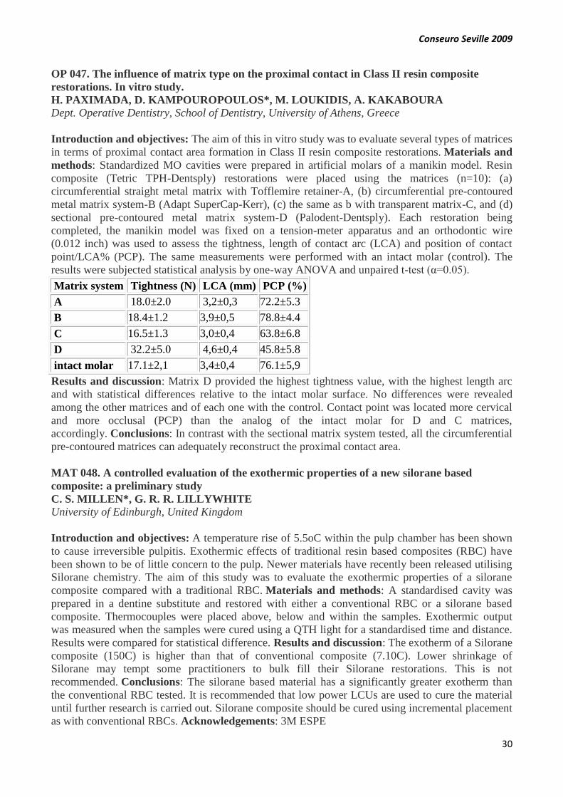
Conseuro Seville 2009
30
OP 047. The influence of matrix type on the proximal contact in Class II resin composite
restorations. In vitro study.
H. PAXIMADA, D. KAMPOUROPOULOS*, M. LOUKIDIS, A. KAKABOURA
Dept. Operative Dentistry, School of Dentistry, University of Athens, Greece
Introduction and objectives: The aim of this in vitro study was to evaluate several types of matrices
in terms of proximal contact area formation in Class II resin composite restorations. Materials and
methods: Standardized MO cavities were prepared in artificial molars of a manikin model. Resin
composite (Tetric TPH-Dentsply) restorations were placed using the matrices (n=10): (a)
circumferential straight metal matrix with Tofflemire retainer-A, (b) circumferential pre-contoured
metal matrix system-B (Adapt SuperCap-Kerr), (c) the same as b with transparent matrix-C, and (d)
sectional pre-contoured metal matrix system-D (Palodent-Dentsply). Each restoration being
completed, the manikin model was fixed on a tension-meter apparatus and an orthodontic wire
(0.012 inch) was used to assess the tightness, length of contact arc (LCA) and position of contact
point/LCA% (PCP). The same measurements were performed with an intact molar (control). The
results were subjected statistical analysis by one-way ANOVA and unpaired t-test (α=0.05).
Matrix system Tightness (N) LCA (mm) PCP (%)
A 18.0±2.0 3,2±0,3 72.2±5.3
B 18.4±1.2 3,9±0,5 78.8±4.4
C 16.5±1.3 3,0±0,4 63.8±6.8
D 32.2±5.0 4,6±0,4 45.8±5.8
intact molar 17.1±2,1 3,4±0,4 76.1±5,9
Results and discussion: Matrix D provided the highest tightness value, with the highest length arc
and with statistical differences relative to the intact molar surface. No differences were revealed
among the other matrices and of each one with the control. Contact point was located more cervical
and more occlusal (PCP) than the analog of the intact molar for D and C matrices,
accordingly. Conclusions: In contrast with the sectional matrix system tested, all the circumferential
pre-contoured matrices can adequately reconstruct the proximal contact area.
MAT 048. A controlled evaluation of the exothermic properties of a new silorane based
composite: a preliminary study
C. S. MILLEN*, G. R. R. LILLYWHITE
University of Edinburgh, United Kingdom
Introduction and objectives: A temperature rise of 5.5oC within the pulp chamber has been shown
to cause irreversible pulpitis. Exothermic effects of traditional resin based composites (RBC) have
been shown to be of little concern to the pulp. Newer materials have recently been released utilising
Silorane chemistry. The aim of this study was to evaluate the exothermic properties of a silorane
composite compared with a traditional RBC. Materials and methods: A standardised cavity was
prepared in a dentine substitute and restored with either a conventional RBC or a silorane based
composite. Thermocouples were placed above, below and within the samples. Exothermic output
was measured when the samples were cured using a QTH light for a standardised time and distance.
Results were compared for statistical difference. Results and discussion: The exotherm of a Silorane
composite (150C) is higher than that of conventional composite (7.10C). Lower shrinkage of
Silorane may tempt some practitioners to bulk fill their Silorane restorations. This is not
recommended. Conclusions: The silorane based material has a significantly greater exotherm than
the conventional RBC tested. It is recommended that low power LCUs are used to cure the material
until further research is carried out. Silorane composite should be cured using incremental placement
as with conventional RBCs. Acknowledgements: 3M ESPE

Conseuro Seville 2009
31
CLIN 049. Minimum Intervention Treatment Plan - Putting MI into practice. Part 1 - MI
Identify: diagnose your patient's susceptibility.
E. REICH*1, A. BANERJEE2, M. BASSO3, M. BLIQUE4, S. DOMÉJEAN-ORLIAGUET5,
C. GAUCHER6, P. KHANDELWAL7, L. LAVOIX7, I, MILÈTIC8, F, ROUSSEL7, J.
ZALBA9,
1University of Cologne, 2King’s College London, 3University of Milan,5 University of Auvergne,
6University of Paris V, 7GC Europe (Belgium), 8University of Zagreb and practices in 4Luxemburg
and 9Spain
Introduction and objectives: The Minimum Intervention (MI) concept is well described in the
literature and summarizes the clinical rationale for the preventive and cause related approach in
cariology. Many studies showed that treatment decisions in cariology varied markedly among
general practitioners (GPs) and that the GPs still wonders “how do I integrate MI in my daily
practice?” The aim of the Pan-European group of academics and GPs - the GC Europe MI Advisory
Board – was to present an evidence based treatment approach for the clinical practice. Materials and
methods: Evidence based literature review. Results and discussion: According to the clinical steps
four parts are used: identify, prevent, restore and recall. Identify encompasses more than looking for
caries. The clinical signs of caries are described according to modified ICDAS criteria in addition to
similar radiological criteria for bitewings. This is added by plaque and/ or gingival index, salivary
factors and possibly other technical data. All this information is than evaluated to assess the patient’s
susceptibility and set up a preventive treatment plan. Acknowledgements: This work was kindly
supported by GC Europe
CLIN 050. Minimum Intervention Treatment Plan - Putting MI into practice. Part 2 - MI
Prevent: Stop caries and prevent it from progressing.
I. MILÈTIC1, A. BANERJEE*2, M. BASSO3, M. BLIQUE4, S. DOMÉJEAN-ORLIAGUET5,
C. GAUCHER6, P. KHANDELWAL7, L. LAVOIX7, E. REICH8, F. ROUSSEL7, J.
ZALBA9,
1University of Zagreb, 2King’s College London , 3University of Milan, 5University of Auvergne,
6University of Paris V, 7GC Europe (Belgium), 8University of Cologne, and dental practices in
4Luxemburg and 9Spain.
Introduction and objectives: The Minimum Intervention (MI) concept is well described in the
literature and summarizes the clinical rationale for the preventive and cause-related approach in
cariology. The aim of the Pan-European group of academics and GPs - the GC Europe MI Advisory
Board – is to present an evidence-based treatment approach for the clinical dental
practice. Materials and methods: Evidence-based literature review. Results and discussion: MI
Prevent deals with “preventive care” and “dental recall frequency”. Depending on the susceptibility
and the risk factors of the patient, preventive treatment regimens are instituted. The standard
approach for all includes oral hygiene recommendations, dietary advice, patient motivation and
maintenance. This standard approach is not sufficient alone to control caries for patients with a high
susceptibility. Those patients with more risk factors need active preventive care. This includes
decontamination (professional mechanical tooth cleaning, antimicrobial gels and rinses, transitional
restoration placement), remineralisation (fluoride, casein-phosphopeptide amorphous calcium
phosphate), xylitol and salivary stimulation, dental sealants (composite or glass ionomer cement).
Repeated diagnosis of the risk factors will be used to assess the need for the precise level of
preventive measures and the amount of recall sessions individualised for each
patient. Acknowledgements: This work was kindly supported by GC Europe

Conseuro Seville 2009
32
CLIN 051. Minimum Intervention Treatment Plan - Putting MI into practice. Part 3 - MI
Restore and MI Recall: Maintain oral health and maximize tooth structure.
M. BASSO1, M. BLIQUE2, J. ZALBA*3, S. DOMÉJEAN-ORLIAGUET4, C. GAUCHER5, A.
BANERJEE6, P. KHANDELWAL7, L. LAVOIX7, I, MILÈTIC8, E. REICH9 F, ROUSSEL7
1University of Milan, 4University of Auvergne, 5University of Paris V, 6King’s College London,
7GC Europe (Belgium), 8University of Zagreb, 9University of Cologne, and practices in
Luxemburg2 and Spain3
Introduction and objectives: The Minimum Intervention (MI) concept is well described in the
literature and summarizes the clinical rationale for the preventive and cause related approach in
cariology. The aim of the Pan-European group of academics and GPs - the GC Europe MI Advisory
Board – was to present an evidence based treatment approach for the clinical practice. Materials and
methods: Evidence based literature review. Results and discussion: MI Restore presents techniques
for conservation of tooth structure when restorations have to be placed. Distinctions are made
between non-invasive and invasive techniques. Adhesive materials are nowadays available for both
indications and the non-invasive or sometimes “repair approach” is shown to be successful in many
indications. MI Recall: To maintain oral health the patient most often needs professional support life,
to compensate his inabilities and control changes in risk factors throughout life. Despite the lack of
scientific evidence regarding optimal recall intervals, there is the need for scheduling recall period
according to the patient’s susceptibility in order to prevent oral disease and to control the
effectiveness of the preventive regimen. Information will be given to remind the audience of the
relevance of customizing the recall period according to the dynamic of the carious process, the
patient demand and some cultural aspects related to national health services context in the different
European countries. Acknowledgements: This work was kindly supported by GC Europe.
OP 052. Microleakage of different bonding systems in Class V composite resin restorations
prepared with Er:YAG laser and diamond bur
O.YAZICIOGLU*, B. EFES GURAY, B. YAMAN, C. DORTER, Y. GOMEC, D. ERDILEK
Istanbul University Faculty of Dentistry Dept. of Operative Dentistry
Introduction and objectives: The aim of this in vitro study was to compare the microleakage of
Er:YAG laser and diamond bur on different bonding systems in Class V restorations. Materials and
methods: Class V cavities (3x3x3 mm) were prepared with Er-YAG laser (VersaWave) and
diamond bur (Komet) on 80 intact human molars. Teeth were randomly distributed into 10 groups
and cavities were restored with CeramX Duo (Dentsply) using 4 different bonding materials, (One
Coat 7.0 Coltène; XP Bond, Dentsply; Clearfil Protect Bond, Kuraray; Adper SE Plus, 3M/ESPE) or
Filtek Silorane and Silorane System Adhesive (3M/ESPE). After the specimens were aged for 7 days
they were immersed in silver nitrate dye and then were sectioned using low speed saw machine.
Microleakage was evaluated by a stereomicroscope and scored for interface between restoration and
teeth. Data were statistically analyzed by One Way ANOVA and Tukey HSD tests. Results and
discussion: Even though no statistically differences were found between any groups (p>0.05),
cavities prepared with diamond bur groups showed slightly higher microleakage scores than Er:YAG
laser groups. Conclusions: The microleakage of different bonding systems was not influenced by the
choice of diamond bur or Er:YAG laser for Class V composite cavity preparation.

Conseuro Seville 2009
33
OP 053.Clinical evaluation of a fissure sealant and a flowable composite.
U. ERDEMIR, H. SAR SANCAKLI*, B.C. YAMAN, E. YILDIZ, T. YUCEL
Istanbul University, Faculty of Dentistry, Dept.of Operative Dentistry
Introduction and objectives: The aim of the study is to evaluate the clinical performance of a
fluoride containing fissure sealant and a flowable composite resin placed on occlusal pits and
fissures. Materials and methods: A fissure sealant; Helioseal F and flowable composite resin; Tetric
EvoFlow were applied on 75 teeth to seal occlusal pits and fissures according to the manufacturers’
instructions. The teeth were evaluated using modified USPHS criteria, at baseline, 1-, 6-, 12- month
intervals by two examiners who were blinded to the materials. Results and discussion: At the first
month, all of the treated teeth were completely intact. After 6 and 12 months the teeth were still
completely sealed and showed the highest values according to the criteria evaluated. Results
demonstrated that after 12 months no loss of restorations and no caries were detected for the Tetric
EvoFlow as well as fluoride containing Helioseal F. Conclusions: After 12 months, Helioseal F and
Tetric EvoFlow showed similar clinical performance in the retention rate and caries preventive effect
of Helioseal F and Tetric EvoFlow fissure sealant.
END 054. Analysis of the extruded material with the continuous wave of condensation
technique
P. CERNADAS-LAGO*, B. MARTIN-BIEDMA, P. VARELA-PATIÑO, J. GONZALEZ-
BAHILLO, M. RUIZ-PIÑON.
International Endodontic Master, Faculty of Medicine and Dentistry, University of Santiago de
Compostela.
Introduction and objectives: Extrusion is the presence of obturation materials in the periapical
tissues. The objectives of this study are to determine the values of extruded material according to the
penetration depth of the SystemB® pluggers, to compare them and to check the adaptation to the
canal walls. Materials and methods: 60 unirradicular teeth were instrumented with Protaper®. They
were divided in two groups: only obturated with gutta-percha and obturated with gutta-percha and
sealer. They were divided in three subgroups according to the plugger penetration. Then, the
specimens were dyed and diafanized. Extrusion was photographed and measured with stereoscopic
microscope, to calculate the volume. Results and discussion: No statistically significant volume
differences were found neither according to the material (p=0,205) nor according to the plugger
penetration (p=0,941), like in literature. Statistically significant differences exist in both groups
(p=0,007 and p=0,036), regarding the frequency of extrusion appearance according to the plugger
penetration depth. Conclusions: Pluggers should be used at 5mm of the working length, to minimize
the extrusion appearance. Acknowledgements: Electronic microscopy unit of the USC. Department
of applied physics of the USC.

Conseuro Seville 2009
34
MAT 055. Determination of the hardening of two types of MTA® based on the thickness.
A. DIAZ-MANCHA*, B. MARTIN-BIEDMA, P. VARELA-PATIÑO, J. GONZALEZ-
BAHILLO, M. RUIZ-PIÑON
International Endodontic Master, Faculty of Medicine and Dentistry, University of Santiago de
Compostela.
Introduction and objectives: The hardness is a physical-chemical property of the MTA® that can
be influenced by other factors. However its relationship with the thickness is not known. The
objectives of this study are, to know the influence the thickness in the hardening of white and gray
MTA® and to identify differences between both and the zones where the measurements were
made. Materials and methods: Twenty samples with plastic cylinders were made and divided in
four groups, determined by the thickness and the colour of the material. They were included in resin
and they were polished with the machine Ecomet 2. The hardness Knoop was determined in the
middle (zone 2) and edges (zone 1,3) of the samples with the microhardness test MXT50. Results
and discussion: Statistically significant differences (ANOVA) between the type of cement, the
thickness or the combination of both in the zone 1 (p=0,117/p=0.798/p=0,642) and 3
(p=0,798/p=0,258, p=0,825) do not exist. However the interaction of the two factors in the zone 2
was significant (p < 0,05). Gary et cols. concluded that 5mm demonstrated greater microhardness
than 2mm. Conclusions: Thickness and type of MTA® do not influence in its hardening.
Acknowledgements: Institute of Ceramics of Galicia.
OP 056. The effect of different finishing and polishing systems on the surface roughness of
different tooth-colored materials
U.ERDEMIR*1, E. YILDIZ1, M. EREN1, A. OZSOY1, F. TOPCU2
1Istanbul University, Faculty Of Dentistry, Dept. of Operative Dentistry, 2Gata Dept. of
Conservative Dentistry and Endodontics
Introduction and objectives: The purpose of this study was to evaluate the effect of various
finishing and polishing systems on the surface roughness of four different types of tooth-colored
materials (Filtek supreme XT, Gradia Direct, Dyract Extra and Fuji IX). Materials and methods:
112 disk-shaped specimens (10 x 2 mm, N = 28 per material) were prepared in a metal mold covered
with a Mylar strip using four different tooth-colored materials. Twenty eight specimens for each
tooth-colored material were randomly divided into four subgroups (n = 7): unpolished (Mylar strip,
control), polished with abrasive disks (Sof-Lex), polished with aluminium oxide and diamond
integrated polisher (Hiluster), and Poli-Pro disk (Premier). The average surface roughness (Ra, µm)
of the control and treated specimens were measured with a surface roughness tester in four different
positions. Results were analyzed by two-way analysis of variance and the Bonferroni post-hoc
comparisons test at a p<0.05. Results and discussion: The order of the surface roughness ranked
from the lowest to the highest according to the tooth-colored materials was: Dyract < Gradia < Filtek
Supreme < Fuji IX; and the ranking for the polishing system was: Mylar strip < Hiluster < Sof-Lex <
Premier. Conclusions: Effectiveness of the finishing and polishing systems on surface roughness
seems to be material dependent.

Conseuro Seville 2009
35
MAT 057. Effect of thermal aging on microtensile bond strength of contemporary self-etching
adhesives.
B. BARACCO1*, MV. FUENTES1, L. CEBALLOS1, S. GONZÁLEZ-LÓPEZ2
1Faculty of Health Sciences, Rey Juan Carlos University, Madrid (Spain), 2Faculty of Dentistry,
University of Granada (Spain).
Introduction and objectives: Self-etching adhesives are considered to be prone to hydrolytic
degradation, especially one-step systems. To evaluate the effect of thermal aging on the microtensile
bond strength (µ-TBS) of one-step self-etching (G-Bond, Xeno V, Bond Force), two-steps self-
etching (Adper Scotchbond SE, Filtek Silorane) and one total-etch (XP Bond) adhesives on sound
dentin. Materials and methods: A block of composite resin (Filtek Z250, except for Filtek Silorane)
was bonded using the adhesives above to exposed occlusal dentin of human extracted molars. One
sample per adhesive was stored in water (24h, 37 °C) and other was thermo-cycled (5000x, 5-55 °C).
All teeth were sectioned in x, y axes to obtain sticks specimens with 1 mm² of bonding area. µ-TBS
test was performed in a universal testing machine. Influence of adhesive system, thermal aging and
their interactions on µ-TBS were analyzed by two-way ANOVA, Student-Newman-Keuls and
Student´s t tests (p<0.05).
Table 1: Mean µ-TBS values for each adhesive system in MPa (SD).
G-
Bond
(GC)
Xeno V
(Dentsply)
Bond Force
(Tokuyama)
Adper
Scotchbond
SE
(3M ESPE)
Filtek
Silorane
(3M
ESPE)
XP Bond
(Dentsply)
24 h
12,9
(4,4)
a1
16,3 (3,7)
a1
23,2 (6,4)
b1
30,7 (6,7)
c1
31,3
(8,9) c1
34,1 (8,8)
c1
5000x
8,1
(0,9)
A2
11,5 (2,1)
A2
14,3 (1,6)
B2
20,9 (2,1)
C2
22,6
(5,1) C2
30,9 (7,6)
D1
Same letters in the same row mean similar µ-TBS values. Same numbers
in the same column mean no influence of thermal aging on µ-TBS value
Results and discussion: µ-TBS values obtained are shown in Table 1. There were statistically
differences among tested adhesives: one-step self-etching adhesives showed the lowest µ-TBS values
while the total-etch adhesive the highest. Thermo-cycling significantly decreased µ-TBS values
except for the total-etch adhesive evaluated. Conclusions: Steps reduction implied a decrease in µ-
TBS results. Self-etching adhesives evaluated exhibited a significantly reduction in µ-TBS values
with thermal aging.

Conseuro Seville 2009
36
OP 058. Radicular section, friction and hybridization in fiber posts retention.
A.IBÁÑEZ-PÁRRAGA*, P.VARELA-PATIÑO, B.MARTIN-BIEDMA, F.GOLDBERG,
J.GONZÁLEZ-BAHILLO, N.BARCIELA-CASTRO, M.RUÍZ-PIÑÓN.
International Endodontic Master, Faculty of Medicine and Dentistry, University of Santiago de
Compostela, Spain
Introduction and objectives: It has been demostrated that post retention is largely contributed by
friction along the canal walls. This study tested the null hipótesis that oval root Canals are more
retentive than circular root Canals in post treatment of endodontically treated teeth. Materials and
methods: Sixty central incisors and maxilary canines were endodontically treated, and prepared for a
post with FRC Postec Plus ® system until drill nº 3. Posts were cemented with Multilink ® following
two differents protocols: (A) using Multilink Primer A + B in canal walls and Monobond-S ® in the
posts, (B) using only the cementating agent. Specimens were then subjected to a microtractional test
using an universal testing machine. Later roots were analized on magnification looking for
guttapercha and cement remains. Results and discussion: Treatment A showed more retention than
treatment B. Independent of treatment Canines group have higher strength values than incisors group
which were less remains found . These results contradict literature giving more significance to
adhesion area and less to friction . Conclusions: Adhesive failure was predominately between post
and cement agent interface. Using silane and adhesive agents increase retention properties. Retention
in oval section canal was higher than circular group. Acknowledgements: Ivoclar Vivadent for
supplying the posts for this study.
END 059. Microscopic analysis of the dentin surface of internal bleached teeth.
T. GONZÁLEZ-GONZÁLEZ*, B. MARTÍN-BIEDMA, P. VARELA-PATIÑO, J.
GONZÁLEZ-BAHILLO, M. RUIZ-PIÑON
International Endodontic Master, Faculty of Medicine and Dentistry, University of Santiago de
Compostela ,Spain.
Introduction and objectives: It is known that bleaching agents produce alterations in the dental
structure. The objectives of our study are analyze this alterations and make a chromatic
analysis. Materials and methods: 9 molars were endodontically treated and internal bleached with
chemically pure sodium perborate. This procedure was repeated once per week during one month.
Vita 3d Easyshade colorimeter was used to study the values of the Cie Lab. A microestructural
analysis was made using electronic microscopy. Results and discussion: Statistically significant
differences were found in the parameters L*, a* and E*. Russell et cols suggest that changes in the
brightness establish a successful bleaching. Microscopically we don´t find any alteration
microestructural, unlike Rotstein et cols. found damages in the organic component of the
matrix. Conclusions: It is an effective technique because we get bleached teeth without dental
alterations. Acknowledgements: Dra. Manuela Neves Lopes. Biomateriais Dentarios, Facultade de
Medicina Dentaria, Universidade de Lisboa, Portugal.

Conseuro Seville 2009
37
STUD 060. Termoplastic obturation technique: bibliographical review
L.VILLAR-ROMERO*, L.PEDROSA-TORRES, B.MARTÍN-BIEDMA, P.VARELA-
PATIÑO, J.GONZÁLEZ-BAHILLO, F.TORRES-CHAVES
International Endodontic Master, Faculty of Medicine and Dentistry, University of Santiago de
Compostela, Spain.
Introduction and objectives: The thermoplastic obturation technique was described by Schilder in
1967. Starting from this, multiple systems were developed that created a more homogeneous
obturation and a better adaptation to the canal wall. The aim is to discover what system gathers the
best conditions. Materials and methods: A search was done in the databases of articles among the
year 2001 and 2008. We will divide the techniques in two groups: heated inside (type System B) or
outside of the canal (type Thermafil). Seven parameters are compared. Results and discussion:
Microleakage: in revised articles there is not a unanimous opinion. Sealing: type Thermafil provides
better results that SystemB. Extrusion: System B provides better result. Curvature/section of the
canal: we don't find differences. The filling of oval canals continues being committed in all the
techniques. Temperature: although Thermafil reaches bigger temperature, the tissular damage it is
not expected since in any technique the temperature transmitted to the tissues it is smaller than 10ºC.
Time: system Thermafil smaller working time. Lateral canals: the difference is not significant
between the techniques. Conclusions: According to these data there is not a superior technique to the
other ones, if not that among them they are supplemented, should be chosen according to the cases.
END 061. A microbiologic evaluation of endodontic disinfection after gaseous ozone treatment
MARIGO L.*, BOLLINO D.,CARDINALI S., LA TORRE G., SOMMA F.
Department of Endodontics, Catholic University of Sacred Hearth – Rome, Italy.
Introduction and objectives: The aim of this study was to evaluate the efficacy of gaseous ozone
during root canal disinfection against Enterococcus faecalis infection. Materials and methods: 28
single-rooted human teeth were prepared endodontically and closed with an apical seal. After
autoclaving, root canals were infected by a broth (TSB) culture of Enterococcus faecalis (ATCC
29212, 108UFC/ml) for three weeks. The teeth were divided in 3 groups: the first group (12 teeth)
was treated applying gaseous ozone for 40 sec. inside root canal. The second group (12 teeth) was
treated using sodium hypoclorite 5% for 5 min. After treatment bacterial growth was evaluated in
each root canal. No treatment was performed on the third group (4 teeth) and teeth were analyzed by
SEM to check Enterococcus faecalis growth. The results were analyzed by T-test (p< 0,05). Results
and discussion: Sodium hypoclorite (complete sterilization) is demonstrated being more effective
then gaseous ozone (decrease of 23% of bacterial growth) with a statistically significant different (p<
0,0001). A biofilm layer was noticed in the canals of third group. Conclusions: Ozone has a little
effect on Enterococcus faecalis and its antibacterial efficacy was not comparable with that of NaOCl
under the test conditions used.

Conseuro Seville 2009
38
PREV 062. Effectiveness of Carisolv before placing fissure sealants
F. CHIVA GARCÍA*, J.C. BÁGUENA GÓMEZ, A.I. NICOLÁS SILVENTE, C. PÉREZ
SÁNCHEZ, M.A. GÓMEZ DE MAYA, M.A. MORALES CANOVAS, A.M. MARTÍNEZ
RODRÍGUEZ
UD Dental Pathology and Therapeutic, Dentistry Faculty, University of Murcia (Spain)
Introduction and objectives: Recent study reported that Carisolv facilitated fissure sealing. The aim
of this study is to confirm if pre-treatment with Carisolv improves the adaptation of fissure sealants
to enamel. Materials and methods: Forty extracted human teeth (molars and premolars) were used
and were randomly divided into four groups of 10 each. Teeth were cleaned with prophylaxis paste.
Group 1: Carisolv was applied for 60” and teeth were copiously washed. Then, fissures were etched
30” with 36% orthophosphoric acid. Group 2: first, teeth were acid-etching, abundantly washing and
then Carisolv was applied 60”. Group 3: acid-etching 30” with 36% orthophosphoric acid. Group 4:
Carisolv for 60” and next, copiously washed. Surface roughness was analyzed in five samples from
each group by stereoscopic microscopy and by SEM examination. Results and discussion: In group
1, most of the debris in the fissures was removed by Carisolv treatment, but it do not increased
surface roughness. After etching, enamel prisms microindentations do not happens. In group 2,
Carisolv filled microretentions of acid-etching previously realized. Teeth of group 3 showed
characteristic pattern of enamel acid-etching and Carisolv (group 4) do not produced any type of
etching. Conclusions: Carisolv-treated surfaces block acid-etching, impeding enamel bonding to
sealants

Conseuro Seville 2009
39
MAT 063. The effect of silane on the bond strengths of composite indirect restorations using
several resin cements.
MV. FUENTES*1, L. CEBALLOS1, S. GONZÁLEZ-LÓPEZ2
1Faculty of Health Sciences, Rey Juan Carlos University, Madrid (Spain), 2Faculty of Dentistry,
Granada University (Spain)
Introduction and objectives: The aim was to determine the effect of silanization of indirect resin
composite on microtensile bond strength results using several resin cements. Materials and
methods: Flat dentin surfaces were created on extracted human third molars. Resin composite
overlays (Filtek Z250) were constructed and received different surface treatment, silanization (RelyX
Ceramic Primer, 3M Espe) or not. Dual cure resin cements studied were: RelyX ARC (3M Espe),
and two self-adhesive cements RelyX Unicem (3M Espe), and G-Cem (GC). The bonded assemblies
were stored in water (24 h, 37 °C) and subsequently prepared for microtensile bond strength testing.
Beams of approximately 1 mm2 were tested in tension at 1 mm/min in a universal tester. Data were
analyzed by 2-way ANOVA and Student-Newman-Keuls test (α=0.05).
Table 1- Means (standard deviations) of microtensile bond
strength data in MPa.
Resin cement Silane n Bond strength Bond strength *
RelyX ARC yes 12 21.7 (6.3)
23.4 (6.8)a no 14 24.8 (9.3)
Rely X Unicem
yes 13 13.6 (8.0) 14,4 (7.1) b
no 11 15.3 (6.2)
G-Cem yes 11 14.6 (6.8)
12.1 (6.7) b no 12 9.7 (5.9)
* Same letters indicate no significant differences (Student-
Newman Keuls, α=0.05)
Results and discussion: Microtensile bond strength values are shown in the table. Two-way
ANOVA revealed a significant influence of the resin cement used (p<0.001). Silanization and
interactions between resin cement and silane application did not affect microtensile bond strength
values (p>0.05). RelyX ARC exhibited the highest results followed by the two self-adhesive resin
cements evaluated. Conclusions: Surface treatment of indirect resin composite with silane did not
improve the bond strength results. Acknowledgements: The authors are grateful to the companies
for providing the materials tested.
Conseuro Seville 2009
40
OP 064. The efficacy of three different bleaching systems using teeth and ivory
O. POLYDOROU1*, R. LANGE2, T. BAYER2, M. ZIMMERMANN2, P.HAHN1
1University Medical Center Freiburg, Dental School and Hospital; 2Deltamed GmbH, Germany
Introduction and objectives: Aim of this in vitro study was to evaluate the use of ivory compared
to extracted human teeth as a material to be used in bleaching studies, by testing three different
products. Materials and methods: Three groups of human teeth and three groups of ivory (n=12),
were stained and bleached with three bleaching systems, respectively: Easywhite Ready (H2O2
30%) for 30min using a plasma unit, Opalescence 35% carbamide peroxide for 60min and “7 day
Bleach” paint-on product (H2O2 6 %) for 10 x 30min. The shade (L*a*b* values) was evaluated
before, after bleaching, and one week later, using a digital camera and a spectrophotometer. Results
and discussion: The digital evaluation showed for the ΔL values the same results for teeth and ivory.
A significant difference was found between the three bleaching products (p<0.05). The
spectrophotometer showed for the ΔL parameter, a similarity for teeth and ivory. Δa and Δb for teeth
and ivory were not found to be similar. Conclusions: Ivory and teeth showed that similar results
were achieved after the three bleaching procedures concerning the ΔL parameter. Although the use
of ivory could be beneficial because of the ability to give a lot of similar samples, and to produce a
good discoloration, further research is necessary.
OP 065. A clinical study of a new Ca3SiO5-based material indicated as a dentine substitute
G.KOUBI1 *, J.C. FRANQUIN1, P. COLON2
1 Dpt. of Operative and Preventive Dentistry, University of Marseilles, France, 2 Dpt. of Operative
Dentistry and Endodontics, University of Paris 7, France.
Introduction and objectives: A new Ca3SiO5-based material (BiodentineTM RD94, Septodont)
has been developed as a bulk dentine substitute before direct and indirect posterior fillings. A three-
year follow-up randomized multicentric clinical study was initiated to evaluate: 1) its longevity as
temporary restoration vs a resin composite (Z100, 3M), and 2) its ability to be combined with an
adhesive filling. Materials and methods: Patients, 18 to 80 years old, were included in this trial and
clinical evaluation was conducted according to USPHS criteria. Observation was scheduled at D0,
D15, 6 months, 1, 2 and 3 years using pulp vitality testing, radiographs and photographs.. Results
and discussion: The clinical study is still on-going. So far, 334 patients have been included (162
female and 172 males, mean age: 47 years) with 81 Class I and 253 Class II cavities. At this stage,
the temporary filling mean longevity has been established to about 12 months. BiodentineTM RD94
was preserved on all dentinal walls without any infiltration or secondary decay. Conclusions: This
new Ca3SiO5-based material could be used as a dentine substitute for definitive dentinal treatment.
As marginal leakage and secondary decays remain a concern in adhesive dentistry,
BiodentineTMRD94 could be preserved if re-intervention is required.
Conseuro Seville 2009
41
END 066. Clinical outcome of root canal fillings using lateral condensation technique after 12
months.
A. PESCHKE*, R. WATZKE; J.-F. ROULET
Dental Clinic, R&D, Ivoclar Vivadent AG, Schaan, Liechtenstein
Introduction and objectives: The aim of this prospective clinical study was to evaluate the
radiographic and clinical performance of Apexit Plus in combination with the lateral condensation
technique. Materials and methods: 41 teeth (106 canals) with radiographically discernible signs of
apical periodontitis (39.0% Non-Vital, 36.6% Revisions, 24.4% Irr. Pulpitis) were treated with a
standard operative procedure. Follow-up appointments after 6 and 12 months entailed radiographic
and clinical evaluation. The radiographic data was analyzed using the PAI scoring system described
by Ørstavik (1986, 1998). Results and discussion: After 12 months 95.1% of the teeth showed an
obvious tendency towards healing and in only 4.9 % no significant radiographic changes were
observed. 61% of the teeth already showed radiographic healing (PAI=1-2), 37% PAI score 3 and
only one case (2%) still a PAI of 4. The pre-operative PAI Score was 3.9 (±0.9) and 3.4(±1.0) at the
immediate post-op radiographic control. After 6 months the mean PAI was 2.6(±1.1) and 2.0 (±0.9)
after 12 months. The reduction of the PAI Scores between each of the different evaluation times was
statistically significant (p<0.01 Wilcoxon Test). Conclusions: Lateral condensation technique using
Apexit Plus showed good clinical success after 12 months. An additional 3 year follow-up is ongoing
to approve the long-term behaviour of this sealer.
END 067. Investigate the root canal anatomy of mandibular premolars in North-west Spanish
population
P. CASTELO*, Z. GÓMEZ, P. VARELA, MARTIN B.
University of Santiago de Compostela
Introduction and objectives: It was observed, during the routine treatment of mandibular premolars
that aberrations were becoming common. Investigators have shown that the pulp canal system is
complex, and canals may branch, divide and rejoin. Although various techniques have been used in
studies evaluating canal morphology, it has been reported that the most detailed information can be
obtained ex vivo by demineralization and staining. Objectives: try to find some tooth with aberration
anatomy and most common anatomy in this population. Materials and methods: Fifty extracted
mandibular premolars were examined. Following the preparation of access cavity, pulp tissue was
removed and the canal systems were stained. The teeth were then rendered clear by demineralization
and immersion in methyl salicylate. Cleared teeth were examined and the following features were
evaluated: type of root canals, lateral canals, and frequency of apical deltas. Results and discussion:
The present study revealed the prevalence of multiple canals in the investigated Spanish north-west
mandibular premolars. Variable root canal morphologies were found in the mandibular first
premolars. Conclusions: The incidence of aberrations in the canal configuration of mandibular
premolars is common and due considerations are to be given during endodontic intervention.

Conseuro Seville 2009
42
OP 068. Clinical comparison of four different whitening systems using hydrogen peroxide.
A. OTEO-ZACCAGNINI*1, J. OTEO-CALATAYUD2, C. OTEO-CALATAYUD1, 2, M.D.
OTEO CALATAYUD3
1Master Course in Esthetic Dentistry, 2 Department of Conservative Dentistry. 3 Department of
Orthodontics. Complutense University of Madrid, Spain
Introduction and objectives: Many of the techniques denominated “Power Bleaching” uses
hydrogen peroxide 35% without the necessity of activate it with light and obtaining good results. The
objective of this study is to evaluate the efficacy of three dental whitening systems using different
kinds of light activation, and to compare them to a dental whitening system using hydrogen peroxide
without any kind of light activation. Materials and methods: 40 patients between 18 and 40 years of
age were included in the study. Patients were distributed randomly in different groups. - Group 1:
Dental whitening system using hydrogen peroxide 35 % with diode light activation (Luma Cool). -
Group 2: Dental whitening system using hydrogen peroxide 9 % with diode light and heat activation
(Metatray). - Group 3: Dental whitening system using hydrogen peroxide 25% with ultraviolet lamp
activation (Zoom 2). - Group 4: Dental whitening system using hydrogen 35 % without any light
activation. (Butler). Results and discussion: Three of the four systems has shown clinically
significant results between initial and final treatment. These systems are LumaCool, Metatray, and
Zoom2, whereas the control group doesn’t show significant statistically results. Zoom2 shows better
results than any of the other groups. This results are statistically significant between groups 1 and 3,
2 and 3, and between groups 3 and 4. But differences between groups 1 and 4, and groups 2 and 4 are
not significant. Conclusions: All whitening systems using light activation has demonstrated good
results in our study. We can assume that Zoom2 has shown better clinical efficacy than any of the
other systems.
OP 069. A clinical trial of at-home tooth bleaching using a 16% carbamide peroxide: 1 hour vs
6 hours of application. A pilot study
P. MASIAN*1, J. OTEO2, C. OTEO2,3, G. ALEGRIA3
1 Master in Dentistry Science, 2 Department of Conservative Dentistry. 3 Master Course in Esthetic
Dentistry. Complutense University of Madrid, Spain
Introduction and objectives: Evaluate the efficacy of a 16% carbamide peroxide at home on the six
anterior maxillary teeth for different periods of application. The periods were 1 hour/day on the teeth
on the right (11, 12, 13) and 6 hour/day on the teeth on the left (21, 22, 23). Materials and methods:
Three examiners out of study evaluated the shade of the six anterior maxillary teeth of 10 patients
using the Vita Shade Guide ordered by value. Patients were instructed to use dispensed gel (Zaris 3M
Espe®) for a period of 1 h/day on the 3 anterior maxillary teeth on the right, and 6 h/day on the 3
anterior maxillary teeth on the left during 2 weeks. Patients were evaluated 2 weeks post-bleaching
following the same protocol that was conducted at the baseline. Results and discussion: Application
of the bleaching gel reflects a statistically significant lightening in tooth shade. There is no
statistically difference (P0,05) between 1 or 6 hours of application. Conclusions: 1 hour of 16%
carbamide peroxide application shows the same results that 6 hours.
Conseuro Seville 2009
43
OP 070. Clinical efficacy of a dental bleaching system with a 25% hydrogen peroxide activated
by u.v. light. A pilot study
G. ALEGRIA*1, J. OTEO2, C. OTEO2,3, P. MASIAN3
1 Master in Dentistry Science , 2 Department of Conservative Dentistry. 3 Master Course in Esthetic
Dentistry. Complutense University of Madrid, Spain
Introduction and objectives: The objective was to evaluate the clinical efficacy of a bleaching
system using a 25% hydrogen peroxide activated by U.V. light. Materials and methods: The six
anterior maxillary teeth of 11 patients were evaluated using the Vita Shade Guide ordered by value
by three different operators out of the study. The investigator applied the bleaching gel to the teeth
on the right for 15 minutes for a total of three times. Then, the teeth were assessed. After that, the
investigator blocked and covered the teeth and the oral and adjacent tissues except the three teeth on
the left, applying the bleaching gel and exposing them to the whitening lamp for 15 minutes, three
times. Then the teeth were evaluated. Results and discussion: Application of the bleaching gel to
the teeth on the right did not result in any increase of lightness in either subject. Application of the
bleaching gel and exposing teeth on the left to the whitening lamp reflects a statistically significant
lightening in tooth shade. Conclusions: Zoom 2 Whitening System is effective to increase lightness
of the teeth.
OP 071. Efficacy of two customized tray-based at-home bleaching systems: a randomized
controlled trial
J. MARQUES*, D. MARQUES, J. SILVEIRA, J. AMARAL, J. CABRITA, A. MATA
GIBBO (Oral Biology and Biochemistry Research Group). University of Lisbon – College of
Dentistry
Introduction and objectives: Aim of this study was to assess the clinical efficacy of two different
whitening products using a customized tray for at-home bleaching. Materials and methods: This
was a two arm parallel RCT in which sixty human subjects attending a Portuguese dental school
were evenly distributed to one of two groups: Ultradent Opalescence 10%PF (OPL) or Vivadent
VivaStyle 10% (VS), both with 10% carbamide peroxide (CP). Bleaching regiment was according
to manufacturer’s protocols. Efficacy was assessed using the VITAPAN classical shade guide
(VITA) on the facial surface of the twelve anterior teeth with A3 or darker shade at pre-treatment
(PT), end-treatment (ET) and six months (6M) under standard light conditions. Shade improvement
(SI) and relapse (SR) were expressed as SI=PT-ET and SR=PT-6M in meanS.E.M. Mean
differences were tested with Student t-test, significance level set at =0,05. Efficacy was considered
according to ADA criteria. This procedure was approved by the local ethical committee. Results and
discussion: At the end of the treatment the SI was 6,2800,177 and 4,2840,196, and a mean SR of
5,5980,265 and 3,3310,266 were observed for OPL and VS respectively. Conclusions: The results
show that both bleaching systems are effective at ET. However, only OPL can be considered
efficient regarding SR at 6M.

Conseuro Seville 2009
44
OP 072. Efficacy of a novel in-office bleaching technique: a pilot study
J. SILVEIRA*, D. MARQUES, J. MARQUES, N. GUILHERME, A. MATA
GIBBO (Oral Biology and Biochemistry Research Group). University of Lisbon – College of
Dentistry
Introduction and objectives: A new in office technique using a 6% hydrogen peroxide (HP) paint
on formulation has recently been developed by our group (EJED 1(1): 70-7, 2006). This pilot study
aims to assess the efficacy of this technique. Materials and methods: 18 subjects attending a dental
school clinic were selected for this study. After screening and dental prophylaxis, the clinical
protocol for the in-office whitening technique using VivaStyle Paint On Plus (Vivadent) with 6% HP
was performed as described previously. Efficacy was assessed using the VITAPAN classical shade
guide (VITA Zahnfabrik) on the facial surface of the twelve anterior maxillary and mandibular teeth
with a gradable VITA shade A3,5 or darker at pre-treatment, end-treatment and six months after the
end-treatment, under corrected operatory light. Efficacy was expressed in MeanS.E.M. of vita
shade guided values. Mean differences were tested with Student t-test, significance level set at
=0,05. This procedure was approved by the local ethical committee. Results and discussion: The
results show a significant (P<0.01) 9.25 +/- 0.33 final shade improvement and a mean of 3.15 +/-
0.52 vita shade color relapse at 6 months when compared to baseline values. Conclusions: The
results suggest that this new technique is effective, offering a viable alternative to high peroxide
concentration in-office procedures.
END 073. Root fractures using fiber posts. A prospective clinical study
J.C. MANCEBO1*, E. JIMÉNEZ-CASTELLANOS2, D. CAÑADAS2
1 Associate professor, 2 Professor. University of Sevilla
Introduction and objectives: The potential of fibre posts to reduce the incidence of root fractures in
comparison with traditional cast posts has been referred to in several in vitro and in vivo
studies. Materials and methods: A prospective clinical study including 131 endodontically treated
teeth have been carried out to assess their survival. One tooth each person, 53 men and 78 women.
The 131 teeth were restored with a fibre post (87 with SNOWPOST®―66,4%―, and 44 with DT
LIGHT POST®―33,6%―) and covered with a crown. The cement used was RelyXUnicem and
dental core was built up with Dentocore Automix, a dual composite. The teeth were clinically and
radiologically evaluated for a period between 1 and 7 years, every 6 months. Results and
discussion: In disagreement with other researches, two cases of root fractures were observed. Both
fractures used SNOWPOST. The first one occurred in a patient with a complete mandibular arcade
and a very reduced maxillary arcade, with bruxing habits, in a maxillary lateral incisor without
ferrule. The oclusal unbalance and the absence of ferrule might be the reasons for its failure. The
second one occurred in a maxillary central incisor, with a previous cast post treatment, in a
completely dentate patient. This tooth had a ferrule at least 2 mm. high. Its failure might be a
consequence of non-detected microfractures by the retreatment. Conclusions: According to our
research root fracture is not frequent by using fiber posts but it occurs if we don’t observe certain
conditions.

Conseuro Seville 2009
45
MAT 074. Odontointegration of a new osteoconductive implant surface. Initial results.
I. VALDIVIA-GANDUR1,2, V. LOZANO DE LUACES3, V. TALLÓN-WALTON*1, A.
MESTRE 4, P. CARVALHO-LOBATO1, M.C. MANZANARES-CÉSPEDES1.
1Muscular and Skeletal Pathology Research Group. Human Anatomy and Embryology Unit,
University of Barcelona, Barcelona, Spain, 2 Anatomy Unit, Faculty of Dentistry, University of
Antofagasta, Chile, 3Ergonomy Unit. Dentistry Department. Faculty of Dentistry, University of
Barcelona, 4Biomedical Engineering Research Centre. Polytechnic University of Catalonia, Spain.
Introduction and objectives: We present the initial results of the reaction of the dental calcified
tissues to a new dental implant surface in an animal model. The surgical method for the implant
placement was designed in order to obtain a stable contact between the implant surface and alveolar
and dental tissues, both calcified and pulpar. The surface has previously showed osteoconductive
properties in studies, both in vitro and in vivo. Materials and methods: Four implants were inserted
the central incisive germs in three pigs, immediately post-extraction of the premolar teeth from
maxillary bones, (3 with surface treatment and 1 without). The curved shape of the incisive permitted
the implant placement through the incisive alveolar space, thus allowing the contact of the implant
surface with the incisor dentin and pulp. The animals were sacrificed 90 days after. The analyses of
the implant-tissue contact and interaction were performed by Backscattering-Scanning Electron
Microscopy and by histology (Manzanares et al (1998)). Results and discussion: The surgical
method proved its feasibility in allowing the contact between the alveolar bone and dental tissues,
and the implant without causing bone fractures, dental lesions or the loss of the implant. Our
observations showed a consistent high percentage (95 to 100%) of direct contact between the treated
implant surface and the dentin tissue, the histological analysis of these samples proved that the dental
pulp tissues were vital: no pulp damage was observed. Conclusions: The surface showed
dentoconductive properties.
MAT 075. Osteointegration and dento-alveolar ankylosis: comparison of calcified tissues
implicated in both process
P. CARVALHO-LOBATO*1, V. TALLÓN-WALTON 1, I. VALDIVIA-GANDUR1,2, M.C.
MANZANARES -CÉSPEDES1, V. LOZANO DE LUACES3
1Muscular and Skeletal Pathology Research Group. Human Anatomy and Embryology Unit,
University of Barcelona, Barcelona, Spain, 2 Anatomy Unit, Faculty of Dentistry, University of
Antofagasta, Chile, 3Ergonomy Unit. Faculty of Dentistry. University of Barcelona.
Introduction and objectives: The objective is the evaluation of the interaction established between
the osseous tissues and the biomaterial, as compared with the ones existing between the alveolar
tissues and the dental tissues, as well as the chronology of both processes. Materials and methods:
The first study consisted in the simultaneous implantation of five types of implants coated with
hydroxiapatite by pulsed laser deposition. In the tibial dyaphisis of Beagle dogs; and the second
consisted in the provocation of an experimental ankylosis. The second maxillary molars ligament
was removed after surgical teeth extraction, the endodontic treatment was done, and then were
replaced .The animals were sacrificed respectively one, two and three months after the surgical
procedure in both experiences. The samples were submitted to a scheduled procedure, in order to
perform the ultrastructural observational studies: scanning microscopy with secondary and
backscattered electrons. Results and discussion: The calcified tissues observed both in the
osteointegration as in the ankylosis dento-alveolar were the same. The calcified tissues present in all
these processes share the same physiological fate: osteoclasts activate the process; chondroid tissue
acts as the stabilizing element in front of the mechanical forces and is substituted by woven bone and
later by lamellar bone when the area reaches mechanical stability. The chronology of the described
processes is essentially the same as our group described previously. Conclusions: No histologycal or
chronological differences were observed between both processes.
Conseuro Seville 2009
46
STUD 076. Adverse effects of dental bleaching: searching for scientific evidence
L. CEZÓN MUÑOZ, M. CURA PEÑA, F. FLORES DORADO*,I. GIRÁLDEZ de LUIS, L.
CEBALLOS GARCÍA.
Universidad Rey Juan Carlos. Madrid. Spain
Introduction and objectives: Bleaching has become one of the most popular esthetic dental
treatments but there are several controversies about its effects. Therefore, the objective is to review
the literature regarding external bleaching adverse effects. Materials and methods: Original full
articles from peer-reviewed journals included in Medline and Pubmed were selected using as search
terms bleach and effects up to November 2008. Results and discussion: Carbamide and hydrogen
peroxide may produce transitional tooth hypersensitivity. Peroxides do not have significant
deleterious effects on enamel and dentin surface morphology, chemistry or microhardness.
Fluoridated bleaching gels seem to reduce these effects. Peroxides do not increase the susceptibility
to acid erosion, tooth paste abrasion or caries lesion formation. A period of at least 7 days should be
allowed after bleaching to carry out an adhesive procedure. Bleaching does not decrease significantly
microhardness of restorative materials. No systemic effect has been attributed to dental
bleaching. Conclusions: Minimal changes in dental tissues and restorative materials are associated to
dental bleaching. More research is needed simulating intraoral conditions and using comparable
protocols.

Conseuro Seville 2009
47
END 077. Adaptation of two obturation systems to the root canal walls
M. COSTA*1, R.MADUREIRA1, L. FORNER2
1ISCS-Norte/Portugal, 2Valencia University. Spain
Introduction and objectives: The microorganisms are considered the principal etiological agents in
the endodontic pathologies. The biomechanics preparation is very important in endodontic treatment,
but it is proved that bacterials still survive in root canal tubules. Filling materials still don’t prevent
apical microinfiltration. Resilon is a polyester-based thermoplastic root filling material, recently
introduced as an alternative to guttapercha. The purpose of this study was to compare the adaptation
and penetration of guttapercha/AHPlus and Resilon System to the root canal walls. Materials
and methods: The sample consists of 20 monorradicular teeth, cleaned and shaped by Hero Shaper
and Hero 642 files. During preparation 5.25% of sodium hypochlorite was used as an irrigant,
finishing with EDTA to remove smear layer. The sample was divided in Group A:
guttapercha/AHPlus, and Group B: Resilon/RealSeal. In both groups, sealers were labeled with
fluorescent Rhodamine B. Sections cut from the coronal, middle and apical thirds of each root were
viewed using a confocal laser scanning microscopy. Depths of dentinal tubule sealer penetration
were observed in statistical analysis.
AHPlus (average) Resilon (average)
Coronal
maximum
penetration
(µm)
1100 627
penetrated
perimeter
(%)
91 62
Middle
maximum
penetration
(µm)
1000 578
penetrated
perimeter
(%)
95 53
Apical
maximum
penetration
(µm)
61 0
penetrated
perimeter
(%)
5 0
Results and discussion: AHPlus presented the best results in the coronal and middle third with
statistically significant. In apical region, AHPlus also had the best results but without statistically
significant.

Conseuro Seville 2009
48
MAT 078. Polymerization shrinkage and bonded interface: a negative relationship
E. CABRERA*, J.C. de la MACORRA
Department of Conservative Dentistry. Faculty of Odontology. Complutense University. Madrid,
Spain
Introduction and objectives: Detrimental effects of polymerization shrinkage on µTBS of bonded
interfaces at outer extremes of longitudinal visible light cured resin composite restorations have been
described. These effects would be extremely relevant in peripheral parts of extended interfaces. This
study tests whether microtensile bond strength to dentin of extended light-cured resin composite
restorations decreases from center to periphery. Materials and methods: Labial dentin of bovine
incisors (n=5) was exposed (500 grit grinding) and restored (Scotch Bond1 XT, VLC Ti Core) in an
only increment of at least 2mm high and 10 x 10 mm in extension. Restorations were sectioned
perpendicularly to interface, producing bar-shaped samples that were submitted to tension until
adhesive separation, its µTBS calculated and the percentage of µTBS values within each specimen
transformed into PTens dependent variable. Spatial relationship of each sample with all others from
same specimen was defined as its MODULE: the result of addition of all vectors with origin in the
center of the sample and ending in the center of all others. MODULE was transformed to PMod: its
percentage within each specimen. Relationship between PTens and PMod was tested through linear
regression analysis. Results and discussion: Linear regression results: ANOVA’s p < 0.00001, r2 =
0.42. Conclusions: There is a statistically significant inverse relationship between distance to the
center of a visible-light-cured mass of composite resin bonded to a flat dentin surface and µTBS of
interface.
MAT 079. Genotoxic Potential and ROS generation of Camphorquinone in Primary Human
Gingival Fibroblasts
J. VOLK*, W. GEURTSEN, G. LEYHAUSEN, A. BECKEDORF, H. RAHMER,G. IBARRA,
D.C. ZIEMANN
Hannover Medical School, Clinic of Conservative/Preventive Dentistry and Periodontology
Introduction and objectives: Objective of the present study was to evaluate whether CQ, even in
the absence of visible-light (VL)-irradiation, exhibits a genotoxic potential in primary human
gingival fibroblasts (HGF). Materials and methods: CQ-dependent cytotoxicity (0.125 to 2.5 mM)
and intracellular reactive oxygen (ROS)-formation (0.25 to 2.5 mM) were assessed by means of
fluorospectrophotometric analyses using propidium iodide and 2´,7´-dichlorodihydrofluorescein
diacetate, respectively. The alkaline comet assay ± human 8 hydroxyguanine DNA-glycosylase 1
(hOGG1), to detect oxidative DNA-lesions, was used to determine the genotoxic potential of CQ
(0.05 to 2.5 mM). The Tukey ANOVA and the Student's t-test (p < 0.05) were used for statistical
analyses. Results and discussion: In concentrations > 0.25 mM, CQ resulted in rapid and significant
ROS-formation in HGF. There was no significant reduction in cell viability during the 3 h incubation
period. However, after 24 h of incubation CQ significantly decreased cell number and cell viability.
Interestingly, CQ induced DNA-damage depend on concentration already after 3 h of incubation. In
addition, experiments with the hOGG1-modified comet-assay (0.5 to 2.5 mM CQ) demonstrated a
CQ-induced increase in oxidative DNA-lesions. Conclusions: CQ, even without VL-irradiation,
exhibits a genotoxic potential in primary HGF cells in vitro. ROS-triggered oxidative DNA-lesions
are at least partly involved in CQ-induced DNA-damage. Acknowledgements: Supported by
DGZMK (Deutsche Gesellschaft für Zahn-, Mund- und Kieferheilkunde).

Conseuro Seville 2009
49
OP 080. Clinical comparison study of two whitening systems with different application
methods.
A. IRSHAED1*, J. OTEO2, C. OTEO2,3, A. OTEO3
1 Master in Dentistry Science , 2 Department of Conservative Dentistry. 3 Master Course in Esthetic
Dentistry. Complutense University of Madrid, Spain
Introduction and objectives: Patients can choose between having their whitening done at home or
in the office. Dental bleaching done “at home” has demonstrated to be an efficacy treatment and
reduces the number of visits to the dental office. To compare two at-home whitening systems over
patients with different application methods, one with brush one other with a traditional system using
trays. Materials and methods: 20 patients were selected between 18 and 40 years of age. Two
groups were formed, one doing the whitening using brushes and the other using thermoplastic trays.
After an initial examination, patients were selected by following our criteria for inclusion. Every
colour shading was carried out using the Vita Classic Guide reordered by brightness. Shading was
performed by 3 different operators and the colour was selected when 2 of the 3 operators matched.
The two dental bleaching systems evaluated were: Hydrogen Peroxide 6% (Vivastyle Paint on Plus,
Ivolcair Vivadent) using brushes over the dental surface, and Carbamide Peroxide 16% (Buttler
Whitening DAY). Night application using thermoplastic trays. Results and discussion: Statistically
results have been found between these two groups. We have obtained better results using
conventional trays than using brushes. Conclusions: Both systems has demonstrated their clinical
efficacy, however the system using trays has shown better results, probably due to a longer contact
time of the peroxide to the teeth.
MAT 081. Micromechanical properties of self-adhesive cements: an one-month evaluation.
E. MAGNI1,2*, N. ILIE2, R. HICKEL2, M. FERRARI1
1Department of Fixed Prosthodontics and Dental Materials, University of Siena, Siena, Italy.
2Department of Restorative Dentistry and Periodontology, Dental School of the Ludwig-Maximilians
University, Munich, Germany
See table on: http://www.efcd.eu/media/pdf/Mat_081_pdf
Introduction and objectives: Limited information is available on the properties of the recently
developed self-adhesive cements. The aim of this study was to assess the micromechanical properties
of self-adhesive cements over one-month. Materials and methods: Five 4mm×4mm×2mm
specimens of G-Cem, MultilinkSprint, Biscem, Maxcem, RelyXUnicem, Breeze and Sac-A
(experimental) were light-cured 30s and stored in deionised water at 37°C. Elastic modulus (E),
elastic work (We/Wtot), creep (Cr) and Vicker’s hardness (VH) were measured after 24h, 1-week
and 1-month with a microhardness-indenter.
Results and discussion: The Table reports means(SD) of the micromechanical
properties. Conclusions: The micromechanical properties differ among self-adhesive cements over
time. Each cement exhibited a specific variation of each property over 1-month.
Conseuro Seville 2009
50
OP 082. Evaluation of composite repair ability of XP Bond
S.W. HWANG, J.W. PARK*
Department of Conservative Dentistry, Yonsei University, Seoul, S. Korea
Introduction and objectives: In some clinical situation, it is needed to repair the composite
restorations for preserve tooth structure and saving time. To obtain good bonding between the old
and new composites, we have to make favorable surface condition. The purpose of this study was to
evaluate the repair of aged composite with XP bond following various surface treatments conditions
with micro-shear bond strength test. Materials and methods: 2 mm thick Ceram-X Mono composite
was cured and aged by thermocycling (5 to 55ºC, 5,000). After aging, the surface was treated by
different roughening methods (sandblasting or bur grinding), and silane treatment. Then XP Bond or
Adper Single Bond 2 was applied and composite was added and effect of postrepair aging was
evaluated. The results were analyzed by one-way ANOVA and Tukey’s test and multiple
comparisons with LSD test. Results and discussion: Individual comparison of the 16 experimental
groups showed no statistical difference. However comparing the effect of the surface roughening,
silane treatment, bonding agents, and post-aging, only the bonding agent showed statistical
difference – XP Bond showed higher bond strength (p<0.05). Conclusions: Following the results of
this experiment, XP bond showed superior repair bond strength than the other. Acknowledgements:
This experiment was supported by Dentsply.
STUD 083. Analysis of an endodontic failure, in a maxillary second molar. Case report and
survey.
Z. GÓMEZ*, P. CASTELO, P. VARELA, B. MARTIN
University of Santiago de Compostela
Introduction and objectives: There are some causes that can cause failures in endodontic
treatments. We present a case of a 34-year-old male who has a water-pipe in a maxillary second
molar as a cause of an endodontic failure; in this case we decided that the election treatment was
extraction of the molar because the patient presented systemic problems. Objectives were: find
failure’s cause, to evaluate technical aspects that could be better in this case. Materials and
methods: After the extraction the teeth were rendered clear by demineralization and immersion in
methyl salicylate (USC´s protocol) and study it. Results and discussion: The radiographic study is
indispensable to value the result in a short period of time and some months after the endodontic
treatment; even some times, with good radiography observations we can find the failure
reason. Conclusions: To have success, is necessary to be in mind all components of the endondontic
treatment: diagnostic, anatomy, access cavities, cleanliness, conformation, disinfection, obturation
and restoration.

Conseuro Seville 2009
51
OP 084.The effect of preparation designs on the marginal and internal gaps in Cerec3 partial
ceramic crowns
D. SEO 1,*, Y. YI 2, B. ROH 1
1Department of Conservative Dentistry, Yonsei University, 2Department of Prosthetic Dentistry,
Myongji Hospital, Kwandong University
Introduction and objectives: The purpose of this study was to evaluate the marginal and internal
gaps in Cerec3 partial ceramic crowns(PCCs) of three different preparation designs in vitro using
microcomputed tomography (CT). Materials and methods: Cerec3 PCCs of three different
preparation designs (n=20) were fabricated according to the following: GroupI-conventional
functional cusp capping/shoulder preparation, GroupII-horizontal reduction of cusps and GroupIII-
complete reduction of cusps/shoulder preparation. After fixation of PCCs, CT scanning was
performed. CT sections were reconstructed 3-dimensionally, then by dividing the total volume of
internal gap by contact surface area, the average internal gap(AIG) was obtained. For 2-dimensional
(2D) CT views, seven bucco-lingual sections and three mesio-distal cross sections were obtained at
predetermined key points. The gaps were measured using CT at each reference point. Statistical
analysis was performed. Results and discussion: For the 3D reconstruction technique, the AIGs
were as followed: GroupI 197.348.2m, GroupII 171.245.1m, and GroupIII 152.727.1m. For
the 2D CT views, the gaps of each group were the smallest on margins, and the largest on
horizontal or angle walls. Following the results, groupI showed larger marginal and internal gaps
compared with the other groups. Conclusions: The simplified designs (GroupII, III) demonstrated
superior results compared to the traditional cusp capping design (GroupI). The marginal gaps were
smaller than the internal gaps in all groups.
CLIN 085. Clinical cases of different types of provisionalization in Porcelain Laminate
Veneers.
S. GOMAR-VERCHER 1*, S. ROMÁN-RICHON2, V. FAUS-MATOSES3, V.J. FAUS-
LLACER4
1,2Master Student in Conservative Dentistry and Endodontics. University Medical and Dental
School Valencia. 3Master Professor in Conservative Dentistry and Endodontics. University Medical
and Dental School Valencia. 4Professor of Conservative Dentistry an Endodontics. Director of the
Master of Conservative Dentistry and Endodontics. University Medical and Dental School Valencia.
Introduction and objectives: One of the most esthetic treatments made with ceramic systems are
Porcelain Laminate Veneers (PLVs), which are usually used in anterior teeth. The choice of
treatment and the sequence to follow must be thoroughly planned before the beginning of treatment,
being provisionalization one of the steps that should be carefully prepared to give esthetic results and
keep gingival health during the performing of the definitive PLVs. Materials and methods: Three
patients were treated, each one with different types of provisional restorations. Patient A with
provisional PLVs 1.2 to 2.2 made with a silicone impression filled with autocure resin. Patient B
with provisional PLVs 1.5 to 2.5 made with a mock-up filled with flow composite. Patient C with
provisional PLVs 1.3 to 2.4 made using CAD-CAM system. Results and discussion: Provisional
made by CAD-CAM was the most esthetic option and had the best fitting with soft-tissue.
Provisionals made with resin need more steps: fill the impression onto the teeth, remove excess of
material and perform chemical adhesion by spot-self-etched areas. Provisional made with autocure
resin can damage soft tissues and they need additional resins with different colors to make them
esthetic. Conclusions: The functional and esthetic results were different using several types of
provisional restorations. The decision of which technique to use depends on how long the
restorations will be required to perform.
Conseuro Seville 2009
52
OP 086. Optical integration and fluorescence: a comparison between different restorative
materials with spectrophotometric analysis.
D. LEFEVER*1, J. MAYORAL 2, J. BASILIO2, M. ROIG3
1Student Master in Esthetic Dentistry, Universistat Internacional de Catalunya, Barcelona, Spain. 2
Professor, Department of Restorative Dentistry, Universitat Internacional de Catalunya, Barcelona,
Spain. 3 Director of Department of Restorative Dentistry, Universitat Internacional de Catalunya,
Barcelona, Spain
Introduction and objectives: Optical integration including fluorescence of restorative materials is
important in order to obtain a better aesthetic outcome. The aim of this study was to evaluate the
optical integration of 3 restorative materials (Amaris, Grandio and an experimental Ormocere
(VOCO, Cuxhaven, Germany)) including fluorescence compared to intact tooth substrate. Materials
and methods: Ten extracted maxillary incisors with mesioincisal preparation were restored with
Grandio, Amaris and Admira composites using the natural layering concept. Before and after
placement of each restoration, the tooth was photographed under standardized conditions (direct,
indirect and black light) with a digital camera and spectrophotometric measurements were made.
Between measurements teeth were allowed to rehydrate for 2 weeks. Differences in L*a*b values
(before and after) were tested with a repeated measures analysis of variance (ANOVA). Differences
in E values were tested with a factorial ANOVA. A subjective analysis of each photograph was
made using 5 evaluators who scored each light condition (direct, indirect and black light) on a visual
scale from 1 to 10. Results and discussion: Differences on fluorescence characteristics were found
between materials and fluorescence of the restorative materials was greater than natural tooth
structure (p<0.05). There were no differences (p>0.05) regarding optical integration between
materials, although Amaris tended to have a better optical integration. Difference in L*a*b values
were found between restorative materials and backgrounds. Conclusions: The tested restorative
materials showed a good optical integration and high values of fluorescence compared to natural
tooth structure.
STUD 087. Endodontic treatment: one or two sessions?
M. CANO*, M.D. LÓPEZ, J.A. GARCÍA, A. ALBA
Dentistry Faculty, University of Murcia (Spain)
Introduction and objectives: Usually, dentist faces the doubt of doing the endodontic treatment in
one or more sessions. Nowadays, nobody has established clear rules because the decision changes
according to tooth’s characteristic, patient’s situation, and dentist’s experience. Making a
classification that joins these factors and that makes this decision easier would be useful. The aim of
this review is to establish a protocol that will help to distinguish between doing the treatment in one
or more sessions. Materials and methods: To achieve these results, we have gone through several
articles and statistical studies that show the successes and failures of the endodontic treatment done
in different number of sessions. Results and discussion: Several sessions of treatment always we
find necrotic pulp, vital pulp with hemorrhage when filling, acute periodontal abscess, inflammation
and pre-operative pain, previous endodontic treatment and wet root canal. One session: when root
canal is dry (no blood, no pus), when the tooth anatomy is not complex and whenever the
professional finds himself qualified. Conclusions: Bearing in mind the assumptions that may come
up when doing an endodontic treatment, we conclude that the general dentist will do the treatment in
several sessions and, exceptionally, just in one.

Conseuro Seville 2009
53
MAT 088. Conversion degree, Bis-GMA release and cytotoxicity of a photopolymerizable
dental composite resin
G. LA TORRE, L. MARIGO, G. NOCCA, F. DE PALMA, A. LUPI, F. SOMMA*, G. RUMI.
Catholic University of Sacred Hearth
Introduction and objectives: The dental composite resins cytotoxicity mainly depends on the
presence of unpolymerized methacrylic monomers that can be released in the oral cavity. Purpose of
this work was the evaluation of the conversion degree, the Bis-GMA monomer release and the in
vitro cytotoxicity of Els (Saremco Dental) composite resin. Materials and methods: Halogen (Blue
light pro, Mectron Medical Technology) or LED (Miniled Satelec, Acteon group) were utilized to
polymerise circular samples; the surfaces of the latter were then analyzed by the FT-IR ATR
technique. The conversion degree was determined evaluating the ratio between the intensity of two
signals (C=C of methacrylic group at 1637 cm-1 and Bis-GMA aromatic ring at 1609 cm-1 utilized
as internal standard). Similar samples were dipped in 75 % alcohol (24 h) to allow the release of Bis-
GMA, then quantified by HPLC technique. Cellular toxicity was evaluated using murine fibroblasts
with MTT ed NRU tests. Results and discussion: No differences of cytotoxicity were detected in
samples cured with halogen or LED source; by the way samples cured with Miniled showed a lower
conversion degree and a consequent greater Bis-GMA release. Conclusions: It was possible to
correlate the Bis-GMA release with the conversion degree of Els composite resin highlighting an
invariance of cytotoxicity – resulted however moderate – in all examined conditions.
OP 089. Residual caries detection using laser fluorescence: an in vivo study
C. ABALOS1,2*, E. GUERRERO2, A. JIMENEZ-PLANAS1.
1Department of Dental Materials, 2Department of Dental Pathology and Therapeutics, University of
Seville (US), Seville, Spain.
Introduction and objectives: Currently, it is difficult to determine the endpoint of caries removal.
The internationally accepted clinical criterion for healthy dentin is its hardness (Kidd, 1996). Laser-
florescence has shown itself to be effective in detecting irreversibly denatured collagen from carious
dentin, differentiating it from healthy collagen. However, it can show false positives when there are
brown areas or dentin close to the pulp. The objective of this investigation is to determine the
performance of DIAGNODent® at the endpoint of caries removal. Materials and methods: This in
vivo study was approved by the University of Seville ethics committee. We chose 71 permanent
molars with dentin caries and carried out two laser-florescence measurements: one with infected
dentin and another once clean. The “Gold Standard” was the hardness of the dentin. The color was
registered, as well as the thickness of the walls and the distance to the pulp via digital radiography
software (Digora®). The sensitivity and the specificity were determined.
DIAGNOdent® n Median (limits) Sensitivity (cut-off >13) Specificity (cut-off
>10 / cut-off>13) Dentin caries 71 41.96 (99-15) 1.00 --
Healthy dentin (A) 71 10.71(55-2) -- 0.76 / 0.82
Stained dentin (B) 9 38.44 (55-14) -- 0.0 / 0.0
Thickness <1mm (C) 5 14.6(18-10) -- 0.4 /0.2
A-(B+C) 57 5.6 (13-2) -- 0.91 / 0.98
Results and discussion: The results are shown in table 1. Conclusions: DIAGNOdent® is a method
with a high level of performance in establishing the endpoint of caries removal. However, it is not
useful in stained dentin and the results can be affected when the thickness is less than 1mm.

Conseuro Seville 2009
54
END 090. Micromorphology of adhesively bonded root canal dentin following different
irrigation protocols
M. FEDERLIN*, B. THONEMANN, G. SCHMALZ
University of Regensburg Dental School
Introduction and objectives: Recently, adhesive obturation materials have been developed to
provide for a more effective seal of endodontically restored teeth. In the present study, the
micromorphology of the adhesive-sealer/root-canal-dentin (RCD) interface was characterized as a
function of the chemo-mechanical modification of RCD through
instrumentation/irrigation. Materials and methods: Root canals in single-rooted teeth were
instrumented and subjected to irrigation with: (1) aqua demin. (control), (2) 5%NaOCl, (3)
5%NaOCl/10%citric-acid, (4) 5%NaOCl/17%EDTA, (5) 5%NaOCl/10%citric-acid/5%NaOCl. An
adhesive interface was created by bonding a conventional adhesive system (Clearfil Protect Bond)
and three adhesive obturation systems (Real Seal, EndoRez, Hybrid Root Seal) to RCD.
Micromorphology of the resin-RCD interface was characterized in cervical, middle and apical root-
canal thirds using low-vacuum-scanning-electron-microscopy (LVSEM) on polished
specimens. Results and discussion: Criteria characteristic of the resin-dentin interdiffusion zone
were identified in adhesively-bonded RCD: resin-tags, lateral-resin-tags, hybrid-layer. The
micromorphology of the interaction-zone strongly depended upon the irrigation protocol and
localization within the root-canal: Smear layer removal with citric-acid or EDTA exposed a
collagen-rich, hybrid-layer-like zone, absent when NaOCl was used as final irrigant after smear layer
removal. Conclusions: Micromorphology of the adhesive-sealer/RCD interface is strongly
influenced by chemo-mechanical modification of the substrate. Therefore, irrigation procedures are
one predominant factor if adhesive obturation of the root canal system is applied.
CLIN 091. Non surgical retreatment of a two rooted lower canine with post treatment disease
subsequent to ineffective surgical and non surgical treatment
G. ROSSI-FEDELE*
Private Practice, London, UK
Introduction and objectives: To report the clinical case presentation and management of a lower
canine with a chronic fistula following previous non surgical and surgical endodontic
treatment. Materials and methods: A 46 years old lady with a non-contributory medical history was
referred for management of a lower canine with complex anatomy. The patient reported a history of
recurrent drainage and swelling in the sulcular area associated to this tooth. Orthograde treatment
was carried out twelve years prior to consultation, while surgical management was carried out five
years after, following the appearance of a fistula; however treatment signs and symptoms were
unchanged. Results and discussion: Intra-oral examination showed that the tooth was heavily
restored and the previous tooth colored restoration appeared to have insufficient margins; a buccal
open fistula was evident in the sulcus related to this tooth. Radiographic exam suggested the
presence of two roots, periapical radiolucency and a radiolucency at the mid-root level of the tooth.
Only one of the roots has been treated in orthograde manner and had apical resection, as well as an
amalgam retrofil; the second root was left untouched.After removal of the previous restoration, the
case was treated in an orthograde manner and, following chemo-mechanical preparation of the root
canal system and calcium hydroxide dressing, the fistula disappeared; subsequently the canals were
filled using gutta-percha and a final restoration using glass-ionomer cement as well as composite was
carried out. The case appeared to settle after the first course of treatment; however 5 years after
further symptoms appeared; this may have developed as consequence of insufficient restoration that
led to marginal leakage and therefore recurrent infection. Conclusions: Better understanding of
endodontic anatomy -as well as the importance of coronal restoration- is required to for an adequate
management of root canal related infections.

Conseuro Seville 2009
55
MAT 092. Effect of autoclave sterilisation of addition and condensation cured silicone
impression materials.
S DEB*, B. MILLAR, T WU.
King’s College London Dental Institute
Introduction and objectives: Current requirements only demand the disinfection of impressions.
However, the sterilisation of dental impressions would appear to be a more favourable option. The
aim of this study was to compare changes in dimensional stability and tear strength of impressions
following sterilisation in a dental autoclave. Materials and methods: Twenty-four specimens were
produced using a standard ruled test block for each of the silicone impression materials (Affinis,
Aquasil and Speedex). Thirty tear strips, for each material, were produced and prepared for the tear
test (ISO 34-1). Specimens were randomly allocated to 3 different groups (autoclaved at 134oC,
disinfected in Perform-ID or untreated). A non-contact scanner and programme was used to analyse
the dimensional changes and a universal testing machine (Intsron 1193) was used to determine tear
strengths. Control groups were impressions subjected to chemical disinfection as well as an untreated
group. Results and discussion: There were no significant differences in the three test dimensions
measured for any of the three impression materials following autoclaving compared to the
disinfected or untreated control groups. The tear strengths were not adversely affected by
autoclaving. Conclusions: These addition and condensation-cured silicone impression materials
tested can be steam autoclaved without adverse effects on dimensional accuracy or tear strength.
MAT 093. A comparison of marginal leakage in vitro for all-ceramic crowns luted with 5
cements.
B. MILLAR*, S DEB
King’s College London Dental Institute
Introduction and objectives: The aim of the study was to compare the dye leakage following
cementation of all-ceramic crowns with 5 current cements. Materials and methods: 30 Authentic®
crowns were fabricated and cemented onto extracted human teeth using 5 cements (a RMGIC: seT,
SDI; a two-stage adhesive: ParaBond® + ParaCore®, ColtèneWhaledent; and three all-in-one self-
adhesives: SmartCem™2, Dentsply; Maxcem Elite™, Kerr; RelyX™ Unicem, 3M
ESPE). Following storage in water and thermal cycling, the teeth were exposed to dye, sectioned
and examined under confocal and optical microscopy. Leakage was determined by two blinded
examiners, and scoring was carried out on a scale of 0-8 per tooth (0 = no leakage) with 6 teeth per
material. Results and discussion: The results showed a wide range of scores between the different
cements. The least leakage was observed for the 2-stage adhesive ParaBond + ParaCore (mean score
0+0), followed by RelyX Unicem (0.8+0.8), SmartCem2 (1.7+2.1), Maxcem Elite, (3.2+1.7) then
seT (5.2+2.5). Statistical analysis was carried out showing that ParaCore, was significantly less
prone to leakage than Maxcem Elite, (P=0.002) and seT (P<0.001). Conclusions: In conclusion, the
choice of luting cement is important in reducing dye leakage. This study favours the use of a separate
adhesive system placed prior to the composite luting resin.
Conseuro Seville 2009
56
OPER 094. Results of preliminary questionnaires to determine the attitudes of dentists towards
postgraduate distance learning courses in operative dentistry.
S. BANERJI*, B. MILLAR, M. COX.
King’s College London Dental Institute
Introduction and objectives: To investigate the attitude of Dental Practitioners in India towards
fulfilling continuing professional development in operative dentistry via a flexible blended learning
online approach. Materials and methods: Following a pilot study a theory of planned behavior
(TpB) questionnaire was developed comprising of 25 questions and using a Likert scale interval level
response format. The questionnaire was distributed in 3 different locations in India and the results
statistically analysed. The respondents provided data regarding their attitudes towards flexible
blended online learning. The significant data is presented. Results and discussion: The view of CPD
via flexible learning was in general very positive, with respondents recognising its benefits for their
careers, profiles among their fellow professionals and for keeping up to date. Most respondents spent
more than 20 days a year on traditional methods of study. Respondents’ most important
considerations when choosing a CPD course were: topic; recognition by their Dental Council;
worldwide status of the course provider; and inclusion of a practical component. Others aspects of
importance were stated to be: cost; location; and course length. Conclusions: Flexible learning in
dentistry was seen to be a popular method of overcoming traditional barriers. Flexible learning
programmes have been launched in EU and India based on this data.
MAT 095. Flexural properties of fiber-reinforced composite posts: the effect of custom
adaptation and span-diameter ratio.
N.M. GRANDE1*, G. PLOTINO1, P. IOPPOLO2, R. BEDINI2, C.H. PAMEIJER3, F.
SOMMA1.
1Department of Endodontics, Catholic University of Sacred Hearth – Rome, Italy. 2Technology and
Health Department, Italian National Institute of Health - Rome, Italy. 3 School of Dental Medicine,
University of Connecticut, Farmington, CT, USA.
Introduction and objectives: To evaluate whether custom modification resulting in an anatomically
shaped post and whether the span/diameter ratio (L/D) would affect the mechanical properties of
fiber-reinforced composite posts. Materials and methods: Preformed glass-fiber posts (Group 1)
and modified glass-fiber posts (Group 2) and glass-fiber rods (Group 3 and 4) (n = 20) were loaded
to failure in a three-point bending test to determine the maximum load (N), flexural strength (MPa)
and flexural modulus (GPa). The span distance tested for Group 3 was 10.0 mm, while for Group 4
was 22.0 mm. Data were subjected to different statistical analysis with significance levels of P <
0.05. Results and discussion: The maximum load, the flexural strength and the flexural modulus
recorded for the different groups are reported in Table 1.Conclusions: The flexural properties of an
anatomically custom modified fiber post were not affected by the modification procedure and the
span-diameter ratio is an important parameter for the interpretation of flexural strength and flexural
modulus values.

Conseuro Seville 2009
57
END 096. Micro-computerized tomographic analysis of root and canal morphology in
maxillary and mandibular molars.
G. PLOTINO1*, N.M. GRANDE1, C. MARCANTONI1, R. PECCI2, R. BEDINI2, F.
SOMMA1.
1Department of Endodontics, Catholic University of Sacred Heart – Rome, Italy. 2Technology and
Health Department, Italian National Institute of Health - Rome, Italy.
Introduction and objectives: Objective of this study was to measure at different level the palatal
root and canal of maxillary molars and the distal root and canal of mandibular molars. Materials
and methods: 30 palatal roots of maxillary molars and 30 distal roots with one canal of mandibular
molars were analyzed with micro-computerized tomography. The cross-sections corresponding to the
apical ten millimeters were analyzed to measure the MesioDistal and BuccoLingual diameters of the
roots and canals, the BL/MD ratios of the canal (ΔC) and the root (ΔR) diameters, the mean taper in
BL and MD dimension. Multiple logistic regression analyses were performed. Results and
discussion:In mandibular molars, BL root and canal diameter was greater than MD. ΔC and ΔR are
high and increased coronally. In all root segments the BL taper was greater than the MD taper. In the
maxillary molars MD root and canal diameter was greater than BL. ΔC and ΔR are small and
remained constant coronally and BL and MD taper were similar. Conclusions: An oval anatomy is
frequently present even in the most apical sections of the distal canal of mandibular molars, while the
palatal canal of maxillary molars is rounder.
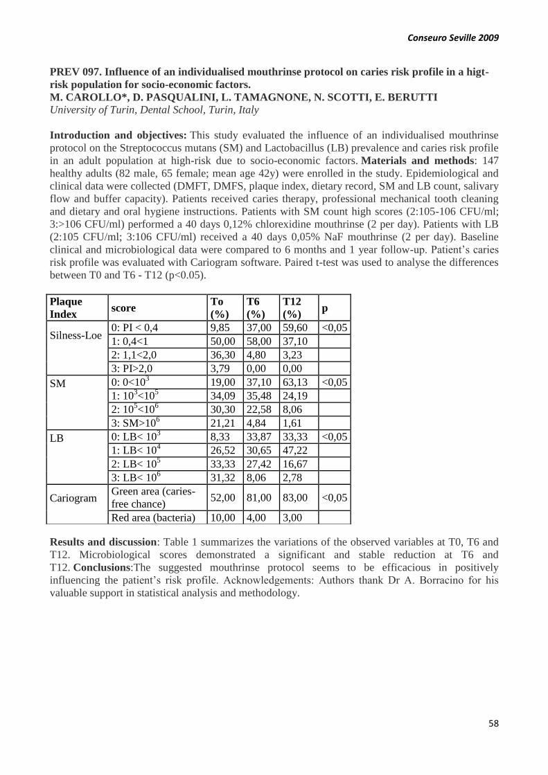
Conseuro Seville 2009
58
PREV 097. Influence of an individualised mouthrinse protocol on caries risk profile in a higt-
risk population for socio-economic factors.
M. CAROLLO*, D. PASQUALINI, L. TAMAGNONE, N. SCOTTI, E. BERUTTI
University of Turin, Dental School, Turin, Italy
Introduction and objectives: This study evaluated the influence of an individualised mouthrinse
protocol on the Streptococcus mutans (SM) and Lactobacillus (LB) prevalence and caries risk profile
in an adult population at high-risk due to socio-economic factors. Materials and methods: 147
healthy adults (82 male, 65 female; mean age 42y) were enrolled in the study. Epidemiological and
clinical data were collected (DMFT, DMFS, plaque index, dietary record, SM and LB count, salivary
flow and buffer capacity). Patients received caries therapy, professional mechanical tooth cleaning
and dietary and oral hygiene instructions. Patients with SM count high scores (2:105-106 CFU/ml;
3:>106 CFU/ml) performed a 40 days 0,12% chlorexidine mouthrinse (2 per day). Patients with LB
(2:105 CFU/ml; 3:106 CFU/ml) received a 40 days 0,05% NaF mouthrinse (2 per day). Baseline
clinical and microbiological data were compared to 6 months and 1 year follow-up. Patient’s caries
risk profile was evaluated with Cariogram software. Paired t-test was used to analyse the differences
between T0 and T6 - T12 (p<0.05).
Results and discussion: Table 1 summarizes the variations of the observed variables at T0, T6 and
T12. Microbiological scores demonstrated a significant and stable reduction at T6 and
T12. Conclusions:The suggested mouthrinse protocol seems to be efficacious in positively
influencing the patient’s risk profile. Acknowledgements: Authors thank Dr A. Borracino for his
valuable support in statistical analysis and methodology.
Plaque
Index score
To
(%)
T6
(%)
T12
(%) p
Silness-Loe
0: PI < 0,4 9,85 37,00 59,60 <0,05
1: 0,4<1 50,00 58,00 37,10
2: 1,1<2,0 36,30 4,80 3,23
3: PI>2,0 3,79 0,00 0,00
SM
0: 0<103 19,00 37,10 63,13 <0,05
1: 103<10
5 34,09 35,48 24,19
2: 105<10
6 30,30 22,58 8,06
3: SM>106 21,21 4,84 1,61
LB
0: LB< 103 8,33 33,87 33,33 <0,05
1: LB< 104 26,52 30,65 47,22
2: LB< 105 33,33 27,42 16,67
3: LB< 106 31,32 8,06 2,78
Cariogram
Green area (caries-
free chance) 52,00 81,00 83,00 <0,05
Red area (bacteria) 10,00 4,00 3,00

Conseuro Seville 2009
59
PREV 098. Influence of an individualized behavioral protocol on patient’s compliance and
caries risk profile in a high-risk population for socio-economic factors.
C. TESSORE*, D. PASQUALINI, L. TAMAGNONE, F. PERA, E.BERUTTI
University of Turin, Dental School, Turin, Italy
Introduction and objectives: This study evaluated the influence of an individualised behavioural
protocol on the compliance and caries risk profile in an adult population. Materials and methods:
147 healthy adults (82 male, 65 female; mean age 42y) were enrolled in the study. Epidemiological
and clinical data (DMFT, DMFS, plaque index, SM and LB count, salivary flow and buffer
capacity), behavioural data (manual dexterity, oral hygiene and dietary habits) were collected.
Patients received caries therapy, professional mechanical tooth cleaning; dietary and oral hygiene
instructions were furnished as individualised objectives. The satisfaction of the suggested objectives
was statistically evaluated. Patients with high scores sugar intake (diet content and frequency)
performed a 40 days 0,05% NaF mouthrinse (2 per day). Baseline clinical and behavioural data were
compared to 6 months and 1 year follow-up. Patient’s caries risk profile was evaluated with
Cariogram software. Paired t-test was used to analyse the differences between T0 and T6 - T12
(p<0.05).
Results and discussion: Table 1 summarizes the variations of the observed variables at T0, T6 and
T12. Patients received a total of 337 objectives. Dietary objectives were satisfacted in 72,86%, while
oral hygiene objectives were satisfacted in 68,78%. Conclusions: The suggested behavioural
protocol seems to be efficacious in positively influencing the patient’s risk profile.
Acknowledgements: Authors thank Dr A. Borracino for his valuable support in statistical analysis
and methodology.
Plaque Index score To (%) T6 (%) T12 (%) p
Silness-Loe
0: PI < 0,4 9,85 37,00 59,60 <0,05
1: 0,4 50,00 58,00 37,10
2: 1,1 36,30 4,80 3,23
3: PI>2,0 3,79 0,00 0,00
Diet contents
0: very low 13,64 11,29 41,67 <0,05
1: low 33,33 58,06 36,11
2: moderate 39,39 30,65 19,44
3: high 13,64 0,00 2,78
Diet frequency
0: 3/day 25,76 22,58 38,89 <0,05
1: 4-5day 30,30 43,55 30,56
2: 6-7/day 25,00 30,65 30,56
3: >7/day 18,94 3,23 0,00
CARIOGRAM
Green area (caries-free chance) 52,00 81,00 83,00 <0,05
Blue area (diet) 16,00 5,00 3,00

Conseuro Seville 2009
60
STUD 099. Undressing color
A. SALAT*, L. JANÉ, M. ROIG
U.I.C. Universidad Internacional de Catalunya
Introduction and objectives: A usual problem to face when building up aesthetic restorations is to
achieve the similar features as in natural teeth. Appropriate aesthetics must be reproduced for a
successful outcome, as a proper shape, surface texture and colour. We will show how to do a
thorough colour measurement for an anterior restoration aided by photography and common digital
technology. It is easy to use basic software to edit digital images of our clinical cases, to obtain an
accurate image that unveils the chromatic tooth features and helpful information to complete our
colour chart. Materials and methods: From an initial picture of the clinical case, using any picture
software (ex. Photoshop, Picture Project) that allows playing with contrast and brightness to modify
the colour curve. With few simple steps a very accurate chromatic map could finally be
achieved. Results and discussion: With a modified image is easy to appreciate the internal tooth
features as opalescence, intensives, characterizations, crack lines, mamelons just to mention a
few. Conclusions: If exploited properly the available digital technology, we will be able to solve
many color matching issues, without needing expensive instruments or large money investments.
OP 100. Root caries treatment in elderly: ART Technique vs. ART with Carisolv®. Clinical
randomized double blind essay. Preliminary results
R. MATEOS-PALACIOS*, J.A. GIL-MONTOYA, J.M. NAVAJAS, R. PULGAR-ENCINAS.
Faculty of Dentistry. University of Granada
Introduction and objectives: Comparing the clinical efficacy in elderly root caries lesions of the
conventional Atraumatic Restorative Treatment (ART) technique versus ART using a chemo-
mechanical method, Carisolv ®. Materials and methods: Thirty-five root-surface caries lesions
from elderly patients were treated. We randomized each lesion to receive one of the two study
treatments: Group1, ART approach (caries removed by hand instruments) and group 2, ART
approach with Carisolv® (caries removed by hand instruments helped by Carisolv®). All lesions
were restored with resin modified glass-ionomer restorative material. Status of the restorations was
assessed at six months intervals. The evaluation criteria used in most ART studies were
adopted. Results and discussion: After 12 months, 38.8% of the ART restorations were assessed as
being “present and sound”, 55.57% had a “slight marginal defect", and 5.26% restorations
disappeared. Among the ART approach with Carisolv® restorations, 40% were “present and sound”,
26.66% had a “slight marginal defect” and 20% restorations disappeared. The survival rates for the
ART approach and ART approach with Carisolv® restorations were 94.7% and 66.6%
respectively. Conclusions: The first results of study show that the use of Carisolv® doesn’t improve
the clinical behaviour of ART restoration in root caries.

Conseuro Seville 2009
61
END 101. Chlorhexidine as intracanal medicament and irrigant
F.CASCALES-MOYA*, G. SÁNCHEZ, R. PARRES, J. REDONDO, R. DELGADO-RUIZ, F.
CHIVA-GARCÍA
Dentistry Faculty, University of Murcia (Spain)
Introduction and objectives: Most important irrigants of the intraradicular canal are sodium
hypochlorite (NaOCl), citric acid and chelants. Calcium hydroxide -Ca(OH)2- is mainly used as
intracanal medicament. However, these products have some disadvantatges like some microbial
resistances. So this, other substances are being investigated to achieve better properties. The
objectives are to study the effects of chlorhexidine (CHX) as irrigant and intracanal
medicament. Materials and methods: A review of latest published literature. Results and
discussion: There are experiments in vitro and in vivo. As irrigant, chlorhexidine has being compare
with NaOCl, especially in the effect in dental tissues, antimicrobial activity and speed of action.
CHX has being also associate with Ca(OH)2 and others medicaments (camphorated p-
monochlorophenol). Antibacterial effects, pain following the treatment, speed of action and their
association with others substances has been investigated and reviewed. There are no differences
when comparing antibacterial effects of 2’5%NaOCl and 0,12%chlorhexidine. NaOCl is more
efficient in removal of debris but the substantivity is shorter than CHX. Conclusions: CHX 2% has
more antibacterial effect and faster than Ca(OH)2, but it don’t induces apex formation. However,
combination of both can be successfully for disinfection in periapical lesions and development of the
antioxidant properties of CHX.
OP 102. In vitro evaluation of microleakage levels of two etch systems in class I cavities
restored with resin composite
L.PEÑATE, A.RUIZ*, M.VALLÉS, M.MERCADÉ, M.ROIG
Department of Operative Dentistry. Universitat Internacional de Catalunya, Barcelona, Spain.
Introduction and objectives: Polymerization shrinkage and a different thermal expansion
coefficient of resin composite compared to dental tissue may cause gaps in the tooth-restoration
interface. Bacteria, fluids, molecules and ions can pass through those gaps, a process which is known
as microleakage. The aim of the present study was to evaluate the microleakage levels of Class I
cavities restored with resin composite using two different etching techniques (total etch / self
etch). Materials and methods: 111 extracted human molars were randomly divided into 6 groups.
After a Class I preparation, the adhesive was applicated (group 1=Xeno III, 2=Excite, 3= AdperTM
ScotchbondTM 1XT, 4=Adper Promp L-Pop, 5= G BondTM and 6= Prime & Bond NT) and the
specimens were restored with resin composite. After thermocycling (2000 cycles), the specimens
were immersed in methylene blue and sectioned. Microscopical photographs of the sectioned
specimens were evaluated by different operators to score dye penetration. Data were analyzed using
a two-factorial ANOVA at p<0.05. Results and discussion: Highest dye penetration was found in
group 5, followed by groups 1, 6 , 2 , 3 and 4. There was no statistically significant difference
between group 1 and 6, group 3 and 4, but statistically significant differences (p<0.05) were found
between group 1 with groups 2, 3, 4 and 5; group 2 with groups 3, 4, 5, 6; group 3 with groups 5 and
6; group 4 with groups 5 and 6; and group 5 with group 6. Conclusions: The level of microleakage
of Class I cavities in molars using total etch system is lower compared to the self etch system.
Conseuro Seville 2009
62
STUD 103. Epidemiological study of dental trauma in Valencian scholars
M. FAUS1*, E. CANDEL1, J. BONET1, V. FAUS2
1 Under graduate students. Valencia University Medical and Dental School. 2 Professor of
Conservative Dentistry and Endodontics. Director of the Master of Conservative Dentistry and
Endodontics.
Introduction and objectives: The aim of the present study was to identify some factors related to
the occurrence of dental trauma in mixed and permanent dentition. Materials and methods: Over a
period of 2 months,1325 people, 6 to 18 years old, from three primary schools were examined in
order to evaluate those factors. Data was collected through clinical examinations, photos and
interviews. Dental trauma was classified according to IADT´s criteria. Results and discussion: The
prevalence of dental trauma was 6%, boys aged between 12-18 years old experienced more injuries
than girls. Non complicated coronal fracture was the most frequent (91,18%).Games were the main
cause of trauma (40%). Color change was observed in 20% of patients. Angle´s class I was the most
frequent in traumatized people. Conclusions: Similar prevalence of injuries and factors related to
trauma that in other studies revised was observed.
OP 104. Fracture resistance of glass-ionomer liner and flowable composite used in open
sandwich Class II amalgam or composite restorations: An in vitro study
B. GÜRAY EFES, C. DÖRTER, Y. GÖMEÇ, M. TIRYAKI*, B. C. YAMAN
Dept. of Operative Dentistry, Faculty of Dentistry. Istanbul University
Introduction and objectives: The aim of this in vitro study was to investigate the effect of a glass-
ionomer liner and a flowable composite used on the gingival floor on the fracture strength of Class II
amalgam and composite restorations. Materials and methods: Class II slot preparations extending
1mm apical to the cemento-enamel junction were made in 42 teeth and randomly assigned to six
groups. Group 1 and 4, restored entirely with nanofill composite (Filtek Supreme XT, 3M/ESPE)
after applying dentin bonding agent (Adper Single Bond 2) or amalgam (World-Cap, Ivoclar). For
the remaining groups, the root-dentin area was restored with light-curing glass-ionomer liner
(Ionoseal, Voco) (Group 2, 5) or flowable composite (Filtek Supreme XT flowable, 3M/ESPE)
(Group 3, 6) and the remainder of the cavity restored with composite (Filtek Supreme XT) (Group 2,
3), or with amalgam (World-Cap) for (Group 5, 6). The restorations were loaded in compression to
failure and the data analyzed using T-test. Results and discussion: Group 6 (3110±220 N) showed
statistically higher fracture resistance than group 1(2690±670 N), 2(2420±590 N), 3 (2300±700 N),
4(2530±800N)(p< 0.05). Conclusions: Use of a glass-ionomer liner or a flowable composite on the
gingival floor did not enhance fracture resistance of the composite restorations, however flowable
composite used in open sandwich Class II amalgam restorations significantly increased fracture
strength.

Conseuro Seville 2009
63
OP 105. Effect of polishing procedures on surface roughness and gloss of resin composites
T.T. TAUBÖCK*, W. BUCHALLA, B. SENER, T. ATTIN
Department for Preventive Dentistry, Periodontology and Cariology, University of Zurich, Zurich,
Switzerland
Introduction and objectives: Effect of one/multi-step polishing systems on roughness (Ra) and
gloss (Gl) of two resin composites and correlation between Ra and Gl was investigated. Materials
and methods: Specimens of microhybrid Tetric and nanofilled Filtek Supreme XT were flattened
(320 grit SiC-paper) and polished for 60 s according to group (n=8): Untreated controls, Sof-Lex,
Astropol, Enhance/Prisma Gloss, Kenda and PoGo. Ra and Gl differences (after-before polishing,
RaD and GlD) were evaluated by ANOVA and Scheffé post-hoc-test (p0.05). Spearman correlation
was conducted between Ra and Gl. Results and discussion: Ra and Gl were inversely correlated
irrespectively of polishing and composite (p0.0001). GlD (p0.0001) but not RaD was significantly
higher for Filtek than for Tetric. Tetric: RaD of control (mean/µm -0.02) was significantly lower
compared to Sof-Lex (-0.18) and Astropol (-0.16). RaD of Sof-Lex was significantly higher than of
Enhance/Prisma Gloss (-0.03) and PoGo (-0.03). Sof-Lex (mean/GU 9.9) showed the significantly
highest GID. Filtek: RaD of control (-0.02) was significantly lower compared to Astropol (-0.13) and
Kenda (-0.12). RaD of Enhance/Prisma Gloss (+0.01) was significantly lower than of Sof-Lex (-
0.10), Astropol and Kenda. GlD of control (2.7) was significantly lower compared to Sof-Lex (15.3),
Astropol (10.6) and PoGo (12.5). Conclusions: Both, composite and polishing system influenced Gl.
OP 106. Nonvital dental bleaching - efficacy comparasion of 15% and 35% hydrogen peroxide
L. CASTRO*, R. MADUREIRA, L. FORNER
ISCS-Norte - Portugal; Facultad de Medicina y Odontología de la Universidad de Valéncia
Introduction and objectives: Today tooth bleaching is based upon hydrogen peroxide as the active
agent. The bleaching efficacy rises with the concentration of the bleaching agent. On the other hand,
higher concentrations seem to raise the risk of side effects. It has been reported in several studies
high concentrations of hydrogen peroxide efficacy, above to 30%, on non-vital bleaching in office.
However there are no studies comparing these concentrations efficacy with lower concentrations of
the same agent. The aim of this work was verify the hydrogen peroxide concentration reduction
effect on the efficacy of this agent on non-vital dental bleaching, by comparing two products with
15% and 35% hydrogen peroxide. Materials and methods: There were created two groups with ten
teeth each and one control group with five teeth as is described in the table. Bleaching percentage per
tooth was calculated after each one of the two bleaching treatment sessions for each group.
Results and discussion: 35% hydrogen peroxide bleaching percentage was always superior with
statistical significance. Conclusions: Reduction of hydrogen peroxide concentration decrease
immediate bleaching efficacy.
Group Bleaching Agent
I 15% hydrogen peroxide
II 35% hydrogen peroxide
III (control) no agent

Conseuro Seville 2009
64
END 107. The apex diameter influence in the MTA sealing ability - open apex simulation
model
P. BERNARDINO*, R. MADUREIRA, L. FORNER
ISCS-Norte/Portugal, Valência University/Spain
Introduction and objectives: In teeth with incomplete root formation by pulp necrosis, the root
development ceases and apical closure cannot be achieved. Root canal treatment at this time is a
significant challenge. MTA (Mineral Trioxide Agregate) has been suggested as a potential material
to form an apical barrier. Aim: The purpose of this work is to evaluate the influence of the apex
diameter in the MTA sealing ability when used as an apical barrier. Materials and methods: Three
experimental groups were created, with 10 single-root teeth each, with an open apex simulation with
different diameters. All canals were instrumented with Protaper® system, followed by hand
instrumentation K-flexofiles until 2mm over extrusion with 60, 80 and 100k-files in groups I, II and
III respectively. All canals received a 5mm MTA apical plug. The teeth were placed in a 50% Silver
Nitrate dye solution to test leakage, which was observed after 1mm sagital sections performed in the
last apical 5mm. Results and discussion: MTA extrusion was most frequent in roots with highest
apical diameter (group III), although there were no statistically significant differences (p<0,05)
between groups concerning its sealing ability. Conclusions: Increased apex diameter is linked to an
increased risk of MTA extrusion but not to an increased apical leakage.
STUD 108. Methodological proposal for analyzing the international scientific production in
Dentistry on the Web of Science
I. JIMÉNEZ-FERNÁNDEZ*1, E. JIMÉNEZ CONTRERAS2, R. PULGAR ENCINAS1
1Stomatology Department, University of Granada, 2 Library and Information Science Department,
University of Granada
Introduction and objectives: The studies in Science Citation Index (SCI) about dental scientific
publications, have been based until now just on analyzing information under category “Dentistry,
Oral Surgery and Medicine” (DOSM) from Journal Citation Reports (JCR). Probably the fact of
excluding the rest of the JCR categories is due to difficulties on design searching techniques to
retrieve this information. The objective on this project is to design retrieval strategies in order to
generate data bases including all SCI dental records not only in DOSM category but in the rest of
medical journals. We will analyze two periods: 1994-1996 and 2004-2006. Materials and methods:
Creating this data base entail two tasks. One is analyzing DOSM from the JRC and as a second task a
breakthrough procedure is proposed. This new technique is hybrid, thematic and author´s
institutional affiliation to recover dental scientific information published on journals not included on
DOSM. Data base including SCI records related to dental scientific publication between 1994-1996
and 2004-2006. Results and discussion: The number of records is 33.618. Conclusions: The
innovative technique presented allows retrieve dental scientific production published on DOSM
journals as well as those other papers not included in this category. This combined analysis
guarantees a better scientific description of the dental research.
Conseuro Seville 2009
65
CLIN 109. LAVA® system. Clinical case
J. PELAEZ*, P.GOMEZ, I. VAZQUEZ, MJ. SUAREZ
Department of Buccofacial Prosthesis. Faculty of Odontology. University Complutense of Madrid
Introduction and objectives: The recent progress in material technology and manufacturing
procedures has extended the indications of all-ceramic systems not only for single crowns
restorations but also for fixed partial dentures (FPDs) in the anterior and posterior sectors. The
objective of this study is to explain the manufacturing process as well as the clinical systematic of
preparation of Lava AllCeramic System posterior fixed partial denture . Materials and methods:
We present a clinical case of a patient with decoloration by tetracycline and absences of 16 and
26. Results and discussion: Once clinical history and the complementary exploration were made, it
was decided to rehabilitate the patient with 3-unit posterior FPD of LAVA® All Ceramic System
(3M ESPE) (CAD/CAM system + zirconium oxide ceramic system). Conclusions: The LAVA
system offers a suitable marginal sealing clinically observed, as well as excellent esthetic results,
allowing in addition a conservative dental preparation. There will be necessary long -term clinical
studies to evaluate the fracture resistance of this new ceramic system.
OP 110. Quantitative margin analysis and shear bond tests for selecting effective experimental
adhesives
U. BLUNCK*, P. ZASLANSKY
DGZ
Introduction and objectives: The purpose of this study was to evaluate the performance of
experimental adhesive systems prior to launching. Materials and methods: 7 groups of 8 Class-V-
cavities were prepared in extracted human incisors (4 mm high, 3 mm wide, 1.5 mm deep, half of
margin length located in enamel). 7 experimental adhesives (Saremco Dental) were combined with
the composite ELS. Replicas were created after 21 days of water storage (TM1) and after a first
thermocycling (TM2), and for 4 adhesives also after one year of water storage (TM3) and after a
second thermocycling (again 2000 cycles, 5° to 55°C) (TM4). Quantitative margin analysis was
performed using an SEM (200x) according to standard criteria. The results were compared with
shear bond strengths (SB) performed by Saremco. Results and discussion: Median values (MPa for
SB, % ”continuous margin” for quantitative margin analysis) for the seven experimental adhesives
(SB/TM1/TM2/TM3/TM4) were I:10.0/36.4/9.5, II:10.0/27.0/10.7, III:10.0/0.0/0.0,
IV:16.0/100.0/100.0/100.0/100.0, V:14.0/92.0/56.9/49.7/20.7, VI: 16.0/100.0/100.0/100.0/100.0,
VII:14.0/100.0/98.5/98.1/71.7. Statistical evaluation (Kruskal-Wallis-Test with Bonferroni
adjustment, p<0.05) showed best results for experimental adhesives IV and VI, from which VI was
launched as cmf adhesive system. Conclusions: Quantitative margin analysis provides detailed and
precise non-destructive measures of bonding effectiveness in experimental products, supporting and
strengthening shear-bond tests (especially after one year water storage). Acknowledgements:
Supported by Saremco Dental.

Conseuro Seville 2009
66
OP 111. In vitro evaluation of color measuring devices.
A. TSILIAGKOU*, E. PAPAZOGLOU, A. KAKABOURA, G. VOUGIOUKLAKIS
Dept. Operative Dentistry, School of Dentistry, University of Athens, Greece
Introduction and objectives: The aim of this in vitro study was to evaluate the validity and
repeatability of three commercially available color measuring devices. Materials and methods:
Three devices were tested: (ES) Vita Easyshade spectrophotometer (Vita), (SS) Spectroshade
spectrophotometer (MHT) and (SV) ShadeVision colorimeter (X-Rite). Each one of the 5 shade tabs
[B1, A2, A3,5, D3, C4 - Vita Classical shade guide (VC)] was placed in a manikin model covered by
a black box and 15 measurements in sequence were performed per device under standardized
conditions. Data in L*,a*,b* values were recorded for all the devices and measurements. The
repeatability was expressed as the coefficient of variance (CV) for L, a*, b* values. Color differences
between every measured tab and its reference (VC) analogue were defined by calculation of
ΔE=[(ΔL*)2+(Δa*)2+(Δb*)2]1/2. ΔE values subjected one-way Anova (a=0.05).The validity per
L*,a*,b* parameter was determined relative to the correspondence values of the reference VC by one
sample t-test (a=0.05).
Device Repeatability ΔE (mean±SD)
CV
(L*) CV (a*)
CV
(b*) B1 A2 A 3,5 D3 C4
ES 0–0,01c 0.05–
0.32 c
0–
0,03 c
3,27±0,26 4,79±0,55 2,38±0,32 4,38±0,44 1,98±0,14
SS 0–0,01 0,16–1,92 0–0,04 2,06±0,19 1,86±0,80 1,09±0,16 2,44±0,12 1.02±0,02
SV 0–0,01 0,03–7,32 0–0,02 1,28±0,25 2,10±0,20 1,46±0,19 2,36±0,16 1,01±0,16
c range of values from all shade tabs tested.
Results and discussion: Concerning repeatability, a* showed higher CV values with wider
distribution than L* and b*, which presented negligible ones. ES exhibited statistically higher ΔE
values for all tab shades compared to SS and SV. In regards of validity, only L* parameter of B1, D3
and C4 tabs measured by SS device proved as valid. Conclusions: 1) All the devices presented high
repeatability in L* and b* parameters but not in a*, 2) The validity of all the devices can be
characterized as poor with the exception of Spectroshade in L* parameter and, 3) Spectroshade and
ShadeVision provided lower color differences (ΔE) than Easyshade relative to the reference Vita
Classical shade guide tabs.

Conseuro Seville 2009
67
CLIN 112. Horizontal root-fractured teeth: report cases
M. HERNÁNDEZ-MASET* 1, P. MICÓ MUÑOZ 2. T. FAYOS SOLER 2, V.J. FAUS-
LLÁCER 3, A SALOM FONTANA 1
1Master Students in Conservative Dentistry and Endodontics. Valencia University Medical and
Dental School. 2 Master Professor in conservative Dentistry and Endodontics. Professor associate of
Conservative Dentistry and Endodontics. Valencia University Medical and Dental School.
3Professor of Conservative Dentistry an Endodontics. Chairman of the Master of Conservative
Dentistry and Endodontics. Valencia University Medical and Dental School.
Introduction and objectives: Horizontal root fractures are common in dental trauma, mainly
occurring on the cervical and middle third of the root of permanent upper incisors. Diagnosis of
horizontal root fractures is reached by the information obtained in clinical radiographic
examinations, and requires different healing patterns, according to the etiology and the location of
fracture lines. The aim of this issue was to report the clinical management and their different
results. Materials and methods: Presentation of different clinical cases, each one with a different
treatment according to clinical conditions which present in the future evolution. Results and
discussion: After the application of the pre-established, all cases had a successful
evolution. Conclusions: We should choose a specific treatment for each case, according to clinical
criteria and the post-evolution.
STUD 113. Importance of post in dental resistance to fractures
V. GARCIA-PEÑALVER*, S. PARRA-CANDEL, E. SANCHEZ-GUIRAO, E. VILLORA-
MORCILLO, J.C. BÁGUENA-GÓMEZ, E. GARCIA-CRUZ
Dentistry Faculty, University of Murcia (Spain)
Introduction and objectives: In endodontically treated teeth of with great loss of coronal structure,
posts are used. However, many dentists don’t use them for the possibility of causing dental fractures.
The aim of this study is to evaluate the dental resistance to fracture according to presence/no
presence of posts, kind of tooth and post types and placing techniques. Materials and methods:
Review of the published literature. Results and discussion: The use of posts is necessary in the
reconstruction of the dental structure to allow restoration retention . In contrast, this additional
retention can cause the loss of tooth resistance. This is solved by placing posts with similar elasticity
to the dentin. Posts are more resistant to fractures in this order: quartz >zirconium >glass fiber
>titanium. The most fractured teeth are second premolars and mesial root of mandibular molars. A
good technique for placing a post would be: post free 5mm in the apical third , 2/3 of the post into
root canal, parallel walls, maximum conservation of dental tissue. Conclusions: Posts shouldn't be
placed randomly in posterior sectors. If we use posts to increase retention, tooth will lose resistance
to fracture. Posts in anterior sectors must be aesthetic.
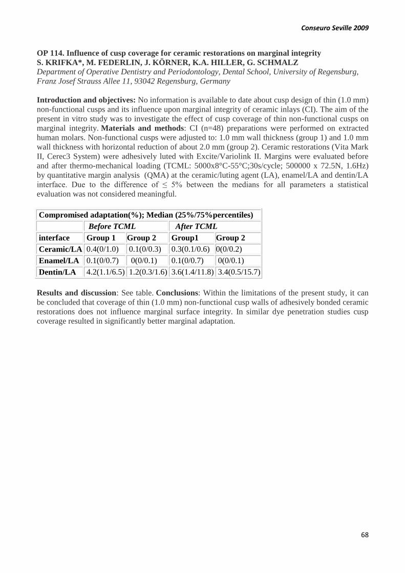
Conseuro Seville 2009
68
OP 114. Influence of cusp coverage for ceramic restorations on marginal integrity
S. KRIFKA*, M. FEDERLIN, J. KÖRNER, K.A. HILLER, G. SCHMALZ
Department of Operative Dentistry and Periodontology, Dental School, University of Regensburg,
Franz Josef Strauss Allee 11, 93042 Regensburg, Germany
Introduction and objectives: No information is available to date about cusp design of thin (1.0 mm)
non-functional cusps and its influence upon marginal integrity of ceramic inlays (CI). The aim of the
present in vitro study was to investigate the effect of cusp coverage of thin non-functional cusps on
marginal integrity. Materials and methods: CI (n=48) preparations were performed on extracted
human molars. Non-functional cusps were adjusted to: 1.0 mm wall thickness (group 1) and 1.0 mm
wall thickness with horizontal reduction of about 2.0 mm (group 2). Ceramic restorations (Vita Mark
II, Cerec3 System) were adhesively luted with Excite/Variolink II. Margins were evaluated before
and after thermo-mechanical loading (TCML: 5000x8°C-55°C;30s/cycle; 500000 x 72.5N, 1.6Hz)
by quantitative margin analysis (QMA) at the ceramic/luting agent (LA), enamel/LA and dentin/LA
interface. Due to the difference of ≤ 5% between the medians for all parameters a statistical
evaluation was not considered meaningful.
Compromised adaptation(%); Median (25%/75%percentiles)
Before TCML After TCML
interface Group 1 Group 2 Group1 Group 2
Ceramic/LA 0.4(0/1.0) 0.1(0/0.3) 0.3(0.1/0.6) 0(0/0.2)
Enamel/LA 0.1(0/0.7) 0(0/0.1) 0.1(0/0.7) 0(0/0.1)
Dentin/LA 4.2(1.1/6.5) 1.2(0.3/1.6) 3.6(1.4/11.8) 3.4(0.5/15.7)
Results and discussion: See table. Conclusions: Within the limitations of the present study, it can
be concluded that coverage of thin (1.0 mm) non-functional cusp walls of adhesively bonded ceramic
restorations does not influence marginal surface integrity. In similar dye penetration studies cusp
coverage resulted in significantly better marginal adaptation.

Conseuro Seville 2009
69
END 115. The effect of two techniques of instrumentation associated to ultrasonic irrigation to
remove the smear layer of root canals
F. TADEU*, R. MADUREIRA, L. FORNER
Portuguese Dental Association
Introduction and objectives: The mechanical instrumentation of the root canal creates an irregular
layer known as smear layer. The meta-analysis made, tell us that the advantages or disadvantages of
the presence of smear layer are controversial. With scanning electronic microscope, it was studied
the capacity to remove the smear layer from the root canals, provoked by the instrumentation with
the manual step-back and rotatory technique associated or not to the passive ultrasonic irrigation
(PUI). Materials and methods: A total of 80 palatin and distal root canals of molars had been
divided randomly in four distinct groups. After each instrument, the root canals were flushed with a
3% NaOCl solution. After splitting the roots longitudinally, the specimens were examined under
scanning electronic microscope.
Group
Manual
step-back
technique
Rotatory
technique
(ProTaperTM)
PUI
A x
B x
C x x
D x x
Results and discussion: The association of PUI to any one of the instrumentation techniques had the
elimination of smear layer with bigger effectiveness, while that, without the aid of ultrasonic, the
rotatory technique left 50% of the canals without smear layer and the manual technique left almost
canals walls with debris. Conclusions: The statistic analysis showed that PUI increase the capacity
of the cleaning of smear layer from root canals.

Conseuro Seville 2009
70
OP 116. Influence of composite thickness and curing light intensity on the degree of conversion
of microhybrid resin composites: an in vitro study.
N. SCOTTI*, A. VENTURELLO, G. MIGLIARETTI, F. PERA, D. PASQUALINI, F.
GEOBALDO , E. BERUTTI
University of Turin
Introduction and objectives: Modern curing lights are available with outputs exceeding 800
mW/cm2, resulting in recommended shorter exposure times. However, this shorter irradiation may
lead to an insufficient degree of conversion of the composite resin, especially at the bottom of the
restorations. The aim of this in vitro study is to compare the degree of conversion obtained with
high-intensity halogen and LED curing units related to different composite thickness, evaluating the
efficacy of shortened irradiation time. Materials and methods: A micro-hybrid resin composite,
shade A3, was placed in 18 aluminium rings with the same diameter (10mm) and different thickness
(1mm, 2mm, 3mm). A tungsten halogen light and a LED lights were employed. The degree of
conversion at the bottom surface of each sample was accomplished by ATR FT-IR analysis
immediately after the high-intensity curing program. Conversion values were compared with the
ANOVA for repeated measures (p<0.05).
DEGREE OF CONVERSION
(mean and SD)
LAMP 1mm 2mm 3mm
Halogen (Swiss
Master Light)
0,35
(±0,02)
0,25
(±0,07)
0,19
(±0,03)
LED (Translux
Power Blue)
0,38
(±0,03)
0,33
(±0,04)
0,26
(±0,05)
Results and discussion: The results are expressed in table 1. The statistical analysis showed a
significant difference between the sample thicknesses but not between different curing
lights. Conclusions: The mean degree of conversion obtained in all sample was quite low,
suggesting that, despite the high intensity of the light, shortened irradiation times aren’t sufficient for
the composite polymerization.

Conseuro Seville 2009
71
STUD 117. Experimental evaluation of the adaptation of fiber post to canals shaped with
ProTaper rotary instruments
N. SCOTTI, R. ROTA*, L. TAMAGNONE, D. PASQUALINI, V. BOBBA, A .
VENTURELLO, G. MIGLIARETTI, E. BERUTTI
University of Turin
Introduction and objectives: Adaptation of the fiber post to the dowel space may be a determinant
factor in the clinical performance of fiber-reinforced composite posts, especially in clinical situations
with extreme loss of the root-canal dentin. The primary objective was to evaluate adaptation of fiber
post to dowel space, prepared with two different techniques. The secondary objective was to examine
the correlation between cement thickness and post bond strength, through the push-out
test. Materials and methods: Thirty-two single-rooted teeth were treated endodontically, 16 root
canal filled, and a dowel space was prepared with Largo vs Largo and post dedicated drills. Each
specimen was sectioned into six 1 mm slices. Cement’s thickness was determined using specific
software. Push-out values were compared with the Mann-Whitney U test (p<0.05). A linear
regression model was used to correlate cement thickness with the work required to dislocate the
post. Results and discussion: The Largo drill technique showed a greater cement thickness
(p<0.001), with no differences in push-out values. The linear regression model showed a positive
association between cement thickness and work required (p<0.001). Non-obturated specimens had
higher push-out values than obturated ones (p<0.001). Conclusions: Bond strength does not appear
to be influenced by the dowel space preparation technique, although work necessary to dislocate the
fiber post is proportional to the cement thickness.

Conseuro Seville 2009
72
STUD 118. Hydrogen peroxide penetration in pulp chamber following different teeth bleaching
treatments
N. SCOTTI, L. BERGANDI, F. STUFFER, R. BECCIO, A. DALMAZZO*, D. PASQUALINI,
D. GHIGO, E. BERUTTI
University of Turin
Introduction and objectives: In order to minimize the irritating effects on the pulp by bleaching
agents, it is mandatory to define which bleaching technique leads to the lowest penetration of free
radicals in the pulp chamber. The aim of this in vitro study is to evaluate the penetration of free
radicals in the pulp chamber after different bleaching techniques. Materials and methods: Twelve
extracted teeth were treated with different bleaching protocols. Acetate buffers were placed into the
pulp chamber to absorb and stabilize any peroxide that might penetrate. The penetration of H2O2
into the pulp chamber was determined spectrophotometrically by a UV-visible microplate reader set
to 596 nm, at room temperature. The results were analyzed by a one-way analysis of variance and
Tukey's test. P < 0.05 was considered significant.
Table 1. Penetration of hydrogen peroxide in pulp chamber.
Group
(n = 3)
Time of
application
(min)
Pulpal
peroxide
(% w/w)
p value
% of pulpal
peroxide vs.
total peroxide
released
Opalescence
Extra-boost 38% 10 2.512 ± 0.06
p <
0.0001 6.9
Opalescence
Extra-boost 38% 20* 2.511 ± 0.04
p <
0.0001 6.9
Opalescence
15% 10 0.925± 0.02 p < 0.001 7.4
Opalescence
15% 240 1.674 ±0.04 p < 0.001 16.2
* After a 10 minutes application, the bleaching gel was replaced with a new one for
10 minutes further
Results and discussion: Results are expressed in table 1. The release of peroxide from Opalescence
Extra-boost 38% was similar after 10 and 20 min. Of course, it is difficult to compare the release
from Opalescence 15% after 10 and 240 min, due to the likely transformation of the relatively
unstable hydrogen peroxide after the longer time. Conclusions: The penetration of peroxide into the
pulp chamber wasn’t function of application time, while it was proportional to the percentage of
bleaching agent. Bleaching agents with high peroxide concentration seem to rapidly release the
peroxide saturating the tooth.
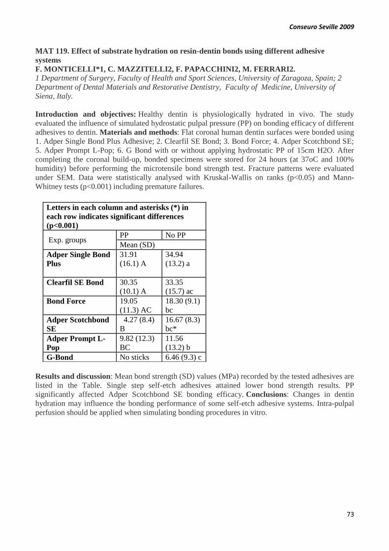
Conseuro Seville 2009
73
MAT 119. Effect of substrate hydration on resin-dentin bonds using different adhesive
systems
F. MONTICELLI*1, C. MAZZITELLI2, F. PAPACCHINI2, M. FERRARI2.
1 Department of Surgery, Faculty of Health and Sport Sciences, University of Zaragoza, Spain; 2
Department of Dental Materials and Restorative Dentistry, Faculty of Medicine, University of
Siena, Italy.
Introduction and objectives: Healthy dentin is physiologically hydrated in vivo. The study
evaluated the influence of simulated hydrostatic pulpal pressure (PP) on bonding efficacy of different
adhesives to dentin. Materials and methods: Flat coronal human dentin surfaces were bonded using
1. Adper Single Bond Plus Adhesive; 2. Clearfil SE Bond; 3. Bond Force; 4. Adper Scotchbond SE;
5. Adper Prompt L-Pop; 6. G Bond with or without applying hydrostatic PP of 15cm H2O. After
completing the coronal build-up, bonded specimens were stored for 24 hours (at 37oC and 100%
humidity) before performing the microtensile bond strength test. Fracture patterns were evaluated
under SEM. Data were statistically analysed with Kruskal-Wallis on ranks (p<0.05) and Mann-
Whitney tests (p<0.001) including premature failures.
Results and discussion: Mean bond strength (SD) values (MPa) recorded by the tested adhesives are
listed in the Table. Single step self-etch adhesives attained lower bond strength results. PP
significantly affected Adper Scotchbond SE bonding efficacy. Conclusions: Changes in dentin
hydration may influence the bonding performance of some self-etch adhesive systems. Intra-pulpal
perfusion should be applied when simulating bonding procedures in vitro.
Letters in each column and asterisks (*) in
each row indicates significant differences
(p<0.001)
Exp. groups PP No PP
Mean (SD)
Adper Single Bond
Plus
31.91
(16.1) A
34.94
(13.2) a
Clearfil SE Bond 30.35
(10.1) A
33.35
(15.7) ac
Bond Force 19.05
(11.3) AC
18.30 (9.1)
bc
Adper Scotchbond
SE
4.27 (8.4)
B
16.67 (8.3)
bc*
Adper Prompt L-
Pop
9.82 (12.3)
BC
11.56
(13.2) b
G-Bond No sticks 6.46 (9.3) c

Conseuro Seville 2009
74
END 120. Osteogenic differentiation of dental pulp stem cells
F.J. RODRIGUEZ*, M. BLANQUER, C. INSUASTI, L. MESEGUER, P. BLEDA, F.
INIESTA, R. OÑATE, S. MARTINEZ, J.M. MORALEDA
University of Murcia
Introduction and objectives: Literature states that human postnatal dental pulp stem cells
(HDPSCs) have the ability to differentiate into osteoblastasts. Our purpose is to differentiate
HDPSCs with three different medium and compare their osteogenic ability. Materials and methods:
Human dental pulp was extracted from teeth of healthy adult subjects aged 21 to 45 years. The pulp
was gently removed and immersed in a digestive solution for 1 h at 37Cº. After digestion, cells were
placed in three flasks with 3 different mediums. Medium 1: Osteodiff (Miltenyi®); Medium 2: α-
MEM supplemented with 15% fetal bovine serum, 100 U/ml penicillin, 0.1 mg/ml streptomyc0in,
and 0.25 lg/ml. amphotericin B; Medium 3: α-MEM medium, supplemented with 20% fetal bovine
serum, 100 mM 2P-ascorbic acid, 2 mM L-glutamine, 100 U/ml penicillin, 0.1 mg/ml streptomyc0in,
and 0.25 lg/ml.amphotericin B. Flasks were incubated at 37ºC in a 5% CO2 and the medium changed
twice week. Results and discussion: After 35 days, HDPSCs formed and developed mineralization
nodules (clusters), as revealed by Alizarin red staining. This staining was bigger with Medium 1 that
medium 2 and medium 3. Conclusions: This study confirms the ability of HDPSCs to differentiate
into osteoblasts , and proves the superior efficacy of the Osteodiff Medium (Miltenyi®) over two
locally made osteo-differentiation published formulations.
PREV 121. Effect of new protective chemical agents on microtensile bond strength of luting
resins to dentine
M. E. ÇÖMLEKOĞLU*, M. DÜNDAR, M. ÖZCAN, B. H. ŞEN
Ege Univesity, School of Dentistry
Introduction and objectives: To test the microtensile bond strengths (MTBS) of newly developed
pretreatment methods for dentin using different chemical agents to form a resistant layer against
demineralization. Materials and methods: Twenty-seven human molars (N=27) were embedded
and flat dentine surfaces were exposed. They were divided into three luting resin groups [SuperBond
C&B (S), Variolink II (V) and Multilink (M)] (n=9/group). Each group was further divided into 3
subgroups of surface treatments, namely TiF4, CAPE and control. Flat dentine surfaces were ground
with 400 grit silicon carbide papers. Conditioned ceramic specimens (5x5x5 mm) were bonded using
the luting agents. After thermocycling (5o-55o, 20s, x1000), each tooth was sectioned into multiple
beams (1x1mm). The sticks (n=108) were fixed to a universal testing machine and stressed until
failure (1 mm/min). MTBS (MPa) were calculated. Failure modes were evaluated under SEM. Two-
way ANOVA and Bonferroni tests were used at 95% CI. Results and discussion: The effect of
cement type on MTBS was significant with V and S presenting higher values (15.7±3.4, 14.9±3.9,
respectively) than that of M (10.5±3.2) (p<0.05). The MTBS values of the experimental chemical
agent groups were significantly lower (CAPE: 12.9±2.2, TiF4:12.9±3.8) than the control group
(15.4±3.8) (p<0.05). Failure types [(adhesive (a), cohesive (c), mixed (m)] for TiF4, CAPE and
control were 20a/4c/12m, 19a/17m and 12a/5c/19m, respectively. Conclusions: The experimental
chemical agent application on the prepared dentine decreased the microtensile bond strength values
of ceramic to dentine.

Conseuro Seville 2009
75
OP 122. Dentinal permeability modifications produced by four dentin adhesives applied with
and without positive liquid pressure in a modified Pashley system
M. MELO*, A. PASCUAL-MOSCARDÓ, F. GIL-LOSCOS, F. ALPISTE-ILLUECA , A.
FYGUEROA-GARCÍA
Dentistry-Dental Materials. University of Valencia
Introduction and objectives: Dentinal sensitivity (DS) is a frequent problem in patients. Several
theories have been proposed to explain the mechanism of DS. The most widely accepted is the so-
called Hydrodynamic Theory. According to this theory dentinal adhesives can be used to treat DS by
creating a coat on the exposed dentine that reduces dentinal permeability. The aim of this in vitro
study is to analyze the modification of dentinal permeability produced by four dentin adhesives
applied with and without positive liquid pressure in a modified Pashley system, using scanning
electron microscopy (SEM) to confirming our results. On the other hand, we compared the materials
to find the best adhesive in reducing dentinal permeability. Materials and methods: Two self etch
adhesives (AdheSE® y G BondTMTM) and two total etch adhesives (AdperTM ScotchbondTM
1XT y ENA BOND) were applied on dentinal discs and introduced in the modified perfusion system.
In half of the discs adhesives were applied with liquid pressure and in the remaining half without
liquid pressure. SEM was used to complement perfusion results. Results and discussion: In all
cases, liquid pressure negatively affects adhesive capability to reduce dentinal permeability. Self etch
adhesives showed the best capability to reduce dentinal permeability. Conclusions: Our results
suggest that dentinal permeability should be introduced systematically in perfusion studies.
OP 123. Comparison of amalgam repair strategies in situations with and without dentin
exposure
M. OZCAN, B. GOKCE*, M.E. COMLEKOGLU,M. DUNDAR
Ege Univesity, School of Dentistry
Introduction and objectives: To test the bond strength strategies on amalgam repairs with and
without surrounding dentin. Materials and methods: Coronal parts of sound human canines (N=50,
n=10 per group) were embedded in autopolymerized PMMA. Cylindrical preparations (diameter: 2.3
mm, depth: 3 mm) were made and filled with a non-Gamma 2 amalgam (Cavex). Dentin surfaces
were exposed, specimens were ground finished using 1200-grit silicone carbide papers and they were
randomly assigned to one of the following conditioning methods: Group 1: Silicacoating amalgam
(30 µm CoJet Sand), etching dentin, silane application on amalgam, adhesive (Clearfil SE Bond) on
dentin, opaquer on amalgam, resin composite (Clearfil Photo Posterior) on both; Group 2: Etching
dentin, silicacoating amalgam, silane application on amalgam, adhesive on dentin, opaquer on
amalgam, resin composite on both; Group 3: Etching, adhesive on dentin, opaquer on amalgam, resin
composite on both. The specimens bonded only on amalgam without involvement of dentin acted as
control groups and they were further assigned to one of the following conditioning methods: Group
4: Silicacoating, opaquer, resin composite and Group 5: No conditioning, opaquer, resin composite.
Specimens were kept in water at 370C for five weeks prior to shear bond test (1mm/min). Failure
types were classified by two calibrated operators. ANOVA and Tukey-Kramer adjustment tests were
made at 95% CI. Results and discussion: Surface conditioning method had a significant effect on
the results (p<0.05) (ANOVA). Unconditioned control group (Gr 5) showed the least favorable
results (1.4 MPa). The highest results were obtained in Group 1 (39.9±4 MPa) being significantly
higher (p<0.05) than those of other groups. Silicacoating resulted in exclusively (100%) cohesive
failures in the amalgam. Conclusions: For reliable repair of amalgam-dentin complex, first amalgam
then dentin must be conditioned.

Conseuro Seville 2009
76
MAT 124. The use, application and hazardous effects of hydrofluoric acid in dentistry
M. DUNDAR*, M. OZCAN
Ege Univesity, School of Dentistry.
Introduction and objectives: To review the chemical properties, clinical applications of
hydrofluoric acid (HF) in restorative dentistry and to highlight the possible hazardous effects of this
agent as well as to recommend the treatment approach for potential risks, based on the review of the
literatures existing in chemical and medical fields. Materials and methods: All original scientific
full papers listed in PubMed and Medline about HF were included in this review using the search
terms: Hydrofluoric acid, Acid etching, Surface conditioning, Ceramics, Fracture, Repair,
Hydrofluoric acid burns, Hydrofluoric acid treatment, hydrofluoric acid etching, workplace
poisoning, poisoning, fatalities, ceramics intra-oral repair, Fluorohydric acid, Fluoric acid, and
Hydrogen fluoride solution between 1970-2007. Scientific reports, medical and chemical textbooks
and handbooks were also used. Product information was referred with instruction articles, and the
internet pages of the manufacturers. Results and discussion: 133 relevant articles were reviewed by
two operators. HF is particularly aggressive for soft tissues. This is not based on the pH value, but on
its toxicity. Although HF is considered to be a dangerous, harmful, irritating compound, both
laboratory evaluations and clinical procedures concerning its use for glass ceramic cementation and
intraoral ceramic repair have been reported without any complications. No biological risks in vivo
have been reported in dentistry but HF burns have been reported in dermatology literature in the
form of case reports. Conclusions: HF is still the most effective medium for conditioning ceramics
with glassy matrix before bonding resin composite. Exposure to HF can produce harmful health
effects that may not be immediately apparent but in the dental literature no such cases were found.
MAT 125. Marginal fit of Cr- Ni- Ti restaurations: influence of casting technique and finish
line design
P. GÓMEZ COGOLLUDO*, J. PELÁEZ, JF LÓPEZ LOZANO, MJ SUÁREZ
Faculty of Dentistry. Complutense University of Madrid
Introduction and objectives: The study was focused on researching the influence of casting
technique and finish line design in marginal fit of Cr- Ni- Ti restorations. Materials and methods:
Sixty standardized specimens of brass were prepared to receive metal-ceramic crowns, divided into
two groups according to the cervical finish line: chamfer and rounded shoulder. Every group was
divided in three subgroups according to the casting process. Induction and centrifuge, burner and
centrifuge and induction and vacuum/pressure. All groups were casting with a Cr- Ni-Ti alloy
(Tillite®) and veneered with Vita VM® 13. The marginal fit was measured at four points using an
image analysis software: buccal, lingual, mesial, and distal. Results and discussion: Burner and
centrifuge (45,87 μm) and Induction and centrifuge (48,62 μm) showed the lowest marginal
discrepancies and significant differences were detected between these groups and induction and
vacuum/pressure (71,08 μm). Chamfer finish line showed the lower values of marginal discrepancy
(54.12 m), obtained the rounded shoulder similar values (56.26 m). No significant differences
were observed between the two finish lines. No significant interactions between casting technique
and finish line were demonstrated. Lingual surface showed the best marginal fit. Conclusions: The
accuracy of fit achieved for the sixth groups analyzed was within the range of clinical acceptance.
Conseuro Seville 2009
77
END 126. Push out-strength of fiber posts placed in pre-existing root canal fillings
M. DIMITROULI*, W. GEURTSEN, H. GÜNAY, A.-K. LÜHRS
Department of Conservative Dentistry, Periodontology & Preventive Dentistry, Hannover Medical
School
Introduction and objectives: Purpose of this study was to compare the bond strength of two fiber
post systems (RelyX Unicem/RelyX Fiber Post (RLX), Variolink II/ DT Light SL (VDW)) in
relation to root canal fillings. Materials and methods: 160 human teeth were divided into 4 groups:
guttapercha/AH Plus (GAP), guttapercha/Guttaflow (GGF), old ‘pre-existing’ root canal fillings
(ORF), canals without filling (WRF, control). After canal preparation, the posts were cemented and
half the specimens were thermocycled (TC, 5000 cycles, 5- 55°C). All specimens were exposed to
the ‘push-out’-test (1 mm/min). Results and discussion: Before/after TC, type of root canal filling
did not affect bond strength. VDW-WRF before TC had the highest bond strength (16.5±6.4 MPa).
After TC, VDW-GGF showed a decreased bond strength (9.5±3.2 vs 5.3±2.4, p=0.005). TC did not
affect the bond strength of RLX. Groups ORF and WRF revealed significant differences for VDW
and RLX (ORF: 16.3±6.0 vs 7.0±2.4, p=0.001, WRF: 16.5±6.4 vs 8.0±5.0, p=0.004) before TC.
After TC, no differences were assessed. Microscopic evaluation of posts showed mainly adhesive
failures between post and cement for VDW or mixed fractures for RLX. Conclusions: Fibre post
systems can be used without regard of the root canal sealer. The etch&rinse system caused higher
bond strength values than the self-adhesive system.
OP 127. Gingiva-shaded compomers: clinical results after 24 months
A.-K. LÜHRS, W. GEURTSEN*, S. GUHR, H. GÜNAY
Department of Conservative Dentistry, Periodontology & Preventive Dentistry, Hannover Medical
School
Introduction and objectives: Periodontal recessions with wedge-shaped defects often generate
aesthetic and functional problems. The restoration of these defects with tooth-coloured materials
sometimes leads to inferior aesthetic results (‘long clinical crowns’). These problems may be reduced
by using gingiva-shaded compomers. Purpose of this study was the evaluation of their clinical long
term performance. Materials and methods: 18 wedge-shaped defects were restored with a gingival-
shaded material and a self-etch adhesive. After 16 and 24 months, all restorations were monitored
and assessed by two calibrated examiners (mod. USPHS-criteria, probing depth, Wilcoxon-test for
statistics) and a questionnaire regarding the patients’ satisfaction with the treatment outcome was
used. Results and discussion: At baseline 18 (retention: 100%), after 16 months 17 (94.4%), and
after 24 months 16 restorations (88.8%) were evaluated. Regarding “anatomic form”, at baseline
83.3% of the restorations scored A in contrast to 41.2% at 16 months and 58.8% at 24 months
(p<0.05). For “gingival reaction” A-scores were 83.3% vs. 23.5% vs. 18.8% (p=0.002). No
significant differences were detected regarding probing depths. The patients’ satisfaction with the
treatment outcome was high. Conclusions: Retention rate using a self-etch adhesive dropped by
more than 11% within 24 months. Regarding anatomic form, a recall should be performed at regular
intervals for re-finishing the cervical restoration margins.

Conseuro Seville 2009
78
OP 128. Shear bond strength of self adhesive resin cements in vitro
A.-K. LÜHRS*, S. GUHR, W. GEURTSEN
Department of Conservative Dentistry, Periodontology & Preventive Dentistry, Hannover Medical
School
Introduction and objectives: Self-adhesive luting resins facilitate the placement of indirect
restorations. The purpose of this study was to evaluate their shear bond strength to enamel and
dentin. Materials and methods: 60 molars were randomly assigned to 12 test-groups (each n=10)
and proximal surfaces were ground flat to get an enamel and dentin surface with a diameter of at
least 4 mm. Lithiumdisilicate-ceramic specimens were bonded to the surfaces either with Variolink /
Syntac (VSC), Panavia F2.0 (PAF), RelyX unicem (RLX), Maxcem Elite (MCE), iCem (IC) or an
experimental self-adhesive resin cement (EXP). The shear bond strength (crosshead speed 1
mm/min) was measured after 24h-storage in NaCl (37°C). The fracture modes were determined with
a stereomicroscope (magnification 8 to 50 fold). For statistics, t-test was used. Results and
discussion: VSC revealed the highest shear bond strength within the enamel groups (42.9 ± 9 MPa),
IC the lowest (10.5 ± 4.2 MPa, p<0.001). The highest dentin shear bond strength was determined for
VSC (39.2 ± 8.9 MPa, p<0.001), the lowest for EXP (7.8 ± 3.9 MPa, p< 0.001). Self-adhesive resin
cements fractured mainly between resin and enamel/dentin. Conclusions: The shear bond strength of
self-adhesive resin cements was partially inferior compared to etch&rinse composite resin cements.
OP 129. Clinical spectrophotometric comparison of the efficacy of an in-office and at-home
dental bleaching technique.
O. BENTOLILA*, J. MAYORAL, N. BENBACHIR, I. KREJCI, M. ROIG.
Universitat International de Catalunya
Introduction and objectives: Objective: To evaluate and compare the efficacy of an in-office and
at-home bleaching procedure and to assess the tooth shade stability. Materials and methods: Method
and Materials: in the present study forty patients were randomly divided in two groups, group A
(n=20) using an in-office bleaching technique with hydrogen peroxide at 25% activated by a lamp
(ZOOM Advanced Power, Discus Dental, USA.), and group B (n=20) using an at-home bleaching
system with carbamide peroxide at 10% (Nite White, Discus Dental, USA). Digital images and
spectrophotometric measurements (L*,a* and b*) were taken at baseline, after the bleaching
treatment , at 15 days ,1 months and 3 months after bleaching. Differences in L* a* b* values were
tested with a repeated measures analysis of variance (ANOVA). Differences in Æ values were tested
with an ANOVA statistical analysis. Results and discussion: Results: In group A and B, significant
differences (p<0.05) were detected in L*, as well as in a* and b* values, between initial and post
bleaching and between initial and 1 months post-op and between initial and 3 months. In contrast,
there was no significant difference between post bleaching and 3 months in group B. While in group
A the L* value decreased from post bleaching to 3 months post-op. Conclusions: The at-home
bleaching technique proved to be more efficient over a period of three months compared to the in-
office technique.

Conseuro Seville 2009
79
MAT 130. Influence of surface treatment on adherence energy of Nickel-Chromium alloy used
in bonded prosthetics
L.DAHAN*, M.DEGRANGE
University of Paris V
Introduction and objectives: Literature mainly addressed that tribochemical treatment followed by
application of silane increase the adherence and the water resistance of resin-bonded joints. The aim
of this study is 1) to compare 2 tribochemical systems on the adherence of nickel-chromium:
Rocatec (laboratory device) and CoJet (chairside device) and 2) to evaluate the effect of silane
condensation by heat treatment on the water resistance of resin-bonded joint. Materials and
methods: Five different surface treatments were done on 50 nickel-chromium samples: 1) Rocatec
with silane, 2) Rocatec with polycondensated silane, 3) Cojet with silane , 4) CoJet with
polycondensated silane 5) sandblasting with alumina particles of 50μm. The surface energy were
calculated by measuring contact angles, the adherence was evaluated by the Double Cantilever Beam
test on a resin-bonded joint using SuperBond. Surface topographies were controlled with a
profilometer and the evolution of silane polycondensation by FTIR spectroscopy. Results and
discussion: There is no significant difference between CoJet and Rocatec (p<0,05). The heat
treatment of silane increases significantly the water resistance of resin-bonded joint (reduction rate of
adherence energy in water decrease from 37% to 17%). Conclusions: Within the limits of our study,
use CoJet and heat silane is a reliable and predictable surface treatment of base alloy to enhance
the energy adherence and increase the survival rate of resin-bonded prosthetics.
MAT 131. Prediction of color change after tooth bleaching using color CIELAB parameters
and linear models.
J. SANTANA-DÍAZ, J.M. LÓPEZ-GARCÍA*, L.J. HERRERA-MALDONADO, M.M.
PÉREZ-GÓMEZ, R. PULGAR-ENCINAS
Stomatology Department; Architecture and Computer Technology Department; Optics Department.
University of Granada.
Introduction and objectives: As yet, the color variation that can be achieved with different
techniques for bleaching teeth is not predictable. The purpose of this study was to develop a new
mathematical algorithm that allows objectively predicting the effectiveness of the bleaching
agent. Materials and methods: Fifty-three volunteers (16 with tetracycline stain) were selected to
participate in this clinical study. All patients were instructed to bleach their teeth for 2 hours, once
daily, for 2 weeks with a 20% carbamide peroxide tooth-bleaching agent (Opalescence 20% PF®,
Ultradent Products). Tooth color measurement was realized using a spectroradiometer (SpectraScan
PR-704®, Photo-Research) at baseline, at 7 and 15 days following initiation of the bleaching
procedure. Results and discussion: L*, a* and b*, WIO values for pre-, during and post-bleaching
were obtained. Color differences, ∆E* between tooth and reference white (0,0,100), color
coordinates, were calculated. Linear models were used to obtain the relationship between initial and
final color parameters analyzed. The better predictive model was obtained for ∆E*: ∆E*f =-0.49*
∆E*i +4.20. Conclusions: The analysis shows it is possible to predict the tooth color change after the
bleaching procedure using the initial tooth color measurement. E* was the most direct and precise
measurement to obtain the color change estimations after this bleaching process.
Conseuro Seville 2009
80
STUD 132. Obturation capacity of IRM, MTA and Silver Amalgam as a retrograde obturation
materials.
J.ALONSO, A.MOLINA, D.PEÑARROCHA*, M.BETORET
Dental School, University of Valencia, Spain
Introduction and objectives: To compare apical sealing capacity of three root-end filling materials:
IRM, MTA and silver amalgam. Sealing effectiveness was determined by their ability to inhibit ink
penetration. Materials and methods: The root canals of 60 extracted human maxillary lateral
incisors were instrumented using Protaper system and obturated with Thermafill. Each tooth was
apically resected and an apical cavity 3mm deep and 1mm wide was ultrasonically prepared. Teeth
were randomly divided into 3 groups and each group was respectively retrofilled with amalgam,
MTA or IRM. Afterwards all teeth were varnished except for the 3 apical millimetres to assure that
any filtration would occur across the obturating material. The teeth where immersed in Indian ink for
72h and kept at 37ºC. They were made transparent using methyl salicylate, observed with a
microscope at x24 magnification and ink filtration was quantified. Results and discussion: Silver
amalgam resulted to be the root-end filling material with the worst sealing ability, whilst IRM and
MTA showed no statistically significant differences. Conclusions: Silver amalgam is the obturation
material provides a worst apical seal than IRM and MTA, while these two have very similar sealing
capacities. However, the extrapolation of these results into a clinical practice is questionable.
OP 133. Caries incidence of individual permanent tooth surface
M. DEMIRCI 1, S. TUNCER1*, Ö. UYSAL 2.
1 Faculty of Dentistry, Departments of Conservative Dentistry, İstanbul University, 2 Cerrahpaşa
Medicine Faculty, Department of Bioistatistics, İstanbul University
Introduction and objectives: The aim of study was to evaluate the incidence of caries at individual
tooth surfaces of anterior and posterior teeth. Materials and methods: Caries was detected
according to the WHO criteria (1987): caries is recorded as presented when a lesion in a pit or
fissure, or on a smooth tooth surface, has a detectably softened floor, undermined enamel or softened
wall. On approximal surface, the examiner must be certain that the explorer has entered a
lesion. Results and discussion: 2382 tooth with caries was examined, 753 were anterior and 1629
were posterior teeth. Mesial surfaces of maxillary incisor and lateral teeth contributed most to caries
(59.3% and 58.5%, respectively). Distal surfaces of maxillary canine, mandibular incisor, lateral and
canine teeth showed higher caries rate (74.5%, 77.5%, 74.2% and 67,6%, respectively). Distal
surfaces of maxillary and mandibular first premolars (68%, 67%) and second premolars (59.4%,
64.1%) contributed most to caries. Most caries rates was detected on occlusal fissures of maxillary
first, second molars (52.7%, 60.9%) and mandibular first and second molars (55.6%,
66.3%). Conclusions: Approximal, especially distal surfaces of anterior and premolar teeth were
more prone to caries than the other surfaces. On the other hand occlusal surfaces especially fissures
of molars was more susceptible to caries.

Conseuro Seville 2009
81
MAT 134. Microtensile bond strength of fiber post: effect of surface roughness.
P. BALDISSARA1*, C. MONACO1, L.F. VALANDRO2
University of Bologna, Bologna, Italy1 and Federal University of Santa Maria, Brazil2
Introduction and objectives: The breakdown of a fibre post restoration often occurs as a
consequence of adhesive failure. In order to improve the bonding between fibre composite and
cements, different post surface treatment have been proposed: the aim of this study was to evaluate
the effect of the increased surface roughness of a new fibre post. Materials and methods: 50 fibre
posts in 5 groups: 1) DT Illusion#2(Dentsply); 2) RelyX Posts#2 (3M-ESPE); 3) Unicore#4
(Ultradent); 4) Postec Plus#1 (Ivoclar); 5) Prosthetic OverPost#3 (Overfibers), the last having a
special surface texture at least 3-times rougher than the other posts. Surface roughness was
determined for each post. PanaviaF and Photocore composite (Kuraray) were used to make a build-
up following the manufacturer directions. The specimens were thermocycled, stored in 100%
humidity, and 4 slices for microtensile test were obtained from each post. Results and discussion:
Mean bonding values of groups 1) to 4) were not significantly different, whereas group 5) value was
(15.2 MPa, p<0.05, 1-way ANOVA). The results strongly suggest that surface roughness, increasing
both micromechanical retention and surface area, has a great effect on the post/cement adhesive
strength. Conclusions: A surface texture of 3.8 Ra of a new post type significantly increases the
adhesive bonding without any further surface treatment.
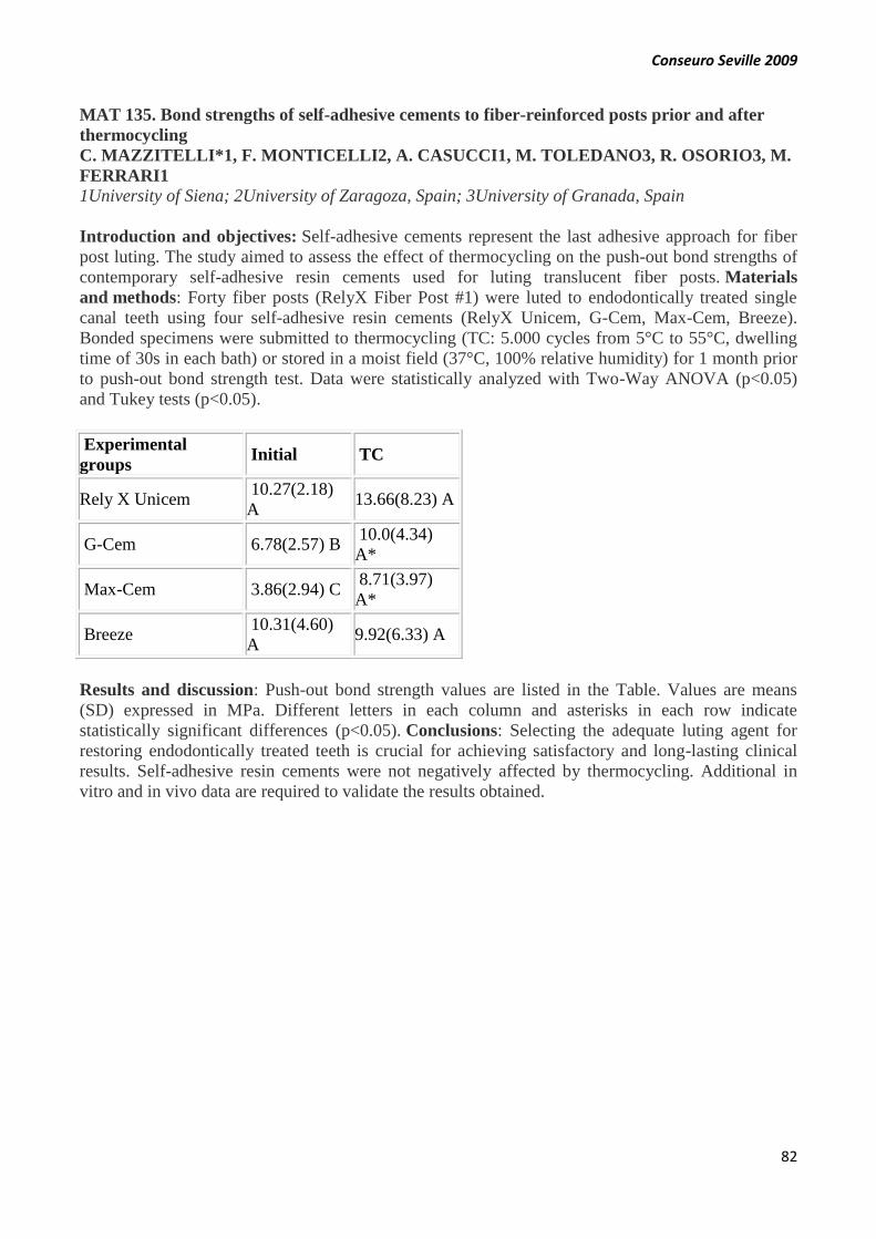
Conseuro Seville 2009
82
MAT 135. Bond strengths of self-adhesive cements to fiber-reinforced posts prior and after
thermocycling
C. MAZZITELLI*1, F. MONTICELLI2, A. CASUCCI1, M. TOLEDANO3, R. OSORIO3, M.
FERRARI1
1University of Siena; 2University of Zaragoza, Spain; 3University of Granada, Spain
Introduction and objectives: Self-adhesive cements represent the last adhesive approach for fiber
post luting. The study aimed to assess the effect of thermocycling on the push-out bond strengths of
contemporary self-adhesive resin cements used for luting translucent fiber posts. Materials
and methods: Forty fiber posts (RelyX Fiber Post #1) were luted to endodontically treated single
canal teeth using four self-adhesive resin cements (RelyX Unicem, G-Cem, Max-Cem, Breeze).
Bonded specimens were submitted to thermocycling (TC: 5.000 cycles from 5°C to 55°C, dwelling
time of 30s in each bath) or stored in a moist field (37°C, 100% relative humidity) for 1 month prior
to push-out bond strength test. Data were statistically analyzed with Two-Way ANOVA (p<0.05)
and Tukey tests (p<0.05).
Experimental
groups Initial TC
Rely X Unicem 10.27(2.18)
A 13.66(8.23) A
G-Cem 6.78(2.57) B 10.0(4.34)
A*
Max-Cem 3.86(2.94) C 8.71(3.97)
A*
Breeze 10.31(4.60)
A 9.92(6.33) A
Results and discussion: Push-out bond strength values are listed in the Table. Values are means
(SD) expressed in MPa. Different letters in each column and asterisks in each row indicate
statistically significant differences (p<0.05). Conclusions: Selecting the adequate luting agent for
restoring endodontically treated teeth is crucial for achieving satisfactory and long-lasting clinical
results. Self-adhesive resin cements were not negatively affected by thermocycling. Additional in
vitro and in vivo data are required to validate the results obtained.
Conseuro Seville 2009
83
END 136. In vivo evaluation of the root canal length determinated by an apex locator in vital
and necrotic pulp
F. KOCANI*, B. KAMBERI, M. AHMETAJ, S.H. MRASORI
University of Prishtina, Kosovo. Faculty of Medicine/ Stomatology, dep. of Endodontics
Introduction and objectives: The ideal apical endpoint of a root canal is considered to be the apical
constriction or cemento-dentinal junction of a tooth with completed root formation. Manufacturers
claim that third-generation apex locators locate the apical foramen in the presence of moisture, pulp
tissue, blood, exudates, or solutions such as sodium hypochlorite or EDTA. They also claim that no
special instruments are necessary to operate the device; a standard endodontic file is used as the
intracanal probe. For this study, a third-generation electronic apex locator, Neosono Ultima EZ
(Amadent, U.S.) was chosen. Materials and methods: In this study 37 teeth were used, 17 out of
them with vital pulp, and 20 teeth with necrotic pulp in which was apical radiolucency. Initially, a K-
file was placed in the root canal, and afterwards was done the Computerized Dental Radiography.
NiTi-files, were used as instruments in which a stopper made out of rubber was placed in the
endometric length, which was calculated with ApexLocator, calibrated monitor from 0.0-0.1mm. The
measurements were conducted in a wet ambient with NaOCl solution in a distance of 0.5mm.In this
position, digital radiography was done with CDR (Schick Tecnology, USA). The instrument which
was used (NiTi-file) was fixated with epoxy resin, while the rest of it which was left was carefully
cut. The extraction of the tooth was carefully done in order to not be created any kind of movement
of in the instrument. Results and discussion: Under the certain issues of this experiment, with the
use of Ni-Ti instruments, the length of the canal measured in electronic method NT, in 66% of the
canals (36 canals) was the same with the actual length of root canal. In 14.8% (8 canals), the length
measured in the electronic method was 0.5% shorter than the actual root canal. In 13% (7 canals) the
electronic measurement was 0.5mm longer than the actual root canal. The tip of the endodontic
instrument was went beyond the foramen in 5.6% of the cases (3 canals). In cases when it was over
passed, it was 1-2 mm from the actual length of the root canal. These results suggest that in 94.4%
(51 canals), with Ultima EZ apex locator, and with NiTi- instruments, the measurements were +/-
0.5mm of the actual length of the root canal. Conclusions: Since, Ultima EZ apex locator gives
accurate readings, thus the exposure of patient in rtg, waves. Apex locators have a lot of advantages
compared to the radiographic method.
END 137. Variations in the number of roots and canals in the 1-st maxillar premolar
B. KAMBERI*, F. KOCANI, M. AHMETAJ, S.H. MRASORI.
University of Prishtina, Kosovo. Faculty of Medicine/ Stomatology, Dep. of Endodontics
Introduction and objectives: The success of the endodontic therapy depends from the bio-
mechanical instrumentation and three dimensional root canal obturation. The failure of these
treatments, are very often as the results of number of the roots and morphological variation, except
the main root canal it can be present also the accessory and supplementary (lateral) canals and this is
trait for every tooth. The narrowness of the maxilla first and second premolar root, the longitudinal
groove along their length are responsible for the differences in the number and typography of cavum
dentis. Knowledge of these variations advances the concept for the successful endodontic treatment.
In this study we research the variations of the number of roots. Materials and methods: 342 teeth
were performed and macroscopically analysed. Results and discussion: The results of this research
show that in 57% of the cases are present two roots, in 40% of the cases are present single roots and
in 3% are three roots. In the cases with single root according to Vertucci classification, in 65% is
type IV (2), 30% with type II (2-1) and 5% type I (1). Conclusions: If the compression of roots
(mesio-bucal way) it will results in change of intra-canal system.

Conseuro Seville 2009
84
MAT 138. Zirconia surface pre-treatments: a combined AFM and SEM analysis
A. CASUCCI*1, F. PAPACCHINI1, C. MAZZITELLI1, E. OSORIO2, M. TOLEDANO2,R.
OSORIO2, A. BORRACCHINI1, M. FERRARI1
1 University of Siena, Italy; 2University of Granada, Spain
Introduction and objectives: Zirconia surface conditionings have been proposed to enhance
micromechanical retentions with resin cements. Zirconia morphology after different
chemo/mechanical surface pre-treatments was analyzed with AFM and SEM. Materials and
methods: Fourteen zirconia discs (Lava: Ø10 mm x1 mm) were divided into 7 groups (n=2)
according to the surface pre-treatment: 1) No surface pre-treatment; 2) Sandblasting (125 µm for
10s); 3) 9.5% Hydrofluoric acid (90 s); 4) Selective Infiltration Etching (SIE); 5) Experimental
Etching Solution, EES (10 min); 6) EES (30 min); 7) EES (60 min). After ultrasonic cleaning for 30
min, each disc was evaluated with an Atomic Force Microscope (Multimode Nanoscope IIIa) in the
tapping mode (tip: 1-10 Ohm-Cm phosphorous (n) dopes Si). Fields of view at 5x5 µm scan size
were considered. The average surface roughness (Ra) after different surface treatments was
determined with multiple measurements (Software Nanoscope V530R35R). Data were statistically
analyzed with Kruskall-Wallis (p<0.05) and Mann-Withney tests (p<0.05). Then, each disc was gold
sputtered (Polaron-Range SC7620) and analyzed under SEM (JEOL) at 12.000x.
Mean Ra
(nm)
No surface pre-treatment 6,94 (1,34)°A
Sandblasting 7,11 (1,09)A
Hydrofluoric acid 5,23 (0,88)A
Selective Infiltration Etching 26,02 (8,83)B
Experimental etching solution for
10s 54,22 (28,97)C
Experimental etching solution for
30s 81,79 (8,03)C
Experimental etching solution for
60s
103,02
(31,19)C
Results and discussion: The experimental etching solution at 10, 30 and 60 s increased zirconia
surface roughness (p<0.05) (Table 1). Mean (SD) surface roughness values (Ra) recorded with
Atomic Force Microscopy after different zirconia surface conditioning approaches. Different letters
indicate statistically significant differences (p<0.05). Conclusions: AFM and SEM have
complementary capabilities that allow for a more reliable evaluation of zirconia morphology after
chemo/mechanical surface treatments.

Conseuro Seville 2009
85
CLIN 139. Improvement of the periodontal interface in restorative treatments.
J.A. CARDOSO*, P. J. ALMEIDA, J. SAMPAIO FERNANDES, C. L. SILVA, A. PINHO
Oporto School Dental Medicine
Introduction and objectives: Gingival factors present a significant influence in the esthetic and
biological success at the perio-restorative interface. The Periodontal Biotype can condition several
aspects of restorative procedures: tissue handling for impressions, placement of the finishing line,
emergence profile, light transmission in the cervical area and tendency to gingival retraction. The
authors will present a case where the periodontal biotype and been surgically enhanced to improve
long term soft tissue stability in a perio-restorative interface. Materials and methods: Male patient,
30 years old, with two porcelain fused to metal crowns on both upper central incisors, placed 15
years ago. Marginal discrepancies were causing unpleasant esthetics and chronic gingival
inflammation. After discussing treatment options with the patient, the following treatment sequence
was adopted: removal of old PFM crowns; direct custom made provisionals; lab-made indirect
provisionals; connective tissue graft using the tunnel technique to augment tissue thickness;
replacement of old metallic posts with direct fiber posts and abutment reconstruction; surface
smoothing of the scar tissue and conditioning of the emergence profile by modification of the
provisional restorations; placement o two full ceramic crowns with zirconia copings. Conclusions:
Optimizing a Periodontal Biotype can enhance the esthetic and biological result at perio-restorative
interfaces. Although these procedures can make treatment more complex and extended, the short to
long-term benefits can be very significant.
OP 140. Water sorption and solubility of five contemporary resin composites
E. PAPAZOGLOU*, T. SOULTIS, G. MOUNTOURIS, A. KAKABOURA
Dept. Operative Dentistry, School of Dentistry, University of Athens, Greece
Introduction and objectives: The aim of this in vitro investigation was to study water sorption and
solubility of five contemporary resin composites. Materials and methods: The materials selected
for study were: the nanohybrids Premise (Kerr), Clearfil Majesty Posterior (Kuraray), Tetric
EvoCeram (Ivoclar); the micro-filled Heliomolar and the hybrid Quixx Posterior Restorative
(Dentsply). Water sorption and solubility were studied according ISO 4049. Disk-shaped specimens
(n=8) 15mmX0.5mm were fabricated using a steel mold. Water sorption was calculated from the
formula Wα = (d1 - f1)/V, where d1 is the specimen weight after hydration, f1 is the final dry weight
and V is the volume. Solubility was calculated from the formula Wδ = (m2 - f1)/V where m2 is the
dry weight before hydration. One-way ANOVA and Tukey’s multiple comparison tests were used to
compare the results (α=0.05). Results and discussion: Water sorption (μg/mm3) was: 18.41±0.83
(A) Heliomolar, 15.66±1.06(B) Tetric EvoCeram, 12.81±2.62 (C) Premise, 10.03±1.05 (D) Clearfil
Majesty Posterior, 9.11±0.51(D) Quixx Posterior Restorative. Groups with different letters are
significantly different. Solubility (μg/mm3) was: 3.81±0.57 (A) Tetric EvoCeram, 3.25±0.44 (A)
Heliomolar, 2.51±0.68 (B) Premise, 2.36±0.21 (B) Quix Posterior Restorative, 1.07±0.11 (C)
Clearfil Majesty Posterior. All recorded values are below the ISO 4049 accepted
values. Conclusions: The nanohybrids and the hybrid composite presented lower water sorption than
the micro-filled composite. For water sorption, two nanohybrids and the hybrid showed lower values
than the micro-filled composite. The three nanohybrids showed differences in water solubility and
sorption performance.

Conseuro Seville 2009
86
END 141. Comparison between lateral condensation modified technique and warm gutapercha
technique for fill internal resorption.
A.M. CUENCA-ABELA*1, M.M. GALVAN SALAZAR1, M. CANO GONZÁLVEZ1, R.
DELGADO RUIZ2.
1 Dentistry students, 2 Associate Professor Restorative Dentistry, School of Medicine and Dentistry,
Murcia University, Murcia
Introduction and objectives: Dentine resorption is a condition caused by a pulp inflammation; the
pulp is transformed into a highly vascularized tissue with a significant reabsortive activity. This is
manifested by an increase in the diameter of the pulp chamber and root canal. In the root form when
this disease presents communication with the periodontium the clinical manifestations may appear.
Due to the nature and evolution of this disease, the procedure endodontic cleaning, and sealing three-
dimensional conformation may become complicated. Purpose: to compare the quality of the filled
canal by the two techniques with radiographic standardized image. To compare the quality of the
canal filling techniques at optical microscope. Materials and methods: 20 lateral incisors were
selected. After the preparation, cleaning and conditioning of the root canals, were to create a
longitudinal cut to divide the teeth in two halves. Once sectioned, teeth were carved with a round
burn, making a circular cavity in the middle third of both halves at the same level. Then use a
methacrylate adhesive replenishment. The root portion of the teeth was included in acrylic blocks.
Following this, randomized selection of two groups was made: Group A. Technique with hot gutta-
percha: This was applied to half of the sample (10 incisors). Group B. Lateral modified condensation
technique: This was applied to the other half of the sample (10 incisors). Finally the results were
confirmed by two radiographic projections (vestibular and proximal). Besides was made the optical
microscope analysis, to confirm in greater detail, the padding obtained. Statistical analysis was used
by non parametric tests. And Tukey`s test to confirm validation. Results and discussion:
Radiographic image show complete radio opacity in the group A. 70% more filled Group A in the
microscope. Conclusions: The hot Gutta-percha technique is indicated to fill the internal root
resorption in. Is necessary to use a larger sample. Is necessary to compare another hot gutta-percha
technique to fill internal root resorption.
STUD 142. Is Resilon the definitive root canal filler?
P. SAORIN*, J.A.MORENO, M. PECCI, C.PEREZ-ALBACETE, A.I. NICOLAS-SILVENTE,
F. CHIVA-GARCIA
Dentistry Faculty, University of Murcia (Spain)
Introduction and objectives: Within endodontic filling materials, gutta-percha is the filling material
par excellence. Recently, a new root canal filler has appeared: Resilon/Epiphany (thermoplastic
synthetic polymer), that is considered an efficient alternative vs. gutta-percha. The aim of this work
is to evaluate the effectiveness of Resilon/Epiphany. Materials and methods: Review of the
published literature. Results and discussion: The advantages of Resilon, compared with gutta-
percha, are better apical sealing, better spreading through the side and better resistance to vertical
fracture. However, we couldn´t say the Resilon/Epiphany has antimicrobial power (though it´s
effective against S. aureus during the first 24 hours), or who has better coronal seal. The main
controversy, related to the possible failure in the future, is its biodegradability, as its surface is
eroded in contact with bacteria and enzymes, humid environments, and sodium hypochlorite 5'25%
(> 5 minutes). The main advantage of Resilon/Epiphany -monoblock form with dentin- would be
altered by degradation of the material surface and create a possible failure of endodontic
treatment. Conclusions: More investigations are necessary for concluding that Resilon is the ideal
material for endodontic, because is possible that degradation of bonding to dentine happens.
Conseuro Seville 2009
87
MAT 143. Influence of load and indentation site on nanomechanical properties of enamel
I. GIRÁLDEZ*, M. A. GARRIDO, L. CEBALLOS, J. RODRÍGUEZ
Faculty of Health Sciences and School of Experimental Sciences and Technology, Rey Juan Carlos
University, Madrid (Spain).
Introduction and objectives: The aim was to determine the influence of load and indentation site on
modulus of elasticity and hardness, using CSM method. Materials and methods:
Enamel sections of 2 mm thick from bovine incisors (n=4) were studied by nanoindentation tests
using a Berkovich indenter. CSM (continuous stiffness measurement) method was used to determine
the elastic modulus (E) and hardness (H). Indentations were carried out at intervals of 100 µm (n=10)
from the enamel surface to the dentin-enamel junction up to a total penetration depth of 2µm. The
results were analyzed with ANOVA and Student-Newman-Keuls tests (p<0.05). Results and
discussion: E and H values significantly decreased with the load (p<0.05). E values were
significantly different among the four loads selected: 1 mN 87.5 GPa (±26.1); 10 mN 80.3 GPa
(±12.8); 100 mN 73.4 GPa (±6.4); 200 mN 68.7 Gpa (±5.1). The same tendency was observed for H
values: 1 mN 4.6 GPa (±2.8); 10 mN 3.7 GPa (±1.1); 100 mN 3.1 GPa (±0.5); 200 mN 2.7 GPa
(±0.4). E and H decreased as the indentations approached the dentin-enamel junction
(p<0.05). Conclusions: E and H values determined by nanoindentation are very sensitive to load
applied and the location of the indentation.
OP 144. Effects of blood contamination on microleakage of different adhesive systems
Y. KORKMAZ 1*, C. CELIK 1, E. KARABULUT 2
1 Baskent University School of Dentistry Department of Conservative Dentistry. 2 Hacettepe
University School of Medicine Department of Biostatistics
Introduction and objectives: The aim of this study was to evaluate the effect of blood
contamination before and after adhesive application on microleakage. Materials and methods:
Class V preparations at cement-enamel junction were made on the buccal and lingual surfaces of 45
freshly extracted human teeth. The teeth were randomly assigned into three groups for different
adhesive systems; etch and rinse adhesive system (XPBond ) and two different one-step self-etching
adhesive systems (XenoV) and (FuturabondNR). The specimens were than further subdivided into
three groups (n=10): Group A contamination before adhesive application, Group B contamination
after adhesive application, Group C no contamination, which was the control. Contamination of the
surfaces was performed with fresh blood and blood was removed by a gentle air blast in each group.
The adhesive systems were applied according to their manufacturer's recommendations. The
specimens were restored with Filtek Supreme XT nano-composite. After thermocycling (X500, 5-
55C0) and immersion in 0.5% basic fuchsin, the dye penetration was evaluated under
stereomicroscope. Statistical analyses were performed with the Kruskal-Wallis and Mann-Whitney
and Wilcoxon tests. Results and discussion: No statistically significant differences were found
among subgroups on the enamel or on the dentin for Xeno V and XP Bond (p > 0.05); however for
Futurabond NR blood contamination before adhesive application exhibited higher microleakage than
other subgroups on dentin. (p<0.05). Conclusions: Blood contamination may increase the
microleakage depending on the adhesive system.

Conseuro Seville 2009
88
STUD 145. Intra-oral piercing: dental complications
J. JIMENA-ESPAÑA, I. SÁNCHEZ-SERNA*, A.B. RODRÍGUEZ-ESPIN, J. MATÉ-
SANCHEZ
Dentistry Faculty, University of Murcia, Spain
Introduction and objectives: In the last decade intra-oral piercing has gained acceptance as a sign
of individuality, marginality, decoration, or group membership. It has been observed that the trend of
wearing jewelry inserted into the intra-oral tissues may result in significant damage to the dental
structure. The purpose of this study is to review current relative literature on intra-oral piercing
regarding its dental complications in order to determine the prevalence of damage to dentition among
piercers. Materials and methods: The current study was based on scientific literature regarding oral
piercing published within the last decade. Results and discussion: After reviewing the scientific
literature, the most common dental problem registered was teeth fractures (40%): chipping of teeth
and teeth cracked or with cusp fractures. Selective dental abrasion was registered as well as galvanic
currents produced by the appliance. Conclusions: Every day dental injury by piercings is more
common. Is therefore necessary that dentists inform patients on the potential complications and the
risk to oral health of intra-oral piercings.
END 146.
WITHDRAWN
MAT 147. Using restorative composite for assembling composite inlays – a study of the degree
of conversion.
C. BERNARD, M.A. DERBANNE*, S. LE GOFF, M. DEGRANGE
Unité de Recherche en Biomatériaux Innovants et Interfaces
Introduction and objectives: There is no consensus on the material to use to bond composite inlays.
Some recommend using light curing restorative composite while other favor dual cure resin cements
(DCRC) considering light absorption by the inlay. Our objective is to study the degree of conversion
(DC) of light curing composite used as a bonding agent for composite inlays. Materials and
methods: 10 indirect composite inlays where made on 10 anatomically similar, freshly extracted
human third molars with a maximum depth of 2mm (Enamel HFO – Micerium). Those where
bonded with 2 steps etch and rinse adhesive (One Step Plus – Bisico) and light curing restorative
composite (Enamel HFO – Micerium) with a LED curing unit. The teeth where then sectioned in half
and microhardness of the bonded joint was measured. In order to correlate microhardness with
degree of conversion, DC was measured by FTIR-ATR on composite samples through polymerized
composite discs of varying thickness, microhardness was then recorded. Results and discussion:
There is a correlation with microhardness and DC (R²=0.91). The DC was 20% to 30% lower under
the bonded inlays except on the center of the tooth. Despite conducting light curing with high
energies, light absorbance by the composite induces a notable loss in terms of DC of the bonded
joint. Conclusions: These results lead us to prefer DCRCs instead of light curing restorative
composite to bond composite inlays.

Conseuro Seville 2009
89
MAT 148. Implant surface modification to prevent dimensional bone changes after immediate
implant placement in dogs.
J.L. CALVO GUIRADO1, R.A. DELGADO RUIZ2, J.E. MATÉ SÁNCHEZ2, F. CHIVA1,
A.B. RODRIGUEZ ESPÍN2, A.I. NICOLÁS SILVENTE2, J.C. BAGUENA-GOMEZ*1, L.
LÓPEZ MARÍ2.
1 Professor, 2Associate professor, Faculty of Medicine and Dentistry. University of Murcia. Spain
Introduction and objectives: To study the effect of implant surface modification in dimensional
alterations of the alveolar bone ridge that occurred following tooth extraction as well as processes of
bone modelling and remodelling in immediate placement. Materials and methods: Six Beagle dogs
were included in the study. In both quadrants of the mandible and maxilla incisions were made in the
crevice region of the 3rd and 4th premolars and molar 1st. Minimum buccal and lingual flaps were
elevated. The third, four premolars and first molar were hemi-sected. The distal roots were removed
and 72 SPI Element implants 4 mm Ø x 9.5 m length (Thommen Medical, Waldenburg, Switzerland)
were placed. Split mouth of maxilla and mandible were made, right side with conditioned surface
implants and left side implants without conditioned surface. The extraction sites were covered with
the mobilized gingival tissue. The extractions of the roots and the sacrifice of the dogs were
staggered in such a manner that all dogs contributed with sockets representing 2, 4 and 12 weeks of
healing. The animals were sacrificed and tissue blocks containing the extraction socket were
dissected, decalcified in EDTA, embedded in paraffin and cut in the buccal–lingual plane. The
sections were stained in haematoxyline–eosine and examined in the microscope. Results and
discussion: The microscopic healing patterns at weeks 2,4, and 12 for the two implant types with
conditioned and non conditioned surface showed similar bone findings. The mean percentage of the
new formed bone in contact with the implant (BIC) was significant greater at 1 month for
conditioned SLA at buccal plate (38.33 ± 1.4 %) and lingual plate (49 ± 1.4%) than the non
conditioned surface at buccal (36.83 ±1.6 %) and lingual plate (38 ± 1.6 %). Conclusions: The
bundle bone was resorbed and replaced with woven bone more in the lingual cortical bone. Lingual
crestal bone was resorbed more than buccal plate at 12 weeks follow up period. Conditioned implant
surface increase bone apposition and reduce healing osteointegration process.

Conseuro Seville 2009
90
MAT 149. Use of collagenized porcine bone vs. bovine bone in sinus lift procedures
J.L. CALVO GUIRADO1, R.A. DELGADO RUIZ2, J.E. MATÉ SÁNCHEZ2, F. CHIVA1,
A.B. RODRIGUEZ ESPÍN2, A.I. NICOLÁS SILVENTE2, J.C. BAGUENA1, J. LOPEZ-
TORTOSA*2.
1 Professor, 2Associate professor , Faculty of Medicine and Dentistry. University of Murcia. Spain
Introduction and objectives: The purpose of this study is to assess the effectiveness of collagenized
porcine bone MP3 vs bovine bone acts as a bone substitute in the maxillary sinus lift after 2 years of
follow-up. Materials and methods: Twenty-four patients with sinus lift bilateral, 12 men and 12
women with an average age of 25 to 68 years, divided into test and control group. Group 1
underwent sinus lift with bovine bone (Bio Oss) as a control group and Group 2 was placed in the
contralateral sinus porcine bone + collagen MP3 as a group test. Eight months after the biopsies were
taken with hole drills before placing implants. Forty-eight implants NanoTite Certain Prevail
(Biomet 3i, Palm Beach Gardens, FL, USA) were placed. Results and discussion: The average
height obtained in the test group of MP3 (Osteobiol, Tecnoss Dental, Italy) was 13 mm in the control
group of bovine bone Bio Oss (Geistlish, Switzerland) was 11.5 mm. The site treated with porcine
bone was found vital bone with bone neoformation and small portions of biomaterial, within the
control group where the presence of biomaterial was higher and a less bone neoformation.
There have been no inflammatory processes and not any implant was lost during the 2 years of
study. Conclusions: The surgical procedure for lifting the maxillary sinus using a mixture of porcine
bone + collagen compared with bovine bone alone, resulting in an increase in bone formation. The
porcine bone is biocompatible and can be used as bone substitute material in these procedures
without the interference of biological processes of bone formation.
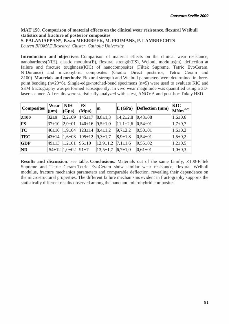
Conseuro Seville 2009
91
MAT 150. Comparison of material effects on the clinical wear resistance, flexural Weibull
statistics and fracture of posterior composites
S. PALANIAPPAN*, B.van MEERBEEK, M. PEUMANS, P. LAMBRECHTS
Leuven BIOMAT Research Cluster, Catholic University
Introduction and objectives: Comparison of material effects on the clinical wear resistance,
nanohardness(NIH), elastic modulus(E), flexural strength(FS), Weibull modulus(m), deflection at
failure and fracture toughness(KIC) of nanocomposites (Filtek Supreme, Tetric EvoCeram,
N’Durance) and microhybrid composites (Gradia Direct posterior, Tetric Ceram and
Z100). Materials and methods: Flexural strength and Weibull parameters were determined in three-
point bending (n=20*6). Single-edge-notched-bend specimens (n=5) were used to evaluate KIC and
SEM fractography was performed subsequently. In vivo wear magnitude was quantified using a 3D-
laser scanner. All results were statistically analyzed with t-test, ANOVA and post-hoc Tukey HSD.
Composites Wear
(µm)
NIH
(Gpa)
FS
(Mpa) m E (GPa) Deflection (mm)
KIC
MNm-3/2
Z100 32±9 2,2±09 145±17 8,8±1,3 14,2±2,8 0,43±08 1,6±0,6
FS 37±10 2,0±01 140±16 9,5±1,0 11,1±2,6 0,54±01 1,7±0,7
TC 46±16 1,9±04 123±14 8,4±1,2 9,7±2,2 0,50±01 1,6±0,2
TEC 43±14 1,6±03 105±12 9,3±1,7 8,9±1,8 0,54±01 1,5±0,2
GDP 49±13 1,2±01 96±10 12,9±1,2 7,1±1,6 0,55±02 1,2±0,5
ND 54±12 1,0±02 91±7 13,5±1,7 6,7±1,0 0,61±01 1,0±0,3
Results and discussion: see table. Conclusions: Materials out of the same family, Z100-Filtek
Supreme and Tetric Ceram-Tetric EvoCeram show similar wear resistance, flexural Weibull
modulus, fracture mechanics parameters and comparable deflection, revealing their dependence on
the microstructural properties. The different failure mechanisms evident in fractography supports the
statistically different results observed among the nano and microhybrid composites.

Conseuro Seville 2009
92
MAT 151. Resin water sorption and hygroscopic expansion reduce the interfacial gap and
microleakage.
J.I. ROSALES LEAL*, H. GONZALEZ MOREIRA, M.A. CABRERIZO VÍLCHEZ
University of Granada.
Table: see this link http://www.efcd.eu/media/pdf/Mat_151_Table.pdf
Introduction and objectives: The objective was to evaluate the effect of the water sorption and
hygroscopic expansion on the interfacial gap and microleakage. Materials and methods: Four resins
were evaluated: Ormocer (Admira [Voco]); Compomer (Dyract Extra [Dentsply]); Hybrid (Spectrum
[Dentsply]); Nanohybrid (Exthet•X [Dentsply]). Water sorption and hygroscopic expansion were
evaluated after water immersion for 24 h; 1 week and 4 weeks and it effect on the interfacial gap size
and microleakage variations. Water sorption was evaluated by weighting resins discs after the three
immersion periods. Hygroscopic expansion was measured under white light confocal microscope
from the volumetric variations of resins fillings in cylindrical cavities prepared in dentin discs.
Interfacial gap size was obtained from the cylindrical cavities resin filled under white light confocal
microscope. The microleakage study was done in class v cavities prepared in extracted third molars.
A multifactor ANOVA and Tukey tests were applied. Results and discussion: see
table. Conclusions: Water immersion induces water sorption and hygroscopic expansion.
Hygroscopic expansion reduces the interfacial gap and cavity microleakage. The effect of
hygroscopic expansion on the gap size and microleakage is dependent on the resin material. Ormocer
and Compomer suffer higher hygroscopic expansion and then higher gap and microleakage reduction
over time. Hybrid and microhybrid composites suffer lower hygroscopic expansion and then lower
gap and microleakage reduction over time. Acknowledgements: This work was supported by the
“Ministerio Español de Educación y Ciencia” (project MAT2007-66117, Junta de Andalucía (project
P07-FQM-02517) and the European Social Fund (ESF).

Conseuro Seville 2009
93
MAT 152. Mechanical characterization of several self-adhesive resin cements by
nanoindentation
M.V. FUENTES, M.A. GARRIDO, L. CEBALLOS*, J. RODRÍGUEZ
Faculty of Health Sciences and School of Experimental Sciences and Technology, Rey Juan Carlos
University, Madrid, Spain
Introduction and objectives: The objective of this study was to evaluate the hardness and Young's
modulus of several self-adhesive resin cements using nanoindentation.Materials and methods:
Three self-adhesive resin cements (dual-cured) were tested: RelyX Unicem (3M Espe), Maxcem
Elite (KerrHawe), an experimental resin cement (Voco), and a self-etching resin cement (chemically-
cured) Multilink Automix (Ivoclar Vivadent). Disc-shaped specimens were prepared for each resin
cement using a teflon mold, 6 mm in diameter and 1.5 mm thick. The dual-cured resin cements were
polymerized for 40s (Bluephase, Ivoclar Vivadent). Specimens were stored in dry conditions (37ºC,
7 days). After that, 15 indentations were performed on each specimen with a Berkovich indenter
(maximum load: 100mN). Young's modulus and hardness of the cements were determined from the
load–displacement curves following the Oliver–Pharr methodology.
Mean hardness and Young's modulus (SD) of resin luting
agents tested.
Resin cement Hardness (GPa)
Young's modulus
(GPa)
RelyX Unicem 0.373 (0.042) a 10.74 (0.45) a
Maxcem Elite 0.375 (0.060)a 9.77 (1.90) a
Experimental cement
Voco 0.334 (0.055)a 9.79 (1.32) a
Multilink Automix 0.091 (0.034)b 2.94 (0.82) b
For each column, means with the same letter are statistically
similar after Student-Newman-Keuls multiple comparisons
(p>0.05)
Results and discussion: Hardness and modulus of elasticity are shown in the table. One-way
ANOVA revealed a significant influence of the resin cement used (p<0.001). The chemically cured
cement, Multilink, presented the lowest values of stiffness and hardness. Dual-cured cements tested
showed similar values of hardness and modulus. Conclusions: All self-adhesive resin cements (dual-
cured) tested showed comparable hardness and modulus values after 7 days in a dry condition.
Acknowledgements: The authors are grateful to the companies for providing the materials tested.

Conseuro Seville 2009
94
PREV 153. Dental and periodontal condition in patients with systemic diseases
1B.U. KRÓL*, 1W. STOKOWSKA, 2A. POPŁAWSKA2
1Department of Conservative Dentistry Medical University in Białystok , Poland, 2 Department of
Endocrinology and Internal Diseases Medical University in Białystok , Poland
Introduction and objectives: The paper aims at evaluating the frequency of occurrence and
intensity of tooth periodontium pathologies in patients with thyroid hyperactivity, thyroid
hypofunction and non-toxic goitres. Materials and methods: A total of 101 adult subjects were
examined. The parameters evaluated were: the number of teeth preserved, dental caries (by DMFT
and its components), oral cavity hygiene (by Pl.I.) and the periodontal condition (by CPI). Results
and discussion: Compared to the control group (71.7%) we observed significantly lower number of
teeth preserved in the hyperthyroid disease group (32.1%) followed by the hypothyroid (43.9%). A
markedly higher DMFT value compared to the control group (24.2%) was observed in the
hyperthyroid (28.4%), whereas a significantly higher Pl.I. value compared to the control group
(1.2%) was recorded in hypothyroid patients (2.0) and in the hyperthyroid (1.8). Clinically healthy
periodontal tissue was only observed in the non-toxic goitre group of patients. The most advanced
periodontal disease was observed in patients with hyperthyroidism. Conclusions: The patients’
involvement in their systemic disease treatment might have been the reason for neglecting oral
hygiene, greater caries intensity and periodontium pathology. Acknowledgements: We thank Prof.
D. Waszkiel for help during data analysis and Prof. I. Kinalska, Prof. M. Górska, Prof. M.
Szelachowska, Dr A. Zonenberg for assistance and advice.
MAT 154. Details of the surface roughness in zirconium implants available in the market
JE. MATÉ*1, R. DELGADO1, JL. CALVO2, F. CHIVA2, AB. RODRIGUEZ1, AI.
NICOLÁS1, JC. BAGUENA1.
1Associated professor Murcia University. Department of PTD. 2 Senior lecturer of Murcia
University. Department of integrated of adults.
Introduction and objectives: In the last times have developed the implants of zirconia stabilized
with Itrium to achieve an improvement in the aesthetic results in comparison with the ones of TiO2.
The superficial roughness of the material is the one who delimits the biological interaction.
Objectives: to describe the quality characteristics of the different zirconium implants available,
through SEM., and to quantify surface roughness of different zirconia available implants, through
perfilometric analysis. Materials and methods: 6 zirconium implants with different surface changes
through physical or chemical treatments. 1 titanium implant with a double acid etching and
sandblasting as skipper. Comparing the surface roughness in three areas of the implant (collar,
middle third and apical third) through the analysis of mechanical and optical surfaces (nm and
micrometers) (Perfilometer Veeco model dektak 3). Study with electron microscopy (JEOL-6100)
scanning and imaging management program (q500Mc Leica). Results and discussion: The surface
roughness varies depending on the surface treatment, being the highest value that found the chemical
and physical processing combined (70% higher). Followed by physical treatments (53%) and finally
chemical treatment (32%). Conclusions: The physical-chemical treatment provides greater surface
roughness. The chemical treatment alters very little surface zirconium.

Conseuro Seville 2009
95
MAT 155. Comparison of the surface micro texturing at 3 nano-particle composites after
standardized polishing.
DELGADO RUIZ RA*1, MATÉ SÁNCHEZ JE1, CALVO GUIRADO JL2, CHIVA F2,
RODRIGUEZ ESPÍN AB1, NICOLÁS SILVENTE AI1, BAGUENA JC2
1Associate professor, 2Professor, Faculty of Medicine and Dentistry. University of Murcia. Spain
Introduction and objectives: To study the effect of surface polishing in surface microtexturing of
three nano particle composites, and to compare the surface roughness values of the nano-composite
after polishing. Materials and methods: Was selected three nano-particle composites: A.Filtek
supreme XT®, B. Esthetics®, C. Gradia®. 30 central teeth with a class V cavity preparation and
random fill, in 3 samples: group 1 (10 teeth filled with composite A), group 2 (10 teeth filled with
composite B), group 3 (10 teeth filled with composite C). Polishing with a standardized protocol. P
value <0.05 . Statistical analysis was perform with non parametric Kruskall Wallis , and Tukey
test. Results and discussion: The roughness values in nano particle composites are lower in C. (
80%), B (60%), A ( 55%). Conclusions: The nano-composite Gradia shows a better polished flat
surface compare with the other two nano-composites. . The surface roughness depends of the nano
particle and also of polishing technique used. Is necessary to continue this research with a more
wider sample to obtain statistical significance and power.
END 156. Histopathologic differences between periradicular lesions of endodontally treated
and non treated teeth
J. BAHILLO*, B. MARTIN-BIEDMA, T.GARCIA-CABALLERO, P.VARELA,
J.GONZALEZ-BAHILLO
Division of Dental Pathology and Therapeutics. Department of Estomatology. School of Dentistry.
Introduction and objectives: The aim of this study is to determinate the histological differences
between periradicular lesions of endodontically treated and non treated teeth. Apical periodontitis is
produced in most cases by intraradicular infection and the chronic inflammatory periapical lesion is
the most common pathology found in the alveolar bone of the jaws. Materials and methods: The
periradicular lesions were radiographed to measure the size of the radiolucidity and those teeth that
had received root canal treatment were identified. The biopsies were obteined during periapical
surgery or by tooth extraction. Removed samples were fixed in 10% neutral formalin, embedded in
paraffin, cutted and stained with hematoxylin and eosin for radiographic examination. Results and
discussion: The histologically studied samples had a simple stratified squamous epithelium with a
lymphoplasmocitary content and inflammatory infiltrate. In some samples it can also be seen
Russel’s bodies. Conclusions: There are not histological significant differences between
periradicular lesions of endodontically treated and non treated teeth.
Conseuro Seville 2009
96
OP 157. Effect of ozone treatment on enamel and dentin bond strength
S. GURGAN *, A. KIREMITCI, F. YALCIN CAKIR, T. ALPASLAN
Hacettepe University, School of Dentistry, Department of Restorative Dentistry, Ankara, Turkey
Introduction and objectives: Ozone has been recognized as a powerful sterilizing agent in
dentistry. There have been concerns regarding potential adverse effects of application of ozone prior
to bonding procedures. The aim of this study was to evaluate the effect of ozone application on
dentin and enamel shear bond strength of two different adhesive systems. Materials and methods:
Forty human enamel and 40 human dentin samples were used. Ten samples per group were prepared
as follows: (I) Ozone application (Ozonytron X) with subsequent application of an etch & rinse
adhesive (Excite), (II) Ozone application with a two step self-etch adhesive (AdheSe), (III) etch &
rinse adhesive application, (IV) self-etch adhesive application. Specimens were restored with a
nanohybrid composite resin (Tetric Evo Ceram) according to the Ultradent method. After storage in
water for 24h, shear bond strength was measured with a universal testing machine. Data were
analyzed using Mann Whitney U test. Results and discussion: No decrease in shear bond strength
was detected for ozone –pretreated enamel specimens compared to untreated controls. However
Ozone treatment resulted in significantly decreased bond strength (p<0.05) on dentin specimens for
both adhesive systems. Conclusions: Ozone pre-treatment may provide comparable bond strengths
depending on the tooth substrates applied.
STUD 158. Discrepancies in the visual determination of dental color
D. ROIG-FERRANDO*, C.M. GEA-ROS, J.C. CABEZOS-GARCIA, F. CHIVA-GARCIA, R.
DELGADO-RUIZ
Dentistry Faculty, University of Murcia (Spain)
Introduction and objectives: The visual determination of dental colour is a routine task in the
dental clinic. This study aims to assess the discrepancy concerning the selection of the tooth
color. Materials and methods: Descriptive observational double-blind study. Dental students
(n=96) took the colour of four teeth (21, 22, 23 and 33) of the same patient under controlled
conditions (illumination, time, place). There were 2 groups: a) with the light of the equipment (LE),
b) with daylight (D). The colour was taken according to the shade guide Vitapan ® Classical and
compared with the results obtained according to the spectrophotometric system Vita Easyshade
®. Results and discussion: There was a great variability in the selection of color (% of agreement
with spectrophotometer):Tooth 21 (LE): 8.9%, Tooth 21 (D): 14.9% , Tooth 22 (LE): 12.9% , Tooth
22 (D): 15.8% , Tooth 23 (LE): 24.0% , Tooth 23 (D): 19.8%, Tooth 33 (LE): 29.7% , Tooth 33 (D):
31.7% . Conclusions: Obtaining the dental colour is a fairly difficult and complex task. Although it
would be desirable to enhance learning it in dentistry schools, clinical experience does not seem to
influence the choice of colour. Besides, it would be desirable to make use of spectrophotometer
systems.

Conseuro Seville 2009
97
END 159. Comparative study of collagen exposure in root dentine treated with two irrigation
protocols.
E. MARÍN-ALTUVE, M. BOLAÑOS-CARMONA, S. GONZÁLEZ-LÓPEZ*
Facultad de Odontología de la Universidad de Granada
Introduction and objectives: We studied the percentage of collagen exposed in dentine of each root
third after applying irrigation protocol 1: 5% NaOCL + 17% EDTA as final irrigant; or protocol 2:
20% Citric acid + 2% CHX. Materials and methods: We used 48 single-root human teeth divided
into 2 groups (n=24). Group A underwent manual biochemical preparation and Group B was
prepared using the ProFile® system. Irrigation protocols 1 and 2 were applied in each group and
RealSeal™ or EndoREZ® were used as filling materials. Roots were cut into 1-mm slices, which
were abraded and stained with Masson‘s trichrome method. Light microscopic study was used to
calculate the percentage of collagen exposed.
Table: Percentages of exposed collagen
Third
5% NaOCL + 17% EDTA 20% Citric acid + 2% CHX
Material Instrumentation Material Instrumentation
RealSeal EndoREZ Manual Rotary RealSeal EndoREZ Manual Rotary
Cervical
59.0(29.5
) 9.3(12.9)
38.0(33.2)
30.3(35.4)
79.4(17.4)
80.0(14.6)
77.7(16.6)
81.6(15.3)
Middle 50.0(26.5)
12.0(14.4)
29.1(26.6)
32.9(31.3)
64.3(22.5)
77.7(27.9)
58.8(27.2)
83.2(17.9)
Apical 56.2(35.0) 4.3(12.1) 31.2(38.0) 29.4(37.3) 93.9(9.0) 86.0(27.5) 83.2(27.5) 96.6(4.3)
Total 55.0(29.9) 8.5(13.2) 32.7(32.2) 30.8(33.8) 79.2(20.7) 81.2(23.7) 73.2(25.9) 87.1(15.0)
Results and discussion: The highest percentage of collagen was exposed with 20% Citric Acid +
2% CHX as irrigant and rotary instrumentation.

Conseuro Seville 2009
98
MAT 160. Demineralization effects of phosphoric acid on surface and subsurface enamel
bovine bleached with in-office hydrogen peroxide
*TORRES-RODRIGUEZ C1, GONZALEZ-LOPEZ S2, BOLANOS-CARMONA MV3,
SANCHEZ-SANCHEZ MP4, RODRIGUEZ-NAVARRO ALEJANDRO5
1 Assistant Professor. Oral Health Department. Faculty of Dentistry. (National University of
Colombia), 2 Associate Professor of Pathology and Dental Therapeutics (University of Granada),
3 Associate Professor of Integrated Pediatric Dentistry (University of Granada), 4 Associate
Professor, Department of Inorganic Chemistry. (University of Granada), 5 Associate Professor,
Department of Mineralogy and Petrology. (University of Granada).
Introduction and objectives: To measure the demineralization capacity of 37% phosphoric acid on
superficial and subsuperficial bovine enamel after short office bleaching with 30% and 38%
hydrogen peroxide and abrasion of 0, 25, 50 and 100 mµ. Materials and methods: Three equal-
sized 4 x 4 mm sections were obtained from the coronal enamel of ten bovine incisors embedded in
resin. One sample specimen from each crown was assigned to one of three groups (n=10): Group I,
no bleaching agent (control group); Group II bleached with 38% hydrogen peroxide for 20 min
(Opalescence Boost Ultradent) and Group III with 30% hydrogen peroxide (Office Iluminé
Dentsplay) for 60 min. At 24 h, each specimen was measured with a micrometer (Mitutoyo 350-
MHN1-25-DM) and immersed in 37% phosphoric acid solution, then aliquots were collected at 30 s
and 60 s, 5-ml. Specimens were abraded 25 µ with 1200-grit silicon carbide paper discs and again
immersed in 37% phosphoric acid solution. This process was repeated for 50- and 100-µ abrasions.
Ca2+ concentrations were measured by atomic absorption spectrophometry. Results and discussion:
The amount of Ca2+ removed was similar at all depth levels between specimens bleached and not
bleached with 30% hydrogen peroxide (Illuminé Office®). More Ca2+ was removed at 25 µm depth
in specimens bleached with 38% hydrogen peroxide (Opalescence Boost. Ultradent®) than in
unbleached specimens. Conclusions: 38% hydrogen peroxide bleaching increases the decalcifying
effect of phosphoric acid on subsurface enamel. 30% PH did not modify the decalcifying capacity of
phosphoric acid at surface and subsurface levels.
END 161. Effect of different irrigation protocols on the adhesive interface in endodontics
E. MARÍN-ALTUVE, M. BOLAÑOS-CARMONA*, S. GONZÁLEZ-LÓPEZ
Facultad de Odontología de la Universidad de Granada
Introduction and objectives: To compare the bond strength of RealSeal™ and EndoREZ® to
dentine of each root third treated with 5% NaOCL + 17% EDTA [Protocol 1] or 20% Citric Acid +
2% CHX [Protocol 2] using the push-out test. Materials and methods: Forty human single-root
teeth were divided into two groups (n=20), applying irrigation Protocol I in one group and Protocol
II in the other and filling with RealSeal™ or EndoREZ®. Roots were transversally cut to obtain 1-
mm sections. Regional bond strength was measured with the push-out test using an Instron 3345
system.
Table: Bond strength in Mpa
Third 5% NaOCL + 17% EDTA 20% Citric acid + 2% CHX
RealSeal EndoREZ RealSeal EndoREZ
Cervical 2.9(5.8) 8.0(10.5) 1.4(4.6) 8.7(11.3)
Middle 4.5(7.5) 9.2(14.1) 1.6(4.2) 5.9(10.1)
Apical 5.9(8.9) 17.3(20.7) 1.1(4.0) 5.8(10.8)
Total 4.2(7.4) a 11.2(15.7) b 1.4(4.3) a 6.9(10.7) b
Results and discussion: The best results were obtained with 5% NaOCL + 17% EDTA (p=0.001)
using EndoREZ filling material. No significant differences were found among the root thirds
Conseuro Seville 2009
99
MAT 162. Biomaterials of bone graft. Prospective study to 2 years.
JE. MATÉ1, R. DELGADO 1, JL. CALVO2, F. CHIVA2, AB. RODRIGUEZ1, AI. NICOLÁS
1, JC. BAGUENA1, E. GARCIA-CRUZ*1.
1Associated professor Murcia University. Department of PTD. 2 Senior lecturer. Murcia University.
Department of integrated of adults.
Introduction and objectives: The need for new horizons in dealing with dental implants makes the
preoperative preparation of the field work (bone, soft tissue). Based on the gold-standard (graft
authology) materials xenografts trying to level the playing field achieved by it. To evaluate the
success of xenografts compared with graft authology. Materials and methods: Graft authology
obtained the calotte, menton, iliac crest and mandibular branch. Xenografts materials: bio-oss and
osteobiol. Patients in private practice to which they are performed bone graft surgery after recessive.
The waiting time for placement of implants is 6 months. Implants placed: BIOMET 3i, and Zimmer.
Charging time of the implant in the upper jaw at 5 to 6 months; in the jaw to 3 months. Results and
discussion: GRAFT AUTOLOGOUS CASES: Follow-up to two years. Survival of the implants
100%. Average loss of bone: <1.6 mm. XENOGRAFTS CASES: Follow-up to two years. Survival
of the implants 100%. Average loss of bone: <1.4 mm. Conclusions: Gold Standard: authology.
Ideal for long-term recovery of large volumes. Improvements of the short-term particulate
xenografts. Recovery of smaller volumes without compromise of the soft
tissues. Acknowledgements: Dra. M. Maté: Statistical analysis.
MAT 163. Cut enamel shear bond strength of silorane
A.B. RODRIGUEZ-ESPIN*, A.I. NICOLAS-SILVENTE, F. CHIVA-GARCIA, J.E. MATE-
SANCHEZ DE VAL, R.A. DELGADO RUIZ, J.C. BAGUENA-GOMEZ
UD Dental Pathology & Therapeutic, Dentistry faculty, University of Murcia (Spain)
Introduction and objectives: Recently, a dental composite of low-shrink (Filtek Silorane) has been
introduced in the dental market. The purpose of this study is to evaluate the in vitro cut enamel shear
bond strength (SBS) and to determine the type of bonding fracture. Materials and methods: Forty-
five bovine incisors were used in three groups: group A (n=15): Filtek Z250 + Scotchbond 1XT etch-
and-rinse adhesive, group B (n=15): Filtek Z250 + Scotchbond SE self-etching adhesive, and group
C (n=15): Filtek Silorane + Silorane self-etch adhesive. Enamel was slightly drilled (ground). Resin
cylinders (stubs) were bonded to enamel surface using the bonding agent according to
manufacturer’s instructions. The SBS were registered with an Instron testing machine, operating at a
crosshead speed of 1 mm/min. The bonding fracture was observed with scanning electron
microscope (SEM). The data was statistically analysed using ANOVA (p 0.05). Results and
discussion: The mean SBS (MPa) were: A = 14.48 4.11; B= 5.50 2.59; C = 8.21 2.91. SBS of
both B and C were significantly lower than A (p < 0.01). No significant differences between B and
C. Conclusions: Filtek Silorane adhesive presented similar shear bond strength that self-etching
adhesive, but lower than etch-and-prime adhesive.

Conseuro Seville 2009
100
MAT 164. Evaluation of the effect of different polishing systems on restorative materials.
R. ABREU-RODRIGUEZ*, A. PASCUAL-MOSCARDO.
Departamento de Estomatologia – Materiales dentales. Clínica Odontológica Facultad de Medicina
y Odontología de Valencia
Introduction and objectives: The systems used for finishing and polishing composite resins and
compomers are numerous and affect the success of filling from a mechanical, biological and
aesthetic point of view. Objective: to evaluate “in vitro” the effect of different polishing systems on
some restorative materials. Materials and methods: The surface of some aesthetic restorative
materials finished and polished with different methods or systems (n: 2520): Sof – Lex Pop On®,
Brushes Sof Lex®, Enhance®, Super Snap®, One Gloss®, Pogo®, Filtek Supreme®, Quixfil®,
Ceram X Duo®, Tetric Ceram®, Gradia Direct®, Spectrum TPH®, Inten S®, Renamel®, Esthet
X®, Dyract Extra®, Premise® and Compoglass F®. In each specimen was measured the surface
roughness and was related with scanning electron microscopy (SEM) images. Results and
discussion: The best combinations were: Filtek Supreme® Sof Lex®, Ceram X Duo® - Super
Snap®, Ceram X Duo® - Sof Lex Pop On® and Renamel® - Pogo®.a highly polished restoration is
necessary to help promote a plaque – free environment. Mann Whitney – Kruskal Wallis (α:
0,05). Conclusions: The surface roughness and SEM revealed that the effect of all systems studied
(compomers, composite resins and polishing systems) were clinically acceptable.
MAT 165. Intact enamel shear bond strength of silorane and its changes on enamel surface
A.I. NICOLAS-SILVENTE*, A.B. RODRIGUEZ-ESPIN, , F. CHIVA-GARCIA, R.A.
DELGADO RUIZ, J.E. MATE-SANCHEZ DE VAL, , A. VICENTE HERNANDEZ
UD Dental Pathology & Therapeutic, Dentistry faculty, University of Murcia (Spain)
Introduction and objectives: Recently, new restorative systems have been introduced in the dental
operative field. One of them is Filtek Silorane, which is supposed to offer low shrink rates. Till now
there are few studies about the shear bond strength (SBS) of this product. The aim of this study was
to compare the SBS of Filtek Silorane with traditional acid-etching technique and with a self-etching
adhesive system in intact enamel and evaluate changes in enamel surface. Materials and methods:
45 bovine upper incisors with intact vestibular enamel surface were used for the SBS test divided is
three groups (n=15). I: (37%-phosphoric acid, Adper Scothbond 1XT and Filtek Z250), II (Adper
Scotchbond SE and Filtek Z250) and III (Filtek Silorane adhesive system and Filtek Silorane paste).
Scanning Electron Microscope (SEM) observations were also carried out to observe the enamel
surface changes. The data were statistically analysed using ANOVA (p 0.05). Results and
discussion: The mean SBS (MPa) were: A=13,55 2.98; B=11.98 3,77; C=7.92 2,91. SBS of C
was significantly lower than A (p = 0.007). No statistical differences were found between other
groups. Conclusions: Filtek Silorane adhesive presented similar SBS that self-etching adhesive, but
lower than etch-and-prime adhesive.

Conseuro Seville 2009
101
STUD 166. Protaper instruments´ fracture with and without previous manual debridement
J. GUERRERO-GIRONES*, V. GARCÍA-PEÑALVER, A. SOLER-GIMÉNEZ, J.C.
BÁGUENA-GÓMEZ, A.I. NICOLÁS-SILVENTE, J.E. MATÉ-SÁNCHEZ.
UD Dental Pathology & Therapeutic, Dentistry faculty, University of Murcia (Spain)
Introduction and objectives: Several studies have evaluated rotary instrument’s fractures, because
the clinical practice showed more fracture incidence in rotary techniques than manual techniques.
The aim of this study is to quantify the number of fractured ProTaper rotary files in extracted teeth
working with and without previous manual instrumentation. Materials and methods: Two
homogeneous groups were used of extracted teeth regarding the quantity, types of tooth and angle
curvature of their roots. Both groups were instrumented with permeability K-files (0.8, 10, 15) and
with ProTaper files (S1, S2). First group was instrumented only with K-file to determine work
endodontic length and then we used S1, S2-files. However, at second group we used K-files to
determine work endodontic length and to clean the canals and then S1,S2-files too. Irrigation
between different files was always used. Finally, we quantified the number of fractured ProTaper
files (S1, S2) in each group. Results and discussion: ANOVA statistical analysis didn’t show
significant differences between both groups in our preliminary results (with or without manual
instrumentation). Conclusions: Even when we permeabilize or we instrument we have the same
results. This situation may be because the sample is still insufficient.
STUD 167. Ozone and teeth bleaching. The reality
A.OLIVARES-RUEDA ,A. ROS-CLEMENTE, , R.DELGADO-RUIZ, A RODRIGUEZ-
ESPIN
Restorative Dentistry Department, Faculty of Dentristry , Murcia University, Spain
Introduction and objectives: The ozone in dentistry has received various applications such as a
disinfectant for surfaces, arrest of bleeding, to provide oxygen in the surgical wound to improve
wound healing, antiseptic to treat stomatitis, endodontic canal disinfection, alveolitis, remineralizing
to affect the arrest of caries and desensitizing. Objectives: to evaluate the effectiveness of ozone as a
bleaching agent. Materials and methods: In vitro study of 30 upper anterior teeth extracted, check
and register of the start colour with Vita Easyshade® will use to evaluate and quantify the colour
change. The sample was divided into three groups of 10 teeth each, a) teeth that are applied
carbamide peroxide at 16% , b) Ozone (HealozoneTM Kavo) c) carbamide peroxide at 16% + Ozone
(HealozoneTM Kavo). After two sessions of bleaching. Quantify the obtained changes by Vita
Easyshade®. Results and discussion: The teeth treated with carbamide peroxide clarified his tone in
1 degree, and teeth treated with carbamide peroxide and ozone clarified its colour 2 or 3 degrees, the
ozone alone don’t produce any value change. Conclusions: The combination of carbamide peroxide
at 37% used with ozone appears to be more effective than the carbamide peroxide only. The ozone
don’t affect in two apply the final colour of this sample teeth.
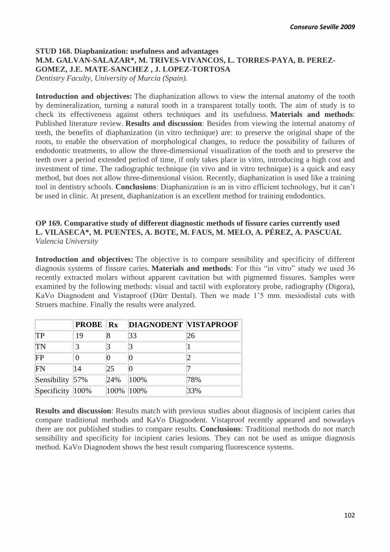
Conseuro Seville 2009
102
STUD 168. Diaphanization: usefulness and advantages
M.M. GALVAN-SALAZAR*, M. TRIVES-VIVANCOS, L. TORRES-PAYA, B. PEREZ-
GOMEZ, J.E. MATE-SANCHEZ , J. LOPEZ-TORTOSA
Dentistry Faculty, University of Murcia (Spain).
Introduction and objectives: The diaphanization allows to view the internal anatomy of the tooth
by demineralization, turning a natural tooth in a transparent totally tooth. The aim of study is to
check its effectiveness against others techniques and its usefulness. Materials and methods:
Published literature review. Results and discussion: Besides from viewing the internal anatomy of
teeth, the benefits of diaphanization (in vitro technique) are: to preserve the original shape of the
roots, to enable the observation of morphological changes, to reduce the possibility of failures of
endodontic treatments, to allow the three-dimensional visualization of the tooth and to preserve the
teeth over a period extended period of time, if only takes place in vitro, introducing a high cost and
investment of time. The radiographic technique (in vivo and in vitro technique) is a quick and easy
method, but does not allow three-dimensional vision. Recently, diaphanization is used like a training
tool in dentistry schools. Conclusions: Diaphanization is an in vitro efficient technology, but it can’t
be used in clinic. At present, diaphanization is an excellent method for training endodontics.
OP 169. Comparative study of different diagnostic methods of fissure caries currently used
L. VILASECA*, M. PUENTES, A. BOTE, M. FAUS, M. MELO, A. PÉREZ, A. PASCUAL
Valencia University
Introduction and objectives: The objective is to compare sensibility and specificity of different
diagnosis systems of fissure caries. Materials and methods: For this “in vitro” study we used 36
recently extracted molars without apparent cavitation but with pigmented fissures. Samples were
examined by the following methods: visual and tactil with exploratory probe, radiography (Digora),
KaVo Diagnodent and Vistaproof (Dürr Dental). Then we made 1’5 mm. mesiodistal cuts with
Struers machine. Finally the results were analyzed.
PROBE Rx DIAGNODENT VISTAPROOF
TP 19 8 33 26
TN 3 3 3 1
FP 0 0 0 2
FN 14 25 0 7
Sensibility 57% 24% 100% 78%
Specificity 100% 100% 100% 33%
Results and discussion: Results match with previous studies about diagnosis of incipient caries that
compare traditional methods and KaVo Diagnodent. Vistaproof recently appeared and nowadays
there are not published studies to compare results. Conclusions: Traditional methods do not match
sensibility and specificity for incipient caries lesions. They can not be used as unique diagnosis
method. KaVo Diagnodent shows the best result comparing fluorescence systems.
Conseuro Seville 2009
103
END 170. Effects of chlorhexidine on the dentin of ferric sulphate pulpotomized primary teeth.
E. RODRÍGUEZ-PRIEGO*, M. BOLAÑOS-CARMONA, S. GONZÁLEZ-LÓPEZ, M.
BRIONES-LUJÁN
Universidad de Granada
Introduction and objectives: Ferric sulphate (FS) is used in primary teeth pulpotomy. FS can
jeopardize the bond strength of some adhesives. Viscostat®’s (V) manufacturer (Ultradent Products,
UT, USA) recommends cleaning the V treated dentin with Clorhexidine (CHX). This study tests the
null hypothesis that 2% CHX (Hibimax®, AstraZeneca S.A., Spain) does not modify the µTBS,
micromorphology or molecular structure of primary dentin prior treated with V. Materials and
methods: Study groups: 1.: VTE: Viscostat + Total etching (37% PA + Excite (Ivoclar Vivadent,
Schaan, Liechtenstein). 2.: VCTE: V + CHX + Total etching. 3.: VSE: V + self-etching (Adper™
Scotchbond™ SE, 3M ESPE, UK). 4.: VCSE: V + CHX + self-etching. µTBS test (n= 16). 1 mm2
dentin/adhesive bars were tested in tension in an INSTRON® 3345. Microscopy: two teeth per group
were visualized by OM and VP-SEM. FTIR. Thin dentin slices (125-130 µm) were soaked with FS
and CHX. Two mg per group of dentin dust were analysed. Results and discussion: µTBS-test
results (MPa): VCTE (11.68+/- 6.17)* < VTE(13.23+/-7.07)< VSE (13.88+/-6.79) < VCSE(16.70+/-
8.03)*. * P <0.05(ANOVA, Tukey’s HDS test). Ferric ions remained on the FS treated dentin
surface even after cleaning with CHX. Conclusions: CHX improved the adhesion of a self-etching
primer to V prior treated primary dentin.
END 171. New system of sponge contained drug delayed antibiotic release in endodontics
M. RUIZ PIÑÓN*1, P. VARELA PATIÑO2, B. MARTÍN BIEDMA, J. BLANCO MÉNDEZ3,
A.J. PÉREZ ESTEVEZ3.
1Lecturer, MA program in Endodontics, 2Departament of Dental Pathology and Therapeutics,
Faculty of Odontology, 3Departament of Pharmacy and Pharmaceutical Technology, 4 Department
of Microbiology. University of Santiago de Compostela, Spain
Introduction and objectives: The aim of this study was to develop a state-of-the-art in sponge
contained drug for its delayed release within the root canal. Materials and methods: At a first stage
different concentrations of antibiotics (Amoxicillin) in sponge polymer were tested on Mueller-
Hinton culture media to assess their effectiveness, release and manipulation features. Enterococos
faecalis was used. In subsequent studies a formulation containing 0.44 mg of amoxicillin per mg of
sponge which released antibiotics for antibiotics for 3 to 4 days was used. The second part of the
study was carried out in Vitro on using 14 extracted uniradicular teeth. Before inserting the sponges
with amoxicillin in the teeth. Results and discussion: As for the samples obtained after scraping the
root canal walls, bacterial growth was only observed after 24 hours in one of the two teeth studied
while both samples came out positive after 48 hours. Conclusions: Further analysis of different
concentrations and antibiotics are necessary to reinforce the preliminary goods results of this new
means of antibiotic transport.

Conseuro Seville 2009
104
MAT 172. Management of pretest failures in microtensile bond strength tests.
J. BASCONES*,, J.C. de la MACORRA.
Department of Conservative Dentistry, Faculty of Odontology, Complutense University of Madrid,
Spain.
Introduction and objectives: Number of pre-test failures (PTF) in tensile bond strength tests is
difficult to interpret. Objective is to compare PTF of various adhesives when tensile bond strength
(TBS) of interface and tensile cohesive strength (TCS) of dentin adjacent to interfaces were
tested. Materials and methods: 40 third molars were randomly assigned to a group (Adper Prompt
L-Pop (APLP), Xeno V (XV), Adhese One (A1), Adper ScotchBond SE (SBSE), Clearfil SE Bond
(CSE), Optibond All in One (OPT), Clearfil DC Bond (DCB), Clearfil S3 Bond (CS3)). Oclusal
enamel and roots were removed and teeth perfused (20 cm distilled H2O). Two horizontal parallel
grooves (2 mm wide, 2 mm deep, 2mm separation) were drilled in oclusal dentin, and were rstored
(Filtek Z250), before crown was rebuilt (2 mm height). Compound bars (resin-dentin) were obtained
from grooves (to assess TBS at interface) and from between grooves (to assess TCS at the same
horizontal plane than TBS). Bars were rounded before testing and PTF annotated. Only specimens
that failed before mechanical test were included in analysis. To test if number of valid/PTF
specimens was independent of test (TBS/TCS), Goodman’s Tau for independence coefficient was
calculated.
Materials
/ test
number
of valid
and
PTF
specimens
Independence
test results
(Tau,
significance)
(TBS or
TCS) PTF valid Tau p
APLP TBS 24 4 0.52 0.00001
TCS 1 10
XV TBS 6 19 0.1 0.63
TCS 3 14
A1 TBS 23 1 0.63 0.000001
TCS 3 13
SBSE TBS 13 12 0.22 0.003
TCS 1 15
CSE TBS 0 15 N/A N/A
TCS 0 18 " "
OPT TBS 0 23 " "
TCS 0 16 " "
DCB TBS 0 18 " "
TCS 0 14 " "
CS3 TBS 2 14 0.06 0.18
TCS 0 14
Results and discussion: see table. Conclusions: When APLP, A1 and SBSE were tested, specimens
prepared to measure TBS failed prematurely significantly more times than the ones prepared to
measure TCS of adjacent dentin. This didn’t happen with the rest of materials.

Conseuro Seville 2009
105
END 173. Comparison of behavior of pain after mechanical or manual endodontic
instrumentation
A.M. ARIAS-PANIAGUA1*, M. AZABAL2, J.J. HIDALGO1, J.C. de la MACORRA1.
1Department of Conservative Dentistry, Faculty of Odontology, Complutense University of Madrid,
Spain. 2Private practice.
Introduction and objectives: Shaping of root canals has changed in recent years due to the
increased use of rotary nickel-titanium files. These new shaping techniques have many advantages
when compared with the traditional manual method using stainless steel files, but it is not known if
postoperative pain behaviour differs according to the shaping technique we use. Aim of study is to
establish the influence of the shaping technique in the incidence, degree, length and type of
postendodontic pain. Materials and methods: Endodontic treatments were carried with mechanical
instrumentation (n = 44) or manual shaping techniques (n=44) in a single visit and data about pre-
treatment patient’s conditions and tooth diagnostic factors were collected. Patients were given a
questionnaire to be filled and returned, where they should record the presence or absence of
postendodontic pain, its duration, need of previous stimulus (none, chewing) and level of
discomfort. Results and discussion: We observed significantly more postoperative pain (X2 test, p =
0.05) in the group treated with manual shaping techniques. However, in this group in duration of
pain was shorter (Mann-W test, p = 0.008). Differences in level of discomfort were not statistically
significant (X2 test, p = 0.8) between groups. Conclusions: Incidence of postoperative pain is
significantly lower in teeth shaped with rotary instrumentation, but length of pain, if present, is
significantly prolonged in these cases.
STUD 174. Microperforated needle for root canal disinfection with ozone
M. ABUIN*, A. TORRADO, B. MARTIN, J.D GONZÁLEZ-BAHILLO, J.C PÉREZ, V. LÜDI
Universidad de Santiago de Compostela, Spain
Introduction and objectives: In general medicine, the ozone-oxygen gases medley is proving itself
as a highly efficient treatment. Ozone is a broad-spectrum germicide effective against all types of
bacteria, fungi, viruses and even spores. Dentistry is not foreing to this capacity. The objective is to
investigate new systems of root canal disinfection during endodontic treatments. Materials and
methods: We patented a silicon needle (ES: 200701651 U), straight, flexible and microperforated in
all his length. This patent is based on a bibliographic review about root canal disinfection with
ozone-oxygen gasses. The new needle has a minor caliber in comparison with present ones. Allows
us to come closer to the apex. Treats the root canal in all his length and three-dimensionally. Allows
the use of high ozone concentrations. It can be used, moreover, with irrigation solutions, in which
case the risk of creating periapical damage is much lower. Results and discussion: Other needles or
cannulae used by other equipments have only one ozone exit located apically. If we rely on the
physical-chemical properties of ozone, it is inactivated at the first point of contact. Moreover the
other techniques use oxygen from the patients breathe and in low concentrations. Conclusions:
These needles show better clinical results than other designs.

Conseuro Seville 2009
106
STUD 175. Microperforated trays for oral treatments with ozone
A.TORRADO*, M. ABUIN, V. LÜDI, J.C. PÉREZ, B. MARTIN, J.D GÓNZALEZ-BAHILLO
Universidad de Santiago de Compostela, Spain
Introduction and objectives: International scientific community recognized ozone as one of the
most powerful oxidants in nature and its antiviral, antifungical and antibacterial capacity have been
widely proved. The principal aim is to prove the major efficiency of these patent in comparison with
present devices. Materials and methods: Based on researches on present equipments, a
microperforated tray was patented (ES: 200701858U) for ozone applications in oral treatments such
as incipient caries, teeth withening and periodontal disease. The silicon tray is microperforated along
its entire surface. It has a double layer with inner cabin and a connection to the ozone entrance (luer
or luer lock). The tray contacts with the whole surface to treat. Ozone will be obtained from medical
oxygen at high concentrations. Results and discussion: The advantage of this equipment is that
higher ozone concentrations that can be achieved. Due to physical-chemical characteristics of this
gas, aspiration is not required. A wider surface will be treated more equally. Present devices are
more complex and its aspiration systems aspire more than they eject. Usually they obtain oxygen
from the patient his breathe and at low concentrations. Conclusions: The microperforated tray its
design is simpler. There is no risk of injury in the respiratory epithelium.
END 176. Glucose and dye penetration of intraorifice sealing materials in root canal treatment
E. BAILÓN-SÁNCHEZ*, S. GONZÁLEZ-CASTILLO, M.P. GONZÁLEZ-RODRÍGUEZ,
C.M. FERRER-LUQUE
University of Granada. School of Dentistry. Department of Estomatology.
Introduction and objectives: Prevention of coronal leakage in root canal therapy has been
accomplished by temporary restorative materials. The use of a material to seal the orifice, in addition
to the temporary restoration after root canal filling mitigates bacterial leakage. The purpose of this
study was to evaluate the sealing capacity of three different intraorifice barriers with two different
models of leakage. Materials and methods: The coronal portions of 60 single-rooted teeth extracted
were removed to obtain roots with 15mm in length. The canals were prepared with hand
instrumentation technique and were filled using the cold lateral condensed technique of the gutta-
percha and AH Plus. The four coronal milimetres of the filling material were removed with System
B. ProRoot MTA, Cavit G and Tetric EvoFlow were placed into the orifice. The roots were mounted
on a glucose penetration and dye leakage models. The penetration was measured at 45 days in mmol
L-1 (glucose) and millimetres (dye). Results and discussion: The results obtained with three sealing
materials were similar with the glucose penetration models; however ProRoot MTA showed
significantly lower dye leakage scores (0.95±0.93) that the other materials (p<0.05). Conclusions:
Cavit G and Tetric EvoFlow, in both models, exhibited a similar performance at 45
days. Acknowledgements: Thanks to Dr. Rafael Poyatos for help in measurement to the samples.
Conseuro Seville 2009
107
END 177. Evaluation of dentine adhesive systems to seal the pulp chamber using two leakage
models
S. GONZÁLEZ-CASTILLO*, E. BAILÓN-SÁNCHEZ, M.P. GONZÁLEZ-RODRÍGUEZ,
C.M. FERRER-LUQUE
University of Granada. School of Dentistry. Department of Estomatology.
Introduction and objectives: An adequate coronal seal is critical for the success of root canal
therapy. A variety of temporary materials have been tested in an attempt to provide a coronal barrier
to prevent microleakage in endodontic, but none of the materials were able to reduce leakage
completely. The purpose of this study was to compare the sealing properties of two different dentine
adhesive placed in the pulp chamber with a glucose penetration model and a dye penetration test.
Introduction and objectives: Sixty extracted human premolars were instrumented to a size 40 K-file
and obturated with gutta-percha and AH Plus, using the lateral condensed technique. The access
cavities were filled with Clearfil SE Bond+Cavit G, Primer & Bond NT+Cavit G, or Cavit G alone.
Microleakage was assessed by methylene blue dye penetration (millimetres) and glucose penetration
model (in mmol L-1). Results and discussion: The results obtained with the glucose penetration
model, at 45 days, were similar for the three and there were no significant differences (p>0.05)
between them. With the dye penetration test, Cavit G alone showed the lower leakage scores
(2,16mm). Conclusions: Under conditions of this study, the use of the dentin adhesive systems did
not enhance the temporary seal of the pulp chamber access. Acknowledgements: Thanks to
Dr.Rafael Poyatos for help in measurement to the samples.
END 178. Eradication of Enterococcus faecalis biofilms by chlorhexidine and cetrimide
M.T. ARIAS-MOLIZ *, C.M. FERRER-LUQUE, M.P. GONZÁLEZ-RODRÍGUEZ, P.
BACA.
University of Granada. School of Dentistry. Department of Estomatology.
Introduction and objectives: Enterococcus faecalis is often isolated in root canals with persistent
periapical disease. Its ability to grow as biofilms might allow E. faecalis to survive in these hard
conditions. The exo-polysaccharide matrix of biofilms prevents the diffusion of the irrigants; cationic
surfactants would be able to disrupt this matrix. The aim of our study was to evaluate the minimal
biofilm eradication concentration of chlorhexidine (CHX), cetrimide (CTR) and several
combinations of both against E. faecalis biofilms.Biofilms of E. faecalis grew aerobically in
polystyrene surfaces for 24h at 37ºC and were exposed 1 minute to ten serial twofold dilutions of 4%
CHX, 0.1% CTR and 4% CHX combined with constant concentrations of CTR (0.01, 0.02 and
0.05%). The antibacterial activity of solutions was evaluated by determining the viable cell counts
and logarithmic killing of cultures. Results and discussion: 4% CHX and 0.05% CTR did not
eradicate E. faecalis biofilms. In contrast, the combinations of: 0.125% CHX + 0.01% CTR,
0.0625% CHX + 0.02 % CTR and all CHX dilutions + 0.05% CTR were able to eradicate them after
1 minute. Conclusions: The combination of CHX with CTR showed synergistic activity in
eradicating E. faecalis biofilms. Acknowledgements: To Francisca Castillo for her technical
assistance.

Conseuro Seville 2009
108
STUD 179. Relationship between smoking and pulpar pathology
H. GONZÁLEZ-GONZÁLEZ*, M. PORLÁN-COSTA, A.B. RODRIGUEZ-ESPIN, J.
LOPEZ-TORTOSA
Dentistry Faculty, University of Murcia (Spain)
Introduction and objectives: Smoking has local and systemic effects on the microcirculation and
inmune system. Therefore, smoking impairs the body’s response to infection including pulp
response. Objective: to discuss the possible relationship between cigarette smoking and endodontic
treatment. Materials and methods: Literature review. Results and discussion: There are evidences
linking smoking with periapical pathology. It seems probably that nicotine causes release of
neuropeptide CGRP in pulp tissue, which is associated to significant changes in blood flow,
inflammation and pulpal pain. However, other studies show no statistically association between
smoking and endodontic disease as apical periodontitis is likely to be multifactorial. Current cigarette
smokers have the highest percentages of teeth with calculus, more decayed or filled coronal and root
surfaces and have lost more teeth than never smokers but it was suggested that this factors are more
likely to be due to other oral factors associated with smoking such as reduced flow of saliva and
gingival exudate than a direct effect of smoking. Conclusions: It is not possible to state that smoking
increases the prevalence of apical periodontitis.
END 180. An audit of intra-oral digital radiographs.
B. ROBINSON,* R. AUSTIN
King’s College Hospital Dental Institute London
Introduction and objectives: The objective of this study was to improve the quality of digital
radiographs taken during endodontic treatment at King’s College Hospital Dental Institute in three
phases. The first phase compared the Schick CDR system with Digora Optime. The second and third
phases involved ways of improving the quality of the digital radiographs produced by the Schick
CDR system. Materials and methods: The FGDP Guidelines on Selection Criteria for Dental
Radiography and Guidance Notes for Dental Practitioners on the Safe Use of X-Ray Equipment-
National Radiological Protection Board enabled the use of a 3 point quality scale (1 excellent, 2,
diagnostically acceptable, 3 unacceptable), which took into consideration sensor angulation,
positioning, contrast and focusing. The recommended FGDP guidelines are not less than 70% images
scoring excellent. For the first phase 50 exposures recorded with the Schick CDR system were
compared with 50 recorded using Digora Optime. For the second and third phases 50 radiographs for
each phase were evaluated with images generated by the Schick system with training provided
between the phases. Results and discussion: Images produced by the Schick system showed an
inferior quality compared with the images generated by the Digora method. Both systems failed to
reach the desired quality FGDP standard of 70% excellent (Schick 55% Digora 69%). Comparison of
the results in the second and third phases showed that training the operator improved the quality but
recommended the purchase of a size 1 or 0 Schick sensors to improve positioning
errors. Conclusions: This study was carried out in order to minimise the ionising radiation dose to
patients and to maximise the clinical and administrative benefits of using a digital system. It
demonstrated an improvement in the quality of radiographs across all criteria measured up to and
beyond the desired standard, from 55% of radiographs scoring excellent in the first phase to 80% in
the third phase. As a result of the study it was decided to install the Schick CDR system because of
the speed it produced images even though the first phase of this study demonstrated inferior image
quality.

Conseuro Seville 2009
109
OP 181. Influence of patient’s age and initial color in dental bleaching effectiveness
P. FENOY*, L. JANE, M. ROIG.
Universitat International de Catalunya
Introduction and objectives: Dental bleaching is a very common treatment in Aesthetics dentistry.
Dental color changes with age because of enamel and dentin amended. Young patients have thick,
opaque enamel while in old patients enamel becomes thin and translucent. Dentin with age becomes
yellowish, with thin and translucent enamel. All these changes in enamel and dentin makes tooth
become darker. These changes in enamel and dentin are irreversible with bleaching. The aim of the
study was to determine the influence of patient’s age and dental initial color in the bleaching
result. Materials and methods: We used Vivastyle paint on plus® ( Ivoclar Vivadent), a 6%
Hydrogen Peroxide. Twenty-eight patients were classified in three groups of age: group I (25-34
years), group II (35-44 years) and group III (45-55 years). They used dental bleaching twice a day
for a week. Dental color was measured with Vitapan Classical Guide and black and white digital
Photographs. We measured dental color after treatment, a month and three month after treatment.
Successful bleaching table was used to obtain a percentage of bleaching. Results and discussion:
Group I patients obtained a higher percentage of bleaching than group II and III patients. Bleaching
results were better for dark initial colors. Conclusions: Young patients obtained better results in
dental bleaching than old patients. Initial color influenced the results.
MAT 182. In vitro evaluation of the antimicrobial activity of several self-etching adhesive
systems against Pseudomonas Aeruginosa and Enterococcus Faecalis.
D. RUBIO*, V. VERA, E. GARCÍA BARBERO, J. GARCÍA BARBERO, A. E. GARCÍA
BARBERO
Departamento Odontología Conservadora. Universidad Complutense de Madrid
Introduction and objectives: The purpose of this study was to determine in vitro the antimicrobial
activities of several self- etching adhesive systems against two endodontic and periodontal
pathogens, Pseudomonas aeruginosa and Enterococcus faecalis. Materials and methods: The
diffusion method on Brain Heart Infusion (BHI) agar was employed, previously inoculated with both
microorganisms. Susceptibility paper discs (SensiDisc®) were placed in the agar plates filled with
four adhesives: Futurabond DC® (Voco), Adhe SE® (Ivoclar Vivadent), Xeno III® (Dentsply
Maillefer) and GC G- Bond® (GC). After 48 hours of incubation at 37º the zones of growth
inhibition were observed and measured with Image Tool 3.0. Results and discussion: For the group
Pseudomonas aeruginosa, Adhe SE® showed the widest diameter (2,003 mm; SD +/-
0,655),followed by Futurabond DC® (1,808 mm; SD+/- 0,476), GC® (1,00 mm; SD +/- 0,322) and
Xeno III® (0'857 mm; SD +/- 0,388). For the group Enterococcus faecalis, Futurabond DC® showed
the widest diameter (4,799 mm; SD +/- 0,830), followed by Xeno III® (1,143 mm; SD +/- 0,345),
GC® (0,761 mm; SD +/- 0,358) and Adhe SE® (0,487 mm; SD +/- 0'198). Conclusions: Futurabond
DC® is an inhibition agent for both bacteria with wide haloes. Adhe SE® inhibits both bacteria with
narrow haloes. Xeno III® and GC® show very narrow haloes.
Conseuro Seville 2009
110
MAT 183. Energy densities and microhardness ratio of nanohybrid composites
K. GRITSCH*1,4, C. ROSSARY1, P. FARGE2,4, N. SINTES-ZYDOWICZ3, P. COLON1,5,
B. GROSGOGEAT1,4
1Laboratoire des Multimatériaux et Interfaces, UMR CNRS 5615, Université de Lyon, France,
2Département de Formation et Centre de Recherche en Biologie Humaine, Université de Lyon,
France, 3Laboratoire des Matériaux Polymères et des Biomatériaux, UMR CNRS 5223, Université
de Lyon, France, 4SCTD/Hospices Civils Lyon, Lyon, France, 5 Faculté de Chirurgie Dentaire
Garancière. Université Paris Diderot, France.
Introduction and objectives: The aim of this study was to control the hardness ratio (bottom
surface microhardness/top surface microhardness) of samples of nanohybrid composites cured by
LEDs. Materials and methods: Two LEDs, Bluephase (Ivoclar Vivadent) and Elipar Freelight 2
(3M ESPE), were used to polymerize 3mm-thick samples of two nanohybrid resins, Ceram X
(Dentsply Caulk) and Tetric EvoCeram (Ivoclar Vivadent). Hardness ratio was measured at various
combinations of power and energy densities. The power density was obtained by modulating the
distance and controlled by a digital radiometer (Cure Rite/Dentsply Caulk). The energy densities
were obtained by varying the exposure duration. The microhardness (Vickers Hardness) of both top
and bottom surfaces was measured with a microdurometer (Model 5014/Buehler), and the ratio
calculated. Results and discussion: CeramX always reached the greatest values of microhardness
(good spectral concordance between the absorption spectrum of the photoinitiator included and the
emission spectrum of the LEDs). All combinations of resin/LED presented a ratio greater or equal to
80% at energy densities greater or equal to 20J/cm². Conclusions: An energy density of 20J/cm²
need to be reached to insure a hardness ratio of 80%. Acknowledgements: The authors wish to thank
Ivoclar Vivadent, 3M ESPE and Dentsply Caulk for the supply of materials.
MAT 184. Elaboration of growth factors-loaded microspheres as colloidal carriers for dentin-
pulp complex regeneration
N. KALAJI 1*, N. SHEIBAT-OTHMAN 1, P. LAURENT 2, I ABOUT 2, H. FESSI 1
1Université de Lyon 2 Laboratoire IMEB-ERT 30, Faculte d’Odontologie, Universite de la
Mediterranee, France
Introduction and objectives: The application of growth factors such as FGF (Fibroblast Growth
Factor) and TGFβ (transforming growth factor) in dental tissue engineering necessitates the use of a
delivery system. A wide range of drug delivery carriers have been applied to accommodate different
molecules to regenerate dental tissues. Materials and methods: In this work FGF, TGFβ was
encapsulated in PLGA microspheres by a w/o/w double emulsion solvent extraction technique.
Microspheres size, colloidal stability, surface properties and morphology were studied. Release
kinetics and Encapsulation efficiency were determined in vitro. Biological activity was studied by
MTT test. Results and discussion: It was found that FGF was encapsulated into these microspheres
with good encapsulation efficiency and that there biological activity was kept after encapsulation
process. Smooth and spherical microspheres were obtained after the drying process. FGF was
released in a multiphasic fashion including an initial burst effect. Conclusions: These findings
provide baseline data for potential uses of microencapsulated FGF and TGFβ in dental pulp healing
and dental tissue-engineering applications. Subsequently FGF-microspheres will be implicated as
direct pulp capping materials into in vitro entire tooth culture model of human immature third
molars, pulp lesion will be done. Pulp responses after applying these FGF and microspheres will be
evaluated. In vitro results of this application will be obtained soon.

Conseuro Seville 2009
111
OP 185. Influence of saliva contamination on microleakage of three adhesive systems
A. AMORIM*, M. HENRIQUES, P. MELO
Faculdade de Medicina Dentária do Porto (FMDUP) - Portugal
Introduction and objectives: Moisture control of the operating field is a critical factor that enhances
restoration´s quality and clinical success. The purpose of this study was to evaluate the influence of
saliva contamination on microleakage of three adhesive systems. Materials and methods: Class V
cavity preparations were made at the CEJ, on buccal and lingual surfaces, in 60 molars. Teeth were
randomly divided for 3 adhesive systems: Prime&Bond®NT, FuturabondNR and FujiBOND LC.
Fresh saliva contamination was done before adhesive polymerization. Three decontamination
protocols were tested: (1) cotton-pellet drying; (2) rinsing, air drying and adhesive reaplication; (3)
air-stream drying. Specimens were restored with Grandio®. After termocycling 1500 cycles, dye
penetration (blue-of-methylene 2%) was evaluated in an optical microscope (16X). Qui-Square
Independence Test for statistical analysis was used at p<0.05. Results and discussion: No
statistically significant differences were found for Prime&Bond®NT. There were statistical
significant differences for FuturabondNR, showing less leakage in decontamination protocol 1
(p<0,001) and protocol 3 (p=0.002) at enamel, and for protocol 2 (p=0.002) at dentin, and for
FujiBOND LC at dentin margins (p<0,001) for decontamination protocol 2. Conclusions: Saliva
contamination had no influence in microleakage of any adhesive systems studied, except when the
cotton-pellet drying decontamination is applied in dentine margins of FuturabondNR and in enamel
margins of FujiBOND LC.
CLIN 186. Management of postoperative hypersensitivity following composite restorations
J. SABBAGH*1, J.-C. FAHD1, R. McCONNELL2
1 Lebanese University, Beirut Lebanon, 2 Cork University, Ireland
Introduction and objectives: The use of resin composite restorative materials has increased due to
the high aesthetic demands of our patients and the environmental issues raised with the use of silver
amalgam. There has been an increase in their use for the restoration of posterior teeth. Many have
reported an increase in postoperative sensitivity resulting in which may last for a fews days but can
in some cases last many weeks. Materials and methods: Using clinical cases, the possible causes of
tooth hypersensitivity are reviewed, and the management and options of treatment are outlined
showing a well defined clinical protocol. Results and discussion: Fluid flow within dentine is the
main physiological factor held responsible of this sensitivity. Another explanation could be the
inflammatory reaction of the pulp due to bacterial contamination. Several factors are responsible for
postoperative hypersensitivity when composite materials are used, including polymerization
shrinkage, poor light-curing outcome and inadequate bonding procedure as well as other factors
which may cause such a fluid movement. Conclusions: Achieving a composite restoration needs 2.5
times more than an amalgam restoration. The use of adequate materials, following a well defined
protocol, will reduce considerably postoperative sensitivity.

Conseuro Seville 2009
112
END 187. Fracture protection of endodontic treated teeth.
F. le COUTRE, D. PAHNCKE, D. BEHREND, H. LANG
University of Rostock
Introduction and objectives: The destabilizing effect of endodontic treatment upon teeth is still
controversial. The stability of the crown-root-complex is mainly determined by the remaining
substance. The aim of this study was to find an easy way to stabilize and to protect endodontically
treated teeth. Materials and methods: In the present study 33 human, freshly extracted molars were
assigned to 3 different groups: A: Control group (untreated teeth), B: Endodontic treatment, m-o-d
preparation and composite resin restoration, C: Endodontic treatment, m-o-d preparation flattening
and partial crown. Group C was built up with composite resin. Afterwards crowns were flattened and
restored using a partial ceramic coverage (“occlusal shield”). All teeth were subjected to a linear
loading until fracture by using a universal testing machine. After the tests every tooth was valued:
easy to repair, difficult to repair, unable to repair. Results and discussion: The statistical evaluation
showed a significant difference between groups A and B (p<0.05) but no difference between groups
A and C (p>0.05). Results in group C were superior to group B but the difference was not
statistically significant (p=0,054). Conclusions: The results of our study show that stabilization of
endodontically treated teeth can be achieved by a simple replacement of the most upper part of the
crown.