construction and characterisation of a full-length infectious molecular clone from a fast...
TRANSCRIPT
Construction and characterisation of a full-length infectious molecularclone from a fast replicating, X4-tropic HIV-1 CRF02.AG
primary isolate
Denis M. Tebit,a Leopold Zekeng,b Lazare Kaptue´,b
Hans-Georg Kra¨usslich,a,* and Ottmar Herchenro¨derc
a Abteilung Virologie, Universitat Heidelberg, Im Neuenheimer Feld 324, D-69120 Heidelberg, Germanyb University of Yaounde Teaching Hospital, University of Yaounde 1, Yaounde 812, Cameroon
c Institut fur Virologie, Medizinische Fakultat “Carl Gustav Carus,” D-01307, TU Dresden, Germany
Received 7 January 2003; returned to author for revision 27 March 2003; accepted 22 April 2003
Abstract
Based on our previous analysis of HIV-1 isolates from Cameroon, we constructed a full-length infectious molecular clone from a primaryisolate belonging to the CRF02.AG group of recombinant viruses which dominate the HIV-epidemic in West and Central Africa. The virusderived by transfection of the proviral clone pBD6-15 replicated with similar efficiency compared to its parental isolate and used CXCR4as coreceptor as well. Furthermore, HIV-1 BD6-15 exhibited similar replication properties and virus yield as the reference B-type HIV-1strain NL4-3. Sequence analysis revealed open reading frames for all structural and accessory genes apart fromvpr. Phylogenetic andbootscanning analyses confirmed that BD6-15 clusters with CRF02.AG recombinant strains from West and Central Africa with similarcross-over points as described for the CRF02.AG prototype strain lbNG. Thus, pBD6-15 represents the first non-subtype B infectiousmolecular clone of a fast replicating, high producer, X4-tropic primary HIV-1 isolate, which had only been briefly passaged in primary cells.© 2003 Elsevier Science (USA). All rights reserved.
Keywords: HIV-1 CRF02.AG; Primary isolate; Infectious molecular clone
Introduction
Sub-saharan Africa is the most affected region in thehuman immunodeficiency virus type 1 (HIV-1) pandemicharbouring about 70% of all persons infected with HIV. InCentral Africa, the presence of HIV-1 and HIV-2 of virtu-ally all groups (HIV-1 group M, O, and N) and subtypes hasbeen reported (Takehisa et al., 1998; Heyndrickx et al.,2000). To date, nine subtypes of HIV-1 group M have beendescribed with different subtypes dominating the epidemicin different parts of the world. Furthermore, coinfectionwith more than one subtype has led to the generation ofrecombinants between different subtypes which have re-
placed their parental strains in some areas. Despite thepresence of many different subtypes in Central Africa, oneform of HIV-1 group M, the circulating recombinant form(CRF), termed CRF02.AG, currently dominates the HIVepidemic in this region. Genotypic studies showed thatapproximately 60% of all viral isolates of West and CentralAfrican origin are CRF02.AG viruses (Takehisa et al., 1998;Heyndrickx et al., 2000; Montavon et al., 2000; Carr et al.,2001). Subtype A and G viruses which are the putativeparental forms of CRF02.AG recombinants are also highlyprevalent in this region (Peeters et al., 2000). Several studieshave reported near full-length sequences of CRF02.AG vi-ruses of West and Central African origin (Howard andRasheed, 1996; Montavon et al., 2000; Carr et al., 2001).While providing important information regarding the mo-lecular epidemiology of HIV-1, these sequence analyses donot permit a detailed molecular characterisation of this virus
* Corresponding author. Fax:�49-6221-56-5003.E-mail address: [email protected]
(H.-G. Krausslich).
R
Available online at www.sciencedirect.com
Virology 313 (2003) 645–652 www.elsevier.com/locate/yviro
0042-6822/03/$ – see front matter © 2003 Elsevier Science (USA). All rights reserved.doi:10.1016/S0042-6822(03)00381-7
group. Such studies require infectious molecular clones thatproduce replication-competent virus following transfectionof permissive target cells.
Much of the currently available information on HIV-1replication has been obtained from experiments with a fewinfectious molecular clones. Besides their role in basic sci-ence, such infectious clones are also important tools invaccine studies as well as for the development of antiviraldrugs and for the analysis of antiviral resistance. Mostreplication-competent clones that exist to date representHIV-1 subtype B viruses, whose role in the epidemic islimited to Europe and North America. Non-subtype B vi-ruses from regions bearing the burden of HIV have not beenstudied intensively until very recently. More than half of theapproximately 10 infectious molecular clones generated sofar are subtype B. More recently, infectious molecularclones have also been generated for subtypes C, D, and theCRF01.AE recombinant form (Gao et al., 1998; Salminen etal., 2000; Ndung’u et al., 2001; Mukai et al., 2002). Tworecent reports described replication-competent molecularclones from CRF02.AG recombinant viruses of Ghanaianorigin (Kusagawa et al., 2001; Takahoko et al., 2001). Ingeneral, most non-B subtype derived infectious clones havesome limitations. For example, although having been gen-erated from PBMCs, they may represent chimeras from twodifferent isolates (Ndung’u et al., 2001). Furthermore, com-parison of the virus derived from the molecular clone withits parental isolate or other reference strains is often lacking(Gao et al., 1998). In the cases where these comparisonswere done, the replication kinetics and virus production wassignificantly reduced for the virus derived from the infec-tious clone, making these reagents difficult to work with(Mukai et al., 2002). In the case of the CRF02.AG viruses,the virus derived from the first replication-competent clonewas restricted in its host range, infecting T cell lines but notperipheral blood mononuclear cells (PBMCs) (Kusagawa etal., 2001). The virus derived from the other clone replicatedin PBMCs, albeit with slow replication kinetics compared toother HIV isolates (Takahoko et al., 2001).
Previously we have characterised a panel of HIV-1 groupM primary isolates from Western Cameroon (Tebit et al.,2002). To generate research tools for studying the dominantviruses in Cameroon in particular and West and CentralAfrica in general, we selected one of these isolates, termed99CMBD6, based on its CRF02.AG genotype in gag, pol,and env for full-length amplification and construction of theinfectious molecular clone pBD6-15. Virus derived fromtransfection of this proviral clone replicated with similarefficiency and kinetics compared to the parental virus iso-late. Furthermore, HIV-1 BD6-15 exhibited similar replica-tion properties and virus yield as the reference B-typeHIV-1 strain NL4-3 (Adachi et al., 1986). Thus, pBD6-15represents the first non-subtype B infectious molecularclone of a fast replicating, high producer, X4-tropic primaryHIV-1 isolate, which had only been briefly passaged inprimary cells.
Results and discussion
The blood sample 99CMBD6 originated from a 32-year-old female Cameroonian suffering from AIDS. We havepreviously reported a partial characterisation of the primaryisolate 99CMBD6, which was determined to be a memberof the CRF02.AG lineage (Tebit et al., 2002). We decided togenerate a full-length infectious molecular clone from thisisolate and compare its characteristics with that of the pa-rental isolate and a reference strain. The redundancy of theviral LTRs necessitated the amplification of the full-lengthproviral genome of 99CMBD6 in two fragments. The 5�fragment extended from the 5� LTR to the reverse transcrip-tase (RT) coding region in the pol gene, while the secondfragment extended from the RT coding region to the 3�LTR. Following long PCR and cloning of the respective 5�and 3� fragments, eight clones were obtained. Four of theseclones contained the 5� fragment (TOPO5�frag1–4) and theother four contained the 3� fragment (TOPO3�frag1–4).Subsequently, the 5� fragments were excised from theTOPO5�frag plasmids and inserted into the TOPO3�fragplasmids, thus generating a total of 16 full-length clones.
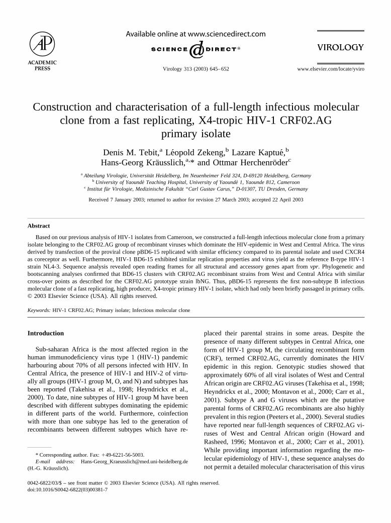
Virus production, virion protein composition, and infec-tivity were tested for all 16 clones following transfection of293T cells. Filtered supernatants from the transfections of14 of the 16 full-length clones contained p24 antigen asdetermined by antigen capture ELISA. These culture mediawere therefore used to infect HIV-permissive C8166 cells.Infectivity was determined by measuring p24 antigen andmonitoring for syncytia formation. Only 1 of the 14 trans-fection supernatants was shown to contain infectious virus.This infectious molecular clone was termed pBD6-15 andwas further characterised with respect to its protein compo-sition, replication properties in different cell types, biolog-ical phenotype, and coreceptor usage.
To determine the protein composition of the BD6-15virus and of viruses obtained from some of the defectivemolecular clones, we performed immunoblot analysis usingthe serum of patient 99CMBD6 for detection. Viruses wereharvested from 293T cells transfected with the respectivefull-length clone and were concentrated by ultracentrifuga-tion through a sucrose cushion. Fig. 1 shows the compara-tive analysis of particle fractions derived from pBD6-15,three of the noninfectious full-length clones (pBD6-5,pBD6-11, pBD6-13), the reference B-type clone pNL4-3,and an HIV-1 group O clone. While transfection ofpBD6-11 did not lead to production of particulate HIVantigen (Fig. 1, lane 4), transfection of pBD6-5 (lane 3) andpBD6-13 (lane 5) yielded particles which exhibited a sim-ilar protein profile as pBD6-15 (lane 6), but were producedat lower efficiency. The patient serum detected the Gag-derived proteins Pr55, matrix (MA), and capsid (CA) aswell as the Pol-derived protein integrase (IN) in all cases.However, the ratio of Gag cleavage products to Pr55polyprotein appeared to be significantly reduced in the twodefective viruses, indicating that there may be a processing
646 D.M. Tebit et al. / Virology 313 (2003) 645–652
defect. A very similar protein profile as for BD6-15 wasdetected for the B-type isolate NL4-3. Less cross-reactivitywas observed for the O-type virus, whose MA and INproteins were not detected by the patient serum.
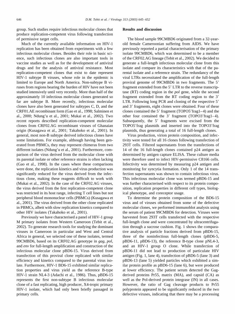
To determine the replication properties of the virus de-rived from the infectious molecular clone pBD6-15, weinfected PBMCs and the T cell line C8166 with virusproduced by transfection of 293T cells and with the parentalisolate 99CMBD6. Virus derived from transfection of 293Tcells with pNL4-3 was used as a control. Fig. 2A shows thatthe growth kinetics and virus production were very similar
for all three viruses in PBMCs. Furthermore, transfection-derived BD6-15 virus also replicated with identical kineticsin C8166 cells as its parental isolate, proving that thismolecular clone faithfully reproduced the properties of theprimary virus (Fig. 2B). Remarkably, BD6-15 exhibitedonly a minor delay in replication kinetics in C8166 cellswhen compared with the widely used and rapidly growingB-type laboratory strain NL4-3.
The biological phenotype and coreceptor usage of trans-fection-derived BD6-15 virus and the parental isolate weredetermined on MT-2 and GHOST cells, respectively. Bothviruses exhibited identical properties: they induced syncytiaon MT-2 cells and exclusively used the coreceptor CXCR4(data not shown). The two previously described CRF02.AGmolecular clones, on the other hand, were nonsyncytiuminducing and used CCR5 as coreceptor for entry (Takahokoet al., 2001; Kusagawa et al., 2001), properties shared bymost infectious clones derived from primary isolates.
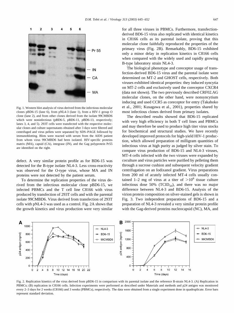
The described results showed that BD6-15 replicatedwith very high efficiency in both T cell lines and PBMCsand may therefore be used to produce high titer virus stocksfor biochemical and structural studies. We have recentlydeveloped improved protocols for high-yield HIV-1 produc-tion, which allowed preparation of milligram quantities ofinfectious virus at high purity as judged by silver stain. Tocompare virus production of BD6-15 and NL4-3 viruses,MT-4 cells infected with the two viruses were expanded bycoculture and virus particles were purified by pelleting themthrough a sucrose cushion and subsequent velocity gradientcentrifugation on an Iodixanol gradient. Virus preparationsfrom 200 ml of acutely infected MT-4 cells usually con-tained 1–2 mg of virus at a titer of �108 tissue cultureinfectious dose 50% (TCID50), and there was no majordifference between NL4-3 and BD6-15. Analysis of thevirion protein composition on silver-stained gels is shown inFig. 3. Two independent preparations of BD6-15 and apreparation of NL4-3 revealed a very similar protein profilewith the Gag-derived proteins nucleocapsid (NC), MA, and
Fig. 1. Western blot analysis of virus derived from the infectious molecularclones pBD6-15 (lane 6), from pNL4-3 (lane 1), from a HIV-1 group Oclone (lane 2), and from other clones derived from the isolate 99CMBD6which were noninfectious (pBD6-5, pBD6-11, pBD6-13, respectively;lanes 3, 4, and 5). 293T cells were transfected with the respective molec-ular clones and culture supernatants obtained after 3 days were filtered andcentrifuged and virus pellets were separated by SDS–PAGE followed byimmunoblotting. Blots were reacted with serum from the AIDS patientfrom whom virus 99CMBD6 had been isolated. HIV-specific proteinsmatrix (MA), capsid (CA), integrase (IN), and the Gag polyprotein Pr55are identified on the right.
Fig. 2. Replication kinetics of the virus derived from pBD6-15 in comparison with its parental isolate and the reference B-strain NL4-3. (A) Replication inPBMCs; (B) replication in C8166 cells. Infection experiments were performed as described under Materials and methods and p24 antigen was monitoredevery 2–3 days for 2 weeks (C8166) and 3 weeks (PBMCs), respectively. The data were obtained from a single experiment done in quadruplicate. Error barsrepresent standard deviation.
647D.M. Tebit et al. / Virology 313 (2003) 645–652
CA as well as the Gag polyprotein as major constituents,confirming the high purity of these preparations. Thus,pBD6-15 is the first infectious molecular clone from aprimary isolate that can be used for the large-scale produc-tion of high-titered virus at high yield and purity. Theseproperties and the fact that it uses CXCR4 as a coreceptormay permit its use as a reference tool in future studies.
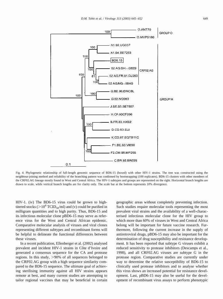
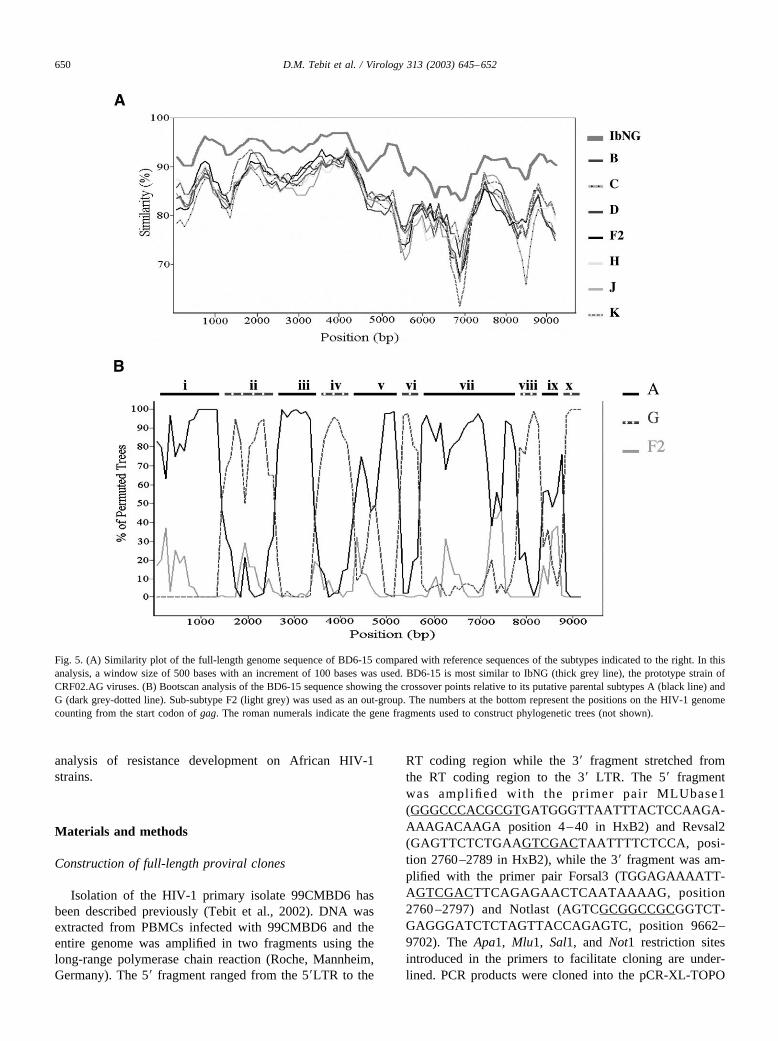
We subsequently determined the complete sequence ofthe HIV proviral genome in pBD6-15. Phylogenetic analy-sis of the full-length sequence by the neighbour-joiningmethod showed that BD6-15 clusters with other referenceCRF02.AG viruses from West and Central Africa with ahigh bootstrap value (Fig. 4; BD6-15 boxed). This wasfurther confirmed by using the similarity plot program in theSIMPLOT package in which BD6-15 was compared toreference isolates from eight different clades. BD6-15 ap-peared to be most similar to the CRF02.AG prototype iso-late IbNg throughout its genomic sequence (Fig. 5A). Todetermine the putative cross-over points, the bootscanninganalysis program of the SIMPLOT package was used. Atotal of nine cross-over points between subtypes A and Gwas observed, indicating that BD6-15 is a complex A/Grecombinant virus. The cross-over points were in the fol-lowing positions of the BD6-15 genome (Fig. 5B): nucleo-tide 1600 (subtype A to subtype G), 2550 (G to A), 3450 (Ato G), 4050 (G to A), 5150 (A to G), 5650 (G to A), 7550(A to G), 7950 (G to A), and 8450 (A to G). Phylogenetictrees constructed with reference sequences of all subtypes
(data not shown) were used to support the various segmentsshown in roman numerals in Fig. 5B. Nine of the 10 frag-ments were confirmed to belong to either subtype A or G bya significant bootstrap value �70% (data not shown). Frag-ment (ix) which stretches from nucleotide 7951 to 8450 wasonly tentatively classified as subtype A due to its lowbootstrap value of 52%.
The full-length sequence of BD6–15 is composed of9774 nucleotides with the arrangement 5�LTR, gag, pol, vif,vpr, tat, vpu, rev, env, nef, and 3�LTR. All reading frameswere open except for vpr, which had an in-frame stop codonlocated 30 codons upstream of the normal vpr stop codon.This in-frame stop codon was not detected in the parentalisolate upon direct pool sequencing of a PCR amplimer,indicating that it may be due to a PCR-induced error or thatit represents a minor population in the primary isolate notdetectable by direct sequencing. Previous studies have alsoshown the presence of in-frame stop codons in accessorygenes and especially in vpr in other proviral clones (Gao etal., 1998). In the LTR, the putative binding sites for tran-scriptional factors NF-�B, SP1, and TATA were conserved.A 16-nucleotide insertion downstream of the primer bindingsite reported in some subtype A, G, and almost all knownCRF02.AG viruses was observed in BD6-15 as well. Anal-ysis of the envelope region showed some minor deletionsand insertions, respectively, at the carboxyl-terminal ends ofthe gp41 and gp120 sequences when compared to otherreference CRF02.AG sequences. The V3 loop sequence ofBD6-15 consisted of 37 amino acids and exhibited a singleamino acid difference compared to its parental isolate. Thecrown of the V3 loop contained the GPGQAF motif whichis dominant among African HIV-1 isolates. In the aminoterminal region of nef, an insertion of 13 codons was ob-served. Ten of these codons represented a duplication of thesequence located only a few codons upstream. This inser-tion, which is not subtype specific, has been found in manyisolates and was also observed in the recently describedCRF02.AG molecular clone (Kusagawa et al., 2001). Itvaries from 6 to 13 amino acids in length in differentisolates.
In summary, pBD6-15 represents a full-length infectiousmolecular clone derived from a typical X4-tropic primaryisolate of the CRF02.AG genotype, the virus which domi-nates the HIV-epidemic in West and Central Africa. Severalproperties make pBD6-15 a unique tool for the functionalcharacterisation of CRF02.AG viruses, for testing their re-sponse to antiretroviral therapy and their probability forresistance development, and for their application in vaccinestudies: (i) The virus used for construction of the full-lengthclone had been isolated from PBMC and was passagedexclusively in PBMC. (ii) The virus obtained by transfec-tion of pBD6-15 exhibited identical replication propertiescompared with its parental isolate, and (iii) replicated al-most as efficiently as the laboratory-adapted B-type virusNL4-3, the most widely used tool in molecular studies of
Fig. 3. Analysis of purified virus preparations on SDS gels stained withsilver. Virus derived from MT-4 cells infected with BD6-15 or NL4-3 waspurified by velocity gradient centrifugation and separated by SDS–PAGE,and viral proteins were stained with silver nitrate. Lane 1 corresponds tomarker proteins (molecular mass in kDa shown to the left), lanes 2 and 3correspond to two independent purifications of BD6-15, and lane 4 corre-sponds to a purification of HIV-1 NL4-3. The positions of the Pr55gagprecursor and the structural proteins NC, MA, and CA are shown to theright.
648 D.M. Tebit et al. / Virology 313 (2003) 645–652
HIV-1. (iv) The BD6-15 virus could be grown to high-titered stocks (�108 TCID50/ml) and (v) could be purified inmilligram quantities and to high purity. Thus, BD6-15 andits infectious molecular clone pBD6-15 may serve as refer-ence virus for the West and Central African epidemic.Comparative molecular analysis of viruses and viral clonesrepresenting different subtypes and recombinant forms willbe helpful to delineate the functional differences betweenthese viruses.
In a recent publication, Ellenberger et al. (2002) analysedprevalent and incident HIV-1 strains in Cote d’ Ivoire andgenerated a consensus sequence for the CA and proteaseregions. In this study, �90% of all sequences belonged tothe CRF02.AG group with a high sequence similarity com-pared to the BD6-15 sequence. The ultimate goal of achiev-ing sterilising immunity against all HIV strains appearsremote at best, and many current studies are attempting totailor regional vaccines that may be beneficial in certain
geographic areas without completely preventing infection.Such studies require molecular tools representing the mostprevalent viral strains and the availability of a well-charac-terised infectious molecular clone for the HIV group towhich more than 60% of viruses in West and Central Africabelong will be important for future vaccine research. Fur-thermore, following the current increase in the supply ofantiretroviral drugs, pBD6-15 may also be important for thedetermination of drug susceptibility and resistance develop-ment. It has been reported that subtype G viruses exhibit areduced sensitivity to protease inhibitors (Descamps et al.,1998), and all CRF02.AG viruses are subtype G in theprotease region. Comparative studies are currently underway to determine the relative susceptibility of BD6-15 toclinically used protease inhibitors and to analyse whetherthis virus shows an increased potential for resistance devel-opment. Last, pBD6-15 may also be useful for the devel-opment of recombinant virus assays to perform phenotypic
Fig. 4. Phylogenetic relationship of full-length genomic sequence of BD6-15 (boxed) with other HIV-1 strains. The tree was constructed using theneighbour-joining method and reliability of the branching pattern was confirmed by bootstrapping (100 replicates). BD6-15 clusters with other members ofthe CRF02.AG lineage mostly found in West and Central Africa. The HIV-1 subtypes and groups are represented on the right. Horizontal branch lengths aredrawn to scale, while vertical branch lengths are for clarity only. The scale bar at the bottom represents 10% divergence.
649D.M. Tebit et al. / Virology 313 (2003) 645–652
analysis of resistance development on African HIV-1strains.
Materials and methods
Construction of full-length proviral clones
Isolation of the HIV-1 primary isolate 99CMBD6 hasbeen described previously (Tebit et al., 2002). DNA wasextracted from PBMCs infected with 99CMBD6 and theentire genome was amplified in two fragments using thelong-range polymerase chain reaction (Roche, Mannheim,Germany). The 5� fragment ranged from the 5�LTR to the
RT coding region while the 3� fragment stretched fromthe RT coding region to the 3� LTR. The 5� fragmentwas amplifi ed with the primer pair MLUbase1(GGGCCCACGCGTGATGGGTTAATTTACTCCAAGA-AAAGACAAGA position 4–40 in HxB2) and Revsal2(GAGTTCTCTGAAGTCGACTAATTTTCTCCA, posi-tion 2760–2789 in HxB2), while the 3� fragment was am-plified with the primer pair Forsal3 (TGGAGAAAATT-AGTCGACTTCAGAGAACTCAATAAAAG, position2760 –2797) and Notlast (AGTCGCGGCCGCGGTCT-GAGGGATCTCTAGTTACCAGAGTC, position 9662–9702). The Apa1, Mlu1, Sal1, and Not1 restriction sitesintroduced in the primers to facilitate cloning are under-lined. PCR products were cloned into the pCR-XL-TOPO
Fig. 5. (A) Similarity plot of the full-length genome sequence of BD6-15 compared with reference sequences of the subtypes indicated to the right. In thisanalysis, a window size of 500 bases with an increment of 100 bases was used. BD6-15 is most similar to IbNG (thick grey line), the prototype strain ofCRF02.AG viruses. (B) Bootscan analysis of the BD6-15 sequence showing the crossover points relative to its putative parental subtypes A (black line) andG (dark grey-dotted line). Sub-subtype F2 (light grey) was used as an out-group. The numbers at the bottom represent the positions on the HIV-1 genomecounting from the start codon of gag. The roman numerals indicate the gene fragments used to construct phylogenetic trees (not shown).
650 D.M. Tebit et al. / Virology 313 (2003) 645–652
TA vector (Invitrogen, Groningen, The Netherlands) andcorrect clones were confirmed by restriction enzyme di-gests. The 5� fragments were excised from the resultingclones and inserted into the plasmids carrying the 3� frag-ments using the newly introduced Apa1 and Sal1 restrictionsites. Cloning was done in a chessboard fashion as describedpreviously (Herchenroder et al., 1995).
Cells, transfections, and infections
293 T cells were maintained in Dulbecco’s minimalEagle’s medium supplemented with 10% inactivated fetalcalf serum, 100 U/ml penicillin, 100 �g/ml streptomycin,and 2 mM glutamin. Virus particles were produced bytransfecting 293 T cells with 15 �g of plasmid DNA usingthe calcium phosphate method. Culture supernatants wereharvested after 3 days, filtered through a 0.45-�m filter, andanalysed by p24 antigen ELISA. Only p24 antigen ELISA-positive cultures were used for further biological analysis.
HIV-1 permissive C8166 and MT-4 cells were main-tained in RPMI 1640 with the supplements described above.PBMCs were isolated from HIV-negative donors as de-scribed (Tebit et al., 2002). Transfection-derived cell-freevirus supernatants were used to infect C8166 cells. Infec-tivity was monitored by observing for syncytia formationand p24 antigen determination. To generate virus stocksfrom clones BD6-15, NL4-3, and the primary isolate99CMBD6, C8166 cells were infected with transfectionderived virus for 5 days and the resulting supernatant wasused to determine the TCID50 by serial 10-fold dilutions ofvirus on quadruplicate cultures of C8166.
Replication properties of BD6-15, its 99CMBD6 paren-tal isolate, and NL4-3 were analysed comparatively inC8166 cells and PBMCs. To this end, 5 � 105 C8166 cellsand 1.5 � 106 IL2/PHA-stimulated PBMCs were infectedwith 500 TCID50 or 1500 TCID50 virus supernatant inquadruplicate in a 48-well plate and incubated overnight at37°C. The next day, the cells were washed twice with PBS,resuspended in 1 ml RPMI 1640, and monitored for 2 and 3weeks, respectively. Virus supernatants were taken every 2to 3 days to measure p24 antigen production. The biologicalphenotype and coreceptor usage of transfection-derived vi-rus and of the primary isolate were determined, respec-tively, on MT-2 cells and GHOST cells as described previ-ously (Tebit et al., 2002).
Sequencing and sequence analysis
The entire genome of BD6-15 was sequenced by theprimer walking method using the dye terminator cycle se-quencing kit on a CEQ 2000 capillary sequencer (Beck-mann Coulter, CA, USA). Contigs were assembled usingthe Contig Assembler of the Vector NTI Suite (Informax,Oxford, UK). Sequences obtained were aligned with HIV-1subtype reference sequences using the CLUSTAL � align-
ment program. Phylogenetic analysis was performed by theneighbour-joining method. Statistical robustness of treesand reliability of the branching pattern were confirmed bybootstrapping. Similarity and bootscanning analysis to de-termine recombination events were done with the SIM-PLOT version 2.5 (Ray, 1999).
Purification of virus particles and analysis of viralproteins
Culture medium from 293T cells transfected with therespective molecular clones was filtered through a 0.45-�mpore-size filter and subsequently centrifuged through acushion of 20% (w/w) sucrose in PBS at 130,000 g for 90min at 4°C. Virus pellets were resuspended in loadingbuffer, subjected to sodium dodecyl sulfate (SDS)–polyac-rylamide gel electrophoresis (PAGE), and proteins trans-ferred to a nitrocellulose membrane (Schleicher & Schuell)by electroblotting. Membranes were reacted with serumfrom the AIDS patient from whom the virus 99CMBD6 wasisolated. Peroxidase-conjugated anti-human IgG antibody(Jackson Immunochemicals) was used as the secondaryantibody and immune complexes were visualised by en-hanced chemiluminescence (ECL System, Amersham) ac-cording to the manufacturer’s instructions.
High-titer virus preparations of BD6-15 and NL4-3 wereproduced essentially as described (Welker et al., 2000).Briefly, MT-4 cells were initially infected with cell-freevirus and were subsequently expanded by cocultivation withnoninfected cells. Virus preparations were harvested beforea pronounced cytopathic effect was observed. Virus-con-taining media were filtered and centrifuged through a su-crose cushion as described above. Concentrated virus wasfurther purified by velocity gradient centrifugation on lo-dixanol gradients as described (Dettenhofer and Yu, 1999)and collected by ultracentrifugation. Gradient-purified viruswas resolved by SDS–PAGE and stained with silver asdescribed (Heukeshoven and Dernick, 1988).
Sequence data
The complete sequence of the BD6-15 infectious molec-ular clone has been deposited in the GenBank under theAccession No. AY271690.
Acknowledgments
We are grateful to U. Tessmer for performing gel anal-ysis and silver stain. This work was supported in partthrough a grant from the Deutsche Forschungsgemeinschaftto H.-G.K. (SFB544, Project A6). D.M.T. was supported bythe Deutscher Akademischer Austauschdienst.
651D.M. Tebit et al. / Virology 313 (2003) 645–652
References
Adachi, A., Gendelman, H.E., Koenig, S., Folks, T., Willey, R., Rabson,A., Martin, M.A., 1986. Production of acquired immunodeficiencysyndrome-associated retrovirus in human and nonhuman cells trans-fected with an infectious molecular clone. J. Virol. 59 (2), 284–291.
Carr, J.K., Torimiro, J.N., Wolfe, N.D., Eitel, M.N., Kim, B., Sanders-Buell, E., Jagodzinski, L.L., Gotte, D., Burke, D.S., Birx, D.L., Mc-Cutchan, F.E., 2001. The AG recombinant lbNG and novel strains ofgroup M HIV-1 are common in Cameroon. Virology 286 (1), 168–181.
Descamps, D., Apetrei, C., Collin, G., Damond, F., Simon, F., Brun-Vezinet, F., 1998. Naturally occurring decreased susceptibility ofHIV-1 subtype G to protease inhibitors. AIDS 12, 1109–1111.
Dettenhofer, M., Yu, X.F., 1999. Highly purified human immunodeficiencyvirus type 1 reveals a virtual absence of Vif in virions. J. Virol. 73 (2),1460–1467.
Ellenberger, D.L., Bin, L., Lupo, D.L., Owen, M.S., Nkengasong, J.,Kadio-Morokro, M.S., Smith, J., Robinson, H., Ackers, M., Greenberg,A., Folks, T., Butera, S., 2002. Generation of a consensus sequencefrom prevalent and incident HIV-1 infections in West Africa to guideAIDS vaccine development. Virology 302, 155–163.
Gao, F., Robertson, D.L., Carruthers, C.D., Morrison, S.G., Jian, B., Chen,Y., Barre-Sinoussi, F., Girard, M., Srinivasan, A., Abimiku, A.G.,Shaw, G.M., Sharp, P.M., Hahn, B.H., 1998. A comprehensive panel ofnear-full-length clones and reference sequences for non-subtype Bisolates of human immunodeficiency virus type 1. J. Virol. 72, 5680–5698.
Herchenroder, O., Turek, R., Neumann-Haefelin, D., Rethwilm, A.,Schneider, J., 1995. Infectious proviral clones of chimpanzee foamyvirus (SFVcpz) generated by long PCR reveal close functional relat-edness to human foamy virus. Virology 214 (2), 685–689.
Heukeshoven, J., Dernick, R., 1988. Improved silver staining procedure forfast staining in PhastSystem development unit. I. Staining of sodiumdodecyl sulfate gels. Electrophoresis 9 (1), 28–32.
Howard, T.M., Rasheed, S., 1996. Genomic structure and nucleotide se-quence analysis of a new HIV type 1 subtype A strain from Nigeria.AIDS Res. Hum. Retroviruses 12, 1413–1425.
Heyndrickx, L., Janssens, W., Zekeng, L., Musonda, R., Anagonou, S.,Van der Auwera, G., Coppens, S., Vereecken, K., De Witte, K., VanRampelbergh, R., Kahindo, M., Morison, L., McCutchan, F.E., Carr,J.K., Albert, J., Essex, M., Goudsmit, J., Asjo, B., Salminen, M., Buve,A., van Der Groen, G., 2000. Simplified strategy for detection ofrecombinant human immunodeficiency virus type 1 group M isolatesby gag/env heteroduplex mobility assay. Study Group on Heterogeneityof HIV Epidemics in African Cities. J. Virol. 74, 363–370.
Kusagawa, S., Takebe, Y., Yang, R., Motomura, K., Ampofo, W., Brand-ful, J., Koyanagi, Y., Yamamoto, N., Sata, T., Ishikawa, K., Nagai, Y.,Tatsumi, M., 2001. Isolation and characterization of a full-length mo-lecular DNA clone of Ghanaian HIV type 1 intersubtype A/G recom-binant CRF02_AG, which is replication competent in a restricted hostrange. AIDS Res. Hum. Retroviruses 17 (7), 649–655.
Montavon, C., Toure-Kane, C., Liegeois, F., Mpoudi, E., Bourgeois, A.,Vergne, L., Perret, J.L., Boumah, A., Saman, E., Mboup, S., Delaporte,E., Peeters, M., 2000. Most env and gag subtype A HIV-1 virusescirculating in West and West Central Africa are similar to the prototypeAG recombinant virus IBNG. J. Acquir. Immune Defic. Syndr. 20,363–374.
Mukai, T., Komoto, S., Kurosu, T., Palacios, J.A., Li, Y.G., Auwanit, W.,Tatsumi, M., Ikuta, K., 2002. Construction and characterization of aninfectious molecular clone derived from the CRF01_AE primary iso-late of HIV type 1. AIDS Res. Hum Retroviruses. 18 (8), 585–589.
Ndung’u, T., Renjifo, B., Essex, M., 2001. Construction and analysis of aninfectious human Immunodeficiency virus type 1 subtype C molecularclone. J. Virol. 75 (11), 4964–4972.
Peeters, M., Esu-Williams, E., Vergne, L., Montavon, C., Mulanga-Ka-beya, C., Harry, T., Ibironke, A., Lesage, D., Patrel, D., Delaporte, E.,2000. Predominance of subtype A and G HIV type 1 in Nigeria, withgeographical differences in their distribution. AIDS Res. Hum. Retro-viruses 16, 315–325.
Ray, S.C. 1999. Simplot for window http://www.med.lhu.edu/dept/scray/download/simplot.
Salminen, M.O., Ehrenberg, P.K., Mascola, J.R., Dayhoff, D.E., Merling,R., Blake, B., Louder, M., Hegerich, S., Polonis, V.R., Birx, D.L.,Robb, M.L., McCutchan, F.E., Michael, N.L., 2000. Construction andbiological characterization of infectious molecular clones of HIV-1subtypes B and E (CRF01_AE) generated by the polymerase chainreaction. Virology 278 (1), 103–110.
Takahoko, M., Tobiume, M., Ishikawa, K., Ampofo, W., Yamamoto, N.,Matsuda, M., Tatsumi, M., 2001. Infectious DNA clone of HIV type 1A/G recombinant (CRF02_AG) replicable in peripheral blood mono-nuclear cells. AIDS Res. Hum. Retroviruses 17 (1), 1083–1087.
Takehisa, J., Zekeng, L., Ido, E., Mboudjeka, I., Moriyama, H., Miura, T.,Yamashita, M., Gurtler, L.G., Hayami, M., Kaptue, L., 1998. Varioustypes of HIV mixed infections in Cameroon. Virology 245, 1–10.
Tebit, D.M., Zekeng, L., Kaptue, L., Salminen, O., Krausslich, H-G.,Herchenroder, O., 2002. Genotypic and phenotypic analysis of HIV-1primary isolates from Western Cameroon. AIDS Res. Hum. Retrovi-ruses. 18 (1), 37–48.
Welker, R., Hohenberg, H., Tessmer, U., Huckhagel, C., Krausslich, H.-G.,2000. Biochemical and structural analysis of isolated mature cores ofhuman immunodeficiency virus type 1. J. Virol. 74 (3), 1168–1177.
652 D.M. Tebit et al. / Virology 313 (2003) 645–652