construction and comprehensive analysis of dysregulated
TRANSCRIPT

Research ArticleConstruction and Comprehensive Analysis of DysregulatedLong Noncoding RNA-Associated Competing Endogenous RNANetwork in Moyamoya Disease
Xuefeng Gu ,1,2 Dongyang Jiang,3 Yue Yang,4,5 Peng Zhang ,6 Guoqing Wan,1,2
Wangxian Gu,2 Junfeng Shi,2 Liying Jiang,2 Bing Chen,7 Yanjun Zheng,2 Dingsheng Liu ,8
Sufen Guo ,4,5 and Changlian Lu 2
1Research Department, Shanghai University of Medicine & Health Science Affiliated Zhoupu Hospital, Shanghai, China2Shanghai Key Laboratory of Molecular Imaging, Shanghai University of Medicine & Health Sciences, Shanghai, China3Department of Cardiology, Pan-Vascular Medicine Institute, Shanghai Tenth People’s Hospital, Tongji University Schoolof Medicine, Shanghai, China4Key Laboratory of Cancer Prevention and Treatment of Heilongjiang Province, Mudanjiang Medical University,Mudanjiang, China5Department of Pathology, Hongqi Hospital Affiliated to Mudanjiang Medical University, Mudanjiang, China6School of Clinical Medicine, Shanghai University of Medicine & Health Sciences, Shanghai, China7Department of Neurosurgery, Affiliated Hospital of Guangdong Medical University, Zhanjiang, Guangdong, China8Department of Oncology and Hematology, Shanghai University of Medicine & Health Sciences Affiliated Zhoupu Hospital, China
Correspondence should be addressed to Dingsheng Liu; [email protected], Sufen Guo; [email protected],and Changlian Lu; [email protected]
Received 22 March 2020; Accepted 9 May 2020; Published 13 June 2020
Guest Editor: Lei Chen
Copyright © 2020 Xuefeng Gu et al. This is an open access article distributed under the Creative Commons Attribution License,which permits unrestricted use, distribution, and reproduction in any medium, provided the original work is properly cited.
Background. Moyamoya disease (MMD) is a rare cerebrovascular disease characterized by chronic progressive stenosis or occlusionof the bilateral internal carotid artery (ICA), the anterior cerebral artery (ACA), and the middle cerebral artery (MCA). MMD issecondary to the formation of an abnormal vascular network at the base of the skull. However, the etiology and pathogenesis ofMMD remain poorly understood. Methods. A competing endogenous RNA (ceRNA) network was constructed by analyzingsample-matched messenger RNA (mRNA), long non-coding RNA (lncRNA), and microRNA (miRNA) expression profiles fromMMD patients and control samples. Then, a protein-protein interaction (PPI) network was constructed to identify crucial genesassociated with MMD. Gene Ontology (GO) and Kyoto Encyclopedia of Genes and Genomes pathway (KEGG) enrichmentanalyses were employed with the DAVID database to investigate the underlying functions of differentially expressed mRNAs(DEmRNAs) involved in the ceRNA network. CMap was used to identify potential small drug molecules. Results. A total of 94miRNAs, 3649 lncRNAs, and 2294 mRNAs were differentially expressed between MMD patients and control samples. Asynergistic ceRNA lncRNA-miRNA-mRNA regulatory network was constructed. Core regulatory miRNAs (miR-107 and miR-423-5p) and key mRNAs (STAT5B, FOSL2, CEBPB, and CXCL16) involved in the ceRNA network were identified. GO andKEGG analyses indicated that the DEmRNAs were involved in the regulation of the immune system and inflammation inMMD. Finally, two potential small molecule drugs, CAY-10415 and indirubin, were identified by CMap as candidate drugs fortreating MMD. Conclusions. The present study used bioinformatics analysis of candidate RNAs to identify a series of clearlyaltered miRNAs, lncRNAs, and mRNAs involved in MMD. Furthermore, a ceRNA lncRNA-miRNA-mRNA regulatory networkwas constructed, which provides insights into the novel molecular pathogenesis of MMD, thus giving promising clues forclinical therapy.
HindawiComputational and Mathematical Methods in MedicineVolume 2020, Article ID 2018214, 12 pageshttps://doi.org/10.1155/2020/2018214

1. Introduction
Moyamoya disease (MMD) is a rare cerebrovascular diseasecharacterized by chronic progressive occlusion or stenosis ofthe bilateral internal carotid artery (ICA), the anterior cere-bral artery (ACA), and the middle cerebral artery (MCA) [1,2]. MMD is secondary to the formation of an abnormal vas-cular network at the base of the skull. Because the abnormalvascular network of the skull base looks like “smoke” oncerebral angiography images, it is called “moyamoya dis-ease” [3]. The MMD incidence rate in Eastern Asian coun-tries is higher [4], and it mainly occurs in children andyoung adults, peaking at the ages of 5 to 9 and 35 to 45 years[5]. MMD can seriously affect the mental and physicalhealth of patients. However, the etiology and pathogenesisof MMD remain poorly understood; it may be related togenetics, inflammation, immune response, and environmen-tal factors [6–11].
Many studies have reported that the ring finger protein213 (RNF213) gene is an important susceptibility gene forMMD in East Asia, especially the p.R4810K variant [12–17]. However, MMD also occurs in patients without muta-tions in RNF213. To date, new candidate risk-MMD genes,such as the vascular smooth muscle cell-specific isoform ofα-actin (ACTA2) [18, 19], endothelial nitric oxide synthase(eNOSase) [20], soluble guanylyl cyclase alpha subunit(GUCY1A3) [21], matrix metalloproteinases (MMPs) [22–26], tissue inhibitor of metalloproteinases (TIMPs) [23, 24],transforming growth factor β1 (TGF-β1) [27], Sortilin 1(SORT1) [28], Connexin 43 (Cx43) [29], and caveolin-1(Cav-1) [30, 31], have been continuously reported to be asso-ciated with MMD.
Moreover, with the development of microarray andsequencing technology, investigators have begun to explorefactors other than direct disease-causing genes, includingnoncoding RNAs (ncRNAs). Gao et al. revealed the expres-sion profile of lncRNAs and mRNAs in MMD patients in2016 [9], and Dai et al. analyzed miRNAs in the serum ofMMD patients and healthy controls in 2012 [32]. miRNAscan posttranscriptionally regulate gene expression by bindingto MREs (miRNA-response elements) of their target tran-script. mRNAs, lncRNAs, and other RNA transcripts couldact as endogenous miRNA sponges to inhibit miRNA func-tion. These interactions illustrate the famous ceRNAhypothesis presented by Salmena in 2011 [33], which gaveus a new “language” in different types of RNA transcripts.After that, the ceRNA hypothesis was applied to many fields[34]. The Linc2GO database was constructed by Liu et al. in2013 [35]. StarBase v2.0 was published by Li et al. to predictmiRNA-ceRNA interactions [36]. Moreover, continuedanalysis of ceRNA networks would deepen our knowledgeabout how different subtypes of noncoding RNAs work witheach other.
In this study, a comprehensive analysis of the miRNA,mRNA, and lncRNA expression profiles in MMD was done,and then, we constructed MMD-specific ceRNA networksusing a large cohort from an online database. As far as weknow, this is the first study to establish a ceRNA lncRNA-miRNA-mRNA network in MMD, which provides novel
insight into the molecular pathogenesis of MMD, thus givingpromising clues for clinical therapy. In addition, core regula-tory miRNAs (miR-107 and miR-423-5p) and key mRNAs(STAT5B, FOSL2, CEBPB, and CXCL16) were enriched inimmune system/inflammation biological processes, indicat-ing their potential role in MMD.
2. Materials and Methods
2.1. Data Collection. miRNA microarray data were down-loaded from Gene Expression Omnibus (GEO, https://www.ncbi.nlm.nih.gov/geo/) in NCBI (The National Center forBiotechnology Information). GEO is an unrestricted openaccess repository that provides high-throughput microarrayand next-generation sequence datasets that have been sub-mitted by researchers around the world. GSE45737 is amiRNA expression profile of the serum from 10 MMDpatients and 10 normal healthy controls [32]. The lncRNAand mRNA expression profiles in blood samples from 15MMD patients and 10 healthy controls were kindly providedby a collaborating academician, Zhao [9].
2.2. Identification of Differentially Expressed RNAs in MMDPatients Compared to Healthy Controls. R software with pack-ages ggplot2, edgeR, and pheatmap (http://bioconductor.org/bioclite. R) was adopted to identify differentially expressedRNAs (DERs). In brief, datasets were standardized after con-version of formats, variance normalization, and the additionof missing values as well as statistical testing of differentiallyexpressed probes. The expression levels of all targets, includ-ing mRNA, miRNA, and lncRNA, within the datasets weresubjected to analysis with R. The threshold was set as a Pvalue < 0.05 and ∣log2FC ∣ >1. According to these criteria,DERs were identified for further analysis.
2.3. Gene Ontology and Pathway Enrichment Analyses. TheDatabase of Annotation, Visualization and Integrated Dis-covery (DAVID, http://david.ncifcrf.gov) is a public databasewith comprehensive online tools for functional annotation.Kyoto Encyclopedia of Genes and Genomes (KEGG) is a col-lection of databases that contain information about genomes,biological pathways, diseases, and chemical substances [37].Gene Ontology (GO) is an international standardized genefunctional classification system that offers a dynamicallyupdated controlled vocabulary and a strictly defined conceptto comprehensively describe properties of genes and theirproducts in any organism. GO has three ontologies: molecu-lar function, cellular component, and biological process [38].
In the present study, GO and KEGG pathway enrichmentanalyses were performed using DAVID. P < 0:05 was consid-ered statistically significant.
2.4. Construction of the ceRNA (lncRNA-miRNA-mRNA)Regulatory Network. The prediction of miRNA-mRNA inter-actions was performed on the open-source platform Encyclo-pedia of RNA Interactomes (ENCORI, http://starbase.sysu.edu.cn) [36]. The unique algorithm of ENCORI enables allobtained interactions to be confirmed by at least one othermajor RNA-RNA prediction website, such as miRanda, Pic-Tar, or TargetScan. In addition to sequence matching, the
2 Computational and Mathematical Methods in Medicine

prediction was approved by multidimensional sequencingdata. All these features make ENCORI a reliable sourcefor predicting RNA-RNA interaction, especially themiRNA-mRNA interaction. Two other databases, miRcode(http://www.mircode.org) and DIANA (http://carolina.imis.athena-innovation.gr), were applied in the study for pre-dicting miRNA-lncRNA interactions. Afterwards, all inter-actions were input into Cytoscape (version 3.7.2, http://cytoscape.org) to visualize ceRNA regulatory networks.The flow chart can be seen in Figure 1.
2.5. Protein-Protein Interaction (PPI) Network. All DEGswere imported into STRING 10.5, which is a search tool usedto identify gene interactions (https://string-db.org/). ThePPIs were used to construct a network, which was visualizedby using Cytoscape software 3.6 (http://www.cytoscape.org).The color of edges in the network indicate protein-proteinassociations: light blue and purple indicate known interac-tions from curated database and experimentally determined,respectively; dark green/red/dark blue indicate predictedinteractions by gene neighborhood/gene fusions/gene cooc-currence, respectively; and light green/black/blue indicatetext mining/coexpression/protein homology.
2.6. Gene Expression Signature Analysis with a ConnectivityMap. The DEGs were used to perform gene expression signa-ture analysis with connectivity maps (CMap, clue.io). Theupregulated and downregulated genes were used as tags,changed into probe IDs referred to Affymetrix U133 Gene-Chip and uploaded into the CMap database to calculate theirvalues from other drug-target datasets. According to the sim-ilarity of gene expression profiles, pairs of gene expressionsignatures and targeted drugs were used to obtain a value.If the value was a positive number, the target drug wouldhave an effect that was similar to that of the MMD-inducedgene expression signature. If the value was a negative num-ber, the targeted drug would have an effect that was oppositethat of theMMD-induced gene expression signature; namely,the targeted drug might have an effect that could be useful intreatment.
2.7. Statistical Analysis. We used SPSS 11.0 (SPSS, Chicago,IL) to analyze the dataset from the microarray experiments.All data are represented as the mean ± SD. Statistical signifi-cance was determined at P < 0:05.
3. Result and Discussion
3.1. Differentially Expressed mRNAs, miRNAs, and lncRNAsbetween MMD Patients and Healthy Controls. After differen-tial expression analysis, a total of 2294 DEmRNAs werescreened between MMD patients and healthy controls, 865of which were downregulated and 1429 of which were upreg-ulated in MMD patients. (Table S1, Figure 2(a)). Severalgenes reported in previous studies in MMD, such as HIF1α(log2FC = 1:214), SORT1 (log2FC = 1:628), and MMP9(log2FC = 2:40), are marked in Figure 2. HIF1α was foundto be overexpressed in the intima of the MCA of MMDpatients. HIF1α is a master transcriptional regulator of theadaptive response to hypoxia. Under hypoxic conditions,
HIF1α translocates to the nucleus, where the HIF1 complex(HIFα/HIFβ) binds to the hypoxia-response element andactivates the expression of many genes that can increaseoxygen delivery and respond to oxygen deprivation inMMD [7]. MMP9 belongs to a family of zinc-bindingproteolytic enzymes that are capable of degrading all thecomponents of the extracellular matrix in a variety ofphysiologic and pathophysiological conditions. Fujimuraet al. inferred that the higher expression of MMP9 in MMDpatients may play an integrated role in physiologic andpathologic angiogenesis and to the instability of the cerebralvascular structure [39]. SORT1 is another gene reported tobe associated with MMD. Increased expression of SORT1inhibited endothelial cell tube formation and regulatedmajor angiogenic factors and MMP9 expression, implyingthat SORT1 participated in the pathogenesis of MMD [28].
In addition, 94 DEmiRNAs and 3649 DElncRNAs fromGEO datasets were identified. Representative DERs areshown in Figure S1 (a-d).
3.2. Construction of a Competing Endogenous RNARegulatory Network. The ENCORI database was employedto screen potential interactions between DERs. A synergistic,competitive module of the ceRNA network was constructedseparately according to upregulated or downregulatedDEmRNAs, which contained 84 nodes in the upregulatedgroup and 66 nodes in the downregulated group. In addition,there were 68 mRNA-miRNA interactions and 16 lncRNA-miRNA interactions in the upregulated group (Figure 3(a)).In the downregulated group, there were 61 interactionsbetween mRNAs and miRNAs and 35 interactions betweenlncRNAs and miRNAs (Figure 3(b)). The ceRNA networkwas generated using Cytoscape, as previously discussed.
Based on the network organization, we found that miR-107 competed with 16 mRNAs and 4 lncRNAs (LINC02434,AL589642.1, AC003092.1, and AL035425.3) in the module(Figure 3(b)). A previous study showed that miR-107 isupregulated in response to low-oxygen conditions [40]. Sub-sequently, miR-107 was found to be abnormally expressed inseveral cancers, such as PDAC. When miR-107 expressionwas downregulated in PDAC, cell migration and invasionwere inhibited, implying the important role of miR-107 intumor cell activity [41]. Furthermore, they found that theexpression of caveolin-1 was upregulated by a miR-107inhibitor. Caveolin-1 was reported to be associated with neg-ative remodeling in MMD through the inhibition of angio-genesis in endothelial cells and the induction of apoptosisin VSMCs [30, 31]. Another study by Meng et al. found thatmiR-107 can inhibit endothelial progenitor cell (EPC) differ-entiation via HIF1β [42]. HIF1β is another subunit of HIF1that generally heterodimerizes with HIF1α. Together, theyplay key roles during hypoxic conditions, which are similarto the conditions in MMD: low oxygen because of vascularocclusion. EPCs can differentiate into mature endothelialcells and play important roles in the recovery of endothelialfunction and tissue repair. The role of EPCs reflects themixed state of vascular obstruction and abnormal angiogen-esis in the pathogenesis of MMD [43]. The ceRNA networknear miR-107 reveals that FoxC1 is one of the potential
3Computational and Mathematical Methods in Medicine

downstream target genes, and it is necessary in the process ofvascular development, involving arterial specification andlymphatic sprouting. Abnormal expression of FoxC1 leadsto unusual angiogenesis in many tissues [44, 45].
In addition, miR-423-5p competed with 36 mRNAs(CXCL16, FOSL2, etc.) and 4 lncRNAs (NEAT1, HCG18,AL137145.2, and LINC00963) in the module (Figure 3(a)).miR-423-5p was reported to play important roles in the inhi-bition of the cell proliferation and invasion of cancer cells
such as colon cancer and ovarian carcinoma [46, 47]. There-fore, the downregulation of miR-423-5p in MMD patientsmay increase the proliferation of vascular smooth musclecells, which is one likely reason for vessel occlusion. Inaddition, numerous studies focusing on NEAT1’s role incancer biology found that this lncRNA plays a crucial rolein carcinogenesis [48]. NEAT1 mainly works as a ceRNAby sponging antitumor miRNAs [49]. NEAT1 is alsoinvolved in immune system responses, viral diseases, and
Differentially expressedlncRNAs (DElncRNAs)
Differentially expressedmiRNAs (DEmiRNAs)
Differentially expressedmRNAs (DEmRNAs)
miRcode miRTarBase/TargetScan/miRDB
PreditedlncRNAs
PredictedmRNAs
Intersected DElncRNAs Intersected DEmRNAsRegulation mediated byDEmiRNAs
lncRNA-miRNA-mRNA ceRNA network CMap analysis
Figure 1: Flowchart of the lncRNA-miRNA-mRNA ceRNA network analysis.
Increased mRNA
log 2
(fol
d ch
ange
)
HIF1A
MMP9
SORT1
HIF1A
1
2
3
4
(a)
–log
2 (fo
ld ch
ange
)
Decreased mRNA
1
2
3
(b)
Figure 2: Differential mRNA expression between MMD patients and controls. Log2FC > 1 (P < 0:05) (MMP9, SORT1, and HIF1α aremarked in red). X-axis shows that the probes of mRNA are arranged in sequence.
4 Computational and Mathematical Methods in Medicine

OR2A1-AS1
NUTM2A-AS1 hsa-miR-483-5p
AC074386.1
AL451085.2
hsa-miR-3196
MLLT1
AL133216.1
LINC00963
MAPK1
OTX1
hsa-miR-3679-5p
FOSL2
MIR4435-2HG
MAFG ANP32A
KDM6B
RAB7A
NEAT1
MARK2
SIRPA
ERF
HCG18
PAK2
SLC45A4
TOM1
BCORL1
ARF3
PFKFB3
KAT6A
TRIOBP WDR26
PDAP1
AL137145.2ZDHHC18
ATG9AFKBP5INTS3
RBM47
ABCC5SLC25A44
TANC2
WIPF2
PPP1R12B
SRGAP2
KIAA0319L
ZFP36L1
CEBPB
ARHGEF11
ARPC5
DBN1
ADAM19
KREMEN1NBEAL2
IMPDH1
ATP9A
CUX1
NPEPPS
ABL2
CHD7
ARID3A
KLF7
AC234582.1
TRIB1
AL008729.2
METTL9 NFKBIZ
PBX2ZBTB34
hsa-miR-423-5p
STAT5B
CXCL16 GALNT2
KDM5B NCOA2FAM160B1
PHF20L1
MGRN1
SLC40A1PCGF3
PHKA2hsa-miR-574-5p
FBXL20
RC3H1
LCOR
ADAM19
(a)
hsa-miR-142-3pATXN7
SLC37A3
OSR2AC099343.3
TRNT1
KATNAL1
HR
ANKRD13B
hsa-miR-140-3p
SAR1A
LINC00265AHDC1
PROSER1
LINC00685
LINC01679
ZNF337-AS1FTX
AL365181.3
LINC00958
hsa-miR-4306
RP11-395B7.2
AC002059.1
SOX13
HSPH1
AC003092.1
CASP2
SVEP1
INO80D
RET
hsa-miR-107
FOXC1
TIPARP
FAM120C
PRPS1
PCDHA5
DTNA
hsa-miR-320d AC092718.4AC100861.1
hsa-miR-320b
AL359265.3
MAP7D2
LEF1
BCAT1AL391988.1
POLR1C
MAPT-IT1
hsa-miR-320a
NR2F1-AS1AL021707.1
FAM189B
SH3BP4
FASN
PWWP2B
LINC02434
PER3
AL589642.1
DDX54
ATP13A3
ENPP2
SEC14L1MAP4
HMGA2
LY9
AL035425.3
(b)
Figure 3: The lncRNA-miRNA-mRNA ceRNA network in MMD. (a) ceRNA network based on upregulated mRNAs involved ceRNA. (b).ceRNA network based on downregulated mRNAs involved ceRNA. Notes: red rectangles represent DElncRNAs, green rectangles representDEmiRNAs, and yellow rectangles represent DEmRNAs.
5Computational and Mathematical Methods in Medicine
Actin filament−based process
Axon guidance
Cell migration
Cell morphogenesis involved in differentiation
Cellular developmental process
Dendrite development
Dendrite morphogenesis
Endocytosis
Epithelium development
Hematopoietic or lymphoid organ development
Immune system development
Immune system process
Leukocyte aggregation
Leukocyte cell−cell adhesion
Leukocyte differentiation
Lymphocyte activation
Lymphocyte aggregation
Lymphocyte homeostasis
Positive regulation of cellular biosynthetic process
Positive regulation of cellular metabolic process
Regulation of cytoskeleton organization
Regulation of RNA biosynthetic process
T cell activation
T cell aggregation
T cell differentiation
0.01 0.02 0.03 0.04Rich factor
Term
Gene number51015
2025
2.5
3.0
3.5
4.0
−log10 (Pvalue)
(a)
Term
Apoptotic process involved in development
Apoptotic process involved in morphogenesis
Cellular response to interleukin−4
Development of primary female sexualcharacteristics
Developmental programmed cell death
Embryonic morphogenesis
Mesenchymal cell development
Mesenchyme development
Mesenchyme morphogenesis
Mesoderm development
Microtubule cytoskeleton organization
Modulation of transcription in other organisminvolved in symbiotic interaction
Neural crest cell development
Organ regeneration
Palate development
Paraxial mesoderm development
Paraxial mesoderm formation
Paraxial mesoderm morphogenesis
Positive regulation of stem cell differentiation
Regulation of stem cell differentiation
Renal system development
Response to interleukin−4
Sensory organ development
Stem cell development
Stem cell differentiation
0.0 0.1 0.2 0.3 0.4Rich factor
Gene number23
45
2.5
3.0
3.5
4.0
−log10 (Pvalue)
(b)
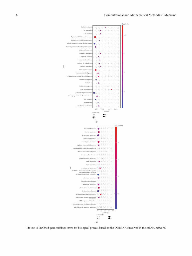
Figure 4: Enriched gene ontology terms for biological process based on the DEmRNAs involved in the ceRNA network.
6 Computational and Mathematical Methods in Medicine

neurodegeneration disorders [50]. To study FOSL2, alsonamed Fra 2, Maurer et al. created Fra 2 knockout miceand found that the mice developed pulmonary arterialocclusion due to vascular SMC proliferation and inflam-mation and pulmonary fibrosis [51, 52]. All of the aboveresults imply that the ceRNA lncRNA-miRNA-mRNA reg-ulatory network we constructed provides many new cluesregarding MMD pathogenesis.
3.3. Functional Annotation of the mRNAs Involved in theceRNA Network. After the ceRNA network was establishedwith the help of the DAVID database, functional annotationand pathway analysis of this small group of DEmRNAs wereperformed to identify potential candidate pathways or bio-logical processes related to MMD.
As shown in Figure 4, some of pathways require ourattention, and processes related to the immune responseand inflammatory reaction, including immune system pro-cess, T cell aggregation, T cell activation, lymphocyte aggre-gation, and lymphocyte activation, were significantlyenriched. Additionally, another enrichment also occurredin biological processes associated with cell development anddifferentiation, including paraxial mesoderm developmentand mesenchymal cell differentiation; these results suggestimportant roles for these biological activities in MMD. TheKyoto Encyclopedia of Genes and Genomes showed thatDEmRNAs were enriched in chemokine signaling, ErbB sig-naling, axon guidance, and vascular smooth muscle contrac-tion (Table 1).
Recently, many studies have shown that immunologica-l/inflammatory factors are involved in the occurrence and
development of MMD. According to IHC staining, therewere T cells and macrophages infiltrating in the stenosedand thickened vascular intima of MMD patients [53]. Theabnormal deposition of IgG in the elastic layer of the ICAand MCA suggests that the infiltration of immune cells andthe damage to the immune functions are related to MMD[54]. Moreover, the overexpression of inflammatory factorsin MMD patients, such as MCP-1, IL-1β, and SDF-1α, sug-gests that inflammation may also affect the progression ofMMD [55]. Consistently, in this study, several mRNAs thatencode critical inflammatory molecules, such as chemokinesand cytokines, were dysregulated and were determined to beDEmRNAs in MMD patients. Nevertheless, although variedmRNAs were clearly enriched in terms of GO analysis, therewere few found in the ceRNA network. However, severalimportant genes involved in the regulation of inflammationin MMD were modulated by ceRNAs. CXCL16 is consideredto be an important pathogenic mediator of atherosclerosis(clinical severity is graded according to the severity of carotidstenosis) [56]. CXCL16 is a vascular-derived factor thatinduces angiogenesis [57]. CXCL16 also exists in a solubleform and interacts with its specific chemokine receptor,CXCR6, to recruit the migration of activated T cells intothe inflammatory tissue [58]. As shown in Figure 3(b), fourpotential lncRNAs, including LINC00963, NEAT1, HCG18,and AL137145.2, could act as ceRNAs to regulate CXCL16through miR-107. The work on this interesting ceRNA net-work remains to be done in the future.
3.4. Protein-Protein Interaction (PPI) Network. As shown inFigure 5, a PPI network for DEmRNA-involved ceRNA
Table 1: KEGG pathway enrichment of all DEmRNAs involved in the ceRNA network.
(a) KEGG pathway enrichment of decreased mRNA involved in the ceRNA network
KEGG ID KEGG term P value Symbols
05216 Thyroid cancer 0.0012614 RET, LEF1
00230 Purine metabolism 0.0353649 PRPS1, POLR1C
04141 Protein processing in the endoplasmic reticulum 0.0365787 HSPH1, SAR1A
(b) KEGG pathway enrichment of increased mRNA involved in the ceRNA network
KEGG ID KEGG term P value Symbols
04012 ErbB signaling pathway 0.000117 ABL2, PAK2, MAPK1, STAT5B
04270 Vascular smooth muscle contraction 0.004943 PPP1R12B, MAPK1, ARHGEF11
04380 Osteoclast differentiation 0.006506 FOSL2, MAPK1, SIRPA
04360 Axon guidance 0.006648 PAK2, MAPK1, SRGAP2
05221 Acute myeloid leukemia 0.012833 MAPK1, STAT5B
05131 Shigellosis 0.014609 MAPK1, ARPC5
04062 Chemokine signaling pathway 0.018775 MAPK1, STAT5B, CXCL16
05211 Renal cell carcinoma 0.018968 PAK2, MAPK1
05220 Chronic myeloid leukemia 0.020529 MAPK1, STAT5B
04810 Regulation of actin cytoskeleton 0.025711 PAK2, MAPK1, ARPC5
04666 Fc gamma R-mediated phagocytosis 0.032876 MAPK1, ARPC5
04660 T cell receptor signaling pathway 0.042378 PAK2, MAPK1
7Computational and Mathematical Methods in Medicine

KDM6B
ARID3A
TOM1MARK2GALNT2KDM5B TRIOBPPHF20L1 MAFG
METTL9ATP9AABCC5
KIAA0319LSIRPA
NBEAL2KLF7TRIB1
OTX1
KAT6A CXCL16FOSL2PFKFB3 SLC40A1
CUX1
TANC2NCOA2CEBPB
STAT5BABL2ARHGEF11
PDAP1 DBN1
SRGAP2
LCOR NPEPPS WIPF2ARPC5
PCGF3ZBTB34
FAM160B1PAK2 ZFP36L1
ADAM19
BCORL1
FKBP5
ERFMAPK1
RAB7A
KREMEN1IMPDH1
PPP1R12BMGRN1FBXL20
RBM47ZDHHC18INTS3
NFKBIZRC3H1 SLC25A44
PBX2PHKA2
CHD7WDR26SLC45A4 ANP32A
ATG9A
ARF3
MLLT1
Figure 5: The protein-protein interaction network. The red and green nodes represent the regulated and downregulated genes, respectively.(For interpretation of the references to color in this figure legend, the reader is referred to the online version of this article.)
Table 2: List of the top 10 potential small molecular drugs predicted by CMap. Scores indicate the strong negative correlation found betweenMMD and drugs.
Score Type ID Name Description
1 -88.5 cp BRD-K86727142 Embelin HCV inhibitor
2 -88.74 cp BRD-K11630072 Carmofur Thymidylate synthase inhibitor
3 -90.86 cp BRD-A61858259 CAY-10415 Insulin sensitizer
4 -91.54 cp BRD-K04923131 GSK-3-inhibitor-IX Glycogen synthase kinase inhibitor
5 -91.85 cp BRD-K53959060 Indirubin CDK inhibitor
6 -93.79 cp BRD-K50720187 Flupirtine Glutamate receptor antagonist
7 -93.87 cp BRD-A14985772 Ascorbyl-palmitate Antioxidant
8 -94.57 cp BRD-K28143534 Cyproheptadine Histamine receptor antagonist
9 -95.78 cp BRD-K79404599 Enzastaurin PKC inhibitor
10 -97.25 cp BRD-K89687904 PKCbeta-inhibitor PKC inhibitor
8 Computational and Mathematical Methods in Medicine

networks was constructed by Cytoscape software. It is impor-tant to highlight that some striking genes, such as MAKP1,STAT5B, CEBPB, FOSL2, PAK2, and ABL2, play vital keyroles in MMD. These interesting genes were also shown inTable 1, such as ABL2, PAK2, MAPK1, and STAT5B wereenriched in the ErbB signaling pathway. After the identifica-
tion of the overlap between the above genes, chemokine sig-naling, T cell receptor signaling, and ErbB signaling shedsome light on the pathogenesis of MMD.
3.5. Potential Small Molecule Drugs. All the DEmRNAsinvolved in the ceRNA regulatory network in MMD were
Indirubin
(a)
CAY-10415
(b)
Figure 6: Potential molecular drugs. (a) Indirubin. (b) CAY-10415.
Chromosome
NEAT1 transcript
NEAT1
miR-423-5p
miR-423-5p
STAT5B
CEBPB
CXCL16
FOSL2
Immune system/inflammation
Pathogenesis of MMD
Chr 11
p15.
5
p15.
4
p15.
3p1
5.2
p15.
1
p14.
3
p14.
1
p13
p12
p11.
2p1
1.12
p11.
11q1
1q1
2.1
q13.
1q1
3.2
q13.
4
q14.
1
q14.
2q1
4.3
q21
q22.
1
q22.
3
q23.
1
q23.
3
q24.
1q2
4.2
q24.
3q2
5
Figure 7: The potential mechanism of a DElncRNA sponging miR-423-5p.
9Computational and Mathematical Methods in Medicine

analyzed by CMap to identify small molecule drugs. Strongnegative correlations were found between MMD and enzas-taurin, cyproheptadine, flupirtine, indirubin, and mitoglita-zone (CAY-10415); strong positive correlations were foundbetween MMD and flavokavain-b, CGS-20625, vinburnine,apicidin, and cytochalasin-d (Table S5). The drugs thathad a strong negative correlation with the pathogenesis ofMMD might have therapeutic effects on MMD (Table 2).CAY-10415 and indirubin gained our attention. Thestructures of the two potential molecular drugs wereinvestigated using the PubChem database (Figure 6).CAY-10415 is a member of a new class of compoundsthat modulate mitochondrial pyruvate carrier (MPC), akey controller of cellular metabolism that influencesmTOR activation [59]. It is commonly known that CAY-10415 can be used as an insulin sensitizer, and it can playthis role without activating PPARᵧ. Therefore, CAY-10415can avoid negative side effects observed in currently usedinsulin sensitizers, such as pioglitazone and rosiglitazone.CAY-10415 has been used in Alzheimer’s disease patients[60]. It is generally accepted that insulin sensitizers cannot only improve diabetes but also improve blood lipiddisorders, reduce the level of free fatty acids in plasma,reduce the effect of fat toxicity, and indirectly protect thefunction of β cells [61]. By inhibiting the proliferationand migration of vascular smooth muscle cells and reducingthe intima-media thickness of arteries, it can play aprotective role in the intima. Likewise, indirubin, a redisomer of indigo, is the active ingredient of the traditionalChinese drug Danggui Longhui Wan, which was used for thetreatment of chronic myelocytic leukemia (CML) [62].Enzyme-based in vitro studies have observed that indirubinand its derivatives, such as indirubin-3′-monoxime, indirubin-5-sulfonate, and indirubin-3′-monoxime-5-sulphonic acid,are potential inhibitors of CDKs [63]. Furthermore,different indirubin derivatives showed antiangiogenesisactivity by blocking VSMC proliferation and endothelialcell function through the inhibition of the STAT signalingpathway and reduction of neointima formation in vivo [64].All of the above findings suggest that CAY-10415 andindirubin may be used in MMD patients to avoid vascularaberration and occlusion.
4. Conclusions
In summary, using bioinformatics analysis of candidateRNAs, the present study identified a series of clearly alteredlncRNAs, miRNAs, and mRNAs involved in MMD. Further-more, a ceRNA lncRNA-miRNA-mRNA regulatory networkwas constructed, which provides a novel insight into themolecular pathogenesis of MMD, thus giving promisingclues for clinical therapy. In addition, core regulatory miR-NAs (miR-107 and miR-423-5p) and key mRNAs (STAT5B,FOSL2, CEBPB, and CXCL16) were enriched in immunesystem/inflammation biological processes, indicating theirpotential role in MMD (Figure 7). In the future, more atten-tion should be paid to the validation of competing endoge-nous RNA interactions with experimental techniques.
Finally, two potential small molecule drugs, CAY-10415and indirubin, were identified by CMap to be candidatedrugs for treating MMD.
Data Availability
(1) The miRNA microarray data used to support the findingsof this study have been deposited in the Gene ExpressionOmnibus (GEO, https://www.ncbi.nlm.nih.gov/geo/) inNCBI (GSE45737). (2) The lncRNA and mRNA microarraydata included in this study are available upon request by con-tact with the corresponding author. The data were kindlyprovided by a collaborating Prof. Zhao.
Conflicts of Interest
The authors declare no conflicts of interest.
Authors’ Contributions
Xuefeng Gu, Dongyang Jiang and Yue Yang contributedequally to this work.
Acknowledgments
This work was supported by the National Natural ScienceFoundation of China (81772829, 81272376, and 81830052),the Key projects for collaborative innovation of ShanghaiUniversity of Medicine & Health Sciences, Construction pro-ject of Shanghai Key Laboratory of Molecular Imaging(18DZ2260400), the Shanghai Municipal Education Com-mission (Class II Plateau Disciplinary Construction Programfor Medical Technology of SUMHS, 2018-2020), the NaturalScience Foundation of Heilongjiang Province (LH2019H119),the Natural Science Foundation of Guangdong Province(2016A030313680), Academic Leader Training Program ofPudong New District Health Bureau of Shanghai (PWRd2015-09), and the Funding Scheme for Training Young Teachersin Shanghai Colleges (ZZJKYX19009).
Supplementary Materials
Table S1: DEmRNA list in excel file. Table S2: DElncRNA listin excel file. Table S3: increased miRNA list in excel file.Table S4: decreased miRNA list in excel file. Table S5: drugsin excel file. Figure S1: DElncRNA and DemiRNA in tiffformat. (Supplementary Materials)
References
[1] T. Kondo, “Moyamoya disease,” Canadian Medical Associa-tion Journal, vol. 190, no. 46, article E1364, 2018.
[2] S. Shang, D. Zhou, J. Ya et al., “Progress in moyamoya disease,”Neurosurgical Review, vol. 43, no. 2, pp. 371–382, 2020.
[3] M. Fujimura, O. Y. Bang, and J. S. Kim, “Frontiers of Neurologyand Neuroscience,” inMoyamoya disease, vol. 40, pp. 204–220,2016.
[4] J. S. Kim, “Moyamoya disease: epidemiology, clinical features,and diagnosis,” Journal of Stroke, vol. 18, no. 1, pp. 2–11, 2016.
10 Computational and Mathematical Methods in Medicine

[5] X. Y. Bao, Q. N. Wang, Y. Zhang et al., “Epidemiology ofmoyamoya disease in China: single-center, population-basedstudy,” World Neurosurgery, vol. 122, pp. e917–e923, 2019.
[6] S. Newman, J. H. Boulter, J. G. Malcolm, I. Pradilla, andG. Pradilla, “Outcomes in patients with moyamoya syndromeand sickle cell disease: a systematic review,” World Neurosur-gery, vol. 135, pp. 165–170, 2020.
[7] Q. Ma, L. Li, B. Yu et al., “Circular RNA profiling of neutrophiltranscriptome provides insights into asymptomatic moya-moya disease,” Brain Research, vol. 1719, pp. 104–112, 2019.
[8] M. Zhao, F. Gao, D. Zhang et al., “Altered expression of circu-lar RNAs in moyamoya disease,” Journal of the NeurologicalSciences, vol. 381, pp. 25–31, 2017.
[9] F. Gao, L. Yu, D. Zhang, Y. Zhang, R. Wang, and J. Zhao,“Long noncoding RNAs and their regulatory network: poten-tial therapeutic targets for adult moyamoya disease,” WorldNeurosurgery, vol. 93, pp. 111–119, 2016.
[10] J. Yu, J. Zhang, J. Li, J. Zhang, and J. Chen, “Cerebral hyperper-fusion syndrome after revascularization surgery in patientswith moyamoya disease: systematic review andmeta-analysis,”World Neurosurgery, vol. 135, pp. 357–366.e4, 2020.
[11] O. Y. Bang, M. Fujimura, and S. K. Kim, “The pathophysiologyof moyamoya disease: an update,” Journal of Stroke, vol. 18,no. 1, pp. 12–20, 2016.
[12] F. Kamada, Y. Aoki, A. Narisawa et al., “A genome-wide asso-ciation study identifies RNF213 as the first moyamoya diseasegene,” Journal of Human Genetics, vol. 56, no. 1, pp. 34–40,2011.
[13] W. Liu, D. Morito, S. Takashima et al., “Identification ofRNF213 as a susceptibility gene for moyamoya disease andits possible role in vascular development,” PLoS One, vol. 6,no. 7, article e22542, 2011.
[14] S. Miyatake, N. Miyake, H. Touho et al., “Homozygousc.l4576G>A variant of RNF213 predicts early-onset and severeform of moyamoya disease,” Neurology, vol. 78, no. 11,pp. 803–810, 2012.
[15] E. H. Kim, M. S. Yum, Y. S. Ra et al., “Importance of RNF213polymorphism on clinical features and long-term outcome inmoyamoya disease,” Journal of Neurosurgery, vol. 124, no. 5,pp. 1221–1227, 2016.
[16] W. Liu, T. Hitomi, H. Kobayashi, K. O. U. J. I. H. HARADA,and A. Koizumi, “Distribution of moyamoya disease suscepti-bility polymorphism p.R4810K in RNF213 in east and south-east Asian populations,” Neurologia medico-chirurgica,vol. 52, no. 5, pp. 299–303, 2012.
[17] W. Liu, H. Hashikata, K. Inoue et al., “A rare Asian founderpolymorphism of raptor may explain the high prevalence ofmoyamoya disease among east Asians and its low prevalenceamong Caucasians,” Environmental Health and PreventiveMedicine, vol. 15, no. 2, pp. 94–104, 2010.
[18] D. C. Guo, C. L. Papke, V. Tran-Fadulu et al., “Mutations insmooth muscle alpha-actin (ACTA2) cause coronary arterydisease, stroke, and Moyamoya disease, along with thoracicaortic disease,” The American Journal of Human Genetics,vol. 84, no. 5, pp. 617–627, 2009.
[19] C. Roder, V. Peters, H. Kasuya et al., “Analysis of ACTA2 inEuropean moyamoya disease patients,” European Journal ofPaediatric Neurology, vol. 15, no. 2, pp. 117–122, 2011.
[20] Y. S. Park, K. T. Min, T. G. Kim et al., “Age-specific eNOS poly-morphisms in moyamoya disease,” Child's Nervous System,vol. 27, no. 11, pp. 1919–1926, 2011.
[21] S. Wallace, D. C. Guo, E. Regalado et al., “Disrupted nitricoxide signaling due to GUCY1A3 mutations increases risk formoyamoya disease, achalasia and hypertension,” ClinicalGenetics, vol. 90, no. 4, pp. 351–360, 2016.
[22] H. Li, Z. S. Zhang, W. Liu et al., “Association of a functionalpolymorphism in the MMP-3 gene with moyamoya diseasein the Chinese Han population,” Cerebrovascular Diseases,vol. 30, no. 6, pp. 618–625, 2010.
[23] X. Wang, Z. Zhang, W. Liu et al., “Impacts and interactions ofPDGFRB, MMP-3, TIMP-2, and RNF213 polymorphisms onthe risk of moyamoya disease in Han Chinese human sub-jects,” Gene, vol. 526, no. 2, pp. 437–442, 2013.
[24] Y. S. Park, Y. J. Jeon, H. S. Kim et al., “The GC + CC genotypeat position -418 in TIMP-2 promoter and the -1575GA/-1306CC genotype in MMP-2 is genetic predisposing factorsfor prevalence of moyamoya disease,” BMC Neurology,vol. 14, no. 1, 2014.
[25] H. S. Kang, J. H. Kim, J. H. Phi et al., “Plasma matrix metallo-proteinases, cytokines and angiogenic factors in moyamoyadisease,” Journal of Neurology, Neurosurgery, and Psychiatry,vol. 81, no. 6, pp. 673–678, 2010.
[26] S. Sonobe, M. Fujimura, K. Niizuma et al., “Increased vascularMMP-9 in mice lacking RNF213: moyamoya disease suscepti-bility gene,” NeuroReport, vol. 25, no. 18, pp. 1442–1446,2014.
[27] C. Roder, V. Peters, H. Kasuya et al., “Polymorphisms inTGFB1 and PDGFRB are associated with moyamoya diseasein European patients,” Acta Neurochirurgica, vol. 152, no. 12,pp. 2153–2160, 2010.
[28] H. Y. Sung, J. Y. Lee, A. K. Park et al., “Aberrant promoterhypomethylation of Sortilin 1: a moyamoya disease bio-marker,” Journal of Stroke, vol. 20, no. 3, pp. 350–361, 2018.
[29] J. Liao, T. Hong, J. Xu, E. Zeng, B. Tang, and W. Lai, “Expres-sion of Connexin43 in cerebral arteries of patients with moya-moya disease,” Journal of Stroke and Cerebrovascular Diseases,vol. 27, no. 4, pp. 1107–1114, 2018.
[30] O. Y. Bang, J. W. Chung, S. J. Kim et al., “Caveolin-1, Ringfinger protein 213, and endothelial function in Moyamoyadisease,” International Journal of Stroke, vol. 11, no. 9,pp. 999–1008, 2016.
[31] J. W. Chung, D. H. Kim, M. J. Oh et al., “Cav-1 (Caveolin-1)and arterial remodeling in adult moyamoya disease,” Stroke,vol. 49, no. 11, pp. 2597–2604, 2018.
[32] D. Dai, Q. Lu, Q. Huang et al., “Serum miRNA signature inmoyamoya disease,” PLoS One, vol. 9, no. 8, article e102382,2014.
[33] L. Salmena, L. Poliseno, Y. Tay, L. Kats, and P. P. Pandolfi, “AceRNA hypothesis: the Rosetta Stone of a hidden RNA lan-guage?,” Cell, vol. 146, no. 3, pp. 353–358, 2011.
[34] D. S. Sardina, S. Alaimo, A. Ferro, A. Pulvirenti, andR. Giugno, “A novel computational method for inferring com-peting endogenous interactions,” Briefings in Bioinformatics,vol. 18, no. 6, pp. 1071–1081, 2017.
[35] K. Liu, Z. Yan, Y. Li, and Z. Sun, “Linc2GO: a human LincRNAfunction annotation resource based on ceRNA hypothesis,”Bioinformatics, vol. 29, no. 17, pp. 2221-2222, 2013.
[36] J.-H. Li, S. Liu, H. Zhou, L.-H. Qu, and J.-H. Yang, “star-Base v2.0: decoding miRNA-ceRNA, miRNA-ncRNA andprotein-RNA interaction networks from large-scale CLIP-Seqdata,” Nucleic Acids Research, vol. 42, no. D1, pp. D92–D97,2013.
11Computational and Mathematical Methods in Medicine

[37] M. Kanehisa and S. Goto, “KEGG: Kyoto encyclopedia ofgenes and genomes,” Nucleic Acids Research, vol. 28, no. 1,pp. 27–30, 2000.
[38] R. Huntley, E. Dimmer, D. Barrell, D. Binns, and R. Apweiler,“The gene ontology annotation (GOA) database,” Nature Pre-cedings, 2009, https://doi.org/10.1038/npre.2009.3154.1.
[39] M. Fujimura, M. Watanabe, A. Narisawa, H. Shimizu, andT. Tominaga, “Increased expression of serum matrixmetalloproteinase-9 in patients with moyamoya disease,” Sur-gical Neurology, vol. 72, no. 5, pp. 476–480, 2009.
[40] R. Kulshreshtha, M. Ferracin, S. E. Wojcik et al., “AmicroRNAsignature of hypoxia,” Molecular and Cellular Biology, vol. 27,no. 5, pp. 1859–1867, 2007.
[41] J. Xiong, D. Wang, A. Wei et al., “Deregulated expression ofmiR-107 inhibits metastasis of PDAC through inhibitionPI3K/Akt signaling via caveolin-1 and PTEN,” ExperimentalCell Research, vol. 361, no. 2, pp. 316–323, 2017.
[42] S. Meng, J. Cao, L. Wang et al., “MicroRNA 107 partly inhibitsendothelial progenitor cells differentiation via HIF-1β,” PLoSOne, vol. 7, no. 7, article e40323, 2012.
[43] K. H. Jung, K. Chu, S. T. Lee et al., “Circulating endothelialprogenitor cells as a pathogenetic marker of moyamoyadisease,” Journal of Cerebral Blood Flow and Metabolism,vol. 28, no. 11, pp. 1795–1803, 2008.
[44] S. Seo, H. P. Singh, P. M. Lacal et al., “Forkhead box transcrip-tion factor FoxC1 preserves corneal transparency by regulatingvascular growth,” Proceedings of the National Academy of Sci-ences of the United States of America, vol. 109, no. 6, pp. 2015–2020, 2012.
[45] S. Seo, H. Fujita, A. Nakano, M. Kang, A. Duarte, and T. Kume,“The forkhead transcription factors, Foxc1 and Foxc2, arerequired for arterial specification and lymphatic sproutingduring vascular development,” Developmental Biology,vol. 294, no. 2, pp. 458–470, 2006.
[46] W. Jia, T. Yu, Q. An, X. Cao, and H. Pan, “MicroRNA-423-5pinhibits colon cancer growth by promoting caspase-dependentapoptosis,” Experimental and Therapeutic Medicine, vol. 16,no. 2, pp. 1225–1231, 2018.
[47] X. Tang, X. Zeng, Y. Huang et al., “miR-423-5p serves as adiagnostic indicator and inhibits the proliferation and invasionof ovarian cancer,” Experimental and Therapeutic Medicine,vol. 15, no. 6, pp. 4723–4730, 2018.
[48] C. Klec, F. Prinz, and M. Pichler, “Involvement of the longnoncoding RNA NEAT1 in carcinogenesis,”Molecular Oncol-ogy, vol. 13, no. 1, pp. 46–60, 2019.
[49] K. Zhou, C. Zhang, H. Yao et al., “Knockdown of long non-coding RNA NEAT1 inhibits glioma cell migration and inva-sion via modulation of SOX2 targeted by miR-132,”MolecularCancer, vol. 17, no. 1, p. 105, 2018.
[50] F. Prinz, A. Kapeller, M. Pichler, and C. Klec, “The implica-tions of the long non-coding RNA NEAT1 in non-cancerousdiseases,” International Journal of Molecular Sciences, vol. 20,no. 3, 2019.
[51] Y. Asano, “Recent advances in animal models of systemic scle-rosis,” The Journal of Dermatology, vol. 43, no. 1, pp. 19–28,2016.
[52] B. Maurer, N. Busch, A. Jüngel et al., “Transcription factor fos-related antigen-2 induces progressive peripheral vasculopathyin mice closely resembling human systemic sclerosis,” Circula-tion, vol. 120, no. 23, pp. 2367–2376, 2009.
[53] J. Masuda, J. Ogata, and C. Yutani, “Smooth muscle cell prolif-eration and localization of macrophages and T cells in theocclusive intracranial major arteries in moyamoya disease,”Stroke, vol. 24, no. 12, pp. 1960–1967, 1993.
[54] R. Lin, Z. Xie, J. Zhang et al., “Clinical and immunopatholog-ical features of moyamoya disease,” PLoS One, vol. 7, no. 4,article e36386, 2012.
[55] G. Ni, W. Liu, X. Huang et al., “Increased levels of circulatingSDF-1α and CD34+ CXCR4+ cells in patients with moyamoyadisease,” European Journal of Neurology, vol. 18, no. 11,pp. 1304–1309, 2011.
[56] J. W. Shi, H. L. Yang, D. X. Fan et al., “The role of CXC chemo-kine ligand 16 in physiological and pathological pregnancies,”American Journal of Reproductive Immunology, vol. 83, no. 4,article e13223, 2020.
[57] X. Yu, R. Zhao, S. Lin et al., “CXCL16 induces angiogenesis inautocrine signaling pathway involving hypoxia-inducible fac-tor 1α in human umbilical vein endothelial cells,” OncologyReports, vol. 35, no. 3, pp. 1557–1565, 2016.
[58] A. Ma, X. Pan, Y. Xing, M. Wu, Y. Wang, and C. Ma, “Eleva-tion of serum CXCL16 level correlates well with atheroscle-rotic ischemic stroke,” Archives of Medical Science, vol. 10,no. 1, pp. 47–52, 2014.
[59] A. Ghosh, T. Tyson, S. George et al., “Mitochondrial pyruvatecarrier regulates autophagy, inflammation, and neurodegener-ation in experimental models of Parkinson's disease,” ScienceTranslational Medicine, vol. 8, no. 368, article 368ra174, 2016.
[60] R. Shah, D. Matthews, R. Andrews et al., “An evaluation ofMSDC-0160, a prototype mTOT modulating insulin sensi-tizer, in patients with mild Alzheimer's disease,” Current Alz-heimer Research, vol. 11, no. 6, pp. 564–573, 2014.
[61] N. Rohatgi, H. Aly, C. A. Marshall et al., “Novel insulin sensi-tizer modulates nutrient sensing pathways and maintains β-Cell phenotype in human islets,” PLoS One, vol. 8, no. 5, articlee62012, 2013.
[62] J. L. Lai, Y. H. Liu, C. Liu et al., “Indirubin inhibits LPS-induced inflammation via TLR4 abrogation mediated bythe NF-kB and MAPK signaling pathways,” Inflammation,vol. 40, no. 1, pp. 1–12, 2017.
[63] A. V. Schwaiberger, E. H. Heiss, M. Cabaravdic et al., “Indiru-bin-3′-monoxime blocks vascular smooth muscle cell prolifer-ation by inhibition of signal transducer and activator oftranscription 3 signaling and reduces neointima formationin vivo,” Arteriosclerosis, Thrombosis, and Vascular Biology,vol. 30, no. 12, pp. 2475–2481, 2010.
[64] T. Blažević, A. V. Schwaiberger, C. E. Schreiner et al.,“12/15-lipoxygenase contributes to platelet-derived growthfactor-induced activation of signal transducer and activatorof transcription 3,” The Journal of Biological Chemistry,vol. 288, no. 49, pp. 35592–35603, 2013.
12 Computational and Mathematical Methods in Medicine