construction ofan ecori restriction map mycoplasma localization
TRANSCRIPT
JOURNAL OF BACTERIOLOGY, Nov. 1992, p. 7289-7296 Vol. 174, No. 220021-9193/92/227289-08$02.00/0Copyright X 1992, American Society for Microbiology
Construction of an EcoRI Restriction Map of Mycoplasmapneumoniae and Localization of Selected Genes
RAINER WENZEL, ELSBETH PIRKL, AND RICHARD HERRMANN*Microbiology, ZMBH, University of Heidelberg, Im Neuenheimer Feld 282, D-6900 Heidelberg, Germany
Received 30 March 1992/Accepted 17 July 1992
A restriction map of the genome of Mycoplasma pneumoniae, a small human pathogenic bacterium, wasconstructed by means of an ordered cosmid library which spans the complete bacterial chromosome. Thepositions of 143 endonuclease EcoRI restriction fragments were determined and aligned with the physical map.In addition, restriction sites for the rare-cutting enzymes XhgoI (25 sites), ApaI (13 sites), NotI (2 sites), and Sfil(2 sites) were included. The resulting map consists of 185 restriction sites, has a mean resolution of 4.4 kbp, andpredicts a genome size of 809 kbp. In addition, several genes were identified and mapped to their respectivegenomic EcoRl restriction fragments.
Until recently, the construction of genetic maps and thedetermination of microbial genome sizes were time-consum-ing procedures. DNA size measurements were performed byrenaturation kinetics or electron microscopy techniques (2,6, 9, 18). Genetic linkage maps, like the one constructed forEscherichia coli (1), were assembled from analyses of con-jugational recombination, complementation, and generalizedtransduction and by mapping of transposon insertions (for areview, see reference 31).The introduction of pulsed-field gel electrophoresis (7, 46,
49) and various two-dimensional agarose gel systems (5, 44,64) enabled the rapid construction of low-resolution physicalmaps of bacterial genomes with rare-cutting enzymes suchas NotI, SfiI, or AvrII (examples are given in references 3,35, 48, and 50; for a review, see reference 49a). Thesestrategies have the advantage of being fast and easy toperform. However, because of their low resolution, theinformation extracted from these physical maps is limited.Another more laborious approach uses ordered clone librar-ies for the construction of genomic restriction maps of morefrequently cutting enzymes such as EcoRI, BamHI, andHindIII. The best-known example of such an analysis is theeight-enzyme restriction map of the E. coli K-12 W3110genome constructed by Kohara et al. (25).Members of the class Mollicutes, i.e., bacteria lacking a
cell wall, are known to possess the smallest genomes of allbacteria so far examined (28, 43). Recent studies showedthat their genome sizes range from 600 to 1,800 kbp (13, 36,39, 53, 63). Several physical maps have been established bymeans of pulsed-field gel electrophoresis or two-dimensionalgel electrophoresis, (e.g., for Mycoplasma mycoides subsp.mycoides [40], Mycoplasma strain PG50 [41], Mycoplasmacapricolum [62], Mycoplasma mobile [5], Mycoplasma gen-italium [13], and Ureaplasma urealyticum [10]). Myco-plasma pneumoniae, a member of the class Mollicutes andthe object of this study, causes primary atypical pneumoniain humans. The complete genome of this bacterium has beencloned into a set of 34 overlapping or adjacent cosmids, twoX phages, and one plasmid by using a fast chromosomewalking technique (59). As a first step toward establishing adetailed physical map, an XhoI restriction map composed of25 fragments was constructed with this cosmid clone collec-
* Corresponding author.
tion (59). The genome size was determined by summation ofthe sizes of the individual unique EcoRI restriction frag-ments of the gene bank (this enzyme was used to constructthe library) and of the genomic XhoI restriction fragments.These summations yielded values of 835 and 849 kbp. In anindependent study by Krause and Mawn (27), the genomesizes were calculated to be 775 and 794 kbp, respectively. Aphysical map was also constructed and comprised 13 ApaIsites, 2 NotI sites, and 2 SfiI sites. In addition, the positionof four genes, coding for rRNA (rrn), deoxyribose-phos-phate aldolase (deoC), a high-molecular-weight protein(hmw3) (26), and the adhesin P1 (23, 55), have been localizedon this physical map.
In order to characterize the genome of M. pneumoniaemore precisely, we decided to establish a detailed EcoRIrestriction map. Our strategy consists of the following steps:(i) determination of number and size of all M. pneumoniaeEcoRI restriction fragments represented by the cosmid col-lection; (ii) construction of EcoRI restriction maps of indi-vidual cosmid clones by a modification of the techniquedescribed by Smith and Birnstiel (51); and (iii) assembly ofthe EcoRI restriction map of the complete genome byalignment of the data from the individual cosmid clones.
MATERIALS AND METHODS
Bacterial strains, growth of M. pneumoniae M129 B18,and construction of the cosmid library are described else-where (57).
Restriction enzymes. Restriction endonucleases were pur-chased from Boehringer or New England Biolabs and usedas recommended by the manufacturers.
Isolation of tRNA. About S x 108 cells of a stationary M.pneumoniae culture were harvested by centrifugation at12,500 x g for 15 min at 4°C, washed once in phosphate-buffered saline, and resuspended in 900 ,u of 50mM Tris (pH8)-50 mM EDTA-200 mM NaCl. Following incubation for 5min at 90°C, 10 ,u of diethylpyrocarbonate and 20 RI of 10%sodium dodecyl sulfate (SDS) were added to the suspension.To shear chromosomal DNA, the solution was passed sev-eral times through a 21-gauge needle. After phenol-chloro-form extraction, nucleic acids were precipitated with ethanoland resuspended in 100 ,ul of denaturing gel loading buffer(80% formamide, 50 mM Tris borate [pH 8.3], 1 mM EDTA,0.1% [wt/vol] xylene cyanol, 0.1% [wt/vol] bromophenol
7289
on Novem
ber 17, 2018 by guesthttp://jb.asm
.org/D
ownloaded from
7290 WENZEL ET AL.
TABLE 1. Synthetic oligonucleotides
Designation Sequence Specificity
o.SP6TW 5'-CACATACGATTTAGGTGACAC-3' SP6 promoter pcosRW2o.RW7 5'-TTCAAAATAGAGTGTTGTGGG-3' tRNATrP T, M.p.ao.P1-5 5'-GAGAATCCAAGTGGACTTGG-3' P1 gene 5' end, M.p.o.P1-3 5'-GGAGCCCCTGGCTTTGGTGG-3' P1 gene 3' end, M.p.o.P1-M2 5'-CCCCCACCACTAAGCACAC-3' P1 gene RepMP2/3 region, M.p.o.MP16S 5'-COTAGCAGGTAATGGCTAGAG-3' 16S rRNA, M.p.o.28K5 5'-GGCATCAACAATAACGGCTAAGGC-3' ORF4 5' end, M.p.130 Ni 5'-CCGGGGAATTCGGATCCATGAAATCGAAGCTAAAG-3' ORF6 5' end, M.p.o.P 30-1 5'-CAGTTTAAGCTTTCTTCGAGGTGG-3' P30 gene 5' end, M.p.o.P 30 2 5'-CCACCTCAACCCGGTATGGCGCCTC-3' P30 gene 3' end, M.p.
a M.p., M. pneumoniae.
blue) (32). Separation of low-molecular-weight RNAs isachieved by electrophoresis on 6% polyacrylamide gelscontaining urea (32). The tRNAs were identified by using anE. coli tRNA mixture as a size marker and were eluted fromthe gel by standard procedures (32).
Oligonucleotides. The oligonucleotides (Table 1) were syn-thesized by a Biosystems 380A machine according to thephosphoramidite method with a solid carrier (8). Oligonucle-otides were purified as described by Ferretti et al. (15).
Gel electrophoresis and Southern blotting. The restrictedDNA was analyzed on 0.4 to 1.5% agarose gels. Followingethidium bromide staining, the gels were transferred tonitrocellulose (BA85; Schleicher & Schuell) or nylon mem-branes (Biodyne; Pall) according to the instructions of thesuppliers.Dot blots of the cosmid collection were prepared by
spotting 2 p,l of the appropriate cosmid DNA solutions (10,ug/ml) onto nitrocellulose filters. The DNA was denatured in1.5 M NaCl-0.5 M NaOH and subsequently neutralized in1.5 M NaCl-0.5 M Tris-HCl (pH 8.0). After being dried andbaked at 80°C for 2 h, the ifiters were used for hybridization.
Radioactive labelling of nucleic acids. Nick translation ofDNA with [a-32P]dATP was done with the Amersham Inter-national kit. Reactions were carried out for 90 min at 15°Cand stopped by the addition of EDTA and phenol. DNA wasseparated from unincorporated nucleotides by SephadexG-100 gel filtration and denatured by heating to 90°C for 5min before use. 5' Labelling of oligonucleotides and dephos-phorylated tRNA was done by the method of Maxam andGilbert (33).
Preparation of the high-molecular-weight size standard.Forty micrograms of EcoRI-linearized pUC18 DNA (56) wasligated at a concentration of 500 ,ug/ml overnight at 16°C with6 Weiss units (56a) of T4 DNA ligase. In some cases it wasnecessary to digest the ligation product partially with EcoRIto reduce the size of the highly polymerized concatamers tothe range of 2.6 to 40 kbp. Approximately 0.5 jtg of DNAwas loaded into a 5-mm slot of an agarose gel.
Hybridization. Standard procedure. After the nitrocellu-lose (or nylon) filters were prehybridized in-50% formam-ide-Sx SSC (lx SSC is 150 mM NaCl plus 15 mM sodiumcitrate [pH 7.2])-5x Denhardt's solution (lx Denhardt'ssolution is 0.02% Ficoll, 0.02% polyvinylpyrrolidine, and0.02% bovine serum albumin)-0.1% SDS-100 ,ug of dena-tured herring sperm DNA per ml at 37°C for at least 4 h,hybridization was performed with 50% formamide-5 xSSC-Sx Denhardt's solution-0.1% SDS by adding the ap-propriate 32P-labelled probe and incubating at 37°C over-night. The filters were washed three times for 30 min with 1xSSC-0.1% SDS at 680C. Autoradiographic signals were
obtained by exposing X-ray films (Kodak) overnight to thefilters. When 5' 32P-labelled oligonucleotides were used asprobes, prehybridization and hybridization steps were per-formed with 5x SSC-5x Denhardt's solution at 37°C. Thewash steps (two washes for 15 min at 37°C) were performedwith 5 x SSC-0.1% SDS.
Low-stringency hybridizations. For the identification ofgenes with probes derived from species other than M.pneumoniae, the protocol was slightly modified in order toenable cross hybridizations between conserved DNA re-gions. Low-stringency conditions were achieved by varia-tion of the formamide concentration (between 20 and 40%) inthe hybridization solution, reduction of hybridization tem-perature to 30°C, and use of washing solutions containinghigh salt concentraitons (2x SSC-0.1% SDS) combined withwash temperatures of 500C.EcoRI restriction mapping of the cosmid collection. Individ-
ual cosmid clones were linearized by digestion with endonu-clease SfiI (see Fig. 3). Linearized cosmid DNA (0.75 ,ug)was then digested partially with 2U ofEcoRI in the presenceof either 5.5 or 11 jig of sonicated calf thymus DNA in a totalvolume of 50 ,ul. Ten-microliter samples were taken after0.5, 1, 2, 4, and 8 min and loaded onto a 0.6% agarose gel.Plasmid pUC18 multimers served as size markers. Afterelectrophoresis, the gel was blotted onto nylon filters andhybridized against radioactively labelled pcosRW2 DNA.Computer analysis. Computer analyses ofDNA sequences
were performed with the program package HUSAR (Heidel-berg Unix Sequence Analysis Resources Release 2.0) at theGerman Cancer Research Center, Heidelberg, Germany,and data bank searches were carried out by using theGenBank and EMBL DNA libraries and the Dayhoff proteinsequence library.
Nucleotide sequence accession numbers. The DNA se-quence data were submitted to the EMBL data library. Theaccession numbers are X67651 for the M. pneumoniae rplPgene for 50S ribosomal protein L16, X67652 for the M.pneumoniae rpsC gene for 30S ribosomal protein S3, andX67653 for the M. pneumoniae atpA gene for ATP synthaseFl sector, a-subunit.
RESULTS
Size determination of all EcoRI restriction fragments. In aprevious publication, we reported the determination of thegenome size ofM. pneumoniae by summation of the sizes ofall unique EcoRI restriction fragments present in the cosmidcollection (59). Since these data are essential for construc-tion of the EcoRI map, we describe here the experimentalbasis for this calculation. Agarose (0.4 and 1.5%) gels were
J. BACTERIOL.
on Novem
ber 17, 2018 by guesthttp://jb.asm
.org/D
ownloaded from

PHYSICAL AND GENETIC MAPS OF M. PNEUMONUE 7291
4- VECTOR
18802 BP-
16116 BP_
13430 BP-
10744 13P .,
8058 BP-*= -
5372 BP-VECTOR =
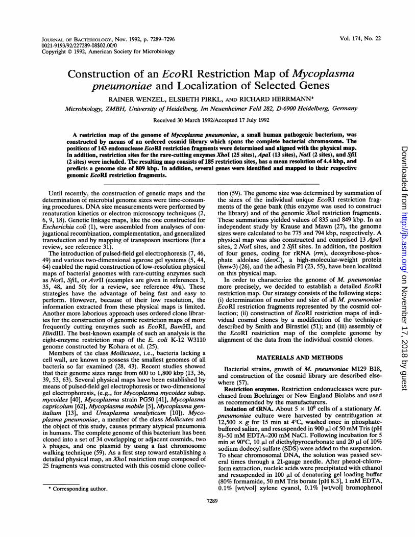
FIG. 1. Size determination ofM. pneumoniae EcoRP restriction fragments, represented by 17 EcoRI digested cosmids and plasmid pSPT7(P7) (57, 59). The fragments were separated on 0.4% (left) and 1.5% (right) agarose gels. SM, size marker (left, pUC18 polymers [monomer,2,686 bp]; right, phage A DNA x BstEII [8,454, 7,242, 6,369, 5,686, 4,822, 4,324, 3,675, 2,323, 1,929, 1,371, 1,264, 702, 224, and 117 bp]).
used to resolve EcoRI restriction fragments of the cosmidlibrary. An example of such a gel is shown in Fig. 1.
Figure 2 shows the size distribution of all fragments largerthan 300 bp in the cosmid library. Fragments smaller than300 bp were neglected. As described recently (59), severalother clones, including a 16.5-kbp EcoRI fragment cloned inthe plasmid pSPT7 and the two phage X recombinants XXand XV, link the terminal cosmid clones pcosMPE7 andpcosMPGT9 and encompass the complete array of EcoRIfragments.EcoRI restriction mapping of the individual cosmid clones
and assembly of the genomic EcoRI map. Our method is amodification of the technique originally introduced by Smithand Birnstiel (51). A brief description of it is shown in Fig. 3.Instead of end-labelled fragments, the partially digestedcosmids were blotted and probed with radioactively labelledvector (pcosRW2) DNA. An example is shown in Fig. 4. Thesizes of various DNA intermediates were determined byusing linear polymers of pUC18 as size markers. The differ-ence in sizes of two neighboring intermediates was evaluatedby comparing them with the EcoRI fragments produced bycomplete EcoRI digestion of the cosmid in question. Such acomparison was not always possible either because thefragment sizes were too large to be accurately estimated orbecause there were too many EcoRI fragments with similarlengths in a cosmid. Generally, these problems could besolved by conventional restriction analysis with enzymessuch as BamHI or HindIII in combination with EcoRI.Another way to analyze the regions in question was toperform restriction mapping from the other end (T7) of thecloned fragment, since two NotI restriction sites flank thecloned DNA in pcosRW2 (Fig. 3). In all cases in which no
additional NotI site is present in the cloned DNA, the insertcould be excised as one contiguous fragment. If the terminalEcoRI fragments are known from parallel analyses fromoverlapping cosmids, then they can be used as probes inhybridizations against Southern blots of the partially EcoRI-digested NotI insert fragment.The cosmids pcosMPP1/P2 and pcosMPGT9, which each
contained one Sfil site in the cloned region, caused problemsin the restriction analysis. In pcosMPP1/P2, the SfiI site ispresent in the T7-terminal 20.1-kbp EcoRI fragment of bothclones. This means that the fragment size is reduced by anSfiI-EcoRI subfragment in a partial EcoRI restriction analy-sis. In the case of pcosMPGT9, the SfiI site in the SP6-terminal 25-kbp EcoRI fragment cuts the cloned DNA awayfrom the vector. The EcoRI restriction map of this cosmidwas established by data derived from other studies in whichrepetitive DNA elements in M. pneumoniae were analyzed(45). The final map is shown in Fig. 2.Ambiguities remain in the pcosMPK5/E9/E30 region be-
cause of the unfavorable position of the cloned inserts withrespect to the SP6 and T7 promoters, making it difficult todefine the order of some fragments. In pcosMPK5, the orderof the 6.6- and 0.87-kbp fragments could not be established.An 0.48-kbp EcoRI fragment in pcosMPE9/E30 has threepossible locations. These ambiguities are indicated by brack-ets in Fig. 2.
In the cosmid clone pcosMPG7/GT9, two EcoRI frag-ments with very similar sizes are present which are indistin-guishable in our analysis. Therefore, the two possible posi-tions of these fragments are shown (Fig. 2).The map also contains a 0.1-kbp EcoRI fragment located
between the 5.7- and 2.7-kbp EcoRI fragments in
VOL. 174, 1992
on Novem
ber 17, 2018 by guesthttp://jb.asm
.org/D
ownloaded from

7292 WENZEL ET AL.
Al RI A68 Bi
E7 C9 R2 D9 A3
AA A X X XA XA X2.8 - 1.7 - 4.4 - 0.6 - 1.85 - 5.7 - 0.1- 2.7 -5.7 - 1.1 - 0.7 - 1.4 - 1.9 - 5.5 - 0.75 - 7.65 - 15 - 2.3 - 15.5 - 2.3 - 9.5 - 0.7 - 1.2 - 6.5 - 10.7 - 7.5 - 9 - 14 -12.6-2.9-3.7-0.55-1 -0.46-9.6-3.8-2-1.8-
__ Em i1 EEm" Em E) m Pi PMnElE El doC EmOE6 P1 C04
Xx Xv
D12 E9 C12 H3
A3 KS E30 D2 G12A A X A X X A X
0.4 - 2.2 - 4.4 - 6.8 - 2.3 - 4.9 - 0.96 - 3.8 - 4.8 - 1.4 - 4 - (6.6 - 0.87) - 2.5 - 16 - 4 -10.5t1.8-2.4-0.48)-6.4 -1.8 -82 - 4.6 - 5.4 - 8.4 - 2.2 - 4.8 - 7.3 - 1.3 - 1.8 - 0.73 - 0.73 - 7.7 - IO - 4.4 - 8 - 3.5 - 10 -
gyA gyrs tuf StP&
F4 P1 D7 Fll
G12 P2 H8 A5 A19
X SXA X N X X A X N10.5 - 7 - 5.5 - 2.1 - 10.5. 0.58 - 3.6 - 1.6 - 4.7 - 6 - 20.1 - 1.3 - 16 - 1.8 - 0.44 - 1.65 - 5.2 - 0.43 - 3.2 - 0.43 - 92 - 0.95 - 4.7 - 4-4 - 82 - 5.5 - 4 - 24 - 5 - 28 - 19 -
1Em Emmm P30 JtpINk(1)s~~~~~~~~~~~~~~~~~~~~~~R (1 ) 4E
H91 FIO
EliX
*1.9-1.8-22.3-6.6-6-4 -6.9 -
FIO A65 K4 GT9
H10 K8 G7 ALv- Xx -pPT7X X X X X X 2.6 26 ASXA XX
14.3 - 2.7 - 0.7 - 6.2 - 5.1 - 12.3 - 0.95- 0.5 - 7 - 3.3 - 18 - 6.7 - 7.3 - 6.3 - 2.6 - 5.4 - 3.3 - 8 -10.8 - 1.1 - 7.5 - 3.3 --- 4 -2- 4- 25 - 16.5 -WI 2.7 2.7 3 c
zplp
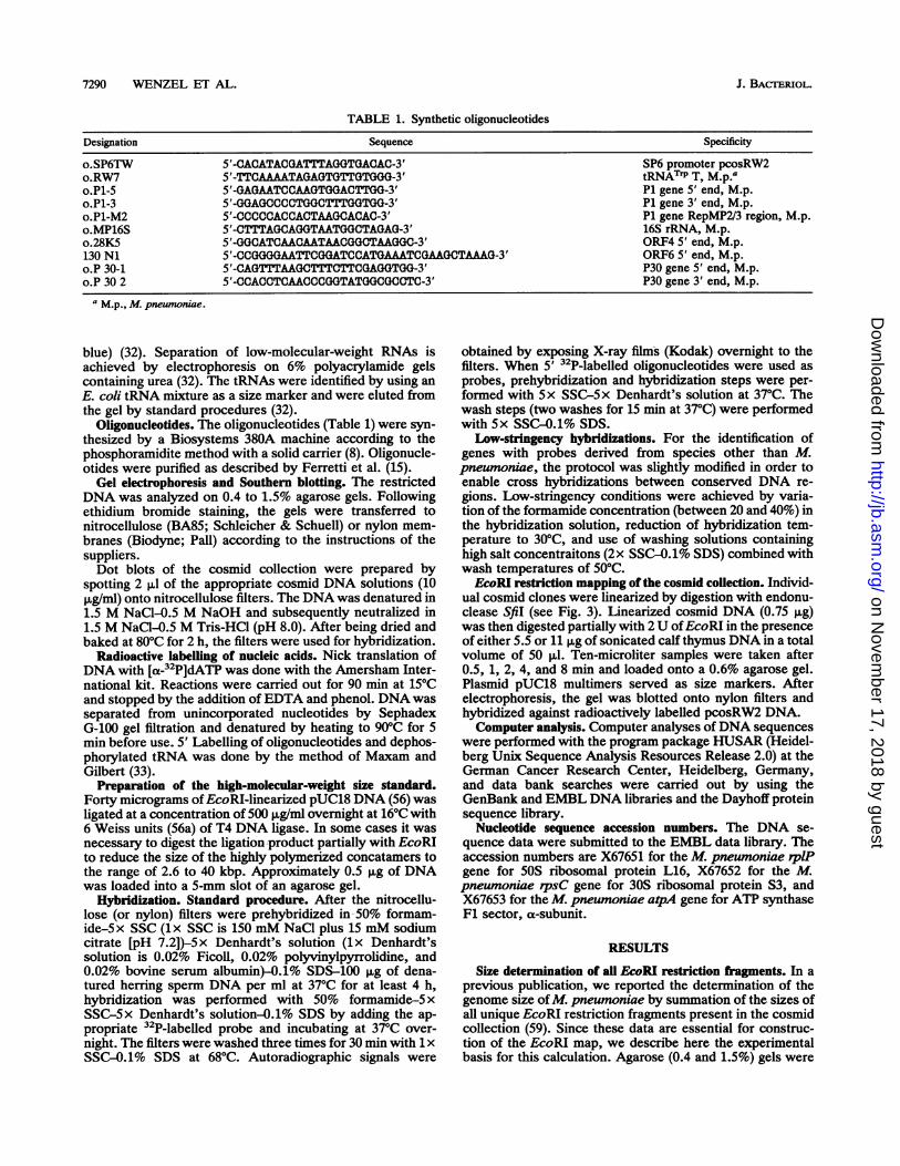
FIG. 2. EcoRI restriction map of the M. pneumoniae genome. The map was linearized between the 16.5-kbp (pSPT7) and the 2.8-kbpEcoRI (pcosMPE7) fragments. The numbers indicate the sizes of individual fragmnents (in kilobase pairs). For orientation, positions of thecosmid clones from the library are shown (horizontal bars). Recognition sites for endonucleases XhoI (X), ApaI (A), NotI (N), and SfiI (S)are indicated. Restriction fragments containing identified genes are marked accordingly (for details, see text and Table 2). For completeness,the positions of four different repetitive DNA sequences (RepMP1, RepMP2/3, RepMP4, and RepMP5; indicated by boxed 1, 2/3, 4, and 5,respectively) are included (45).
pcosMPA1/E7 which is known from a published 10-kbpDNA sequence of this region (23). Furthermore, sequenceanalysis of certain repetitive DNA elements, called RepMP1(58), revealed seven 85-bp EcoRI fragments in the cosmidspcosMPR1/R2, -P1, -P2/F4, -A1/E7, -H91, -GT9, and -C9which were not located on the map.The restriction sites for the enzymes XhoI (25 sites) ApaI
(13 sites), SfiI (2 sites), and NotI (2 sites), which have beenpublished already, were mapped onto this EcoRI map bydouble digest of individual cosmids with these endonu-cleases. A total of 150 EcoRI fragments, including the seven
85-bp EcoRI fragments, were counted, and a genome size of809 kbp was determined. This calculation differs by 26 kbpfrom the former determination, which was based on thecomparison of the cosmid's EcoRI restriction pattern. Thissize difference probably arises from duplicating detection ofseveral fragments that are located on overlapping regions ofneighboring cosmids.
Identification and mapping of M. pneumoniae genes. Inorder to construct a genetic map, all M. pneumoniae geneswhich have been cloned and sequenced so far were localizedon the EcoRI map by using either the cloned genes or
oligonucleotides as specific probes in cross hybridizationexperiments. An alternative approach tested whether heter-ologous cloned genes could be useful in identifying thecorresponding M. pneumoniae genes on a routine basis.Gene probes derived from M. pneumoniae (sequences)
were initially hybridized against dot blots of the completecosmid library. Positive clones were characterized in moredetail by Southern blotting of the endonuclease EcoRI-digested cosmid(s) in question. With M. pneumoniae probesall of the positive signals were unambiguous, and a furtherconfirmation by DNA sequencing was unnecessary. How-
ever, this procedure had to be modified for analyses withheterologous gene probes as follows. Hybridization to ge-nomic Southern blots of EcoRI-digested M. pneumoniaeDNA was performed under various low-stringency hybrid-ization conditions (see Materials and Methods). If positivesignals (i.e., one or two cross-reacting EcoRI fragments)were obtained, the cosmid library was screened and thecorresponding cosmid was identified. After subcloning, theregion in question had to be sequenced. Finally, computeranalyses of the DNA sequences were performed with re-spect to open reading frames, amino acid sequences, andhomologies to known protein sequences of other bacterialspecies present in data bases; the existence of the counter-part gene in M. pneumoniae was considered to be provedwhen significant homologies, at least 40% identity, weredetected.Table 2 summarizes all of the genes which have been
mapped so far, and Fig. 2 shows their positions on the EcoRIrestriction map. Most of the analyses done with homologousgene probes were straightforward and need no further ex-planation. But some others are briefly described below.P1 operon. The P1 gene that encodes the major adhesin
protein P1 (20, 21, 55) is flanked by two open reading frames,ORF4 and ORF6 (23), which have coding capacities for a 28-and a 130-kDa protein. Substantial regions of the P1 gene(RepMP2/3 and RepMP4) and of ORF6 (RepMP5) appear asrepetitive DNA sequences (12, 45, 54, 58, 60). In order tomap these genes, it was necessary to use oligonucleotideswhich hybridized only to the unique regions, such as o.P1-5and o.P1-3, which are specific for the 5' and 3' ends of the P1gene, respectively, o.130 Ni, with a specificity for ORF6,and o.28K5, with a specificity for ORF4. An example of aprobe that gives false positives is o.Pl-M2, which is derived
J. BACTERIOL.
on Novem
ber 17, 2018 by guesthttp://jb.asm
.org/D
ownloaded from

PHYSICAL AND GENETIC MAPS OF M. PNEUMONIAE 7293
EcoARl ERIEooRAEooRl EooRI EcoRiT7 4000
5500 F8200,
4400 _
4700
950 _
9200
<lS I EoI I IT
pco&RW2
Linewrizatlon |x Sfil
SP6
A---
. ....~AfI.
74, ' 2,88
. 4-I
, -1, 0
ri('
Ndi EcoRl EcoAl Ecol EcoR EcRI EoRl Nsflls --1 1 1I I I I I 8Sold-IIEI iI si
T ape I ER EcoRi
Partialx EcoRI
Digest
Ndal E60RI EoRiE coRI EcoRI EooRI E60RISR -II I
NotI EooRl ERoAI EcoRI EoRN Eb*RI EoARl
81lT7 SpEPNotI EGaRl EaRl EooRi EnORI EcoRi EnoAl
Ti7 aPeI I
Ndil ElooRRlE aERlRIERl EwRlSol
I I I
Nail EoARI liooRl EWoM EioRI8*I -*l I|
IT7 IPS EIoIT li;DoRI
Ndil EoARl EGnaRl EGn~~~I] I I
ITi IPS I naIso~~~~~FfT 7 SPssoRNall EoRI EcoAl
-b
Tm1i7 sp I oAI
Nmal
I
)RI
NdlEaRI
T 7 SPa EiCORINadl
; SPS
FIG. 3. Schematic drawing of the principle of the EcoRI restric-tion mapping of individual cosmid clones. Construction of thecloning vector pcosRW2 has been described previously (57). EcoRIrestriction fragments produced by the partial digest are orderedaccording to size. Only those fragments which hybridize to radio-actively labelled vector DNA are shown. SP6 and T7 indicate thepositions of the bacteriophage promoters.
FIG. 4. EcoRI restriction mapping of cosmid pcosMPD7. Thepartially EcoRI-digested DNA was separated on a 0.6% agarose geland blotted onto nylon. The filter was hybridized with 32P-labelledpcosRW2 DNA. This vector also cross-reacts with pUC18 polymerswhich serve as size standards (numbers on the right). For an exactsize determination, less-exposed X-ray films were used. SM, §izemarker; the numbers at the top represent reaction times (in minutes)at which the samples were taken. The numbers on the left indicatethe order of EcoRI restriction fragments in pcosMPD7; SP6 and T7positions are indicated. All sizes are in base pairs.
from a repetitive region of the P1 gene (RepMP2/3) andreacts with many EcoRI fragments in a Southern blot (Table1 and Fig. 2).tRNAs. tRNAs were identified by hybridizing radioac-
tively labelled total tRNA preparations from M. pneumoniaecells (see Materials and Methods) to dot blots of the cosmidcollection. Several cosmids appeared to be positive. A tRNAgene cluster coding for five tRNAs and the single tRNA withthe anti-codon UCA gene were mapped to the 28-kbp EcoRIfragment of the cosmid pcosMPAl9/Fll. The position of thesingle tRNA cluster was confirmed by subcloning and DNAsequencing (47). The tRNATrP(UCA) gene was located byhybridization with the sequence-specific oligonucleotideo.RW7 (22). The other presumptive tRNA loci are not shownon the map. The final confirmation awaits completion byDNA sequencing of the hybridization-positive EcoRI frag-ments.ATPase operon (atp). An example for the use of a heterol-
ogous probe to identify the corresponding M. pneumoniae isthe FlFo-ATPase. The probe was a cloned fragment of theATPase operon from Mycoplasma strain PG50, an operonwhich has been cloned and partially sequenced (42).Under our hybridization conditions (40% formamide-5 x
SSC at 37°C), a strong positive signal appeared with cosmidpcosMPD2 and was mapped to a 4.8-kbp EcoRI fragment.DNA sequence analysis of about 450 nucleotides from oneDNA strand of the cloned fragment and a computer-assistedanalysis of the data revealed an open reading frame with anamino acid sequence showing significant identity (50%) to aregion ofatpA ofMycoplasma strain PG50 and of E. coli (24)(Fig. 5).
Additional experiments were performed with heterologousgene probes to identify other genes which are widely spread
F.ln-
VOL. 174, 1992 s., (; , - ~~~~. ; ;~;U. ..i ;
NW
on Novem
ber 17, 2018 by guesthttp://jb.asm
.org/D
ownloaded from

7294 WENZEL ET AL.
TABLE 2. M. pneumoniae genes localized onto the EcoRI map
Gene Gene product Map position (cosmid/ Probe (organism) Reference and/orEcoRI fragment, kbp) source
P1 P1 protein, adhesin pcosMPE7/5.7 o.P1-5, o.P1-3, o.P1-M2 (M.p.') 21, 55ORF4 Open reading frame for a 28-kDa protein pcosMPE7/1.1 o.28 K5 (M.p.) 23ORF6 Open reading frame for a 130-kDa protein pcosMPE7/2.7 o.130N 1 (M.p.) 23rrn Unique rRNA operon pcosMPR2/9.5 o.MP16S (M.p.) 19deoC Deoxyribose-phosphate aldolase pcosMPD9/14 Plasmid pll-6 (M.p.) P.-C. Hu (30)gyrA Gyrase subunit A pcosMPK5/0.96 Personal communication, K. F. 11
Bott (M.p.)gyrB Gyrase subunit B pcosMPK5/3.8 Personal communication, K. F. 11
Bott (M.p.)tuf Elongation factor protein EF-Tu pcosMPK5/16 Plasmid (M.p.) S. Razin (65)atpA ATP synthase Fo sector a-subunit pcosMPD2/4.8 Plasmid pMYC 1193 (strain C. Christiansen (42)
G50)P30 Protein, 30 kDa, surface exposed pcosMPH8/9.2 o.30-1, o.30-2 (M.p.) 14tRNA cluster tyrT; gluT, lysT, leuT, glyT pcosMPF11/28 Total tRNA; sequencing (M.p.) 47tRNA trpT translates UGA into tryptophan pcosMPF11/28 o.RW7 22rplP 50S ribosomal subunit protein L16 A v/8; pcosMPGT9/25 Random sequencingrpsC 30S ribosomal subunit protein S3 X v/8 Random sequencing
a M.p., M. pneumoniae.
among bacteria. Examples include genes such as the dnaAprotein gene (16), the heat shock protein gene hsp9O (4, 29),and the cheWgene product, which plays a role in chemotaxis(52). The probes derived from Salmonella typhimurium(cheW) and E. coli (dnaA and htpG). In these hybridizationexperiments, either too many cross-reacting fragments weredetected at low stringency or none were detected at morestringent hybridization conditions; in the case of the htpGgene, a weak signal appeared at low stringency (37°C, 20%formamide-5 x SSC). DNA sequence analysis however,revealed a region with some limited homology to the probe atthe DNA level but no open reading frame with significanthomology to hsp9O.Ribosomal protein genes. Finally, genes for ribosomal
proteins S3 and L16 were identified by sequence analysis(one strand only) of a region which initially was difficult toclone into E. coli plasmids or cosmids (57). Deduced aminoacid sequences (181 amino acids) showed identity of 49 to64% (depending on the analyzed region) between the M.pneumoniae and the M. capricolum (37) and E. coli (66)ribosomal proteins S3 and L16 (data not shown).
DISCUSSION
The construction of a physical EcoRI restriction map ofthe M. pneumoniae genome with a cloned library in combi-nation with the fast and simple mapping technique of Smithand Birnstiel (51) was easy to perform and yielded clear andreliable results. In most cases, no additional information hadto be provided by other types of experiments. A total of 185restriction sites (143 EcoRI, 25 XhoI, 13 ApaI, 2 NotI, and 2SfiI sites) have been mapped with a mean resolution of 4.4kbp and with a calculated genome size of 809 kbp. If the 143EcoRI sites are counted separately, a mean fragment lengthof 5.7 kbp is determined. This value is only fairly consistentwith the theoretical value of 3,086 bp calculated for agenome with a GC content of41% (34), even considering thatfragments smaller than 300 bp were neglected.The EcoRI restriction map serves as a basis for the
construction of a genetic one. This was achieved by theidentification of several genes and their locations to partic-ular restriction fragments. Our selection of genes was basedmainly on three criteria. The first criterion is the relevance of
10 20 30 40 50 60
1 MOLNSTEISELIKQRIAQFNVVSEANNEGTIVSVSDGVIRIHGLADCMQGEMISLPGNRY E. COLI
61 AIALNLERDSVGAVVMGPYADLAEGMKVKCTGRI LEVPVGRGLLGRVVNTLGAPIDGKGP E. COLI
121 I E. COLI
1 ----------------------------L--------LH----------------YLSPY M. PG501 --------------------- NSSVAQIVHQLEVTDSMKYTTVVCATCSDPASMIYLTPF M. PNEUMONIAE
181 DAI INQRDSGIKCIYVAIGQKASTISNVVRKLEEHGALANTIVVVATASESAALQYLARM E. COLI
S V LE L T VV AT S A YL P CONSENSUS
8 TGITIAEEWMESGNDVLIVYDDLTKHAVSYRENSLLLRRPPGREAYPGDVFYLHSRLLER M. PG5040 TGITIAEYWLKQGKDVLIEFDDLSKH.IAYRTLSLLLRRPPGREAF.GDVFYLHSRLLER M. PNEUMONIAE
241 PVALMGEYFRDRGEDALI IYDDLSKQAVAYRQISLLLRRPPGREAFPGDVFYLHSRLLER E. COLI
TGITIAEYW G DVLI YDDLSKHAVAYR SLLLRRPPGREAFPGDVFYLHSRLLER CONSENSUS
68 AARVN----------ENYG-GGSITALPIIETQAGDISAYIPTNVISITDGOIFLSSELF M. PG50100 ACRLN --------- EEHG-GGSITALPIIETQAGDISAYIPTNVISITDGQLF.VSNLF M. PNEUMONIAE301 AARVNAEYVEAFTKGEVKGKTGSLTALPI IETOAGDVSAFVPTNVISITDGQI FLETNLF E. COL I
** * * * ** *********** ** *********** *
AARVN E G GGSITALPIIETQAGDISAYIPTNVISITDGQIFL SNLF CONSENSUS
117 NQGVRPAEDIGPSVSRVGSAAEIKSIKQVSGTLKLELAQYYELESFAKFGSDLDETTKAT N. PG50150 NSG--------------------------------------------------------- M. PNEUMONIAE361 NAGIRPAVNPGISVSRVGGAAQTKIMKKLSGGIRTALAQYRELAAFSQFASDLDDATRKQ E. COLI
N G RPA CONSENSUS
177 LDQOGAKI IQMLIQKQFNPLEOVDQAILLLTIKSHLLKWLPLDSIYNF------------- M. PG50421 LDHGQKVTELLKQKOYAPMSVAOQSLVLFAAERGYLADVELSKIGSFEAALLAYVDRDHA E. COLI
481 PLMOEINQTGGYNDEIEGKLKGILDSFKATQSW E. COL I
FIG. 5. Partial amino acid homology between the genes for thea-subunit of the ATPase from M. pneumoniae, Mycoplasma strainPG50 (42), and E. coli (24). For Mycoplasma strain PG50, onlypartial sequence data were available. Identical amino acids areindicated by asterisks; dots indicate identity between two of threeamino acids. Homology is present across the complete sequencedDNA region of M. pneunoniae which in E. coli is represented byamino acid positions 202 to 363.
J. BACTERIOL.
on Novem
ber 17, 2018 by guesthttp://jb.asm
.org/D
ownloaded from

PHYSICAL AND GENETIC MAPS OF M. PNEUMONIAE 7295
certain genes with respect to evolutionary or systematicrelationship studies within the rather heterogeneous classMollicutes (e.g., the elongation factor Tu). The secondaspect concerns the possibility of comparing the M. pneu-
moniae map with other bacterial genetic maps. This means
that only those genes which have previously been charac-terized can be taken into consideration. Examples includerRNA operons or ribosomal proteins. The last aspect is a
more practical one and concerns gene probes which are
available from related species. There is an additional diffi-culty in analyzing M. pneumoniae since this species, as an
exception among members of the class Mollicutes, has a
relatively high GC content of 41%, compared with theaverage GC range of members of the class Mollicutes of 24 to30% (43). Therefore, with gene probes from other Myco-
plasma species, the advantages arising from the close rela-tionship are countered by the different codon usages of theseextremely AT-rich organisms. It may be more promising touse probes from less-related species, such as Bacillus sub-tilis, which have similar GC contents. This was shown byColman et al. (11), who used a B. subtilis gene probe for theidentification of the gyrA and gyrB genes, which code for thegyrase subunits A and B, respectively.The failure to detect dnaA, htpG, and cheW with heterol-
ogous probes does not indicate that these genes are absentfromM. pneumoniae. It only illustrates a negative result thatis based on cross hybridization difficulties with heterologousprobes. In fact, Fujita et al. (17) succeeded in cloning thednaA gene from M. capricolum by using degenerate syn-
thetic oligonucleotides which were derived from sequences
of regions conserved among different bacterial dnaA genes.
In summary, we concluded that the cross hybridizationstrategy with heterologous probes is not the method ofchoice for fast identification and localization of a largernumber of genes for the purpose of constructing a geneticmap. Because of the reduced stringency in the hybridizationconditions, we frequently obtained several signals or even
unique signals that turned out to be falsely positive. There-fore, it is essential in any instance to sequence the DNA ofthe fragment in question. The faster way of identifying genesis to sequence DNA regions by a shotgun strategy followedby the identification of possible coding regions and thesearch for amino acid homologies to known genes by com-
putational analysis. Mycoplasmas, which are known to havesmall genomes, appear to be especially well suited for thisstrategy since their chromosomes should not contain toomuch unnecessary information (38). As far as M. pneumo-
niae is concerned, where a complete ordered cosmid libraryexists, sequencing of the genome in an ordered fashion is a
promising alternative to a shotgun approach.
ACKNOWLEDGMENTS
We thank M. Nassal, R. Frank, and B. Borchert for synthesis ofthe oligonucleotides; K. F. Bott, C. Christiansen, P.-C. Hu, 0.
Rasmussen, S. Razin, and J. Stock for supplying the gene probesand unpublished information; L. Runkel for critically reading themanuscript; and S. Nazareth for preparing the manuscript.
This work was supported by the Bundesministerium fir Fors-chung und Technologie (BCT 0381/5).
REFERENCES1. Bachmann, B. J. 1990. Linkage map of Escherichia coli K-12,
edition 8. Microbiol. Rev. 54:130-197.2. Bak, A. L., F. T. Black, C. Christiansen, and E. A. Freundt.
1969. Genome size of mycoplasmal DNA. Nature (London)224:1209-1210.
3. Bancroft, I., C. P. Wolk, and E. V. Oren. 1989. Physical and
genetic maps of the genome of the heterocyst-forming cyano-bacterium Anabaena sp. strain PCC 7120. J. Bacteriol. 171:5940-5948.
4. Bardweli, J. C. A., and E. A. Craig. 1987. Eukaryotic Mr 83,000heat shock protein has a homologue in Escherichia coli. Proc.Natl. Acad. Sci. USA 84:5177-5181.
5. Bautsch, W. 1988. Rapid physical mapping of the Mycoplasmamobile genome by two-dimensional field inversion gel electro-phoresis techniques. Nucleic Acids Res. 16:11461-11467.
6. Bode, H. R., and H. J. Morowitz. 1967. Size and structure of theMycoplasma hominis H39 chromosome. J. Mol. Biol. 23:191-199.
7. Cantor, C. R., C. L. Smith, and M. K. Mathew. 1988. Pulsed-field gel electrophoresis of very large DNA molecules. Annu.Rev. Biophys. Biophys. Chem. 17:287-304.
8. Caruthers, M. H. 1982. New methods for synthesizing deoxy-oligonucleotides. Genet. Eng. 4:1-16.
9. Chow, L. T. 1977. Sequence arrangement of the Escherichia colichromosome and of putative insertion sequences, as revealedby electron microscopic heteroduplex studies. J. Mol. Biol.113:611-621.
10. Cocks, B. G., L. E. Pyle, and L. R. Finch. 1989. A physical mapof the genome of Ureaplasma urealyticum 960T with ribosomalRNA loci. Nucleic Acids Res. 17:6713-6719.
11. Colman, S. D., P.-C. Hu, and K. F. Bott. 1990. Mycoplasmapneumoniae DNA gyrase genes. Mol. Microbiol. 4:1129-1134.
12. Colman, S. D., P.-C. Hu, and K. F. Bott. 1990. Prevalence ofnovel repeat sequences in and around the P1 operon in thegenome of Mycoplasma pneumoniae. Gene 87:91-96.
13. Colman, S. D., P.-C. Hu, W. Litaker, and K. F. Bott. 1990. Aphysical map of the Mycoplasma genitalium genome. Mol.Microbiol. 4:683-687.
14. Dallo, S. F., A. Chavoya, and J. B. Baseman. 1990. Characteri-zation of the gene for a 30-kilodalton adhesin-related protein ofMycoplasma pneumoniae. Infect. Immun. 58:4163-4165.
15. Ferretti, L., S. S. Karnik, H. G. Khorana, M. Nassal, and D. D.Oprian. 1986. Total synthesis of a gene for bovine rhodopsin.Proc. Natl. Acad. Sci. USA 83:599-603.
16. Fujita, M. O., H. Yoshikawa, and N. Ogasawara. 1989. Structureof the dnaA region of Pseudomonas putida: conservationamong three bacteria, Bacillus subtilis, Escherichia coli and P.putida. Mol. Gen. Genet. 215:381-387.
17. Fujita, M. O., H. Yoshikawa, and N. Ogasawara. 1992. Structureof the dnaA and DnaA-box region in the Mycoplasma capri-colum chromosome: conservation and variations in the courseof evolution. Gene 110:17-23.
18. Gillis, M., J. de Ley, and M. de Cleene. 1970. The determinationof molecular weight of bacterial genome DNA from renaturationrates. Eur. J. Biochem. 12:143-153.
19. Gfibel, U. B., A. Geiser, and E. J. Stanbridge. 1987. Oligonucle-otide probes complementary to variable regions of ribosomalRNA discriminate between Mycoplasma species. J. Gen. Mi-crobiol. 133:1969-1974.
20. Hu, P.-C., R. M. Cole, Y. S. Huing, J. A. Graham, D. E.Gardner, A. M. Collier, and W. A. Clyde, Jr. 1982. Mycoplasmapneumoniae infection: role of a surface protein in the attach-ment organelle. Science 216:313-315.
21. Inamine, J. M., T. P. Denny, S. Loechel, U. Schaper, C. H.Huang, K. F. Bott, and P.-C. Hu. 1988. Nucleotide sequence ofthe P1 attachment-protein gene of Mycoplasma pneumoniae.Gene 64:217-229.
22. Inamine, J. M., K.-C. Ho, S. Loechel, and P.-C. Hu. 1990.Evidence that UGA is read as a tryptophan codon rather than asa stop codon by Mycoplasma pneunoniae, Mycoplasma geni-talium, and Mycoplasma gallisepticum. J. Bacteriol. 172:504-506.
23. Inamine, J. M., S. Loechel, and P.-C. Hu. 1988. Analysis of thenucleotide sequence of the P1 operon of Mycoplasma pneumo-niae. Gene 73:175-183.
24. Kanazawa, H., T. Kayano, K. Mabuchi, and M. Futai. 1981.Nucleotide sequence of the genes coding for alpha, beta andgamma subunits of the proton-translocating ATPase of Esche-richia coli. Biochem. Biophys. Res. Commun. 103:604-612.
VOL. 174, 1992
on Novem
ber 17, 2018 by guesthttp://jb.asm
.org/D
ownloaded from

7296 WENZEL ET AL.
25. Kohara, Y., K. Akiyama, and K. Isono. 1987. The physical mapof the whole E. coli chromosome: application of a new strategyfor rapid analysis and sorting of a large genomic library. Cell50:495-508.
26. Krause, D. C., D. K. Leith, R. M. Wilson, and J. B. Baseman.1982. Identification of Mycoplasma pneumoniae proteins asso-ciated with hemadsorption and virulence. Infect. Immun. 35:809-817.
27. Krause, D. C., and C. B. Mawn. 1990. Physical analysis andmapping of the Mycoplasma pneunoniae chromosome. J. Bac-teriol. 172:4790-4797.
28. Krawiec, S., and M. Riley. 1990. Organization of the bacterialchromosome. Microbiol. Rev. 54:502-539.
29. Lindquist, S., and E. A. Craig. 1986. The heat-shock proteins.Annu. Rev. Biochem. 55:1151-1191.
30. Loechel, S., J. Inamine, and P.-C. Hu. 1989. Nucleotide se-quence of the deoC gene of Mycoplasma pneumoniae. NucleicAcids Res. 17:801.
31. Low, K. B. 1987. Mapping techniques and determination ofchromosome size, p. 1184-1189. In F. C. Neidhardt, J. L.Ingraham, K. B. Low, B. Magasanik, M. Schaechter, and H. E.Umbarger (ed.), Eschenichia coli and Salmonella typhimurium:cellular and molecular biology, vol. 2. American Society forMicrobiology, Washington, D.C.
32. Maniatis, T., E. F. Fritsch, and J. Sambrook 1982. Molecularcloning: a laboratory manual. Cold Spring Harbor Laboratory,Cold Spring Harbor, N.Y.
33. Maxam, A. M., and W. Gilbert. 1980. Sequencing end-labeledDNA with base-specific chemical cleavages. Methods Enzymol.65:499-560.
34. McClelland, M. 1988. Recognition sequences of type II restric-tion systems are constrained by the G+C content of hostgenomes. Nucleic Acids Res. 16:2283-2294.
35. McClelland, M., J. Jones, Y. Patel, and M. Nelson. 1987.Restriction endonucleases for pulsed field mapping of bacterialgenomes. Nucleic Acids Res. 15:5985-6005.
36. Neimark, H. C., and C. S. Lange. 1990. Pulse-field electropho-resis indicates full-length Mycoplasma chromosomes rangewidely in size. Nucleic Acids Res. 18:5443-5448.
37. Ohkubo, S., A. Muto, Y. Kawauchi, F. Yamao, and S. Osawa.1987. The ribosomal protein gene cluster of Mycoplasma capri-colum. Mol. Gen. Genet. 210:314-322.
38. Peterson, S. N., N. Schramm, P.-C. Hu, K. F. Bott, and C. A.Hutchison. 1991. A random sequencing approach for placingmarkers on the physical map of Mycoplasma genitalium. Nu-cleic Acids Res. 19:6027-6031.
39. Pyle, L. E., L. N. Corcoran, B. G. Cocks, A. D. Bergemann,J. C. Whitley, and L. R. Finch. 1988. Pulsed-field electrophore-sis indicates larger-than-expected sizes for Mycoplasma ge-nomes. Nucleic Acids Res. 16:6015-6025.
40. Pyle, L. E., and L. R. Finch. 1988. A physical map of thegenome of Mycoplasma mycoides subspecies mycoides Y withsome functional loci. Nucleic Acids Res. 16:6027-6039.
41. Pyle, L. E., T. Taylor, and L. R. Finch. 1990. Genomic maps ofsome strains within the Mycoplasma mnycoides cluster. J. Bac-teriol. 172:7265-7268.
42. Rasmussen, 0. F., and C. Christiansen. 1990. A 23 kb region ofthe Mycoplasma PG50 genome with three identified geneticstructures, p. 315-323. In G. Stanek, G. H. Cassell, J. G. Tully,and R. F. Whitcomb (ed.), Recent advances in mycoplasmol-ogy. Proceedings of the 7th Congress of the InternationalOrganization for Mycoplasmology, Baden near Vienna, 1988.Suppl. 20. Fischer Verlag, Stuttgart, Germany.
43. Razin, S. 1985. Molecular biology and genetics of mycoplasmas(Mollicutes). Microbiol. Rev. 49:419-455.
44. Romling, U., D. Grothues, W. Bautsch, and B. Tfimmier. 1989.A physical genome map of Pseudomonas aeruginosa PAO.EMBO J. 8:4081-4089.
45. Ruland, K., R. Wenzel, and R. Herrmann. 1990. Analysis ofthree different repeated DNA elements present in the P1 operonof Mycoplasma pneumoniae: size, number and distribution onthe genome. Nucleic Acids Res. 18:6311-6317.
46. Schwartz, D. C., and C. R. Cantor. 1984. Separation of yeast
chromosome-sized DNA by pulsed-field gel electrophoresis.Cell 37:67-75.
47. Simoneau, P., R. Wenzel, R. Herrmann, and P.-C. Hu. 1990.Nucleotide sequence of a tRNA cluster from Mycoplasmapneumoniae. Nucleic Acids Res. 18:2814.
48. Sitzmann, J., and A. Klein. 1991. Physical and genetic map ofthe Methanococcus voltae chromosome. Mol. Microbiol. 5:505-513.
49. Smith, C. L., and C. R. Cantor. 1986. Pulsed-field gel electro-phoresis of large DNA molecules. Nature (London) 319:701-702.
49a.Smith, C. L., and G. Condemine. 1990. New approaches forphysical mapping of small genomes. J. Bacteriol. 172:1167-1172.
50. Smith, C. L., J. G. Econome, A. Schutt, S. Klco, and C. RCantor. 1987. A physical map of the Escherichia coli K12genome. Science 236:1448-1453.
51. Smith, H. O., and M. L. Birmstiel. 1976. A simple method forDNA restriction site mapping. Nucleic Acids Res. 3:2387-2398.
52. Stock, A., J. Mottonen, T. Chen, and J. Stock. 1987. Identifica-tion of a possible nucleotide binding site in CheW, a proteinrequired for sensory transduction in bacterial chemotaxis. J.Biol. Chem. 262:535-537.
53. Su, C. J., and J. B. Baseman. 1990. Genome size of Mycoplasmagenitalium. J. Bacteriol. 172:4705-4707.
54. Su, C.-J., A. Chavoya, and J. B. Baseman. 1988. Regions ofMycoplasmapneumoniae cytadhesin P1 structural gene exist asmultiple copies. Infect. Immun. 56:3157-3161.
55. Su, C. J., V. V. Tryon, and J. B. Baseman. 1987. Cloning andsequence analysis of cytadhesin P1 gene from Mycoplasmapneumoniae. Infect. Immun. 55:3023-3029.
56. Vieira, J., and J. Messing. 1982. The pUC plasmids, anM13mp7-derived system for insertion mutagenesis and sequenc-ing with synthetic universal primers. Gene 19:259-268.
56a.Weiss, B., A. Jacquemin-Sablon, T. R. Live, G. C. Fareed, andC. C. Richardson. 1968. Enzymatic breakage and joining ofdeoxyribonucleic acid. VI. Further purification and propertiesof polynucleotide ligase from Escherichia coli infected withbacteriophage T4. J. Biol. Chem. 243:4543-4551.
57. Wenzel, R., and R. Herrmann. 1988. Physical mapping of theMycoplasmapneumoniae genome. Nucleic Acids Res. 16:8323-8336.
58. Wenzel, R., and R. Herrmann. 1988. Repetitive DNA elementsin Mycoplasmapneumoniae. Nucleic Acids Res. 16:8337-8350.
59. Wenzel, R., and R. Herrmann. 1989. Cloning of the completeMycoplasmapneumoniae genome. Nucleic Acids Res. 17:7029-7043.
60. Wenzel, R, E. Pirkl, K. Ruland, and R. Hernmann. 1990.Multiple copies of Mycoplasma pneumoniae gene P1 se-quences. Zentralbl. Bakteriol. Mikrobiol. Hyg. Suppl. 20:193-198.
61. Whitley, J. C., and L. R. Flnch. 1989. Location of sites oftransposon Tn916 insertion in the Mycoplasma mycoides ge-nome. J. Bacteriol. 171:6870-6872.
62. Whitley, J. C., A. Muto, and L. R. Finch. 1991. A physical mapfor Mycoplasma capricolum Cal. kid with loci for all knowntRNA species. Nucleic Acids Res. 19:399-400.
63. Ye, F., F. Laigret, J. C. Whitley, C. Citti, L. R. Finch, P. Carle,J. Renaudin, and J. M. Bov6. 1992. A physical and genetic mapof the Spiroplasma citri genome. Nucleic Acids Res. 20:1559-1565.
64. Yee, T., and M. Inouye. 1982. Two-dimensional DNA electro-phoresis applied to the study of DNA methylation and theanalysis of genome size in Myxococcus xanthus. J. Mol. Biol.154:181-196.
65. Yogev, D., S. Sela, H. Bercovier, and S. Razin. 1990. Nucleotidesequence and codon usage of the elongation factor Tu(EF-Tu)gene from Mycoplasma pneumoniae. Mol. Microbiol. 4:1303-1310.
66. Zurawski, G., and S. M. Zurawski. 1985. Structure of theEscherichia coli S10 ribosomal protein operon. Nucleic AcidsRes. 13:4521-4526.
J. BAcTERiOL.
on Novem
ber 17, 2018 by guesthttp://jb.asm
.org/D
ownloaded from