contamination in the micro lab - swacm - southwestern …€¦ · · 2016-10-10contamination in...
TRANSCRIPT

Contamination Wars: Contamination in the Micro Lab
Amanda T. Harrington, PhD, D(ABMM) Assistant Professor, Pathology, University of Illinois at Chicago Director, Clinical Microbiology Laboratory, University of Illinois
Hospital and Health Science System SWACM 2016
Disclaimers
• All of these stories are true—some of the names have been changed to protect the innocent

Case 1
An observant technologist brings a wound culture workup to the attention of the director on daily microbiology bench rounds. In addition to Staphylococcus aureus, she has identified a Gram negative rod that she is not familiar with. The biochemical identification reveals the organism to be Ralstonia pickettii.

Ralstonia picketii
• Aerobic GNR • Oxidase positive • Non-fermenter • Grows on MAC • Commonly found in water and soil • R. pickettii species most commonly associated with human
infection • Bacteremia, meningitis, endocarditis, osteomyelitis, CF • Nosocomial infections—contaminated blood products,
sterile water, saline, treatment fluids and venous catheters • Other species have been re-assigned to Cuprividas • Phenotypically similar to P. fluorescens and B. cepacia
complex

The Story Unfolds…
Over the next several days Ralstonia pickettii is recovered from several more wound and respiratory cultures. The biochemical identification has been confirmed via sequence based identification from the first culture work up.
At rounds, they begin to be suspect….

Pseudoepidemic
An outbreak, usually in a closed community or an institution, of a disorder with no demonstrable physical cause
• Common source?
• Commonly involved organisms – Acinetobacter
– Stenotrophomonas
– Burkholderia
– Pseudomonas
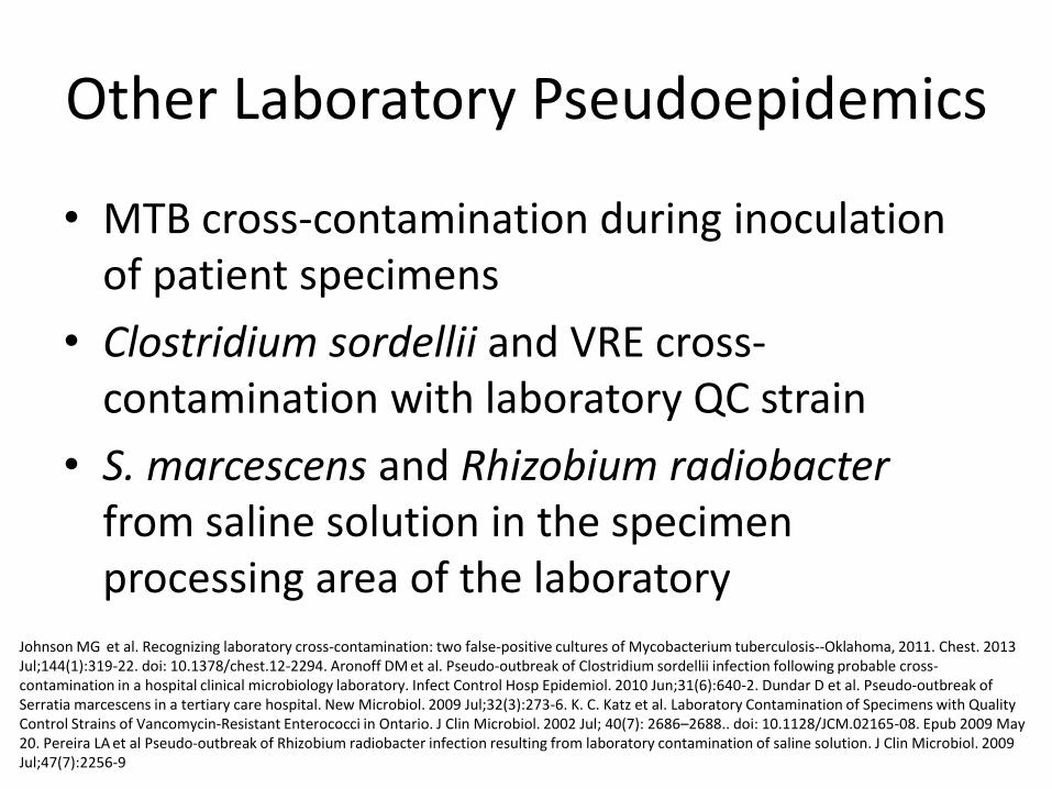
Other Laboratory Pseudoepidemics
• MTB cross-contamination during inoculation of patient specimens
• Clostridium sordellii and VRE cross-contamination with laboratory QC strain
• S. marcescens and Rhizobium radiobacter from saline solution in the specimen processing area of the laboratory
Johnson MG et al. Recognizing laboratory cross-contamination: two false-positive cultures of Mycobacterium tuberculosis--Oklahoma, 2011. Chest. 2013 Jul;144(1):319-22. doi: 10.1378/chest.12-2294. Aronoff DM et al. Pseudo-outbreak of Clostridium sordellii infection following probable cross-contamination in a hospital clinical microbiology laboratory. Infect Control Hosp Epidemiol. 2010 Jun;31(6):640-2. Dundar D et al. Pseudo-outbreak of Serratia marcescens in a tertiary care hospital. New Microbiol. 2009 Jul;32(3):273-6. K. C. Katz et al. Laboratory Contamination of Specimens with Quality Control Strains of Vancomycin-Resistant Enterococci in Ontario. J Clin Microbiol. 2002 Jul; 40(7): 2686–2688.. doi: 10.1128/JCM.02165-08. Epub 2009 May 20. Pereira LA et al Pseudo-outbreak of Rhizobium radiobacter infection resulting from laboratory contamination of saline solution. J Clin Microbiol. 2009 Jul;47(7):2256-9

Ralstonia in Water
• Biofilm formation
• Resistant to biocides
• Laboratory-based and industrial ultrapure water systems

Case Resolution
Removal of Common Source • Disposal • Autoclave • Process improvement/review
In the water jacket of CO2 incubator • Many attempts
– Bleach – Alcide – Heat (>80oC)
• Be suspicious

Case 2
The Infectious Diseases team consults on a patient whose CSF was reported to have rare Gram negative rods seen on Gram stain. The patient was put on antimicrobial therapy based on these findings. Three days later the culture work up is reported as no growth. On rounds the ID team is wondering about the discrepancy.

Review of CSF Gram Stain

The Story Unfolds…
Over the next several days additional CSF samples from the patient and two additional patients are intermittently positive with GNR on Gram stain but negative for growth in culture. After closer review, all patients are from the same unit, neurology ICU.
At rounds, they begin to be suspect….

Investigation and Intervention
• No recent changes in tube
• Red top tubes kept in open bins
• Change to individually bagged tubes
• Gram stain from inside of tube
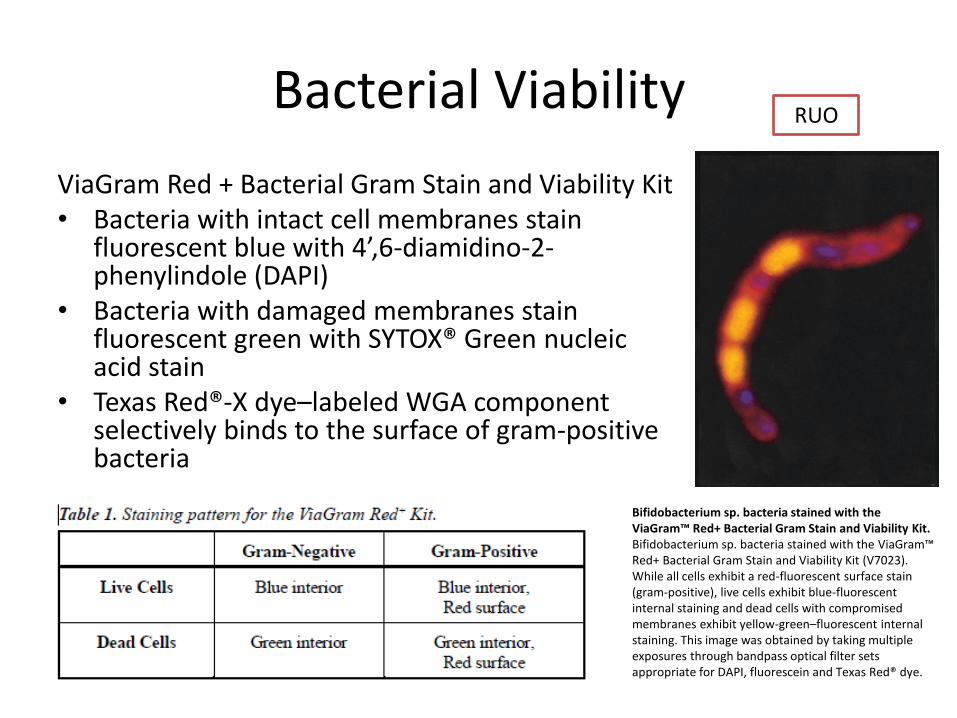
Bacterial Viability
ViaGram Red + Bacterial Gram Stain and Viability Kit • Bacteria with intact cell membranes stain
fluorescent blue with 4’,6-diamidino-2-phenylindole (DAPI)
• Bacteria with damaged membranes stain fluorescent green with SYTOX® Green nucleic acid stain
• Texas Red®-X dye–labeled WGA component selectively binds to the surface of gram-positive bacteria
Bifidobacterium sp. bacteria stained with the ViaGram™ Red+ Bacterial Gram Stain and Viability Kit. Bifidobacterium sp. bacteria stained with the ViaGram™ Red+ Bacterial Gram Stain and Viability Kit (V7023). While all cells exhibit a red-fluorescent surface stain (gram-positive), live cells exhibit blue-fluorescent internal staining and dead cells with compromised membranes exhibit yellow-green–fluorescent internal staining. This image was obtained by taking multiple exposures through bandpass optical filter sets appropriate for DAPI, fluorescein and Texas Red® dye.
RUO

Another Pseudoepidemic
• Three patients admitted over a 2-week period with musculoskeletal complaints had one or more
• Joint fluid specimens submitted for culture. • Anaerobic chopped meat-glucose broth (CMGB) tubes yielded one or
more organisms not typically associated with septic arthritis (Enterobacter, Enterococcus, Escherichia hermannii, and Pseudomonas diminuti)
• Laboratory investigation yielded isolates from sham-inoculated CMGB tubes.
• PFGE analysis demonstrated that a single strain of E cloacae was isolated from four CMGB tubes representing all three patients, and a single strain of E faecium was isolated from CMGB tubes representing two patients and the sham-inoculated tube.
• Molecular typing clearly demonstrated clonality among the isolates and indicated that a common source of contamination, most likely the CMGB tubes, was responsible for these cases.
Morris T et al. A pseudoepidemic due to laboratory contamination deciphered by molecular analysis. Infect Control Hosp Epidemiol. 1995 Feb;16(2):82-7.

Case Resolution
• Limited response from the manufacturer
• Switch to LP kits (short term)
• Use a dedicated and sterilely packaged set of tubes for CSF
• Impact to patient care and lab operations

Case 3
Dear all, I am working in a microbiology lab and each summer we face a “fly invasion" problem. We have done everything we can to keep the lab “fly free”. Is there any efficient way that we can keep flies out of our lab? Thank you. Sincerely, Lord of the Flies

Suggestions from the Message Board
• Are your doors closed... windows closed? • Can you work in a hood or a designated room? • Does anyone in your lab/building work with
Drosophila? • Does anyone regularly bring fruit for lunch? • What samples do you work with?
• Have you tried to clean up your lab from bottom to top
and discharge or clean everything contaminated with the flies? They must have some place where they can propagate.

Helpful Suggestions
• Cider vinegar trap
• Dish washing soap trap
• Sticky fly traps
• Store media in an enclosed area
• Commercially prepared products

Case Resolution
• Enclosed storage
• Sticky paper

Case 4
Hospital A uses PCR based testing for its active surveillance program for MRSA. One of the units notices a spike in their unit’s positivity rate. Some of the swabs samples sent for PCR are also set up for culture, and MRSA is recovered in the majority of cases. The unit changes their collection practices; however, their positivity rate only drops for one month and then spikes again a month later. They ask the microbiology laboratory to help investigate.

Self Contained Automated Systems
• Single-use reagents; closed PCR systems
• Eliminates the need for separate processing rooms
• Limited technical expertise required

The Story Unfolds
The microbiology lab continues to attempt to confirm PCR positive results using culture. This time MRSA is only recovered in a small number of samples and ½ of the PCR results are not confirmed upon repeat testing. Although all of the QC checks are within range, the laboratory begins to suspect…

Contamination Control
Aseptic Cleaning Technique
• Cleaning should be carried out periodically before and after PCR work. This applies to – All work surfaces including bench tops, pipettors,
fridge/freezer handles, and any other touch points.
– Wiping down using 10% bleach
– Use a DI water-dampened paper towel to remove bleach residue
– Followed by a 70% alcohol dampened paper towel to help quickly dry the surfaces.
https://www.luminexcorp.com/blog/2014/10/14/10-ways-minimize-contamination-molecular-laboratory/

Contamination Control
Dedicated Consumables and Equipment • Using consumables and equipment dedicated to each room. Each room and/or
work area should have its own centrifuge, vortexers, pipettors, gloves, coats, etc. – Never “borrow” a post-PCR pipettor for use in a pre-PCR room without thoroughly
decontaminating the pipettor first
Use of Aerosol-Resistant Pipettes • Aerosols can lead to cross-contamination from sample-to-sample. • Aerosol-resistant pipette tips have a barrier, which acts as a seal when exposed to
potential liquid contaminants, trapping them inside the barrier. Pipetting Technique • Proper pipetting technique ensures that the accurate volume is aspirated and
dispensed and avoids splashing when dispensing liquid. • Open and close all sample tubes and reaction plates carefully so samples don’t
splash out. • Spinning tubes before opening can prevent aerosols when opening tubes. • Keep reactions and components capped whenever possible.
https://www.luminexcorp.com/blog/2014/10/14/10-ways-minimize-contamination-molecular-laboratory/

Contamination Control
Frequently Changing Gloves
• Always wear fresh gloves when working in a PCR area.
• Change gloves frequently, especially if you suspect they have become soiled with solutions containing template DNA.
https://www.luminexcorp.com/blog/2014/10/14/10-ways-minimize-contamination-molecular-laboratory/

Strategies for Monitoring
Wipe Tests
• Proactively monitor the laboratory environment for contamination before it becomes an issue. – At a minimum, wipe tests should be performed on
a monthly basis. This frequency should be increased if any contamination is suspected.
– Instructions are provided by some manufacturers
– College of American Pathologists (MIC.64850 Sample/Amplicon Contamination).
https://www.luminexcorp.com/blog/2014/10/14/10-ways-minimize-contamination-molecular-laboratory/

Strategies for Monitoring
Positivity Rate Monitoring • Positive and negative controls
– A no-template control (NTC) is used to check for the absence of contamination in the reagents, consumables, and environment.
• Monitor positivity rate. – Any sudden increase in positive specimens for which a
cause cannot be determined (i.e. seasonal outbreaks), should be investigated.
– College of American Pathologists (MOL.20550 Test Result Statistics).
https://www.luminexcorp.com/blog/2014/10/14/10-ways-minimize-contamination-molecular-laboratory/
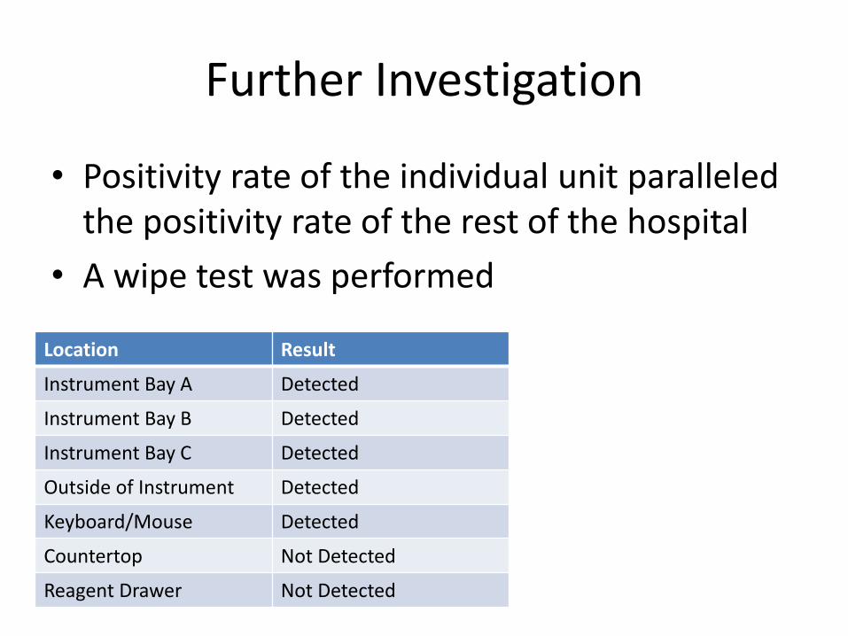
Further Investigation
• Positivity rate of the individual unit paralleled the positivity rate of the rest of the hospital
• A wipe test was performed
Location Result
Instrument Bay A Detected
Instrument Bay B Detected
Instrument Bay C Detected
Outside of Instrument Detected
Keyboard/Mouse Detected
Countertop Not Detected
Reagent Drawer Not Detected

Further Investigation
Systems-based Issues
1. Instrument located in a busy specimen processing area
a) Tech re-education about proper technique
b) Tech in service about molecular diagnostic techniques
2. Improper decontamination methods a) Immediate thorough decontamination
b) Tech re-education about proper technique

Case Resolution
• After a second round of wipe testing, target was not detected at all sites
• Increased frequency of cleaning and monitoring
• Change from spray to wipes
Case 6
A clinical microbiology laboratory with a molecular diagnostics section is having a problem. The internal control to their HCV viral load assay keeps failing. The laboratory also performs HCV genotyping using an assay that requires the lab to open the tubes after an initial amplification process. The laboratory suspects there is an issue with the instrument. The field service engineer suggests…

Potential Sources of Contamination
A typical PCR generates as many as 109 copies of target sequence and if aerosolized, even the smallest aerosol will contain as many as 106 amplification products
• Specimens with large numbers of target
organisms/templates in clinical specimens that may result in cross-contamination,
• Plasmid clones derived from organisms that have been previously analyzed and that may be present in large numbers in the laboratory environment (research labs)
• Repeated amplification of the same target sequence, which leads to accumulation of amplification products in the laboratory environment

Contamination Control
Laboratory Construction • At a minimum, two areas should be designated for PCR testing: Pre- and Post-PCR • Dead Air Box (DAB) with UV light
– Houses consumables and dedicated small equipment (mini-centrifuge, vortex, pipettors, tips, and tubes) needed
Pre-PCR • PCR master mix preparation • Sample preparation/addition to master mix.
– Dedicated refrigerator/freezer for kit storage – Separate equipment for storage of samples
Post-PCR • Post amplification steps and analysis • Physically separated from the pre-PCR room. The post-PCR room is where the
– Thermal cyclers for amplification – Any instrumentation needed for post-PCR analysis
https://www.luminexcorp.com/blog/2014/10/14/10-ways-minimize-contamination-molecular-laboratory/
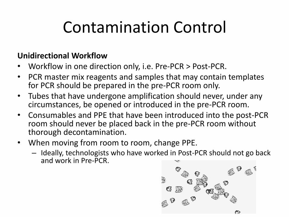
Contamination Control
Unidirectional Workflow • Workflow in one direction only, i.e. Pre-PCR > Post-PCR. • PCR master mix reagents and samples that may contain templates
for PCR should be prepared in the pre-PCR room only. • Tubes that have undergone amplification should never, under any
circumstances, be opened or introduced in the pre-PCR room. • Consumables and PPE that have been introduced into the post-PCR
room should never be placed back in the pre-PCR room without thorough decontamination.
• When moving from room to room, change PPE. – Ideally, technologists who have worked in Post-PCR should not go back
and work in Pre-PCR.

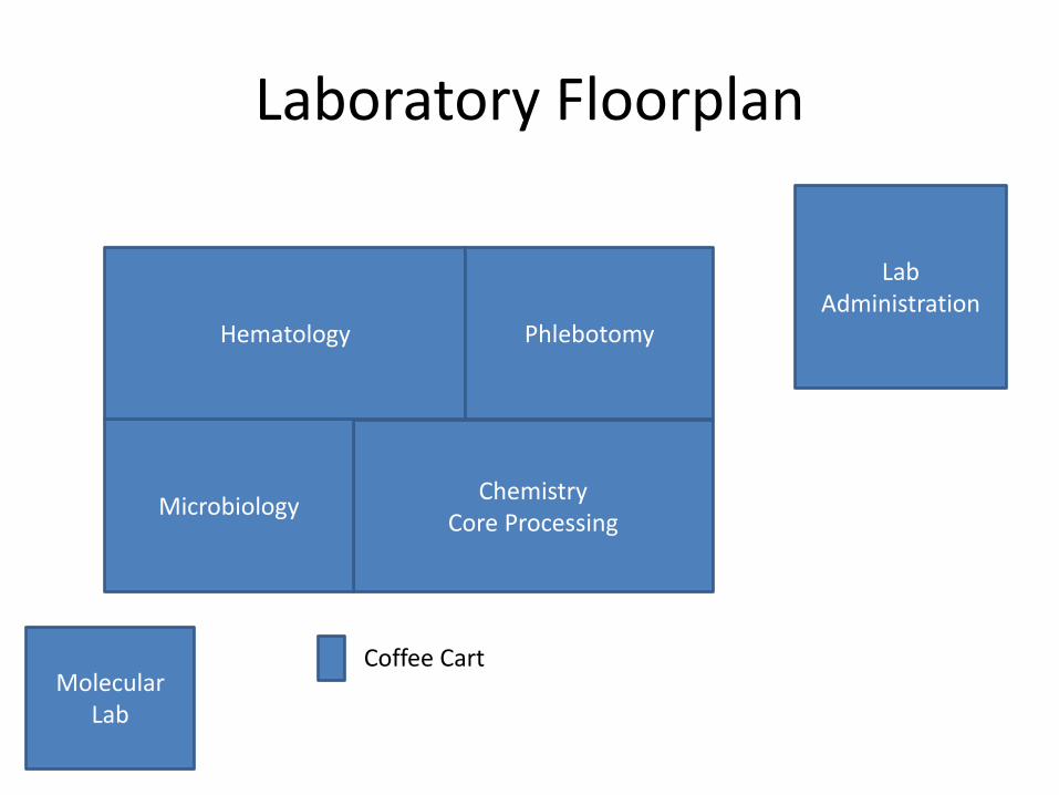
Laboratory Floorplan
Chemistry Core Processing
Molecular Lab
Lab Administration
Microbiology
Hematology Phlebotomy
Coffee Cart
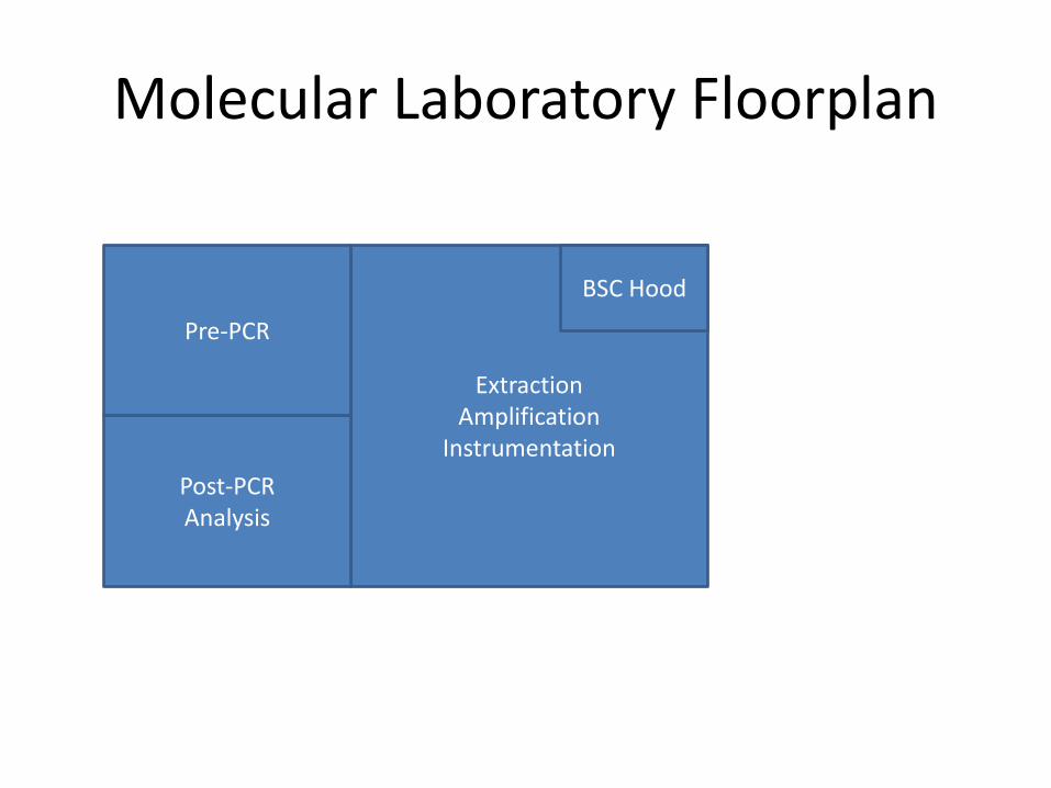
Molecular Laboratory Floorplan
Extraction Amplification
Instrumentation
Post-PCR Analysis
Pre-PCR
BSC Hood

Contamination Control
Environmental Control • Both areas should have independent environmental control
and not use common ductwork for air conditioning. • Rooms should be equipped with air-lock doors. • If physical barriers or separate rooms cannot be established
for Pre-PCR and Post-PCR work, all efforts should be taken to set up the work areas as far apart as possible. – Treat these two work areas as if they are in separate rooms. – Wear different personal protective equipment (PPE) in each
area.
https://www.luminexcorp.com/blog/2014/10/14/10-ways-minimize-contamination-molecular-laboratory/

Wipe Test
• Sampled several locations in the molecular lab
• All results came out as HCV not detected
• One exception = 100 IU/ml on the door handle
• …And then we remembered something

The UNG Enzyme
Some basic nucleotide biology • E.Coli enzyme Uracil-DNA-glycosylase (UDG) • The base cytosine (C) can undergo spontaneous deamination • Converts the cytosine to a uracil U • C pairs to G, U pairs to A • Creates a point mutation wherever it occurs
– one template strand being the original, correct sequence – the other strand misdirecting a replicating polymerase to insert an A for what
should be a G.
• Base excision repair = a way to detect and repair this specific damage event
• Uracil-N-glycosylase enzyme searches through dsDNA for any uracil residues and cuts out any uracil bases it finds – DNA backbone intact but a hole in the series of bases – Acts to block progress of a DNA polymerase using the strand as a template.
http://www.mlo-online.com/the-elephant-in-the-room-contamination-control-in-molecular-testing.php

The UNG Enzyme
Contamination control measure
Two steps: 1. DNA amplification products contain at least some percentage of uracil • Most DNA polymerases, when presented with dUTP as a nucleotide, will
accept it in place of dTTP, albeit with sometimes slightly lower efficiency. • Since only a single uracil residue per amplicon strand is all that is needed
to mark it for ung cleavage, this lower incorporation efficiency is balanced by using a mixture of mostly dTTP with some dUTP in the reaction mix.
• Most of the time the polymerase will incorporate a T against an A template location, at full efficiency; only occasionally will it pause to incorporate a U.
• Overall good replication efficiency is maintained, but every amplicon will have at least some uracil residues.
http://www.mlo-online.com/the-elephant-in-the-room-contamination-control-in-molecular-testing.php

The UNG Enzyme
Contamination control measure
Two steps: 2. Adding ung enzyme to every PCR reaction • Master mix preparation or as part of the reaction setup • Pre-incubate the reactions containing ung for a few minutes prior to
an initial high temperature strand denaturation step. • Ung will act to cleave any carried-over, uracil-containing
contaminant template material. • Also acts to permanently inactivate the ung so it can’t interfere with
the production of new, uracil-containing amplicons. • Vendors provide different enzymes
http://www.mlo-online.com/the-elephant-in-the-room-contamination-control-in-molecular-testing.php

Back to the Wipe Test
• Enzyme removes 103-105 copies of amplicon
• Any detectible product means
Laboratory Floorplan
Chemistry Core Processing
Molecular Lab
Lab Administration
Microbiology
Hematology Phlebotomy
Coffee Cart

Wipe Test Results
Chemistry Core Processing
Molecular Lab
Lab Administration
Microbiology
Hematology Phlebotomy
Coffee Cart

Case Resolution
• Move instrument to new, clean, physically separated space
• Re-locate all supplies to closed space
• Implementation of strict cleaning protocols
• Implementation of periodic wipe tests

Contamination Prevention Measures
Cleaning agents that are suitable for and dedicated to decontaminating nucleic acid contamination • NA removing agents
– (License to kill, DNAZap™, DNA remover, DNA-exit plus DNA-free™)
• 10% solution of sodium hypochloride • UV irradiation
Use of modified primers that use ribonucleotides • Use of UNG with dUTP • Iso-psoralen and long wavelength UV photoactivation • Sterilizing the PCR-mixture directly before amplification starts (UV-
Induced thymine dimers) Good laboratory QA/QC • Good monitoring program
– Rate monitoring – Environmental study
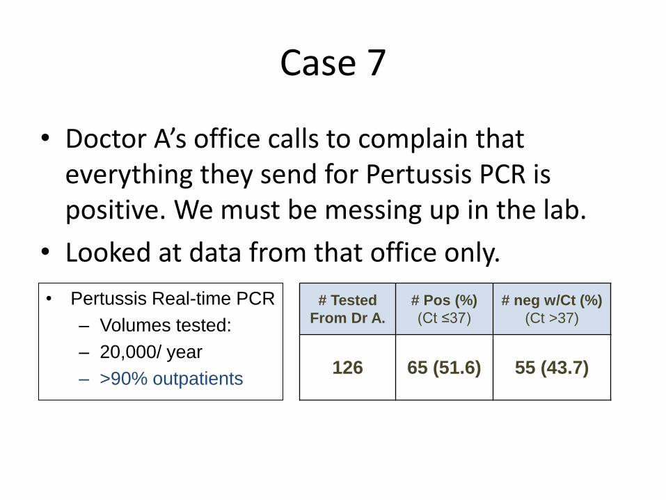
Case 7
• Doctor A’s office calls to complain that everything they send for Pertussis PCR is positive. We must be messing up in the lab.
• Looked at data from that office only.
• Pertussis Real-time PCR
– Volumes tested:
– 20,000/ year
– >90% outpatients
# Tested
From Dr A.
# Pos (%)
(Ct ≤37)
# neg w/Ct (%)
(Ct >37)
126 65 (51.6) 55 (43.7)

Contamination of Environment with Target
Ct values 21-25

Pediatric Office: Common Practices
Vaccine Prep Area
Collection Kits

DNA in Vaccines
Detection of Bordetella pertussis DNA in Acellular Vaccines and in Environmental Samples from Pediatric Physician Offices A
Leber, et al.. 2010. Interscience Conference in Antimicrobial Agents and Chemotherapeutics (ICAAC), Boston, MA.

Vaccine DNA in Environment
Detection of Bordetella pertussis DNA in Acellular Vaccines and in Environmental Samples from Pediatric Physician Offices
A Leber, et al.. 2010. Interscience Conference in Antimicrobial Agents and Chemotherapeutics (ICAAC), Boston, MA.

Office Practice and POC Testing
Danielle Holland is given a FluMist influenza vaccination in St. Leonard, Md. AP Oct 2005

The War Against Contamination
• Thoughtful investigation
• Strategic planning
• Time consuming
• Costly
• Minimize impact to patient care