contamination pattern of listeria monocytogenes and other listeria spp. in a salmon slaughterhouse...
TRANSCRIPT
ELSEVIER International Journal of
Food Microbiology 25 (1995) 19-27
lntwnational Journal of~ood Microbiiogy
Contamination pattern of Listeria monocytogenes and other Listeria spp. in a salmon slaughterhouse
and smoked salmon processing plant
Liv Marit Rflrvik a, * , Dominique A. Caugant b, Magne Yndestad a
a Deparrment of Pharmacology, Microbiology and Food Hygiene, Norwegian College of Veterinary Medicine, P.O. Box 8146 Dep., 0033 Oslo, Norway
b Department of Bacteriology, National Institute of Public Health, Oslo, Norway
Received 3 April 1994; accepted 14 June 1994
Abstract
A smoked salmon processing plant including a smokehouse and a slaughterhouse was examined for the occurrence of Listeria monocytogenes and other Listeria spp. From a total of 47.5 samples the overall frequency of L. monocytogenes was 16%, while other Listeria spp. were found in 22% of the samples. L. monoqtogenes was most often detected in samples from the smokehouse, where 29% of the environmental and 26% of the fish samples during processing contained the bacteria. 17% of the fish raw material to the smokehouse were contaminated, while 11% of the samples from vacuum-packed smoked salmon were positive for L. monocytogenes. The slaughterhouse was sporadically contami- nated, but L. monocytogenes was not found in 50 samples of slaughtered fish. L. monocyto- genes was found in the seawater outside the slaughterhouse. Multilocus enzyme elec- trophoresis divided the isolated L. monocytogenes strains into 11 electrophoretic types (ETs). One ET, ET-6, which is the most common ET in Norway, seemed to have colonized the smokelhouse. Isolates from the seawater, from the slaughterhouse and from fish coming into the smokehouse, before filleting, were other ETs.
Keywords: Listeria monocytogenes; Salmon; Multilocus enzyme electrophoresis
1. Introduction
Since .Listetiu monocytogenes in the early 1980s was recognized as a food-borne pathogen, listeriosis outbreaks and sporadic cases have been linked to several food
* Corresponding author. Fax: (+ 47) 22 964850.
0168-1605/95/$09.50 0 1995 Elsevier Science B.V. All rights reserved SSDZO168-1605(94)00080-k?
20 L.M. Rpvik et al. /International Journal of Food Microbiology 25 (1995) 19-27
items. Documented sources of the bacteria to patients have been cheese, vegeta- bles, and meat products (Schuchat et al., 1991; Hellesnes et al., 1992). Raw fish and shellfish consumption has been epidemiologically linked to one outbreak (Lennon et al., 1984), and undercooked fish has been assumed to be the source of a sporadic case of listeriosis (Facinelli et al., 1989).
L. monocytogenes has been detected in a wide range of sea products (Dillon et al., 1992). In two investigations L. monocytogenes was found in 12% and 9% of the samples of cold smoked salmon, respectively (Jemmi, 1990; Rorvik et al., 1991).
While liquid smoke has an antimicrobial effect (Messina et al., 1988), the cold smoking process does not seem to affect L. monocytogenes (Guyer and Jemmi, 1991). The bacteria can grow well on vacuum-packed, smoked salmon during storage at 4°C (Guyer and Jemmi, 1991; Rorvik et al., 1991). Thus consumption of contaminated smoked salmon may represent a health hazard.
To prevent the occurrence of L. monocytogenes in smoked salmon it is impor- tant to identify the sources for the bacteria in processing plants. In this study we investigated the contamination routes of L. monocytogenes to vacuum-packed smoked salmon in a processing plant where the products had been found contami- nated.
2. Materials and methods
2.1. Processing ptant
The plant included a salmon slaughterhouse with a smokehouse located in the same building. The slaughterhouse received salmon from transport boats. The boats were pumping seawater into the fish tanks during transport and unloading at the slaughterhouse. Slaughter of the fish was performed by CO, stunning, exsan- guination, bleeding, exvisceration, cooling (in water), sorting out, and packaging in boxes with ice. Seawater taken from deep sea at some distance from the shore was used during the slaughtering process. Smoked salmon was produced using the fish from the slaughterhouse of the plant as well as from other slaughterhouses, cooled or thawed. Processing of smoked salmon included filleting, salting, drying/smok- ing, cooling, trimming (removal of small bones and skin), slicing and vacuum-pack- ing.
2.2. Samples
Four hundred and seventy-five samples were collected on six occasions during a period of eight months, in 1991-92.
Seawater from the processing plant’s environment where the transport boats pumped water into the fish tanks, was sampled with a NIVA water sampler (Ormerod et al., 1982) 1 meter under the surface, into sterile bottles. Seawater from the fish tanks was collected in sterile bottles. Fresh municipal water and seawater used during the slaughtering process were sampled directly in sterile
L.M. R@rvik et al./Intemational Journal of Food Microbiology 25 (1995) 19-27 21
bottles from water taps. All water samples were collected in volumes of 500 ml. Ice was sampled in sterile plastic bags and thawed, 500 ml were used for each sample.
Slaughtered salmon in the slaughterhouse, and salmon coming in to the smoke- house, before filleting, were swabbed using five swabs per sample.
Filleted salmon (50 g) after each step of the process in the smokehouse, and vacuum-packed smoked salmon (50 g) were rinsed in equal amounts of peptone water (water with 0.1% Bacto peptone, Difco Laboratories, Detroit, MI, USA and 0.85% NaCl, Merck, Darmstadt, Germany) in a plastic bag for 45 s; the peptone water was, used as sample.
The processing equipment and environment in the slaughterhouse and the smokehouse were examined by swabbing. Cotton plugs were placed in the sinks for 4 h, two plugs were used for each sample. Wooden splinters for the smoke oven were collected, 25 g for each sample.
2.3. Sample analysis
Examinations for L. monocytogenes and other Listeria spp. were carried out according to the Nordic Committee on Food Analysis (1990). A two-step enrich- ment method was used: each sample was incubated in primary Listeria Enrichment Broth (LEBl, CM863 and supplement, SR142E, Gxoid, Basingstoke, UK) at 30°C for 24 h. Volumes of 0.1 ml of LEBl were then transferred to secondary Listeria Enrichment Broth (LEB2, CM863 and supplement, SR143E, Oxoid) and incubated at 30°C for 24 h. LEB2 was plated on Oxford agar (Listeria Selective agar base and supplement, SR140E, Oxoid) and incubated at 30°C for 48 h. Ten suspected Listeria colonies on Oxford agar were streaked on blood agar plates and incubated at 37°C for 20 h. Two hemolytic colonies and one Listeria-like non hemolytic colony from each sample were identified by hemolysis, catalase reaction, motility at 22”C, microscopy, fermentation of rhamnose and xylose, and CAMP reaction with Staphylococcus aureus and Rhodococcus equi.
For water volumes of 500 ml were filtered (White Grid 45 pm, Gelman Sci., MI), and the filters were incubated in 20 ml LEBl. The five swabs of each sample were incubated in 50 ml LEBl. For two cotton plugs, each wooden splinter sample, and for 25 ml of the peptone water rinse from the fish, 225 ml LEBl were used.
A quantative analysis of L. monocytogenes in the vacuum-packed, smoked salmon samples was performed by spreading 0.1 ml of the peptone water/LEBl mixture, before incubation, directly on Oxford agar. Incubation and identification of suspected colonies were as described above.
Usually, two isolates from each positive sample were preserved frozen at - 70°C.
2.4. Serotyping
L. monocytogenes isolates were serotyped by Bacto-Listeria-0 antisera serotype 1 and 4 (Difco Laboratories, Detroit, Mi., USA). Isolates non typable with these sera were: examined by Bacto-Listeria-0 polyvalent serum (Difco).
22 L.M. R@ruik et al. /International Journal of Food Microbiology 25 (1995) 19-27
2.5. Multilocus enzyme electrophoresis (MEE)
Ninety of the L. monocytogenes isolates were examined by multilocus enzyme electrophoresis. Methods of protein extracts preparation, starch-gel electrophore- sis and selective enzyme staining were as described earlier (Kolstad et al., 1992) and according to Selander et al. (1986). Twenty-one enzymes were assayed: IPO, indophenol oxidase; 6PG, 6-phosphogluconate dehydrogenase; G6P, glucose 6- phosphate dehydrogenase; ADK, adenylate kinase; PGI, phosphoglucose iso- merase; GDH, glutamate dehydrogenase; PE2, PE3 and PE4, three peptidases; NSP, nucleoside phosphorylase; AC1 and AC2, two aiid phosphatases; PGM, phosphoglucomutase; G3P, glyceraldehyde 3-phosphate dehydrogenase; IDH, isoc- itrate dehydrogenase; EST, esterase; MPI, mannose phosphate isomerase; FUM, fumarase; ALD, alanine dehydrogenase; LAD, lactate dehydrogenase; CAT, cata- lase.
Genetic distance between pairs of electrophoretic types (ETs) was expressed as the proportion of enzyme loci at which dissimilar alleles occurred (mismatches), and clustering was performed from a matrix of genetic distances by the average linkage method (Sneath and Sokal, 1973).
3. Results
3. I. Preualence of Listeria
The proportion of samples positive for L. monocytogenes and other Listeria spp. m shown in Table 1. Overall, 16% per cent of the samples contained L. monocyto- genes, and 22% harboured other Listeria spp.
L. monocytogenes was isolated from 9% of the samples and other Listeria spp. were recovered from 36% of the samples from the environmental seawater which was used by the salmon transport boats. Neither L. monocytogenes nor other Listeria spp. were found in seawater from the slaughterhouse or in the fresh water and ice used in the plant.
None of the 50 samples of slaughtered fish in the slaughterhouse or stored in boxes was positive for L. monocytogenes, even though each sample represented several fish. Six (7%) of the 83 environmental samples taken in the slaughterhouse revealed L. monocytogenes; five of six positive samples were from the sink and one was from the processing environment.
Four of 24 (17%) thawed or cooled fish entering the smokehouse, before filleting, contained L. monocytogenes. The positive samples included fish both from the slaughterhouse of this processing plant (three of 15 samples) and from another slaughterhouse (one of nine samples).
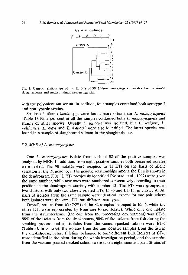
L. monocytogenes was frequently isolated in the smokehouse, where 29% of the environmental samples and 26% of the fish samples were contaminated. The frequencies of L. monocytogenes and other Listeria spp. after each step of the process in the smokehouse are listed in Table 2. The fish was heavily contaminated
L.M. R(rvik et al. /International Journal of Food Microbiology 2.5 (1995) 19-27 23
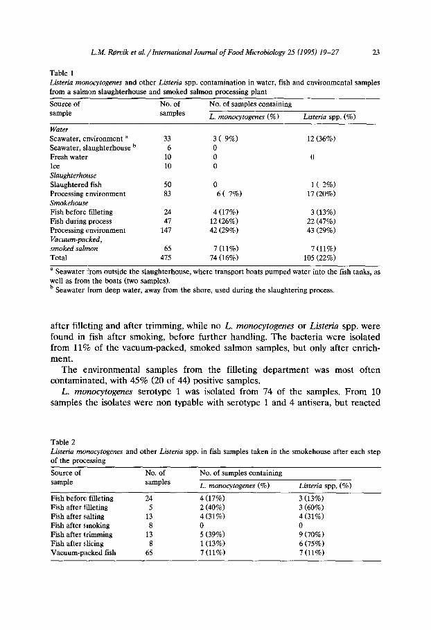
Table 1 List&a monocytogenes and other Listeria spp. contamination in water, fish and environmental samples from a salmon slaughterhouse and smoked salmon processing plant
Source of No. of No. of samples containing sample samples L. monocytogenes (%) Listeria spp. (“lo)
Water
Seawater, environment a Seawater, slaughterhouse b Fresh water Ice Slaughterhouse Slaughtered fish Processing environment Smokehouse Fish before filleting Fish during process Processing environment Vacuum-packed, smoked salmon
Total
33 6
10 10
50 0 1 ( 2%) 83 6( 7%) 17 (20%)
24 4 (17%) 3 (13%) 47 12 (26%) 22 (47%)
147 42 (29%) 43 (29%)
65 7 (11%) 475 74 (16%)
3( 9%) 0 0
0
12 (36%)
0
7 (11%) 105 (22%)
a Seawater from outside the slaughterhouse, where transport boats pumped water into the fish tanks, as well as from the boats (two samples). b Seawater From deep water, away from the shore, used during the slaughtering process.
after filleting and after trimming, while no L. monocytogenes or Listeriu spp. were found in fish after smoking, before further handling. The bacteria were isolated from 11% of the vacuum-packed, smoked salmon samples, but only after enrich- ment.
The environmental samples from the filleting department was most often contaminated, with 45% (20 of 44) positive samples.
L. monocytogenes serotype 1 was isolated from 74 of the samples. From 10 samples ithe isolates were non typable with serotype 1 and 4 antisera, but reacted
Table 2 Listeria monocytogenes and other Listeria spp. in fish samples taken in the smokehouse after each step of the processing
Source of No. of No. of samples containing sample samples L. monocytogenes (%) Listeria spp. (%)
Fish before filleting 24 4 (17%) 3 (13%) Fish after filleting 5 2 (40%) 3 (60%) Fish after z,alting 13 4 (31%) 4 (31%) Fish after r(moking 8 0 0 Fish after trimming 13 5 (39%) 9 (70%) Fish after slicing 8 1(13%1 6 (75%) Vacuum-packed fish 65 7 (11%) 7 (11%)
L. M. Rervik et al. /International Journal of Food Microbiology 25 (1995) 19-27
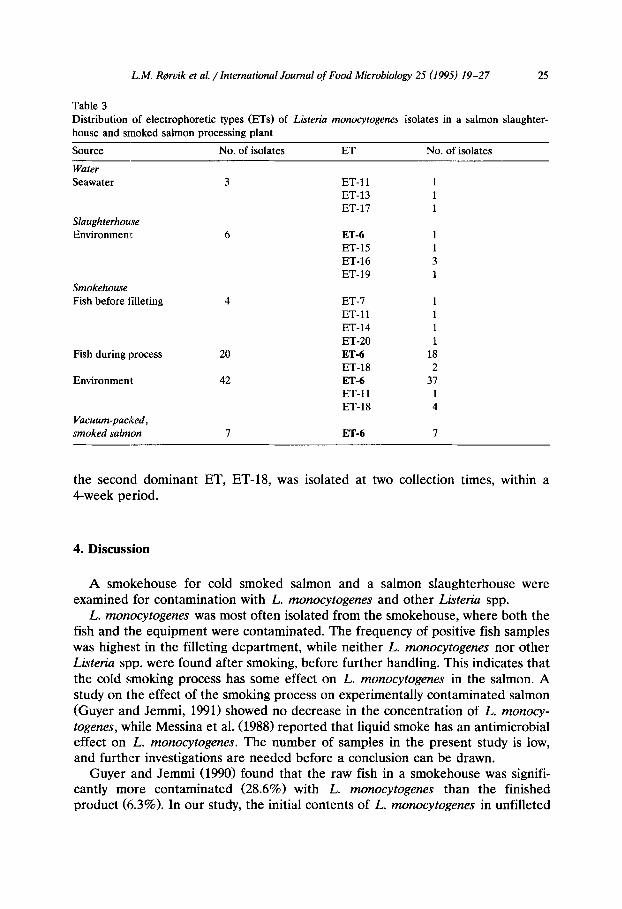
Genetic distance
.5 .4 .3 .2 .1 0
ET
L!z
Cluster A 6 13 7
14 15 16 17
Cluster B 18 11 19 20
Fig. 1. Genetic relationships of the 11 ETs of 90 Listeria monocytogenes isolates from a salmon slaughterhouse and smoked salmon processingg plant.
with the polyvalent antiserum. In addition, four samples contained both serotype 1 and non typable strains.
Strains of other Listetiu spp. were found more often than L. monocytogenes (Table 1). Nine per cent of all the samples contained both L. monocytogenes and strains of other species. Usually L. innocua was isolated, but L. seeligeri, L. welshimeri, L. gruyi and L. ivanovii were also identified. The latter species was found in a sample of slaughtered salmon in the slaughterhouse.
3.2. MEE of L. monocytogenes
One L. monocytogenes isolate from each of 82 of the positive samples was analysed by MEE. In addition, from eight positive samples both preserved isolates were tested. The 90 isolates were assigned to 11 ETs on the basis of allelic variation at the 21 gene loci. The genetic relationships among the ETs is shown in the dendrogram (Fig. 1). ETs previously identified (Kolstad et al., 1992) were given the same number, while new ones were numbered consecutively according to their position in the dendrogram, starting with number 13. The ETs were grouped in two clusters, with only two closely related ETs, ET-6 and ET-13, in cluster A. All pairs of isolates from the same sample were identical, except for one pair, where both isolates were the same ET, but different serotypes.
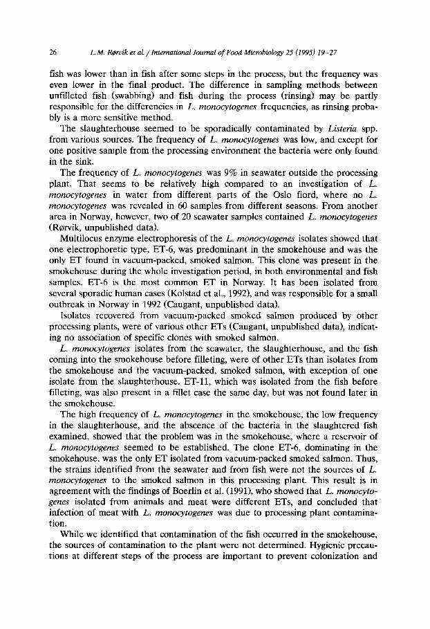
Overall, strains from 63 (78%‘0).of the 82 samples belonged to ET-6, while the other ETs were represented by from one to six isolates. While only one isolate from the slaughterhouse (the one from the processing environment) was ET-6, 88% of the isolates from the smokehouse, 90% of the isolates from fish during the smoking process and all isolates from the vacuum-packed salmon were ET-6 (Table 3). In contrast, the isolates from the four positive samples from the fish in the smokehouse, before filleting, belonged to four different ETs. Isolates of ET-6 were identified in the plant during the whole investigation period, and the samples from the vacuum-packed smoked salmon were taken eight months apart. Strains of
L.M. R@rvik et al. /International Journal of Food Microbiology 25 (1995) 19-27 25
Table 3 Distribution of electrophoretic types (ETs) of Lbteria monocytogenes isolates in a salmon slaughter- house and smoked salmon processing plant
Source No. of isolates ET No. of isolates
Water Seawater
Slaughterhouse Environmem
Smokehouse
Fish before filleting
Fish during process
Environment
Vacuum-packed,
smoked salmon
3 ET-11 ET-13 ET-17
6 ET-6 ET-15 ET-16 ET-19
4 ET-7 ET-11 ET-14 ET-20
20 ET-6
ET-18 42 ET-6
ET-11 ET-18
7 ET-6
1 1 1 1
18 2
37 1 4
7
the second dominant ET, ET-B, was isolated at two collection times, within a 4-week period.
4. Discussion
A smokehouse for cold smoked salmon and a salmon slaughterhouse were examined for contamination with L. monocytogenes and other Listeria spp.
L. monocytogenes was most often isolated from the smokehouse, where both the fish and tlhe equipment were contaminated. The frequency of positive fish samples was highest in the filleting department, while neither L. monocytogenes nor other Listeria spp. were found after smoking, before further handling. This indicates that the cold smoking process has some effect on L. monocytogenes in the salmon. A study on the effect of the smoking process on experimentally contaminated salmon (Guyer and Jemmi, 1991) showed no decrease in the concentration of L. monocy- togenes, while Messina et al. (1988) reported that liquid smoke has an antimicrobial effect on L. monocytogenes. The number of samples in the present study is low, and further investigations are needed before a conclusion can be drawn.
Guyer and Jemmi (1990) found that the raw fish in a smokehouse was signifi- cantly more contaminated (28.6%) with L. monocytogenes than the finished product (6.3%). In our study, the initial contents of L. monocytogenes in unfilleted
26 L.M. R(rvik et al. /International Journal of Food Microbiology 25 (1995) 19-27
fish was lower than in fish after some steps in the process, but the frequency was even lower in the final product. The difference in sampling methods between unfilleted fish (swabbing) and fish during the process (rinsing) may be partly responsible for the differencies in L. monocytogenes frequencies, as rinsing proba- bly is a more sensitive method.
The slaughterhouse seemed to be sporadically contaminated by L&e& spp. from various sources. The frequency of L. monocytogenes was low, and except for one positive sample from the processing environment the bacteria were only found in the sink.
The frequency of L. monocytugenes was 9% in seawater outside the processing plant. That seems to be relatively high compared to an investigation of L. monocytogenes in water from different parts of the Oslo fiord, where no L. monocytogenes was revealed in 60 samples from different seasons. From another area in Norway, however, two of 20 seawater samples contained L. monocytogenes (Rorvik, unpublished data).
Multilocus enzyme electrophoresis of the L. monocytogenes isolates showed that one electrophoretic type, ET-6, was predominant in the smokehouse and was the only ET found in vacuum-packed, smoked salmon. This clone was present in the smokehouse during the whole investigation period, in both environmental and fish samples. ET-6 is the most common ET in Norway. It has been isolated from several sporadic human cases Kolstad et al., 19921, and was responsible for a small outbreak in Norway in 1992 (Caugant, unpublished data).
Isolates recovered from vacuum-packed smoked saImon produced by other processing plants, were of various other ETs (Caugant, unpublished data), indicat- ing no association of specific clones with smoked salmon.
L. monocytogenes isolates from the seawater, the slaughterhouse, and the fish coming into the smokehouse before filleting, were of other ETs than isolates from the smokehouse and the vacuum-packed, smoked salmon, with exception of one isolate from the slaughterhouse. ET-11, which was isolated from the fish before filleting, was also present in a fillet case the same day, but was not found later in the smokehouse.
The high frequency of L. monocytogenes in the smokehouse, the low frequency in the slaughterhouse, and the abscence of the bacteria in the slaughtered fish examined, showed that the problem was in the smokehouse, where a reservoir of L. monocytogenes seemed to be established. The clone ET-6, dominating in the smokehouse, was the only ET isolated from vacuum-packed smoked salmon. Thus, the strains identified from the seawater and from fish were not the sources of L. monocytogenes to the smoked salmon in this processing plant. This result is in agreement with the findings of Boerlin et al. (1991), who showed that L. monocyto- genes isolated from animals and meat were different ETs, and concluded that infection of meat with L. monocytogenes was due to processing plant contamina- tion.
While we identified that contamination of the fish occurred in the smokehouse, the sources of contamination to the plant were not determined. Hygienic precau- tions at different steps of the process are important to prevent colonization and
L.M. RQrvik et al. /International Journal of Food Microbiology 25 (1995) 19-27 27
spread of L. monocytogenes in processing plants. Such precautions were reinforced as a result of our investigation.
Acknowledgements
The authors wish to thank Brit Heidenreich and Pia Stavnes for excellent technical assistance. The project was supported by a grant from the Norwegian Fisheries Research Council (1902-251.007).
References
Boerlin, P. ;and Piffaretti, J.C. (1991) Typing of human, animal, food and environmental isolates of Listeria monocytogenes by multilocus enzyme electrophoresis. Appl. Environ. Microbial. 57, 1624- 1629.
Dillon, R. and Patel, T. (1992) Listeria in seafoods: A review. J. Food Protect. 55, 1009-1015. Facinelli, B., Varalda, P.E., Toni, M., Casolari, C. and Fabio, U. (19891 Ignorance about listeria. Br.
Med. J. 299, 738. Guyer, S. and Jemmi, T. (1990) Betribsuntersuchungen zum Vorkommen von Lbteria monocytogenes in
gerluchertem Laths. Arch. Lebensmittelhyg. 41, 129-152. Guyer, S. and Jemmi, T. (1991) Behaviour of List&a monocytogenes during fabrication and storage of
experimentally contaminated smoked salmon. Appl. Environ. Microbial. 57, 1523-1527. Hellesnes, I., Blystad, H. and Bevanger, L. (1992) An outbreak of listeriosis in Trondelag. Norwegian
Infectious Diseases Notification Systems, 20, 43. Jemmi, T. (1990) Zum Vorkommen von Lbteria monocytogenes in importierten geraucherten und
fermentierten Fischen. Arch. Lebensmittelhyg. 41, 107-109. Kolstad, J., Caugant, D.A. and Rorvik, L.M. (1992) Differentiation of Listeria monocytogenes isolates
by using plasmid profiling and multilocus enzyme electrophoresis. Int. J. Food Microbial. 16, 247-260.
Lennon, D., Lewis, B., Mantell, C., Becroft, D., Dove, B., Farmer, K., Tonkin, S., Yeates, N., Stamp, R. and Micldeson, K. (1984) Epidemic perinatal listeriosis. Ped. Infect. Dis. 3, 30-34.
Messina, MC., Ahmad, H.A., Marchello, J.A., Gerba, C.P. and Paquette, M.W. (1988) The effect of liquid smoke on Listeria monocytogenes. J. Food Protect. 51, 629-631.
Nordic Committee on Food Analysis (19901 Ltiteria monocytogenes. Detection in foods. Leaflet no. 136, Esbo, Finland.
Ormerod, K., Bonde, G.J. and Kristensen, K.K. (1982) Bacteriological examination. In: M.J. Suess (Ed.), Biological, Bacteriological and Virological Examination of Water for Pollution Control, Vol. 3. Pergamon Press, Oxford, pp. 273-461.
Rorvik, L.M. and Yndestad, M. (1990) Listeria monocytogenes in foods in Norway. Int. J. Food Microbial. 13, 97-104.
Rorvik, L.M., Skjerve, E. and Yndestad, M. (1991) Growth of Listeria monocytogenes in vacuum-packed, smoked salmon, during storage at 4°C. Int. J. Food Microbial. 14, 111-118.
Schuchat, A., Swaminathan, B. and Broome, C. (1991) Epidemiology of human listeriosis. Clin. Microbial. Rev. 4, 169-183.
Selander, R.K., Caugant, D.A., Ochman, H., Musser, J.M., Gilmour, M.N. and Whittam, T.S. (19861 Methods of multilocus enzyme electrophoresis for bacterial population genetics and systematics. Appl. Environ. Microbial. 51, 873-884.
Sneath, P.H.A. and Sokal, R.R. (19731 Numerical taxonomy. W.H. Freeman & Co., San Fransisco, CA, USA.