continuation format page -...
TRANSCRIPT

Principal Investigator/Program Director (Last, First, Middle): Weiner, Michael/Morris, John C.
PHS 398/2590 (Rev. 09/04) Page Continuation Format Page
Introduction to Revised Application: Response to Summary Statement for 3 U01 AG024904-02S1 We appreciate the careful consideration the Review Committee gave our original application,
as well as the conclusion that “having neuropathological confirmation as well as standard neuropathological assessments that can be correlated with the radiographic and biomarker findings is an important add-on to the ADNI study.” The Committee had two major concerns with the application and also budget issues. 1. “considerable overlap with the mission of existing ADCCs or ADRCs, (where) routine
neuropathological assessment is already being carried out on the test subjects” Each ADCC and ADRC will perform its own neuropathological assessment on ADNI
participants at their site who come to autopsy. The ADCC/ADRC neuropathological assessments, however, are inappropriate for ADNI’s purposes because they are non-uniform. Each ADCC/ADRC uses an unique protocol for tissue preservation, staining, and immunohistochemistry. Additionally, individual neuropathologists interpret neuropathological lesions differently and apply different neuropathological criteria. These multiple sources of variability result in substantial differences in quantitative counts of senile plaques and neurofibrillary tangles by ADCC/ADRC neuropathologists examining sections of midfrontal cortex from the same cases1 and produce disagreement about whether Alzheimer’s disease (AD) is present or not.2 Although such differences likely do not notably affect the neuropathological diagnosis of AD in individuals with well-established disease, problems arise for cases with less advanced dementia or in nondemented individuals who demonstrate AD neuropathology. Because the ADNI sample is composed entirely of these less clear-cut cases, the nonstandard neuropathological assessments performed in the ADCCs/ADRCs pose enormous problems for ADNI. For example, brains of individuals with mild cognitive impairment (MCI) often demonstrate numerous diffuse senile plaques with only some neuritic plaques and few neocortical tangles. In some ADRCs, these findings have been interpreted to represent AD3 but as “normal brain” at others.4 There is a notable lack of uniformity in the neuropathological assessment of the stages of AD targeted by ADNI: the developers of the most recent consensus criteria for the neuropathological diagnosis of AD concede that criteria for early-stage AD and MCI “remain to be determined”5. It is essential that ADNI have standard and uniform neuropathological assessments for its exceptionally valuable sample, hence this revised application to establish a Neuropathology Core for ADNI.
The need for standardization of the neuropathological assessment of AD has long been recognized.6 A critical goal of ADNI is to develop standards for neuroimaging across ADNI sites, rather than to rely on the disparate imaging protocols in the various sites. Similarly, ADNI has adopted the Uniform Data Set (UDS) for the standard cognitive and clinical examination of participants, rather than accepting the widely varying protocols developed at individual ADCCs and ADRCs. Because no neuropathological assessment analogous to the UDS is available, this application will provide an uniform neuropathological assessment for all ADNI participants coming to autopsy. The ADNI Neuropathology Core assessment thus circumvents the problems of substantial inter-rater variability in neuropathologic assessment and diagnosis that currently is the state among ADCCs/ADRCs by providing standardized clinicopathological correlations for the multicenter ADNI study. It also will serve as a central tissue repository to facilitate and promote collaborative research using ADNI material.
[Only 29 of the 59 ADNI sites represent ADCCs/ADRCs. The ADNI Neuropathology Core will not only provide standard neuropathological assessments on autopsied cases from non-ADCC/ADRC ADNI sites, it will perform the only neuropathologic assessment for almost all of the non-ADCC/ADRC sites. The results of the ADNI Neuropathology Core assessment will be made available to each ADNI site]. 2. “only a very small subset of subjects will come to postmortem examination. The numbers in the
application are likely an overestimate and no adjustments are built in for death rate among the three groups.”
Principal Investigator/Program Director (Last, First, Middle): Weiner, Michael/Morris, John C.
PHS 398/2590 (Rev. 09/04) Page Continuation Format Page
We attempted in the original application to provide data for death rates in the three groups, based on the National Alzheimer Coordinating Center (NACC) dataset of over 73,000 participants evaluated in ADCCs/ADRCs (i.e., comparable to ADNI participants). The NACC rates are supported by our own experience in the Washington University ADRC (n=741 individuals across the three groups: normal, MCI/CDR 0.5, and mild AD). We proposed LOWER death rates for the ADNI Neuropathology Core application than those documented by NACC and our ADRC in an effort to be conservative; we presented adjusted death rates for each of the three patient groups. The case for an adequate number of ADNI autopsies was not compelling to the reviewers, so we add additional justification here.
Our predicted death rates, especially for MCI, may have been too conservative. The annual death rates in a community sample from Rochester, MN, are 6% for 243 nondemented individuals (mean age 78.8 y; mean education 13.1 y) and 9.7% for 243 individuals with MCI (mean age 78.8 y; mean education 13.3 y)7; (personal communication, Ronald C. Petersen, PhD, MD). Increased mortality for individuals with MCI has been noted by others. A five-year follow-up of nondemented individuals (n=10,263) in the Canadian Study of Health and Aging with mean age of 79 y revealed that 30% of “no cognitive impairment” individuals died and 49% of “cognitive impairment, no dementia” died,8 the annual death rates from this study (6% for normals, 10% for CIND) are comparable to the Rochester, MN, series and also with those reported from the Kungsholmen Project.9 The Religious Orders Study (ROS) reported that 12.8% of 587 nondemented persons (mean age 74.3 y; mean education 18.3 y) died over 4.5 years, whereas 29.9% of 211 persons with MCI (mean age 78.6; mean education 17.9, mean MMSE 27.4) died in the same period.10 On an annual basis, these death rates approximate 2.8% for nondemented individuals and 6.6% for individuals with MCI; these rates are highly relevant for ADNI, as the ROS sample is equivalent in terms of demographic features, diagnostic classifications, and rates of research participation to ADNI participants.
We now estimate annual death rates of 1% for controls, 2% for MCI, and 5% for AD participants. The ROS data indicate that these rates still are conservative. Nonetheless, death rates likely will be lower in the initial years of ADNI enrollment (i.e., in the current funding cycle). Already, 98 participants have been enrolled since the first subject entered ADNI in August 2005 and enrollment is expected to close in February 2007. Given the possibility that enrollment is slower than expected, coupled with the lower death rates in ADNI’s initial years, a conservative approach regarding death rates (and hence autopsies, as we expect a 50% autopsy rate) is preferred.
However, ADNI death rates may approach those of the ROS (ie, 2.5% for controls, 6.5% for MCI, possibly 10% for AD) in the ADNI sample by the end of this funding cycle (8/31/09). Should a competitive renewal for ADNI be successful the Neuropathology Core will accession an increasing number of valuable autopsies as the participants grow older and die. It is critical to establish the Core now, not only to capitalize on autopsies that occur during the current cycle but also to ensure that the Core’s infrastructure is fully operational so that autopsies in future cycles can be accommodated. Clinicopathological correlations in even a small subset of ADNI participants now will be extremely important to verify, for example, the accuracy of ADNI’s diagnostic classifications so that adjustments can be made, if appropriate, before the entire sample is enrolled.
If the ADNI Neuropathology Core is established in the current cycle, we pledge to accession ADNI brains (using resources of the Washington University ADRC) even should competitive renewals for ADNI be unsuccessful. This commitment ensures that clinicopathological data from ADNI participants continue to captured for correlation with the rich ADNI dataset of cognitive, biomarker, and imaging measures even if the parent grant expires. (We will seek other sources of funding to offset the additional burden ADNI brains will bring to our ADRC Neuropathology Core, but nonetheless remain committed to obtaining clinicopathological correlations in the ADNI sample, even if ADNI is not refunded). 3. Budget

Principal Investigator/Program Director (Last, First, Middle): Weiner, Michael/Morris, John C.
PHS 398/2590 (Rev. 09/04) Page Continuation Format Page
Although 50% or greater of ADNI brains will be harvested and examined neuropathologically at existing ADCCs/ADRCs, these assessments will be nonstandard and thus the findings will be of limited utility for research purposes. The variability across neuropathology cores in the ADCCs/ADRCs can only be resolved by adoption of uniform criteria and protocols across all ADCCs/ADRCs (there are no current efforts in this regard) or by examination of all ADNI brains by a central Neuropathology Core, as proposed in this application. Thus, our budget is for the sole purpose of providing this “gold standard” neuropathology assessment for ADNI participants. It does not duplicate (overlap) the assessments performed by individual ADCCs/ADRCs because the assessments are not comparable. Only the ADNI Neuropathology Core assessment will be appropriate for research purposes.
We have revised the budget to address the concerns of reviewers. The costs for autopsies, including transportation costs for decedents, have been reduced. The salary support for Deborah Carter has been reduced from 30% to 15%. Because of shortened time period in which the Core can function if awarded (12/1/06 to 8.31./09), the number of anticipated autopsies has been lowered to 36. The consultant expenses have been eliminated. These reductions result in a notably lower budget; Year 2 direct costs fro this application are $127,305 compared with $165, 433 in the original application (Year 2 costs are compared because it is the first full 12 month budget year). 4. Expansion of Specific Aim 2 ADNI now proposes to obtain amyloid imaging studies in ~20% of participants using positron emission tomography (PET) with the 11C benzothiazole derivative, Pittsburgh Compound-B (PIB). Specific Aim 2 of this revised application now includes the opportunity to determine the relationship between PIB imaging and molecular neuropathology in ADNI individuals who come to autopsy. Significant revisions from the last application are indicated by italics. List of commonly used abbreviations: AD: Alzheimer’s disease ADNI: Alzheimer’s Disease Neuroimaging Initiative (U01AG024904; MW Weiner, PI), the parent
grant for this supplemental application ADNI-NPC: The Neuropathology Core for ADNI (i.e., the focus of this application) ADCS: Alzheimer’s Disease Cooperative Study (U01AG10483; L Thal, PI), a clinical trials
consortium of academic Alzheimer’s disease research programs administered at the University of California, San Diego (UCSD). The sites and investigators participating in ADNI overlap to a great extent with those participating in the ADCS, and thus ADNI’s Clinical Core and its Data Coordinating Center are based at UCSD to capitalize on the existing ADCS infrastructure.
ADCs: Alzheimer Disease Centers, a network of ~30 academic programs supported by the National Institute on Aging (NIA) to foster AD research. The ADC program includes both Alzheimer Disease Research Centers (ADRCs; P50 grants) and Alzheimer Disease Center Cores (ADCCs; P30 grants). Almost all ADCs are performance sites for both ADNI and ADCS.
DAT: Dementia of Alzheimer type MCI: Mild cognitive impairment, a purported transitional stage between normal cognitive aging and
clinically diagnosed AD. NACC: National Alzheimer’s Coordinating Center (U01AG016976; W Kukull, PI) the repository for a
Minimal Data Set (MDS) of clinical and cognitive data generated by the ADCs. WUADRC: Washington University’s ADRC (P50AG05681; JC Morris, PI), including its
Administration, Neuropathology, and Data Management and Statistics Cores which serve as the infrastructure for ADNI-NPC.
A. SPECIFIC AIMS
The Alzheimer's Disease Neuroimaging Initiative (ADNI; U01AG024904, Michael W. Weiner, PI) has as its overarching goal the development of surrogate imaging markers for the clinical progression
Principal Investigator/Program Director (Last, First, Middle): Weiner, Michael/Morris, John C.
PHS 398/2590 (Rev. 09/04) Page Continuation Format Page
of mild cognitive impairment (MCI) and early-stage Alzheimer's disease (AD). In pursuit of this goal, ADNI will conduct serial neuroimaging studies over 2-3 years in MCI individuals (n=400), aged 55-90 years, in comparison with similarly aged nondemented individuals (n=200) and individuals with mild AD (n=200) at ~50 ADNI sites. The funding period for ADNI is 9/30/04-8/31/09; the initial ADNI participant was enrolled beginning in August 2005. Three major specific aims will be addressed by ADNI: 1) to develop uniform standards for acquiring longitudinal magnetic resonance imaging (MRI) and positron emission tomography (PET) data and (in approximately 20% of the ADNI sample) a cerebrospinal fluid biomarker profile for MCI, AD, and nondemented aging; 2) determine those imaging methods that provide maximum power to distinguish treatment effects in trials of individuals with MCI and early-stage AD; and 3) create an accessible data repository that describes longitudinal changes in brain structure and metabolism and provides clinical, cognitive, and biomarker data to validate the imaging surrogates. The full ADNI application can be accessed at www.loni.ucla.edu/ADNI/; its administrative structure and design are summarized in Section C. (Preliminary Studies).
This supplemental application is to establish an ADNI Neuropathology Core (ADNI-NPC). It is an extension of the ADNI specific aims in that it will provide the "gold standard" validation of the clinical diagnoses and imaging surrogates through neuropathological examination of ADNI participants who come to autopsy. The Specific Aims of this application are to: 1. Provide and implement training materials and protocols to assist clinicians at ADNI sites in obtaining voluntary consent for brain autopsy in ADNI participants; 2 Establish a central laboratory to provide uniform neuropathological assessments in all autopsied ADNI participants in accordance with standard criteria and to promote clinical-neuroimaging-neuropathological correlations, including determining the relationship between the molecular neuropathology and structural and functional changes as detected by Pittsburgh Compound-B (PIB), in early Alzheimer’s disease 3. Maintain a state-of-the-art resource for fixed and frozen brain tissue obtained from autopsied ADNI participants to support ADNI's biomarker studies (John Q. Trojanowski, Biomarker Core Leader) and develop a process wherein investigators may have access to the tissue and data for research purposes; and 4. Interact with ADNI's Data Coordinating Center (Ron Thomas, Leader) to ensure appropriate entry of the Core's data into ADNI's database, promote data sharing and collaborative research, and integrate the ADNI-NPC with all ADNI components to support its administration, operations, and progress toward goals.
To accomplish these aims, the ADNI-NPC capitalizes on the existing infrastructure of the Washington University Alzheimer Disease Research Center (WU ADRC; P50AG05681, JC Morris, PI), funded continuously by the National Institute on Aging since 1985. The ADRC’s Administrative (Dr. Morris), Neuropathology (Dr. Cairns), and Data Management and Statistics (Dr. Grant) Cores provide the framework and support for the ADNI-NPC. The Form developed by the National Alzheimer Coordinating Center (NACC; U01AG016976,W. Kukull, PI) for all Alzheimer Disease Centers (ADCs) to report neuropathological findings from autopsied cases will be the primary data collection instrument. In this way, the ADNI-NPC uses standard criteria for neuropathological diagnoses of dementing illness and existing protocols and procedures to achieve these diagnoses. NOTE: The ADNI-NPC will not interfere with or supercede neuropathological activities at any ADNI site. It will use brain tissue obtained at the sites to provide a uniform, gold-standard, neuropathological assessment to support the clinical classifications and research aims of ADNI. Only

Principal Investigator/Program Director (Last, First, Middle): Weiner, Michael/Morris, John C.
PHS 398/2590 (Rev. 09/04) Page Continuation Format Page
the neuropathological data from ADNI-NPC, not from neuropathology cores of individual ADCCs/ADRCs, will be entered in the ADNI database so the ADNI-NPC effort is not duplicative of existing activities. B. BACKGROUND AND SIGNIFICANCE
The rationale for the ADNI-NPC is based on four principles: 1) neuropathological examination is essential to validate the clinical diagnoses in the ADNI study groups; 2) variability in methods and interpretation of lesions among individual neuropathologists require a central laboratory, using state-of-the-art methods and up-to-date criteria, to establish uniform and standard neuropathological diagnoses; 3) clinical-neuroimaging-neuropathological correlations in any ADNI participant who comes to autopsy will be of exceptional value; and 4) the archiving of fixed and frozen brain tissue will facilitate biomarker studies of the earliest stages of AD.
AD is the most common neurodegenerative disease in the population aged over 60 years. Although clinical diagnostic accuracy has improved, the differential diagnosis remains problematic, particularly in the early stages of disease. Distinguishing between AD and other neurodegenerative diseases associated with later stages of dementia may also be difficult. This uncertainty can only reliably be resolved by neuropathologic examinination of the brain after death, or rarely by biopsy11-14. Consensus neuropathologic diagnostic criteria for the dementing illnesses have been established that more closely reflect the underlying molecular pathology and it is increasingly being recognized that more than one pathologic process may operate alone or in combination with others. For example, neuronal cytoplasmic aggregates of alpha-synuclein (Lewy bodies) are routinely found in combination with tau and beta-amyloid deposits in AD 13,15-19. To date, only neuropathological examination can robustly identify the lesions caused by protein misfolding and the presence of one or more disease processes in the brain. A centralized uniform neuropathologic examination is essential because different centers, including participating ADRCs, use varying methods of tissue preservation, pretreatment, staining protocols and different antibodies to detect pathological inclusions. For example, the detection of parenchymal amyloid deposits, the earliest detectable pathological change in AD, is undertaken using histological methods (silver impregnations and thioflavin S) in some centers and by immunohistochemical methods using different anti-amyloid antibodies (e.g. 10D5, 4G8) in others. Although each ADRC will continue to use its own unique methods, for ADNI cases to be comparable pathologically, a unified staining regimen is required and this can only be undertaken by uniform quality control at the ADNI-NPC. In addition, neuropathologists vary in interpreting the same brain tissue20,21. Multi-center quality assessment studies continue to show extreme variation in staining, and therefore diagnostic classification, between centers22-24.
Clinical and structural imaging studies have identified early changes which presage AD, such as reduced hippocampal volume25,26 and functional studies have shown reduced hippocampal metabolism in MCI and AD27, but these studies typically lack neuropathology and it is this uncertainty of the underlying pathological process which may account for unexplained variance in the data. The ADNI-NPC will facilitate the most robust clinical, neuroimaging, and pathological correlations, such as our recent study of hippocampal volume reduction in neuropathologically confirmed cases of AD28. The ADNI studies will provide exceptional value by relating clinical, neuroimaging, biomarker, and pathological data from comprehensively assessed individuals, studied longitudinally with standard clinical, cognitive, and behavioral batteries and with imaging protocols, in the earliest stages of AD (many in the prodromal MCI stage). It also may provide novel insights into the multi-dimensional pathogenesis of AD. The WU ADRC has a productive track record in clinicopathological correlative studies of AD.29-40 We also have been active in developing neuropathologic criteria for the disorder.
A recent report41 of an ADCS clinical trial in MCI individuals to determine whether vitamin E or donepezil could delay the clinical diagnosis of AD underscores the interpretative difficulties caused by the absence of neuropathological validation. In 769 individuals with MCI who were randomly assigned to vitamin E, donepezil, or a multivitamin, there were no significant differences among the treatment groups in progression to AD at 3 years41. However, the donepezil group had reduced

Principal Investigator/Program Director (Last, First, Middle): Weiner, Michael/Morris, John C.
PHS 398/2590 (Rev. 09/04) Page Continuation Format Page
likelihood of progression to AD for the first year of the study, and individuals with one or more apolipoprotein E (apoE) ε4 alleles benefited from donepezil throughout the 3-year study. One interpretation of these analyses is that the donepezil benefit stems from a subset of individuals in this study for whom MCI was caused by underlying neuropathological AD (consistent with the apoE results). Without neuropathological examination, however, this interpretation cannot be confirmed in spite of its critical importance for the understanding of MCI. Neuropathologic Diagnostic Criteria for AD. The original Director of the WU ADRC, Dr. Leonard Berg, contributed to the discussions that led to the original proposed criteria for the neuropathological criteria for AD 42; a former ADRC Neuropathology Core Leader, Dr. Daniel W. McKeel, Jr, contributed to the criteria proposed by the Consortium to Establish a Registry for Alzheimer’s Disease (CERAD)43; and a Core investigator, Joseph L. Price, PhD, participated in the working group sponsored by the National Institute on Aging and Reagan Institute Working Group (NIA-Reagan Institute)44 to develop neuropathological criteria for AD. A significant change from previous diagnostic criteria of Khachaturian 45 and the Consortium to Establish a Registry for Alzheimer’s Disease (CERAD) 46 was the decision to incorporate neurofibrillary changes in the NIA Reagan criteria. The work of Dr. Price, Braak and Braak, and others established that neurofibrillary changes are an early event in the pathogenesis of AD and correlate with dementia severity47-55. Thus, the staging of neurofibrillary changes in the Braak and Braak scheme55 together with the traditional assessment of neuritic plaques were incorporated in the NIA-Reagan Institute criteria. With these criteria, the probability of dementia being caused by different degrees of pathology severity were defined. A significant omission from the criteria, however, was the assessment of pathological changes in incipient AD or in the absence of dementia, the very entities that are the focus of the ADNI proposal. Consensus Neuropathologic Criteria have been developed for other neurodegenerative diseases which may be present alone or in combination with AD pathology and these will be applied to ADNI-NPC cases, where appropriate. These criteria specify brain regions for sampling, special stains and specific antibodies for immunohistochemistry, and methods for quantitatively evaluating the distribution and severity of lesions. They include the synucleinopathies: Parkinson’s disease (PD), PD with dementia (PDD), dementia with Lewy bodies (DLB), the Lewy body variant of AD (LBVAD), and multiple system atrophy (MSA)56-67; the tauopathies: progressive supranuclear palsy (PSP), corticobasal degeneration (CBD), Pick’s disease (PiD), frontotemporal dementia with parkinsonism linked to chromosome 17 (FTDP-17)68-72, as well as diseases without tau-positive inclusions: frontotemporal lobar degeneration (FTLD) and FTLD with motor neuron-type inclusions (FTLD-MND-type)73-76; and cerebrovascular disease (CVD)77. We will follow established guidelines to properly assess the pathology of other neurodegenerative diseases including argyrophilic grain disease (AGD)78-80, neuronal intermediate filament inclusion disease (NIFID)81,82, and chromosome 9-linked dementia with valosin-containing protein inclusions.83,84 Cases with suspect prion disease will be forwarded to the National Prion Disease Pathology Surveillance Center (NPDPSC). Instructions for forwarding tissue specimens to NPDPSC may be found at http://www.cjdsurveillance.com/. Abnormal Filamentous Proteins in Non-demented Aging and “Preclinical” Alzheimer’s Disease. To determine the sequential evolution of the two hallmark lesions of AD, neurofibrillary tangles (NFT) and Aβ-containing neuritic plaques in non-demented subjects and AD cases, Core members have used stereological methods to map and quantify the distribution and density of NFTs and Aβ plaques in a unique series of cases whose pre-mortem cognitive status had been assessed with the Clinical Dementia Rating (CDR)85, including 39 non-demented cases (CDR = 0; age, 51–88 years), 15 very mildly demented cases (CDR = 0.5), and 8 severely demented (CDR = 3) cases (Figures 2 and 3) 86. The initial formation of tangles and plaques in healthy aging appeared to be independent of each other. Tangles were found in all the non-demented cases, especially in hippocampal and parahippocampal areas; the average tangle concentration increased exponentially with age. In contrast, plaques were absent in some brains up to age 88 years, and the earliest plaque

Principal Investigator/Program Director (Last, First, Middle): Weiner, Michael/Morris, John C.
PHS 398/2590 (Rev. 09/04) Page Continuation Format Page
formation in other cases occurred in the neocortex, in patches of diffuse plaques. Widely distributed neuritic as well as diffuse plaques throughout neocortex and limbic structures characterized a further group of non-demented cases. In these cases there was also a substantial increase over other non-demented cases, both in the number of tangles and in the rate of increase in tangles with age, suggesting an interaction between amyloid and neurofibrillary change at this stage. Such cases closely resemble CDR 0.5 cases, and it was proposed that they represent “preclinical” Alzheimer’s disease.
Figure 1. Map of neurofibrillary tangles from sections through the temporal lobe of Case A (left panel) with CDR = 0. This case, like many others, had no plaques. CA numbers = hippocampal fields. CDR = Clinical Dementia Rating. On the right, densities of tangles in there limbic areas, as a function of age in non-demented (CDR = 0) cases. The lines connecting the filled symbols represent the average value for all cases within each 5 years of age. Spearman rank correlation r = 0.78, 0.77, and 0.70, for the entorhinal cortex (EC), perirhinal cortex (A35), and CA1 respectively 87. Figure 2. Maps of the distribution of Aβ plaques in sections through the temporal lobe of 4 CDR = 0 cases. (A, B, and C) Representative cases with patches of diffuse plaques in the temporal cortex. The case in A has only a very small patch of plaques, whereas B and C have large, multiple patches. (D) A representative case with widespread neuritic and diffuse Aβ plaques. Small dots represent diffuse plaques and larger spots represent neuritic plaques. CA = hippocampal fields 88.
Abnormal Filamentous Inclusions Link AD with Other Neurodegenerative Diseases. A growing consensus is emerging that hitherto seemingly unrelated neurodegenerative diseases share common
Principal Investigator/Program Director (Last, First, Middle): Weiner, Michael/Morris, John C.
PHS 398/2590 (Rev. 09/04) Page Continuation Format Page
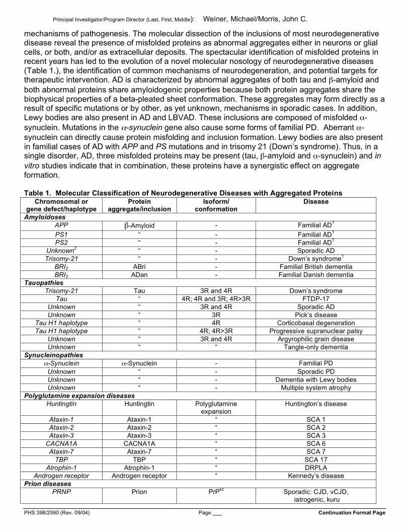
mechanisms of pathogenesis. The molecular dissection of the inclusions of most neurodegenerative disease reveal the presence of misfolded proteins as abnormal aggregates either in neurons or glial cells, or both, and/or as extracellular deposits. The spectacular identification of misfolded proteins in recent years has led to the evolution of a novel molecular nosology of neurodegenerative diseases (Table 1.), the identification of common mechanisms of neurodegeneration, and potential targets for therapeutic intervention. AD is characterized by abnormal aggregates of both tau and β-amyloid and both abnormal proteins share amyloidogenic properties because both protein aggregates share the biophysical properties of a beta-pleated sheet conformation. These aggregates may form directly as a result of specific mutations or by other, as yet unknown, mechanisms in sporadic cases. In addition, Lewy bodies are also present in AD and LBVAD. These inclusions are composed of misfolded α-synuclein. Mutations in the α-synuclein gene also cause some forms of familial PD. Aberrant α-synuclein can directly cause protein misfolding and inclusion formation. Lewy bodies are also present in familial cases of AD with APP and PS mutations and in trisomy 21 (Down’s syndrome). Thus, in a single disorder, AD, three misfolded proteins may be present (tau, β-amyloid and α-synuclein) and in vitro studies indicate that in combination, these proteins have a synergistic effect on aggregate formation.
Table 1. Molecular Classification of Neurodegenerative Diseases with Aggregated Proteins
Chromosomal or gene defect/haplotype
Protein aggregate/inclusion
Isoform/ conformation
Disease
Amyloidoses APP β-Amyloid - Familial AD1 PS1 “ - Familial AD1 PS2 “ - Familial AD1
Unknown2 “ - Sporadic AD
Trisomy-21 “ - Down’s syndrome1 BRI2 ABri - Familial British dementia BRI2 ADan - Familial Danish dementia
Tauopathies Trisomy-21 Tau 3R and 4R Down’s syndrome
Tau “ 4R; 4R and 3R; 4R>3R FTDP-17 Unknown “ 3R and 4R Sporadic AD Unknown “ 3R Pick’s disease
Tau H1 haplotype “ 4R Corticobasal degeneration Tau H1 haplotype “ 4R; 4R>3R Progressive supranuclear palsy
Unknown “ 3R and 4R Argyrophilic grain disease Unknown “ “ Tangle-only dementia
Synucleinopathies α-Synuclein α-Synuclein - Familial PD
Unknown “ - Sporadic PD Unknown “ - Dementia with Lewy bodies Unknown “ - Multiple system atrophy
Polyglutamine expansion diseases Huntingtin Huntingtin Polyglutamine
expansion Huntington’s disease
Ataxin-1 Ataxin-1 “ SCA 1 Ataxin-2 Ataxin-2 “ SCA 2 Ataxin-3 Ataxin-3 “ SCA 3
CACNA1A CACNA1A “ SCA 6 Ataxin-7 Ataxin-7 “ SCA 7
TBP TBP “ SCA 17 Atrophin-1 Atrophin-1 “ DRPLA
Androgen receptor Androgen receptor “ Kennedy’s disease Prion diseases
PRNP Prion PrPsc Sporadic: CJD, vCJD, iatrogenic, kuru

Principal Investigator/Program Director (Last, First, Middle): Weiner, Michael/Morris, John C.
PHS 398/2590 (Rev. 09/04) Page Continuation Format Page
PRNP Prion PrPsc Familial: CJD, FFI, GSS Other diseasse Superoxide dismutase 1 Superoxide dismutase - Familial amyotrophic lateral sclerosis
Unknown Neuronal intermediate filament proteins
- NIFID
Protease Inhibitor 12 Neuroserpin - FENIB Legend: 1Both familial and sporadic forms of AD and adult Down’s syndrome cases show both extracellular aggregates of β-amyloid and intraneuronal abnormal aggregates of tau protein; 2the ε4 allele of the apolipoprotein E gene is a risk factor for AD; 3R, the predominant number of tau isoforms with three microtubule-binding domains; ABri, amyloid Bri; ADan, amyloid Danish; APP, amyloid precursor protein; BRI2, an integral transmembrane glycoprotein gene; CACNA1A, α(1A) subunit of voltage-gated calcium channel type P/Q gene; CJD, Creutzfeldt-Jakob disease; vCJD, variant Creutzfeldt-Jakob disease; SOD1; copper/zinc-superoxide dismutase 1; DRPLA, dentatorubropallidoluysian atrophy; FENIB, familial encephalopathy with neuroserpin inclusion bodies; FFI, fatal familial insomnia; FTDP-17, frontotemporal dementia with parkinsonism linked to chromosome 17; FTL, ferritin light polypeptide gene; GSS, Gerstmann-Sträussler-Scheinker disease; NIFID, neuronal intermediate filament inclusion disease; PD, Parkinson’s disease; PRNP, prion protein gene; PrPsc, protease resistant prion protein; PS1, presenilin 1 gene; SCA, spinocerebellar ataxia; TBP, TATA-binding protein gene. C. PROGRESS REPORT/PRELIMINARY STUDIES 1. A synopsis of ADNI’s administrative structure and clinical design is shown below. The ADNI Data Coordinating Center is contained within the Clinical Core at the University of California, San Diego (See Letter of Support from Dr. Leon Thal). Goals of the ADNI Longitudinal Multisite Observational Study • Develop “standards” for imaging • Improve methods for clinical trials • Determine the optimum methods for acquiring and processing images • “Validate” imaging and biomarker data by correlating with neuropsych and behavioral data • Provide a data base and biological samples for PHARMA
ADNI Administrative Structure • Administrative Core: M Weiner • Clinical Core: L Thal, R Petersen, M Albert, P Tariot, D Salmon – Based at ADCS at UCSD • Neuroimaging Core – MRI: C Jack, N Schuff, A Dale, N Fox, C DeCarli, M Bernstein, J Felmlee – PET: W Jagust, N Foster, E Reiman, R Koeppe • Informatics: A Toga UCLA/LONI • Biomarker Core: J.Q. Trojanowski, L Shaw • Statistics: L Beckett • 50+ performance sites
ADNI Study Design
• MCI (n = 400): 0, 6, 12, 18, 24, 30, 36 months • Mild AD (n = 200): 0, 6, 12, 18, 24 months • Controls (n = 200): 0, 6, 12, 24, 36 months • Clinical, MRI (1.5 T) at: – MCI - All except 30 months – AD - All except 18 months – Nl - Baseline, 6 months, then yearly • FDG PET at same timepoints in a 50% subset • 3.0 T MRI at same timepoints in a 25% subset • Blood and urine at baseline then yearly for biomarkers • Immortalized cell lines at baseline
Principal Investigator/Program Director (Last, First, Middle): Weiner, Michael/Morris, John C.
PHS 398/2590 (Rev. 09/04) Page Continuation Format Page
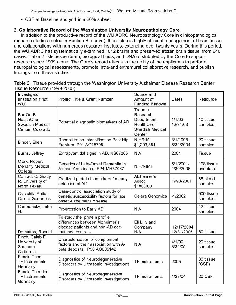
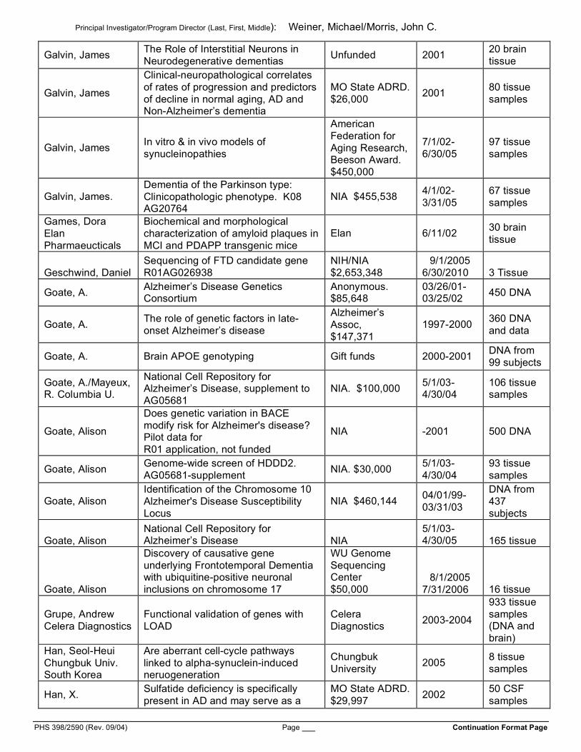
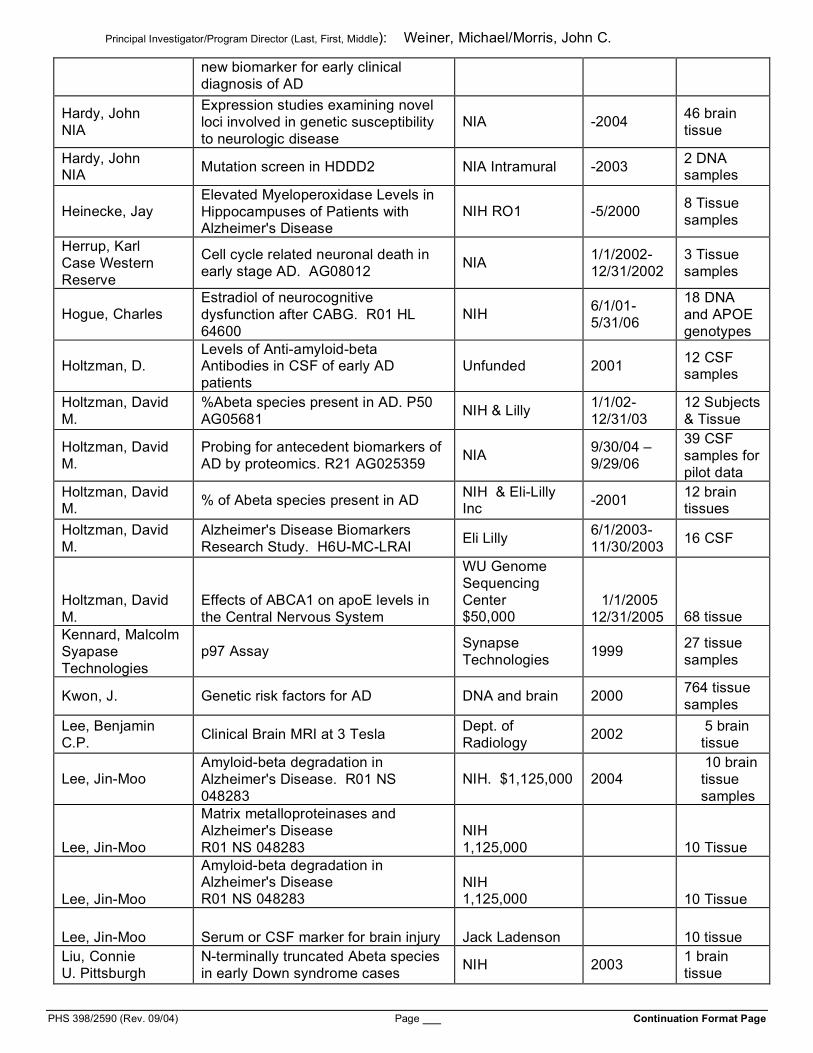
• CSF at Baseline and yr 1 in a 20% subset 2. Collaborative Record of the Washington University Neuropathology Core In addition to the productive record of the WU ADRC Neuropathology Core in clinicopathological research studies (noted in Section B, above), there also is highly efficient management of brain tissue and collaborations with numerous research institutes, extending over twenty years. During this period, the WU ADRC has systematically examined 1042 brains and preserved frozen brain tissue from 640 cases. Table 2 lists tissue (brain, biological fluids, and DNA) distributed by the Core to support research since 1999 alone. The Core’s record attests to the ability of the applicants to perform neuropathological assessments, promote intra-and extramural collaborative research, and publish findings from these studies. Table 2. Tissue provided through the Washington University Alzheimer Disease Research Center Tissue Resource (1999-2005).
Investigator (institution if not WU)
Project Title & Grant Number Source and Amount of Funding if known
Dates Resource
Bar-Or, B. HealthOne Swedish Medical Center, Colorado
Potential diagnostic biomarkers of AD
Trauma Research Department, HealthOne Swedish Medical Center
1/1/03-12/31/03
10 tissue samples
Binder, Ellen Rehabilitation Intensification Post Hip Fracture. P01 AG15795
NIH/NIA $1,203,854
8/1/1998-5/31/2004
20 tissue samples
Burns, Jeffrey Extrapyamidal signs in AD. NS07205 NIA 2004 Tissue
Clark, Robert Meharry Medical College
Genetics of Late-Onset Dementia in African-Americans. R24-MH57067 NIH/NIMH 5/1/2001-
4/30/2006 198 tissue and data
Conrad, C, Gracy R. University of North Texas,
Oxidized protein biomarkers for early detection of AD
Alzheimer’s Assoc $180,000
1998-2001 85 blood samples
Cravchik, Anibal Celera Genomics
Case-control association study of genetic susceptibility factors for late onset Alzheimer's disease
Celera Genomics -1/2002 900 tissue samples
Csernansky, John G. Progression to Early AD NIA 2004 42 tissue
samples
Demattos, Ronald
To study the protein profile differences between Alzheimer’s disease patients and non-AD age-matched controls.
Eli Lilly and Company N/A
12/17/2004 12/31/2005 60 tissue
Finch, Caleb E. University of Southern California
Characterization of complement factors and their association with A-beta deposits. P50 AG005142
NIA 4/1/00-3/31/05-
29 tissue samples
Funck, Theo TF Instruments Germany
Diagnostics of Neurodegenerative Disorders by Ultrasonic Investigations TF Instruments 2005 30 tissue
(CSF)
Funck, Theodor TF Instruments Germany
Diagnostics of Neurodegenerative Disorders by Ultrasonic Investigations TF Instruments 4/28/04 20 CSF
Principal Investigator/Program Director (Last, First, Middle): Weiner, Michael/Morris, John C.
PHS 398/2590 (Rev. 09/04) Page Continuation Format Page
Galvin, James The Role of Interstitial Neurons in Neurodegenerative dementias Unfunded 2001 20 brain
tissue
Galvin, James
Clinical-neuropathological correlates of rates of progression and predictors of decline in normal aging, AD and Non-Alzheimer’s dementia
MO State ADRD. $26,000 2001 80 tissue
samples
Galvin, James In vitro & in vivo models of synucleinopathies
American Federation for Aging Research, Beeson Award. $450,000
7/1/02-6/30/05
97 tissue samples
Galvin, James. Dementia of the Parkinson type: Clinicopathologic phenotype. K08 AG20764
NIA $455,538 4/1/02-3/31/05
67 tissue samples
Games, Dora Elan Pharmaeucticals
Biochemical and morphological characterization of amyloid plaques in MCI and PDAPP transgenic mice
Elan 6/11/02 30 brain tissue
Geschwind, Daniel Sequencing of FTD candidate gene R01AG026938
NIH/NIA $2,653,348
9/1/2005 6/30/2010 3 Tissue
Goate, A. Alzheimer’s Disease Genetics Consortium
Anonymous. $85,648
03/26/01-03/25/02 450 DNA
Goate, A. The role of genetic factors in late-onset Alzheimer’s disease
Alzheimer’s Assoc, $147,371
1997-2000 360 DNA and data
Goate, A. Brain APOE genotyping Gift funds 2000-2001 DNA from 99 subjects
Goate, A./Mayeux, R. Columbia U.
National Cell Repository for Alzheimer’s Disease, supplement to AG05681
NIA. $100,000 5/1/03-4/30/04
106 tissue samples
Goate, Alison
Does genetic variation in BACE modify risk for Alzheimer's disease? Pilot data for R01 application, not funded
NIA -2001 500 DNA
Goate, Alison Genome-wide screen of HDDD2. AG05681-supplement NIA. $30,000 5/1/03-
4/30/04 93 tissue samples
Goate, Alison Identification of the Chromosome 10 Alzheimer's Disease Susceptibility Locus
NIA $460,144 04/01/99-03/31/03
DNA from 437 subjects
Goate, Alison National Cell Repository for Alzheimer’s Disease NIA
5/1/03-4/30/05 165 tissue
Goate, Alison
Discovery of causative gene underlying Frontotemporal Dementia with ubiquitine-positive neuronal inclusions on chromosome 17
WU Genome Sequencing Center $50,000
8/1/2005 7/31/2006 16 tissue
Grupe, Andrew Celera Diagnostics
Functional validation of genes with LOAD
Celera Diagnostics 2003-2004
933 tissue samples (DNA and brain)
Han, Seol-Heui Chungbuk Univ. South Korea
Are aberrant cell-cycle pathways linked to alpha-synuclein-induced neruogeneration
Chungbuk University 2005 8 tissue
samples
Han, X. Sulfatide deficiency is specifically present in AD and may serve as a
MO State ADRD. $29,997 2002 50 CSF
samples
Principal Investigator/Program Director (Last, First, Middle): Weiner, Michael/Morris, John C.
PHS 398/2590 (Rev. 09/04) Page Continuation Format Page
new biomarker for early clinical diagnosis of AD
Hardy, John NIA
Expression studies examining novel loci involved in genetic susceptibility to neurologic disease
NIA -2004 46 brain tissue
Hardy, John NIA Mutation screen in HDDD2 NIA Intramural -2003 2 DNA
samples
Heinecke, Jay Elevated Myeloperoxidase Levels in Hippocampuses of Patients with Alzheimer's Disease
NIH RO1 -5/2000 8 Tissue samples
Herrup, Karl Case Western Reserve
Cell cycle related neuronal death in early stage AD. AG08012 NIA 1/1/2002-
12/31/2002 3 Tissue samples
Hogue, Charles Estradiol of neurocognitive dysfunction after CABG. R01 HL 64600
NIH 6/1/01-5/31/06
18 DNA and APOE genotypes
Holtzman, D. Levels of Anti-amyloid-beta Antibodies in CSF of early AD patients
Unfunded 2001 12 CSF samples
Holtzman, David M.
%Abeta species present in AD. P50 AG05681 NIH & Lilly 1/1/02-
12/31/03 12 Subjects & Tissue
Holtzman, David M.
Probing for antecedent biomarkers of AD by proteomics. R21 AG025359 NIA 9/30/04 –
9/29/06
39 CSF samples for pilot data
Holtzman, David M. % of Abeta species present in AD NIH & Eli-Lilly
Inc -2001 12 brain tissues
Holtzman, David M.
Alzheimer's Disease Biomarkers Research Study. H6U-MC-LRAI Eli Lilly 6/1/2003-
11/30/2003 16 CSF
Holtzman, David M.
Effects of ABCA1 on apoE levels in the Central Nervous System
WU Genome Sequencing Center $50,000
1/1/2005 12/31/2005 68 tissue
Kennard, Malcolm Syapase Technologies
p97 Assay Synapse Technologies 1999 27 tissue
samples
Kwon, J. Genetic risk factors for AD DNA and brain 2000 764 tissue samples
Lee, Benjamin C.P. Clinical Brain MRI at 3 Tesla Dept. of
Radiology 2002 5 brain tissue
Lee, Jin-Moo Amyloid-beta degradation in Alzheimer's Disease. R01 NS 048283
NIH. $1,125,000 2004 10 brain tissue samples
Lee, Jin-Moo
Matrix metalloproteinases and Alzheimer's Disease R01 NS 048283
NIH 1,125,000
10 Tissue
Lee, Jin-Moo
Amyloid-beta degradation in Alzheimer's Disease R01 NS 048283
NIH 1,125,000
10 Tissue
Lee, Jin-Moo Serum or CSF marker for brain injury Jack Ladenson 10 tissue Liu, Connie U. Pittsburgh
N-terminally truncated Abeta species in early Down syndrome cases NIH 2003 1 brain
tissue

Principal Investigator/Program Director (Last, First, Middle): Weiner, Michael/Morris, John C.
PHS 398/2590 (Rev. 09/04) Page Continuation Format Page
Mancuso, David Calcium independent phsopholipases A2 in AD
Internal funding Washington Univ.
2005 20 tissue samples
Morris, John C. Neuropathology of Nondemented Aging. U01 AG16976
NACC collaborative
7/1/01-6/30/2003
20 brain samples
Mucke, Lennart
Molecular markers for Alzheimer disease cognitive impairment P50 AG023501
NIH 6,143,429
5/15/2004 3/31/2009 4 Tissue
Nowotny, P./Goate, A.
Association of Nicastrin with early onset AD
Psychiatry, Washington Univ.
4/1/02-3/30/04
1000 tissue samples
Pastor, P. Analysis of the 17q21 region in familial fronto-temporal dementia and in sporadic tauopathies. #40240
AFAR,PSP Europe, Society for PSP (USA). $80,000
7/1/2003-6/30/2004
51 tissue samples
Perry, George Case Western Reserve
Oxidative damage in mild cases of Alzheimer disease NIA 7 tissue
samples
Pulliam, Joseph U. Kentucky
Validation of HFE mutation as a risk factor for Alzheimer's disease, UK ADRC pilot
NIA. $20,000 5/1/02-4/30/03
55 tissue samples
Roe, Catherine Gamma-secretase activity as a modulator of cancer and AD risk-A genetic test
NCI-Siteman Cancer Ctr. .$35,000
2004 323 tissue samples
Roe, Catherine Gamma-secretase activity as a modulator of cancer and AD risk-a genetic test of this hypothesis
Siteman Cancer Center Pilot grant $26,400
7/1/04-5/30/06 713 tissue
Sharma, Vijay Imaging beta amyloid in the brain Internal funding 2005 8 tissue samples
Sharma, Vijay Imaging b-Amyloid Plaques in the Brain
Unfunded
2 tissue
Sheline, Yvette. Age-related hippocampal volume loss in major depressive disorder. R01 MH60697
NIH 7/1/2000-6/30/2005
30 DNA and APOE genotypes
Sheline, Yvette. Age-related hippocampal volume loss in major depressive disorder. R01 MH60697
NIH 7/1/2000-6/30/2005
30 DNA and APOE genotypes
Shulman, Howard SurroMed/NeuroDx
Discovery of biomarkers for AD SurroMed/NeuroDX 2005 43 tissue
samples
Shulman, Howard Discovery of Biomarkers for Alzheimer's Disease
SurroMed/ NeuroDx 2005
40 tissue (CSF)
Song, Sheng-Kwei Neuronal injury in AD brain detected using diffusion MRI. P50AG05681 NIA, $26,750 5/1/02-
4/30/03- 20 brain tissue
Song, Sheng-Kwei Neuronal injury in AD brain detected using diffusion MRI. P50AG05681 NIA, $26,750 5/1/02-
4/30/03- 20 brain tissue
Srivastava, RAK Regulation of cholesterol trafficking in the brain
MO State ADRD, $30,000 2000 6 brain tissue
samples Stephan, D/ Reiman, E. Translational Genomics/ASU
Neurogenomics of Alzheimer's Disease and Aging. R01 AG023193 NIA $28,695 10/1/03-
9/30/08 5 Tissue samples

Principal Investigator/Program Director (Last, First, Middle): Weiner, Michael/Morris, John C.
PHS 398/2590 (Rev. 09/04) Page Continuation Format Page
Sun, Grace U Missouri
Proteins and gene expression in AD DHHS 1 P01 AG18357 NIH. 5 million 5/1/2001-
4/30/2006 10 brain tissue
Sun, Grace U. of Missouri
Immunohistochemistry identification of cPLA2 and COXII in control and AD brain
Alzheimer’s Assoc. 2000 5 tissue
samples
Surgochov, Andrei
Sera and CSF synoretin in AD, alternative splicing of nicotinicacetylcholine receptor subunit pre-mRNA in dopaminergic pathways. R01R01 NS43762
NIH 2002 Tissue samples
Sweet, R./Goate, A. U. Pittsburgh
Psychosis of Alzheimer’s Disease U01 AG16976
NIA, NACC $7,580
7/1/02-6/30/04
30 data/tissue
Tu, Pang-hsien Channel abnormality in AD Unfunded 2004 Tissue
Tu, Pang-hsien Alteration of calcineurin pathway indiffuse lewy body disease
MO State ADRD pilot 2004 Tissue
Wilkins, Consuelo Vitamin D deficiency in normal aging and AD. P01 AG03991 NIA, $53,000 1/1/2003-
12/31/2003 50 tissue samples
Willkins, Consuelo Vitamin D deficiency in normal aging and AD. BIRCWH Scholar’s Award, K12 HD01459
NIH, BIRCWH 7/1/03-6/30/05
30 tissue samples
Wolozin, Ben Boston University
Effects of statins on human grain pathology
Unfunded (NIH grant application)
2005 Tissue
Wolozin, Benjamin Effects of Statins on Human Brain Pathology
Unfunded 2005 9 tissue
Wu, Jane The Role of Alternative Splicing in Neurodegeneration. R01 AG017518 NIA 9/30/99-
7/31/04 50 Tissue samples
Zhukareva, Victoria U. Pittsburgh
Taupathies: Genotype and Phenotype. P01AG17586 and R01 AG14586
NIA, $233,603
3/15/01-2/28/05
2 HDDD2 subjects, brain tissue
3. NACC Supplementary Grant: Seven Centers Collaborative Study of the Neuropathology of Non-demented Aging. A supplementary grant (PI JC Morris) from NACC funded a multi-center (Duke University, Mayo Clinic, University of Kentucky, University of Rochester, Washington University, Oregon Health Sciences University, and University of California, San Diego) study of the neuropathologic correlates of cognitive impairment in non-demented aging. The WU ADRC served as the co-ordinating center and its Neuropathology Core as the Central laboratory. Tissue from one hundred and eight brains (97 nondemented individuals and 11 with MCI at last assessment before death) was sent from the participating centers to the Core for systematic and uniform staining on the cases. The following stains were used: hematoxylin and eosin, modified Bielschowsky, and Gallyas silver impregnations. Immunohistochemistry was performed using the following antibodies: Aβ and tau. Computerized stereological methods were used to assess the morphology, distribution, and density of lesions in the selected brain areas of all cases. These data show a high frequency of neuropathological AD in nondemented individuals as ascertained by the major neuropathological criteria (Figure 3).89 This NACC project demonstrates the ability of the Core to successfully obtain brain tissue from other sites and to generate data and diagnoses as a Central Core, and thus serves as the model for the ADNI- NPC.

Principal Investigator/Program Director (Last, First, Middle): Weiner, Michael/Morris, John C.
PHS 398/2590 (Rev. 09/04) Page Continuation Format Page
3. Multi-center on-going collaborations with the Neuropathology Core. Dr Cairns also has had a long track record of collaborative research and since becoming Leader of the Neuropathology Core of the Washington University ADRC (10/04) has helped to initiate a number of new collaborations. Dr Cairns was the Medical Research Council of the United Kingdom Brain Bank Co-ordinator and helped to initiate a network of 19 brain banks in Europe, BrainNet Europe, a "Network of Excellence" funded by the European Commission in the 6th Framework Program "Life Science" (LSHM-CT-2004-503039). Brain Net Europe may be accessed at (http://www.brainnet-europe.org/). Dr Cairns and Dr Trojanowski (University of Pennsylvania) are undertaking a review of cases of frontotemporal dementia with ubiquitin inclusions in collaboration with 12 NACC Neuropathology Cores with the aim of identifying atypical dementias. In collaboration with Drs Sandra Weintraub (PI) and Eileen Bigio (Northwestern University, Chicago, Il.), Charles White (University of Texas Southwestern, TX), and Dr Julie Schneider (Rush University Medical Center, Chicago, Il), a Midwest Consortium for Frontotemporal Lobar Degeneration NACC pilot project application (“Clinical Phenotypes of FTD are Determined by Neuropathology and Biochemical Abnormalities”; U01 AG16976) was awarded in 2005 with Drs Cairns and Morris representing the WU site. D. RESEARCH DESIGN AND METHODS The ADNI-Neuropathology Core will play a pivotal role in the mission of ADNI by implementing the four specific Aims described below. SPECIFIC AIM 1: Provide and implement training materials and protocols in obtaining voluntary consent for brain autopsy in ADNI participants. The ADNI-NPC will utilize materials already developed by successful programs for conveying information about brain autopsy and the forms for voluntary autopsy consent. Examples of “autopsy information packets” from the WU ADRC and the University of Pennsylvania ADC are provided in the Appendix. A standard packet includes an Autopsy Fact Sheet, brochures (including one developed specifically for African Americans), religious views regarding autopsy, information on procedures to follow at time of death, and autopsy consent forms. These standard materials will be modified slightly to reflect their use in the ADNI study. They will be presented at the ADNI Steering Committee Meetings (held 3 times each year) by Dr. Morris, who will instruct ADNI clinicians in their use, and then distribute them to all ADNI sites for dissemination to ADNI participants (see below). These materials also will be considered for web-based access for ADNI participants. The protocol used at Washington University for successfully obtaining autopsy consent has been published 90 (see Appendix) and has been implemented successfully elsewhere (see Letter of Support from Dr. Robert Green). It will serve as the model for introducing the consideration of
Figure 3. The CERAD and NIA-Reagan criteria agree very well along their ordinal scales (96%, kappa = 0.95).
60 6053
61
19 19
4739
19 18
2 3
0%
10%
20%
30%
40%
50%
60%
70%
80%
90%
100%
CERAD NIA-Reagan Khachaturian Wash U
No ADNo AD No ADNo AD
AD AD
Def AD High Prob
Intermed Prob
Low Prob
Prob. AD
Poss. AD
Figure 1. Percent classification by four sets of neuropathologic criteria in 97 CDR 0 cases3

Principal Investigator/Program Director (Last, First, Middle): Weiner, Michael/Morris, John C.
PHS 398/2590 (Rev. 09/04) Page Continuation Format Page
autopsy and obtaining voluntary autopsy consent at each ADNI site. In brief, an ADNI physician will lead a discussion about autopsy with all participants (demented and non-demented) at their initial assessment (study partners and families are welcomed in the discussion and required for AD participants). There are 3 objectives of the discussion: 1) to convey information about the value of brain autopsy in confirming the clinical diagnosis and advancing knowledge regarding MCI and AD; 2) to initiate consideration of the individual’s wishes as regards autopsy; and 3) to answer questions, misconceptions, or concerns about autopsy. The involvement of the physician in these discussions emphasizes the importance of autopsy. The discussions are repeated on an annual basis if the individual has not decided about autopsy, but are terminated once a decision is reached. There is no pressure for the individual to decide; they are encouraged to involve family members, clergy, physicians, or other resource persons in their decision-making. Participants are assured that a decision not to have autopsy in no way jeopardizes their research participation or any other patient rights. When voluntary consent is granted, more detailed information is provided about procedures to follow at time of death, including telephone numbers to call and other guidelines (e.g., for transportation of the deceased to the place of autopsy). Participants are strongly encouraged to share this information with next-of-kin, legally authorized representative (LAR), and private physicians. In many states, final legal authorization by the LAR or next-of-kin must be obtained at time of death. Wallet-sized cards (see Appendix) with a summary of instructions and contact information are provided (as many cards as needed).
The proposed procedures for obtaining provisional consent for autopsy are effective. The autopsy rate for all ADCs is 43% (personal communication, Walter Kukull). Since the WU ADRC was established in 1985, its overall autopsy WU ADRC rate is 56%; in the recently completed 5-year funding cycle (5/1/00-4/30/05) the rate was 62%. The procedures are summarized in the flow chart (Figure 4 on next page). With the adoption of these procedures across the ADNI sites, we expect at least a 50% autopsy rate for ADNI participants (it may well be higher as ADNI participants are committed to research). Each ADNI site will be encouraged to establish an autopsy coordinator (typically a research nurse or coordinator) who processes the autopsy consent, provides information as needed, and monitors the need to update any information (e.g., change in residence) at the ADNI participant’s longitudinal assessments. The coordinator also will develop procedures for that site to facilitate autopsies outside of usual hours (e.g., evenings and weekends). The actual procedures are expected to vary in accordance with local needs and resources (one model used by many ADCs is to provide 24-hour telephone access). At time of death, the autopsy coordinator (or a suitable representative) facilitates arrangements to ensure the completion of the autopsy. The coordinator notifies the ADNI-NPC, which in turn verifies that the site neuropathologist has the dissection protocol (see Aim 2, below) and necessary materials to send the requisite tissue to the ADNI-NPC. The ADNI-NPC, in addition to instructing site personnel at each ADNI Steering Committee Meeting in these procedures, will be available at any time to answer questions. Contact information (24-hour pager) for an ADNI-NPC personnel will be provided to all ADNI sites. Transportation costs from point of death to the autopsy suite, costs of the autopsy procedure, and shipment of materials are covered by the ADNI-NPC so that the decedent’s family and the individual ADNI site do not incur extra expense. Our experience at Washington University is that outright refusal of autopsy by participants is rare (2%). The most common outcome (about 66%) is that the participant and family consider the consent without decision. The percentage (32%) who provide formal consent during life is about half of our autopsy rate (62%) suggesting that many who are “undecided” during life ultimately consent to autopsy at time of death. Although the number of ADNI autopsies expected in the current grant period is relatively small, the implementation of the voluntary consent procedures now almost certainly will translate into increased numbers of autopsies in subsequent funding cycles.

Principal Investigator/Program Director (Last, First, Middle): Weiner, Michael/Morris, John C.
PHS 398/2590 (Rev. 09/04) Page Continuation Format Page
Figure 4. Autopsy Consent Protocol Anticipated ADNI death and autopsy rates
All-cause, age-specific death rates per 1000 population by 5-year age group (both sexes; 2001, US Vital Statistics) are as follows: 55-59y, 7.7; 60-64y, 12.1; 65-69y, 18.7; 70-74y, 28.8; 75-79y, 44.9; 80-84y, 71.5; 85+y, 151.1. Although higher death rates are found in AD, ADNI participants (age 55-90y) are anticipated to be unusually healthy as they are committed to fulfilling the demands of the longitudinal imaging and other studies. Moreover, those with AD will be in the mild stage of the illness and thus have a lower death rate than the general AD population. Nonetheless, we anticipate sufficient deaths will occur in ADNI participants in this funding cycle to fulfill the Specific Aims of the ADNI-NPC. We base this conclusion on a review of death rates in a sample of healthy (except for the presence in some of AD) elderly volunteer participants in the longitudinal studies of the WU ADRC. Since the inception of the cohort, for all individuals age 55y-90y at entry, death occurred within 5

Principal Investigator/Program Director (Last, First, Middle): Weiner, Michael/Morris, John C.
PHS 398/2590 (Rev. 09/04) Page Continuation Format Page
years of enrollment in 69 of 741 (9%) non-demented (CDR 0) individuals, in 118 of 798 (15%) individuals with MCI (CDR 0.5), and in 195 of 654 (30%) individuals with mild AD (CDR 1). These data roughly translate to an annual death rate of 2% for CDR 0, 3% for CDR 0.5, and 6% for CDR 1 individuals. Comparable rates are reported from the Minimum Data Set (MDS) of NACC which currently has data from 73,037 subjects enrolled from all National Institute on Aging-funded ADCs: MDS annual death rates for individuals age 55-90y are 1.5% for non-demented controls, 1.7% for individuals with MCI, and 8% for individuals with AD (Walter Kukull, personal communication). In addition, annual death rates in the Religious Orders Study (participants comparable to the ADNI sample) are 2.8% for nondemented elderly and 6.6% for individuals with MCI.10 Other studies of MCI also demonstrate increased mortality for this condition.7-9
In this application, we use conservative estimates because the demanding ADNI protocol may select for healthy participants. We thus assume annual death rates of 1% for non-demented ADNI participants, 2% for MCI individuals, and 5% for AD individuals. Over 5 years, these rates would yield 10 non-demented, 40 MCI, and 50 AD deaths. Assuming a 50% autopsy rate across ADNI, over the funding cycle for ADNI-NPC (12/1/06 – 8/31/09), and adjusting for a slightly greater death rate in the last 2 years of the grant as the sample ages and progresses in dementia severity, we anticipate that there will be autopsies in at least 4 non-demented controls, 12 MCI individuals, and 20 AD individuals (Table 3).
Almost all existing ADCs (n=30) also are ADNI sites, and thus the ADNI sample will largely correspond to the individuals entered into NACC’s MDS. It is likely that the death rates, and thus the number of autopsies, will be greater than these conservative estimates as the death rates may be more comparable to those reported by the MDS and autopsies may increase as sites improve their consent rates beyond 50%. Although there will be a shortened time frame of this supplement in relation to the parent grant, the number of autopsies are expected to average between 10-15 per year (lower in initial years, higher in later years). Table 3. Predicted minimum number of deaths and autopsies during ADNI-NPC study period. Clinical staging Normal
MCI
DAT Total
No. ADNI participants 200 400 200 800 Annual death rate (%) 1% 2% 5% - Predicted deaths in 5 years 10 40 50 100 Predicted autopsies (12/01/06 – 8/31/09)
4 12 20 36
Aim 2: To Provide central, uniform neuropathologic diagnoses to validate clinical assessment and facilitate clinicopathological correlations, including determining the relationship between the molecular neuropathology, structural, and functional changes, including Pittsburgh Compound-B (PIB), in early Alzheimer’s disease. Rationale for Aim 2: To facilitate systematic clinicopathologic studies and to initiate innovative biochemical investigations into the pathogenesis of fibrillary lesions, standard brain sampling, processing, and staining protocols are necessary. Although most ADCs undertake their own neuropathological assessments, there is variation between centers in the methods of fixation, sampling of tissue blocks, section thickness, stains, and antibodies used for immunohistochemistry (IHC). Although each center has its own protocol, there is a need for standardized sampling, staining and IHC of tissues from ADNI participants. In those centers that do not undertake neuropathology the administration of brain tissue donation, harvesting, and neuropathology, will have to be coordinated centrally. A central ADNI-NPC will provide standard tissue sampling, staining, IHC, and consensus neuropathological diagnosis.
Principal Investigator/Program Director (Last, First, Middle): Weiner, Michael/Morris, John C.
PHS 398/2590 (Rev. 09/04) Page Continuation Format Page
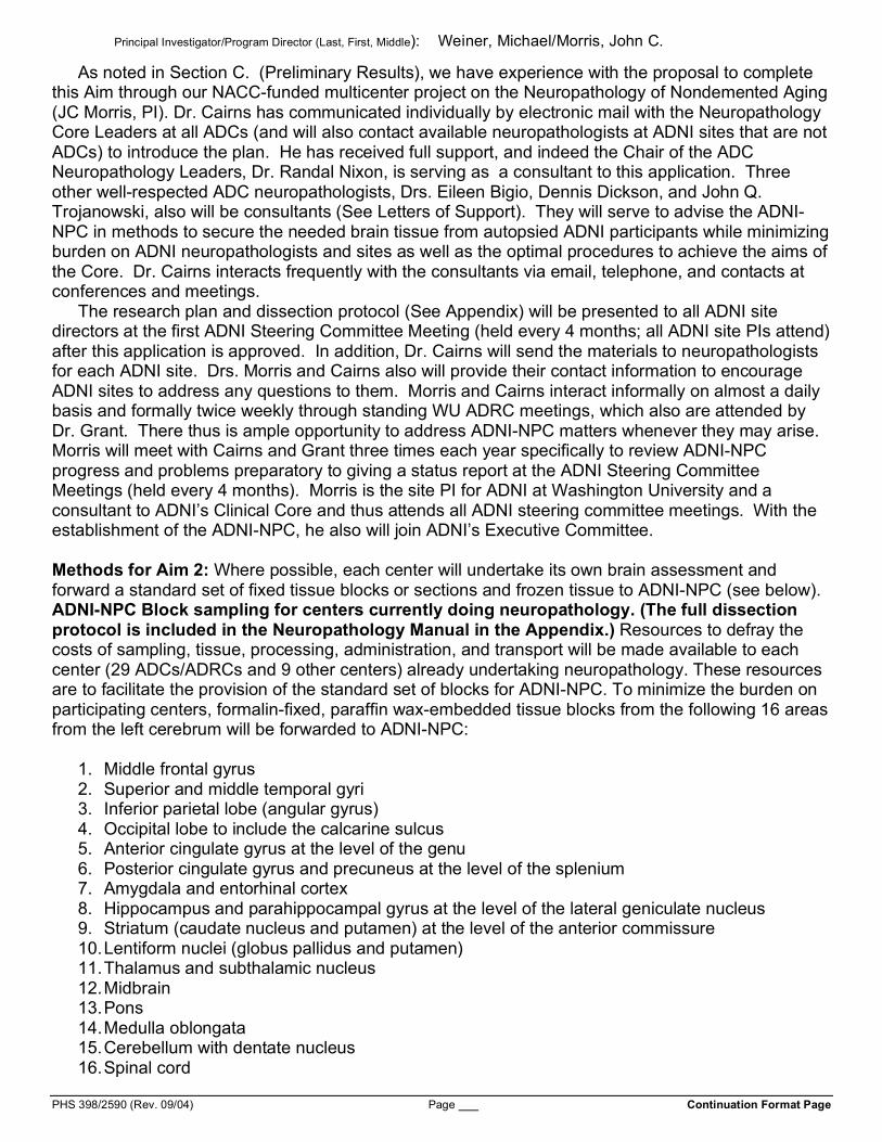
As noted in Section C. (Preliminary Results), we have experience with the proposal to complete this Aim through our NACC-funded multicenter project on the Neuropathology of Nondemented Aging (JC Morris, PI). Dr. Cairns has communicated individually by electronic mail with the Neuropathology Core Leaders at all ADCs (and will also contact available neuropathologists at ADNI sites that are not ADCs) to introduce the plan. He has received full support, and indeed the Chair of the ADC Neuropathology Leaders, Dr. Randal Nixon, is serving as a consultant to this application. Three other well-respected ADC neuropathologists, Drs. Eileen Bigio, Dennis Dickson, and John Q. Trojanowski, also will be consultants (See Letters of Support). They will serve to advise the ADNI-NPC in methods to secure the needed brain tissue from autopsied ADNI participants while minimizing burden on ADNI neuropathologists and sites as well as the optimal procedures to achieve the aims of the Core. Dr. Cairns interacts frequently with the consultants via email, telephone, and contacts at conferences and meetings. The research plan and dissection protocol (See Appendix) will be presented to all ADNI site directors at the first ADNI Steering Committee Meeting (held every 4 months; all ADNI site PIs attend) after this application is approved. In addition, Dr. Cairns will send the materials to neuropathologists for each ADNI site. Drs. Morris and Cairns also will provide their contact information to encourage ADNI sites to address any questions to them. Morris and Cairns interact informally on almost a daily basis and formally twice weekly through standing WU ADRC meetings, which also are attended by Dr. Grant. There thus is ample opportunity to address ADNI-NPC matters whenever they may arise. Morris will meet with Cairns and Grant three times each year specifically to review ADNI-NPC progress and problems preparatory to giving a status report at the ADNI Steering Committee Meetings (held every 4 months). Morris is the site PI for ADNI at Washington University and a consultant to ADNI’s Clinical Core and thus attends all ADNI steering committee meetings. With the establishment of the ADNI-NPC, he also will join ADNI’s Executive Committee. Methods for Aim 2: Where possible, each center will undertake its own brain assessment and forward a standard set of fixed tissue blocks or sections and frozen tissue to ADNI-NPC (see below). ADNI-NPC Block sampling for centers currently doing neuropathology. (The full dissection protocol is included in the Neuropathology Manual in the Appendix.) Resources to defray the costs of sampling, tissue, processing, administration, and transport will be made available to each center (29 ADCs/ADRCs and 9 other centers) already undertaking neuropathology. These resources are to facilitate the provision of the standard set of blocks for ADNI-NPC. To minimize the burden on participating centers, formalin-fixed, paraffin wax-embedded tissue blocks from the following 16 areas from the left cerebrum will be forwarded to ADNI-NPC:
1. Middle frontal gyrus 2. Superior and middle temporal gyri 3. Inferior parietal lobe (angular gyrus) 4. Occipital lobe to include the calcarine sulcus 5. Anterior cingulate gyrus at the level of the genu 6. Posterior cingulate gyrus and precuneus at the level of the splenium 7. Amygdala and entorhinal cortex 8. Hippocampus and parahippocampal gyrus at the level of the lateral geniculate nucleus 9. Striatum (caudate nucleus and putamen) at the level of the anterior commissure 10. Lentiform nuclei (globus pallidus and putamen) 11. Thalamus and subthalamic nucleus 12. Midbrain 13. Pons 14. Medulla oblongata 15. Cerebellum with dentate nucleus 16. Spinal cord

Principal Investigator/Program Director (Last, First, Middle): Weiner, Michael/Morris, John C.
PHS 398/2590 (Rev. 09/04) Page Continuation Format Page
In the unusual situation where it is impractical to forward a tissue block (e.g., if the block is used for stereology), 10 paraffin wax sections from each will be provided to ADNI-NPC for systematic neuropathology and diagnosis. Frozen tissue. To provide tissue for biochemical studies and to advance the aims of the Biomarkers Study, snap frozen tissue will be dissected, frozen, and sent to ADNI-NPC. The following coronal slices (0.5 to 1cm thick) will be taken:
1. Frontal lobe to include striatum; 2. Frontal and temporal lobe at the level of the mamillary body; 3. Temporal and parietal lobes at the level of the lateral geniculate nucleus; 4. Occipital lobe to include the calcarine sulcus.
ADNI-NPC Tissue harvesting from centers that do not perform neuropathology For those centers (n=21) that do not currently undertake neuropathology, and do not plan to do so during the period of this project, ADNI-NPC will assist in arrangements in consultation with the local center to harvest the brain and to ship tissue to ADNI-NPC where a systematic neuropathological evaluation will be performed. Tissue Processing. Routine sampling for histology includes taking blocks which are fixed in buffered neutral formalin. All remaining tissue is snap frozen and stored in zip-lock bags at -75°C in locked freezers. The Neuropathology Core currently samples: olfactory bulbs, middle frontal gyrus, orbitofrontal cortex, olfactory cortex, anterior cingulate gyrus, caudate nucleus and putamen at the level of the nucleus accumbens, anterior commissure and nucleus basalis of Meynert, lentiform nucleus (globus pallidus, putamen), superior and middle temporal gyri, inferior temporal gyrus, amygdala, hippocampus pes to include entorhinal cortex, hippocampus at level of lateral geniculate nucleus, hypothalamus, thalamus and subthalamic nucleus, precentral gyrus, postcentral gyrus, posterior cingulate gyrus and precuneus, Broca’s area, posterior cingulate gyrus, inferior frontal gyrus, Wernicke’s area, midbrain, pons, medulla oblongata, cerebellum with dentate nucleus, vermis, and spinal cord from all dementia cases and cervical, thoracic, lumbar and sacral spinal cord from all FTD and movement disorder cases, when possible. Any additional pathology or abnormality is also sampled. In addition, thick blocks (~7mm) are taken from the frontal lobe, temporal lobe, hippocampus, striatum, and pons, and fixed in formalin for tissue microarray experiments. Histology. Buffered formalin and frozen tissue will be sent to the ADNI-NPC and treated in the same manner as that described in the ADRC Neuropathology Manual (Appendix). On all cases, the following stains will be performed on the blocks indicated above, or as requested by the neuropathologist: hematoxylin and eosin and modified Bielschowsky silver impregnation. Routine immunohistochemistry will be performed using the following antibodies: ubiquitin (1510), tau (PHF1, AT8), β-amyloid (4G8, 10D5), and α-synuclein (LB509). In cases with ubiquitin-positive inclusions, the following additional IHC will be performed: α-internexin and valosin-containing protein (VCP). Histology Review. Dr. Cairns reviews the histological slides in a systematic manner. The data are entered into the NACC Neuropathology Data Form and transmitted by Dr. Grant to the ADNI Coordinating Center. The NACC Neuropathology Protocol is included in the Appendix. The final neuropathologic diagnosis and neuropathologic report will be forwarded to ADNI for entry into the central database and to the center that made available the tissue. Neuropathologic assessment and diagnostic criteria. The operational criteria for the classification of AD and other pathologies defined by the NACC (see Appendix) will be applied to all ADNI-NPC cases (and are currently applied to all WU ADRC cases). The neuropathological diagnosis will be determined by Dr. Cairns using consensus neuropathologic criteria for AD91-93, and for nonAD disorders as described in Section B (Background and Significance). The NACC Neuropathology Form includes an entry for the diagnosis of AD by each of the 3 sets of widely used criteria: Khachaturian, CERAD, and NIA-Reagan. ADNI-NPC cases thus will be diagnosed in accordance with each of these criteria, as no consensus currently exists in favor of one set in relation to the
Principal Investigator/Program Director (Last, First, Middle): Weiner, Michael/Morris, John C.
PHS 398/2590 (Rev. 09/04) Page Continuation Format Page
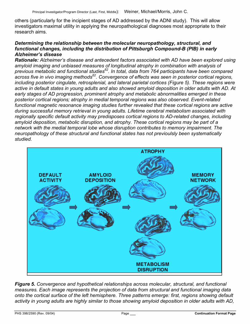
others (particularly for the incipient stages of AD addressed by the ADNI study). This will allow investigators maximal utility in applying the neuropathological diagnoses most appropriate to their research aims. Determining the relationship between the molecular neuropathology, structural, and functional changes, including the distribution of Pittsburgh Compound-B (PIB) in early Alzheimer’s disease Rationale: Alzheimer’s disease and antecedent factors associated with AD have been explored using amyloid imaging and unbiased measures of longitudinal atrophy in combination with analysis of previous metabolic and functional studies62. In total, data from 764 participants have been compared across five in vivo imaging methods62. Convergence of effects was seen in posterior cortical regions, including posterior cingulate, retrosplenial, and lateral parietal cortices (Figure 5). These regions were active in default states in young adults and also showed amyloid deposition in older adults with AD. At early stages of AD progression, prominent atrophy and metabolic abnormalities emerged in these posterior cortical regions; atrophy in medial temporal regions was also observed. Event-related functional magnetic resonance imaging studies further revealed that these cortical regions are active during successful memory retrieval in young adults. Lifetime cerebral metabolism associated with regionally specific default activity may predisposes cortical regions to AD-related changes, including amyloid deposition, metabolic disruption, and atrophy. These cortical regions may be part of a network with the medial temporal lobe whose disruption contributes to memory impairment. The neuropathology of these structural and functional states has not previoulsly been systematically studied.
Figure 5. Convergence and hypothetical relationships across molecular, structural, and functional measures. Each image represents the projection of data from structural and functional imaging data onto the cortical surface of the left hemisphere. Three patterns emerge: first, regions showing default activity in young adults are highly similar to those showing amyloid deposition in older adults with AD,
Principal Investigator/Program Director (Last, First, Middle): Weiner, Michael/Morris, John C.
PHS 398/2590 (Rev. 09/04) Page Continuation Format Page
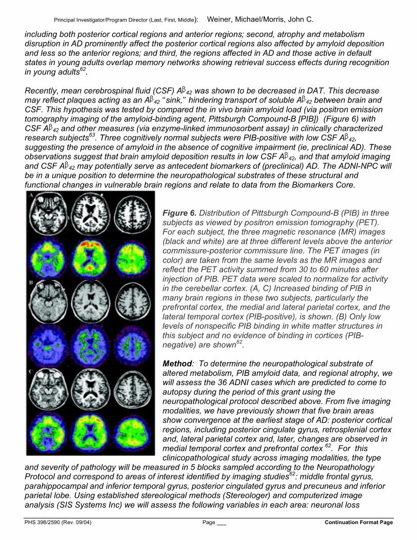
including both posterior cortical regions and anterior regions; second, atrophy and metabolism disruption in AD prominently affect the posterior cortical regions also affected by amyloid deposition and less so the anterior regions; and third, the regions affected in AD and those active in default states in young adults overlap memory networks showing retrieval success effects during recognition in young adults62. Recently, mean cerebrospinal fluid (CSF) A 42 was shown to be decreased in DAT. This decrease may reflect plaques acting as an A 42 sink, hindering transport of soluble A 42 between brain and CSF. This hypothesis was tested by compared the in vivo brain amyloid load (via positron emission tomography imaging of the amyloid-binding agent, Pittsburgh Compound-B [PIB]) (Figure 6) with CSF A 42 and other measures (via enzyme-linked immunosorbent assay) in clinically characterized research subjects63. Three cognitively normal subjects were PIB-positive with low CSF A 42, suggesting the presence of amyloid in the absence of cognitive impairment (ie, preclinical AD). These observations suggest that brain amyloid deposition results in low CSF A 42, and that amyloid imaging and CSF A 42 may potentially serve as antecedent biomarkers of (preclinical) AD. The ADNI-NPC will be in a unique position to determine the neuropathological substrates of these structural and functional changes in vulnerable brain regions and relate to data from the Biomarkers Core.
Figure 6. Distribution of Pittsburgh Compound-B (PIB) in three subjects as viewed by positron emission tomography (PET). For each subject, the three magnetic resonance (MR) images (black and white) are at three different levels above the anterior commissure-posterior commissure line. The PET images (in color) are taken from the same levels as the MR images and reflect the PET activity summed from 30 to 60 minutes after injection of PIB. PET data were scaled to normalize for activity in the cerebellar cortex. (A, C) Increased binding of PIB in many brain regions in these two subjects, particularly the prefrontal cortex, the medial and lateral parietal cortex, and the lateral temporal cortex (PIB-positive), is shown. (B) Only low levels of nonspecific PIB binding in white matter structures in this subject and no evidence of binding in cortices (PIB-negative) are shown62. Method: To determine the neuropathological substrate of altered metabolism, PIB amyloid data, and regional atrophy, we will assess the 36 ADNI cases which are predicted to come to autopsy during the period of this grant using the neuropathological protocol described above. From five imaging modalities, we have previously shown that five brain areas show convergence at the earliest stage of AD: posterior cortical regions, including posterior cingulate gyrus, retrosplenial cortex and, lateral parietal cortex and, later, changes are observed in medial temporal cortex and prefrontal cortex 62. For this clinicopathological study across imaging modalities, the type
and severity of pathology will be measured in 5 blocks sampled according to the Neuropathology Protocol and correspond to areas of interest identified by imaging studies62: middle frontal gyrus, parahippocampal and inferior temporal gyrus, posterior cingulated gyrus and precuneus and inferior parietal lobe. Using established stereological methods (Stereologer) and computerized image analysis (SIS Systems Inc) we will assess the following variables in each area: neuronal loss

Principal Investigator/Program Director (Last, First, Middle): Weiner, Michael/Morris, John C.
PHS 398/2590 (Rev. 09/04) Page Continuation Format Page
(stereology), synapse loss (quantitative image analysis using synatophysin immunohistochemistry), β-amyloid (10D5 and 8G8 immunohistochemistry), phosphorylated tau (PHF1 and AT8), and α-synuclein (LB509) deposition by immunohistochemistry and quantitative image analysis. These data will be correlated with imaging data across the different modalities with the assistance of the Biostatistics Core. Aim 3: Maintain a state-of-the-art brain tissue resource to advance collaborative research and to facilitate ADNI’s Biomarker Study headed by John Trojanowski, Center for Neurodegenerative Disease Research, University of Pennsylvania. (See Letter of Support). Rationale for Aim 3: To facilitate systematic clinicopathologic studies and biochemical investigations, both fixed and unfixed brain tissue from ADNI subjects who come to autopsy will be essential. It is unlikely that any one of the participating sites alone will accrue sufficient numbers of cases to undertake robust clinicopathologic studies on ADNI subjects. Therefore, to maximize the value of longitudinally assessed ADNI cases, a central Neuropathology Core is necessary to coordinate and centralize tissues that will be used in collaborative studies by ADNI participants. Only the ADNI-NPC, by a concerted effort and participation of multiple sites, will generate sufficient numbers of cases to undertake clinicopathologic and biochemical studies. A bank of well-characterized fresh and fixed ADNI brain samples for diagnosis and research is essential for the success of this project. In particular, banked frozen tissue is essential for determining the earliest biochemical changes in the brain in AD. Special focus will be placed on the detection of oligomeric species of Aβ, possible harbingers of fibrillary Aβ by immunohistochemistry in frozen brain 94. These data will be correlated with morphological and imaging data to reveal the spectrum of changes associated with the pathogenesis of AD.
Frozen brain tissue is also essential to determine changes in solubility of proteins which characterize AD and other neurodegenerative diseases. In particular, protein extraction, separation by electrophoresis, and Western blotting will be used by the state-of-the-art ADNI-NPC laboratory to determine tau isoforms and thus distinguish biochemically between the different 3R and 4R tauopathies. Tissue Collection and Characterization. Formalin-fixed, paraffin wax-embedded material, and frozen tissue will be available for clinicopathological and biochemical studies. Areas rich in pathology will be identified by IHC using anti-ubiquitin, anti-tau-, and synuclein, and anti-Aβ antibodies. To confirm the presence of lesions in the frozen tissue block used for protein extraction, a section will be taken and subject to IHC. Only where lesions have been demonstrated in the frozen block will biochemistry be performed. Sequential Biochemical Fractionation: Using modification of well-established methods, grey and underlying white matter will be dissected from the superior frontal gyrus and weighed 95. Tissue will be homogenized in 2ml/g high salt (HS) buffer (50mmol/L Tris pH 7.5) containing 2 mmol/L EDTA, 0.75 mol/L NaCl, and a cocktail of protease inhibitors, and centrifuged at 40,000 x g for 30 minutes at 4ºC. Supernatants will be saved as the HS fraction and pellets will be washed by re-extraction in HS buffer. Resulting pellets will subjected to two sequential extractions for each buffer of increasing protein extraction strength. Pellets will be homogenized in 2ml/g Triton-X (TX) buffer containing HS, 1% Triton X-100, and protease inhibitors and centrifuged as for the HS fraction. Supernatants will be saved as the TX fraction. Pellets will be homogenized in RIPA buffer containing 50 mmol/L Tris, 150 mmol/L NaCL, 0.5% sodium deoxycholate, 0.1% sodium SDS and 1% NP40 adjusted to pH 8.0, and centrifuged and pelleted as above. The supernatants will be saved as the RIPA fraction. Pellets will be resuspended in 2% SDS in 50mmol/L Tris pH 7.6 with protease inhibitors, and centrifuged as above but at 15ºC. Supernatants will be preserved as the SDS fraction. Pellets will then be homogenized in 2ml/g BUST buffer (2.5 mmol/L TCEP, 2% SDS, 8mol/L urea, 50 mmol/L Tris pH 7.6 with protease inhibitors), centrifuged at 40,000 x g for 30 minutes at 15oC. The supernatants will be saved as the BUST fraction. Pellets will be resuspended by sonication in 70% formic acid and centrifuged at 40,000 x g for 1 hour at 4ºC. Myelin precipitate will be removed using HS buffer with

Principal Investigator/Program Director (Last, First, Middle): Weiner, Michael/Morris, John C.
PHS 398/2590 (Rev. 09/04) Page Continuation Format Page
20% sucrose prior to the RIPA extraction. Protein concentration will be determined using the Coomassie protein assay (Pierce, Rockford, Il) and bovine serum albumin as a standard. SDS sample buffer (10 mmol/L Tris, pH 6.8, 1mmol/L EDTA, 40 mmol/L dithiothreitol, 1% SDS, 10% sucrose) will be added to samples of HS, TX, RIPA, and FA, and sample buffer without SDS (10 mmol/L Tris, pH 6.8, 1mmol/L EDTA, 40 mmol/L dithiothreitol, 10% sucrose) will be added to SDS-soluble samples, followed by heating at 100ºC for 5 minutes, with the exception of the BUST samples which will not require the addition of sample buffer or heating. Western Blot Analysis: Proteins will be separated by 12% SDS-polyacrylamide gel electrophoresis and subsequently transferred electrophoretically to nitrocellulose membrane (Schleicher & Schuell, Keene, NH) in buffer containing 25 mmol/L Tris, 190 mmol/L glycine, and 10% methanol. Membranes will be blocked with a 5% solution of powdered skim milk dissolved in Tris-buffered saline (50 mmol/L Tris, pH 7.6, 150 mmol/L NaCl), incubated with primary antibody conjugated to horseradish peroxidase, developed with Chemiluminescence Reagents (Perkin Elmer Life Sciences,Inc., Boston, MA) and exposed onto X-Omat Blue XB-1 films (Kodak, Rochester, NY). Western blotting using monoclonal antibodies to protein epitopes of interest (Aβ, tau, synuclein, ubiquitin) will be used to determine fractions rich in ubiquitinated- and insouluble-protein complexes. SPECIFIC AIM 4: Interact with ADNI’s Data Coordinating Center to ensure entry of Core data into ADNI’s database, promote data sharing and collaborative research, and integrate ADNI-NPC with all ADNI components. The Core’s data manager, Dr. Grant, has served as the data manager for the WU ADRC for 20 years and is fully knowledgeable about the forms, data entry procedures, and data access protocols to be used for the ADNI-NPC. Dr. Grant has provided data from the ADRC to the original Minimum Data Set established for the ADCs by the NIA since the initial collection in 1998 and subsequently to the NACC. We are very experienced with the NACC Neuropathology Form to be used in the ADNI-NPC. Drs. Grant and Morris developed a web-based data entry application for ADRC neuropathologists to enter data for the NACC forms. Dr. Grant also is very familiar with the ADNI Data Coordinating Center. Since 1995, our ADRC has conducted 7 clinical trial protocols involving 89 research participants for the Alzheimer’s Disease Cooperative Study at the University of California, San Diego (“home” of the ADNI Data Coordinating Center). Data from the ADNI-NPC will be entered on a timely basis into the ADRC’s computerized database to facilitate rapid identification of errors and the application of correction procedures while the original information sources still are readily available. The data entry screens are customized for the NACC Neuropathology Form and are SAS/FSP applications; data entry occurs by ADNI-NPC personnel on PCs connected to the computer servers in the ADRC’s computing resource. The SAS data entry screens include error checking codes and integrity constraints. Edits are checked with SAS audit trails and SAS PROC COMPARE. Types of data include autopsy status, ADNI site contributing the tissue with an ADNI-NPC identification number, postmortem interval, brain weight, cause(s) of death, and NACC protocol findings. Sites will send data to the ADNI-NPC either by fax on a secure line or by password protected electronic files. Data are maintained according to Health Insurance Portability and Accountability Act (HIPAA) guidelines. The servers are backed-up daily and copies of backups are kept at a secure off-site location. The data manager will maintain a file to track status of tissue and data transmission both from the ADNI sites and to the ADNI Data Coordinating Center. Data will be transmitted to the Coordinating Center at least quarterly (more often as the case material allows).
The WU ADRC Neuropathology Core already maintains its own database to meet its daily requirements for archiving and as a tool for research investigators. In addition, a centralized database is maintained by the ADRC Data Management and Biostatistics Core using the relational database SAS software. There is restricted access to these data via a secure virtual private network (VPN). The Core adheres to HIPAA compliant electronic record privacy guidelines. The Department of

Principal Investigator/Program Director (Last, First, Middle): Weiner, Michael/Morris, John C.
PHS 398/2590 (Rev. 09/04) Page Continuation Format Page
Pathology maintains a staff of four full-time computer systems analysts who maintain department-wide virus scans and undertake routine maintenance.
A relational database will be established for Core data at the Coordinating Center. To maintain inter-center compatibility, the database will be constructed with unique identifiers that are used by ADNI and will be compatible with the individual ADNI site databases and accessible by ADNI investigators. Procedures for Accessing Autopsy Data and Tissue from the ADNI Neuropathology Core: Data. Data generated by the ADNI-NPC will be transmitted securely to the ADNI Coordinating Center for storage, management and distribution according to the ADNI procedures. These de-identified data will be available with the relevant clinical, biological and imaging data on the Coordinating Center’s public access web site. Tissue. The process by which investigators request access to ADNI NPC tissue is based on the established and successful procedures in place at the WU ADRC. Qualified investigators will initiate requests for ADNI autopsy material by providing basic information (including a 3 page research summary and NIH biosketch) about their research project to the ADNI NPC Tissue Committee (see below). The instructions and forms are web-based (www.alzheimer.wustl.edu/adrc2/ResourcesDB/Intro.asp) for easy access. Prospective investigators will be encouraged to consult with Drs. Morris and Cairns. Written reviews of the request from at least 2 members of the ADNI-NPC Tissue Committee, chaired by Dr. Cairns, or other experts recruited for a particular protocol will be provided for discussion and approval by email vote of the Tissue Committee conducted monthly or as requests dictate. The Tissue Committee will forward its recommendations to the ADNI Executive Committee (see below) for final approval. The criteria used by reviewers will be: scientific merit, feasibility, appropriateness of principal investigator qualifications, burden on ADNI samples, and appropriateness to ADNI goals/themes. ADNI Neuropathology Core Tissue Committee ADNI Executive Committee:
Morris, John C., Washington Univ. Sch of Med. Michael Weiner, UC San Francisco/SFVAMC Cairns, Nigel, Washington Univ. Sch. of Med. Arthur Toga, UC Los Angeles Bigio, Eileen, Northwestern Univ. Laurel Beckett, UC Davis Dickson, Dennis, Mayo Clinic William Jagust, UC Berkeley Nixon, Randal, Oregon Health Sciences Univ. Leon Thal, UC San Diego Trojanowski, John Q., Univ. of Pennsylvania John Trojanowski, Univ. of Pennsylvania
Ron Thomas, UC San Diego Clifford Jack, Mayo Clinic Peter Snyder, Industry (Pfizer) Ron Petersen, Mayo Clinic Subject selection for approved tissue requests will be achieved through discussion with the requesting investigator, the ADNI NPC staff, and the ADNI Coordinating Center. All samples leaving Washington University are de-identified by use of a code (generated for this shipment only) and with the execution of a Limited Data Use Agreement (See Appendix). Upon shipment, a final list of samples shipped is shared with the ADNI Coordinating Center for purposes of reporting and tracking the transfer. In return for the use of ADNI autopsy tissue, we ask the following of investigators: acknowledgment of the ADNI grant number in publications and presentations, productivity reports on publications or funding that were derived from the project, and no third-party sharing without notification. We do not charge for sharing materials/data unless the request requires effort beyond what can be subsumed under normal ADNI NPC budgeted effort (as recommended by NIA). If the request justifies a charge, the cost is kept to a minimum and based on actual expenses (effort and materials). E. HUMAN SUBJECTS 1. Risk to Subjects
Principal Investigator/Program Director (Last, First, Middle): Weiner, Michael/Morris, John C.
PHS 398/2590 (Rev. 09/04) Page Continuation Format Page
a. Human Subject Involvement. Individuals 55-90 years old who are either cognitively normal (n=200), have mild cognitive impairment (MCI; n=400) or who have mild Alzheimer’s disease (AD; n=200) will be enrolled by participating sites in the ADNI study. Subjects of both sexes will be recruited by ADNI sites without regard to race, ethnicity or religious background. Except for dementia in the AD group, subjects are generally healthy when enrolled in the ADNI. All subjects enrolled in ADNI are eligible to have brain autopsy through the ADNI-Neuropathology Core (ADNI-NPC). Over the period of this grant, we estimate that at least 36 individuals will come to autopsy (4 cognitively normal, 12 MCI, and 20 AD). b. Source of Materials. Where possible, each center will process their own brain tissue and forward a set of fixed tissue blocks and frozen tissue to the ADNI-NPC. For centers that do not currently undertake neuropathology, the ADNI-NPC will make arrangements in consultation with the local center to harvest the brain and ship tissue to ADNI-NPC where a systematic neuropathological evaluation will be performed. The respective ADNI Site will obtain standard information related to cause of death, and other relevant information. For sites that do not do their own neuropathology, the ADNI-NPC will submit a neuropathologic report to the site (identified by code only) for their records and for feedback to the next-of-kin or legal representative according the state statutes governing the site. All procedures will be undertaken for research purposes only and filed in a research file. There will be no billing of insurance companies or Medicare. c. Potential Risks. There is no direct risk to living persons incurred by the activities of the ADNI-NPC. In the highly unlikely event confidentiality is breached, knowledge of the participant’s neuropathologic diagnosis conceivably could affect family members. 2. Recruitment and Informed Consent. a. Recruitment and Informed Consent. All ADNI sites will give information to the participant and their family at the first time of assessment and annually thereafter about the ADNI autopsy program. Consent for autopsy will be obtained according to the state statutes governing each site. The harvesting of brain tissue will be done in accordance with the procedures governing the pathology department at each site which will include a legal consent for autopsy and includes permission to use the brain tissue for research purposes. b. Protection Against Risk. Subject confidentiality is strictly protected. Tissue and clinical data will be identified by code number, date of birth, and date of expiration. The master list of other personal identifiers will be held by the individual site and not available to the ADNI-NPC. Subject paper files and other research materials reside at the individual ADNI sites and will be protected in compliance with HIPAA regulations. 3. Potential Benefits of the Proposed Research to the Subjects and Others a. Potential Benefits to Subjects. Subjects do not benefit from this research. b. Potential Benefits to Society. Society will benefit from the knowledge generated by ADNI to develop surrogate imaging markers for progression of MCI and AD as validated by neuropathologic assessment. 4. Importance of the Knowledge to be Gained. It is important to have standardized neuropathological criteria. Until reliable biomarkers of disease progression are validated, the gold standard for the diagnosis of AD remains the neuropathologic assessment of CNS tissue at autopsy. In relation to these anticipated benefits and the importance of knowledge to be gained, the minimal risks involved in this research are reasonable. INCLUSION OF WOMEN AND MINORITIES Women and members of minority groups will be actively recruited during the ADNI study. The subjects assessed by the ADNI-NPC are expected to reflect of the entire ADNI sample. ADNI expects 12% enrollment of minorities (which reflects the aged minority population of the country) and
Principal Investigator/Program Director (Last, First, Middle): Weiner, Michael/Morris, John C.
PHS 398/2590 (Rev. 09/04) Page Continuation Format Page
the sample to be 58% female based on participating site enrollment history. ADNI is developing a separate plan for minority recruitment. Enrollment is monitored and tracked with additional support given to sites as needed to enhance minority recruitment. INCLUSION OF CHILDREN—Children will not be studied. F. VERTEBRATE ANIMALS - None G. LITERATURE CITED
Reference List
1. Mirra SS, Gearing M, McKeel DW, Jr., Crain BJ, Hughes JP, van Belle G, Heyman A. Interlaboratory comparison of neuropathology assessments in Alzheimer's disease: A study of the Consortium to Establish a Registry for Alzheimer's Disease (CERAD). J Neuropathol Exp Neurol 1994; 53:303-315.
2. Tierney MC, Fisher RH, Lewis AJ, Zorzitto ML, Snow WG, Reid DW, Nieuwstraten P. The NINCDS-ADRDA Work Group criteria for the clinical diagnosis of probable Alzheimer's disease: A clinicopathologic study of 57 cases. Neurology 1988; 38:359-364.
3. McKeel DW, Price JL, Miller JP, Grant EA, Xiong C, Berg L, Morris J.C. Neuropathologic criteria for diagnosing Alzheimer Disease in persons with pure dementia of Alzheimer type. J Neuropathol Exp Neurol 2004; 63:1028-1037.
4. Jack CR, Dickson DW, Parisi JE, Xu YC, Cha RH, O'Brien PC, Edland SD, Smith GE, Boeve BF, Tangalos EG, Kokmen E, Petersen RC. Antemortem MRI findings correlate with hippocampal neuropathology in typical aging and dementia. Neurology 2002; 58:750-757.
5. National Institute on Aging, Reagan Institute Working Group on Diagnostic Criteria for the Neuropathological Assessment of Alzheimer's Disease. Consensus recommendations for the postmortem diagnosis of Alzheimer's disease. Neurobiol Aging 1997; 18:S1-S2.
6. Wisniewski HM, Rabe A, Zigman W, Silverman W. Neuropathological diagnosis of Alzheimer's disease. J Neuropathol Exp Neurol 1989; 48:606-609.
7. Leep Hunderfund AN, Leibson CL, Slusser TC, Roberts R, Petersen RC. Survival in mild cognitive impairment. Neurology 2005; 64:A166.
8. Tuokko H, Frerichs R, Graham J, Rockwood K, Kristjansson B, Fisk J, Bergman H, Kozma A, McDowell I. Five-year follow-up of cognitive impairment with no dementia. Arch Neurol 2003; 60:577-582.
9. Frisoni GB, Fratiglioni L, Fastbom J, Viitanen M, Winblad B. Mortality in nondemented subjects with cognitive impairment: The influence of health-related factors. Am J Epidemiol 1999; 150:1031-1044.
10. Bennett DA, Wilson RS, Schneider JA, Evans DA, Beckett LA, Aggarwal NT, Barnes LL, Fox JH, Bach J. Natural history of mild cognitive impairment in older persons. Neurology 2002; 59:198-205.
11. Mirra SS, Heyman A, McKeel D, Sumi SM, Crain BJ, Brownlee LM, Vogel FS, Hughes JP, van Belle G, Berg L. The Consortium to Establish a Registry for Alzheimer's Disease (CERAD). Part II. Standardization of the neuropathologic assessment of Alzheimer's disease. Neurology 1991; 41:479-486.
12. McKhann GM, Albert MS, Grossman M, Miller B, Dickson D, Trojanowski JQ. Clinical and pathological diagnosis of frontotemporal dementia: report of the Work Group on Frontotemporal Dementia and Pick's Disease. Arch Neurol 2001; 58:1803-1809.
13. Trojanowski JQ, Dickson D. Update on the neuropathological diagnosis of frontotemporal dementias. J Neuropathol Exp Neurol 2001; 60:1123-1126.
14. Hyman BT, Trojanowski JQ. Consensus recommendations for the postmortem diagnosis of Alzheimer disease from the National Institute on Aging and the Reagan Institute Working Group on diagnostic criteria for the neuropathological assessment of Alzheimer disease. J Neuropathol Exp Neurol 1997; 56:1095-1097.
15. Kuusisto E, Parkkinen L, Alafuzoff I. Morphogenesis of Lewy bodies: dissimilar incorporation of alpha-synuclein, ubiquitin, and p62. J Neuropathol Exp Neurol 2003; 62:1241-1253.
16. McKeith IG, Galasko D, Kosaka K, Perry EK, Dickson DW, Hansen LA, Salmon DP, Lowe J, Mirra SS, Byrne EJ, Lennox G, Quinn NP, Edwardson JA, Ince PG, Bergeron C, Burns A, Miller BL, Lovestone S, Collerton D, Jansen EN, Ballard C, De Vos RA, Wilcock GK, Jellinger KA, Perry RH. Consensus
Principal Investigator/Program Director (Last, First, Middle): Weiner, Michael/Morris, John C.
PHS 398/2590 (Rev. 09/04) Page Continuation Format Page
guidelines for the clinical and pathologic diagnosis of dementia with Lewy bodies (DLB): report of the consortium on DLB international workshop. Neurology 1996; 47:1113-1124.
17. McKhann GM, Albert MS, Grossman M, Miller B, Dickson D, Trojanowski JQ. Clinical and pathological diagnosis of frontotemporal dementia: report of the Work Group on Frontotemporal Dementia and Pick's Disease. Arch Neurol 2001; 58:1803-1809.
18. Mirra SS, Heyman A, McKeel D, Sumi SM, Crain BJ, Brownlee LM, Vogel FS, Hughes JP, van Belle G, Berg L. The Consortium to Establish a Registry for Alzheimer's Disease (CERAD). Part II. Standardization of the neuropathologic assessment of Alzheimer's disease. Neurology 1991; 41:479-486.
19. Price JL, Morris JC. Tangles and plaques in nondemented aging and "preclinical" Alzheimer's disease. Ann Neurol 1999; 45:358-368.
20. Bancher C, Paulus W, Paukner K, Jellinger K. Neuropathologic diagnosis of Alzheimer disease: consensus between practicing neuropathologists? Alzheimer Dis Assoc Disord 1997; 11:207-219.
21. Mirra SS, Gearing M, McKeel DW, Jr., Crain BJ, Hughes JP, van Belle G, Heyman A. Interlaboratory comparison of neuropathology assessments in Alzheimer's disease: a study of the Consortium to Establish a Registry for Alzheimer's Disease (CERAD). J Neuropathol Exp Neurol 1994; 53:303-315.
22. Bancher C, Paulus W, Paukner K, Jellinger K. Neuropathologic diagnosis of Alzheimer disease: consensus between practicing neuropathologists? Alzheimer Dis Assoc Disord 1997; 11:207-219.
23. Geddes JW, Tekirian TL, Soultanian NS, Ashford JW, Davis DG, Markesbery WR. Comparison of neuropathologic criteria for the diagnosis of Alzheimer's disease. Neurobiol Aging 1997; 18:S99-105.
24. Mirra SS, Gearing M, McKeel DW, Jr., Crain BJ, Hughes JP, van Belle G, Heyman A. Interlaboratory comparison of neuropathology assessments in Alzheimer's disease: a study of the Consortium to Establish a Registry for Alzheimer's Disease (CERAD). J Neuropathol Exp Neurol 1994; 53:303-315.
25. Csernansky JG, Wang L, Joshi S, Miller JP, Gado M, Kido D, McKeel D, Morris JC, Miller MI. Early DAT is distinguished from aging by high-dimensional mapping of the hippocampus. Dementia of the Alzheimer type. Neurology 2000; 55:1636-1643.
26. Csernansky JG, Wang L, Swank J, Miller JP, Gado M, McKeel D, Miller MI, Morris JC. Preclinical detection of Alzheimer's disease: hippocampal shape and volume predict dementia onset in the elderly. Neuroimage 2005; 25:783-792.
27. Mosconi L, Tsui W-H, De Santi S, Li J, Rusinek H, Convit A, Li Y, Boppana M, de Leon MJ. Reduced hippocampal metabolism in MCI and AD; Automated FDG-PET image analysis. Neurology 2005; 64:1860-1867.
28. Csernansky JG, Hamstra J, Wang L, McKeel D, Price JL, Gado M, Morris JC. Correlations between antemortem hippocampal volume and postmortem neuropathology in AD subjects. Alzheimer Dis Assoc Disord 2004; 18:190-195.
29. Morris JC, Fulling K. Early Alzheimer's disease. Diagnostic considerations. Arch Neurol 1988; 45:345-349.
30. Price JL, Davis PB, Morris JC, White DL. The distribution of tangles, plaques and related immunohistochemical markers in healthy aging and Alzheimer's disease. Neurobiol Aging 1991; 12:295-312.
31. McKeel DW, Jr., Ball MJ, Price JL, Smith DS, Miller JP, Berg L, Morris JC. Interlaboratory histopathologic assessment of Alzheimer neuropathology: different methodologies yield comparable diagnostic results. Alzheimer Dis Assoc Disord 1993; 7:136-151.
32. Morris JC, McKeel DW, Jr., Fulling K, Torack RM, Berg L. Validation of clinical diagnostic criteria for Alzheimer's disease. Ann Neurol 1988; 24:17-22.
33. Morris JC, McKeel DW, Jr., Storandt M, Rubin EH, Price JL, Grant EA, Ball MJ, Berg L. Very mild Alzheimer's disease: informant-based clinical, psychometric, and pathologic distinction from normal aging. Neurology 1991; 41:469-478.
34. Morris JC, Storandt M, McKeel DW, Jr., Rubin EH, Price JL, Grant EA, Berg L. Cerebral amyloid deposition and diffuse plaques in "normal" aging: Evidence for presymptomatic and very mild Alzheimer's disease. Neurology 1996; 46:707-719.
35. Morris JC, Storandt M, Miller JP, McKeel DW, Price JL, Rubin EH, Berg L. Mild cognitive impairment represents early-stage Alzheimer disease. Arch Neurol 2001; 58:397-405.
36. McKeel DW, Jr., Price JL, Miller JP, Grant EA, Xiong C, Berg L, Morris JC. Neuropathologic criteria for diagnosing Alzheimer disease in persons with pure dementia of Alzheimer type. J Neuropathol Exp Neurol 2004; 63:1028-1037.
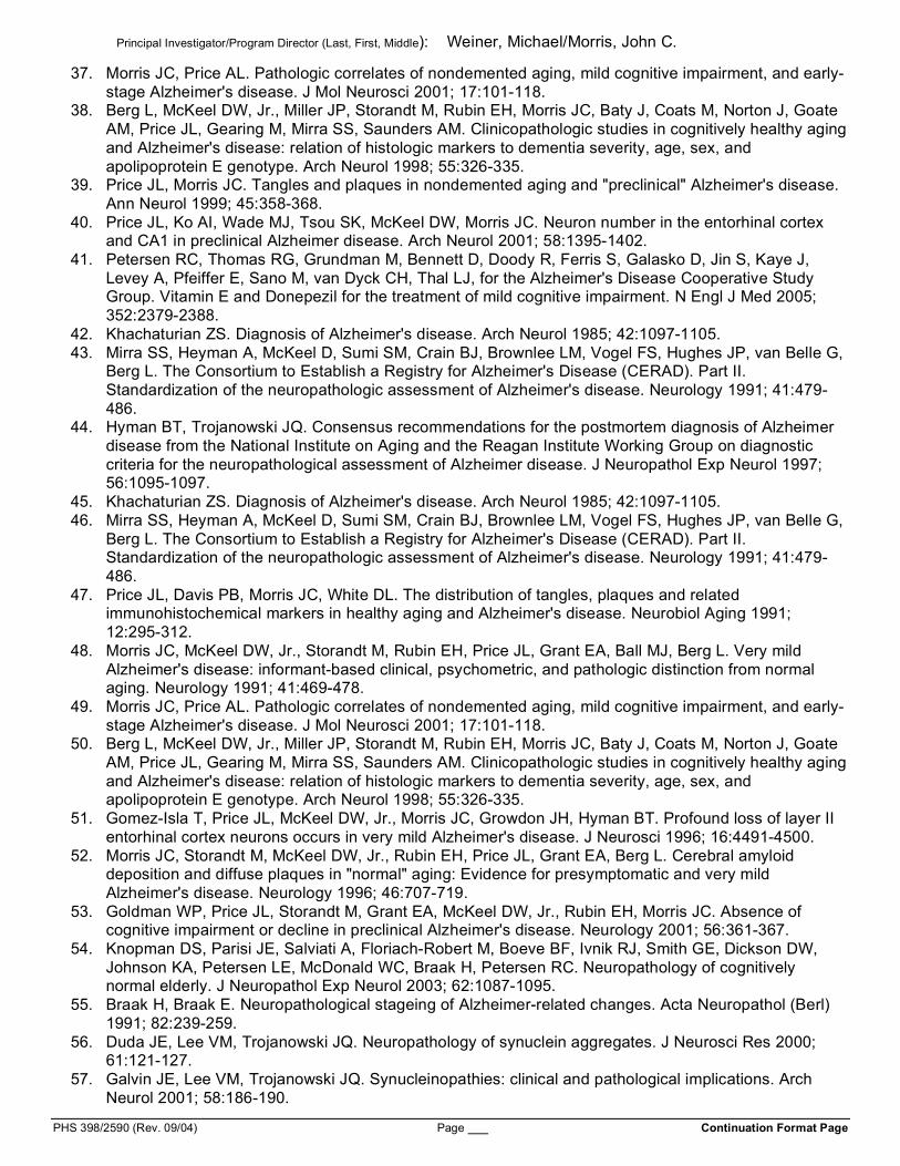
Principal Investigator/Program Director (Last, First, Middle): Weiner, Michael/Morris, John C.
PHS 398/2590 (Rev. 09/04) Page Continuation Format Page
37. Morris JC, Price AL. Pathologic correlates of nondemented aging, mild cognitive impairment, and early-stage Alzheimer's disease. J Mol Neurosci 2001; 17:101-118.
38. Berg L, McKeel DW, Jr., Miller JP, Storandt M, Rubin EH, Morris JC, Baty J, Coats M, Norton J, Goate AM, Price JL, Gearing M, Mirra SS, Saunders AM. Clinicopathologic studies in cognitively healthy aging and Alzheimer's disease: relation of histologic markers to dementia severity, age, sex, and apolipoprotein E genotype. Arch Neurol 1998; 55:326-335.
39. Price JL, Morris JC. Tangles and plaques in nondemented aging and "preclinical" Alzheimer's disease. Ann Neurol 1999; 45:358-368.
40. Price JL, Ko AI, Wade MJ, Tsou SK, McKeel DW, Morris JC. Neuron number in the entorhinal cortex and CA1 in preclinical Alzheimer disease. Arch Neurol 2001; 58:1395-1402.
41. Petersen RC, Thomas RG, Grundman M, Bennett D, Doody R, Ferris S, Galasko D, Jin S, Kaye J, Levey A, Pfeiffer E, Sano M, van Dyck CH, Thal LJ, for the Alzheimer's Disease Cooperative Study Group. Vitamin E and Donepezil for the treatment of mild cognitive impairment. N Engl J Med 2005; 352:2379-2388.
42. Khachaturian ZS. Diagnosis of Alzheimer's disease. Arch Neurol 1985; 42:1097-1105. 43. Mirra SS, Heyman A, McKeel D, Sumi SM, Crain BJ, Brownlee LM, Vogel FS, Hughes JP, van Belle G,
Berg L. The Consortium to Establish a Registry for Alzheimer's Disease (CERAD). Part II. Standardization of the neuropathologic assessment of Alzheimer's disease. Neurology 1991; 41:479-486.
44. Hyman BT, Trojanowski JQ. Consensus recommendations for the postmortem diagnosis of Alzheimer disease from the National Institute on Aging and the Reagan Institute Working Group on diagnostic criteria for the neuropathological assessment of Alzheimer disease. J Neuropathol Exp Neurol 1997; 56:1095-1097.
45. Khachaturian ZS. Diagnosis of Alzheimer's disease. Arch Neurol 1985; 42:1097-1105. 46. Mirra SS, Heyman A, McKeel D, Sumi SM, Crain BJ, Brownlee LM, Vogel FS, Hughes JP, van Belle G,
Berg L. The Consortium to Establish a Registry for Alzheimer's Disease (CERAD). Part II. Standardization of the neuropathologic assessment of Alzheimer's disease. Neurology 1991; 41:479-486.
47. Price JL, Davis PB, Morris JC, White DL. The distribution of tangles, plaques and related immunohistochemical markers in healthy aging and Alzheimer's disease. Neurobiol Aging 1991; 12:295-312.
48. Morris JC, McKeel DW, Jr., Storandt M, Rubin EH, Price JL, Grant EA, Ball MJ, Berg L. Very mild Alzheimer's disease: informant-based clinical, psychometric, and pathologic distinction from normal aging. Neurology 1991; 41:469-478.
49. Morris JC, Price AL. Pathologic correlates of nondemented aging, mild cognitive impairment, and early-stage Alzheimer's disease. J Mol Neurosci 2001; 17:101-118.
50. Berg L, McKeel DW, Jr., Miller JP, Storandt M, Rubin EH, Morris JC, Baty J, Coats M, Norton J, Goate AM, Price JL, Gearing M, Mirra SS, Saunders AM. Clinicopathologic studies in cognitively healthy aging and Alzheimer's disease: relation of histologic markers to dementia severity, age, sex, and apolipoprotein E genotype. Arch Neurol 1998; 55:326-335.
51. Gomez-Isla T, Price JL, McKeel DW, Jr., Morris JC, Growdon JH, Hyman BT. Profound loss of layer II entorhinal cortex neurons occurs in very mild Alzheimer's disease. J Neurosci 1996; 16:4491-4500.
52. Morris JC, Storandt M, McKeel DW, Jr., Rubin EH, Price JL, Grant EA, Berg L. Cerebral amyloid deposition and diffuse plaques in "normal" aging: Evidence for presymptomatic and very mild Alzheimer's disease. Neurology 1996; 46:707-719.
53. Goldman WP, Price JL, Storandt M, Grant EA, McKeel DW, Jr., Rubin EH, Morris JC. Absence of cognitive impairment or decline in preclinical Alzheimer's disease. Neurology 2001; 56:361-367.
54. Knopman DS, Parisi JE, Salviati A, Floriach-Robert M, Boeve BF, Ivnik RJ, Smith GE, Dickson DW, Johnson KA, Petersen LE, McDonald WC, Braak H, Petersen RC. Neuropathology of cognitively normal elderly. J Neuropathol Exp Neurol 2003; 62:1087-1095.
55. Braak H, Braak E. Neuropathological stageing of Alzheimer-related changes. Acta Neuropathol (Berl) 1991; 82:239-259.
56. Duda JE, Lee VM, Trojanowski JQ. Neuropathology of synuclein aggregates. J Neurosci Res 2000; 61:121-127.
57. Galvin JE, Lee VM, Trojanowski JQ. Synucleinopathies: clinical and pathological implications. Arch Neurol 2001; 58:186-190.

Principal Investigator/Program Director (Last, First, Middle): Weiner, Michael/Morris, John C.
PHS 398/2590 (Rev. 09/04) Page Continuation Format Page
58. Forman MS, Giasson BI, Duda JE, Miller B, Lee VMY, Trojanowski JQ. Convergence of tau and synuclein pathology in dementia patients. Faseb Journal 2003; 17:A658.
59. Goedert M. Alpha-synuclein and neurodegenerative diseases. Nat Rev Neurosci 2001; 2:492-501. 60. Spillantini MG, Schmidt ML, Lee VM, Trojanowski JQ, Jakes R, Goedert M. Alpha-synuclein in Lewy
bodies. Nature 1997; 388:839-840. 61. Spillantini MG, Crowther RA, Jakes R, Cairns NJ, Lantos PL, Goedert M. Filamentous alpha-synuclein
inclusions link multiple system atrophy with Parkinson's disease and dementia with Lewy bodies. Neurosci Lett 1998; 251:205-208.
62. McKeith IG, O'Brien JT, Ballard C. Diagnosing dementia with Lewy bodies. Lancet 1999; 354:1227-1228.
63. McKeith IG, Galasko D, Kosaka K, Perry EK, Dickson DW, Hansen LA, Salmon DP, Lowe J, Mirra SS, Byrne EJ, Lennox G, Quinn NP, Edwardson JA, Ince PG, Bergeron C, Burns A, Miller BL, Lovestone S, Collerton D, Jansen EN, Ballard C, De Vos RA, Wilcock GK, Jellinger KA, Perry RH. Consensus guidelines for the clinical and pathologic diagnosis of dementia with Lewy bodies (DLB): report of the consortium on DLB international workshop. Neurology 1996; 47:1113-1124.
64. Hansen L, Salmon D, Galasko D, Masliah E, Katzman R, DeTeresa R, Thal L, Pay MM, Hofstetter R, Klauber M, . The Lewy body variant of Alzheimer's disease: a clinical and pathologic entity. Neurology 1990; 40:1-8.
65. Gilman S, Low PA, Quinn N, Albanese A, Ben Shlomo Y, Fowler CJ, Kaufmann H, Klockgether T, Lang AE, Lantos PL, Litvan I, Mathias CJ, Oliver E, Robertson D, Schatz I, Wenning GK. Consensus statement on the diagnosis of multiple system atrophy. J Neurol Sci 1999; 163:94-98.
66. Hughes AJ, Daniel SE, Blankson S, Lees AJ. A clinicopathologic study of 100 cases of Parkinson's disease. Arch Neurol 1993; 50:140-148.
67. Schroder R, Watts GD, Mehta SG, Evert BO, Broich P, Fliessbach K, Pauls K, Hans VH, Kimonis V, Thal DR. Mutant valosin-containing protein causes a novel type of frontotemporal dementia. Ann Neurol 2005; 57:457-461.
68. Forman MS, Schmidt ML, Kasturi S, Perl DP, Lee VM, Trojanowski JQ. Tau and alpha-synuclein pathology in amygdala of Parkinsonism-dementia complex patients of Guam. Am J Pathol 2002; 160:1725-1731.
69. Forman MS, Giasson BI, Duda JE, Miller B, Lee VM, Trojanowski JQ. Convergence of tau and alpha-synuclein pathology in dementia patients. Journal of Neuropathology and Experimental Neurology 2002; 61:484.
70. Lee VM, Trojanowski JQ. Neurodegenerative tauopathies: human disease and transgenic mouse models. Neuron 1999; 24:507-510.
71. Hutton M, Lendon CL, Rizzu P, Baker M, Froelich S, Houlden H, Pickering-Brown S, Chakraverty S, Isaacs A, Grover A, Hackett J, Adamson J, Lincoln S, Dickson D, Davies P, Petersen RC, Stevens M, de Graaff E, Wauters E, van Baren J, Hillebrand M, Joosse M, Kwon JM, Nowotny P, Heutink P, . Association of missense and 5'-splice-site mutations in tau with the inherited dementia FTDP-17. Nature 1998; 393:702-705.
72. McKhann GM, Albert MS, Grossman M, Miller B, Dickson D, Trojanowski JQ. Clinical and pathological diagnosis of frontotemporal dementia: report of the Work Group on Frontotemporal Dementia and Pick's Disease. Arch Neurol 2001; 58:1803-1809.
73. Brun A, Englund B, Gustafson L, Passant U, Mann DMA, Neary D, Snowden JS. Clinical and Neuropathological Criteria for Frontotemporal Dementia. Journal of Neurology Neurosurgery and Psychiatry 1994; 57:416-418.
74. McKhann GM, Albert MS, Grossman M, Miller B, Dickson D, Trojanowski JQ. Clinical and pathological diagnosis of frontotemporal dementia: report of the Work Group on Frontotemporal Dementia and Pick's Disease. Arch Neurol 2001; 58:1803-1809.
75. Kertesz A, Kawarai T, Rogaeva E, George-Hyslop P, Poorkaj P, Bird TD, Munoz DG. Familial frontotemporal dementia with ubiquitin-positive, tau-negative inclusions. Neurology 2000; 54:818-827.
76. Bigio EH, Johnson NA, Rademaker AW, Fung BB, Mesulam MM, Siddique N, Dellefave L, Caliendo J, Freeman S, Siddique T. Neuronal ubiquitinated intranuclear inclusions in familial and non-familial frontotemporal dementia of the motor neuron disease type associated with amyotrophic lateral sclerosis. J Neuropathol Exp Neurol 2004; 63:801-811.
77. Roman GC, Tatemichi TK, Erkinjuntti T, Cummings JL, Masdeu JC, Garcia JH, Amaducci L, Orgogozo JM, Brun A, Hofman A, Moody DM, Obrien MD, Yamaguchi T, Grafman J, Drayer BP, Bennett DA,

Principal Investigator/Program Director (Last, First, Middle): Weiner, Michael/Morris, John C.
PHS 398/2590 (Rev. 09/04) Page Continuation Format Page
Fisher M, Ogata J, Kokmen E, Bermejo F, Wolf PA, Gorelick PB, Bick KL, Pajeau AK, Bell MA, DeCarli C, Culebras A, Korczyn AD, Bogousslavsky J, Hartmann A, Scheinberg P. Vascular Dementia - Diagnostic-Criteria for Research Studies - Report of the Ninds-Airen International Workshop. Neurology 1993; 43:250-260.
78. Braak H, Braak E. Argyrophilic grains: characteristic pathology of cerebral cortex in cases of adult onset dementia without Alzheimer changes. Neurosci Lett 1987; 76:124-127.
79. Togo T, Sahara N, Yen SH, Cookson N, Ishizawa T, Hutton M, de Silva R, Lees A, Dickson DW. Argyrophilic grain disease is a sporadic 4-repeat tauopathy. J Neuropathol Exp Neurol 2002; 61:547-556.
80. Zhukareva V, Shah K, Uryu K, Braak H, Del Tredici K, Sundarraj S, Clark C, Trojanowski JQ, Lee VM. Biochemical analysis of tau proteins in argyrophilic grain disease, Alzheimer's disease, and Pick's disease : a comparative study. Am J Pathol 2002; 161:1135-1141.
81. Bigio EH, Lipton AM, White CL, III, Dickson DW, Hirano A. Frontotemporal and motor neurone degeneration with neurofilament inclusion bodies: additional evidence for overlap between FTD and ALS. Neuropathol Appl Neurobiol 2003; 29:239-253.
82. Cairns NJ, Grossman M, Arnold SE, Burn DJ, Jaros E, Perry RH, Duyckaerts C, Stankoff B, Pillon B, Skullerud K, Cruz-Sanchez FF, Bigio EH, Mackenzie IR, Gearing M, Juncos JL, Glass JD, Yokoo H, Nakazato Y, Mosaheb S, Thorpe JR, Uryu K, Lee VM, Trojanowski JQ. Clinical and neuropathologic variation in neuronal intermediate filament inclusion disease. Neurology 2004; 63:1376-1384.
83. Watts GD, Wymer J, Kovach MJ, Mehta SG, Mumm S, Darvish D, Pestronk A, Whyte MP, Kimonis VE. Inclusion body myopathy associated with Paget disease of bone and frontotemporal dementia is caused by mutant valosin-containing protein. Nat Genet 2004; 36:377-381.
84. Schroder R, Watts GD, Mehta SG, Evert BO, Broich P, Fliessbach K, Pauls K, Hans VH, Kimonis V, Thal DR. Mutant valosin-containing protein causes a novel type of frontotemporal dementia. Ann Neurol 2005; 57:457-461.
85. Morris JC. The Clinical Dementia Rating (CDR): current version and scoring rules. Neurology 1993; 43:2412-2414.
86. Price JL, Morris JC. Tangles and plaques in nondemented aging and "preclinical" Alzheimer's disease. Ann Neurol 1999; 45:358-368.
87. Price JL, Morris JC. Tangles and plaques in nondemented aging and "preclinical" Alzheimer's disease. Ann Neurol 1999; 45:358-368.
88. Price JL, Morris JC. Tangles and plaques in nondemented aging and "preclinical" Alzheimer's disease. Ann Neurol 1999; 45:358-368.
89. Morris JC, Price JL, McKeel DW, Higdon R, Buckles VD, and NNA Study Group. The neuropathology of nondemented aging. Neurobiol Aging 2004; 25:137.
90. Norton JB, Morris JC. Obtaining consent for autopsy in dementia research: The Memory and Aging Project experience. Am J Alz Dis 1996; July/August.
91. Hyman BT, Trojanowski JQ. Consensus recommendations for the postmortem diagnosis of Alzheimer disease from the National Institute on Aging and the Reagan Institute Working Group on diagnostic criteria for the neuropathological assessment of Alzheimer disease. J Neuropathol Exp Neurol 1997; 56:1095-1097.
92. Khachaturian ZS. Diagnosis of Alzheimer's disease. Arch Neurol 1985; 42:1097-1105. 93. Mirra SS, Heyman A, McKeel D, Sumi SM, Crain BJ, Brownlee LM, Vogel FS, Hughes JP, van Belle G,
Berg L. The Consortium to Establish a Registry for Alzheimer's Disease (CERAD). Part II. Standardization of the neuropathologic assessment of Alzheimer's disease. Neurology 1991; 41:479-486.
94. Lacor PN, Buniel MC, Chang L, Fernandez SJ, Gong Y, Viola KL, Lambert MP, Velasco PT, Bigio EH, Finch CE, Krafft GA, Klein WL. Synaptic targeting by Alzheimer's-related amyloid beta oligomers. J Neurosci 2004; 24:10191-10200.
95. Cairns NJ, Zhukareva V, Uryu K, Zhang B, Bigio E, Mackenzie IR, Gearing M, Duyckaerts C, Yokoo H, Nakazato Y, Jaros E, Perry RH, Lee VM, Trojanowski JQ. alpha-internexin is present in the pathological inclusions of neuronal intermediate filament inclusion disease. Am J Pathol 2004; 164:2153-2161.
H. CONSORTIUM/CONTRACTUAL ARRANGEMENTS - None

Principal Investigator/Program Director (Last, First, Middle): Weiner, Michael/Morris, John C.
PHS 398/2590 (Rev. 09/04) Page Continuation Format Page
I. RESOURCE SHARING Intellectual Property and Data Sharing. Our ADNI NPC is committed to the sharing of intellectual property and research resources (e.g. tissue and data) with minimum restriction. (Please see Tables 2 for listing of tissue shared with investigators within Washington University and with extramural academic institutions and industry, both nationally and internationally.) Intellectual property and data generated from this project will be administered in accordance with both ADNI, Washington University and NIH policies, including the Bayh-Dole Act of 1980, the NIH Data Sharing Policy and Implementation Guidance of March 5, 2003, the RFA-AG-04-011, and the Health Insurance Portability and Accountability Act (HIPAA). Inventions: Ownership of sole or joint inventions developed from this Center will be owned by the institution(s) employing the inventor(s). Inventorship shall be determined by U.S. Patent Law, Title 35, United States Code. University and participating investigators/institutions will disclose any inventions developed under the project and such inventions will be reported and managed as provided by NIH policies. Sole inventions will be administered by the institution employing the inventor. Joint inventions shall be administered based on mutual consultation between the parties. Similar procedures will be followed for copyrights. Materials: Materials generated from this Center will be disseminated in accordance with ADNI and Washington University/participating institutional and NIH policies. Depending on such policies, materials will be transferred to others under the terms of a material transfer agreement or simple letter of agreement using the least restrictive language possible. See Appendix for standard material transfer agreement (MTA). Data Sharing: Data generated by this ADNI core will be transmitted to the ADNI Coordinating Center for sharing and distribution according to the procedures established by the ADNI leadership and placement on their public access website. Subjects participating in the ADNI are informed in their consent documents that: data, images and biological samples collected will be coded to prevent disclosure of protected health information (PHI) and any research health information, in compliance with HIPAA regulations; and these data and biological samples may be shared with other institutions or companies as approved by the ADNI’s Executive Committee. Autopsy tissue and data will be shared as allowed by the respective autopsy consents, provisional and postmortem. J. LETTERS OF SUPPORT
Leon J. Thal, University of California, San Diego Robert C. Green, Boston University Randal R. Nixon, Oregon Health Sciences University Eileen H. Bigio, Northwestern University Dennis W. Dickson, Mayo Clinic John Q. Trojanowski, University of Pennsylvania Ronald C. Petersen, Mayo Clinic