control of nuclear and nucleolar localization of nuclear inclusion … · control of nuclear and...
TRANSCRIPT

Control of Nuclear and Nucleolar Localization of NuclearInclusion Protein a of Picorna-Like Potato virus Ain Nicotiana Species W
Minna-Liisa Rajamaki1 and Jari P.T. Valkonen
Department of Applied Biology, University of Helsinki, Helsinki FIN-00014, Finland
The multifunctional nuclear inclusion protein a (NIa) of potyviruses (genus Potyvirus; Potyviridae) accumulates in the
nucleus of virus-infected cells for unknown reasons. In this study, two regions in the viral genome-linked protein (VPg)
domain of NIa in Potato virus A (PVA) were found to constitute nuclear and nucleolar localization signals (NLS) in plant cells
(Nicotiana spp). Amino acid substitutions in both NLS I (residues 4 to 9) and NLS II (residues 41 to 50) prevented nuclear
localization, whereas mutations in either single NLS did not. Mutations in either NLS, however, prevented nucleolar
localization and prevented or diminished virus replication in protoplasts, accumulation in infected plant tissues, and/or
systemic movement in plants. One NLS mutant was partially complemented by the wild-type VPg expressed in transgenic
plants. Furthermore, NLS I controlled NIa accumulation in Cajal bodies. The VPg domain interacted with fibrillarin, a
nucleolar protein, and depletion of fibrillarin reduced PVA accumulation. Overexpression of VPg in leaf tissues interfered
with cosuppression of gene expression (i.e., RNA silencing), whereas NLS I and NLS II mutants, which exhibited reduced
nuclear and nucleolar localization, showed no such activity. These results demonstrate that some of the most essential viral
functions required for completion of the infection cycle are tightly linked to regulation of the NIa nuclear and nucleolar
localization.
INTRODUCTION
It is largely unknown why proteins encoded by positive-strand
RNA viruses that replicate in membranous structures in the
cytoplasm are localized in the nucleus (Salonen et al., 2004;
Miller andKrijinse-Locker, 2008). There are indications that these
viruses target the nucleus to recruit or sequester certain nuclear
components or suppress host defense (Hiscox, 2003, 2007). For
example, infection with animal viruses can induce changes in the
nucleocytoplasmic distribution of host proteins (Hiscox, 2003).
The 2b protein ofCucumbermosaic virus, a single-stranded RNA
virus (genus Cucumovirus, family Bromoviridae), localizes to the
nucleus and suppresses RNA silencing, a fundamental antiviral
mechanism in plants (Lucy et al., 2000). By contrast, transloca-
tion of the P19 protein of Tomato bushy stunt virus (genus
Tombusvirus, family Tombusviridae) to the nucleus by plant ALY
proteins appears to downregulate the suppressor activity of P19
(Canto et al., 2006). A few viral proteins are also found in the
nucleolus. The nucleolus is involved in rRNA synthesis, ribosome
biogenesis, cell cycle regulation, stress responses, and gene
silencing (Olson et al., 2000; Pontes et al., 2006; Boisvert et al.,
2007). Viral proteins in the nucleolus either redistribute nucleolar
proteins to alter nucleolar functions or recruit nucleolar proteins
to facilitate viral replication (Hiscox, 2007). In plant cells infected
with Groundnut rosette virus (GRV; genus Umbravirus), the GRV
ORF3 protein enters the nucleus and accumulates in the nucle-
olus and Cajal bodies (CBs), small subnuclear bodies, or struc-
tures (Ryabov et al., 2004; Kim et al., 2007b). This viral protein
interacts with the nucleolar protein fibrillarin, which is required for
long-distance viral movement in plants (Kim et al., 2007a, 2007b;
Canetta et al., 2008).
Potyviruses (genusPotyvirus, family Potyviridae) constitute the
largest group of plant-infecting RNA viruses (Rajamaki et al.,
2004). They belong to the picorna-like supergroup of positive-
sense single-stranded RNA viruses (Goldbach, 1986) and en-
code a large polyprotein that is subsequently cleaved by three
virus-encoded proteinases to yield up to 10 mature proteins
(Rajamaki et al., 2004) (Figure 1). Additionally, a 6- to 7-kDprotein
is produced from the P3 protein–coding region by frameshifting
(Chung et al., 2008). Nuclear inclusion protein b (NIb; replicase)
and the main viral proteinase, NIa, localize to the nucleus where
they may induce nuclear inclusions (Knuhtsen et al., 1974;
Dougherty and Hiebert, 1980; Edwardson and Christie, 1983;
Restrepo et al., 1990; Martin et al., 1992; Hajimorad et al., 1996).
Furthermore, NIa proteins of Tobacco etch virus (TEV) and Turnip
mosaic virus have been detected in the nucleolus (Restrepo et al.,
1990; Beauchemin et al., 2007). NIa is processed in cis at an
internal suboptimal proteolytic cleavage site by the C-proximal
proteinase domain (NIa-Pro) (Carrington and Dougherty, 1987). In
infected cells, the majority of NIa accumulates (as the unpro-
cessed form) in thenucleus (Carringtonet al., 1993). In the nucleus,
Turnip mosaic virus NIa interacts with the translation initiation
factor eIF(iso)4E and the poly(A) binding protein (Beauchemin
et al., 2007; Beauchemin and Laliberte, 2007).
1 Address correspondence to [email protected] author responsible for distribution of materials integral to thefindings presented in this article in accordance with the policy describedin the Instructions for Authors (www.plantcell.org) is: Minna-LiisaRajamaki ([email protected]).WOnline version contains Web-only data.www.plantcell.org/cgi/doi/10.1105/tpc.108.064147
The Plant Cell, Vol. 21: 2485–2502, August 2009, www.plantcell.org ã 2009 American Society of Plant Biologists
NIa is a multifunctional protein; thus, the ramifications of its
nuclear and nucleolar localization are of particular interest. The
NIa-Pro domain has sequence-nonspecific DNase activity and
proteinase activity (Carrington and Dougherty, 1987; Anindya
and Savithri, 2004), whereas all other known functions of NIa are
regulated by the viral genome-linked protein (VPg) constituting
the N-proximal domain. VPg has NTP binding activity, is uridyly-
lated by the viral RNA polymerase (NIb), and may act as a primer
for synthesis of viral RNA (Murphy et al., 1996; Schaad et al.,
1996; Puustinen and Makinen, 2004; Anindya et al., 2005). VPg
is covalently linked to the 59-end of the viral genomic RNA via
a conserved Tyr (residue 60) in Tobacco vein mottling virus
(Murphy et al., 1991), which in Potato virus A (PVA) corresponds
to residue 63. Substitution of this Tyr residue with other amino
acids abolishes infectivity of both viruses (Murphy et al., 1996;
Germundsson et al., 2007). The intrinsically disordered structure
of VPg (Grzela et al., 2008; Rantalainen et al., 2008) provides
flexibility for many types of interactions with viral and host
proteins (Hong et al., 1995; Li et al., 1997; Wittmann et al.,
1997; Schaad et al., 2000; Guo et al., 2001; Dunoyer et al., 2004;
Leonard et al., 2004; Thivierge et al., 2008). Interactions between
eIF4E or eIF(iso)4E (Wittmann et al., 1997; Schaad et al., 2000;
Robaglia and Caranta, 2006) and VPg or NIa are important for
virus infectivity (Leonard et al., 2000; Robaglia and Caranta,
2006). VPg is also involved in viral cell-to-cell and long-distance
movement (Nicolas et al., 1997; Schaad et al., 1997; Rajamaki
and Valkonen, 1999, 2002, 2004).
The control of NIa nuclear localization has been studied in TEV
(Carrington et al., 1991; Schaad et al., 1996). The TEVNIa nuclear
localization signal (NLS) is located at the N-proximal part of the
VPg. Initially, it was thought to consist of two regions defined by
residues 1 to 11 and 43 to 72, both of which are required for
efficient nuclear localization of a b-glucuronidase/NIa (GUS-NIa)
fusion protein (Carrington et al., 1991). However, further studies
showed that residues 40 to 49 constitute the NLS, whereas the
N-terminal residues contribute to optimal nuclear translocation
of NIa (Carrington et al., 1991; Schaad et al., 1996). Substitution
of residues K41, K43, R44, and T48 in VPg reduce NIa nuclear
localization and TEV infectivity (Schaad et al., 1996), indicating
that NIa nuclear localization may be important for potyvirus
infection.
The aim of this study was to investigate further the regulatory
mechanisms of nuclear and nucleolar localization of NIa and to
establish whether NIa localization is important for infection of
plants. Two regions at theN-proximal part of the VPg in PVAwere
found to control both nuclear and nucleolar localization ofmarker
fusion proteins. One of these regions also controlled protein
localization to CBs. Mutations in the NLS regions of VPg reduced
PVA infectivity in protoplasts and plants. Overexpression of VPg
complemented a noninfectious mutant of PVA and delayed gene
silencing (cosuppression) in transgenic plants. These results
demonstrate that some of the most essential viral functions
required for completion of the infection cycle are tightly linked to
the control of NIa nuclear and nucleolar localization.
RESULTS
Nuclear Localization of PVA NIa and VPg
The majority of NIa is detected in the nucleus of PVA-infected
cells in systemically infected leaves of Solanum commersonii
(Rajamaki and Valkonen, 2003) and tobacco (Nicotiana tabacum)
(Figure 2A), as determined by immunostaining with antibodies
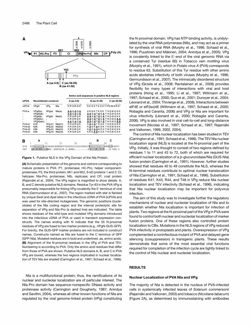
Figure 1. Putative NLS in the VPg Domain of the NIa Protein.
(A) Schematic presentation of the genome and cistrons corresponding to
mature proteins in PVA: P1, proteinase; HC-Pro, helper component-
proteinase; P3, the third protein; 6K1 and 6K2, 6-kD proteins 1 and 2; CI,
helicase; NIa-Pro, proteinase; NIb, replicase; and CP, coat protein
(Rajamaki et al., 2004). The VPg region is magnified to show details. A,
B, and C denote putative NLS domains. Residue Tyr-63 in the PVA VPg is
presumably responsible for linking VPg covalently the 59 terminus of viral
RNA (Germundsson et al., 2007). The region marked with star is flanked
by unique SwaI and ApaI sites in the infectious cDNA of PVA (icPVA) and
was used for site-directed mutagenesis. The genomic positions (nucle-
otides) of the NIa coding region and the internal proteolytic site for
separation of VPg and NIa-Pro (a dashed line) are indicated. The table
shows residues of the wild-type and mutated VPg domains introduced
into the infectious cDNA of PVA or used in transient expression con-
structs. The names ending with N indicate that the 55 N-proximal
residues of VPg are fused to two marker proteins (e.g., VPgN-GUS-GFP).
For brevity, the GUS-GFP marker proteins are not included in construct
names. Constructs named as NIa are fused to the C terminus of GFP
(GFP-NIa). Mutated residues are in bold and underlined. aa, amino acids.
(B) Alignment of the N-proximal residues in the VPg of PVA and TEV.
Numbering is according to PVA. Only the amino acid residues that differ
from those of PVA are shown. Putative NLS domains A, B, and C in PVA
VPg are boxed, whereas the two regions implicated in nuclear localiza-
tion of TEV NIa are shaded (Carrington et al., 1991; Schaad et al., 1996).
2486 The Plant Cell

against VPg and NIa-Pro. To examine whether NIa and VPg are
targeted to the nucleus in the absence of PVA infection, the
proteins were expressed as fusions to the C terminus of green
fluorescent protein (GFP-NIa and GFP-VPg) (Figure 3) in leaves
of Nicotiana benthamiana by agroinfiltration. To prevent the
cleavage between VPg and NIa-Pro domains and to study
localization of unprocessed NIa, the P1 position of the internal
cleavage site was mutagenized [substitution E189H; construct
GFP-NIa(E/H); see Supplemental Figure 1A online]. Fluores-
cence microscopy revealed that GFP-NIa, GFP-NIa(E/H), and
GFP-VPg accumulated predominantly in the nucleus, with little or
no fluorescence observed in the cytoplasm (Figure 2C; see
Supplemental Figure 1Bonline). By contrast, GFP alone localized
to both the cytoplasm and nucleus (Figure 2B).
Regionsof theVPgDomainControllingNuclearLocalization
Residues 41 to 50 (KKGKTKGKTH) form the putative NLS of PVA
NIa, based on comparison with the TEV NIa NLS (Figure 1B)
(Schaad et al., 1996). Therefore, the full-length VPg domain of
PVA NIa, the N-terminal part of VPg (VPgN; residues 1 to 55), the
putative NLS (residues 41 to 50) within the VPg domain, and the
NLS of TEV VPg (residues 40 to 49) were each expressed as a
fusion with GUS-GFP (Figure 3). The large GUS-GFP fusion
protein was used to prevent spontaneous movement of GFP to
the nucleus (Grebenok et al., 1997). The constructs were intro-
duced for expression in the epidermal cells of N. benthamiana
leaves by particle bombardment and/or agroinfiltration. VPgN-
GUS-GFP was detected almost exclusively in the nucleus,
whereas GUS-GFP was localized in the cytoplasm (Figure 4),
showing that the N-terminal 55 residues of VPg were sufficient to
target GUS-GFP to the nucleus and hence contained the NLS of
PVA NIa. By contrast, the majority of VPg-GUS-GFP, PVAnls-
GUS-GFP, and TEVnls-GUS-GFP were detected in the cyto-
plasm (see Supplemental Figure 2 online). In PVAnls-GUS-GFP
and TEVnls-GUS-GFP, the residues constituting an NLS may
have been masked by the putative conformation of the large
GUS-GFP protein, which may have prevented nuclear localiza-
tion. Constructs based on VPgN-GUS-GFP and GFP-NIa were
thus used for further analysis.
The four residues critical for nuclear localization of TEV NIa are
Lys-41, Lys-43, Arg-44, and Thr-48 (Schaad et al., 1996). They
correspond to Lys-42, Lys-44, Thr-45, and Thr-49, respectively,
in PVA NIa and define the putative NLS region designated as B
(Figure 1). In TEV, substitution of two or three of the residues Lys-
41, Lys-43, or Arg-44 with Ala perturbs nuclear transport of NIa,
whereas substitution of single residues has little effect (Schaad
et al., 1996). When two or three of the corresponding residues of
the putative NLS region B in PVA NIa were substituted with Ala in
GFP-NIa and VPgN-GUS-GFP (mutations b1N, b2N, b3N, and
b4N; Figure 1), all mutant proteins showed similarly reduced
nuclear localization compared with VPgN-GUS-GFP (Figures 4A
and 4C to 4F) and GFP-NIa (see Supplemental Figure 1B online).
Because the region B in PVA contains three Lys residues in
Figure 2. Nuclear Localization of NIa of PVA in Cells of N. tabacum and N. benthamiana.
(A) Immunohistochemical localization with antibodies against VPg in upper noninoculated leaves ofN. tabacum systemically infected with PVA at 20 DAI
as described (Rajamaki and Valkonen, 2003). Immunostaining signals (red) were detected mainly in the nucleus.
(B) Localization of GFP (not fused to NIa) in the cytoplasm and nucleus of epidermal cells of an agroinfiltrated leaf of N. benthamiana 3 DAIF.
(C) GFP-NIa transiently expressed by agroinfiltration and detected by epifluorescence microscopy 3 DAIF.
(D) The 4’,6-diamidino-2-phenylindole (DAPI) staining indicates the positions of nuclei in cells shown in (C). Red autofluorescence caused by chlorophyll
is visible in (B) due to filter settings that differ from those in (C). Bars = 25 mm.
Nucleolar Localization of Potyviral NIa 2487

addition to Lys-42 and Lys-44 and nuclear localization was not
fully abolished by the aforementioned constructs, an additional
construct was made to substitute all five Lys residues of the
putative NLS region B with Ala. Whereas a substantial portion of
the resultant protein VPgN(b)-GUS-GFP and GFP-NIa(b) re-
mained in the cytoplasm, similar to the aforementioned B-site
mutant proteins, a fraction of these proteins entered the nucleus
(Figure 4I; see Supplemental Figure 1B online), indicating that
another region at the N terminus of VPg contributed to nuclear
localization.
We concluded that one of the additional putative NLS regions
could be at theN terminus of VPg,which contains basic residues.
The effect of the four N-terminal-most basic amino acid residues
was studied, and the corresponding part of the N terminus was
designated as region A (Figure 1). Substitution of the residues
(Lys-4, Arg-5, Arg-7, and Lys-9) in this region with Ala signifi-
cantly reduced, but did not abolish, nuclear localization of the
resultant proteins VPgN(a)-GUS-GFP and GFP-NIa(a) (Figure
4H; see Supplemental Figure 1B online). When these substitu-
tions in NLS region A were combined with the five substitutions
made in the NLS region B, the resultant protein VPgN(ab)-GUS-
GFP did not localize to the nucleus (Figure 4J). The two Lys
residues (Lys-54 and Lys-55) in a region designated as C (Figure
1) were substituted with Ala [construct VPgN(bc)-GUS-GFP],
which had no detectable effect on the already reduced nuclear
localization of VPgN(b)-GUS-GFP (Figure 4K). As expected, no
nuclear localization was observed for VPgN(abc)-GUS-GFP.
These results indicated that the VPg domain of PVA NIa contains
a bipartite NLS defined by region A (designated as NLS I) and
region B (designated as NLS II).
NLS Regions in VPg Control Protein Targeting to the
Nucleolus and CBs
Fluorescence from VPgN-GUS-GFP and GFP-NIa in the nucleus
was concentrated in subnuclear bodies, presumably the nucle-
olus and CBs. The subnuclear bodies were identified using the
fibrillarin 2 protein, which localizes to the nucleolus and CBs (Gall
2003), and the U2B0 protein, which localizes to CBs (Beven et al.,
1995). The genes encoding these proteins were derived from
Arabidopsis thaliana, and the subnuclear localization of the
corresponding proteins in N. benthamiana has been reported
(Kim et al., 2007b). The proteinswere taggedwith red fluorescent
protein (AtFib2-mRFP and AtU2B0-mRFP) and coexpressed
with VPgN-GUS-GFP, GFP-VPg, GFP-NIa, or GFP-NIa(E/H) in
N. benthamiana leaves by agroinfiltration. The intensive green
fluorescence from VPgN-GUS-GFP, GFP-VPg, GFP-NIa, and
GFP-NIa(E/H) in a large nuclear body and smaller subnuclear
bodies colocalized with the red fluorescence from AtFib2-mRFP
(Figure 5A; see Supplemental Figure 3 online), indicating that the
large body was the nucleolus and the smaller bodies were CBs.
Redfluorescence fromAtU2B0-mRFPcolocalizedwith the smaller
subnuclear bodies expressing GFP fluorescence from VPgN-
GUS-GFP (Figure 5E), which confirmed that they were CBs.
VPgN(b)-GUS-GFP, in which the five Lys residues of NLS II
(region B) were substituted with Ala, was not detected in the
nucleolus (Figure 5B, Table 1). Substitution of two or three
residues in NLS II (constructs b1N, b2N, b3N, and b4N; Figure 1)
reduced but did not abolish nucleolar targeting. Among these
mutants, the lowest rate of nucleolar localization was observed
with Lys-42/Lys-44 substitutions (b1N and b3N), whereas mu-
tants with a single Lys substitution (b2N and b4N) were not
greatly affected (Table 1). Substitution of the four basic residues
in NLS I (region A) with Ala abolished nucleolar targeting, as
shown by expression and localization of VPgN(a)-GUS-GFP
(Figure 5D). In accordance with these results, nucleolar localiza-
tion of GFP-NIa(a) and GFP-NIa(b), containing substitutions at
four and five residues of NLS I and NLS II, respectively, was
reduced but to a lesser extent thanwith the corresponding fusion
proteins containing the VPg N terminus only (see Supplemental
Figure 3 online).
VPgN(b)-GUS-GFP still accumulated in CBs (Figures 5B and
5C), in contrast with VPgN(a)-GUS-GFP, which showed no signal
in CBs (Figure 5D). These results were confirmed using AtU2B0-mRFP for colocalization (Figures 5F and 5G) and showed that
NLS I directed protein localization to CBs.
Figure 3. Schematic Presentation of the Constructs Used for Protein
Expression and Localization Studies.
(A) pRT-GFP, (B) pRT-GFP-VPg, (C) pRT-GFP-NIa, (D) pRT-GUS-GFP,
(E) pRT-PVAnls-GUS-GFP (PVAnls refers to region B in Figure 1), (F)
pRT-TEVnls-GUS-GFP, (G) pRT-VPgN-GUS-GFP, (H) pRT-VPg-GUS-
GFP, (I) pROK2-AtFib2-mRFP, and (J) pROK2-AtU2B0-mRFP. pRT-
GFP-NIa(E/H) with an amino acid substitution E189H in the internal
cleavage site to prevent cleavage between the VPg and NIa-Pro domains
was prepared by site-directed mutagenesis of pRT-GFP-NIa. NLS
mutants (Figure 1) were prepared by site-directed mutagenesis of pRT-
GFP-NIa and pRT-VPgN-GUS-GFP. The arrow represents the Cauli-
flower mosaic virus (CaMV) 35S promoter. Protein coding regions are
marked by boxes.
2488 The Plant Cell
Interaction of PVA VPg with the Nucleolar Protein Fibrillarin
Fibrillarin is a major nucleolar protein also present in CBs (Gall,
2003). The ORF3 protein of GRV (Kim et al., 2007a) and some
proteins of animal viruses interact with fibrillarin or certain other
nucleolar proteins (reviewed in Hiscox, 2007), which prompted
us to test whether PVA NIa could interact with fibrillarin. Two
copies (Nb Fib2a andNb Fib2b) of the fibrillarin genewere cloned
from N. benthamiana and predicted to encode proteins of 314
residues differing at two positions (10 [S versus G, respectively]
and 253 [M versus I, respectively]). The sequence of Nb Fib2a
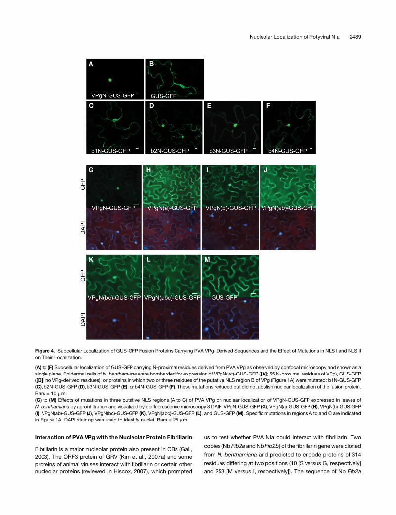
Figure 4. Subcellular Localization of GUS-GFP Fusion Proteins Carrying PVA VPg–Derived Sequences and the Effect of Mutations in NLS I and NLS II
on Their Localization.
(A) to (F) Subcellular localization of GUS-GFP carrying N-proximal residues derived from PVA VPg as observed by confocal microscopy and shown as a
single plane. Epidermal cells ofN. benthamianawere bombarded for expression of VPgN(wt)-GUS-GFP ([A]; 55 N-proximal residues of VPg), GUS-GFP
([B]; no VPg-derived residues), or proteins in which two or three residues of the putative NLS region B of VPg (Figure 1A) were mutated: b1N-GUS-GFP
(C), b2N-GUS-GFP (D), b3N-GUS-GFP (E), or b4N-GUS-GFP (F). These mutations reduced but did not abolish nuclear localization of the fusion protein.
Bars = 10 mm.
(G) to (M) Effects of mutations in three putative NLS regions (A to C) of PVA VPg on nuclear localization of VPgN-GUS-GFP expressed in leaves of
N. benthamiana by agroinfiltration and visualized by epifluorescencemicroscopy 3 DAIF. VPgN-GUS-GFP (G), VPgN(a)-GUS-GFP (H), VPgN(b)-GUS-GFP
(I), VPgN(ab)-GUS-GFP (J), VPgN(bc)-GUS-GFP (K), VPgN(abc)-GUS-GFP (L), and GUS-GFP (M). Specific mutations in regions A to and C are indicated
in Figure 1A. DAPI staining was used to identify nuclei. Bars = 25 mm.
Nucleolar Localization of Potyviral NIa 2489

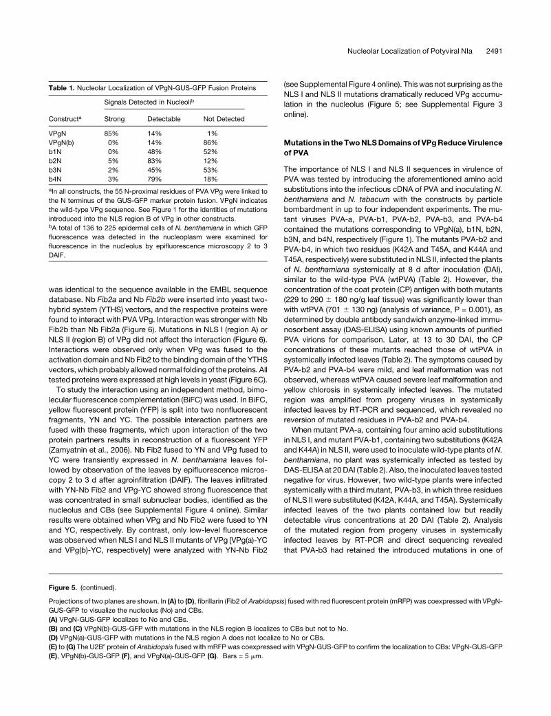
Figure 5. Subnuclear Localization of VPgN-GUS-GFP Fusion Protein Constructs Expressed in Leaves of N. benthamiana by Agroinfiltration and
Detected by Confocal Microscopy 2 to 3 DAIF.
2490 The Plant Cell
was identical to the sequence available in the EMBL sequence
database. Nb Fib2a and Nb Fib2b were inserted into yeast two-
hybrid system (YTHS) vectors, and the respective proteins were
found to interact with PVA VPg. Interaction was stronger with Nb
Fib2b than Nb Fib2a (Figure 6). Mutations in NLS I (region A) or
NLS II (region B) of VPg did not affect the interaction (Figure 6).
Interactions were observed only when VPg was fused to the
activation domain andNb Fib2 to the binding domain of the YTHS
vectors,which probably allowed normal folding of theproteins. All
tested proteins were expressed at high levels in yeast (Figure 6C).
To study the interaction using an independent method, bimo-
lecular fluorescence complementation (BiFC) was used. In BiFC,
yellow fluorescent protein (YFP) is split into two nonfluorescent
fragments, YN and YC. The possible interaction partners are
fused with these fragments, which upon interaction of the two
protein partners results in reconstruction of a fluorescent YFP
(Zamyatnin et al., 2006). Nb Fib2 fused to YN and VPg fused to
YC were transiently expressed in N. benthamiana leaves fol-
lowed by observation of the leaves by epifluorescence micros-
copy 2 to 3 d after agroinfiltration (DAIF). The leaves infiltrated
with YN-Nb Fib2 and VPg-YC showed strong fluorescence that
was concentrated in small subnuclear bodies, identified as the
nucleolus and CBs (see Supplemental Figure 4 online). Similar
results were obtained when VPg and Nb Fib2 were fused to YN
and YC, respectively. By contrast, only low-level fluorescence
was observed when NLS I and NLS II mutants of VPg [VPg(a)-YC
and VPg(b)-YC, respectively] were analyzed with YN-Nb Fib2
(see Supplemental Figure 4 online). Thiswas not surprising as the
NLS I and NLS II mutations dramatically reduced VPg accumu-
lation in the nucleolus (Figure 5; see Supplemental Figure 3
online).
Mutations in theTwoNLSDomainsof VPgReduceVirulence
of PVA
The importance of NLS I and NLS II sequences in virulence of
PVA was tested by introducing the aforementioned amino acid
substitutions into the infectious cDNA of PVA and inoculating N.
benthamiana and N. tabacum with the constructs by particle
bombardment in up to four independent experiments. The mu-
tant viruses PVA-a, PVA-b1, PVA-b2, PVA-b3, and PVA-b4
contained the mutations corresponding to VPgN(a), b1N, b2N,
b3N, and b4N, respectively (Figure 1). The mutants PVA-b2 and
PVA-b4, in which two residues (K42A and T45A, and K44A and
T45A, respectively) were substituted in NLS II, infected the plants
of N. benthamiana systemically at 8 d after inoculation (DAI),
similar to the wild-type PVA (wtPVA) (Table 2). However, the
concentration of the coat protein (CP) antigen with both mutants
(229 to 290 6 180 ng/g leaf tissue) was significantly lower than
with wtPVA (701 6 130 ng) (analysis of variance, P = 0.001), as
determined by double antibody sandwich enzyme-linked immu-
nosorbent assay (DAS-ELISA) using known amounts of purified
PVA virions for comparison. Later, at 13 to 30 DAI, the CP
concentrations of these mutants reached those of wtPVA in
systemically infected leaves (Table 2). The symptoms caused by
PVA-b2 and PVA-b4 were mild, and leaf malformation was not
observed, whereas wtPVA caused severe leaf malformation and
yellow chlorosis in systemically infected leaves. The mutated
region was amplified from progeny viruses in systemically
infected leaves by RT-PCR and sequenced, which revealed no
reversion of mutated residues in PVA-b2 and PVA-b4.
When mutant PVA-a, containing four amino acid substitutions
in NLS I, andmutant PVA-b1, containing two substitutions (K42A
and K44A) in NLS II, were used to inoculate wild-type plants ofN.
benthamiana, no plant was systemically infected as tested by
DAS-ELISA at 20DAI (Table 2). Also, the inoculated leaves tested
negative for virus. However, two wild-type plants were infected
systemically with a third mutant, PVA-b3, in which three residues
of NLS II were substituted (K42A, K44A, and T45A). Systemically
infected leaves of the two plants contained low but readily
detectable virus concentrations at 20 DAI (Table 2). Analysis
of the mutated region from progeny viruses in systemically
infected leaves by RT-PCR and direct sequencing revealed
that PVA-b3 had retained the introduced mutations in one of
Figure 5. (continued).
Projections of two planes are shown. In (A) to (D), fibrillarin (Fib2 of Arabidopsis) fused with red fluorescent protein (mRFP) was coexpressed with VPgN-
GUS-GFP to visualize the nucleolus (No) and CBs.
(A) VPgN-GUS-GFP localizes to No and CBs.
(B) and (C) VPgN(b)-GUS-GFP with mutations in the NLS region B localizes to CBs but not to No.
(D) VPgN(a)-GUS-GFP with mutations in the NLS region A does not localize to No or CBs.
(E) to (G) The U2B0 protein of Arabidopsis fused with mRFP was coexpressed with VPgN-GUS-GFP to confirm the localization to CBs: VPgN-GUS-GFP
(E), VPgN(b)-GUS-GFP (F), and VPgN(a)-GUS-GFP (G). Bars = 5 mm.
Table 1. Nucleolar Localization of VPgN-GUS-GFP Fusion Proteins
Constructa
Signals Detected in Nucleolib
Strong Detectable Not Detected
VPgN 85% 14% 1%
VPgN(b) 0% 14% 86%
b1N 0% 48% 52%
b2N 5% 83% 12%
b3N 2% 45% 53%
b4N 3% 79% 18%
aIn all constructs, the 55 N-proximal residues of PVA VPg were linked to
the N terminus of the GUS-GFP marker protein fusion. VPgN indicates
the wild-type VPg sequence. See Figure 1 for the identities of mutations
introduced into the NLS region B of VPg in other constructs.bA total of 136 to 225 epidermal cells of N. benthamiana in which GFP
fluorescence was detected in the nucleoplasm were examined for
fluorescence in the nucleolus by epifluorescence microscopy 2 to 3
DAIF.
Nucleolar Localization of Potyviral NIa 2491

the two plants. In the other plant, VPg residue A45 had reverted
back to Thr (wild type).
In N. tabacum, virulence of all mutant viruses tested was
significantly compromised. Only one plant inoculated with PVA-
b2 and three plants inoculated with PVA-b4 were systemically
infected at 20 DAI, and virus concentrations were low (Table 2).
The progeny viruses analyzed from the systemically infected
leaves by RT-PCR and direct sequencing had retained the
introduced mutations. No infection was detected with PVA-a,
PVA-b1, and PVA-b3 (Table 2).
Expression of VPg Partially Complements an NLSMutant of
PVA in Transgenic Plants
Two transgenic lines of N. benthamiana (tt2 and tt7) expressing
PVA VPg were inoculated with wtPVA and the mutants PVA-a,
PVA-b1, PVA-b2, PVA-b3, and PVA-b4 to detect possible in
trans complementation of the mutations. Little difference in
systemic infection and virus accumulation was observed be-
tween wild-type, tt2, and tt7 plants following inoculation with
wtPVA, PVA-b2, or PVA-b4, all of which showed high or relatively
high levels of virulence (Table 2). Also, the low virulence of PVA-
b3 and lack of detectable infectivity of PVA-a were not comple-
mented in the transgenic lines compared with wild-type plants
(Table 2). In two transgenic plants systemically infected with
PVA-b3, the progeny viruses had retained the original mutations,
but in two other plants, one of the three amino acid substitutions
in VPg reverted to the wild type. The concentrations of wtPVA
decreased from 13 to 20 DAI in the transgenic lines, probably
because the initially high titers of PVA RNA and expression of
the virus-homologous VPg transgene mRNA induced RNA
silencing–based resistance to PVA, but no plant systemically
infected with wtPVA or the aforementioned PVA mutants recov-
ered from infection during the experiment until 28 DAI.
By contrast, infectivity of mutant PVA-b1 was noticeably
improved in VPg-expressing transgenic plants. Although this
mutant failed to infect any wild-type plant systemically,;50%of
the transgenic plants were systemically infected with virus titers
no different from those of wtPVA at 20 DAI (Table 2). The progeny
viruses were analyzed by RT-PCR and direct sequencing from
systemically infected leaves, and in all cases PVA-b1 had
retained the introduced mutations.
To exclude to some extent the possibility of mutations having
occurred elsewhere in the viral genome, the NIb region was
sequenced from viral progenies in 19 plants because NIa andNIb
interact (Hong et al., 1995; Li et al., 1997; Guo et al., 2001).
However, no mutations were found in NIb.
Mutations in the Two NLS Domains of VPg Reduce
Multiplication of PVA in Protoplasts
ThewtPVA,mutants PVA-b1, PVA-b2, PVA-b3, andPVA-b4, and
an additional mutant, norPVA, with a frameshift in the open
reading frame (to prevent any expression of CP from the PVA
genome under the 35S promoter), were inoculated into proto-
plasts ofN. tabacum. NoPVACPwas detected in the protoplasts
inoculated with PVA-b1, PVA-b3, or norPVA (Table 3). The
mutants PVA-b2 and PVA-b4 multiplied to detectable levels,
but the amounts of CP antigen were approximately fourfold and
twofold lower than with wtPVA, respectively (Table 3).
Cosuppression of the gfp Gene in the Presence of VPg and
NLS Mutants
Leaves of GFP transgenic N. benthamiana line 16c were coinfil-
trated with Agrobacterium tumefaciens strains for expression of
GFP and VPg, VPg(a) (K4A/R5A/R7A/K9A), VPg-b1 (K42A/
K44A), VPg-b2 (K42A/T45A), VPg-b3 (K42A/K44A/T45A),
VPg-b4 (K44A/T45A), or HC-Pro or GUS as controls. GFP
Figure 6. Interaction of PVA VPg with the Fibrillarin 2 Proteins Nb Fib2a
and Nb Fib2b of N. benthamiana in YTHS.
Nb Fib2a and Nb Fib2b differ at two residues (10 [S versus G, respec-
tively] and 253 [M versus I, respectively]).
(A) Differences in protein–protein interactions indicated by differences in
the growth rate of yeast on the selection medium 8 d after plating.
(B) Relative differences in growth rate of yeast cotransformed for
expression of different combinations of proteins: ++, growth in 2 to
4 d; +, growth in 5 to 8 d; –, no growth; nt, not tested.
(C) Immunodetection of the fusion proteins expressed in yeast by protein
gel blotting using monoclonal antibodies to the GAL4 activation domain
(AD) or DNA binding domain (DB). Lanes: 1, empty vector; 2, Nb Fib2a; 3,
VPg; 4, VPg-b3; 5, Nb Fib2b; 6, VPg(a); 7, VPg(b); 8, empty vector; 9, Nb
Fib2a; 10, Nb Fib2b; 11, VPg; 12, VPg-b3; 13, VPg(a); 14, VPg(b). The
sizes of molecular mass markers (kD) are indicated to the left.
2492 The Plant Cell

fluorescence was readily visible in the leaf tissue coinfiltrated
with GFP and VPg up to 4 dDAIF (Figure 7A) but faded thereafter.
By contrast, GFP fluorescence was barely visible at any time in
tissues infiltratedwith GFP and VPg-a, VPg-b1, VPg-b2, VPg-b3,
or VPg-b4, except for the fluorescence in veins due to the GFP
expressed from the gfp transgene in the 16c line. Similarly,
tissues coinfiltrated with GFP and GUS showed only temporal
faint expression of GFP fluorescence, which started to decrease
at 3 DAIF due to the expected activated gfp cosuppression. A
bright red border eventually appeared around the infiltrated
areas. Coinfiltration with GFP and HC-Pro resulted in strong and
long-lasting GFP fluorescence (Figure 7A) that remained visible
until the termination of the experiment (8 DAIF).
In the analyses done at 4 DAIF, the levels of GFP mRNA
detected in tissues coinfiltrated with GFP and VPg or HC-Pro
were higher than with GUS and GFP or GFP and VPg(a), VPg-b1,
VPg-b3, VPg-b4 (Figure 7B), or VPg-b2. The gfp-specific small
interfering RNAs (siRNAs) were detected in tissues infiltrated
Table 2. Amounts of PVA CP Antigen in Systemically Infected Leaves of N. benthamiana and N. tabacum Inoculated with Wild-Type PVA and
Mutants Containing Amino Acid Substitutions in Two NLSs Situated in the VPg Domain of NIa (NLS I and NLS II, Respectively, Separated by a Dash)
wtPVA PVA-b1 PVA-b2 PVA-b3 PVA-b4 PVA-a
KRQRQK - KKGKT KRQRQK - KAGAT KRQRQK - KAGKA KRQRQK - KAGAA KRQRQK - KKGAA AAQAQA - KKGKT
naAmount
(mg/g)b n
Amount
(mg/g) n
Amount
(mg/g) n
Amount
(mg/g) n
Amount
(mg/g) n
Amount
(mg/g)
N. benthamianac
8 DAI wt 5/5 701 (6130) 0/5 – 4/5 229 (6180) 0/5 – 5/5 290 (6180) 0/19 –
tt2 5/5 831 (691) 0/5 – 5/5 181 (6150) 0/5 – 5/5 259 (678) 0/10 –
tt7 4/5 739 (6105) 0/5 – 2/5 271 (6257) 0/5 – 3/5 413 (6123) 0/10 –
13 DAI wt 13/13 547 (6161) 0/13 – 12/13 375 (6173) 1/13 7.0 12/13 383 (6147) 0/19 –
tt2 9/9 601 (6184) 6/11 127 (6202) 10/10 447 (6240) 0/11 – 10/10 388 (6108) 0/10 –
tt7 9/10 621 (6235) 1/10 5.0 6/11 430 (6226) 2/10 556 (6181) 8/11 444 (6171) 0/10 –
20 DAI wt 13/13 597 (6155) 0/13 – 11/13 490 (6123) 2/13 170 (6115) 13/13 462 (6105) 0/19 –
tt2 9/9 272 (6165) 7/11 281 (6137) 10/10 418 (6108) 2/11 158 (615) 10/10 327 (691) 0/10 –
tt7 9/10 240 (6179) 3/10 197 (633) 7/11 370 (6118) 2/10 366 (6226) 7/11 299 (694) 0/10 –
N. tabacum
13 DAI wt 13/13 9.4 (64.4) 0/9 – 1/13 0.7 0/9 – 2/13 1.1 (60.1) 0/5 –
20 DAI wt 13/13 12.5 (65.7) 0/9 – 1/13 6.9 0/9 – 3/13 2.6 (61.6) 0/5 –
aThe number of plants systemically infected as detected by DAS-ELISA among the total number of plants inoculated by particle bombardment in two
experiments.bMean amounts of PVA CP antigen (mg/g leaf tissue) as determined by DAS-ELISA (SD in parentheses) using known amounts of purified PVA particles
for comparison.cNontransgenic (wt) plants and plants of two VPg-transgenic lines (tt2 and tt7) of N. benthamiana expressing PVA VPg (Germundsson et al., 2007)
were used.
Table 3. Infectivity of the PVA-nls Mutants in N. tabacum Protoplasts at 3 DAI
Virusa
Experiment 1 Experiment 2
Virus Amount (ng)b Relative Amount Virus Amount (ng) Relative Amount
wtPVA 11.4 6 0.9 100% 31.4 6 3.6 100%
PVA-b1 0.0 0% 0.0 0%
PVA-b2 2.6 6 1.6 23% 3.8 6 0.5 12%
PVA-b3 0.0 0% 0.0 0%
PVA-b4 5.6 6 0.6 49% 5.3 6 0.3 17%
norPVA 0.0 0% 0.0 0%
Mock 0.0 0% 0.0 0%
aRecombinant PVA genomes used to inoculate protoplasts of N. tabacum by electroporation. The PVA mutants designated as b1 to b4 contain amino
acid substitutions in the nuclear localization site B of the VPg domain of NIa. The amino acid substitutions are identical to those tested for their effect
on nuclear and nucleolar localization of marker proteins (b1N to b4N) explained in Figure 1. wtPVA, wild-type PVA; PVAnor, a noninfectious PVA
mutant obtained by introducing a frameshift mutation in the middle of the region encoding the first protein (P1) at the 59-proximal part of the viral RNA;
mock, protoplasts electroporated with buffer (no viral RNA).bMean amounts of PVA CP antigen (ng) per 106 inoculated protoplasts as determined by DAS-ELISA using known amounts of purified PVA particles
for comparison. Relative amounts (%) were calculated by comparison with the amounts of wtPVA. Absorbance values (A405) of norPVA and mock were
0.002 to 0.026.
Nucleolar Localization of Potyviral NIa 2493
with GFP andGUS, VPg(a), VPg-b1, VPg-b3, VPg-b4 (Figure 7B),
or VPg-b2, but not in tissues infiltrated with GFP and VPg or HC-
Pro (Figure 7B). Only low levels of gfp-specific siRNA were
detected in tissues coinfiltrated with GUS and GFP at 4 DAIF,
which was probably because cosuppression had only recently
initiated and GFP expression levels were low. The experiment
was performed two to five times with wild-type VPg and different
VPg mutants, and results were similar each time.
Fibrillarin Silencing Reduces PVA Accumulation
To examine whether fibrillarin plays a role in PVA infection, as it
does in infection of GRV (Kim et al., 2007a), fibrillarin expression
was suppressed inN. benthamiana plants using a Tobacco rattle
virus (TRV)-based vector carrying a fragment of the Nb Fib2a
gene of N. benthamiana (TRV-Fib2). N. benthamiana plants were
also inoculated with an empty TRV vector as a control. To
monitor initiation of silencing, a few plants were inoculated with
the TRV vector carrying a fragment of the phytoene desaturase
gene (TRV-PDS), which provides a silencing phenotype charac-
terized by loss of chlorophyll (Ratcliff et al., 2001). At the time
point when PDS silencing was manifested in plants inoculated
with TRV-PDS (see Supplemental Figure 5 online), the mean
expression level of fibrillarin mRNA in the leaves of TRV-Fib2–
infected plants was;25 to 86% (n = 6) that of the leaves in the
corresponding position of TRV-infected plants, as determined by
real-time RT-PCR (see Supplemental Figure 6 online). The top
leaves of plants systemically infected with TRV-Fib2 exhibited
severe curling and malformation, which resulted in severe
stunting of the plants (see Supplemental Figure 5 online). There-
fore, the lower, nearly fully expanded upper leaves exhibiting the
aforementioned depletion of Nb Fib2 expression were mechan-
ically inoculated with PVA, and leaves at the corresponding
position in TRV-infected control plants were inoculated as con-
trols. PVA accumulation was 52 to 66% in the inoculated leaves
of fibrillarin-silenced plants compared with nonsilenced TRV-
infected control plants as tested 6 DAI in three experiments
(Table 4). PVA was also detected in the upper noninoculated
leaves in both types of plants, and differences in virus concen-
tration between fibrillarin-silenced and nonsilenced upper leaves
were similar to those observed in inoculated leaves (Table 4).
DISCUSSION
Our data show that nuclear localization of NIa is controlled by
residues 4 to 9 (NLS I) and 41 to 50 (NLS II) of the VPg domain in
PVA. Both regions were needed for efficient nuclear localization,
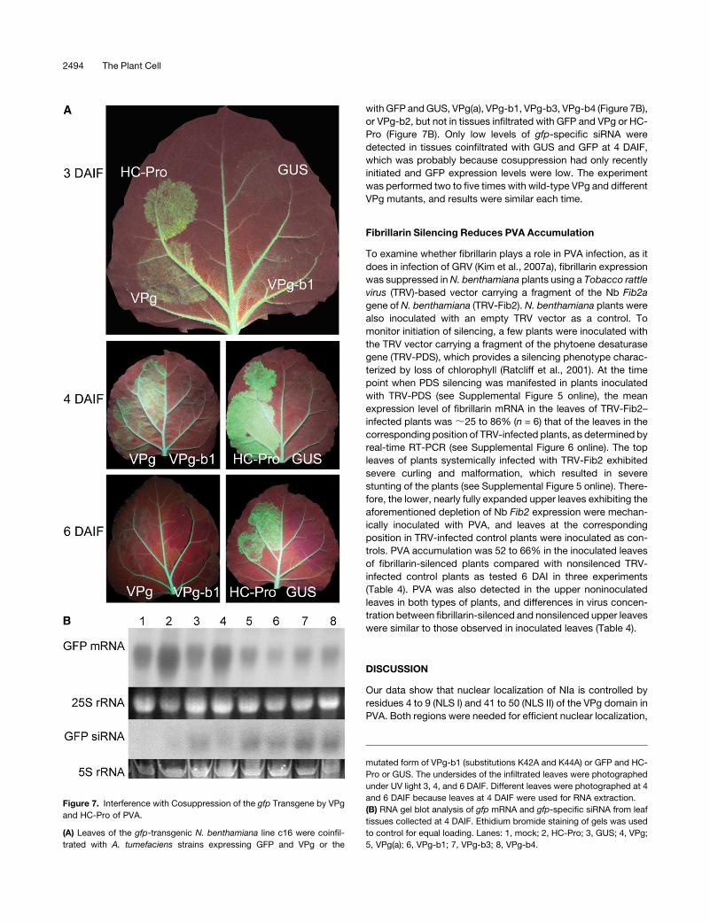
Figure 7. Interference with Cosuppression of the gfp Transgene by VPg
and HC-Pro of PVA.
(A) Leaves of the gfp-transgenic N. benthamiana line c16 were coinfil-
trated with A. tumefaciens strains expressing GFP and VPg or the
mutated form of VPg-b1 (substitutions K42A and K44A) or GFP and HC-
Pro or GUS. The undersides of the infiltrated leaves were photographed
under UV light 3, 4, and 6 DAIF. Different leaves were photographed at 4
and 6 DAIF because leaves at 4 DAIF were used for RNA extraction.
(B) RNA gel blot analysis of gfp mRNA and gfp-specific siRNA from leaf
tissues collected at 4 DAIF. Ethidium bromide staining of gels was used
to control for equal loading. Lanes: 1, mock; 2, HC-Pro; 3, GUS; 4, VPg;
5, VPg(a); 6, VPg-b1; 7, VPg-b3; 8, VPg-b4.
2494 The Plant Cell
and NLS I seemed particularly important (Figure 4; see Supple-
mental Figure 1B online). These results provide further evidence
for a bipartite NLS in potyviral NIa. In a previous study, the NIa of
TEV was expressed in transgenicN. tabacum plants, and as with
PVA in our study, the region corresponding toNLS II was found to
control nuclear localization (Carrington et al., 1991; Schaad et al.,
1996). In addition, data suggested that the N-terminal residues of
NIa contributed to optimal nuclear localization of TEV NIa
(Carrington et al., 1991). Hence, results for these two potyviruses
are consistent with regard to the existence of NLS I and NLS II in
the VPg domain. Moreover, both NLS regions are highly con-
served in genus Potyvirus (see Supplemental Figure 7 online).
Our results also show that NLS I and NLS II control nucleolar
localization of PVA NIa. Mutation of one of the two NLS regions
was sufficient to abolish nucleolar targeting, unlike the situation
for nuclear localization. Secondly, nucleolar targeting was re-
duced and eventually abolished by an increasing number of
basic residue substitutions (Lys residues replaced by Ala) in NLS
II, whereas nuclear localization was similarly reduced but not
abolished by these mutations (Table 1, Figures 4 and 5; see
Supplemental Figures 1B and 3 online). Hence, nucleolar and
nuclear localization were controlled independently by the same
NLS regions. Although TEV NIa has been observed in the
nucleolus (Schaad et al., 1996), detailed studies on nucleolar
targeting of NIa have not been reported. Thus, our current
findings on nucleolar and nuclear localization of a potyviral
protein were made in a study area that has generally gained
little attention in plant virology (Kim et al., 2007a, 2007b).
PVA NIa was found to accumulate in CBs, which are highly
dynamic structures originally described as nucleolar accessory
bodies (reviewed in Pontes and Pikaard, 2008). Localization to
CBs was controlled by NLS I, whereas mutations in NLS II had
little effect. Hence, mutations in NLS II prevented nucleolar
localization but allowed high levels of accumulation in CBs. This
was particularly pronounced in experiments involving only the
N-proximal part of VPg fused to marker proteins. Similar results
were obtained using NIa, but the lower intensity of signals in CBs
using these constructs made comparison of differences more
difficult.
There is only a single precedent of a plant virus protein
localized in CBs. The ORF3 protein of GRV (unrelated to
potyviruses) accumulates in nucleoli and CBs where it interacts
with fibrillarin (Kim et al., 2007a, 2007b), amajor nucleolar protein
also present in CBs (Gall, 2003). It was therefore relevant to test
for possible interactions betweenNIa and fibrillarin. Data showed
that the VPg domain of PVA NIa interacted with fibrillarin in vivo.
This interaction was detected initially by YTHS and subsequently
studied in plant cells by BiFC. In GRV, interaction between the
ORF3 protein and fibrillarin recruits some fibrillarin to the cyto-
plasm for assembly of virus-like particles that are competent for
long-distance movement (Kim et al., 2007a, 2007b; Canetta
et al., 2008). However, it seems unlikely that the interaction
between VPg and fibrillarin would recruit fibrillarin to the cyto-
plasm because the interaction between VPg and fibrillarin was
observed only in nucleoli and CBs but not in the cytoplasm.
Another difference was observed in nucleolar trafficking of PVA
NIa and GRV ORF3. Whereas ORF3 uses a mechanism that
involves the appearance of multiple CB-like structures followed
by fusion of these structures with nucleoli (Kim et al., 2007a,
2007b), nomultiple CB-like structures were observed with NIa or
mutated NIa proteins. Despite these differences, results with
PVA NIa and GRV ORF3 both indicate that interactions of these
viral proteins with fibrillarin are important for virus infection; the
underlying mechanism, however, may differ. Reduction of fibril-
larin expression by ;30 to 80% in leaves by means of virus-
induced gene silencing (VIGS) reduced PVA accumulation by
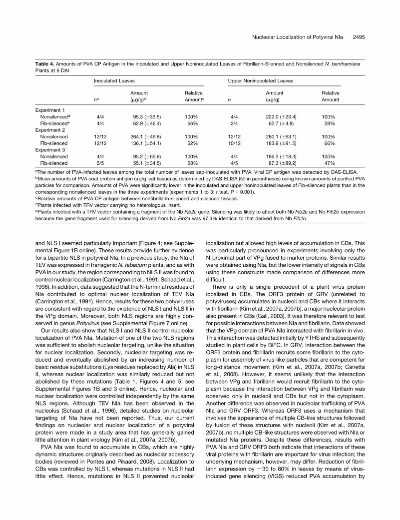
Table 4. Amounts of PVA CP Antigen in the Inoculated and Upper Noninoculated Leaves of Fibrillarin-Silenced and Nonsilenced N. benthamiana
Plants at 6 DAI
Inoculated Leaves Upper Noninoculated Leaves
naAmount
(mg/g)bRelative
Amountc n
Amount
(mg/g)
Relative
Amount
Experiment 1
Nonsilencedd 4/4 95.3 (633.5) 100% 4/4 222.0 (623.4) 100%
Fib-silencede 4/4 62.9 (646.4) 66% 2/4 62.7 (64.8) 28%
Experiment 2
Nonsilenced 12/12 264.1 (649.8) 100% 12/12 280.1 (663.1) 100%
Fib-silenced 12/12 136.1 (654.1) 52% 10/12 183.9 (691.5) 66%
Experiment 3
Nonsilenced 4/4 95.2 (665.9) 100% 4/4 186.3 (618.3) 100%
Fib-silenced 5/5 55.1 (634.5) 58% 4/5 87.3 (689.2) 47%
aThe number of PVA-infected leaves among the total number of leaves sap-inoculated with PVA. Viral CP antigen was detected by DAS-ELISA.bMean amounts of PVA coat protein antigen (mg/g leaf tissue) as determined by DAS-ELISA (SD in parentheses) using known amounts of purified PVA
particles for comparison. Amounts of PVA were significantly lower in the inoculated and upper noninoculated leaves of Fib-silenced plants than in the
corresponding nonsilenced leaves in the three experiments (experiments 1 to 3; t test, P = 0.001).cRelative amounts of PVA CP antigen between nonfibrillarin-silenced and silenced tissues.dPlants infected with TRV vector carrying no heterologous insert.ePlants infected with a TRV vector containing a fragment of the Nb Fib2a gene. Silencing was likely to affect both Nb Fib2a and Nb Fib2b expression
because the gene fragment used for silencing derived from Nb Fib2a was 97.3% identical to that derived from Nb Fib2b.
Nucleolar Localization of Potyviral NIa 2495
;50%but did not prevent systemic spread of the virus. In similar
experiments with GRV, fibrillarin depletion was manifested as
inhibition of systemic spread of the virus (Kim et al., 2007a,
2007b).
Substitution of the basic amino acids Lys-42 and Lys-44 with
Ala in NLS II caused the greatest reduction of nucleolar locali-
zation and the greatest loss of virulence in PVA in Nicotiana
plants (Tables 1 to 3). For example, the mutant viruses PVA-b1
and PVA-b3 failed to multiply in N. tabacum protoplasts. These
negative effects on virulence may have been caused by the lack
of nucleolar localization; still, residues in theNLS II region defined
in this study have also been implicated in NTP binding and
uridylylation of PVA VPg (Puustinen and Makinen, 2004). How-
ever, the region of VPg defined asNLS I in this study has not been
implicated in any other potyviral functions. Mutations in NLS I
inhibited nucleolar and CB localization of NIa and infection of
N. tabacum and N. benthamiana plants. A larger part of the
N terminus of VPg (16 residues) has been implicated in interac-
tions with a plant protein, PVIP, that contains a motif common to
proteins involved in transcriptional regulation via chromosome
remodeling (Dunoyer et al., 2004). These findings from indepen-
dent experiments link nucleolar localization of NIa and the
corresponding regions of the VPg domain with important viral
functions.
The severely debilitated functions of PVA-b1 (Lys-42 and Lys-
44mutated to Ala in NLS II) were complemented by the wild-type
VPg expressed in transgenic lines of N. benthamiana, which was
not observed with PVA-b3 that carried a third mutation (T45A).
These findings are essentially novel because functional defects
due to mutations in VPg are usually not complemented in trans
(Schaad et al., 1996, 1997; Nicolas et al., 1997; Keller et al., 1998;
Rajamaki and Valkonen, 1999). Only in one study was multipli-
cation of a TEV mutant containing the amino acid substitutions
analogous to PVA-b1 stimulated in protoplasts of a transgenicN.
tabacum line expressing the wild-type 6K2/NIa polyprotein of
TEV (Schaad et al., 1996). It seems that Lys-42 and Lys-44 in the
PVA NIa control a specific function that is distinct from other
functions by complementation in trans.We hypothesized that the
function could be related to circumvention or suppression of host
defense because the PVA-b2 and PVA-b4 mutants substituted
only for one of the two residues (Lys-42 or Lys-44) showed
reduced amplification but were able to mediate systemic infec-
tion in a large proportion of inoculated plants. The virulence of
these mutants was also more severely reduced in N. tabacum
than N. benthamiana, consistent with studies showing that N.
tabacum is more resistant to viruses than N. benthamiana (Yang
et al., 2004; Rajamaki et al., 2005). Alternatively, an interaction
with host factors controlled by Lys-42 and Lys-44 of NIa may
stimulate host functions necessary for viral infection. Interac-
tion of NIa with eIF(iso)4E has been observed in the nucleus
(Beauchemin et al., 2007); however, we did not consider this
relevant because to our knowledge, the interaction between
PVA NIa with eIF(iso)4E is not affected by the aforementioned
substitutions in NIa.
The hypothesis that nuclear localization of VPg/NIa might play
a role in interference with host antiviral defense was supported
by the finding that VPg interfered with gfp cosuppression and
was hence able to control gene silencing, a mechanism involved
in regulation of cellular gene expression and antiviral defense
(Pontes and Pikaard, 2008; Ruiz-Ferrer and Voinnet, 2009).While
the ability of VPg to suppress gene silencing was temporal and
weaker than that of HC-Pro, the well-known gene silencing
suppressor of potyviruses (Brigneti et al., 1998; Johansen and
Carrington, 2001), it was consistently observed in five indepen-
dent experiments. It therefore seems that VPg is an auxiliary
factor involved in interference of RNA silencing by potyviruses.
This effect of VPgwas absent in similar experiments performed in
a previous study probably because the mRNAs for GFP and
VPg expression both included the PVA 59-untranslated region
and the homologous sequences added to the silencing pressure
(Germundsson et al., 2007). Previous studies have implied that
the P1 protein of potyviruses also acts as an auxiliary factor for
suppression of RNA silencing (Rajamaki et al., 2005).
RNA silencing is an inducible antiviral defense mechanism of
particular importance in plants; therefore, impaired PVA infec-
tivity following mutations that abolish any detectable effects of
VPg on gene cosuppression is consistent with involvement of
VPg in suppression of RNA silencing during virus infection. The
infectivity of PVA carrying some of these mutations in VPg was
complemented in trans in transgenic plants expressing wild-type
VPg, which was to be expected if the mutations compromised
suppression of RNA silencing; the viral proteins capable of
suppressing RNA silencing have so far been identified mainly
by assayswhere they are expressed in trans outside the infection
context (Dıaz-Pendon and Shou-Wei, 2008). Previous studies
show that VPg is transported from lower infected leaves and
accumulates in the companion cells in sink leaves prior to
systemic infection, which was hypothesized to be needed for
suppression of host defense and initiation of infection (Rajamaki
and Valkonen, 2003). Results of this study are consistent with this
hypothesis and suggested that the mutations, and hence VPg/
NIa, played a major role at an early phase of infection because
mutations severely compromised infectivity. However, once
infection was established in inoculated leaves, the mutants
were able to infect the plants systemically, which suggests that
the proposed auxiliary functions of VPg in silencing suppression
are not an absolute requirement for infection in N. benthamiana.
Studies on mutated forms of VPg showed that suppression of
silencing was linked to localization to the nucleus, nucleoli, and
CBs. These findings on the linkage between VPg nuclear local-
ization and its silencing suppression function also seem conceiv-
able based on the pivotal roles of nucleoli and CBs as processing
centers for small RNAs, including siRNAs and microRNAs that
regulate gene expression posttranscriptionally (Pontes et al.,
2006; Brosnan et al., 2007; Pontes and Pikaard, 2008). siRNA
and CBs are also involved in transcriptional gene silencing by
methylation (Pontes and Pikaard, 2008). There are a few previous
examples of viral proteins whose activity in suppression of RNA
silencing depends on nuclear localization. The 2b protein of
Cucumber mosaic virus (Bromoviridae) and P0 protein of Beet
western yellows virus (Luteoviridae) interact with Argonaute1, the
core component of the RNA-induced silencing complex, in the
nucleus and inhibit its activity or target it for degradation, respec-
tively (Zhang et al., 2006; Bortolamiol et al., 2007). The P6 protein
of CaMV (Caulimoviridae) inhibits the nuclear protein DRB4,
2496 The Plant Cell

which is required for siRNAsynthesis byDicer-like protein 4 (Haas
et al., 2008). The large number of host proteins known to be
involved in regulation of RNA silencing in the nuclear environment
(e.g., siRNA and microRNA processing in nucleoli and CBs;
Pontes and Pikaard, 2008) makes it challenging to identify the
host factors targeted by VPg for interference with silencing.
Fibrillarin is included in small nuclear and nucleolar ribonucleopro-
teins that accumulate in CBs en route to spliceosomes and
nucleoli, respectively (Pontes and Pikaard, 2008), also suggesting
a role for the fibrillarin–VPg interaction in silencing suppression.
Alternatively, the fibrillarin–VPg interaction may disrupt certain
nucleolar functions (e.g., host transcription or pre-mRNA process-
ing), which might explain the observed shutdown of host gene
expression during potyvirus infection (Wang and Maule, 1995).
Because VPg seems to suppress silencing in the nuclear environ-
ment, we hypothesize that its main target is a host gene.
By contrast, many previously described viral silencing suppres-
sors function in the cytoplasmic environment to sequester virus-
derived siRNA (Dıaz-Pendon andShou-Wei, 2008;Ruiz-Ferrer and
Voinnet, 2009).
Our study provides evidence that the successful infection
cycle of the picorna-like potyvirus PVA requires localization of
viral VPg/NIa to the nucleus, nucleoli, and CBs, which may be a
consequence of the need to interfere with host defenses and
hence facilitate initiation of infection. These findings are sup-
ported by studies indicating that nuclear localization of NIa is
needed for successful infection by TEV (Schaad et al., 1996) and
that poliovirus (family Picornaviridae) regulates nuclear and
nucleolar functions in host cells (Waggoner and Sarnow, 1998;
Izumi et al., 2001;Hiscox, 2003; Banerjee et al., 2005). Hence, the
results reported here may be generally applicable to a large
number of picorna-like plant and animal viruses.
METHODS
Plant Material
Plants of tobacco (Nicotiana tabacum cv Samsun nn), Nicotiana
benthamiana, and transgenicN. benthamiana lines tt2 and tt7 expressing
the VPg of PVA (Germundsson and Valkonen, 2006) were grown from
seeds of our ownstock.Seeds of the transgenic line 16cofN.benthamiana
showing strong constitutive expression of GFP (Brigneti et al., 1998) were
kindly provided by David Baulcombe. Plants were grown in a growth
chamber (Weiss Umweltstechnik) under constant conditions (photo-
period 16 h; light intensity 250 mE s–1 m–2; temperature 22/188C day/
night; relative humidity 75%) and watered daily as needed and fertilized
weekly with 1% fertilizer (N:P:K = 16:9:22; Puutarhan tayslannos, Kemira
GrowHow).
Construction of Mutant PVA cDNAs
Recombinant PVA cDNA clones were constructed based on a vector
containing the full-length infectious cDNA clone of PVA-B11 placed under
the control of theCauliflowermosaic virus (CaMV) 35S promoter (Puurand
et al., 1996). Mutations were introduced into the VPg coding sequence to
substitute basic residues or a Thr residue in the putative NLS regions with
Ala. Site-directed mutagenesis of the VPg coding sequence was per-
formed on a plasmid containing a 1898-nucleotide-long HindIII-ApaI
fragment of the PVA cDNA (Figure 1) as described (Rajamaki and
Valkonen, 1999) using the QuickChange site-directed mutagenesis sys-
tem (Stratagene) and appropriate nucleotide primers (see Supplemental
Table 1 online). Mutations were verified by sequencing. Subsequently, a
1208-nucleotide-long fragment of themutated sequencewas released by
digestion with SwaI andApaI and transferred to the unique SwaI andApaI
sites in the full-length PVA cDNA (Figure 1) as described (Rajamaki and
Valkonen, 1999). PVA mutant PVA-a contained four amino acid substi-
tutions (K4A, R5A, R7A, and K9A) in NLS I in VPg. PVA-mutants PVA-b1
(K42A and K44A), PVA-b2 (K42A and T45A), PVA-b3 (K42A, K44A, and
T45A), and PVA-b4 (K44A and T45A) have two to three amino acid
substitutions in NLS II in VPg (Figure 1). PVAnor contains the GFPuv
sequence in a reverse orientation inside the P1 gene andwas constructed
as described (Rajamaki et al., 2005).
Transient Protein Expression in Leaf Cells from Vectors Introduced
by Particle Bombardment
The recombinant clones to be introduced into plant cells for transient
expression by particle bombardment were constructed in the pRT-GFP
plasmid (Solovyev et al., 2000) kindly provided by Andrey Zamyatnin
(Swedish University of Agricultural Sciences, Uppsala, Sweden), except
the constructs for expression of GFP-VPg and GFP-NIa, which were
based on the plasmid pRT-GFP/TGBp1 (Zamyatnin et al., 2004). All
plasmid transcriptions were under the control of the CaMV 35S promoter
and terminated by the polyadenylation signal. Standard cloning proce-
dures were used (Sambrook and Russell, 2001). The resulting clones
were verified by restriction enzyme digestion and sequencing. Plasmids
were cloned in Escherichia coli DH5a.
To construct pRT-GFP-NIa, pRT-GFP-NIa(a), pRT-GFP-NIa(b1), pRT-
GFP-NIa(b2), pRT-GFP-NIa(b3), and pRT-GFP-NIa(b4), the NIa coding
region was amplified from the corresponding infectious PVA cDNA by
PCR with primers specific for the 59-end and 39-end of the NIa coding
sequence and which contained a BamHI site and a XbaI site, respectively
(see Supplemental Table 1 online). The 39-end primer also included a TAA
translation stop codon at the end. The PCR products were digested with
BamHI and XbaI and cloned in the pRT-GFP/TGBp1 vector digested with
the same enzymes, which yielded plasmids with the NIa fused to the C
terminus of GFP. pRT-GFP-VPg was produced as above but using a
reverse primer specific to the 39-end of the VPg coding sequence. The
internal proteolytic processing site of NIa was mutated in pRT-GFP-NIa
by replacing the Glu residue in the internal cleavage site by His (E189H)
using appropriate primers. The resulting construct was named pRT-GFP-
NIa(E/H).
To prepare pRT-GUS-GFP, a DNA fragment containing the full-length
GUS gene sequence was PCR amplified from pA-GUS (Savenkov and
Valkonen, 2001) using primers specific to the 59-end and 39-end of the
GUS sequence (see Supplemental Table 1 online). The 59-end primer
included an AUG translation initiation codon and a unique KpnI restriction
site, whereas the 39-end primer included a uniqueNcoI restriction site but
lacked the GUS stop codon. The PCR fragment was digested with KpnI
and NcoI and cloned into a similarly digested pRT-GFP vector to yield
plasmid pRT-GUS-GFP, in which the N terminus of GFP was fused
to GUS.
pRT-PVAnls-GUS-GFP and pRT-TEVnls-GUS-GFP (Figure 3) were
constructed as above but using a forward primer containing a KpnI
restriction site and initiation codon (AUG) followed by the nucleotide
sequence for 10 amino acids of the putative PVA VPg NLS II (residues 41
to 50 of PVA VPg; region B in Figure 1) or the TEV VPgNLS (residues 40 to
49 of TEV VPg; Figure 1B) (Schaad et al., 1996), respectively, and theGUS
59-end sequence.
To make pRT-VPg-GUS-GFP, the full-length VPg sequence was am-
plified by PCRwith primers that contained an XhoI or aKpnI site andwere
specific to the 59-end and 39-end of the VPg coding sequence, respec-
tively. The 59-end primer also included an AUG translation initiation codon
before the VPg sequence. The PCR product was digested with XhoI and
Nucleolar Localization of Potyviral NIa 2497

KpnI and cloned into a similarly digested pRT-GUS-GFP vector, yielding a
plasmid with the VPg fused to the N terminus of GUS-GFP. pRT-VPgN-
GUS-GFP was constructed essentially as pRT-VPg-GUS-GFP but using
a 39-end primer that annealed to a sequence at nucleotides 146 to 165
downstream from the NLS II region in VPg. When expressed this con-
struct produced the first 55 residues of VPg fused to the N terminus of
GUS-GFP.
To express fusion proteins in which residues in the NLS I and NLS II
regions in VPg were substituted with Ala, the sequence encoding the
N-proximal region of VPg (residues 1 to 55) was amplified from the
mutated PVA cDNAs (PVA-a, PVA-b1, PVA-b2, PVA-b3, and PVA-b4) and
inserted into pRT-GUS-GFP. The resultant plasmids were designated as
pRT-VPgN(a)-GUS-GFP, pRT-b1N-GUS-GFP, pRT-b2N-GUS-GFP, pRT-
b3N-GUS-GFP, and pRT-b4N-GUS-GFP, respectively (Figures 1 and 3).
Constructs pRT-VPgN(b)-GUS-GFP and pRT-GFP-NIa(b), in which all
basic residues of NLS II were substituted with Ala, were made by site-
directed mutagenesis of the VPg coding sequence in the plasmid pRT-
b1N-GUS-GFP and pRT-GFP-NIa(b1), respectively, using appropriate
primers as described above. The VPg sequence in pRT-VPgN(b)-GUS-
GFP was subjected to further site-directed mutagenesis to obtain the
constructs pRT-VPgN(ab)-GUS-GFP, pRT-VPgN(bc)-GUS-GFP, and
pRT-VPgN(abc)-GUS-GFP, which contained additional amino acid sub-
stitutions in two or three putative NLS regions (A, B, and C; Figure 1).
Plasmid DNA was precipitated on tungsten particles (Bio-Rad) by first
adding 18mL of tungsten particles (60mg/mL) to plasmidDNA (5 to 10mg)
under continuous mixing in a total volume of 40 mL and then adding one-
tenth volume of 3 M sodium acetate and 3 volumes of absolute ethanol.
After incubation at –208C for 1 h, the samples were centrifuged at 2000g
for 30 s and washed carefully three times with absolute ethanol. Full-
grown leaves were detached from 6- to 8-week-old N. benthamiana
plants, placed onmoist Whatman paper in a Petri dish, and inoculated by
particle bombardment using the PDS-100 system (Bio-Rad) as described
(Morozov et al., 1997).
Protein Expression in Leaves Using Agroinfiltration
For transient protein expression in leaves by agroinfiltration, binary
vectors were prepared by transferring the aforementioned expression
cassettes from the pRT vectors to the binary vector pKOH200 (Savenkov
and Valkonen, 2001). The pRT vectors were digested with HindIII, which
releases the expression cassette including the 35S promoter from the
pRT vector backbone. The cassette was subsequently ligated to
pKOH200 linearized with HindIII.
Binary vectors were electroporated into Agrobacterium tumefaciens
strain C58C1 (Ti plasmid pGV2260) using a Bio-Rad Gene Pulser. For
transient expression of the constructs, the underside of a leaf of N.
benthamianawas infiltrated essentially as described by Llave et al. (2000).
Briefly, the Agrobacterium cultures were grown in Luria broth with
appropriate antibiotic selection at 288C for 16 h. The cells were collected
by centrifugation and resuspended in infiltration medium (10 mM MgCl2and 20 mM acetosyringone) to an OD600 0.4 to 0.6 and incubated at room
temperature for 3 h before injection into leaves. For colocalization of two
proteins, the cultures of the corresponding two A. tumefaciens strains
were combined in a 1:1 ratio (v/v) for infiltration.
Fluorescence Microscopy
Epifluorescence microscopy was done with a Leitz Laborlux 12D micro-
scope with an epifluorescence extension (Leitz Ploemopak) and the
appropriate filters (Leica Nilomark Oy) on samples stained with DAPI,
expressing GFP fusion proteins or expressing YFP fluorescence resulting
from BiFC. Confocal microscopy was used to study subcellular localiza-
tion of the GFP- and mRFP-tagged proteins and detect protein–protein
interactions by BiFC. The microscopy was performed with a Leica TCS
SP2 AOBS device using320 and363 water immersion objectives at the
Institute of Biotechnology, University of Helsinki.
VIGS
To suppress expression of fibrillarin in N. benthamiana by VIGS, a
Tobacco rattle virus–based vector was used (kindly provided by David
Baulcombe). A 441-bp fragment from the 59-end of Nb Fib2a was PCR
amplified frompGAD-Fib2a using appropriate primers and cloned into the
SmaI site of TRV RNA2 vector pTV00 (Ratcliff et al., 2001). The vector
named pTV-Fib2 was transformed into A. tumefaciens GV3101 contain-
ing pSa-rep. To induce VIGS, A. tumefaciens strains carrying TRV RNA1
(pBINTRA6) and RNA2 (pTV00, pTV-Fib2, or pTV-PDS) were combined in
a ratio of 1:1 (v/v) and agroinfiltration was performed as above.
Virus Inoculation and Detection in Plants
Plants of N. benthamiana and N. tabacum were inoculated with PVA
cDNA clones using the Helios Gene Gun System (Bio-Rad). Plasmid DNA
was linearized by AgeI digestion. Cartridges were prepared and used for
bombardment of leaves as described (Hamalainen et al., 2000). The first
full-grown leaves were inoculated when plants were 5 to 6weeks old. The
upper leaves of N. benthamiana plants used for VIGS assays were
Carborundum dusted and mechanically inoculated with sap extracted
from PVA-infected plants.
PVA was detected in the inoculated and upper noninoculated leaves
by DAS-ELISA using a monoclonal antibody (MAb 58/0; Adgen) and an
alkaline phosphatase–conjugated MAb 58/0 to PVA CP as described
(Rajamaki et al., 1998).Sampleswereweighedandground inELISAsample
buffer at 1 g per 10 mL. Leaf samples from N. benthamiana were further
diluted 100-fold. Two 100-mL aliquots from each sample were transferred
to an ELISA microtiter plate (Greiner Laborteknik) coated with MAb 58/0.
Known amounts of purified PVA particles (0.32 to 200 ng) were included
in all ELISA plates as a standard to estimate PVA concentrations in leaves.
For sequencing the mutated genomic regions in virus progeny, PVA
virions were immunocaptured from leaf sap prepared as described for
DAS-ELISA. A microcentrifuge tube was coated with the PVA MAb 58/0,
and 100 mL of sap was added followed by incubation at 48C overnight.
cDNA synthesis on viral RNA was performed using Moloney murine
leukemia virus reverse transcriptase (Promega) and random hexamers
[(dN)6] according to the manufacturer’s instructions. Viral sequences were
amplified by PCR using primers targeted to the 39-ends of the cylindrical
inclusion protein (CI) and VPg encoding regions or of the 59- and 39-ends of
the NIb region, followed by direct sequencing of the PCR products.
Preparation and Inoculation of Protoplasts
Protoplasts were prepared frommature leaves ofN. tabacum. PVA cDNA
(7 mg) was introduced into batches of 106 protoplasts by electroporation
with a Bio-Rad Genepulser II as described (Denecke and Vitale, 1995)
except that the electroporation buffer of Salmenkallio-Marttila et al. (1995)
was used. Electroporated protoplasts were incubated at room temper-
ature under dim light for 2 to 3 days, after which they were harvested by
low-speed centrifugation (2000g for 5 min at 208C). The supernatant was
removed and the pellet resuspended in 100 mL ELISA sample buffer by
repeated passage through a 0.5-mm-wide syringe needle. The suspen-
sionwas transferred to an ELISAmicrotiter well precoatedwithMAb 58/0,
and PVA detection was performed as above.
Protein–Protein Interactions Studied by the Yeast
Two-Hybrid System
The VPg coding sequences were amplified from the infectious cDNA
clones of wtPVA, PVA-a, and PVA-b3 or from the plasmid pRT-GFP-NIa
(b) using appropriate primers containing the restriction sites needed for
2498 The Plant Cell

cloning. The fibrillarin genes (open reading frames) Nb Fib2a and Nb
Fib2b were amplified by PCR from cDNA synthesized from total RNA
extracted from leaves of N. benthamiana using primers designed based
on the published sequence (Kim et al., 2007b). The amplification products
were sequenced and cloned in-framewith the GAL4 DNA binding domain
(BD) and activation domain (AD) in the vectors pGBKT7 and pGADT7,
respectively (Matchmaker Gal4 Yeast Two-Hybrid System 3; Clontech)
and verified by sequencing. Saccharomyces cerevisiae (strain AH109)
was cotransformed with the BD and AD plasmids using the lithium
acetate method (Schiestl and Gietz, 1989). Protein–protein interactions
were assessed at 308C on a high-stringency selective medium lacking
Trp, Leu, His, and adenine. The interaction between the Simian virus 40
large T-antigen and the murine p53 protein expressed from plasmids
pGADT7-T and pGBKT7-53, respectively (provided by Clontech), was
used as a positive control, whereas the empty pGADT7 and pGBKT7
vectors (no insert) were used as negative controls.
Expression of the fusion proteins was assessed by growing trans-
formed yeast strains in 5mLof SDmedium (Clontech)without Trp and Leu
at 308C overnight and isolating total proteins essentially as described
(Volland et al., 1994). The protein pellet was dissolved in 100 mL of 23
Laemmli buffer (Laemmli, 1970) and boiled for 5 min. Proteins were
analyzed by electrophoresis in a 12%SDS-polyacrylamide gel and electro-
blotted to a Hybond-P polyvinylidene difluoride membrane (Amersham).
The membranes were probed with monoclonal antibodies (diluted
1:25,000) specific to the GAL4 DB and AD (Clontech). Signals were
detected using horseradish peroxidase–conjugated secondary anti-
bodies (AmershamBiosciences) (dilution 1:150,000) and the Super Signal
West Pico chemiluminescent substrate system (Pierce).
Cosuppression Assays
For cosuppression assays, the VPg coding sequences were amplified by
PCR from the infectious cDNA clones of wtPVA, PVA-a, PVA-b1, PVA-b2,
PVA-b3, and PVA-b4 using appropriate primers including an AUG-start
codon and an TAA-stop codon for VPg (see Supplemental Table 1 online).
PCR products were cloned under the control of the 35S promoter fused to
the PVA 59-untranslated region in the binary pKOH200 vector as de-
scribed (Savenkov and Valkonen, 2001). The resultant plasmids were
named pA-VPg, pA-VPg(a), pA-VPg-b1, pA-VPg-b2, pA-VPg-b3, and pA-
VPg-b4. Other pKOH200-based binary vectors contained theUidA (GUS)
gene carrying a plant intron to prevent GUS expression in A. tumefaciens
(Vancanneyt et al., 1990) (pA-GUS), the helper component proteinase
(HC-Pro) coding sequence of PVA (Savenkov and Valkonen, 2001) (pA-
HCpro), and the gene mgfp4 (pBIN35S-GFP). These constructs have
been described (Kreuze et al., 2005).
Binary vectors were electroporated into A. tumefaciens strain C58C1
with Ti plasmid pGV3850 as described above. Agroinfiltration (Johansen
and Carrington, 2001) was performed using different A. tumefaciens
cultures that were diluted with induction medium (10 mMMgCl2 and 150
mM acetosyringone) to a final optical density of OD600 = 0.5. The cultures
of the strain carrying pBIN35S-GFP and the strains carrying the other
constructs were combined in a 1:3 ratio and used to infiltrate leaves of N.
benthamiana line 16c.
Total RNA was extracted with TRIzol (Invitrogen) according to the
manufacturer’s instructions, and low and high molecular weight RNA
fractions were separated as described (Kreuze et al., 2005). High molec-
ular weight RNA (5 mg) was separated by formaldehyde gel electropho-
resis, transferred to a Hybond-NX or Hybond-N membrane (Amersham
Biosciences), fixed by UV light, and prehybridized and hybridized at 558C
overnight. The membrane was washed three times with 53 SSC (sodium
chloride-sodium citrate buffer) + 0.5% SDS. For siRNA analysis, low
molecular weight RNA (20 mg) was separated in a 15% PAGE gel
containing 8 M urea. siRNA detection was performed as described (Pall
and Hamilton, 2008). An antisense [a-32P]UTP-labeled gfp-specific RNA
probe was synthesized and used to detect gfp mRNA and siRNA as
described (Kreuze et al., 2005).
BiFC
The VPg coding sequences and the fibrillarin genes Nb Fib2a and Nb
Fib2b were amplified by PCR using appropriate primers containing
restriction sites needed for cloning, and the products were cloned into
the binary expression cassettes described by Zamyatnin et al. (2006). The
VPg coding sequences and mutants were cloned in frame with the
N-proximal or C-proximal half of the yfp gene to express the two halves of
YFP (YN and YC, respectively) as a C-terminal fusion with VPg. The
fibrillarin genes were cloned to express YN or YC fused to the N or C
terminus of fibrillarin. All constructs were verified by sequencing. Binary
vectors were electroporated into A. tumefaciens strain C58C1 cells
containing the Ti plasmid pGV2260 as described above. The cultures of
A. tumefaciens were resuspended in the induction media (10 mM MgCl2and 20mMacetosyringone) at OD600 = 0.5, combined in a ratio of 1:1 (v/v),
and agroinfiltrated into leaves of N. benthamiana as described above.
Analysis of Fibrillarin Silencing by Real-Time PCR
Total RNA was extracted with TRIzol (Invitrogen) according to manufac-
turer’s instructions and treated with RQ1RNase-free DNase (Promega) at
378C for 30 min. The enzyme was then inactivated at 658C for 10 min.
cDNA synthesis was performed using Moloney murine leukemia virus
reverse transcriptase (Promega) and random hexamers [(dN)6] accord-
ing to the manufacturer’s instructions. Quantitative real-time PCR
was performed with primers 59-CAGAAGTGGAAGGGACTTGG-39 and
59-GGTGCCTAGCATCCTCAATAA-39 designed by ProbeFinder software
(Roche) according to the Nb Fib2a sequence excluding the N-terminal-
most 441 bp used in the pTV-Fib2 vector. The primers were designed to
amplify both Nb Fib2a and Nb Fib2b. Actin expression in N. benthamiana
was assayed as an internal standard using primers 59-GAGAT-
TCCGCTGCCCAGAA-39 and 59-GAATGCCAGCAGCTTCCATT-39. The
LightCycler 480 SYBR Green I PCR Master Mix (Roche) and LightCycler
480 System (Roche) were used for the analysis. PCR amplification
efficiency and relative gene expression were calculated by setting the
threshold with the 2nd Derivative Maximum and calculating fold changes
with the E-method (E = ETCpT(C)-CpT(S) 3 ER
CpR(S)-CpR(C)), where ET is the
efficiency of target, ER is the efficiency of reference, CpT(C) is the Cp of
the target (control), CpT(S) is the Cp of the target (sample), CpR(C) is the
Cp of the reference (control), and CpR(S) is the Cp of the reference
(sample), using LightCycler 480 Software (release 1.5.0 sp1).
Accession Numbers
Sequence data from this article can be found in the GenBank/EMBL
databases under accession numbers FM244835 (Nb Fib2b), AM269909
(Nb Fib2a), and AJ296311 (full-length infectious cDNA clone of PVA-B11).
Supplemental Data
The following materials are available in the online version of this article.
Supplemental Figure 1. Protein Gel Blot Analysis and Subcellular
Localization of GFP-NIa Fusion Proteins Expressed in Leaves of
Nicotiana benthamiana.
Supplemental Figure 2. Subcellular Localization of VPg-GUS-GFP,
PVAnls-GUS-GFP, and TEVnls-GUS-GFP Fusion Proteins as Ob-
served by Epifluorescence Microscopy 3 DAI.
Supplemental Figure 3. Subnuclear Localization of GFP-NIa and
GFP-VPg Fusion Proteins Expressed in Leaves of Nicotiana ben-
thamiana.
Nucleolar Localization of Potyviral NIa 2499

Supplemental Figure 4. Visualization by Green Fluorescence of the
Interaction between Potato virus A VPg and Fibrillarin in Nucleoli and
Cajal Bodies Using a BiFC Assay.
Supplemental Figure 5. Virus-Induced Gene Silencing of Fibrillarin
and Phytoene Desaturase in Nicotiana benthamiana using Tobacco
rattle virus as a Vector.
Supplemental Figure 6. Expression of Fibrillarin mRNA in the Leaves
of TRV-Fib2–Infected Plants Compared with the Leaves of TRV-
Infected Plants.
Supplemental Figure 7. Multiple Sequence Alignment of the 60
N-Proximal Amino Acid Residues of VPg from 56 Potyviruses Shows
That the NLS Regions Are Conserved in Genus Potyvirus.
Supplemental Table 1. Primers Used for Cloning or Site-Directed
Mutagenesis.
ACKNOWLEDGMENTS
We thank Anna Germundsson for providing seeds of the VPg-transgenic
N. benthamiana lines and participating in preliminary plant experiments,
Sidona Sigorskaite for technical assistance, and Michael Taliansky for
providing the AtFib2-mRFP and AtU2B0-mRFP clones. Financial support
from the Academy of Finland (Grants 204104, 1102003, and 1118766) is
gratefully acknowledged.
Received October 31, 2008; revised July 15, 2009; accepted July 31,
2009; published August 21, 2009.
REFERENCES
Anindya, R., Chittori, S., and Savithri, H.S. (2005). Tyrosine 66 of
Pepper vein banding virus genome-linked protein is uridylylated by
RNA-dependent RNA polymerase. Virology 336: 154–162.
Anindya, R., and Savithri, H.S. (2004). Potyviral NIa proteinase, a
proteinase with novel deoxyribonuclease activity. J. Biol. Chem. 279:
32159–32169.
Banerjee, R., Weidman, M.K., Navarro, S., Comai, L., and Dasgupta,
A. (2005). Modifications of both selectivity factor and upstream
binding factor contribute to poliovirus-mediated inhibition of RNA
polymerase I transcription. J. Gen. Virol. 86: 2315–2322.
Beauchemin, C., Boutet, N., and Laliberte, J.-F. (2007). Visualization
of the interaction between the precursors of VPg, the viral protein
linked to the genome of Turnip mosaic virus, and the translation
eukaryotic initiation factor iso 4E in planta. J. Virol. 81: 775–782.
Beauchemin, C., and Laliberte, J.-F. (2007). The poly(A) binding
protein is internalized in virus-induced vesicles or redistributed to
the nucleolus during Turnip mosaic virus infection. J. Virol. 81: 10905–
10913.
Beven, A.F., Simpson, G.G., Brown, J.W.S., and Shaw, P.J. (1995).
The organization of spliceosomal components in the nuclei of higher
plants. J. Cell Sci. 108: 509–518.
Boisvert, F.-M., van Koningsbruggen, S., Navascues, J., and Lamond,
A.I. (2007). The multifunctional nucleolus. Nat. Rev. Mol. Cell Biol. 8:
574–585.
Bortolamiol, D., Pazhouhandeh, M., Marrocco, K., Genschik, P., and
Ziegler-Graff, V. (2007). The polerovirus F box protein P0 targets
ARGONAUTE1 to suppress RNA silencing. Curr. Biol. 17: 1615–1621.
Brigneti, G., Voinnet, O., Li, W.X., Ji, L.H., Ding, S.W., and Baulcombe,
D.C. (1998). Viral pathogenicity determinants are suppressors of trans-
gene silencing in Nicotiana benthamiana. EMBO J. 17: 6739–6746.
Brosnan, C.A., Mitter, N., Christie, M., Smith, N.A., Waterhouse,
P.M., and Carroll, B.J. (2007). Nuclear gene silencing directs recep-
tion of long-distance mRNA silencing in Arabidopsis. Proc. Natl. Acad.
Sci. USA 104: 14741–14746.
Canetta, E., Kim, S.H., Kalinina, N.O., Shaw, J., Adya, A.K., Gillespie,
T., Brown, J.W.S., and Taliansky, M. (2008). A plant virus movement
protein forms ringlike complexes with the major nucleolar protein,
fibrillarin, in vitro. J. Mol. Biol. 376: 932–937.
Canto, T., Uhrig, J.F., Swanson, M., Wright, K.M., and MacFarlane,
S.A. (2006). Translocation of Tomato bushy stunt virus P19 protein
into the nucleus by ALY proteins compromises its silencing suppres-
sor activity. J. Virol. 80: 9062–9072.
Carrington, J.C., and Dougherty, W.G. (1987). Small nuclear inclusion
protein encoded by a plant potyvirus genome is a protease. J. Virol.
61: 2540–2548.
Carrington, J.C., Freed, D.D., and Leinicke, A.J. (1991). Bipartite
signal sequence mediates nuclear translocation of the plant potyviral
NIa protein. Plant Cell 3: 953–962.
Carrington, J.C., Haldeman, R., Dolja, V.V., and Restrepo-Hartwig,
M.A. (1993). Internal cleavage and trans-proteolytic activities of the
VPg-proteinase (NIa) of tobacco etch potyvirus in vivo. J. Virol. 67:
6995–7000.
Chung, B.Y.-W., Miller, W.A., Atkins, J.F., and Firth, A.E. (2008). An
overlapping essential gene in the Potyviridae. Proc. Natl. Acad. Sci.
USA 105: 5897–5902.
Denecke, J., and Vitale, A. (1995). The use of protoplasts to study
protein synthesis and transport by the plant endomembrane system.
Methods Cell Biol. 50: 335–348.
Dıaz-Pendon, J.A., and Shou-Wei, D. (2008). Direct and indirect roles
of viral suppressors of RNA silencing in pathogenesis. Annu. Rev.
Phytopathol. 46: 303–326.
Dougherty, W.G., and Hiebert, E. (1980). Translation of potyvirus RNA
in a rabbit reticulocyte lysate: Identification of nuclear inclusion
proteins as products of tobacco etch virus RNA translation and
cylindrical inclusion protein as a product of the potyvirus genome.
Virology 104: 174–182.
Dunoyer, P., Thomas, C., Harrison, S., Revers, F., and Maule, A.
(2004). A cysteine-rich plant protein potentiates Potyvirus movement
through an interaction with the virus genome-linked protein VPg.
J. Virol. 78: 2301–2309.
Edwardson, J.R., and Christie, R.G. (1983). Cytoplasmic cylindrical
and nucleolar inclusions induced by potato virus-A. Phytopathology
73: 290–293.
Gall, J.G. (2003). The centennial of the Cajal body. Nat. Rev. Mol. Cell
Biol. 4: 975–980.
Germundsson, A., Savenkov, E.I., Ala-Poikela, M., and Valkonen,
J.P.T. (2007). VPg of Potato virus A alone does not suppress RNA
silencing but affects virulence of a heterologous virus. Virus Genes 34:
387–399.
Germundsson, A., and Valkonen, J.P.T. (2006). P1- and VPg-trans-
genic plants show similar resistance to Potato virus A and may
compromise long distance movement of the virus in plant sections
expressing RNA silencing-based resistance. Virus Res. 116: 208–213.
Goldbach, R. (1986). Molecular evolution of plant RNA viruses. Annu.
Rev. Phytopathol. 24: 289–310.
Grebenok, R.J., Pierson, E., Lambert, G.M., Gong, F.-C., Afonso,
C.L., Haldeman-Cahill, R., Carrington, J.C., and Galbraith, D.W.
(1997). Green-fluorescent protein fusions for efficient characterization
of nuclear targeting. Plant J. 11: 573–586.
Grzela, R., Szolajska, E., Ebel, C., Madern, D., Favier, A., Wojtal, I.,
Zagorski, W., and Chroboczek, J. (2008). Virulence factor of potato
virus Y, genome-attached terminal protein VPg, is a highly disordered
protein. J. Biol. Chem. 283: 213–221.
Guo, D., Rajamaki, M.-L., Saarma, M., and Valkonen, J.P.T. (2001).
2500 The Plant Cell

Towards a protein interaction map of potyviruses: Protein interaction
matrixes of two potyviruses based on the yeast two-hybrid system.
J. Gen. Virol. 82: 935–939.
Haas, G., Azevedo, J., Moissiard, G., Geldreich, A., Himber, C.,
Bureau, M., Fukuhara, T., Keller, M., and Voinnet, O. (2008).
Nuclear import of CaMV P6 is required for infection and suppression
of the RNA silencing factor DRB4. EMBO J. 27: 2102–2112.
Hajimorad, M.R., Ding, X.S., Flasinski, S., Mahajan, S., Graff, E.,
Haldeman-Cahill, R., Carrington, J.C., and Cassidy, B.G. (1996).
NIa and NIb of peanut stripe potyvirus are present in the nucleus of
infected cells, but do not form inclusions. Virology 224: 368–379.
Hiscox, J.A. (2003). The interaction of animal cytoplasmic RNA viruses
with the nucleus to facilitate replication. Virus Res. 95: 13–22.
Hiscox, J.A. (2007). RNA viruses: Hijacking the dynamic nucleolus. Nat.
Rev. Microbiol. 5: 119–127.
Hong, Y., Levay, K., Murphy, J.F., Klein, P.G., Shaw, J.G., and Hunt,
A.G. (1995). A potyvirus polymerase interacts with the viral coat
protein and VPg in yeast cells. Virology 214: 159–166.
Hamalainen, J.H., Kekarainen, T., Gebhardt, C., Watanabe, K.N.,
and Valkonen, J.P.T. (2000). Recessive and dominant genes interfere
with the vascular transport of Potato virus A in diploid potatoes. Mol.
Plant Microbe Interact. 13: 402–412.
Izumi, R.E., Valdez, B., Banerjee, R., Srivastava, M., and Dasgupta,
A. (2001). Nucleolin stimulates viral internal ribosome entry site-
mediated translation. Virus Res. 76: 17–29.
Johansen, L.K., and Carrington, J.C. (2001). Silencing on the spot.
Induction and suppression of RNA silencing in the Agrobacterium-
mediated transient expression system. Plant Physiol. 126: 930–938.
Keller, K.E., Johansen, I.E., Martin, R.R., and Hampton, R.O. (1998).
Potyvirus genome-linked protein (VPg) determines pea seed-borne
mosaic virus pathotype-specific virulence in Pisum sativum. Mol.
Plant Microbe Interact. 11: 124–130.
Kim, S.H., MacFarlane, S., Kalinina, N.O., Rakitina, D.V., Ryabov,
E.V., Gillespie, T., Haupt, S., Brown, J.W.S., and Taliansky, M.
(2007a). Interaction of a plant virus-encoded protein with the major
nucleolar protein fibrillarin is required for systemic virus infection.
Proc. Natl. Acad. Sci. USA 104: 11115–11120.
Kim, S.H., Ryabov, E.V., Kalinina, N.O., Rakitina, D.V., Gillespie, T.,
MacFarlane, S., Haupt, S., Brown, J.W.S., and Taliansky, M.
(2007b). Cajal bodies and the nucleolus are required for a plant virus
systemic infection. EMBO J. 26: 2169–2179.
Knuhtsen, H., Hiebert, E., and Purcifull, D.E. (1974). Partial purification
and some properties of tobacco etch virus induced intranuclear
inclusions. Virology 61: 200–209.
Kreuze, J.F., Savenkov, E.I., Cuellar, W., Li, X., and Valkonen, J.P.T.
(2005). Viral class 1 RNase III involved in suppression of RNA
silencing. J. Virol. 79: 7227–7238.
Laemmli, U.K. (1970). Cleavage of structural proteins during the as-
sembly of the head of bacteriophage T4. Nature 227: 680–685.
Leonard, S., Plante, D., Wittmann, S., Daigneault, N., Fortin, M.G.,
and Laliberte, J.-F. (2000). Complex formation between potyvirus
VPg and translation eukaryotic initiation factor 4E correlates with virus
infectivity. J. Virol. 74: 7730–7737.
Leonard, S., Viel, C., Beauchemin, C., Daigneault, N., Fortin, M.G.,
and Laliberte, J.-F. (2004). Interaction of VPg-Pro of Turnip mosaic
virus with the translation initiation factor 4E and the poly(A)-binding
protein in planta. J. Gen. Virol. 85: 1055–1063.
Li, X.H., Valdez, P., Olvera, R.E., and Carrington, J.C. (1997). Func-
tions of the tobacco etch virus RNA polymerase (NIb): Subcellular
transport and protein-protein interaction with VPg/Proteinase (NIa).
J. Virol. 71: 1598–1607.
Llave, C., Kasschau, K.D., and Carrington, J.C. (2000). Virus-encoded
suppressor of posttranscriptional gene silencing targets a maintenance
step in the silencing pathway. Proc. Natl. Acad. Sci. USA 97:
13401–13406.
Lucy, A.P., Guo, H.S., Li, W.X., and Ding, S.W. (2000). Suppression of
post-transcriptional gene silencing by a plant viral protein localized in
the nucleus. EMBO J. 19: 1672–1680.
Martin, M.T., Garcia, J.A., Cervera, M.T., Goldbach, R.W., and van
Lent, J.W.M. (1992). Intracellular localization of three non-structural
plum pox potyvirus proteins by immunogold labelling. Virus Res. 25:
201–211.
Miller, S., and Krijinse-Locker, J. (2008). Modification of intracellular
membrane structures for virus replication. Nat. Rev. Microbiol. 6:
363–374.
Morozov, S.Yu., Fedorkin, O.N., Juttner, G., Schiemann, J., Baulcombe,
D.C., and Atabekov, J.G. (1997). Complementation of a potato virus X
mutant mediated by bombardment of plant tissues with cloned virus
movement protein genes. J. Gen. Virol. 78: 2077–2083.
Murphy, J.F., Klein, P.G., Hunt, A.G., and Shaw, J.G. (1996). Re-
placement of the tyrosine residue that links a potyviral VPg to the viral
RNA is lethal. Virology 220: 535–538.
Murphy, J.F., Rychlik, W., Rhoads, R.E., Hunt, A.G., and Shaw, J.G.
(1991). A tyrosine residue in the small nuclear inclusion protein of
tobacco vein mottling virus links the VPg to the viral RNA. J. Virol. 65:
511–513.
Nicolas, O., Dunnington, S.W., Gotow, L.F., Pirone, T.P., and
Hellmann, G.M. (1997). Variations in the VPg protein allow a potyvirus
to overcome va gene resistance in tobacco. Virology 237: 452–459.
Olson, M.O.J., Dundr, M., and Szebeni, A. (2000). The nucleolus: An old
factory with unexpected capabilities. Trends Cell Biol. 10: 189–196.
Pall, G.S., and Hamilton, A.J. (2008). Improved northern blot method
for enhanced detection of small RNA. Nat. Protoc. 3: 1077–1084.
Pontes, O., Li, C.F., Nunes, P.C., Haag, J., Ream, T., Vitins, A.,
Jacobsen, S.E., and Pikaard, C.S. (2006). The Arabidopsis chromatin-
modifying nuclear siRNA pathway involves a nucleolar RNA processing
center. Cell 126: 79–92.
Pontes, O., and Pikaard, C.S. (2008). siRNA and miRNA processing:
New functions for Cajal bodies. Curr. Opin. Genet. Dev. 18: 197–203.
Puurand, U., Valkonen, J.P.T., Makinen, K., Rabenstein, F., and
Saarma, M. (1996). Infectious in vitro transcripts from cloned cDNA of
the potato A potyvirus. Virus Res. 40: 135–140.
Puustinen, P., and Makinen, K. (2004). Uridylylation of the potyvirus
VPg by viral replicase NIb correlates with the nucleotide binding
capacity of VPg. J. Biol. Chem. 279: 38103–38110.
Rajamaki, M., Merits, A., Rabenstein, F., Andrejeva, J., Paulin, L.,
Kekarainen, T., Kreuze, J.F., Forster, R.L.S., and Valkonen, J.P.T.
(1998). Biological, serological, and molecular differences among
isolates of potato A potyvirus. Phytopathology 88: 311–321.
Rajamaki, M.-L., Kelloniemi, J., Alminaite, A., Kekarainen, T.,
Rabenstein, F., and Valkonen, J.P.T. (2005). A novel insertion site
inside the potyvirus P1 cistron allows expression of heterologous
proteins and suggests some P1 functions. Virology 342: 88–101.
Rajamaki, M.-L., Maki-Valkama, T., Makinen, K., and Valkonen, J.P.
T. (2004). Infection with potyviruses. In Plant-Pathogen Interactions.
N. Talbot, ed (Oxford, UK: Blackwell Publishing), pp. 68–91.
Rajamaki, M.-L., and Valkonen, J.P.T. (1999). The 6K2 protein and the
VPg of potato virus A are determinants of systemic infection in
Nicandra physaloides. Mol. Plant Microbe Interact. 12: 1074–1081.
Rajamaki, M.-L., and Valkonen, J.P.T. (2002). Viral genome-linked
protein (VPg) controls accumulation and phloem-loading of a potyvirus
in inoculated potato leaves. Mol. Plant Microbe Interact. 15: 138–149.
Rajamaki, M.-L., and Valkonen, J.P.T. (2003). Localization of a
potyvirus and the viral genome-linked protein in wild potato leaves
at an early stage of systemic infection. Mol. Plant Microbe Interact.
16: 25–34.
Nucleolar Localization of Potyviral NIa 2501

Rajamaki, M.-L., and Valkonen, J.P.T. (2004). Detection of a natural
point mutation in Potato virus A that overcomes resistance to vascular
movement in Nicandra physaloides, and studies on seed-transmissi-
bility of the mutant virus. Ann. Appl. Biol. 144: 77–86.
Rantalainen, K.I., Uversky, V.N., Permi, P., Kalkkinen, N., Dunker,
A.K., and Makinen, K. (2008). Potato virus A genome-linked protein
VPg is an intrinsically disordered molten globule-like protein with a
hydrophobic core. Virology 377: 280–288.
Ratcliff, F., Martin-Hernandez, A.M., and Baulcombe, D.C. (2001).
Tobacco rattle virus as a vector for analysis of gene function by
silencing. Plant J. 25: 237–245.
Restrepo, M.A., Freed, D.D., and Carrington, J.C. (1990). Nuclear
transport of plant potyviral proteins. Plant Cell 2: 987–998.
Robaglia, C., and Caranta, C. (2006). Translation initiation factors: A
weak link in plant RNA virus infection. Trends Plant Sci. 11: 40–45.
Ruiz-Ferrer, V., and Voinnet, O. (2009). Roles of plant small RNAs in
biotic stress responses. Annu. Rev. Plant Biol. 60: 485–510.
Ryabov, E.F., Kim, S.H., and Taliansky, M. (2004). Identification of a
nuclear localization signal and nuclear export signal of the umbraviral
long-distance RNA movement protein. J. Gen. Virol. 85: 1329–1333.
Salmenkallio-Marttila, M., Aspegren, K., Akerman, S., Kurten, U.,
Mannonen, L., Ritala, A., Teeri, T.H., and Kauppinen, V. (1995).
Transgenic barley (Hordeum vulgare L.) by electroporation of proto-
plasts. Plant Cell Rep. 15: 301–304.
Salonen, A., Ahola, T., and Kaariainen, L. (2004). Viral RNA replication
in association with cellular membranes. Curr. Top. Microbiol. Immu-
nol. 285: 139–173.
Sambrook, J., and Russell, D.W. (2001). Molecular Cloning: A Labo-
ratory Manual. (Cold Spring Harbor, NY: Cold Spring Harbor Labora-
tory Press).
Savenkov, E.I., and Valkonen, J.P.T. (2001). Potyviral helper compo-
nent-proteinase expressed in transgenic plants enhaces titers of
Potato leaf roll virus but does not alleviate its phloem-limitation.
Virology 283: 285–293.
Schaad, M.C., Anderberg, R.J., and Carrington, J.C. (2000). Strain-
specific interaction of the tobacco etch virus NIa protein with the
translation initiation factor eIF4E in the yeast two-hybrid system.
Virology 273: 300–306.
Schaad, M.C., Haldeman-Cahill, R., Cronin, S., and Carrington, J.C.
(1996). Analysis of the VPg-proteinase (NIa) encoded by tobacco etch
potyvirus: Effects of mutations on subcellular transport, proteolytic
processing, and genome amplification. J. Virol. 70: 7039–7048.
Schaad, M.C., Lellis, A.D., and Carrington, J.C. (1997). VPg of
tobacco etch potyvirus is a host genotype-specific determinant for
long-distance movement. J. Virol. 71: 8624–8631.
Schiestl, R.H., and Gietz, R.D. (1989). High efficiency transformation of
intact yeast cells using single-stranded nucleic acids as a carrier.
Curr. Genet. 16: 339–346.
Solovyev, A.G., Stroganova, T.A., Zamyatnin, A.A., Jr., Fedorkin,
O.N., Schiemann, J., and Morozov, S.Yu. (2000). Subcellular sorting
of small membrane-associated triple gene block proteins: TGBp3-
assisted targeting of TGBp2. Virology 269: 113–127.
Thivierge, K., Cotton, S., Dufresne, P.J., Mathieu, I., Beauchemin,
C., Ide, C., Fortin, M.G., and Laliberte, J.-F. (2008). Eukaryotic
elongation factor 1A interacts with Turnip mosaic virus RNA-depen-
dent RNA polymerase and VPg-Pro in virus-induced vesicles. Virology
377: 216–225.
Vancanneyt, G., Schmidt, R., O’Conner-Sanchez, A., Willmitzer, L.,
and Rocha-Sosa, M. (1990). Construction of an intron-containing
marker gene: splicing of the intron in transgenic plants and its use in
monitoring early events in Agrobacterium-mediated plant transforma-
tion. Mol. Gen. Genet. 220: 245–250.
Volland, C., Urban-Grimal, D., Geraud, G., and Haguenauer-Tsapis,
R. (1994). Endocytosis and degradation of the yeast uracil permease
under adverse conditions. J. Biol. Chem. 269: 9833–9841.
Waggoner, S., and Sarnow, P. (1998). Viral ribonucleoprotein complex
formation and nucleolar-cytoplasmic relocalization of nucleolin in
poliovirus-infected cells. J. Virol. 72: 6699–6709.
Wang, D., and Maule, A.J. (1995). Inhibition of host gene expression
associated with plant virus replication. Science 267: 229–231.
Wittmann, S., Chatel, H., Fortin, M.G., and Laliberte, J.-F. (1997).
Interaction of the viral protein genome linked of Turnip mosaic
potyvirus with the translational eukaryotic initiation factor (iso) 4E of
Arabidopsis thaliana using the yeast two-hybrid system. Virology 234:
84–92.
Yang, S.-J., Carter, S.A., Cole, A.B., Cheng, N.-H., and Nelson, R.S.
(2004). A natural variant of a host RNA-dependent RNA polymerase is
associated with increase susceptibility to viruses in N. benthamiana.
Proc. Natl. Acad. Sci. USA 101: 6297–6302.
Zamyatnin, A.A., Jr., Solovyev, A.G., Bozhkov, P.V., Valkonen, J.P.
T., Morozov, S.Y., and Savenkov, E.I. (2006). Assessment of
the integral membrane protein topology in living cells. Plant J. 46:
145–154.
Zamyatnin, A.A., Jr., Solovyev, A.G., Savenkov, E.I., Germundsson,
A., Sandgren, M., Valkonen, J.P.T., and Morozov, S.Y. (2004).
Transient coexpression of individual genes encoded by the triple gene
block of Potato mop-top virus reveals requirements for TGBp1 traf-
ficking. Mol. Plant Microbe Interact. 17: 921–930.
Zhang, X., Yuan, Y.-R., Pei, Y., Lin, S.-S., Tuschl, T., Patel, D.J., and
Chua, N.-H. (2006). Cucumber mosaic virus-encoded 2b suppressor
inhibits Arabidopsis Argonaute1 cleavage activity to counter plant
defense. Genes Dev. 20: 3255–3268.
2502 The Plant Cell

DOI 10.1105/tpc.108.064147; originally published online August 21, 2009; 2009;21;2485-2502Plant Cell
Minna-Liisa Rajamäki and Jari P.T. Valkonen SpeciesNicotiana in Potato virus A
Control of Nuclear and Nucleolar Localization of Nuclear Inclusion Protein a of Picorna-Like
This information is current as of June 2, 2018
Supplemental Data /content/suppl/2009/08/07/tpc.108.064147.DC1.html
References /content/21/8/2485.full.html#ref-list-1
This article cites 89 articles, 29 of which can be accessed free at:
Permissions https://www.copyright.com/ccc/openurl.do?sid=pd_hw1532298X&issn=1532298X&WT.mc_id=pd_hw1532298X
eTOCs http://www.plantcell.org/cgi/alerts/ctmain
Sign up for eTOCs at:
CiteTrack Alerts http://www.plantcell.org/cgi/alerts/ctmain
Sign up for CiteTrack Alerts at:
Subscription Information http://www.aspb.org/publications/subscriptions.cfm
is available at:Plant Physiology and The Plant CellSubscription Information for
ADVANCING THE SCIENCE OF PLANT BIOLOGY © American Society of Plant Biologists